Note: Descriptions are shown in the official language in which they were submitted.
CA 02498778 2005-03-11
WO 2004/o2~4pL podies that Recognize Hyperproliferative Cells and vi tn Sas
ot~°2sso6
Mal~ing and Using Same
Related Applications
This application claims the benefit of priority of application serial no.
60/410,366, filed September 11, 2002.
Field of the Invention
The invention relates to antibodies that bind to antigens associated with
hyperproliferating cells, and methods of treating hyperproliferative
disorders.
Back ound
Classical antineoplastic therapeutic strategies such as surgery, radiation,
and
chemotherapy not only fail to cure the great majority of neoplasms, but their
employment often leads to severe and debilitating side effects. The potential
of
antibodies as "magic bullets" for cancer therapy has been appreciated for
nearly a century. During the past 25 years, various scientific developments
have made possible the production of unlimited quantities of clinical-grade
murine, chimeric, and humanized monoclonal antibodies (MoAbs).
Immunotherapy as a fourth anti-cancer therapy has already been proven to be
quite effective. Intact, unconjugated MoAbs may: [1] produce anticancer
effects through the immune system on the basis of interactions between the Fc
2 0 portion of antibody and complement proteins and/or effector cells; [2]
induce
regulatory effects by neutralizing circulating ligands or blocking cell
membrane receptors, thereby interfering with ligand/receptor interactions and
signal transduction; [3] serve as immunogens for anti-cancer vaccines through
the anti-idiotype-network cascade. Conjugated MoAbs can serve as carriers of
2 5 other agents such as radioisotopes, natural toxins, chemotherapy drugs,
cytokines, and immune cells. Important aspects of the antigenic target are the
degree to which it is tumor-specific or tumor-associated, whether it
internalizes or not, whether it is shed, the density of expression, and the
physiologic significance of the antigen to the target cell.
3 0 In the 1980s investigators established the safety of antibody
administration,
defined certain predictable antibody-mediated toxicities, and confirmed that
CA 02498778 2005-03-11
WO 2004/024874 PCT/US2003/028806
antibodies could reach tumor targets and produce antitumor effects. However,
clinical use of non-human antibodies in humans is limited due to the
development of an anti-globulin immune response in the host. This limitation
has been overcome with the production of antibodies with varying degrees of
humanization. For example, engineered chimeric human-mouse MoABs have
been developed by replacing the mouse Fc region with the human constant
region. Moreover, the framework regions of variable domains of rodent
irmnunoglobulins were also replaced by their human equivalents. In 1997
rituximab (Rituxan), a mouse-human chimeric anti-CD20, became the first
MoAb approved by regulatory agencies for the treatment of a human
malignancy.
Summary
Isolated human polyclonal and monoclonal antibodies are provided. In one
embodiment, an antibody is designated RM4 (ATCC deposit No. PTA-541 ~)
and selectively binds to an antigen designated AgRM4. In another
embodiment, an antibody is designated RM2 (ATCC deposit No. PTA-5411)
and selectively binds to an antigen designated AgRM2.
Antibodies having significant binding affinity for AgRM4 and AgRM2;
having the binding specificity of the antibody of RM4 and RM2; that compete
2 0 for the binding of the RM4 or RM2 antibody of AgRM4 and AgRM2,
respectively; and that bind to an epitope of AgRM4 or AgRM4 and AgRM2 to
which the antibody RM4 or RM2 binds, are provided. Exemplary antibodies
having the binding specificity of RM4 and RM2 have a binding affinity for
AgRM4 and AgRM2, respectively, within 1000-fold, within 100-fold, and
2 5 within 10-fold of RM4 and RM2 antibodies.
Modified antibodies, such as substitutions, additions and deletions of RM4 and
RM2 are provided. Exemplary modified antibodies deviate from the light
chain or the heavy chain amino acid sequence of RM4 (ATCC deposit No.
PTA-5412) and RM2 (ATCC deposit No. PTA-5411), provided that the
3 0 modified antibody binds to AgRM4 and AgRM2, respectively. Exemplary
deletions include Fab, Fab', Fv, F(ab')2, Fd, and single chain Fv.
Modified antibodies that include attached or incorporated molecular
entitiesare
further provided. Such entities include cytotoxic molecules (e.g., bacteri l
2
CA 02498778 2005-03-11
w0 2ooXin2 plant toxin, alpha, beta or gamma radionuclme, cytotoxic ~ u~~
~S2oo3/o2sso6
cytokine), detectable labels and tags (e.g., radioisotopes, fluorescent
compound, colloidal metal, chemiluminescent compound, bioluminescent
compound, enzyme and paramagnetic labels).
Because AgRM2 and AgRM4 have been found to be expressed in
proliferating cells, for example, in part on the cell surface, the invention
includes antibodies that bind to hyperproliferating cells in any cell, tissue
or
organ type (e.g., breast, colon, gut, or lung cell). Exemplary
hyperproliferating cells include metastatic and non-metastatic cancer or
neoplastic cells (e.g., of the breast, colon, gut, or lung).
Further provided are nucleic acids that encode RM4 (ATCC deposit No. PTA-
5412) and RM2 (ATCC deposit No. PTA-5411), both full length and
subsequences thereof, cells that contain the nucleic acids (e.g., transformed
cells and hybridoma cells) and cells that express invention antibodies.
Antibody combination compositions are also provided. In one embodiment, a
composition includes an RM4 (ATCC deposit No. PTA-5412) or an RM2
(ATCC deposit No. PTA-5411) antibody, and one or more anti-tumor or
immune enhancing agents (e.g., an antibody that binds to an antigen). In
another embodiment, a composition includes an RM4 (ATCC deposit No.
2 o PTA-5412) and RM2 (ATCC deposit No. PTA-5411) antibody.
Fits including compositions of the invention are additionally provided {e.g.,
combination compositions, pharmaceutical compositions). Kits can include
instructions for use in a method of the invention, in vitro, ex vivo or in
vivo.
Pharmaceutical compositions including antibodies of the invention (e:g., RM4
2 5 or RM2), and a pharmaceutically acceptable carrier, are also provided.
Methods of producing antibodies of the invention are provided. In one
embodiment, a nucleic acid that encodes an invention antibody is introduced
into a host cell or a translation extract, and the host cell or extract is
incubated
under conditions whereby the nucleic acid is expressed as a translation
3 0 product, and the antibody isolated.
Also provided are methods of detecting AgRM4 and AgRM2, in a sample in
vitro and in vivo (e.g., in a subject or biological sample from a subject). In
CA 02498778 2005-03-11
WO 2004/024874 , PCT/US2003/028806
one embodiment, a method includes contacting AgRM4 or a sample triat may
contain AgRM4 with RM4 under conditions allowing the antibody to bind
AgRM4; and assaying for the presence of AgRM4. In another embodiment, a
method includes contacting AgRM2 or a sample that may contain AgRM2
with RM2 under conditions allowing the antibody to bind AgRM2; and
assaying for the presence of AgRM2.
Methods of identifying inhibitors and stimulators of AgRM4 and AgRM2
expression are provided. In one embodiment, a method includes contacting a
cell that expresses or is capable of expressing AgRM4 with a test compound;
and detecting expression of said AgRM4. In another embodiment, a method
includes contacting a cell that expresses or is capable of expressing AgRM2
with a test compound; and detecting expression of said AgRM2. A change in
AgRM4 or AgRM2 expression indicates that the test compound is an inhibitor
or stimulator of AgRM4 or AgRM2 expression.
Methods of inhibiting or preventing the proliferation of a cell (e.g., a
proliferating or hyperproliferating cell) in vitro, ex vivo and in vivo (e.g.,
in a
mammalian subject such as a human) that expresses AgRM4 or AgRM2 are
provided. In one embodiment, a method includes contacting the cell with an
amount of antibody (e.g., RM4 or RM2) sufficient to inhibit or prevent
2 0 proliferation of the cell. Exemplary cells include brain, skin, breast,
colon,
gut, lung, and pancreatic cells. Exemplary hypezproliferating cells include
metastatic and non-metastatic cancer cells.
Methods of treating hyperproliferative cell disorders, including tumors,
cancers and neoplasia, are provided. In one embodiment, a method includes
2 5 administering to a subject an amount of antibody sufficient to treat the
hyperproliferative cell disorder. In another embodiment, a method includes
administering to a subject an amount of human monoclonal antibody
designated RM4 (ATCC deposit No. PTA-5412) effective to treat the subject.
In yet another embodiment, a method includes administering to a subject an
3 0 amount of human monoclonal antibody designated RM2 (ATCC deposit No.
PTA-54 ~ 1 ) effective to treat the subject. In still another embodiment, a
method includes administering to a subject an amount of human monoclonal
antibody designated RM4 (ATCC deposit No. PTA-5412) or RM2 (ATCC
deposit No. PTA-5411 ) and an immune enhancing or anti-tumor agent
4
CA 02498778 2005-03-11
WO 2004/024874 PCT/US2003/028806
effective to treat the subject. In still a further embodiment, a method
includes
administering to a subject an amount of human monoclonal antibody
designated RM4 (ATCC deposit No. PTA-5412) and designated RM2 (ATCC
deposit No. PTA-5411) effective to treat the subject.
Tumors treated in accordance with the invention include stage I, II, III, IV
and
V tumors; metastatic and non-metastatic tumors; solid and liquid tumors;
tumors located at least in part in brain, skin, breast, colon, gut, lung, and
pancreas; hematopoetic tumors; sarcomas, carcinomas, melanomas,
myelomas, blastomas, lymphomas and leukemias. Candidate treatment
1 o subjects include subjects undergoing, or having undergone anti-cell
proliferative (e.g., anti-tumor) therapy.
Treatments include reducing one or more adverse symptoms associated with
the tumor. Treatments also include reducing tumor volume, inhibiting an
increase in tumor volume, inhibiting a progression or worsening of the tumor,
stimulating tumor cell lysis or apoptosis, and reducing or inhibiting tumor
metastasis. Treatments fiuther include reducing mortality of the subject.
Methods of screening for the presence of a hyperproliferative disorders are
provided. In one embodiment, a method includes contacting a tissue in vitro
or ih vivo with an RM4 antibody (ATCC deposit No. PTA-5412) or an RM2
2 0 antibody (ATCC deposit No. PTA-5411), and assaying for the presence of
AgRM4 or AgRM2. The presence of AgRM4 or AgRM2 in the tissue
indicates the presence of a hyperproliferative disorder.
5
CA 02498778 2005-03-11
WO 2004/024874 PCT/US2003/028806
Description of Drawings
Figure 1 shows tumor (Pant-1 cells, a pancreatic cancer cell line) necrosis in
mice following injection with RM2. Tumor volume for each week following
injection is illustrated.
Figure 2 shows tumor (Co1o205 cells, a colon cancer cell line) necrosis in
mice following injection with RM4. Tumor volume for each week following
injection is illustrated.
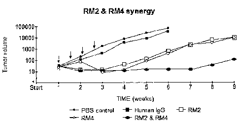
Figure 3 shows tumor (Calu-1 cells, a lung cancer cell line) necrosis in mice
following injection with RM2 and RM4. Arrows indicate injections on days 7,
10, 14 and 18.
Detailed Description
The invention is based, at least in part, on the isolation and
characterization of
human antibodies that selectively bind to hyperproliferative cells, including
tumor cells in vivo. That is, the antibodies preferentially bind to
hyperproliferating cells in comparison to non-proliferating cells. Thus, the
antibodies are useful for detecting and screening for the presence of
hyperproliferative cells and the antigens to which the antibodies bind. In
addition, the antibodies are cytotoxic towards hyperproliferating cells to
which
they bind when administered in a sufficient amount. For example, as
2 0 exemplified herein, an invention antibody, for example, ItM4 (ATCC deposit
No. PTA-5412), is able to induce tumor regression (reduce tumor volume) in
mice bearing tumors (see, for example, Figures 2 and 3). Thus, antibodies of
the invention are useful for treating undesirable, excessive or abnormal cell
proliferation including for example, non-metastatic and metastatic tumors.
2 5 In accordance with invention, isolated antibodies, methods of making the
antibodies and methods of using the antibodies, including therapeutic and
diagnostic methods, are provided. The invention antibodies are capable of
selectively binding to antigens associated with hyperproliferating cells. In
one
embodiment, an invention antibody is an isolated human monoclonal antibody
3 0 designated RM4 that selectively binds to an antigen designated AgluVI4.
Exemplary antibody RM4 is produced by a human IgG secreting cell line
derived using standard somatic cell hybridization technology (ATCC deposit
6
CA 02498778 2005-03-11
WO 2004/024874_ PCT/US2003/028806
No. Y~1~A-X412). The antibody secreting B cell was obtained from pooiea
regional draining lymph nodes of cancer patients and immortalized with RN 15,
a WIL-2 derived human fusion partner. RM4 recognizes a cell surface
(extracellular matrix) component (AgRM4). AgRM4 is expressed at least in
part on the cell surface. AgRM4 is more highly expressed in proliferating
cells than in non-proliferating cells, e.g., hyperproliferating cells. AgRM4
is
present on metastatic or non-metastatic breast, colon, gut and lung cancer
cells.
As used herein, the term "antibody" refers to a protein that binds to other
molecules (antigens) via heavy and light chain variable domains, VH and VL,
respectively. Antibodies include IgG, IgD, IgA, IgM and IgE, subtypes, and
mixtures thereof. The antibodies may be polyclonal or monoclonal, intact
immunoglobulin molecules, two full length heavy chains linked by disulfide
bonds to two full length light chains, or subsequences (i.e. fragments)
thereof,
with our without constant region, that bind to an epitope of an antigen, and
mixtures thereof. Antibodies may comprise heavy or light chain variable
regions, VH or VL, individually, or in any combination.
The terms "protein," "polypeptide" and "peptide" are used interchangeably
herein to refer to two or more covalently linked amino acids, or "residues,"
through an amide bond or equivalent. Polypeptides are of unlimited length and
2 0 the amino acids may be linked by non-natural and non-amide chemical bonds
including, for example, those formed with glutaraldehyde, N-
hydoxysuccinimide esters, bifunctional maleimides, or N,N'-
dicyclohexylcarbodiimide (DCC). Non-amide bonds include, for example,
ketomethylene, aminomethylene, olefin, ether, thioether and the like (see,
e.g.,
Spatola (1983) in Chemistry and Biochemistry of Amino Acids, Peptides and
Proteins, Vol. 7, pp 267-357, "Peptide and Backbone Modifications," Marcel
Decker, NY).
As used herein, the term "isolated," when used as a modifier of an invention
composition (e.g., antibodies, modified forms, subsequences, nucleic acids
3 0 encoding same, cells, vectors, etc.), means that the compositions are made
by
the hand of man or are separated from their naturally occurring in vivo
environment. Generally, compositions so separated are substantially free of
one or more materials with which they normally associate with in nature, for ,
example, one or more protein, nucleic acid, lipid, carbohydrate, cell
membrane.
7
CA 02498778 2005-03-11
WO 2004/024874 PCT/US2003/028806
Thus, an Isolated antibody is typically substantially free of one or more
materials with which it may typically associate with in nature. The term
"isolated" does not exclude alternative physical forms, such as polypeptide
multimers, post-translational modifications (e.g., phosphoryiation,
glycosylation) or derivatized forms.
An "isolated" antibody can also be "substantially pure" when free of most or
all of the materials with which it typically associates with in nature. Thus,
an
isolated molecule that also is substantially pure does not include
polypeptides
or polynucleotides present among millions of other sequences, such as
antibodies of an antibody library or nucleic acids in a genomic or cDNA
library, for example. Of course, a "substantially pure" molecule can be
combined with one or more other molecules. Thus, the term "substantially
pure" does not exclude combination compositions.
Substantial purity can be at least about 60% or more of the molecule by mass.
Purity can also be about 70% or 80% or more, and can be greater, for example,
90% or more. Purity can be determined by any appropriate method, including,
for example, UV spectroscopy, chromatography (e.g., HPLC, -gas phase), gel
electrophoresis (e.g., silver or coomassie staining) and sequence analysis
(nucleic acid and peptide).
2 0 The invention further provides antibodies having the binding specificity
of the
antibodies set forth herein. In one embodiment, the antibody has.the binding
specificity of RM4. In one aspect, the binding is specific for AgRM4.
The invention additionally provides antibodies that compete with the binding
of the antibodies set forth herein, and antibodies that bind to an epitope of
2 5 AgRM4 to which an antibody of the invention binds. In one embodiment, the
antibody competes with the binding of RM4 to an antigen. In another
embodiment, the antibody binds to an epitope of AgRM4 to which an antibody
of the invention binds. In one aspect, the antibody competes with the binding
of RM4 to AgRM4.
3 0 As used herein, the term "bind" or "binding" means that the compositions
referred to have affinity for each other. The term "specific" or "selective,"
and
grammatical variations thereof, when used in reference to binding, means that
the binding between the molecules is such that it can be distinguished from
8
CA 02498778 2005-03-11
WO 2onon~ spe 4fic or non-selective binding to other molecules using an
assay2sucno288o6
as ELISA, immunoprecipitation, coprecipitation, western blotting, two-hybrid
assays and the like. Appropriate controls can be used to distinguish between
"specific" and "non-specific" binding. For example, specific or selective
binding typically has a dissociation constant (KD) of less than about 1 X 10⁻⁵
M or less than about 1 X 10⁻⁶ M, 1 X 10⁻⁷ M, 1 X 10⁻⁸ M, 1 X 10⁻⁹ M, or 1 X
10-i° M. In contrast, non-specific binding typically has significantly
less
affinity, for example, a KD greater than 10-3 M. Thus, selective binding can
be
distinguished from non-selective binding by measuring dissociation constant
of the complex. Selective binding can also be distinguished form non-
selective binding by increasing the stringency of the binding assay. A
particular example of specific binding is that which occurs between an
antibody and an antigen.
As used herein, the term "epitope" means an antigenic determinant to which
an antibody binds. A polypeptide epitope can be as few as three amino acids,
yet generally an epitope has at least five amino acids or more, e.g., at least
eight to 12 amino acids. A "conformational epitope" is an epitope comprised
of a two or three dimensional juxtaposition of amino acids; the amino acids
can be contiguous or non-contiguous on the same polypeptide or on one or
2 0 more different polypeptides.
Antibodies having substantially the same (e.g., within about 10-fold) and
having different binding affinity from the antibodies set forth herein are
also
provided. In one embodiment, an antibody has increased or decreased affinity
for the antigen (e.g., AgRM4) in comparison to a reference antibody (e.g.,
2 5 RM4). In one aspect, an antibody has a binding affinity for AgRM4 within
1000-fold of the RM4 antibody. In additional aspects, the antibodies having
different binding affinity from the antibodies set forth herein are within 2-
5, 5-
10, 10-50, 50-100, 100-1000 and 1000-10,000 fold of RM4 antibody heavy
and light chain sequences.
3 0 ~ Antibodies having significant binding affinity for AgRM4 are also
provided.
As used herein, the term "significant" or "substantial" when used in reference
to binding affinity or activity, means that the dissociation constant (KD) of
the
complex (e.g., antibody-antigen complex) is not less than 10-3 M. In other
words, for significant binding affinity or activity, the KD must be less than
lfl-3
9
CA 02498778 2005-03-11
WO 2004/024874 q _~ _6 _7 8 CT/US2003/028806
M, e.g., i a M, 10 M, 10 M, 10 M, 10- M, etc. n ypicaiiy, trP~ ~~D ~~ all
antibody-antigen complex is about 10-5 M to about 10-6 M or less.
Antibodies of the invention include modified forms of the antibodies set forth
herein, provided that the modified antibody retains, at least a part of, a
function or activity of the unmodified or reference antibody. For example, a
modified RM4 antibody may retain antigen binding specificity, e.g., bind an
epitope present in AgRM4, but have increased or decreased binding affinity
for AgRM4 relative to unmodified RM4.
Thus, invention antibodies further include antibodies having sequences
distinct from the RM4 antibody heavy and light chain sequences. In various
embodiments, an antibody has the binding specificity of RM4, competes for
RM4 binding to AgRM4, and binds to an epitope of AgRM4 to which an
antibody of the invention binds.
The term "modify" and grammatical variations thereof, when used in
reference to a composition such as a polypeptide or nucleic acid, means that
the modified composition deviates from a reference composition. Polypeptide
modifications include amino acid substitutions, additions and deletions, which
are also referred to as "variants." Polypeptide modifications also include one
or more D-amino acids substituted for L-amino acids (and mixtures thereof),
2 0 structural and functional analogues, for example, peptidomimetics having
synthetic or non-natural amino acids or amino acid analogues and derivatized
forms. Polypeptide modifications further include fusion (chimeric)
polypeptide sequences, which is an amino acid sequence having one or more
molecules not normally present in a reference native (wild type) sequence
2 5 covalently attached to the sequence, for example, one or more amino acids.
Modifications include cyclic structures such as an end-to-end amide bond
between the amino and carboxy- terminus of the molecule or intra- or inter-
molecular disulfide bond. Polypeptides including antibodies may be modified
in vitro or in vivo, e.g., post-translationally modified to include, for
example,
3 0 sugar residues, phosphate groups, ubiquitin, fatty acids or lipids.
Thus, the invention provides antibodies having one or more modifications,
provided that the modified antibody retains an activity or function of a
reference antibody (e.g., antigen binding activity). In one embodiment, the
CA 02498778 2005-03-11
WO 2004/024874 PCT/US2003/028806
antibody is modified from a light chain or the heavy chain amino acra
sequence of RM4. In one aspect, a modified antibody has an amino acid
substitution, addition or deletion (e.g., 1-3, 3-5, 5-10 or more) of the
variable
or constant region, heavy or light chain. In another aspect, the modified
antibody comprises a subsequence (e.g., Fab, Fab', Fv, F(ab')2, Fd, or single
chain Fv). In yet another aspect, the substitution is with a human or non-
human amino acid which is structurally similar to the human residue. In a
particular aspect, the substitution is a conservative amino acid substitution.
A "conservative substitution" means the replacement of one amino acid by a
biologically, chemically or structurally similar residue. Biologically similar
means that the substitution is compatible with biological activity, e.g.,
antigen
binding. Structurally similar means that the amino acids have side chains with
similar length, such as alanine, glycine and serine, or having similar size.
Structurally similar substitutions are unlikely to alter antigenicity of the
antibody relative to the unsubstituted antibody. Chemical similarity means
that the residues have the same charge or are both hydrophilic or hydrophobic.
Particular examples include the substitution of one hydrophobic residue, such
as isoleucine, valine, leucine or methionine for another, or the substitution
of
one polar residue for another, such as the substitution of arginine for
lysine,
2 0 glutamic for aspartic acids, or glutamine for asparagine, serine for
threonine,
and the like.
Invention antibodies having a sequence not identical to a sequence of heavy
and light chain amino acid sequences of RM4 include antibodies having an
amino acid sequence with 50%, 60%, 70%, 75%, 80%, 85%, 90%, 95%, 96%,
2 5 97%, 98%, or more identity to a heavy or light chain amino acid sequence
of
RM4. The identity can be over a defined area of the antibody, e.g., one or
more complementarity determining regions (CDRs) or framework region.
The term "identical" or "identity" means that two or more referenced entities
are the same. Thus, where two protein sequences are identical, they have the
3 0 same amino acid sequence. "Areas of identity" means that a portion of two
or
more referenced entities are the same. Thus, where two protein sequences are
identical over one or more sequence regions they share amino acid identity in
these regions. The term "substantial identity" means that the identity is
structurally or functionally significant. That is, the identity is such that
the
11
CA 02498778 2005-03-11
WO 2004/024874 PCT/US2003/028806
molecules are structurally identical or have at least one of the same 3uncuons
(e.g., biological function) even though the molecules differ.
Due to variation in the amount of sequence conservation between structurally
and functionally related proteins, the amount of sequence identity for
substantial identity will depend upon the type of protein, the region and its
function. For proteins there can be as little as 30% sequence identity, but
typically there is more, e.g., 50%, 60%, 75%, 85%, 90%, 95%, 96%, 97%,
98%, identity to a reference sequence. For nucleic acid sequences, ~0%
sequence identity or more typically constitutes substantial homology, but can
vary depending on the comparison region.
The extent of identity between two sequences can be ascertained using a
computer program and mathematical algorithm known in the art. Such
algorithms that calculate percent sequence identity (homology) generally
account for sequence gaps and mismatches over the comparison region. For
example, a BLAST (e.g., BLAST 2.0) search algorithm (see, e.g., Altschul et
al. (1990) J. Mol. Biol. 215:403-10, publicly available through NCBI) has
exemplary search parameters as follows: Mismatch -2; gap open 5; gap
extension 2. For polypeptide sequence comparisons, a BLASTP algorithm is
typically used in combination with a scoring matrix, such as PAM 100, PAM
2 0 250, and BLOSUM 62.
As used herein, the term "subsequence" or "fragment" means a portion of the
full length molecule. For example, a subsequence of an antibody is at least
one amino acid less in length than full length antibody having intact heavy
and
light chain sequence (e.g. one or more internal or terminal amino acid
2 5 deletions from either amino or carboxy-termini). Subsequences therefore
can
be any length up to the full length molecule.
Subsequences include portions which retain at least part of the function or
activity of a full length antibody or a reference antibody sequence. For
example, an antibody subsequence will retain the ability to selectively bind
to
3 0 an antigen (e.g., AgRM4) even though the binding affinity of the
subsequence
may be greater or less than the binding affinity of the full length reference
antibody. Subsequences can comprise a portion of any of the invention
antibody sequences, for example, a portion of VH or VL domain of RM4.
12
CA 02498778 2005-03-11
WO 2004/024874 PCT/US2003/028806
~pecWc examples of antibody subsequences of the invention mcmue, mr
example, Fab, Fab', Fv, F(ab')2, Fd, or single chain antibody (SCA) fragment
(e.g., scFv). Additional fragments are known in the art and described, for
example, in Hudson, Cur. Opin. Bioteclznol. 9:395 (1998).
Pepsin or papain digestion of whole antibodies can be used to generate
subsequences. For example, Fab can be produced by digestion of a whole
antibody with the enzyme papain, to yield a fragment consisting of an intact
light chain and a portion of a heavy chain. (Fab')2 can be produced by
treating
a whole antibody with the enzyme pepsin, without subsequent reduction. An
Fab' antibody fragment can be produced from (Fab')a by reduction with a thiol
reducing agent, which yields a molecule consisting of an intact light chain
and
a portion of a heavy chain. Two Fab' fragments are produced per antibody
molecule treated in this manner.
An Fv fragment is a fragment containing the variable region of a light chain
VL and the variable region of a heavy chain VH expressed as two chains. The
association may be non-covalent or may be covalent, such as a chemical
cross-linking agent or an intermolecular disulfide bond (mbar et al., (1972)
Pnoc. Natl. Acad Sci. USA 69:2659; Sandhu (1992) C~it. Rev. Biotech.
12:437).
2 0 A single chain antibody (SCA) is a genetically engineered or enzymatically
digested antibody containing the variable region of a light chain VL and the
variable region of a heavy chain, optionally linked by a flexible linker, such
as
a polypeptide sequence, in either VL-linker-VH orientation or in VH-linker-VL
orientation. Alternatively, a single chain Fv fragment can be produced by
2 5 linking two variable domains via a disulfide linkage between two cysteine
residues. Methods for producing scFv antibodies are described, for example,
by Whitlow et al., (1991) In: Methods: A Companion to Methods in
Enzymolo~y 2:97; U.S. Patent No. 4,946,778; and Pack et al., (1993)
BiolTeclznology 11:1271.
3 0 Other methods of producing antibody subsequences, such as separation of
heavy chains to form monovalent light-heavy chain fragments, further
cleavage of fragments, or other enzymatic, chemical, or genetic techniques
13
CA 02498778 2005-03-11
WO 2004/024874 PCT/US2003/028806
may also be used, provided that the subsequences have a function ur acnvmy,
e.g., bind to the antigen to which the intact antibody binds.
Modified forms also include derivatized sequences, for example, amino acids
in which free amino groups form amine hydrochlorides, p-toluene sulfonyl
groups, carbobenzoxy groups; the free carboxy groups from salts, methyl and
ethyl esters; free hydroxl groups that form O-acyl or O-alkyl derivatives, as
well as naturally occurring amino acid derivatives, for example,
4-hydroxyproline, for proline, 5-hydroxylysine for lysine, homoserine for
serine, ornithine for lysine, etc. Modifications can be produced using any of
a
variety of methods well known in the art (e.g., PCR based sited-directed,
deletion and insertion mutagenesis, chemical modification and mutagenesis,
cross-linking, etc.).
Antibodies of the invention can be either joined directly or indirectly
through
covalent or non-covalent binding, e.g. via a multimerization domain, to
produce multimers. Specific examples of domains that confer multimer
formation include coiled-coil (e.g., leucine zipper structures) and alpha-
helical
protein sequences. Sequences that mediate protein-protein binding via Van
der Waals' forces, hydrogen bonding or charge-charge bonds are also
contemplated as multimerization domains. One specific example of a
2 0 multimerization domain is p53 residues 319 to 360, which mediates tetramer
formation. Another example is extracellular protein TSP4, a member of the
thrombospondin family, which can form pentamers. Additional specific
examples are the leucine zippers of jun, fos, and yeast protein GCN4.
The antibodies of the invention therefore also include multimers. A multimer
2 5 can be a dimer, trimer, tetramer or other higher order oligomer. Multimers
can
be combinations of the same antibodies (homo-oligomers) or different
antibodies (hetero-oligomers), the different antibodies being human,
humanized or non-human.
Antibodies of the invention can be modified to include one or more functions
3 0 or activities in addition to binding a particular antigen. For example, an
antibody can include a region that binds to a different antigen, or have a
function distinct from antigen binding. Such modified antibodies are referred
to herein as "multifunctional antibodies," and include, for example,
14
CA 02498778 2005-03-11
WO 2004/024874_ PCT/US2003/028806
mulnspecmc (e.g., bispecific, trispecific, tetraspecmc, etc.~ annbuu~~~.
term "multispecific" refers to an antibody that binds to two or more different
antigenic epitopes. The different epitopes may be present on the same antigen
or different antigens. For example, a multispecific antibody oligomer
comprises a mixture of two or more antibodies each having different epitope
binding specificity and which form a multimer. The different epitopes may be
expressed by the same or a different cell.
The term "multifunctional" means that the composition referred to has two or
more activities or functions. Particular non-limiting examples include, for
example, antigen binding, enzyme activity, ligand or receptor binding
(substrates, agonists and antagonists), detection, purification, and toxicity.
The term "detectable label" refers to a molecule that can be conjugated to
another molecule so as to enable detection of the conjugated molecule.
Examples of detectable labels include chelators, photoactive agents,
radionuclides (alpha, beta and gamma emitters), fluorescent agents and
paramagnetic ions. Th term "tag" refers to a molecule conjugated to another
that allows detection or purification. Specific examples of tags include
immunoglobulins, T7, polyhistidine tags, glutathione-S-transferase, a chitin-
binding tag, calmodulin-binding tag, myc tag, and a Xpress epitope (detectable
2 0 by anti-Xpress antibody; Invitrogen, Carlsbad, Calif., USA).
An antibody that has an attached polypeptide with enzyme activity (e.g., green
fluorescent protein, acetyltransferase, galactosidase, glucose oxidase,
peroxidase, horseradish peroxidase (HRP), urease and alkaline phosphatase) is
one particular example of a muiltifunctional antibody. Attached polypeptides
2 5 also include apoptotic factors, differentiative factors, chemokines and
cytokines (interleukins, interferons).
Additional candidate functions for multifunctional antibodies other than
antigen binding include, for example, radioactive (e.g., 3H,'4C, 32P, 33P,
3sS,
i2sh ~3~I) and non-radioactive moieties (e.g., gold particles, colored glass
or
3 0 plastic polystyrene, polypropylene, or latex beads) and amino acid
sequences
(e.g., tags, as set forth herein) for detection.
Detectable moieties also include fluorescent compounds (e.g., fluorescein
isothiocyanate, rhodamine, phycoerytherin, phycocyanin, allophycocyanin, o-
1'S
CA 02498778 2005-03-11
WO 2004/024874 PCT/US2003/028806
phthalderiyde, fluorescamine, and commercially avalailable tluoropnores such
as Alexa Fluor 350, Alexa Fluor 488, Alexa Fluor 532, Alexa Fluor 546,
Alexa Fluor 568, Alexa Fluor 594, Alexa Fluor 647, and BODiPY dyes such
as BODIPY 493/503, BODIPY FL, BODIPY R6G, BODIPY 530/550,
BODIPY TMR, BODIPY 558/568, BODIPY 558/568, BODIPY 564/570,
BODIPY 576/589, BODIPY 581/591, BODIPY TR, BODIPY 630/650,
BODIPY 650/665, Cascade Blue, Cascade Yellow, Dansyl, lissamine
rhodamine B, Marina Blue, Oregon Green 488, Oregon Green 514, Pacific
Blue, rhodamine 6G, rhodamine green, rhodamine red, tetramethylrhodamine
and Texas Red, from Molecular Probes, Inc., Eugene, OR), colloidal metals,
chemiluminescent compounds (e.g., huminol, isohuminol, an aromatic
acridinium ester, an imidazole, an acridinium salt and oxalate esters),
bioluminescent compounds (e.g., luciferin, luciferase and aequorin),
paramagnetic labels (e.g., chromium (III), manganese (II), manganese (III),
iron (II), iron (III), cobalt (II), nickel (II), copper (II), praseodymium
(III),
neodymium (III), samarium (III), gadolinium (III), terbium (III), dysprosium
(III), holmium (III), erbium (III) and ytterbium (III)) which can be detected
by
MRI, and adhesion proteins (e.g., biotin, streptavidin, avidin, and other
lectins).
2 0 Additional candidate functions include cytotoxicity (e.g., bacterial
cholera
toxin, pertussis toxin, anthrax toxin lethal factor, Pseudomonas exotoxin A,
diphtheria toxin, plant toxin ricin, radionuclides such as 47Sc 67Cu, 72Se,
88Y,
90Sr 90Y 97Ru 99TC 105 111In' 125f 131I? 149Tb, 153~m' 186Re' 188Re' 194OS'
> > > o
2o3Pb~ 211Af 212Bi' 213Bi' 212Pb, 223Ra,225AC' 227AC' 228Th, and CytOtOXIC
drugs).
2 5 Modified antibodies therefore also include addition of functional
entities,
covalently or non-covalently attached to the antibodies of the invention.
Multifunctional antibodies can be produced through chemical crosshinking of
the selected molecules (which have been produced by synthetic means or by
expression of nucleic acid that encode the polypeptides), via an amino acid
3 0 linker sequence or through recombinant DNA technology combined with in
vitro, or cellular expression of the polypeptide. Multispecific antibodies can
be similarly produced through recombinant technology and expression, fusion
of hybridomas (e.g., to produce quadromas) that produce antibodies with
different epitopic specificities, or expression of multiple nucleic acid
encoding
3 5 antibody variable chains with different epitopic specificities in a single
cehh.
16
CA 02498778 2005-03-11
WO 2orie~coupiing of such agents can be performed using conventiona ~rrTcu u2u
03/028806
known in the art (see, for example, R. Reisfeld and S. Sell Eds. Monoclonal
Antibodies and Cancer Therany, Alan R. Liss Inc. NY, 1985; and U.S. Pat.
Nos. 5,558,852 and 5,624,659)
Polypeptide sequences can be made using recombinant DNA technology of
polypeptide encoding nucleic acids via cell expression or in vitro
translation,
or chemical synthesis of polypeptide chains using methods known in the art.
Antibodies of the invention, including modified forms and subsequences can
be expressed from recombinantly produced antibody-encoding nucleic acid
(see, e.g., Harlow and Lane, Using Antibodies: A Laboratory Manual, Cold
Spring Harbor Laboratory, 1999; Fitzgerald et al., J.A.C.S. 117:11075 (1995);
Gram et al., P~°oc. Natl. Acad. Sci. USA 89:3576 (1992)).
Antibodies may
also be produced by expressing encoding nucleic acids in mammalian, insect,
and plant cells. Polypeptide sequences including antibodies can also be
produced by a chemical synthesizer (see, e.g., Applied Biosystems, Foster
City, CA).
The invention further provides nucleic acids encoding invention antibodies,
including modified forms thereof. In various embodiments, a nucleic acid
encodes a sequence of a heavy or light chain amino acid sequence set forth of
2 0 RM4. In a particular aspect, a nucleic acid encodes a sequence of a heavy
or
light chain amino acid sequence of RM4.
As used herein, a "nucleic acid," refers to at least two or more ribo- or
deoxy-
ribonucleic acid base pairs (nucleotides) that are linked through a
phosphoester bond or equivalent. Nucleic acids include polynucleotides and
2 5 polynucleosides. Nucleic acids include single, double or triplex, circular
or
linear, molecules. A nucleic acid molecule may belong exclusively or in a
mixture to any group of nucleotide-containing molecules, as exemplified by,
but not limited to: RNA, DNA, cDNA, genomic nucleic acid, non-genomic
nucleic acid, naturally occurnng and non naturally occurring nucleic acid and
3 0 synthetic nucleic acid.
Nucleic acids can be of any length. Nucleic acid lengths typically range from
about 20 nucleotides to 10 Kb, 10 nucleotides to SKb, 1 to 5 Kb or less, 1000
to about 500 nucleotides or less in length. Nucleic acids can also be shorter,
17
CA 02498778 2005-03-11
WO 2004/024874 ' PCT/US2003/028806
for example, 100 to about 500 nucleotides, or from about 12 to 2~, ~~ vo w,
50 to 100, 100 to 250, or about 250 to 500 nucleotides in length.
Nucleic acids further include modifications such as nucleotide and nucleoside
substitutions, additions and deletions, as well as derivatized forms and
fusion
sequences (e.g., encoding recombinant polypeptide). For example, due to the
degeneracy of the genetic code, nucleic acids include sequences and
subsequences degenerate with respect to nucleic acids that encode amino acid
sequences of RM4. Other examples are nucleic acids complementary to a
sequence that encodes an amino acid sequence of RM4. Nucleic acid
1 o deletions (subsequences) have from about 10 to 25, 25 to 50 or 50 to 100
nucleotides. Such nucleic acids are useful for expressing polypeptide
fragments, for genetic manipulation (as primers and templates for PCR
amplification), and as probes to detect the presence or an amount of a
sequence encoding an invention antibody in vitf-o, in a cell, culture medium,
biological sample (e.g., tissue, organ, blood or serum), or in a subject.
In yet another example of nucleic acid modifications, nucleic acids that
hybridize at high stringency to nucleic acids that encode an amino acid
sequence of RM4, a subsequence thereof and nucleic acid sequences
complementary to the encoding nucleic acids, are provided. Hybridizing
2 0 nucleic acids are useful for detecting the presence or an amount of a
sequence
encoding an invention antibody in vitro, or in a cell, culture medium,
biological sample (e.g., tissue, organ, blood or serum), or in a subject.
The term "hybridize" refers to the binding between nucleic acid sequences.
Hybridizing sequences will generally have more than about 50% homology to
2 5 a nucleic acid that encodes an amino acid sequence of RM4. The
hybridization region between hybridizing sequences can extend over at least
about 10-15 nucleotides, 15-20 nucleotides, 20-30 nucleotides, 30-50
nucleotides, 50-100 nucleotides, or about 100 to 200 nucleotides or more.
As is understood by those skilled in the art, the TM {melting temperature) is
the
3 0 temperature at which binding between two nucleic acid sequences is no
longer
stable. For two sequences to bind, the temperature of a hybridization reaction
must be less than the calculated TM for the sequences under the hybridization
conditions. The TM is influenced by the amount of sequence complementarity,
1$
CA 02498778 2005-03-11
WO 2004/024874 PCT/US2003/028806
length, composition (%GC), type of nucleic acid (RNA vs. DNA, ana me
amount of salt, detergent and other components in the reaction (e.g.,
formamide). All of these factors are considered in establishing appropriate
hybridization conditions (see, e.g., the hybridization techniques and formula
for calculating TM described in Sambrook et al., In: Molecular Clonin~,~A
Laboratory Manual, 3rd ed., Cold Spring Harbor Laboratory Press, 2001).
Typically, wash conditions are adjusted to attain the desired degree of
hybridization stringency. Thus, hybridization stringency can be determined
empirically, for example, by washing under particular conditions, e.g., at low
stringency conditions or high stringency conditions. Optimal conditions for
selective hybridization will vary depending on the particular hybridization
reaction involved. An example of high stringency hybridization conditions are
as follows: 2X SSClO.l% SDS at about 37°C or 42°C (hybridization
conditions); O.SX SSC/0.1% SDS at about room temperature (low stringency
wash); O.SX SSC/0.1% SDS at about 42°C (moderate stringency wash); and
0.1 X SSC/0.1% SDS at about 65°C (high stringency wash).
Nucleic acids can be produced using various standard cloning and chemical
synthesis techniques. Such techniques include, but are not limited to nucleic
acid amplification, e.g., polymerase chain reaction (PCR), with genomic DNA
2 0 or cDNA targets using primers (e.g., a degenerate primer mixture) capable
of
annealing to antibody encoding sequence; and chemical synthesis of nucleic
acid sequences. The sequences produced can then be translated in vitxo, or
cloned into a plasmid and propagated and then expressed in a cell (e.g.,
microorganism, such as yeast or bacteria, a eukaryote such as an animal or
2 5 mammalian cell or in a plant).
The invention further provides expression cassettes including a nucleic acid
encoding an invention antibody operably linked to an expression control
element. As used herein, the term "operably linked" refers to a physical or a
functional relationship between the elements referred to that permit them to
3 0 operate in their intended fashion. Thus, an expression control element
"operably linked" to a nucleic acid means that the control element modulates
nucleic acid transcription and as appropriate, translation of the transcript.
19
CA 02498778 2005-03-11
WO 2004/024874 PCT/US2003/028806
Yhysica~ linkage is not required for the elements to be operably lmcu. r m
example, a minimal element can be linked to a nucleic acid encoding an
invention antibody. A second element that controls expression of an operably
linked nucleic acid encoding a protein that functions "in trans" to bind to
the
minimal element can influence expression of the antibody. Because the
second element regulates expression of antibody, the second element is
operably linked to the nucleic acid encoding the antibody even though it is
not
physically linked.
The term "expression control element" refers to nucleic acid that influences
l0 expression of an operably linked nucleic acid. Promoters and enhancers are
particular non-limiting examples of expression control elements. A "promotor
sequence" is a DNA regulatory region capable of initiating transcription of a
downstream (3' direction) coding sequence. The promoter sequence includes
a number of nucleotides necessary to facilitate transcription initiation.
Enhancers also regulate gene expression, but can function a distance from the
transcription start site of the gene to which it is operably linked. Enhancers
function at either 5' or 3' ends of the gene, as well as within the gene
(e.g., in
introns or coding sequences). Additional expression control elements include
leader sequences and fusion partner sequences, internal ribosome binding sites
2 0 (IRES) elements for the creation of multigene, or polycistronic, messages,
splicing signal for introns, maintenance of the correct reading frame of the
gene to permit in-frame translation of mRNA, polyadenylation signal to
provide proper polyadenylation of the transcript of a gene of interest, and
stop
codons.
2 5 Expression control elements include "constitutive" elements such that
transcription of the operably linked nucleic acid occurs without the presence
of a signal or stimuli. Expression control elements that confer expression in
response to a signal or stimuli, which either increases or decreases
expression
of the operably linked nucleic acid, are "regulatable." A regulatable element
3 0 that increases expression of the operably linked nucleic acid in response
to a
signal or stimuli is referred to as an "inducible element." A regulatable
element that decreases expression of the operably linked nucleic acid in
response to a signal or stimuli is referred to as a "repressible element"
(i.e., the
signal decreases expression; when the signal is removed or absent, expression
3 5 is increased).
CA 02498778 2005-03-11
WO 2004/024874 PCT/US2003/028806
Expression control elements include elements active in a particular mssue or
cell type, referred to as "tissue-specific expression control elements."
Tissue-
specific expression control elements are typically active in specific cell or
tissue types because they are recognized by transcriptional activator
proteins,
or other regulators of transcription, that are unique to the specific cell or
tissue
type.
Expression control elements include full-length nucleic acid sequences, such
as native promoter and enhancer elements, as well as subsequences or
nucleotide variants thereof (e.g., substituted/mutated or other forms that
differ
1 o from native sequences) which retain all or part of full-length or non-
variant
control element function (confer regulation, e.g., retain some amount of
inducibility in response to a signal or stimuli).
For bacterial expression, constitutive promoters include T7, as well as
inducible promoters such as pL of bacteriophage 7~, plac, ptrp, ptac (ptrp-lac
hybrid promoter). In insect cell systems, constitutive or inducible promoters
(e.g., ecdysone) may be used. In yeast, constitutive promoters include, for
example, ADH or LEU2 and inducible promoters such as GAL (see, e.g.,
Ausubel et al., In: Current Protocols in Molecular Biolo~y, Vol. 2, Ch. 13,
ed.,
Greene Publish. Assoc. & Wiley Interscience, 1988; Grant et al., (1987) In:
2 0 Methods in Enzymolo~y, 153:516-544, eds. Wu ~ Grossman, 1987, Acad.
Press, N.Y.; Glover, DNA Cloning, Vol. II, Ch. 3, IRL Press, Wash., D.C.,
1986; Bitter (1987) In: Methods in Enzymolo~y, 152:673-684, eds. Berger &
Kimmel, Acad. Press, N.Y.; and, Strathern et al., The Molecular Biology
the Yeast Saccharomyces (1982) eds. Cold Spring Harbor Press, Vols. I and
2 5 II).
For mammalian expression, constitutive promoters of viral or other origins
may be used. For example, SV40, or viral long terminal repeats (LTRs) and
the like, or inducible promoters derived from the genome of mammalian cells
(e.g., metallothionein IIA promoter; heat shock promoter, steroid/thyroid
3 0 hormone/retinoic acid response elements) or from mammalian viruses (e.g.,
the adenovirus late promoter; the inducible mouse mammary tumor virus LTR)
are used.
21
CA 02498778 2005-03-11
w0 2ooneoiri gention also provides stably and transiently transformed cPCS
aiiu20o3/028806
progeny thereof into which a nucleic acid molecule encoding an invention
antibody has been introduced by means of recombinant DNA techniques in
vitro, ex vivo or in vivo. The transformed cells can be propagated and the
introduced nucleic acid transcribed, or encoded protein expressed.
Transformed cells include but are not limited to prokaryotic and eukaryotic
cells such as bacteria, fungi, plant, insect, and animal (e.g., mammalian,
including human) cells. In one particular aspect, the cell is a hybridoma. The
cells may be present in culture, in a cell, tissue or organ ex vivo or a
subject.
A progeny cell may not be identical to the parental cell,since there may be
mutations that occur during replication.
The term "transformed" means a genetic change in a cell following
incorporation of nucleic acid (e.g., a transgene) exogenous to the cell. Thus,
a
"transformed cell" is a cell into which, or a progeny of which a nucleic acid
molecule has been introduced by means of recombinant DNA techniques.
Cell transformation to produce host cells may be carried out as described
herein or using techniques known in the art. Accordingly, methods of
producing cells including the nucleic acids and cells expressing the invention
antibodies are also provided.
2 0 Typically, cell transformation employs a "vector," which refers to a
plasmid,
virus, such as a viral vector, or other vehicle known in the art that can be
manipulated by insertion or incorporation of a nucleic acid. For genetic
manipulation "cloning vectors" can be employed, and to transcribe or translate
the inserted polynucleotide "expression vectors" can be employed. Such
2 5 vectors are useful for introducing nucleic acids, including a nucleic acid
that
encodes an antibody operably linked with an expression control element, and
expressing the antibody in vitro (e.g., in solution or in solid phase), in
cells or
in a subject ifa vivo.
A vector generally contains an origin of replication for propagation in a
cell.
3 0 Control elements, including expression control elements as set forth
herein,
present within a vector, can be included to facilitate transcription and
translation.
22
CA 02498778 2005-03-11
WO 2004/024874 PCT/US2003/028806
Vectors can include a selection marker. A "selection marker" is a gene may
allows for the selection of cells containing the gene. "Positive selection"
refers to a process whereby only cells that contain the selection marker will
survive upon exposure to the positive selection. Drug resistance is one
example of a positive selection marker; cells containing the marker will
survive in culture medium containing the selection drug, and cells lacking the
marker will die. Selection markers include drug resistance genes such as ~zeo,
which confers resistance to 6418; laygf°, which confers resistance to
hygromycin; and puro which confers resistance to puromycin. Other positive
selection marker genes include genes that allow identification or screening of
cells containing the marker. These genes include genes for fluorescent
proteins (GFP and GFP-like chromophores, luciferase), the lacZ gene, the
alkaline phosphatase gene, and surface markers such as CDB, among others.
"Negative selection" refers to a process whereby cells containing a negative
selection marker are killed upon exposure to an appropriate negative selection
agent. For example, cells which contain the herpes simplex virus-thymidine
kinase (HSTI tk) gene (Wigler et al., Cell 11:223 (1977)) are sensitive to the
drug gancyclovir (GANG). Similarly, the gpt gene renders cells sensitive to 6-
thioxanthine.
2 0 Viral vectors included are those based on retroviral, adeno-associated
virus
(AAV), adenovirus, reovirus, lentivirus, rotavirus genomes, simian virus 40
(SV40) or bovine papilloma virus (Cone et al., Proc. Natl. Acad. Sci. USA
81:6349 (1984); Eukaryotic Viral Vectors, Cold Spring Harbor Laboratory,
Gluzman ed., 1982; Sarver et al., Mol. Cell. Biol. 1:486 (1981)). Additional
2 5 viral vectors useful for expression include parvovirus, rotavirus, Norwalk
virus, coronaviruses, paramyxo and rhabdoviruses, togavirus (e.g., sindbis
virus and semliki forest virus) and vesicular stomatitis virus (VSV).
Mammalian expression vectors include those designed for in vivo and ex vivo
expression, such as AAV (U.S. Patent No. 5,604,090). AAV vectors have
3 0 previously been shown to provide expression of Factor IX in humans and in
mice at levels sufficient for therapeutic benefit (Kay et al., Nat. Genet.
24:257
(2000); Nakai et al., Blood 91:4600 (1998)). Adenoviral vectors (U.S. Patent
Nos. 5,700,470, 5,731,172 and 5,928,944), herpes simplex virus vectors (U.S.
Patent No. 5,501,979) retroviral (e.g., lentivirus vectors are useful for
infecting
3 5 dividing as well as non-dividing cells and foamy virues) vectors (U.S.
Patent
23
CA 02498778 2005-03-11
wo 200o/o2~sn~a4 820 5 693 508 5 665 577 6 013 516 and 5 674 703
ana/wiYO°3/o2sso6
> > > > > > > > > > > >
publications W092/05266 and WO92/14829) and papilloma virus vectors
(e.g., human and bovine papilloma virus) have all been employed in gene
therapy (U.S. Patent No. 5,719,054). Vectors also include cytomegalovirus
(CMV) based vectors (U.S. Patent No. 5,561,063). Vectors that efficiently
deliver genes to cells of the intestinal tract have been developed (see, e.g.,
U.S.
Patent Nos. 5,821,235, 5,786,340 and 6,110,456).
Introduction of antibodies and nucleic acid encoding invention antibodies into
target cells can also be carried out by methods known in the art such as
osmotic shock (e.g., calcium phosphate), electroporation, microinjection, cell
fusion, etc. Introduction of nucleic acid and polypeptide in vitro, ex vivo
and
i~ vivo can also be accomplished using other techniques. For example, a
polymeric substance, such as polyesters, polyamine acids, hydrogel, polyvinyl
pyrrolidone, ethylene-vinylacetate, methylcellulose, carboxymethylcellulose,
protamine sulfate, or lactide/glycolide copolymers, polylactide/glycolide
copolymers, or ethylenevinylacetate copolymers. A nucleic acid can be
entrapped in microcapsules prepared by coacervation techniques or by
interfacial polymerization, for example, by the use of hydroxymethylcellulose
or gelatin-microcapsules, or poly (methylmethacrolate) microcapsules,
2 0 respectively, or in a colloid drug delivery system. Colloidal dispersion
systems include macromolecule complexes, nano-capsules, microspheres,
beads, and lipid-based systems, including oil-in-water emulsions, micelles,
mixed micelles, and liposomes.
Liposomes for introducing various compositions into cells, including nucleic
2 5 acids, including, for example, phosphatidylcholine, phosphatidylserine,
lipofectin and DOTAP, are known to those skilled in the art (see, e.g., U.S.
Patent Nos. 4,844,904, 5,000,959, 4,863,740, 4,975,282, GIBCO-BRL,
Gaithersburg, Md). Piperazine based amphilic cationic lipids useful for gene
therapy also are known (see, e.g., U.S. Patent No. 5,861,397). Cationic lipid
3 0 systems also are known (see, e.g., U.S. Patent No. 5,459,127).
Accordingly,
viral and non-viral vector means of delivery into cells or tissue, in vitro,
ira
vivo and ex vivo are included.
The invention therefore also provides methods of producing antibodies of the
invention. In one embodiment, a method includes: introducing a nucleic acid
24
CA 02498778 2005-03-11
WO 2004/024874 PCT/US2003/028806
that encodes the antibody into a host cell or a translation extract; mcuoann~
said host cell or extract under conditions whereby said nucleic acid is
expressed as a translation product including said antibody; and isolating or
purifying the antibody. In one aspect, the nucleic acid encodes RM4. In
another aspect, the nucleic acid encodes modified RM4 (e.g., a variant or
subsequence).
The invention antibodies also can be combined with any other compounds or
agents that may provide an enhanced or synergistic therapeutic benefit. The
invention therefore also provides combination compositions including an
invention antibody and one or more additional compounds or agents and
methods of using the combinations. For example, an invention antibody may
be combined with a compound or agent that has anti-tumor activity or immune
enhancing activity. In a particular example, RM4 is combined with RM2.
As used here, the term "immune enhancing," when used in reference to a
compound, agent, therapy or treatment, means that the compound agent,
therapy or treatment, provides an increase, stimulation, induction or
promotion
of an immune response, humoral or cell-mediated. Such therapies can
enhance immune response generally, or enhance immune response to a
specific target tumor.
2 0 Specific non-limiting examples of immune enhancing agents include
monoclonal, polyclonal antibody and mixtures thereof. Antibodies include
antibodies that bind to tumor-associated antigens (TAA). The term "tumor
associated antigen" or "TAA" refers to an antigen expressed by a tumor cell.
TAAs may be expressed in amounts greater in tumor cells than a normal non-
2 5 tumor cell counterpart, or may be expressed at similar levels, or at
levels less
than a normal cell counterpart.
Particular non-limiting examples of TAAs that can be targeted and TAA
binding antibodies include, for example, human IBD12 monoclonal antibody
which binds to epithelial cell surface H antigen (U.S. Patent No. 4,814,275);
3 0 M195 antibody which binds to leukemia cell CD33 antigen (U.S. Patent No.
6,599,505); monoclonal antibody DS6 which binds to ovarian carcinoma CA6
tumor-associated antigen (U.S. Patent No. 6,596,503); and BR96 antibody
which binds to LeX carbohydrate epitope expressed by colon, breast, ovary,
CA 02498778 2005-03-11
WO 2004/024874 PCT/US2003/028806
and lung carcinomas. Additional anti-tumor antibodies that can be empioyeu
include, for example, Rituxan~, Herceptin (anti-Her-2 neu antibody),
Bevacizumab (Avastin), Zevalin, Bexxar, Oncolym, 17-lA(Edrecolomab),
3F8 (anti-neuroblastoma antibody), MDX-CTLA4, Campath~, Mylotarg and
IMC-C225 (Cetuximab).
Antibody RM2 is produced by a human IgG secreting cell line derived using
standard somatic cell hybridization technology (ATCC deposit No. PTA-5411;
Example 1). RM2 binds to a peptide sequence termed AgRM2 of
approximately 52kDa, as determined by denaturing gel electrophoresis.
AgRM2 is expressed at least in part on the cell surface. AgRM2 is more
highly expressed in proliferating cells than in non-proliferating cells, e.g.,
hyperproliferating cells. AgRM2 is present on metastatic or non-metastatic
lung, skin (melanoma), pancreatic, and brain (neuroblastoma/glioma) cancer
cells.
Other non-limiting examples of TAAs that can be targeted with an antibody
include MUC-1, HER-2/neu, MAGE, p53, T/Tn and CEA (Breast cancer);
MUC-2 and MUC-4, CEA, p53 and the MAGE (colon cancer); MAGE,
MART-1 and gp100 (melanoma); GM2, Tn, sTn, Thompson-Friedenreich
antigen (TF), MUC1, MUC2, beta chain of chorionic gonadotropin (hCG beta),
2 0 HER2/neu, PSMA and PSA (prostate cancer); chorionic gonadotropin
(testicular cancer); and alpha fetoprotein (hepato-cellular carcinoma).
Additional examples of immune enhancing agents include immune cells such
as lymphocytes, plasma cells, macrophages, NK cells and B-cells expressing
antibody against the tumor. Cytokines that enhance or stimulate
2 5 immunogenicity against tumor such as IL-2, IL-1 a, IL-1 (3, IL-3, IL-6, IL-
7,
granulocyte-macrophage-colony stimulating factor (GMCSF), IFN-y, IL-12,
TNF-a, and TNF(3 are also non-limiting examples of immune enhancing
agents. Chemokines including MIP-la, MIP-lei, RANTES, SDF-l, MCP-1,
MCP-2, MCP-3, MCP-4, eotaxin, eotaxin-2, I-309/TCA3, ATAC, HCC-l,
3 o HCC-2, HCC-3, LARC/MIP-3a, PARC, TARO, CK(3, CKj36, CK(37, CK(38,
CK[39, CK[311, CK(312, C10, IL-8, GROa, GROG, ENA-78, GCP-2,
PBP/CTAPIII(3-TG/NAP-2, Mig, PBSF/SDF-1, and lymphotactin are
additional non-limiting examples of immune enhancing agents.
26
CA 02498778 2005-03-11
WO 2004/024874 PCT/US2003/028806
As used rierein, an "anti-tumor," "anti-cancer" or "anti-neoplastic ireavmew,
therapy, activity or effect means any compound, agent, therapy or treatment
regimen or protocol that inhibits, decreases, slows, reduces or prevents
hyperplastic, tumor, cancer or neoplastic growth, metastasis, proliferation or
survival. Anti-tumor compounds, agents, therapies or treatments can operate
by disrupting, inhibiting or delaying cell cycle progression or cell
proliferation;
stimulating or enhancing apoptosis, lysis or cell death, inhibiting nucleic
acid
or protein synthesis or metabolism, inhibiting cell division, or decreasing,
reducing or inhibiting cell survival, or production or utilization of a
necessary
cell survival factor, growth factor or signaling pathway (extracellular or
intracellular). Examples of anti-tumor therapy include chemotherapy,
immunotherapy, radiotherapy (ionizing or chemical), local thermal
(hyperthermia) therapy and surgical resection.
Specific non-limiting examples of chemical agent classes having anti-cell
proliferative and anti-tumor activities include alkylating agents, anti-
metabolites, plant extracts, plant alkaloids, nitrosoureas, hormones,
nucleoside
and nucleotide analogues. Specific examples of drugs include
cyclophosphamide, azathioprine, cyclosporin A, prednisolone, melphalan,
chlorambucil, mechlorethamine, busulphan, methotrexate, 6-mercaptopurine,
2 0 thioguanine, 5-fluorouracil, cytosine arabinoside, AZT, 5-azacytidine (S-
AZC)
and 5-azacytidine related compounds, bleomycin, actinomycin D,
mithramycin, mitomycin C, carmustine, lomustine, semustine, streptozotocin,
hydroxyurea, cisplatin, mitotane, procarbazine, dacarbazine, taxol,
vinblastine,
vincristine, doxorubicin and dibromomannitol.
2 5 The invention further provides kits including one or more antibodies of
the
invention, including pharmaceutical formulations, packaged into suitable
packaging material. In one embodiment, a kit includes an antibody or
modified form of RM4. In another embodiment, a kit includes a nucleic acid
encoding an antibody or modified form of RM4. In additional embodiments, a
3 0 kit includes nucleic acids that further include an expression control
element;
an expression vector; a viral expression vector; an adeno-associated virus
expression vector; an adenoviral expression vector; and a retroviral
expression
vector. In yet an additional embodiment, a kit includes a cell that expresses
an
invention antibody or modified form, e.g., RM4. In still further embodiments,
3 5 a kit includes a compound or agent having anti-tumor or immune-enhancing
27
CA 02498778 2005-03-11
WO 2004/024874 PCT/US2003/028806
activity, for example, an alkylating agent, anti-metabolite, plant aiKaioia,
pram
extract, antibiotic, nitrosourea, hormone, nucleoside analogue, nucleotide
analogue, antibody that binds a TAA.
As used herein, the term "packaging material" refers to a physical structure
housing the components of the kit. The packaging material can maintain the
components sterilely, and can be made of material commonly used for such
purposes (e.g., paper, corrugated fiber, glass, plastic, foil, ampules, etc.).
The
label or packaging insert can include appropriate written instructions, for
example, practicing a method of the invention, e.g., detecting a
hyperproliferative disorder, treating a hyperprolferative disorder, etc. Kits
of
the invention therefore can additionally include instructions for using the
kit
components in a method.
Thus, in additional embodiments, a kit includes a label or packaging insert
including instructions for expressing an invention antibody or a nucleic acid
encoding an invention antibody in cells in vitro, in vivo, or ex vivo. In yet
additional embodiments, a kit includes a label or packaging insert including
instructions for treating a subject (e.g., a subject having or at risk of
having a
cell proliferative disorder such as a tumor) with an invention antibody or a
nucleic acid encoding an invention antibody in vivo, or ex vivo. In further
2 0 embodiments, a kit includes a label or packaging insert including
instructions
for detecting the presence or expression level of AgRM4 in vitro or in vivo.
Instructions can therefore include instructions for practicing any of the
methods of the invention described herein. For example, invention
pharmaceutical compositions can be included in a container, pack, or
2 5 dispenser together with instructions for achninistration to a subject.
Instructions may additionally include indications of a satisfactory clinical
endpoint or any adverse symptoms that may occur, or additional information
required by regulatory agencies such as the Food and Drug Administration for
use on a human subject.
3 0 The instructions may be on "printed matter," e.g., on paper or cardboard
within the kit, on a label affixed to the kit or packaging material, or
attached to
a vial or tube containing a component of the kit. Instructions may comprise
voice or video tape and additionally be included on a computer readable
~s
CA 02498778 2005-03-11
WO 2004/024874 PCT/US2003/028806
medium, such as a disk (floppy diskette or hard disk), optical CD sucn as ~.;u
or DVD-ROM/RAM, magnetic tape, electrical storage media such as RAM
and ROM and hybrids of these such as magnetic/optical storage media.
Invention kits can additionally include a buffering agent, a preservative, or
a
protein/nucleic acid stabilizing agent. The kit can also include control
components for assaying for activity, e.g., a control sample or a standard.
Each component of the kit can be enclosed within an individual container or in
a mixture and all of the various containers can be within single or multiple
packages.
Antibodies, including modified forms, can be used for detection and
diagnostic purposes. The invention therefore also provides methods of
detecting AgRM4. In one embodiment, a method includes: contacting
AgRM4, or a sample that may contain AgRM4 with an antibody that binds to
AgRM4 under conditions allowing binding; and determining the presence of
AgRM4. Detecting AgRM4 indicates the presence of AgRM4. In one aspect,
the detecting is in vitro. In another aspect, the detecting is ifa vivo. Thus,
in
another embodiment, a method of detecting the presence of AgRM4 in a
subject includes: contacting a subject or a sample from a subject with an
antibody that binds to AgRM4 under conditions allowing the antibody to bind
2 0 to AgRM4; and assaying for the presence of AgRM4 in the subject or in the
sample. The presence of AgRM4 indicates the presence of AgRM4 in the
subject. Because antibodies of the invention can be used to detect AgRM4,
the invention further provides methods for detecting expression levels of
AgRM4.
2 5 Methods of identifying compounds that inhibit or stimulate expression of
AgRM4 are provided. In one embodiment, a method includes: contacting a
cell that expresses or is capable of expressing AgRM4 with a test compound;
and detecting expression of AgRM4. A change in expression in the presence
of the test compound indicates that the test compound inhibits or stimulates
3 0 AgRM4 expression.
The antibodies of the invention, including subsequences, modified forms,
encoding nucleic acids, etc., can be incorporated into pharmaceutical
compositions. Such pharmaceutical compositions are useful for
29
CA 02498778 2005-03-11
WO 2004/024874 PCT/US2003/028806
administration to a subject ih vivo or ex vivo, and for providing therapy ror
a
physiological disorder or condition treatable with an invention antibody,
e.g., a
hyperproliferative disorder (tumor) of the brain, lung, skin, pancreas,
breast,
colon or gut.
Pharmaceutical compositions include "pharmaceutically acceptable" and
"physiologically acceptable" carriers, diluents or excipients. As used herein
the terms "pharmaceutically acceptable" and "physiologically acceptable"
include solvents (aqueous or non-aqueous), solutions, emulsions, dispersion
media, coatings, isotonic and absorption promoting or delaying agents,
compatible with pharmaceutical administration. Such formulations can be
contained in a liquid; emulsion, suspension, syrup or elixir, or solid form;
tablet (coated or uncoated), capsule (hard or soft), powder, granule, crystal,
or
microbead. Supplementary compounds (e.g., preservatives, antibacterial,
antiviral and antifungal agents) can also be incorporated into the
compositions.
Pharmaceutical compositions can be formulated to be compatible with a
particular local or systemic route of administration. Thus, pharmaceutical
compositions include carriers, diluents, or excipients suitable for
administration by particular routes. Specific non-limiting examples of routes
of administration for compositions of the invention are parenteral, e.g.,
2 0 intravenous, intradermal, intramuscular, subcutaneous, oral, transdermal
(topical), transmucosal, and rectal administration.
Solutions or suspensions used for parenteral, intradermal, or subcutaneous
application can include: a sterile diluent such as water for injection, saline
solution, fixed oils, polyethylene glycols, glycerine, propylene glycol or
other
2 5 synthetic solvents; antibacterial agents such as benzyl alcohol or methyl
parabens; antioxidants such as ascorbic acid or sodium bisulfite; chelating
agents such as ethylenediaminetetraacetic acid; buffers such as acetates,
citrates or phosphates and agents for the adjustment of tonicity such as
sodium
chloride or dextrose. pH can be adjusted with acids or bases, such as
3 0 hydrochloric acid or sodium hydroxide.
Pharmaceutical compositions for injection include sterile aqueous solutions
(where water soluble) or dispersions and sterile powders for the
extemporaneous preparation of sterile injectable solutions or dispersion. For
CA 02498778 2005-03-11
WO 2004/024874 PCT/US2003/028806
intravenous administration, suitable carriers include physiological saline,
bacteriostatic water, Cremophor ELTM (BASF, Parsippany, NJ) or phosphate
buffered saline (PBS). The carrier can be a solvent or dispersion medium
containing, for example, water, ethanol, polyol (for example, glycerol,
propylene glycol, and liquid polyetheylene glycol, and the like), and suitable
mixtures thereof. Fluidity can be maintained, for example, by the use of a
coating such as lecithin, by the maintenance of the required particle size in
the
case of dispersion and by the use of surfactants. Antibacterial and antifungal
agents include, for example, parabens, chlorobutanol, phenol, ascorbic acid
1 o and thimerosal. Isotonic agents, for example, sugars, polyalcohols such as
mannitol, sorbitol, sodium chloride can be included in .the composition.
Including an agent which delays absorption, for example, aluminum
monostearate and gelatin can prolong absorption of injectable compositions.
Sterile injectable solutions can be prepared by incorporating the active
compound in the required amount in an appropriate solvent with one or a
combination of above ingredients followed by filtered'sterilization.
Generally,
dispersions are prepared by incorporating the active compound into a sterile
vehicle containing a basic dispersion medium and other ingredients as above.
In the case of sterile powders for the preparation of sterile injectable
solutions,
2 0 methods of preparation include, for example, vacuum drying and freeze-
drying which yields a powder of the active ingredient plus any additional
desired ingredient from a previously sterile-filtered solution thereof.
For transmucosal or transdermal administration, penetrants appropriate to the
barrier to be permeated are used in the formulation. Such penetrants are
2 5 generally known in the art, and include, for example, for transmucosal
administration, detergents, bile salts, and fusidic acid derivatives.
Transmucosal administration can be accomplished through the use of nasal
sprays, inhalation devices (e.g., aspirators) or suppositories. Far
transdermal
administration, the active compounds are formulated into ointments, salves,
3 0 gels, creams or patches.
Invention antibodies, including s modified forms and nucleic acids encoding
them, can be prepared with carriers that protect against rapid elimination
from
the body, such as a controlled release formulation or a time delay material
such as ,glyceryl monostearate or glyceryl stearate. The compositions can also
31
CA 02498778 2005-03-11
WO 2004/024874 PCT/US2003/028806
be delivered using implants and microencapsulated delivery systems to
achieve local or systemic sustained delivery or controlled release.
Biodegradable, biocompatable polymers can be used, such as ethylene vinyl
acetate, polyanhydrides, polyglycolic acid, collagen, polyorthoesters, and
polylactic acid. Methods for preparation of such formulations are known to
those skilled in the art. The materials can also be obtained commercially from
Alza Corporation and Nova Pharmaceuticals, Inc. Liposomal suspensions
(including liposomes targeted to cells or tissues using antibodies or viral
coat
proteins) can also be used as pharmaceutically acceptable carriers. These can
be prepared according to methods known to those skilled in the art, for
example, as described in U.S. Patent No. 4,522,811.
Additional pharmaceutical formulations appropriate for administration are
known in the art (see, e.g., Gennaro (ed.), Remington: The Science and
Practice of Pharmacy, 20th ed., Lippincott, Williams 8z Wilkins (2000); Ansel
et al., Pharmaceutical Dosage Forms and Drug Delivery S sty ems, 7th ed.,
Lippincott Williams & Wilkins Publishers (1999); Kibbe (ed.), Handbook of
Pharmaceutical Excipients American Pharmaceutical Association, 3rd ed.
(2000); and Pharmaceutical Principles of Solid Dosage Forms, Technonic
Publishing Co., Inc., Lancaster, Pa., (1993)).
2 0 The pharmaceutical formulations can be packaged in dosage unit form for
ease
of administration and uniformity of dosage. "Dosage unit form" as used
herein refers to physically discrete units suited as unitary dosages
treatment;
each unit contains a predetermined quantity of active compound in association
with the pharmaceutical carrier or excipient calculated to produce the desired
2 5 therapeutic effect.
Thus, the invention provides methods for inhibiting or preventing
proliferation
of a cell that expresses AgRM4. In one embodiment, a method includes
contacting a cell that expresses AgRM4 with an amount of antibody that binds
to AgRM4 sufficient to inhibit or prevent proliferation of the cell. In one
3 0 aspect, the cell is a proliferating cell, e.g., a hyperproliferating cell.
In another
aspect, the hyperproliferating cell is a metastatic or non-metastatic cancer
cell.
In particular aspects, the cell is selected from a brain, lung, skin,
pancreas,
breast colon or gut cell.
32
CA 02498778 2005-03-11
WO 2004/024874 PCT/US2003/028806
The cell may be present in a subject, for example, a mammal (e.g., human
subject) having or at risk of having a hyperproliferative disorder. Thus, the
invention also provides methods of treating a hyperproliferative cell disorder
in a subject wherein at least a portion of the hyperproliferative cells
express
AgRM4. In one embodiment, the antibody is administered to a subject in an
amount sufficient to treat the hyperproliferative cell disorder. In one
aspect,
the hyperproliferative disorder comprises a tumor.
Further provided are methods of treating a subject having or at risk of having
a
tumor. In one embodiment, a method includes administering to the subject an
amount of human monoclonal antibody designated RM4 that selectively binds
to an antigen designated AgRM4, sufficient to treat the subject. In additional
embodiments, a method includes administering to the subject an amount of
antibody having the binding specificity of human monoclonal antibody
designated RM4; administering to the subject an amount of antibody that
competes for the binding of human monoclonal antibody designated RM4 to
AgRM4; and administering to the subject an amount of antibody that binds to
an epitope of AgRM4 to which human monoclonal antibody designated RM4
binds, sufficient to treat the subject. In one aspect, a method also includes
administering an immune enhancing or anti-tumor agent, such as .an anti-
2 0 tumor antibody (e.g., RM2).
As used herein, the term "hyperproliferate," and grammatical variations
thereof, when used in reference to a cell, tissue or organ, refers to
undesirable,
excessive or abnormal cell, tissue or organ proliferation, differentiation or
survival. Proliferative and differ entiative disorder include diseases and
2 5 physiological conditions, both benign and neoplastic, characterized by
undesirable, excessive or abnormal cell numbers, cell growth or cell survival
in a subject. Specific examples of such disorders include metastatic and non-
metastatic tumors and cancers.
The terms "tumor," "cancer," "malignancy," and "neoplasia" are used
3 0 interchangeably herein and refer to a cell or population of cells of any
cell or
tissue origin, whose growth, proliferation or survival is greater than growth,
proliferation or survival of a normal counterpart cell, e.g. a cell
proliferative or
el
differentiative disorder. Such disorders include, for example, carcinoma,
sarcoma, melanoma, neural, and reticuloendothelial nr haematopoietic
33
CA 02498778 2005-03-11
WO 2004/024874 PCT/US2003/028806
neoplastic disorders (e.g., myeloma, lymphoma or leukemia). Tumors can
arise from a multitude of primary tumor types, including but not limited to
breast, lung, thyroid, head and neck, brain, lymphoid, gastrointestinal
(mouth,
esophagus, stomach, small intestine, colon, rectum), genito-urinary tract
(uterus, ovary, cervix, bladder, testicle, penis, prostate), kidney, pancreas,
liver,
bone, muscle, skin, and may metastasize to secondary sites. Tumors can be in
any stage, e.g., a stage I, II, III, IV or V tumor, or in remission.
A "solid tumor" refers to neoplasia or metastasis that typically aggregates
together and forms a mass. Specific examples include visceral tumors such as
melanomas, breast, pancreatic, uterine and ovarian cancers, testicular cancer,
including seminomas, gastric or colon cancer, hepatomas, adrenal, renal and
bladder carcinomas, lung, head and neck cancers and brain tumors/cancers.
Carcinomas refer to malignancies of epithelial or endocrine tissue, and
include
respiratory system carcinomas, gastrointestinal system carcinomas,
genitourinary system carcinomas, testicular carcinomas, breast carcinomas,
prostatic carcinomas, endocrine system carcinomas, and melanomas. The term
also includes carcinosarcomas, e.g., which include malignant tumors
composed of carcinomatous and sarcomatous tissues. Adenocarcinoma
includes a carcinoma of a glandular tissue, or in which the tumor forms a
2 0 gland like structure. Melanoma refers to malignant tumors of melanocytes
and
other cells derived from pigment cell origin that may arise in the skin, the
eye
(including retina), or other regions of the body, including the cells derived
from the neural crest that also gives rise to the melanocyte lineage.
Additional
carcinomas can form from the uterine/cervix, lung" head/neck, colon,
2 5 pancreas, testes, adrenal gland, kidney, esophagus, stomach, liver and
ovary.
Sarcomas refer to malignant tumors of mesenchymal cell origin. Exemplary
sarcomas include for example, lymphosarcoma, liposarcoma, osteosarcoma,
chondrosarcoma, leiomyosarcoma, rhabdomyosarcoma and fibrosarcoma.
Neural neoplasias include glioma, glioblastoma, meningioma, neuroblastoma,
3 0 retinoblastoma, astrocytoma, oligodendrocytoma
A "liquid tumor" refers to neoplasia of the reticuloendothelial or
haematopoetic system, such as a lymphoma, myeloma, or leukemia, or a
neoplasia that is diffuse in nature. Particular examples of leukemias include
34
CA 02498778 2005-03-11
WO 2004/024874 PCT/US2003/028806
acute and chronic lymphoblastic, myeolblastic and multiple myeloma.
Typically, such diseases arise from poorly differentiated acute leukemias,
e.g.,
erythroblastic leukemia and acute megakaryoblastic leukemia. Specific
myeloid disorders include, but are not limited to, acute promyeloid leukemia
(APML), acute myelogenous leukemia (AML) and chronic myelogenous
leukemia (CML); lymphoid malignancies include, but are not limited to, acute
lymphoblastic leukemia (ALL), which includes B-lineage ALL and T-lineage
ALL, chronic lymphocytic leukemia (CLL), prolymphocytic leukemia (PLL),
hairy cell leukemia (HLL) and Waldenstrom's macroglobulinemia (WM).
Specific malignant lymphomas include, non-Hodgkin lymphoma and variants,
peripheral T cell lymphomas, adult T cell leukemia/lymphoma (ATL),
cutaneous T-cell lymphoma (CTCL), large granular lymphocytic leukemia
(LGF), Hodgkin's disease and Reed-Sternberg disease.
Methods of the invention include providing a detectable or measurable
improvement in the subjects condition: a therapeutic benefit. A therapeutic
benefit is any objective or subjective transient or temporary, or longer term
improvement in the condition or a reduction in the severity or adverse
symptom of the condition. Thus, a satisfactory clinical endpoint is achieved
when there is an incremental or a partial reduction in the severity or
duration
2 0 or frequency of one or more associated adverse symptoms or complications,
or
inhibition or reversal of one or more of the physiological, biochemical or
cellular manifestations or characteristics of the condition. A therapeutic
benefit or improvement ("ameliorate" is used synonymously) therefore need
not be complete destruction of all target hyperproliferating cells (e.g.,
tumor)
2 5 or ablation of all adverse symptoms or complications associated with the
disorder. For example, inhibiting an increase in tumor cell mass
(stabilization
of a disease) can reduce mortality and prolong lifespan even if only for a few
days, weeks or months, and even though some or most of the tumor remains.
Specific non-limiting examples of therapeutic benefit include a reduction in
3 0 tumor volume (size or cell mass), inhibiting an increase in tumor volume,
slowing or inhibiting tumor progression or metastasis, stimulating tumor cell
lysis or apoptosis,. Examination of a biopsied sample containing a tumor
(e.g.,
blood or tissue sample), can establish whether a reduction in numbers of tumor
cells or inhibition of tumor cell proliferation has occurred. Alternatively,
for a
~5
CA 02498778 2005-03-11
WO 2004/024874 PCT/US2003/028806
solid tumor, invasive and non-invasive imaging methods can ascertain a
reduction in tumor size, or inhibiting increases in tumor size.
Adverse symptoms and complications associated with tumor, neoplasia, and
cancer that can be reduced or decreased include, for example, nausea, lack of
appetite, lethargy, pain and discomfort. Thus, a reduction in the severity,
duration or frequency of adverse symptoms, an improvement in the subjects
subjective feeling, such as increased energy, appetite, psychological well
being, are examples of therapeutic benefit
The doses or "sufficient amount" for treatment to achieve a therapeutic
benefit
l0 or improvement are effective to ameliorate one, several or all adverse
symptoms or complications of the condition, to a measurable or detectable
extent, although preventing or inhibiting a progression or worsening of the
disorder, condition or adverse symptom, is a satisfactory outcome. Thus, in
the
case of a hyperproliferative condition or disorder, the amount of antibody
will
be sufficient to provide a therapeutic benefit to the subject or to ameliorate
the
condition or symptom. The dose may be proportionally increased or reduced
as indicated by the status of the disease being treated or the side effects of
the
treatment.
Doses also considered effective are those that result in reduction of the use
of
2 0 another therapeutic regimen or protocol. For example, an antibody of the
invention is considered as having a therapeutic benefit if its administration
results in less chemotherapeutic drug, radiation or immunotherapy being
required for tumor treatment.
Of course, as is typical for treatment protocols, some subjects will exhibit
2 5 greater or less response to treatment. For example, appropriate amounts
will
depend upon the condition treated (e.g., the tumor type or stage of the
tumor),
the therapeutic effect desired, as well as the individual subject (e.g., the
bioavailability within the subject, gender, age, etc.).
The invention antibodies also can be administered in association with any
3 0 other therapeutic regimen or treatment protocol. Other treatment protocols
include drug treatment (chemotherapy), surgical ressection, hyperthermia,
radiotherapy, and immunetherapy, as set forth herein and known in the art.
The invention therefore provides methods in whioch the antibodies of the
36
CA 02498778 2005-03-11
WO 2004/024874 PCT/US2003/028806
invention are used in combination with any anti-cell proiiferative therapeutic
regimen or treatment protocol, such as those set forth herein or known in the
art.
Radiotherapy includes internal or external delivery to a subject. For example,
alpha, beta, gamma and X-rays can administered to the subject externally
without the subject internalizing or otherwise physically contacting the
radioisotope. Specific examples of X-ray dosages administered range from
daily doses of 50 to 200 roentgens for prolonged periods of time (3 to
5/week),
to single doses of 2000 to 6000 roentgens. Dosages vary widely, and depend
on duration of exposure, the half life of the isotope, the type of radiation
emitted, the cell type and location treated and the progressive stage of the
disease.
The term "subject" refers to animals, typically mammalian animals, such as a
non-human primate (gorillas, chimpanzees, orangutans, macaques, gibbons), a
domestic animal (dogs and cats), a farm animal (horses, cows, goats, sheep,
pigs), experimental animal (mouse, rat, rabbit, guinea pig) and humans.
Subjects include disease model animals (e.g., such as mice and non-human
primates) for testing in vivo efficacy of antibodies of the invention (e.g., a
tumor animal model). Human subjects include adults, and children, for
2 0 example, newborns and older children, between the ages of 1 and 5, 5 and
10
and 10 and 18.
Subjects include humans having or at risk of having a hyperproliferative
disorder, such as subjects having a cell or tissue that expresses AgRM4, or
subjects that have a family history of, are genetically predisposed to, or
have
2 5 been previously afflicted with a hyperproliferative disorder. Thus,
subjects at
risk for developing cancer can be identified with genetic screens for tumor
associated genes, gene deletions or gene mutations. Subjects at risk for
developing breast cancer lack Brcal, for example. Subjects at risk for
developing colon cancer have deleted or mutated tumor suppressor genes,
3 0 such as adenomatous polyposis coli (APG~, for example.
Unless otherwise defined, all technical and scientific terms used herein have
the same meaning as commonly understood by one of ordinary skill in the art
to which this invention relates. Although methods and materials similar or
~7
CA 02498778 2005-03-11
WO 2equivage t to those described herein can be used in the practice o iesung
oi~o2sso6
the invention, suitable methods and materials are described herein.
All publications, patents and other references cited herein are incorporated
by
reference in their entirety. In case of conflict, the present specification,
including definitions, will control.
As used herein, singular forms "a", "and," and "the" include plural referents
unless the context clearly indicates otherwise. Thus, for example, reference
to
"an antibody" includes a plurality of such antibodies and reference to "a
sequence" can include reference to all or a part of or one or more sequences,
and so forth.
A number of embodiments of the invention have been described. Nevertheless,
it will be understood that various modifications may be made without
departing from the spirit and scope of the invention. Accordingly, the
following examples are intended to illustrate but not limit the scope of
invention described in the claims.
Examples
Example 1
This example describes characteristics of human monoclonal antibody RM2
(ATCC deposit No. PTA-5411 ).
2 0 Generation of RM2:
Pooled regional draining lymph nodes from patients with colon and pancreatic
cancers were obtained from surgical specimens at biopsy, processed under
sterile conditions and then stimulated in vitro with pokeweed mitogen (PWM;
Borrebaeck, C. (ed) InVitro Immunization in Hybridoma Technolo~y; Elsevier
Publisher, New York, 1988). Briefly, nodal segments were immersed in
serum-free RPMI 1640 media, trimmed free of extraneous tissue and capsular
components, and teased with nugent forceps to make a single cell suspension.
Cells remaining in suspension after larger aggregates settled were removed
and washed twice (500 x g for 5 min). All dissections and cell preparations
3 0 were performed at room temperature. The isolated lymphocytes were
resuspended at 5 x 10~6/ml in RPMI 1640 medium supplemented with 10%
38
CA 02498778 2005-03-11
WO 2004/024874 PCT/US2003/028806
FCS and PWM (Borrebaeck, C. (ed), supra) and subsequently incubated
overnight at 37c in S%C02/95%air prior to fusion.
Using 35% Polyethylede Glycol 1500, 3.3 x 10~7 patient's lymphocytes were
fused with 1.6 x 10~7 TMr-RN15 cells using standard hybridoma generation
protocols (Harlow and Lane, supra). After the fusion, the cells were added to
96-well microtiter plates at 1 x 10~5 cells/well. The day after the fusion the
growth medium was replaced by RPMI 1640 medium supplemented with 10%
FCS and 2mM glutamine, including hypoxanthine (1 x 10~-4M), amethopterin
(4 x 10~-7M), and thymidine (1.6 x 10~-SM) [HAT media]. After 2 to 4
weeks in culture, hybrids visible to the eye were further analyzed for human
antibody production. Those found to secrete antibody were expanded and
cloned by limiting dilution without any feeder layer cells in standard RPMI
1640 medium supplemented with 10% FCS.
39
CA 02498778 2005-03-11
WO 2004/024874 PCT/US2003/028806
Table 1 - RM2 EIA Cell Line Reactivity Profile
Cell Line Reactivity
Brain
U-87 MG +
MC-IXC +
Lung
SK-LU-1 +
A549 +
Calu-1 +
NCI-H661 +
Melanoma
A375 +
MeWo +
SK-MEL-28 +
Pancreatic
PANC-1 +
Capan-1 +
Breast
SK-BR-3 -
MCF-7 -
Colon
Co1o205 -
HT-29 -
LoVo -
Caco-2 -
SK-CO-1 -
Hematopoietic
Daudi -
Raj i -
Ovary
Caov-4 -
SK-OV-3 -
Skin
A431 -
Immunohistochemistry:
Tissue thin sections (5u thick) from fresh surgical biopsy specimens were
prepared by cryostat and mounted on glass slides for evaluation (Harlow and
Lane, supra). RM2 was added first to the slides, incubated for 45 min, washed,
and incubated another 45 min with a biotinylated secondary goat anti-human
IgG, followed by avidin-horseradish peroxidase. The sections were counter
stained with Evan's blue/hematoxylin and mounted. These results are Shawn
in Table 2.
CA 02498778 2005-03-11
WO 2004/024874 PCT/US2003/028806
Table 2:
Tumor Tissue RM2
Breast 1/1
Colon 0/6 (0/2)
Lung 7/7 (2/2)
Melanoma 9/9 (3/3)
Pancreatic 4/4
Leukemia 0/4
Lymphoma 0/4
Normal Tissue
Adrenal gland 0/3
Breast 0/4
Bronchus 0/3
Esophagus 0/3
Gallbladder 0/3 '
Heart 0/3
Intestine 0/5
Kidney 0/4
Liver 0/3
Lung 0/3
Muscle 0/3
Ovary 0/3
Pancreas 0/3
Prostate 0/3
Skin 0/3
Spleen 0/3
Stomach 0/3
Testis 0/3
Thyroid 0/3
Tongue 0/3
Tonsil 0/3
Urinary Bladder 0/3
RM2 Purification:
Antibody containing supernatants from the RM2 clone were pooled,
concentrated, and purified with Protein G chromatography. The column was
extensively washed to remove non-bound proteins. RM2 antibody was eluted
from the column using low pH and subsequently analyzed for activity.
Biodistribution:
Puri~ed~ RM2 was labeled with 125-I using a standard chlflramine-T procedure.
l.Omg of RM2 was combined with 125-I (14-17 mCi/ug) at an iodine:protein
ratio of 1:10 in 12x75 tubes. Ten ul of chloramine-T per 100ug protein were
41
CA 02498778 2005-03-11
WO 2004/024874 PCT/US2003/028806
added and incubated for 3min at room temperature. The reaction was stopped
with 10u1 of sodium metabisulfite per 100ug protein. Nonbound 125-I was
removed by using a G-50-80 centrifuge column. Specific activities were
between 0.2 - l.OmCi/mg (0.02-O.lmCi/100ug RM2). 4 x 10~6 PANG-1 cells
were implanted subcutaneously on the left flanks of 5 female athymic mice
(nu/nu; 4-6 weeks old). When tumor volumes were about 200-300 mm~3 each
mouse was given 100u1 of 125-I labeled RM2 via tail vein. 48 hours later the
mice were sacrificed and the tumor, blood, and major organs were removed,
weighed, and counted in a gamma scintillation counter. These results are
1 o shown in Table 3.
Table 3:
Organs RM2 (PANC-1)
tumor 30.4
liver 11.9
spleen 11.5
kidney 2.4
lung 3.4
muscle 3.8
heart 2.5
stomach 4.9
intestine 3.1
bone 2.7
blond 8.9
Tumor Regression:
On day zero, fifteen female athymic mice (nu/nu; 4-6 weeks old) were each
injected with 4 x 10~6 PANG-1 cells in the left flank and divided into three
groups of five mice each. On day 7, Group 1 received 100u1 injections of PBS,
Group 2 received 100u1 injections of 100ug of control (irrelevant) IgG, and
Group 3 received 100u1 injections of 100ug of RM2. On day 10, each Group
received a second injection of their respective treatments. On day 14 each
2 0 Group received a third injection and on day 21 each Group received their
last
injection. Each mouse was evaluated and tumor measured on the same day,
once a week. This data is shown in Fig 1. Tumor Volume at each week
following injection expressed numerically is summarized in Table 7.
42
CA 02498778 2005-03-11
WO 2004/024874 . PCT/US2003/028806
Example ~
This example describes the isolation of Human Monoclonal Antibody RM4,
the tissue and antigen binding characteristics of the antibody and the
sequence
of the antibody.
Generation of RM4:
Pooled regional draining lymph nodes from patients with colon cancer were
obtained from surgical specimens at biopsy and processed under sterile
conditions. Briefly, the nodal segments were immersed in serum-free RPMI
1640 media, trimmed free of extraneous tissue and capsular components, and
teased with nugent forceps to make a single cell suspension. Cells remaining
in suspension after larger aggregates settled were removed and washed twice
(500 x g for 5 min). All dissections and cell preparations were performed at
room temperature. The isolated lymphocytes were resuspended at 5 x 10~6/ml
in RPMI 1640 medium supplemented with 10% FCS and incubated overnight
at 37c in ~%C02/95%air prior to fusion.
Using 35% Polyethylede Glycol 1500, 3.3 x 10~7 patient's lymphocytes were
fused with 1.6 x 10~7 RN15 cells using standard hybridoma generation
' protocols (Harlow and Lane, supra). After the fusion, the cells were added
to
96-well microtiter plates at 1 x 10~~ cells/well. The day after the fusion the
2 0 growth medium was replaced by RPMI 1640 medium supplemented with 10%
FCS and 2mM glutamine, including hypoxanthine (1 x 10~-4M), amethopterin
(4 x 10~-7M), and thymidine (1.6 x 10~-SM) [HAT media]. After 2 to 4
weeks in culture, hybrids visible to the eye were further analyzed for human
antibody production. Those found to secrete antibody were expanded and
2 5 cloned by limiting dilution without any feeder layer cells in standard
RPMI
1640 medium supplemented with 10% FCS.
Antibody Assay:
Quantitation of human immunoglobulin was assessed by standard enzyme
immunoassays (EIA) as previously described (Harlow and Lane, supra).
3 0 RM4 Specificity:
To asses the specificity of RM4 the antibody was screened against a panel of
human cell lines and this data is shown in Table 4. The cell lines were
obtained from the American Type Culture Collection (ATCC) and
43
CA 02498778 2005-03-11
WO 2004/024874 PCT/US2003/028806
immobilized on assay plates for analysis. Briefly, logarithmic phase cells
were collected, washed in PBS, resuspended, aliquoted at 2 x 10~5 cells per
well in flat-bottomed Immulon 96-well plates, and placed overnight in a 37c
drying oven (Harlow, E. and Lane, D. Antibodies. A Laboratory Manual;
Cold Spring Harbor Laboratory, New York, 1988). To these cells, RM4
antibody supernatant was added, incubated, washed, and developed with
horseradish peroxidase-conjugated goat anti-human IgG. All tests were done
in triplicate and read on a micro-plate EIA reader.
44
CA 02498778 2005-03-11
WO 2ooa/uole 4?4RM4 EIA Cell Line Reactivity Profile PCT/US2oo3/o2sso6
Cell Line Reactivity
Breast
MDA-MB-361 +
SK-BR-3 +
MCF-7 +
Colon
WiDr +
Co1o205 +
LoVo +
LST174 +
HT-29 +
Gastric
KATO-III +
Lung
SK-LU-1 +
NCI-H661 +
NCI-H1435 +
Calu-1 +
A549 +
Brain .
U-87 MG -
MC-ICX -
Hematopoietic
Raj i -
Daudi -
EB1 -
Melanoma
A375 -
MeWo -
SK-MEL-28 -
Ovary
Caov-4 -
SK-OV-3 -
Pancreatic
Capan-1 -
PANC-1 -
Skin
A431 -
Immunohistochemistry:
Tissue thin sections (5u thick) from fresh surgical biopsy specimens were
prepared by cryostat and mounted on glass slides for evaluation (Harlow and
Lane, supra). RM4 was added first to the slides, incubated for 45 min, washed,
and incubated another 45 min with a biotinylated secondary goat anti-human
IgG, followed by avidin-horseradish peroxidase. The sections were counter
stained with Evan's biue/hematoxylin and mounted. These results are shown
1 o in Table 5.
CA 02498778 2005-03-11
WO 2004/024874 PCT/US2003/028806
lame ~:
Tumor Tissue RM4
Breast 4/4
Colon 8/8 (2/2)
Lung 7/7 (3/3)
Melanoma 0/4 (0/2)
Pancreatic 0/3
Leukemia 0/4
Lymphoma 0/4
Normal Tissue 0/3
Adrenal gland 0/3
Breast 0/3
Bronchus 0/3
Esophagus 0/3
Gallbladder 0/3
Heart 0/4
Intestine 0/3
Kidney 0/3
Liver 0/3
Lung 0/3
Muscle 0/3
Ovary 0/3
Pancreas 0/3
Prostate 0/3
Skin 0/4
Spleen 0/3
Stomach 0/3
Testis 0/3
Thyroid 0/3
Tongue 0/3
Tonsil 0/3
Urinary Bladder 0/3
RM4 Purification:
Antibody supernatants from the RM4 clone were pooled, concentrated, and
purified with Protein G chromatography. The column was extensively washed
to remove non-bound proteins. RM4 antibody was eluted from the column
using low pH and subsequently analyzed for activity.
Biodistribution:
Purified RM4 was labeled with 125-I using a standard chloramine-T procedure.
l .Omg of RM4 was combined with 125-I (14-17 mCi/ug) at an iodine:protein
ratio of 1:10 in 12x75 tubes. Ten ul of chloramine-T per 100ug protein were
added and incubated for 3min at room temperature. The reaction was stopped
with 1 Oul of sodium metabisulfite per 100ug protein. Nonbound 125-I was
~6
CA 02498778 2005-03-11
w0 2oemovea ny using a G-50-80 centrifuge column. Specific activitP sT
ereoo3/o2sso6
between 0.2 - l.OmCi/mg (0.02-O.lmCi/100ug RM3). 4 x 10~6 SK-BR-3 cells
were implanted subcutaneously on the left flanks of 5 female athymic mice
(nu/nu; 4-6 weeks old). When tumor volumes were about 200-300 mm~3 each
mouse was given 1 OOuI of 125-I labeled RM4 via tail vein. 48 hours later the
mice were sacrificed and the tumor, blood, and major organs were removed,
weighed, and counted in a gamma scintillation counter. These results are
shown in Table 6.
Table 6:
Organs RM4 (SK-BR-3)
tumor 8.9
liver 7.2
spleen 7.4
kidney 1.3
lung 3.4
muscle 1.4
heart 1.1
stomach 1.4
intestine 2.3
bone 1.2
blood 4.2
1 o Tumor Regression:
On day zero, fifteen female athymic mice (nu/nu; 4-6 weeks old) were each
injected with 4 x 10~6 Co1o205 cells in the left flank and divided into three
groups of five mice each. On day 7, Group 1 received 100u1 injections of PBS,
Group 2 received 100u1 injections of 100ug of control (irrelevant) IgG, and
Group 3 received 100u1 injections of 100ug of RM4. On day 10, each Group
received a second injection of their respective treatments. On day 14 each
Group received a third injection and on day 21 each Group received their last
injection. Each mouse was evaluated and tumor measured on the same day,
once a week. This data is shown in Fig 2. Tumor Volume at each week
2 o following injection expressed numerically is summarized in Table 7.
Antigen Analysis:
No antigen was detected using standard western blot procedures. This
suggests that either AgRM4 is not a protein, is a conformation dependent
structure, or is bound to the non-protein fraction of cells.
47
CA 02498778 2005-03-11
WO 2004/024874 PCT/US2003/028806
FAGS analysis has shown AgRM4 to cell proliferation dependent. Cells in
stationary phase growth do not express cell surface AgRM4. Cells growing
logarithmically express AgRM4 at high levels.
Table 7:
Week 1 Week 2 Week 3 Week 4 Week 5 Week 6 Week 7 Week 8 Week 9
Colo 205
Control 128146 1132f581 8289f212 4732318969 130483f18640
HumanIgG 120152 308f90 l7GOf395 985012861 329793041 82009119519 170749f31873
RM4 128f4G 77f121 9fG 1614 1615 26112 6062 6131534 2288f948
Panc-1
Control 45~1G 1677f379 1043513728 64244112008 122229140601
HumanIgG 36f22 5921712 12312f2963 42968f9477 97656f25317 1G1964t352G7
RM2 36122 916 513 1015 1015 IOtS 810 106 1015
Example 3
This example describes the synergistic activity of RM4 in combination with
antibody RM2.
In brief, fifteen female athymic mice (nu/nu; 4-6 weeks old) were each
injected with 4 x 10~6 Calu 1 cells (lung tumor) at day 0 as described above.
On day 7, Group 1 received 100u1 injections of PBS, Group 2 received 100u1
injections of 100ug of control {irrelevant) IgG, and Group 3 received 100u1
injections of 100ug of RM4. On day 10, each Group received a second
injection of their respective treatments. On day 14 each Group received a
third injection and on day 21 each Group received their last injection. Each
mouse was evaluated and tumor measured on the same day, once a week. This
data is shown in Fig 3.
48