Note: Descriptions are shown in the official language in which they were submitted.
CA 02499852 2005-03-22
WO 2004/028352 PCT/US2003/031032
METHODS FOR PREDICTION AND PROGNOSIS OF CANCER,
AND MONITORING CANCER THERAPY
[001] This application claims benefit of U.S. Provisional Application Serial
No. 60/415,194, filed
September 30, 2002, the contents of which are incorporated herein by reference
in their entirety.
FIELD OF THE INVENTION
[002] The present invention relates to biomarkers and the use of biomarkers
for the prediction and
prognosis of cancer as well as the use of biomarkers to monitor the efficacy
of cancer treatment.
Specifically, this invention relates to the use of adrenomedullin as a
biomarker for Raf kinase
inhibitors.
BACKGROUND OF THE INVENTION
[003] Many disease states are characterized by differences in the expression
levels of various
genes either through changes in the copy number of the genetic DNA or through
changes in levels
of transcription of particular genes (e.g., through control of initiation,
provision of RNA
precursors, RNA processing, etc.). For example, losses and gains of genetic
material play an
important role in malignant transformation and progression. These gains and
losses are thought to
be driven by at least two kinds of genes, oncogenes and tumor suppressor
genes. Oncogenes are
positive regulators of tumorgenesis, while tumor suppressor genes are negative
regulators of
tumorgenesis (Marshall, Cell 64:313-326, 1991; Weinberg, Science 254:1138-
1146, 1991).
Therefore, one mechanism of activating unregulated growth is to increase the
number of genes
coding for oncogene proteins or to increase the level of expression of these
oncogenes (e.g., in
response to cellular or environmental changes), and another mechanism is to
lose genetic material
or to decrease the level of expression of genes that code for tumor
suppressors. This model is
supported by the losses and gains of genetic material associated with glioma
progression
(Mikkelson, et al., J. Cellular Biochem. 46:3-8, 1991). Thus, changes in the
expression
(transcription) levels of particular genes (e.g., oncogenes or tumor
suppressors) serve as signposts
for the presence and progression of various cancers.
[004] Raf kinase is a protein involved in the Ras signal transduction pathway.
Ras regulates
several pathways which synergistically induce cellular transformation,
including the Raf/Mek/Erk
cascade and the rac and rho pathways. In particular, Ras activates the Raf/Mek
pathway by first
localizing Raf to the plasma membrane, where Raf initiates a mitogenic kinase
cascade (Hall,
Science 264:1413-1414, 1994). Activated Raf phosphorylates and activates Mek
(a known
downstream substrate), which in turn phosphorylates and activates Erk.
Activated Erk then
translocates from the cytoplasm into the nucleus and modulates gene expression
via the
CA 02499852 2005-03-22
WO 2004/028352 PCT/US2003/031032
phosphorylation of transcription factors. Thus, activation of Raf kinase, via
activation of Ras, is
considered an important mechanism by which cancer develops.
[005] A number of studies have suggested that inhibition of Raf kinase is an
important target for
cancer therapy. For instance, dominant negative mutants of Raf, Mek, or Erk
activity significantly
reduce the transforming ability of mutant Ras in a rodent fibroblast
background. Moreover, human
tumor cell lines expressing a dominant negative Mek were deficient in their
ability to grow both in
tissue culture as well as in anchorage independent growth assays when compared
to the parental
cell line. Such mutants also inhibited both the primary and metastatic growth
of human tumor
xenografts in vivo. Additional support for targeting Raf kinase comes from
work with antisense
oligonucleotides. ISIS 5132, a phosphorothioate antisense oligonucleotide
designed to target c-
Raf, was found to inhibit the growth of the A549 human lung xenograft in vivo
(Monia, et al.,
Proc. Natl. Acad. Sci. USA 93: 15481-15484, 1996). When taken together these
data suggest that
Raf kinase protein is a significant contributor to the malignant phenotype
driven by activated ras
signaling. Moreover, the data also suggests that small molecule inhibitors of
Raf kinase activity
will be an important therapeutic mechanism in the treatment of cancer.
[006] Compounds which are used as therapeutics to treat these various diseases
(e.g., cancer)
presumably reverse some or all of these gene expression changes. The
expression change of at
least some of these genes may, therefore, be used as a method to monitor, or
even predict, the
efficacy of such therapeutics. As a result, some or all of these gene
expression changes may be
considered to be, and may be utilized as, a biomarker. By extension, the gene
products may also
be used as biomarkers. Besides being used to monitor or predict the efficacy
of a therapeutic,
biomarkers may also be used to identify patients who are predicted to respond
positively to
therapeutic administration and those that might revert to non-responsive
status. The analysis of
these expression changes may be performed in the target tissue of interest
(e.g., tumor) or in some
surrogate cell population (e.g., peripheral blood leukocytes). In the latter
case, correlation of the
gene expression changes with efficacy (e.g., tumor shrinkage or non-growth)
should be especially
strong for the expression change pattern to be used as a marker for efficacy.
[007] Adrenomedullin is a secreted survival or growth factor that has been
shown to be secreted
by many human tumor types (e.g., lung (non-small cell and small cell),
adrenal, brain, skin,
ovarian, and uterine) and human tumor cell lines (Pio, et al., Peptides
22:1719-1729, 2001; Miller,
et al., J. Biol. Chem. 271:23345-23351, 1996). Adrenomedullin has mitogenic
activities on normal
and malignant cell types, exhibits antiapoptotic and angiogenic capabilities
(Kato, et al.,
Endocrinol. 138:2615-2620, 1997), and its expression is up-regulated in
hypoxic conditions
(Garayoa, et al., Mol. Endocrinol. 14:848-862, 2000).
[008] The present invention describes for the first time a link between
adrenomedullin and Raf
kinase. That is, it has been demonstrated in a human xenograft tumor model
that the expression of
CA 02499852 2005-03-22
WO 2004/028352 PCT/US2003/031032
adrenomedullin is regulated by a Raf kinase inhibitor (Figures 1 and 2). The
modulation of
adrenomedullin expression correlates with the efficacy of the Raf kinase
inhibitor and its
pharmacokinetics. Therefore, adrenomedullin may serve as a valuable biomarker
for tumor
progression and differentiation, and in particular, as a biomarker to monitor
the efficacy of
treatment with a Raf kinase inhibitor.
SUMMARY OF THE INVENTION
[009] The present invention relates to biomarkers and the use of biomarkers
for the prediction and
prognosis of cancer as well as the use of biomarkers to monitor the efficacy
of cancer treatment.
Specifically, this invention relates to the use of adrenomedullin as a
biomarker for a Raf kinase
inhibitor.
[010] In addition, it is an objective of the invention to provide methods and
reagents for the
prediction, diagnosis, prognosis, and therapy of cancer.
[011] In one embodiment of the present invention, the biomarkers comprise one
or more genes
and/or gene products that demonstrate altered expression following exposure to
a drug. In a
further embodiment, the drug is a Raf kinase inhibitor, and in another
embodiment, the biomarker
is adrenomedullin.
[012] Another embodiment of the present invention is a method for screening
the effects of a drug
on a tissue or cell sample comprising the step of analyzing the level of
expression of one or more
genes and/or gene products, wherein the gene expression and/or gene product
levels in the tissue or
cell sample are analyzed before and after exposure to the drug, and a
variation in the expression
level of the gene and/or gene product is indicative of a drug effect or
provides a patient diagnosis
or predicts a patient's response to the treatment. In a further embodiment,
the drug is a Raf kinase
inhibitor. In another embodiment, the gene or gene product is adrenomedullin.
[013] Another aspect of the present invention is a method for discovering
novel drugs comprising
the step of analyzing the level of expression of one or more genes and/or gene
products, wherein
the gene expression and/or gene product levels of the cells are analyzed
before and after exposure
to the drug, and a variation in the expression level of the gene and/or gene
product is indicative of
drug efficacy. In a further aspect, the gene or gene product is
adrenomedullin.
[014] The invention further provides a method for identifying a compound
useful for the treatment
of cancer comprising administering to a subject with cancer a test compound,
and measuring the
activity of the polypeptide, wherein a change in the activity of the
polypeptide is indicative of the
test compound being useful fox the treatment of cancer. In a further
embodiment, the polypeptide
is adrenomedullin, and in another embodiment, the compound is a Raf kinase
inhibitor.
[015] The invention, thus, provides methods which may be used to identify
compounds which
may act, for example, as regulators or modulators such as agonists and
antagonists, partial
CA 02499852 2005-03-22
WO 2004/028352 PCT/US2003/031032
agonists, inverse agonists, activators, co-activators, and inhibitors.
Accordingly, the invention
provides reagents and methods for regulating the expression of a
polynucleotide or a polypeptide
associated with cancer. Reagents that modulate the expression, stability, or
amount of a
polynucleotide or the activity of the polypeptide may be a protein, a peptide,
a peptidomimetic, a
nucleic acid, a nucleic acid analogue (e.g., peptide nucleic acid, locked
nucleic acid), or a small
molecule.
[016] The present invention also provides a method for providing a patient
diagnosis comprising
the step of analyzing the level of expression of one or more genes and/or gene
products, wherein
the gene expression and/or gene product levels of normal and patient samples
are analyzed, and a
variation in the expression level of the gene and/or gene product in the
patient sample is diagnostic
of a disease. The patient samples include, but are not limited to, blood,
amniotic fluid, plasma,
semen, bone marrow, and tissue biopsy. In a further embodiment, the gene or
gene product is
adrenomedullin.
[017] The present invention still further provides a method of diagnosing
cancer in a subject
comprising measuring the activity of the polypeptide in a subject suspected of
having cancer,
wherein if there is a difference in the activity of the polypeptide, relative
to the activity of the
polypeptide in a subject not suspected of having cancer, then the subject is
diagnosed has having
cancer. In a further embodiment, the polypeptide is adrenomedullin.
[018] In another embodiment, the invention provides a method for detecting
cancer in a patient
sample in which an antibody to a protein is used to react with proteins in the
patient sample. In a
still further embodiment, the antibody is specific for adrenomedullin.
[019] Another aspect of the present invention is a method for distinguishing
between normal and
disease states comprising the step of analyzing the level of expression of one
or more genes and/or
gene products, wherein the gene expression and/or gene product levels of
normal and disease
tissues are analyzed, and a variation in the expression level of the gene
and/or gene product is
indicative of a disease state. In a further aspect, the gene or gene product
is adrenomedullin.
[020] In another embodiment, the invention pertains to a method of determining
the phenotype of
cells comprising detecting the differential expression, relative to normal
cells, of at least one gene,
wherein the gene is differentially expressed by at least a factor of two, at
least a factor of five, at
least a factor of twenty, or at least a factor of fifty. In a further
embodiment, the gene encodes
adrenomedullin.
[021] In yet another embodiment, the invention pertains to a method of
determining the phenotype
of cells, comprising detecting the differential expression, relative to normal
cells, of at least one
polypeptide, wherein the protein is differentially expressed by at least a
factor of two, at least a
factor of five, at least a factor of twenty, an up to at least a factor of
fifty. In a further embodiment,
the polypeptide is adrenomedullin.
4
CA 02499852 2005-03-22
WO 2004/028352 PCT/US2003/031032
[022] In another embodiment, the invention pertains to a method for
determining the phenotype of
cells from a patient by providing a nucleic acid probe comprising a nucleotide
sequence having at
least about 10, at least about 15, at least about 25, or at least about 40
consecutive nucleotides,
obtaining a sample of cells from a patient, optionally providing a second
sample of cells
substantially all of which are non-cancerous, contacting the nucleic acid
probe under stringent
conditions with mRNA of each of said first and second cell samples, and
comparing (a) the
amount of hybridization of the probe with mRNA of the first cell sample, with
(b) the amount of
hybridization of the probe with mRNA of the second cell sample, wherein a
difference of at least a
factor of two, at least a factor of five, at least a factor of twenty, or at
least a factor of fifty in the
amount of hybridization with the mRNA of the first cell sample as compared to
the amount of
hybridization with the mRNA of the second cell sample is indicative of the
phenotype of cells in
the first cell sample. In a further embodiment; the nucleic acid probe
comprises the nucleotide
sequence encoding adrenomedullin.
[023] In another embodiment, the invention provides a test kit for identifying
the presence of
cancerous cells or tissues, comprising a probe/primer, for measuring a level
of a nucleic acid in a
sample of cells isolated from a patient. In certain embodiments, the kit may
further include
instructions for using the kit, solutions for suspending or fixing the cells,
detectable tags or labels,
solutions for rendering a nucleic acid susceptible to hybridization, solutions
for lysing cells, or
solutions for the purification of nucleic acids. In a further embodiment, the
probe/primer
comprises the nucleotide sequence encoding adrenomedullin.
[024] In one embodiment, the invention provides a test kit for identifying the
presence of cancer
cells or tissues, comprising an antibody specific for a protein. In certain
embodiments, the kit
further includes instructions for using the kit. In certain embodiments, the
kit may further include
solutions for suspending or fixing the cells, detectable tags or labels,
solutions for rendering a
polypeptide susceptible to the binding of an antibody, solutions for lysing
cells, or solutions for the
purification of polypeptides. In a still further embodiment, the antibody is
specific for
adrenomedullin.
[025J In another embodiment, the invention provides a test kit for monitoring
the efficacy of a
compound or therapeutic in cancerous cells or tissues, comprising a
probe/primer, for measuring a
level of a nucleic acid in a sample of cells isolated from a patient. In
certain embodiments, the kit
may further include instructions for using the kit, solutions for suspending
or fixing the cells,
detectable tags or labels, solutions for rendering a nucleic acid susceptible
to hybridization,
solutions for lysing cells, or solutions for the purification of nucleic
acids. In a further
embodiment, the probe/primer comprises the nucleotide sequence encoding
adrenomedullin.
[026] In one embodiment, the invention provides a test kit for monitoring the
efficacy of a
compound or therapeutic in cancer cells or tissues, comprising an antibody
specific for a protein.
CA 02499852 2005-03-22
WO 2004/028352 PCT/US2003/031032
In certain embodiments, the kit further includes instructions for using the
kit. In certain
embodiments, the kit may further include solutions for suspending or fixing
the cells, detectable
tags or labels, solutions for rendering a polypeptide susceptible to the
binding of an antibody,
solutions for lysing cells, or solutions for the purification of polypeptides.
In a still further
embodiment, the antibody is specific for adrenomedullin.
DESCRIPTION OF THE FIGURES
[027] Figure 1 depicts the expression changes of the adrenomedullin gene in
response to exposure
to a Raf kinase inhibitor using the Affymetrix gene expression analysis
method. The Y-axis
depicts the fold change of expression of adrenomedullin in the compound-
treated, tumor-bearing
animals relative to vehicle treated, tumor-bearing animals. The X-axis depicts
the time, in hours,
on day 9 post-treatment at which time the tumor samples were taken.
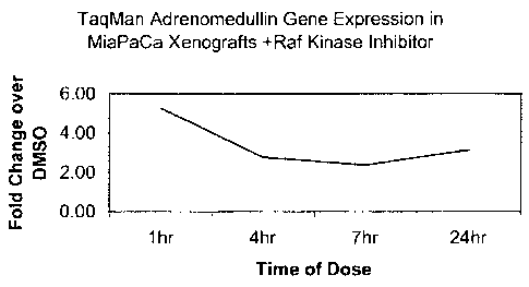
[028] Figure 2 depicts the expression changes of the adrenomedullin gene in
response to exposure
to a Raf kinase inhibitor using the TaqMan gene expression analysis method.
The Y-axis depicts
the fold change of expression of adrenomedullin in the compound-treated, tumor-
bearing animals
relative to vehicle treated, tumor-bearing animals. The X-axis depicts the
time, in hours, on day 9
post-treatment at which time the tumor samples were taken.
DETAILED DESCRIPTION OF THE INVENTION
[029] It is to be understood that this invention is not limited to the
particular methodology,
protocols, cell lines, animal species or genera, constructs, and reagents
described and as such may
vary. It is also to be understood that the terminology used herein is for the
purpose of describing
particular embodiments only, and is not intended to limit the scope of the
present invention which
will be limited only by the appended claims.
[030] It must be noted that as used herein and in the appended claims, the
singular forms "a,"
"and," and "the" include plural reference unless the context clearly dictates
otherwise. Thus, for
example, reference to "a gene" is a reference to one or more genes and
includes equivalents thereof
known to those skilled in the art, and so forth.
[031] Unless defined otherwise, all technical and scientific terms used herein
have the same
meaning as commonly understood to one of ordinary skill in the art to which
this invention
belongs. Although any methods, devices, and materials similar or equivalent to
those described
herein can be used in the practice or testing of the invention, the preferred
methods, devices and
materials are now described.
[032] All publications and patents mentioned herein are hereby incorporated
herein by reference
for the purpose of describing and disclosing, for example, the constructs and
methodologies that
are described in the publications which might be used in connection with the
presently described
6
CA 02499852 2005-03-22
WO 2004/028352 PCT/US2003/031032
invention. The publications discussed above and throughout the text are
provided solely for their
disclosure prior to the filing date of the present application. Nothing herein
is to be construed as
an admission that the inventors are not entitled to antedate such disclosure
by virtue of prior
invention.
Definitions
[033] For convenience, the meaning of certain terms and phrases employed in
the specification,
examples, and appended claims are provided below.
[034] An "address" on an array (e.g., a microarray) refers to a location at
which an element, for
example, an oligonucleotide, is attached to the solid surface of the array.
[035] The term "agonist," as used herein, is meant to refer to an agent that
mimics or up-regulates
(e.g., potentiates or supplements) the bioactivity of a protein. An agonist
may be a wild-type
protein or derivative thereof having at least one bioactivity of the wild-type
protein. An agonist
may also be a compound that up-regulates expression of a gene or which
increases at least one
bioactivity of a protein. An agonist can also be a compound which increases
the interaction of a
polypeptide with another molecule, for example, a target peptide or nucleic
acid.
[036] "Amplification," as used herein, relates to the production of additional
copies of a nucleic
acid sequence. For example, amplification may be carried out using polymerase
chain reaction
(PCR) technologies which are well known in the art. (see, e.g., Dieffenbach
and Dveksler (1995)
PCR Primer, A Laboratory Manual, Cold Spring Harbor Press, Plainview, N.Y.)
[037] "Antagonist," as used herein, is meant to refer to an agent that down-
regulates (e.g.,
suppresses or inhibits) at least one bioactivity of a protein. For example, a
Raf kinase inhibitor is
an example of such an antagonist. An antagonist may be a compound which
inhibits or decreases
the interaction between a protein and another molecule, for example, a target
peptide or enzyme
substrate. An antagonist may also be a compound that down-regulates expression
of a gene or
which reduces the amount of expressed protein present.
[038] The term "antibody," as used herein, is intended to include whole
antibodies, for example,
of any isotype (IgG, IgA, IgM, IgE, etc.), and includes fragments thereof
which are also
specifically reactive with a vertebrate (e.g., mammalian) protein. Antibodies
may be fragmented
using conventional techniques and the fragments screened for utility in the
same manner as
described above for whole antibodies. Thus, the term includes segments of
proteolytically-cleaved
or recombinantly-prepared portions of an antibody molecule that are capable of
selectively
reacting with a certain protein. Non-limiting examples of such proteolytic
and/or recombinant
fragments include Fab, F(ab')2, Fab', Fv, and single chain antibodies (scFv)
containing a V[L]
and/or V[H] domain joined by a peptide linker. The scFv's may be covalently or
non-covalently
CA 02499852 2005-03-22
WO 2004/028352 PCT/US2003/031032
linked to form antibodies having two or more binding sites. The subject
invention includes
polyclonal, monoclonal, or other purified preparations of antibodies and
recombinant antibodies.
[039] The terms "array" or "matrix" refer to an arrangement of addressable
locations or
"addresses" on a device. The locations can be arranged in two-dimensional
arrays, three-
dimensional arrays, or other matrix formats. The number of locations may range
from several to at
least hundreds of thousands. Most importantly, each location represents a
totally independent
reaction site. A "nucleic acid array" refers to an array containing nucleic
acid probes, such as
oligonucleotides or larger portions of genes. The nucleic acid on the array is
preferably single-
stranded. Arrays wherein the probes are oligonucleotides are referred to as
"oligonucleotide
arrays" or "oligonucleotide chips." A "microarray," also referred to herein as
a "biochip" or
"biological chip," is an array of regions having a density of discrete regions
of at least about
100/cm2, and preferably at least about 1000/cm2. The regions in a microarray
have typical
dimensions, for example, diameters, in the range of between about 10-250 pm,
and are separated
from other regions in the array by about the same distance.
[040] "Biological activity" or "bioactivity" or "activity" or "biological
function," which are used
interchangeably, herein mean an effector or antigenic function that is
directly or indirectly
performed by a polypeptide (whether in its native or denatured conformation),
or by any
subsequence thereof. Biological activities include binding to polypeptides,
binding to other
proteins or molecules, activity as a DNA binding protein, as a transcription
regulator, ability to
bind damaged DNA, etc. A bioactivity can be modulated by directly affecting
the subject
polypeptide. Alternatively, a bioactivity can be altered by modulating the
level of the polypeptide,
such as by modulating expression of the corresponding gene.
[041] The term "biological sample," as used herein, refers to a sample
obtained from an organism
or from components (e.g., cells) of an organism. The sample may be of any
biological tissue or
fluid. The sample may be a sample which is derived from a patient. Such
samples include, but are
not limited to, sputum, blood, blood cells (e.g., white cells), tissue or
biopsy samples (e.g., tumor
biopsy), urine, peritoneal fluid, and pleural fluid, or cells therefrom.
Biological samples may also
include sections of tissues such as frozen sections taken for histological
purposes.
[042] The term "biomarker" or "marker" encompasses a broad range of intra- and
extra-cellular
events as well as whole-organism physiological changes. Biomarkers may be
represent essentially
any aspect of cell function, for example, but not limited to, levels or rate
of production of signaling
molecules, transcription factors, metabolites, gene transcripts as well as
post-translational
modifications of proteins. Biomarkers may include whole genome analysis of
transcript levels or
whole proteome analysis of protein levels and/or modifications.
[043] A biomarker may also refer to a gene or gene product which is up- or
down-regulated in a
compound-treated, diseased cell of a subject having the disease compared to an
untreated diseased
CA 02499852 2005-03-22
WO 2004/028352 PCT/US2003/031032
cell. That is, the gene or gene product is sufficiently specific to the
treated cell that it may be used,
optionally with other genes or gene products, to identify, predict, or detect
efficacy of a small
molecule. Thus, a biomarker is a gene or gene product that is characteristic
of efficacy of a
compound in a diseased cell or the response of that diseased cell to treatment
by the compound.
[044] A nucleotide sequence is "complementary" to another nucleotide sequence
if each of the
bases of the two sequences match, that is, are capable of forming Watson-Crick
base pairs. The
term "complementary strand" is used herein interchangeably with the term
"complement." The
complement of a nucleic acid strand may be the complement of a coding strand
or the complement
of a non-coding strand.
[045] "Detection agents of genes" refers to agents that can be used to
specifically detect the gene
or other biological molecules relating to it, for example, RNA transcribed
from the gene or
polypeptides encoded by the gene. Exemplary detection agents are nucleic acid
probes, which
hybridize to nucleic acids corresponding to the gene, and antibodies.
[046] The term "cancer" includes, but is not limited to, solid tumors, such as
cancers of the breast,
respiratory tract, brain, reproductive organs, digestive tract, urinary tract,
eye, liver, skin, head and
neck, thyroid, parathyroid, and their distant metastases. The term also
includes lymphomas,
sarcomas, and leukemias.
[047] Examples of breast cancer include, but are not limited to, invasive
ductal carcinoma,
invasive lobular carcinoma, ductal carcinoma in situ, and lobular carcinoma in
situ.
[048] Examples of cancers of the respiratory tract include, but are not
limited to, small-cell and
non-small-cell lung carcinoma, as well as bronchial adenoma and
pleuropulmonary blastoma.
[049] Examples of brain cancers include, but are not limited to, brain stem
and hypophtalmic
glioma, cerebellar and cerebral astrocytoma, medulloblastoma, ependymoma, as
well as
neuroectodermal and pineal tumor.
[050] Tumors of the male reproductive organs include, but are not limited to,
prostate and
testicular cancer. Tumors of the female reproductive organs include, but are
not limited to,
endometrial, cervical, ovarian, vaginal, and vulvar cancer, as well as sarcoma
of the uterus.
[051] Tumors of the digestive tract include, but are not limited to, anal,
colon, colorectal,
esophageal, gallbladder, gastric, pancreatic, rectal, small-intestine, and
salivary gland cancers.
[052] Tumors of the urinary tract include, but are not limited to, bladder,
penile, kidney, renal
pelvis, ureter, and urethral cancers.
[053] Eye cancers include, but are not limited to, intraocular melanoma and
retinoblastoma.
[054] Examples of liver cancers include, but are not limited to,
hepatocellular carcinoma (liver
cell carcinomas with or without fibrolamellar variant), cholangiocarcinoma
(intrahepatic bile duct
carcinoma), and mixed hepatocellular cholangiocarcinoma.
CA 02499852 2005-03-22
WO 2004/028352 PCT/US2003/031032
[055] Skin cancers include, but are not limited to, squamous cell carcinoma,
Kaposi's sarcoma,
malignant melanoma, Merkel cell skin cancer, and non-melanoma skin cancer.
[056] Head-and-neck cancers include, but are not limited to, laryngeal /
hypopharyngeal /
nasopharyngeal / oropharyngeal cancer, and lip and oral cavity cancer.
[057] Lymphomas include, but are not limited to, A>DS-related lymphoma, non-
Hodgkin's
lymphoma, cutaneous T-cell lymphoma, Hodgkin's disease, and lymphoma of the
central nervous
system.
[058] Sarcomas include, but are not limited to, sarcoma of the soft tissue,
osteosarcoma,
malignant fibrous histiocytoma, lymphosarcoma, and rhabdomyosarcoma.
[059] Leukemias include, but are not limited to, acute myeloid leukemia, acute
lymphoblastic
leukemia, chronic lymphocytic leukemia, chronic myelogenous leukemia, and
hairy cell leukemia.
[060] "A diseased cell of cancer" refers to a cell present in subjects having
cancer. That is, a cell
which is a modified form of a normal cell and is not present in a subject not
having cancer, or a
cell which is present in significantly higher or lower numbers in subjects
having cancer relative to
subjects not having cancer.
[061] The term "equivalent" is understood to include nucleotide sequences
encoding functionally
equivalent polypeptides. Equivalent nucleotide sequences may include sequences
that differ by
one or more nucleotide substitutions, additions, or deletions, such as allelic
variants.
[062] The term "expression profile," which is used interchangeably herein with
"gene expression
profile" and "fingerprint" of a cell refers to a set of values representing
mRNA levels of one or
more genes in a cell. An expression profile preferably comprises values
representing expression
levels of at least about 10 genes, preferably at least about 50, 100, 200 or
more genes. Expression
profiles may also comprise an mRNA level of a gene which is expressed at
similar levels in
multiple cells and conditions (e.g., a housekeeping gene such as GAPDH). For
example, an
expression profile of a diseased cell of cancer refers to a set of values
representing mRNA levels
of 10 or more genes in a diseased cell.
[063] The term "gene" refers to a nucleic acid sequence that comprises control
and coding
sequences necessary for the production of a polypeptide or precursor. The
polypeptide can be
encoded by a full length coding sequence or by any portion of the coding
sequence. The gene may
be derived in whole or in part from any source known to the art, including a
plant, a fungus, an
animal, a bacterial genome or episome, eukaryotic, nuclear or plasmid DNA,
cDNA, viral DNA, or
chemically synthesized DNA. A gene may contain one or more modifications in
either the coding
or the untranslated regions which could affect the biological activity or the
chemical structure of
the expression product, the rate of expression, or the manner of expression
control. Such
modifications include, but are not limited to, mutations, insertions,
deletions, and substitutions of
CA 02499852 2005-03-22
WO 2004/028352 PCT/US2003/031032
one or more nucleotides. The gene may constitute an uninterrupted coding
sequence or it may
include one or more introns, bound by the appropriate splice junctions.
[064] "Hybridization" refers to any process by which a strand of nucleic acid
binds with a
complementary strand through base pairing. For example, two single-stranded
nucleic acids
"hybridize" when they form a double-stranded duplex. The region of double-
strandedness may
include the full-length of one or both of the single-stranded nucleic acids,
or all of one single-
stranded nucleic acid and a subsequence of the other single-stranded nucleic
acid, or the region of
double-strandedness may include a subsequence of each nucleic acid.
Hybridization also includes
the formation of duplexes which contain certain mismatches, provided that the
two strands are still
forming a double-stranded helix. "Stringent hybridization conditions" refers
to hybridization
conditions resulting in essentially specific hybridization.
[065] The term "isolated," as used herein, with respect to nucleic acids, such
as DNA or RNA,
refers to molecules separated from other DNAs or RNAs, respectively, that are
present in the
natural source of the macromolecule. The term "isolated" as used herein also
refers to a nucleic
acid or peptide that is substantially free of cellular material, viral
material, culture medium when
produced by recombinant DNA techniques, or chemical precursors or other
chemicals when
chemically synthesized. Moreover, an "isolated nucleic acid" may include
nucleic acid fragments
which are not naturally occurring as fragments and would not be found in the
natural state. The
term "isolated" is also used herein to refer to polypeptides which are
isolated from other cellular
proteins and is meant to encompass both purified and recombinant polypeptides.
[066] As used herein, the terms "label" and "detectable label" refer to a
molecule capable of
detection, including, but not limited to, radioactive isotopes, fluorophores,
chemiluminescent
moieties, enzymes, enzyme substrates, enzyme cofactors, enzyme inhibitors,
dyes, metal ions,
ligands (e.g., biotin or haptens), and the like. The term "fluorescer" refers
to a substance or a
portion thereof which is capable of exhibiting fluorescence in the detectable
range. Particular
examples of labels which may be used in the present invention include
fluorescein, rhodamine,
dansyl, umbelliferone, Texas red, luminol, NADPH, alpha - beta -galactosidase,
and horseradish
peroxidase.
[067] As used herein, the term "level of expression" refers to the measurable
expression level of a
given nucleic acid. The level of expression of a nucleic acid is determined by
methods well known
in the art. The term "differentially expressed" or "differential expression"
refers to an increase or
decrease in the measurable expression level of a given nucleic acid. As used
herein, "differentially
expressed" or "differential expression" means the difference in the level of
expression of a nucleic
acid is at least 1.4-fold or more in two samples used for comparison, both of
which are compared
to the same normal standard sample. "Differentially expressed" or
"differential expression"
according to the invention also means a 1.4-fold, or more, up to and including
2-fold, 5-fold, 10-
11
CA 02499852 2005-03-22
WO 2004/028352 PCT/US2003/031032
fold, 20-fold, 50-fold or more difference in the level of expression of a
nucleic acid in two samples
used for comparison. A nucleic acid is also said to be "differentially
expressed" in two samples if
one of the two samples contains no detectable expression of a given nucleic
acid, provided that the
detectably expressed nucleic acid is expressed at +/- at least 1.4 fold.
Differential expression of a
nucleic acid sequence is "inhibited" the difference in the level of expression
of the nucleic acid in
two or more samples used for comparison is altered such that it is no longer
at least a 1.4 fold
difference. Absolute quantification of the level of expression of a nucleic
acid may be
accomplished by including a known concentrations) of one or more control
nucleic acid species,
generating a standard curve based on the amount of the control nucleic acid
and extrapolating the
expression level of the "unknown" nucleic acid species from the hybridization
intensities of the
unknown with respect to the standard curve.
[068] As used herein, the term "nucleic acid" refers to polynucleotides such
as deoxyribonucleic
acid (DNA) and, where appropriate, ribonucleic acid (RNA). The term should
also be understood
to include, as equivalents, analogs of either RNA or DNA made from nucleotide
analogs and, as
applicable to the embodiment being described, single-stranded (sense or
antisense) and double-
stranded polynucleotides. Chromosomes, cDNAs, mRNAs, rRNAs, and ESTs are
representative
examples of molecules that may be referred to as nucleic acids.
[069] The term "oligonucleotide" as used herein refers to a nucleic acid
molecule comprising, for
example, from about 10 to about 1000 nucleotides. Oligonucleotides for use in
the present
invention are preferably from about 15 to about 150 nucleotides, more
preferably from about 150
to about 1000 in length. The oligonucleotide may be a naturally occurring
oligonucleotide or a
synthetic oligonucleotide. Oligonucleotides may be prepared by the
phosphoramidite method
(Beaucage and Carruthers, Tetrahedron Lett. 22:1859-62, 1981), or by the
triester method
(Matteucci, et al., J. Am. Chem. Soc. 103:3185, 1981), or by other chemical
methods known in the
art.
[070] The term "patient" or "subject" as used herein includes mammals (e.g.,
humans and
animals).
[071] As used herein, a nucleic acid or other molecule attached to an array is
referred to as a
"probe" or "capture probe." When an array contains several probes
corresponding to one gene,
these probes are referred to as a "gene-probe set." A gene-probe set may
consist of, for example,
about 2 to about 20 probes, preferably from about 2 to about 10 probes, and
most preferably about
S probes.
[072] The "profile" of a cell's biological state refers to the levels of
various constituents of a cell
that are known to change in response to drug treatments and other
perturbations of the biological
state of the cell. Constituents of a cell include, for example, levels of RNA,
levels of protein
abundances, or protein activity levels.
12
CA 02499852 2005-03-22
WO 2004/028352 PCT/US2003/031032
[073] The term "protein" is used interchangeably herein with the terms
"peptide" and
"polypeptide."
[074] An expression profile in one cell is "similar" to an expression profile
in another cell when
the level of expression of the genes in the two profiles are sufficiently
similar that the similarity is
indicative of a common characteristic, for example, the same type of cell.
Accordingly, the
expression profiles of a first cell and a second cell are similar when at
least 75% of the genes that
are expressed in the first cell are expressed in the second cell at a level
that is within a factor of
two relative to the first cell.
[075] "Small molecule," as used herein, refers to a composition with a
molecular weight of less
than about S kD and most preferably less than about 4 kD. Small molecules can
be nucleic acids,
peptides, polypeptides, peptidomimetics, carbohydrates, lipids, or other
organic or inorganic
molecules. Many pharmaceutical companies have extensive libraries of chemical
and/or biological
mixtures, often fungal, bacterial, or algal extracts, which can be screened
with any of the assays of
the invention to identify compounds that modulate a bioactivity.
[076] The term "specific hybridization" of a probe to a target site of a
template nucleic acid refers
to hybridization of the probe predominantly to the target, such that the
hybridization signal can be
clearly interpreted. As further described herein, such conditions resulting in
specific hybridization
vary depending on the length of the region of homology, the GC content of the
region, and the
melting temperature ("Tin") of the hybrid. Thus, hybridization conditions may
vary in salt
content, acidity, and temperature of the hybridization solution and the
washes.
[077] A "variant" of polypeptide refers to a polypeptide having an amino acid
sequence in which
one or more amino acid residues is altered. The variant may have
"conservative" changes,
wherein a substituted amino acid has similar structural or chemical properties
(e.g., replacement of
leucine with isoleucine). A variant may also have "nonconservative" changes
(e.g., replacement of
glycine with tryptophan). Analogous minor variations may include amino acid
deletions or
insertions, or both. Guidance in determining which amino acid residues may be
substituted,
inserted, or deleted without abolishing biological or immunological activity
may be identified
using computer programs well known in the art, for example, LASERGENE software
(DNASTAR).
[078] The term "variant," when used in the context of a polynucleotide
sequence, may encompass
a polynucleotide sequence related to that of a particular gene or the coding
sequence thereof. This
definition may also include, for example, "allelic," "splice," "species," or
"polymorphic" variants.
A splice variant may have significant identity to a reference molecule, but
will generally have a
greater or lesser number of polynucleotides due to alternate splicing of exons
during mRNA
processing. The corresponding polypeptide may possess additional functional
domains or an
absence of domains. Species variants are polynucleotide sequences that vary
from one species to
13
CA 02499852 2005-03-22
WO 2004/028352 PCT/US2003/031032
another. The resulting polypeptides generally will have significant amino acid
identity relative to
each other. A polymorphic variant is a variation in the polynucleotide
sequence of a particular
gene between individuals of a given species. Polymorphic variants also may
encompass "single
nucleotide polymorphisms" (SNPs) in which the polynucleotide sequence varies
by one base. The
presence of SNPs may be indicative of, for example, a certain population, a
disease state, or a
propensity for a disease state.
[079] An aspect of the invention is directed to the identification of agents
capable of modulating
the differentiation and proliferation of cells characterized by aberrant
proliferation. More
specifically, the invention relates to methods of screening candidate
compounds or substances for
their ability to regulate the differential expression of nucleic acid
sequences. That is, if a nucleic
acid sequence is overexpressed in cancer cells, then the candidate compounds
are screened for
their ability to decrease expression, and if a nucleic acid sequence is
underexpressed in cancer
cells, then a test compound is screened for its ability to increase
expression. In addition, the
invention relates to screening assays to identify test compounds or substances
which modulate the
activity of one or more polypeptides which are encoded by the differentially
expressed sequences
described herein. In this regard, the invention provides assays for
determining compounds that
modulate the expression of marker nucleic acids and/or alter the bioactivity
of the encoded
polypeptide.
Screeningfor modulation of differential expression
[080] Drug screening is performed by adding a test compound (e.g., Raf kinase
inhibitor) to a
sample of cells, and monitoring the effect. A parallel sample which does not
receive the test
compound is also monitored as a control. The treated and untreated cells are
then compared by
any suitable phenotypic criteria, including but not limited to microscopic
analysis, viability testing,
ability to replicate, histological examination, the level of a particular RNA
or polypeptide
associated with the cells, the level of enzymatic activity expressed by the
cells or cell lysates, and
the ability of the cells to interact with other cells or compounds.
Differences between treated and
untreated cells indicates effects attributable to the test compound.
[081] Desirable effects of a test compound include an effect on any phenotype
that was conferred
by the cancer-associated marker nucleic acid sequence. Examples include a test
compound that
limits the overabundance of mRNA, limits production of the encoded protein, or
limits the
functional effect of the protein. The effect of the test compound would be
apparent when
comparing results between treated and untreated cells.
[082] The invention thus, also encompasses methods of screening for agents
(e.g., Raf kinase
inhibitor) which inhibit or enhance the expression of the nucleic acid markers
in vitro, comprising
exposing a cell or tissue in which the marker nucleic acid mRNA (e.g.,
adrenomedullin) is
14
CA 02499852 2005-03-22
WO 2004/028352 PCT/US2003/031032
detectable in cultured cells to an agent in order to determine whether the
agent is capable of
inhibiting or enhancing production of the mRNA; and determining the level of
mRNA in the
exposed cells or tissue, wherein a decrease in the level of the mRNA after
exposure of the cell line
to the agent is indicative of inhibition of the marker nucleic acid mRNA
production and an
increase in mRNA levels is indicative of enhancement of maker mRNA production.
[083] Alternatively, the screening method may include in vitro screening of a
cell or tissue in
which marker protein is detectable in cultured cells to an agent suspected of
inhibiting or
enhancing production of the marker protein; and determining the level of the
marker protein in the
cells or tissue, wherein a decrease in the level of marker protein after
exposure of the cells or tissue
to the agent is indicative of inhibition of marker protein production and an
increase on the level of
marker protein is indicative of enhancement of marker protein production.
[084] The invention also encompasses in vivo methods of screening for agents
which inhibit or
enhance expression of the marker nucleic acids, comprising exposing a subject
having tumor cells
in which marker mRNA or protein is detectable to an agent suspected of
inhibiting or enhancing
production of marker mRNA or protein; and determining the level of marker mRNA
or protein in
tumor cells of the exposed mammal. A decrease in the level of marker mRNA or
protein after
exposure of the subject to the agent is indicative of inhibition of marker
nucleic acid expression
and an increase in the level of marker mRNA or protein is indicative of
enhancement of marker
nucleic acid expression.
[085] Accordingly, the invention provides a method comprising incubating a
cell expressing the
marker nucleic acids with a test compound and measuring the mRNA or protein
level. The
invention further provides a method for quantitatively determining the level
of expression of the
marker nucleic acids in a cell population, and a method for determining
whether an agent is
capable of increasing or decreasing the level of expression of the marker
nucleic acids in a cell
population. The method for determining whether an agent is capable of
increasing or decreasing
the level of expression of the marker nucleic acids in a cell population
comprises the steps of (a)
preparing cell extracts from control and agent-treated cell populations, (b)
isolating the marker
polypeptides from the cell extracts, and (c) quantifying (e.g., in parallel)
the amount of an
immunocomplex formed between the marker polypeptide and an antibody specific
to said
polypeptide. The marker polypeptides of this invention may also be quantified
by assaying for its
bioactivity. Agents that induce an increase in the marker nucleic acid
expression may be identified
by their ability to increase the amount of immunocomplex formed in the treated
cell as compared
with the amount of the immunocomplex formed in the control cell. In a similar
manner, agents
that decrease expression of the marker nucleic acid may be identified by their
ability to decrease
the amount of the immunocomplex formed in the treated cell extract as compared
to the control
cell.
IS
CA 02499852 2005-03-22
WO 2004/028352 PCT/US2003/031032
[086] The present invention provides isolated nucleic acid sequences which are
differentially
regulated in cancer, and a method for identifying such sequences. The present
invention provides
a method for identifying a nucleotide sequence which is differentially
regulated in a subject with
cancer, comprising: hybridizing a nucleic acid sample corresponding to RNA
obtained from the
subject to a nucleic acid sample comprising one or more nucleic acid molecules
of known identity;
and measuring the hybridization of the nucleic acid sample to the one or more
nucleic acid
molecules of known identity, wherein a two-fold difference in the
hybridization of the nucleic acid
sample to the one or more nucleic acid molecules of known identity relative to
a nucleic acid
sample obtained from a subject without cancer is indicative of the
differential expression of the
nucleotide sequence in a subject with cancer.
[087] Generally, the present invention provides a method for identifying
nucleic acid sequences
which are differentially regulated in a subject with cancer comprising
isolating messenger RNA
from a subject, generating cRNA from the mRNA sample, hybridizing the cRNA to
a microarray
comprising a plurality of nucleic acid molecules stably associated with
discrete locations on the
array, and identifying patterns of hybridization of the cRNA to the array.
According to the present
invention, a nucleic acid molecule which hybridizes to a given location on the
array is said to be
differentially regulated if the hybridization signal is at least two-fold
higher or lower than the
hybridization signal at the same location on an identical array hybridized
with a nucleic acid
sample obtained from a subject that does not have cancer.
Microarrays for Determining the Level of Expression of Genes
[088] Determining gene expression levels may be accomplished utilizing
microarrays. Generally,
the following steps may be involved: (a) obtaining an mRNA sample from a
subject and preparing
labeled nucleic acids therefrom (the "target nucleic acids" or "targets"); (b)
contacting the target
nucleic acids with an array under conditions sufficient for the target nucleic
acids to bind to the
corresponding probes on the array, for example, by hybridization or specific
binding; (c) optional
removal of unbound targets from the array; (d) detecting the bound targets,
and (e) analyzing the
results, for example, using computer based analysis methods. As used herein,
"nucleic acid
probes" or "probes" are nucleic acids attached to the array, whereas "target
nucleic acids" are
nucleic acids that are hybridized to the array.
(089] Nucleic acid specimens may be obtained from a subject to be tested using
either "invasive"
or "non-invasive" sampling means. A sampling means is said to be "invasive" if
it involves the
collection of nucleic acids from within the skin or organs of an animal
(including murine, human,
ovine, equine, bovine, porcine, canine, or feline animal). Examples of
invasive methods include,
for example, blood collection, semen collection, needle biopsy, pleural
aspiration, umbilical cord
biopsy. Examples of such methods are discussed by Kim, et al., (J. Virol.
66:3879-3882, 1992);
16
CA 02499852 2005-03-22
WO 2004/028352 PCT/US2003/031032
Biswas, et al., (Ann. NY Acad. Sci. 590:582-583, 1990); and Biswas, et al.,
(J. Clin. Microbiol.
29:2228-2233, 1991).
[090] In contrast, a "non-invasive" sampling means is one in which the nucleic
acid molecules are
recovered from an internal or external surface of the animal. Examples of such
"non-invasive"
sampling means include, for example, "swabbing," collection of tears, saliva,
urine, fecal material,
sweat or perspiration, hair.
[091] In one embodiment of the present invention, one or more cells from the
subject to be tested
are obtained and RNA is isolated from the cells. In a preferred embodiment, a
sample of
peripheral blood leukocytes (PBLs) cells is obtained from the subject. It is
also possible to obtain
a cell sample from a subject, and then to enrich the sample for a desired cell
type. For example,
cells may be isolated from other cells using a variety of techniques, such as
isolation with an
antibody binding to an epitope on the cell surface of the desired cell type.
Where the desired cells
are in a solid tissue, particular cells may be dissected, for example, by
microdissection or by laser
capture microdissection (LCM) (see, e.g., Bonner, et al., Science 278:1481,
1997; Emmert-Buck,
et al., Science 274:998, 1996; Fend, et al., Am. J. Path. 154:61, 1999; and
Murakami, et al.,
Kidney Int. 58:1346, 2000).
[092] RNA may be extracted from tissue or cell samples by a variety of
methods, for example,
guanidium thiocyanate lysis followed by CsCI centrifugation (Chirgwin, et al.,
Biochemistry
18:5294-5299, 1979). RNA from single cells may be obtained as described in
methods for
preparing cDNA libraries from single cells (see, e.g., Dulac, Curr. Top. Dev.
Biol. 36:245, 1998;
Jena, et al., J. Immunol. Methods 190:199, 1996).
[093] The RNA sample can be further enriched for a particular species. In one
embodiment, for
example, poly(A)+ RNA may be isolated from an RNA sample. In another
embodiment, the RNA
population may be enriched for sequences of interest by primer-specific cDNA
synthesis, or
multiple rounds of linear amplification based on cDNA synthesis and template-
directed in vitro
transcription (see, e.g., Wang, et al., Proc. Natl. Acad. Sci. USA 86:9717,
1989; Dulac, et al.,
supra; Jena, et al., supra). In addition, the population of RNA, enriched or
not in particular
species or sequences, may be further amplified by a variety of amplification
methods including, for
example, PCR; ligase chain reaction (LCR) (see, e.g., Wu and Wallace, Genomics
4:560, 1989;
Landegren, et al., Science 241:1077, 1988); self sustained sequence
replication (SSR) (see, e.g.,
Guatelli, et al., Proc. Natl. Acad. Sci. USA 87:1874, 1990); nucleic acid
based sequence
amplification (NASBA) and transcription amplification (see, e.g., Kwoh, et
al., Proc. Natl. Acad.
Sci. USA 86:1173, 1989). Methods for PCR technology are well known in the art
(see, e.g., PCR
Technology: Principles and Applications for DNA Amplification (ed. H. A.
Erlich, Freeman Press,
N.Y., N.Y., 1992); PCR Protocols: A Guide to Methods and Applications (eds.
Innis, et al.,
Academic Press, San Diego, Calif., 1990); Manila, et al., Nucleic Acids Res.
19:4967, 1991;
17
CA 02499852 2005-03-22
WO 2004/028352 PCT/US2003/031032
Eckert, et al., PCR Methods and Applications 1:17, 1991; PCR (eds. McPherson
et al., IRL Press,
Oxford); and U.S. Pat. No. 4,683,202). Methods of amplification are described,
for example, by
Ohyama, et al., (BioTechniques 29:530, 2000); Luo, et al., (Nat. Med. 5:117,
1999); Hegde, et al.,
(BioTechniques 29:548, 2000); Kacharmina, et al., (Meth. Enzymol. 303:3,
1999); Livesey, et al.,
Curr. Biol. 10:301, 2000); Spirin, et al., (Invest. Ophtalmol. Vis. Sci.
40:3108, 1999); and Sakai, et
al., (Anal. Biochem. 287:32, 2000). RNA amplification and cDNA synthesis may
also be
conducted in cells in situ (see, e.g., Eberwine, et al. Proc. Natl. Acad. Sci.
USA 89:3010, 1992).
[094] The nucleic acid molecules may be labeled to pernlit detection of
hybridization of the
nucleic acid molecules to a microarray. That is, the probe may comprise a
member of a signal
producing system and thus, is detectable, either directly or through combined
action with one or
more additional members of a signal producing system. For example, the nucleic
acids may be
labeled with a fluorescently labeled dNTP (see, e.g., Kricka, 1992,
Nonisotopic DNA Probe
Techniques, Academic Press San Diego, Cali~), biotinylated dNTPs or rNTP
followed by addition
of labeled streptavidin, chemiluminescent labels, or isotopes. Another example
of labels include
"molecular beacons" as described in Tyagi and Kramer (Nature Biotech. 14:303,
1996).
Hybridization may be also be determined, for example, by plasmon resonance
(see, e.g., Thiel, et
al. Anal. Chem. 69:4948, 1997).
[095J In one embodiment, a plurality (e.g., 2, 3, 4, S, or more) of sets of
target nucleic acids are
labeled and used in one hybridization reaction ("multiplex" analysis). For
example, one set of
nucleic acids may correspond to RNA from one cell and another set of nucleic
acids may
correspond to RNA from another cell. The plurality of sets of nucleic acids
may be labeled with
different labels, for example, different fluorescent labels (e.g., fluorescein
and rhodamine) which
have distinct emission spectra so that they can be distinguished. The sets may
then be mixed and
hybridized simultaneously to one microarray (see, e.g., Shena, et al., Science
270:467-470, 1995).
[096] Microarrays for use according to the invention include one or more
probes of genes
characteristic of small molecule efficacy. In a preferred embodiment, the
microarray comprises
probes corresponding to one or more of genes selected from the group
consisting of genes which
are up-regulated in cancer and genes which are down-regulated in cancer. The
microarray may
comprise probes corresponding to at least 10, preferably at least 20, at least
50, at least 100 or at
least 1000 genes characteristic of small molecule efficacy.
[097] There may be one or more than one probe corresponding to each gene on a
microarray. For
example, a microarray may contain from 2 to 20 probes corresponding to one
gene and preferably
about 5 to 10. The probes may correspond to the full-length RNA sequence or
complement
thereof of genes characteristic of small molecule efficacy, or the probe may
correspond to a
portion thereof, which portion is of sufficient length to permit specific
hybridization. Such probes
may comprise from about 50 nucleotides to about 100, 200, 500, or 1000
nucleotides or more than
18
CA 02499852 2005-03-22
WO 2004/028352 PCT/US2003/031032
1000 nucleotides. As further described herein, microarrays may contain
oligonucleotide probes,
consisting of about 10 to 50 nucleotides, preferably about 15 to 30
nucleotides and more
preferably about 20-25 nucleotides. The probes are preferably single-stranded
and will have
sufficient complementarity to its target to provide for the desired level of
sequence specific
hybridization.
[098] Typically, the arrays used in the present invention will have a site
density of greater than
100 different probes per cm2. Preferably, the arrays will have a site density
of greater than
500/cm2, more preferably greater than about 1000/cmz, and most preferably,
greater than about
10,000/cm2. Preferably, the arrays will have more than 100 different probes on
a single substrate,
more preferably greater than about 1000 different probes, still more
preferably, greater than about
10,000 different probes and most preferably, greater than 100,000 different
probes on a single
substrate.
[099] A number of different microarray configurations and methods for their
production are
known to those of skill in the art and are disclosed in U.S. Patent Nos:
5,242,974; 5,384,261;
5,405,783; 5,412,087; 5,424,186; 5,429,807; 5,436,327; 5,445,934; 5,556,752;
5,405,783;
5,412,087; 5,424,186; 5,429,807; 5,436,327; 5,472,672; 5,527,681; 5,529,756;
5,545,531;
5,554,501; 5,561,071; 5,571,639; 5,593,839; 5,624,711; 5,700,637; 5,744,305;
5,770,456;
5,770,722; 5,837,832; 5,856,101; 5,874,219; 5,885,837; 5,919,523; 6,022,963;
6,077,674; and
6,156,501; Shena, et al., Tibtech 16:301, 1998; Duggan, et al., Nat. Genet.
21:10, 1999; Bowtell, et
al., Nat. Genet. 21:25, 1999; Lipshutz, et al., 21 Nature Genet. 20-24, 1999;
Blanchard, et al., 11
Biosensors and Bioelectronics, 687-90, 1996; Maskos, et al., 21 Nucleic Acids
Res. 4663-69,
1993; Hughes, et al., Nat. Biotechol. (2001) 19:342; the disclosures of which
are herein
incorporated by reference. Patents describing methods of using arrays in
various applications
include: U.S. Pat. Nos. 5,143,854; 5,288,644; 5,324,633; 5,432,049; 5,470,710;
5,492,806;
5,503,980; 5,510,270; 5,525,464; 5,547,839; 5,580,732; 5,661,028; 5,848,659;
and 5,874,219; the
disclosures of which are herein incorporated by reference.
[100] Arrays preferably include control and reference nucleic acids. Control
nucleic acids
include, for example, prokaryotic genes such as bioB, bioC and bioD, cre from
P1 bacteriophage
or polyA controls, such as dap, lys, phe, thr, and trp. Reference nucleic
acids allow the
normalization of results from one experiment to another and the comparison of
multiple
experiments on a quantitative level. Exemplary reference nucleic acids include
housekeeping
genes of known expression levels, for example, GAPDH, hexokinase, and actin.
[101] In one embodiment, an array of oligonucleotides may be synthesized on a
solid support.
Exemplary solid supports include glass, plastics, polymers, metals,
metalloids, ceramics, organics,
etc. Using chip masking technologies and photoprotective chemistry, it is
possible to generate
ordered arrays of nucleic acid probes. These arrays, which are known, for
example, as "DNA
19
CA 02499852 2005-03-22
WO 2004/028352 PCT/US2003/031032
chips" or very large scale immobilized polymer arrays ("VLSIPSTM" arrays), may
include millions
of defined probe regions on a substrate having an area of about 1 cmz to
several cm2, thereby
incorporating from a few to millions of probes (see, e.g., U.S. Patent No.
5,631,734).
[102] To compare expression levels, labeled nucleic acids may be contacted
with the array under
conditions sufficient for binding between the target nucleic acid and the
probe on the array. In a
preferred embodiment, the hybridization conditions may be selected to provide
for the desired
level of hybridization specificity; that is, conditions sufficient for
hybridization to occur between
the labeled nucleic acids and probes on the microarray.
[103] Hybridization may be carried out in conditions permitting essentially
specific hybridization.
The length and GC content of the nucleic acid will determine the thermal
melting point and thus,
the hybridization conditions necessary for obtaining specific hybridization of
the probe to the
target nucleic acid. These factors are well known to a person of skill in the
art, and may also be
tested in assays. An extensive guide to nucleic acid hybridization may be
found in Tijssen, et al.
(Laboratory Techniques in Biochemistry and Molecular Biology, Vol. 24:
Hybridization With
Nucleic Acid Probes, P. Tijssen, ed. Elsevier, N.Y., (1993)).
[104] The methods described above result in the production of hybridization
patterns of labeled
target nucleic acids on the array surface. The resultant hybridization
patterns of labeled nucleic
acids may be visualized or detected in a variety of ways, with the particular
manner of detection
selected based on the particular label of the target nucleic acid.
Representative detection means
include scintillation counting, autoradiography, fluorescence measurement,
colorimetric
measurement, light emission measurement, light scattering, and the like.
[105] One such method of detection utilizes an array scanner that is
commercially available
(Affymetrix, Santa Clara, CA), for example, the 41 T~M Arrayer, the 418"N
Array Scanner, or the
Agilent GeneArrayTM Scanner. This scanner is controlled from a system computer
with an
interface and easy-to-use software tools. The output may be directly imported
into or directly read
by a variety of software applications. Preferred scanning devices are
described in, for example,
U.S. Patent Nos. 5,143,854 and 5,424,186.
[106] For fluorescent labeled probes, the fluorescence emissions at each site
of a transcript array
may be, preferably, detected by scanning confocal laser microscopy.
Alternatively, a laser may be
used that allows simultaneous specimen illumination at wavelengths specific to
the two
fluorophores and emissions from the two fluorophores may be analyzed
simultaneously (see, e.g.,
Shalom et al., Genome Res. 6:639-645, 1996). In a preferred embodiment, the
arrays may be
scanned with a laser fluorescent scanner with a computer controlled X-Y stage
and a microscope
objective. Fluorescence laser scanning devices are described in Shalom et al.,
supra.
[107] Various algorithms are available for analyzing gene expression data, for
example, the type
of comparisons to perform. In certain embodiments, it is desirable to group
genes that are co-
CA 02499852 2005-03-22
WO 2004/028352 PCT/US2003/031032
regulated. This allows for the comparison of large numbers of profiles. A
preferred embodiment
for identifying such groups of genes involves clustering algorithms (for
reviews of clustering
algorithms, see, e.g., Fukunaga, 1990, Statistical Pattern Recognition, 2nd
Ed., Academic Press,
San Diego; Everitt, 1974, Cluster Analysis, London: Heinemann Educ. Books;
Hartigan, 1975,
Clustering Algorithms, New York: Wiley; Sneath and Sokal, 1973, Numerical
Taxonomy,
Freeman; Anderberg, 1973, Cluster Analysis for Applications, Academic Press:
New York).
Biomarker Discovery
[108] Expression patterns may be used to derive a panel of biomarkers that can
be used to predict
the efficacy of drug treatment in the patients. The biomarkers may consist of
gene expression
levels from microarray experiments on RNA isolated from biological samples,
RNA isolated from
frozen samples of tumor biopsies, or mass spectrometry-derived protein masses
in the serum.
[109] Although the precise mechanism for data analysis will depend upon the
exact nature of the
data, a typical procedure for developing a panel of biomarkers is as follows.
The data (gene
expression levels or mass spectra) are collected for each patient prior to
treatment. As the study
progresses, the patients are classified according to their response to the
drug treatment; either as
efficacious or non-efficacious. Multiple levels of efficacy can be
accommodated in a data model,
but a binary comparison is considered optimal, particularly if the patient
population is less than
several hundred. Assuming adequate numbers of patients in each class, the
protein and/or gene
expression data may be analyzed by a number of techniques known in the art.
Many of the
techniques are derived from traditional statistics as well from the field of
machine learning. These
techniques serve two purposes:
1. Reduce the dimensionality of data - In the case of mass spectra or gene
expression
microarrays, data is reduced from many thousands of individual data points to
bout three to ten.
The reduction is based upon the predictive power of the data points when taken
as a set.
2. Training - These three to ten data points are then used to train multiple
machine learning
algorithms which then "learn" to recognize, in this case, patterns of protein
masses or gene
expression which distinguish efficacious drug treatment from non-efficacious.
All patient samples
can be used to train the algorithms.
[110] The resulting, trained, algorithms are then tested in order to measure
their predictive power.
Typically, when less than many hundreds of training examples are available,
some form of cross-
validation is perfornled. To illustrate, consider a ten-fold cross validation.
In this case, patient
samples are randomly assigned to one of ten bins. In the first round of
validation the samples in
nine of the bins are used for training and the remaining samples in the tenth
bin are used to test the
algorithm. This is repeated an additional nine times, each time leaving out
the samples in a
different bin for testing. The results (correct predictions and errors) from
all ten rounds are
21
CA 02499852 2005-03-22
WO 2004/028352 PCT/US2003/031032
combined and the predictive power is then assessed. Different algorithms, as
well as different
panels, may be compared in this way for this study. The "best" algorithm/panel
combination will
then be selected. This "smart" algorithm may then be used in future studies to
select the patients
that are most likely to respond to treatment.
[111] Many algorithms benefit from additional information taken for the
patients. For example,
gender or age could be used to improve predictive power. Also, data
transformations such as
normalization and smoothing may be used to reduce noise. Because of this, a
large number of
algorithms may be trained using many different parameters in order to optimize
the outcome. If
predictive patterns exist in the data, it is likely that an optimal, or near-
optimal, "smart" algorithm
can be developed. If more patient samples become available, the algorithm can
be retrained to
take advantage of the new data.
[112] As an example using mass spectrometry, plasma (1 pl) may be applied to a
hydrophobic
SELDI-target, washed extensively in water, and analyzed by the SELDI-Tof mass
spectrometer.
This may be repeated on 100 or more patient samples. The protein profiles
resulting from the
intensities of some 16,000 m/z values in each sample would be statistically
analyzed in order to
identify sets of specific m/z values that are predictive of drug efficacy.
Identical experiments
using other SELDI-targets, such as ion-exchange or IMAC surfaces, could also
be conducted.
These will capture different subsets of the proteins present in plasma.
Furthermore, the plasma
may be denatured and prefractionated prior to application onto the SELDI
target.
Diagnostic & Prognostic Assays
[113] The present invention provides methods for determining whether a subject
is at risk for
developing a disease or condition characterized by unwanted cell proliferation
by detecting
biomarkers (e.g., adrenomedullin), that is, nucleic acids and/or polypeptide
markers for cancer.
[114) In clinical applications, human tissue samples may be screened for the
presence and/or
absence of biomarkers identified herein. Such samples could consist of needle
biopsy cores,
surgical resection samples, lymph node tissue, or serum. For example, these
methods include
obtaining a biopsy, which is optionally fractionated by cryostat sectioning to
enrich tumor cells to
about 80% of the total cell population. In certain embodiments, nucleic acids
extracted from these
samples may be amplified using techniques well known in the art. The levels of
selected markers
detected would be compared with statistically valid groups of metastatic, non-
metastatic
malignant, benign, or normal tissue samples.
[115] In one embodiment, the diagnostic method comprises determining whether a
subject has an
abnormal mRNA and/or protein level of the biomarkers (e.g., adrenomedullin),
such as by
Northern blot analysis, reverse transcription-polymerase chain reaction (RT-
PCR), in situ
hybridization, immunoprecipitation, Western blot hybridization, or
immunohistochemistry.
22
CA 02499852 2005-03-22
WO 2004/028352 PCT/US2003/031032
According to the method, cells may be obtained from a subject and the levels
of the biomarkers,
protein, or mRNA level, are determined and compared to the level of these
markers in a healthy
subject. An abnormal level of the biomarker polypeptide or mRNA levels is
likely to be indicative
of cancer.
[116] Accordingly, in one aspect, the invention provides probes and primers
that are specific to
the unique nucleic acid markers disclosed herein. Accordingly, the nucleic
acid probes comprise a
nucleotide sequence at least 10 nucleotides in length, preferably at least 15
nucleotides, more
preferably, 25 nucleotides, and most preferably at least 40 nucleotides, and
up to all or nearly all of
the coding sequence which is complementary to a portion of the coding sequence
of a marker
nucleic acid sequence.
[117] In one embodiment, the method comprises using a nucleic acid probe to
determine the
presence of cancerous cells in a tissue from a patient. Specifically, the
method comprises:
1. providing a nucleic acid probe comprising a nucleotide sequence at least 10
nucleotides in
length, preferably at least 15 nucleotides, more preferably, 25 nucleotides,
and most
preferably at least 40 nucleotides, and up to all or nearly all of the coding
sequence which
is complementary to a portion of the coding sequence of a nucleic acid
sequence and is
differentially expressed in tumors cells;
obtaining a tissue sample from a patient potentially comprising cancerous
cells;
providing a second tissue sample containing cells substantially all of which
are non-
cancerous;
4. contacting the nucleic acid probe under stringent conditions with RNA of
each of said first
and second tissue samples (e.g., in a Northern blot or in situ hybridization
assay); and
comparing (a) the amount of hybridization of the probe with RNA of the first
tissue
sample, with (b) the amount of hybridization of the probe with RNA of the
second tissue
sample; wherein a statistically significant difference in the amount of
hybridization with
the RNA of the first tissue sample as compared to the amount of hybridization
with the
RNA of the second tissue sample is indicative of the presence of cancerous
cells in the
first tissue sample.
[118] In one aspect, the method comprises in situ hybridization with a probe
derived from a given
marker nucleic acid sequence (e.g., adrenomedullin). The method comprises
contacting the
labeled hybridization probe with a sample of a given type of tissue
potentially containing
cancerous or pre-cancerous cells as well as normal cells, and determining
whether the probe labels
23
CA 02499852 2005-03-22
WO 2004/028352 PCT/US2003/031032
some cells of the given tissue type to a degree significantly different (e.g.,
by at least a factor of
two, or at least a factor of five, or at least a factor of twenty, or at least
a factor of fifty) than the
degree to which it labels other cells of the same tissue type.
[119] Also within the invention is a method of determining the phenotype of a
test cell from a
given human tissue, for example, whether the cell is (a) normal, or (b)
cancerous or precancerous,
by contacting the mRNA of a test cell with a nucleic acid probe at least 12
nucleotides in length,
preferably at least 15 nucleotides, more preferably at least 25 nucleotides,
and most preferably at
least 40 nucleotides, and up to all or nearly all of a sequence which is
complementary to a portion
of the coding sequence of a nucleic acid sequence, and which is differentially
expressed in tumor
cells as compared to normal cells of the given tissue type; and determining
the approximate
amount of hybridization of the probe to the mRNA, an amount of hybridization
either more or less
than that seen with the mRNA of a normal cell of that tissue type being
indicative that the test cell
is cancerous or pre-cancerous.
[120] Alternatively, the above diagnostic assays may be carried out using
antibodies to detect the
protein product encoded by the marker nucleic acid sequence (e.g.,
adrenomedullin). Accordingly,
in one embodiment, the assay would include contacting the proteins of the test
cell with an
antibody specific for the gene product of a nucleic acid, the marker nucleic
acid being one which is
expressed at a given control level in normal cells of the same tissue type as
the test cell, and
determining the approximate amount of immunocomplex formation by the antibody
and the
proteins of the test cell, wherein a statistically significant difference in
the amount of the
immunocomplex formed with the proteins of a test cell as compared to a normal
cell of the same
tissue type is an indication that the test cell is cancerous or pre-cancerous.
Preferably, the antibody
is specific for adrenomedullin.
[l21] The method for producing polyclonal and/or monoclonal antibodies which
specifically bind
to polypeptides useful in the present invention is known to those of skill in
the art and may be
found in, for example, Dymecki, et al., (J. Biol. Chem. 267:4815, 1992);
Boersma & Van
Leeuwen, (J. Neurosci. Methods 51:317, 1994); Green, et al., (Cell 28:477,
1982); and Arnheiter,
et al., (Nature 294:278, 1981).
[122] Another such method includes the steps of: providing an antibody
specific for the gene
product of a marker nucleic acid sequence, the gene product being present in
cancerous tissue of a
given tissue type at a level more or less than the level of the gene product
in non-cancerous tissue
of the same tissue type; obtaining from a patient a first sample of tissue of
the given tissue type,
which sample potentially includes cancerous cells; providing a second sample
of tissue of the same
tissue type (which may be from the same patient or from a normal control, e.g.
another individual
or cultured cells), this second sample containing normal cells and essentially
no cancerous cells;
contacting the antibody with protein (which may be partially purified, in
lysed but unfractionated
24
CA 02499852 2005-03-22
WO 2004/028352 PCT/US2003/031032
cells, or in situ) of the first and second samples under conditions permitting
immunocomplex
formation between the antibody and the marker nucleic acid sequence product
present in the
samples; and comparing (a) the amount of immunocomplex formation in the first
sample, with (b)
the amount of immunocomplex formation in the second sample, wherein a
statistically significant
difference in the amount of immunocomplex formation in the first sample less
as compared to the
amount of immunocomplex formation in the second sample is indicative of the
presence of
cancerous cells in the first sample of tissue.
[123] The subject invention further provides a method of determining whether a
cell sample
obtained from a subject possesses an abnormal amount of marker polypeptide
which comprises (a)
obtaining a cell sample from the subject, (b) quantitatively determining the
amount of the marker
polypeptide in the sample so obtained, and (c) comparing the amount of the
marker polypeptide so
determined with a known standard, so as to thereby determine whether the cell
sample obtained
from the subject possesses an abnormal amount of the marker polypeptide. Such
marker
polypeptides may be detected by immunohistochemical assays, dot-blot assays,
ELISA, and the
like.
[124] Immunoassays are commonly used to quantitate the levels of proteins in
cell samples, and
many other immunoassay techniques are known in the art. The invention is not
limited to a
particular assay procedure, and therefore, is intended to include both
homogeneous and
heterogeneous procedures. Exemplary immunoassays which may be conducted
according to the
invention include fluorescence polarization immunoassay (FPIA), fluorescence
immunoassay
(FIA), enzyme immunoassay (EIA), nephelometric inhibition immunoassay (NIA),
enzyme-linked
immunosorbent assay (ELISA), and radioimmunoassay (RIA). An indicator moiety,
or label
group, may be attached to the subject antibodies and is selected so as to meet
the needs of various
uses of the method which are often dictated by the availability of assay
equipment and compatible
immunoassay procedures. General techniques to be used in performing the
various immunoassays
noted above are known to those of ordinary skill in the art.
[125] In another embodiment, the level of the encoded product, or
alternatively the level of the
polypeptide, in a biological fluid (e.g., blood or urine) of a patient may be
determined as a way of
monitoring the level of expression of the marker nucleic acid sequence in
cells of that patient.
Such a method would include the steps of obtaining a sample of a biological
fluid from the patient,
contacting the sample (or proteins from the sample) with an antibody specific
for an encoded
marker polypeptide, and determining the amount of immune complex formation by
the antibody,
with the amount of immune complex formation being indicative of the level of
the marker encoded
product in the sample. This determination is particularly instructive when
compared to the amount
of immune complex formation by the same antibody in a control sample taken
from a normal
individual or in one or more samples previously or subsequently obtained from
the same person.
CA 02499852 2005-03-22
WO 2004/028352 PCT/US2003/031032
[126] In another embodiment, the method may be used to determine the amount of
marker
polypeptide present in a cell, which in turn may be correlated with
progression of a
hyperproliferative disorder. The level of the marker polypeptide may be used
predictively to
evaluate whether a sample of cells contains cells which are, or are
predisposed towards becoming,
transformed cells. Moreover,, the subject method may be used to assess the
phenotype of cells
which are known to be transformed, the phenotyping results being useful in
planning a particular
therapeutic regimen. For example, very high levels of the marker polypeptide
in sample cells is a
powerful diagnostic and prognostic marker for a cancer. The observation of
marker polypeptide
levels may be utilized in decisions regarding, for example, the use of more
aggressive therapies.
[127] As set out above, one aspect of the present invention relates to
diagnostic assays for
determining, in the context of cells isolated from a patient, if the level of
a marker polypeptide is
significantly reduced in the sample cells. The term "significantly reduced"
refers to a cell
phenotype wherein the cell possesses a reduced cellular amount of the marker
polypeptide relative
to a normal cell of similar tissue origin. For example, a cell may have less
than about 50%, 25%,
10%, or 5% of the marker polypeptide compared to that of a normal control
cell. In particular, the
assay evaluates the level of marker polypeptide in the test cells, and,
preferably, compares the
measured level with marker polypeptide detected in at least one control cell,
for example, a normal
cell and/or a transformed cell of known phenotype.
[l28] Of particular importance to the subject invention is the ability to
quantitate the level of
marker polypeptide as determined by the number of cells associated with a
normal or abnormal
marker polypeptide level. The number of cells with a particular marker
polypeptide phenotype
may then be correlated with patient prognosis. In one embodiment of the
invention, the marker
polypeptide phenotype of a lesion is determined as a percentage of cells in a
biopsy which are
found to have abnormally high/low levels of the marker polypeptide. Such
expression may be
detected by immunohistochemical assays, dot-blot assays, ELISA, and the like.
[129] Where tissue samples are employed, immunohistochemical staining may be
used to
determine the number of cells having the marker polypeptide phenotype. For
such staining, a
multiblock of tissue may be taken from the biopsy or other tissue sample and
subjected to
proteolytic hydrolysis, employing such agents as protease K or pepsin. In
certain embodiments, it
may be desirable to isolate a nuclear fraction from the sample cells and
detect the level of the
marker polypeptide in the nuclear fraction.
[130] The tissue samples are fixed by treatment with a reagent such as
formalin, glutaraldehyde,
methanol, or the like. The samples are then incubated with an antibody,
preferably a monoclonal
antibody, with binding specificity for the marker polypeptides. This antibody
may be conjugated
to a label for subsequent detection of binding. Samples are incubated for a
time sufficient for
formation of the immunocomplexes. Binding of the antibody is then detected by
virtue of a label
26
CA 02499852 2005-03-22
WO 2004/028352 PCT/US2003/031032
conjugated to this antibody. Where the antibody is unlabeled, a second labeled
antibody may be
employed, for example, which is specific for the isotype of the anti-marker
polypeptide antibody.
Examples of labels which may be employed include radionuclides, fluorescers,
chemiluminescers,
enzymes, and the like.
[131] Where enzymes are employed, the substrate for the enzyme may be added to
the samples to
provide a colored or fluorescent product. Examples of suitable enzymes for use
in conjugates
include horseradish peroxidase, alkaline phosphatase, malate dehydrogenase,
and the like. Where
not commercially available, such antibody-enzyme conjugates are readily
produced by techniques
known to those skilled in the art.
[132] In one embodiment, the assay is performed as a dot blot assay. The dot
blot assay fords
particular application where tissue samples are employed as it allows
determination of the average
amount of the marker polypeptide associated with a single cell by correlating
the amount of
marker polypeptide in a cell-free extract produced from a predetermined number
of cells.
[133] It is well established in the cancer literature that tumor cells of the
same type (e.g., breast
and/or colon tumor cells) may not show uniformly increased expression of
individual oncogenes or
uniformly decreased expression of individual tumor suppressor genes. There may
also be varying
levels of expression of a given marker gene even between cells of a given type
of cancer, further
emphasizing the need for reliance on a battery of tests rather than a single
test. Accordingly, in
one aspect, the invention provides for a battery of tests utilizing a number
of probes of the
invention, in order to improve the reliability and/or accuracy of the
diagnostic test.
[134] In one embodiment, the present invention also provides a method wherein
nucleic acid
probes are immobilized on a DNA chip in an organized array. Oligonucleotides
may be bound to a
solid support by a variety of processes, including lithography. For example, a
chip may hold up to
250,000 oligonucleotides. These nucleic acid probes comprise a nucleotide
sequence at least about
12 nucleotides in length, preferably at least about 15 nucleotides, more
preferably at least about 25
nucleotides, and most preferably at least about 40 nucleotides, and up to all
or nearly all of a
sequence which is complementary to a portion of the coding sequence of a
marker nucleic acid
sequence and is differentially expressed in tumor cells. The present invention
provides significant
advantages over the available tests for various cancers, because it increases
the reliability of the
test by providing an array of nucleic acid markers on a single chip.
[135] The method includes obtaining a biopsy, which is optionally fractionated
by cryostat
sectioning to enrich tumor cells to about 80% of the total cell population.
The DNA or RNA is
then extracted, amplified, and analyzed with a DNA chip to determine the
presence of absence of
the marker nucleic acid sequences.
[136] In one embodiment, the nucleic acid probes are spotted onto a substrate
in a two-
dimensional matrix or array. Samples of nucleic acids may be labeled and then
hybridized to the
27
CA 02499852 2005-03-22
WO 2004/028352 PCT/US2003/031032
probes. Double-stranded nucleic acids, comprising the labeled sample nucleic
acids bound to
probe nucleic acids, may be detected once the unbound portion of the sample is
washed away.
[137] The probe nucleic acids may be spotted on substrates including glass,
nitrocellulose, etc.
The probes can be bound to the substrate by either covalent bonds or by non-
specific interactions,
such as hydrophobic interactions. The sample nucleic acids can be labeled
using radioactive
labels, fluorophores, chromophores, etc.
[138] Techniques for constructing arrays and methods of using these arrays are
described, for
example, in EP No. 0 799 897; PCT No. WO 97/292 12; PCT No. WO 97127317; EP
No. 0 785
280; PCT No. WO 97/02357; U.S. Pat. No. 5,593,839; U.S. Pat. No. 5,578,832; EP
No. 0 728 520;
U.S. Pat. No. 5,599,695; EP No. 0 721 016; U.S. Pat. No. 5,556,752; PCT No. WO
95/22058; and
U.S. Pat. No. 5,631,734.
[139] Further, arrays may be used to examine differential expression of genes
and may be used to
determine gene function. For example, arrays of nucleic acid sequences may be
used to determine
if any of the nucleic acid sequences are differentially expressed between
normal cells and cancer
cells. Increased expression of a particular message in a cancer cell, which is
not observed in a
corresponding normal cell, may indicate a cancer-specific protein.
[140] In one embodiment, nucleic acid molecules may be used to generate
microarrays on a solid
surface (e.g., a membrane) such that the arrayed nucleic acid molecules may be
used to determine
if any of the nucleic acids are differentially expressed between normal cells
or tissue and
cancerous cells or tissue. In one embodiment, the nucleic acid molecules of
the invention may be
cDNA or may be used to generate cDNA molecules to be subsequently amplified by
PCR and
spotted on nylon membranes. The membranes may then be reacted with
radiolabeled target
nucleic acid molecules obtained from equivalent samples of cancerous and
normal tissue or cells.
Methods of cDNA generation and microarray preparation are known to those of
skill in the art and
may be found, for example, in Bertucci, et al., (Hum. Mol. Genet. 8:2129,
1999); Nguyen, et al.,
(Genomics 29:207, 1995);, Zhao, et al., (Gene 156:207); Gress, et al.,
(Mammalian Genome 3:609,
1992); Zhumabayeva, et al., (Biotechniques 30:158, 2001); and Lennon, et al.,
(Trends Genet.
7:314, 1991).
[141] In yet another embodiment, the invention contemplates using a panel of
antibodies which
are generated against the marker polypeptides of this invention. Preferably,
the antibodies are
generated against adrenomedullin. Such a panel of antibodies may be used as a
reliable diagnostic
probe for cancer. The assay of the present invention comprises contacting a
biopsy sample
containing cells, for example, lung cells, with a panel of antibodies to one
or more of the encoded
products to determine the presence or absence of the marker polypeptides.
[142] The diagnostic methods of the subject invention may also be employed as
follow-up to
treatment, for example, quantitation of the level of marker polypeptides may
be indicative of the
28
CA 02499852 2005-03-22
WO 2004/028352 PCT/US2003/031032
effectiveness of current or previously employed cancer therapies as well as
the effect of these
therapies upon patient prognosis.
[143] In addition, the marker nucleic acids or marker polypeptides may be
utilized as part of a
diagnostic panel for initial detection, follow-up screening, detection of
reoccurrence, and post-
treatment monitoring for chemotherapy or surgical treatment.
[144] Accordingly, the present invention makes available diagnostic assays and
reagents for
detecting gain and/or loss of marker polypeptides from a cell in order to aid
in the diagnosis and
phenotyping of proliferative disorders arising from, for example, tumorigenic
transformation of
cells.
[145] The diagnostic assays described above may be adapted to be used as
prognostic assays, as
well. Such an application takes advantage of the sensitivity of the assays of
the invention to events
which take place at characteristic stages in the progression of a tumor. For
example, a given
marker gene may be up- or down-regulated at a very early stage, perhaps before
the cell is
irreversibly committed to developing into a malignancy, while another marker
gene may be
characteristically up- or down-regulated only at a much later stage. Such a
method could involve
the steps of contacting the mRNA of a test cell with a nucleic acid probe
derived from a given
marker nucleic acid which is expressed at different characteristic levels in
cancerous or
precancerous cells at different stages of tumor progression, and determining
the approximate
amount of hybridization of the probe to the mRNA of the cell, such amount
being an indication of
the level of expression of the gene in the cell, and thus an indication of the
stage of tumor
progression of the cell; alternatively, the assay may be earned out with an
antibody specific for the
gene product of the given marker nucleic acid, contacted with the proteins of
the test cell. A
battery of such tests will disclose not only the existence and location of a
tumor, but also will
allow the clinician to select the mode of treatment most appropriate for the
tumor, and to predict
the likelihood of success of that treatment.
[146] The methods of the invention may also be used to follow the clinical
course of a tumor. For
example, the assay of the invention may be applied to a tissue sample from a
patient; following
treatment of the patient for the cancer, another tissue sample is taken and
the test repeated.
Successful treatment will result in either removal of all cells which
demonstrate differential
expression characteristic of the cancerous or precancerous cells, or a
substantial increase in
expression of the gene in those cells, perhaps approaching or even surpassing
normal levels.
[147] In yet another embodiment, the invention provides methods for
determining whether a
subject is at risk for developing a disease, such as a predisposition to
develop cancer, associated
with aberrant activity of a polypeptide, preferably, adrenomedullin, wherein
the aberrant activity
of the polypeptide is characterized by detecting the presence or absence of a
genetic lesion
characterized by at least one of (a) an alteration affecting the integrity of
a gene encoding a marker
29
CA 02499852 2005-03-22
WO 2004/028352 PCT/US2003/031032
polypeptides, or (b) the mis-expression of the encoding nucleic acid. To
illustrate, such genetic
lesions may be detected by ascertaining the existence of at least one of (i) a
deletion of one or
more nucleotides from the nucleic acid sequence, (ii) an addition of one or
more nucleotides to the
nucleic acid sequence, (iii) a substitution of one or more nucleotides of the
nucleic acid sequence,
(iv) a gross chromosomal rearrangement of the nucleic acid sequence, (v) a
gross alteration in the
level of a messenger RNA transcript of the nucleic acid sequence, (vi)
aberrant modification of the
nucleic acid sequence, such as of the methylation pattern of the genomic DNA,
(vii) the presence
of a non-wild type splicing pattern of a messenger RNA transcript of the gene,
(viii) a non-wild
type level of the marker polypeptide, (ix) allelic loss of the gene, and/or
(x) inappropriate post-
translational modification of the marker polypeptide.
[148] The present invention provides assay techniques for detecting lesions in
the encoding
nucleic acid sequence. These methods include, but are not limited to, methods
involving sequence
analysis, Southern blot hybridization, restriction enzyme site mapping, and
methods involving
detection of absence of nucleotide pairing between the nucleic acid to be
analyzed and a probe.
[149] Specific diseases or disorders, for example, genetic diseases or
disorders, are associated
with specific allelic variants of polymorphic regions of certain genes, which
do not necessarily
encode a mutated protein. Thus, the presence of a specific allelic variant of
a polymorphic region
of a gene in a subject may render the subject susceptible to developing a
specific disease or
disorder. Polymorphic regions in genes, may be identified, by determining the
nucleotide
sequence of genes in populations of individuals. If a polymorphic region is
identified, then the
link with a specific disease may be determined by studying specific
populations of individuals, for
example, individuals which developed a specific disease, such as cancer. A
polymorphic region
may be located in any region of a gene, for example, exons, in coding or non-
coding regions of
exons, introns, and promoter region.
[150] In an exemplary embodiment, there is provided a nucleic acid composition
comprising a
nucleic acid probe including a region of nucleotide sequence which is capable
of hybridizing to a
sense or antisense sequence of a gene or naturally occurring mutants thereof,
or 5' or 3' flanking
sequences or intronic sequences naturally associated with the subject genes or
naturally occurring
mutants thereof. The nucleic acid of a cell is rendered accessible for
hybridization, the probe is
contacted with the nucleic acid of the sample, and the hybridization of the
probe to the sample
nucleic acid is detected. Such techniques may be used to detect lesions or
allelic variants at either
the genomic or mRNA level, including deletions, substitutions, etc., as well
as to determine
mRNA transcript levels.
[151] A preferred detection method is allele specific hybridization using
probes overlapping the
mutation or polymorphic site and having about 5, 10, 20, 25, or 30 nucleotides
around the mutation
or polymorphic region. In a preferred embodiment of the invention, several
probes capable of
CA 02499852 2005-03-22
WO 2004/028352 PCT/US2003/031032
hybridizing specifically to allelic variants are attached to a solid phase
support, for example, a
"chip." Mutation detection analysis using these chips comprising
oligonucleotides, also termed
"DNA probe arrays" is described, for example, by Cronin, et al., (Human
Mutation 7:244, 1996).
In one embodiment, a chip may comprise all the allelic variants of at least
one polymorphic region
of a gene. The solid phase support is then contacted with a test nucleic acid
and hybridization to
the specific probes is detected. Accordingly, the identity of numerous allelic
variants of one or
more genes may be identified in a simple hybridization experiment.
[152] In certain embodiments, detection of the lesion comprises utilizing the
probe/primer in a
polymerase chain reaction (PCR) (see, e.g., U.S. Patent Nos. 4,683,195 and
4,683,202), such as
anchor PCR or RACE PCR, or, alternatively, in a ligase chain reaction (LCR)
(see, e.g.,
Landegran, et al., Science 241:1077-1080, 1988; Nakazaw, et al., Proc. Natl.
Acad. Sci. USA
91:360-364, 1994), the latter of which can be particularly useful for
detecting point mutations in
the gene (see, e.g., Abravaya, et al., Nuc. Acid Res. 23:675-682, 1995). In an
illustrative
embodiment, the method includes the steps of (i) collecting a sample of cells
from a patient, (ii)
isolating nucleic acid (e.g., genomic, mRNA, or both) from the cells of the
sample, (iii) contacting
the nucleic acid sample with one or more primers which specifically hybridize
to a nucleic acid
sequence under conditions such that hybridization and amplification of the
nucleic acid (if present)
occurs, and (iv) detecting the presence or absence of an amplification
product, or detecting the size
of the amplification product and comparing the length to a control sample. It
is anticipated that
PCR and/or LCR may be desirable to use as a preliminary amplification step in
conjunction with
any of the techniques used for detecting mutations described herein.
[153] Alternative amplification methods include: self sustained sequence
replication (Guatelli, et
al., Proc. Natl. Acad. Sci. USA 87:1874-1878, 1990), transcriptional
amplification system (Kwoh,
et al., Proc. Natl. Acad. Sci. USA 86:1173-1177, 1989), Q-Beta Replicase
(Lizardi, et al.,
Bio/Technology 6:1197, 1988), or any other nucleic acid amplification method,
followed by the
detection of the amplified molecules using techniques well known to those of
skill in the art.
These detection schemes are especially useful for the detection of nucleic
acid molecules if such
molecules are present in very low numbers.
Predictive Assays
[154] Laboratory-based assays, which can predict clinical benefit from a given
anti-cancer agent,
will greatly enhance the clinical management of patients with cancer. In order
to assess this effect,
a biomarker associated with the anti-cancer agent may be analyzed in a
biological sample (e.g.,
tumor sample, plasma) before, during, and following treatment.
[155] For example, the expression of adrenomedullin mRNA and protein is
altered by treatment
with a Raf kinase inhibitor in laboratory animals bearing xenografted human
tumors. Relative to
31
CA 02499852 2005-03-22
WO 2004/028352 PCT/US2003/031032
the group treated with vehicle, expression of adrenomedullin mRNA and protein
was initially
increased in the Raf kinase inhibitor-treated group. As the plasma
concentration of the Raf kinase
inhibitor increased, expression of adrenomedullin decreased. Adrenomedullin
expression
rebounded as the plasma Raf kinase inhibitor levels declined. Adrenomedullin
is secreted in the
systemic circulation and may be detected in plasma and urine. Thus, changes in
the plasma and
urine concentration of adrenomedullin following treatment with a Raf kinase
inhibitor may be
monitored in patients with cancer. Additionally, adrenomedullin protein levels
may also be
monitored by quantitative immunohistochemistry using paraffin-embedded tumor
biopsies.
[156] Another approach to monitor treatment is an evaluation of serum
proteomic spectra.
Specifically, plasma samples may be subjected to mass spectroscopy (e.g.,
surface-enhanced laser
desorption and ionization) and a proteomic spectra may be generated for each
patient. A set of
spectra, derived from analysis of plasma from patients before and during
treatment, may be
analyzed by an iterative searching algorithm, which can identify a proteomic
pattern that
completely discriminates the treated samples from the untreated samples. The
resulting pattern
may then be used to predict the clinical benefit following treatment.
[157] Global gene expression profiling of biological samples (e.g., tumor
biopsy samples, blood
samples) and bioinformatics-driven pattern identification may be utilized to
predict clinical benefit
and sensitivity, as well as development of resistance to an anti-cancer agent.
For example, RNA
isolated from cells derived from whole blood from patients before and during
treatment may be
used to generate blood cell gene expression profiles utilizing Affymetrix
GeneChip technology and
algorithms. These gene expression profiles may then predict the clinical
benefit from treatment
with a particular anti-cancer agent.
[158] Analysis of the biochemical composition of urine by 1D 'H-NMR (Nuclear
Magnetic
Resonance) may also be utilized as a predictive assay. Pattern recognition
techniques may be used
to evaluate the metabolic response to treatment with an anti-cancer agent and
to correlate this
response with clinical endpoints. The biochemical or endogenous metabolites
excreted in urine
have been well-characterized by proton NMR for normal subjects (Zuppi, et al.,
Clin Chim Acta
265:85-97, 1997). These metabolites (approximately 30-40) represent the by-
products of the major
metabolic pathways, such as the citric acid and urea cycles. Drug-, disease-,
and genetic-stimuli
have been shown to produce metabolic-specific changes in baseline urine
profiles that are
indicative of the timeline and magnitude of the metabolic response to the
stimuli. These analyses
are multi-variant and therefore use pattern recognition techniques to improve
data interpretation.
Urinary metabolic profiles may be correlated with clinical endpoints to
determine the clinical
benefit.
32
CA 02499852 2005-03-22
WO 2004/028352 PCT/US2003/031032
EXAMPLES
[159] The structures, materials, compositions, and methods described herein
are intended to be
representative examples of the invention, and it will be understood that the
scope of the invention
is not limited by the scope of the examples. Those skilled in the art will
recognize that the
invention may be practiced with variations on the disclosed structures,
materials, compositions and
methods, and such variations are regarded as within the ambit of the
invention.
Example 1. Tumor Xenograft Gene Expression Profiling Protocol
A. Tumor implantation and excision
[160] Female nude mice ranging between 11-19 weeks of age, and with an average
weight of
approximately 18-25 grams were used in these studies. Separately, human
pancreatic carcinoma
(MiaPaCa) and lung (WCI-H460) cancer cell lines were grown in tissue culture
to approximately
70% confluency. Cells were harvested on the day of implant (5x10⁶
cells/mouse), and were
suspended in Hanks Balanced Salt Solution from the time of harvest to the time
of implant at
which time each mouse received a 0.2 ml injection of the cell suspension of
the appropriate cell
innoculum. The cells were injected subcutaneously in the right flank of each
mouse and tumors
were monitored for growth. Time of staging (dosing) was determined when tumors
reached a size
of 75-125 mgs (from Day 5 - Day 9 of implant). The vehicle used for the Raf
kinase inhibitor was
12.5% Ethanol,12.5% Cremafor EL, and water. For both the MiaPaCa and WCI-H460
xenografts,
oral dosing was at 80 mg/kg once a day for 9 days. On Day 9, at 1, 4, 7, and
24 hours after dose
administration, mice were euthanized and three drug-treated and three vehicle-
treated tumors were
harvested and snap frozen.
B. RNA extraction and cRNA preparation
[161] Total RNA was extracted from tumor explants using TRIzol reagent (Life
Technologies,
MD) according to a modified vendor protocol which utilizes the RNeasy protocol
(Qiagen, CA).
After homogenization with a Brinkmann Polytron PT10/35 (Brinkmann,
Switzerland) and phase
separation with chloroform, samples were applied to RNeasy columns. RNA
samples were treated
with DNase I using Rnase-free DNase Set (Qiagen, CA).
[162] After elution and quantitation with IJV spectrophotometry, each sample
was reverse
transcribed into double-stranded cDNA using the Gibco Superscript II Choice
System for RT-PCR
according to vendor protocol (Invitrogen, CA).
[163] Samples were organically extracted and ethanol precipitated.
Approximately 1 pg cDNA
was then used in an in vitro transcription reaction incorporating biotinylated
nucleotides using an
RNA labeling kit (Enzo Diagnostics, NY). The resulting cRNA was treated with
RNeasy clean-up
protocol and then quantified using UV spectrophotometry. The cRNA (15 pg) was
fragmented in
33
CA 02499852 2005-03-22
WO 2004/028352 PCT/US2003/031032
the presence of MgOAc and KOAc at 94°C. Fragmented RNA (10 p,g) was
loaded onto each
array, one cRNA sample per array. Arrays were hybridized for 16 hours at
45°C rotating at 60 rpm
in an Affymetrix GeneChip Hybridization Oven 640.
C. Microarray Suite S.0 analysis
[164] Following hybridization, arrays were stained with Phycoerythrin-
conjugated Streptavidin,
placed in an Agilent GeneArray Scanner and then exposed to a 488 nm laser,
causing excitation of
the phycoerythrin. The Microarray Suite 5.0 software digitally converts the
intensity of light given
off by the array into a numeric value indicative of levels of gene expression.
Because each array
represented a single animal sample, treated animals were compared to the
vehicle animals and
relative fold changes of genes were obtained. Those genes increased or
decreased by at least 2-
fold change were considered significant and chosen for further analysis.
D. cDNA preparation for TaqMan Analysis
[165] Each RNA sample was reverse transcribed using the GibcoBRL Superscript
II First Strand
Synthesis System for RT-PCR according to vendor protocol (Life Technologies,
MD). The final
concentration of RNA in the reaction mix was 50 ng/pL. Reverse transcription
was performed
with 50 ng Random Hexamers.
E. TaqMan quantitative analysis using fluorescent probes
[166] Specific primers and probes for adrenomedullin (SEQ ID NO: 1 and 7) were
designed
according to PE Applied Biosystems recommendations and are listed below:
forward primer: 5'-(GTGAATGTCTCAGCGAGGTGTAA)-3' (SEQ ID NO: 2)
reverse primer: 5'-(CCTTCTTCCACACAGGAGGTAATC)-3' (SEQ ID NO: 3)
probe: 5'-(FAM) -TTCGCCGCGTGGAATGTGAGTGT-(TAMRA)-3' (SEQ 117 NO: 4)
where FAM = 6-carboxy-fluorescein and TAMRA = 6-carboxy-tetramethyl-rhodamine.
[167] Quantitation experiments were performed on 25 ng reverse transcribed RNA
from each
sample. 18S ribosomal RNA was measured as a control using the Pre-Developed
TaqMan Assay
Reagents (PDAR)(PE Applied Biosystems, CA). Assay reaction mix was as follows:
final
TaqMan Universal PCR Master lx
Mix (2x)
(PE Applied Biosystems, CA)
PDAR control - 18 S RNA (20x) 1 x
Forward primer 300 nM
Reverse primer 300 nM
Probe 200 nM
cDNA 25 ng
34
CA 02499852 2005-03-22
WO 2004/028352 PCT/US2003/031032
Water to 25 pL
F. TaqMan quantitative analysis using SYBR green
[168] Specific primers and probes for adrenomedullin were designed according
to PE Applied
Biosystems Primer Express program recommendations and are listed below:
forward primer: 5'-(GTGAATGTCTCAGCGAGGTGTAA)-3' (SEQ m NO: 5)
reverse primer: 5'-(CCTTCTTCCACACAGGAGGTAATC)-3' (SEQ ID NO: 6)
probe: SYBR Green
[169] Quantitation experiments were performed on 25 ng reverse transcribed RNA
from each
sample. 18S ribosomal RNA was measured as a control using the Pre-Developed
TaqMan Assay
Reagents (PDAR)(PE Applied Biosystems, CA). Assay reaction mix was as follows:
final
TaqMan SYBR Green PCR Master Mix (2x) lx
(PE Applied Biosystems, CA)
Forward primer 300 nM
Reverse primer 300 nM
cDNA 25 ng
Water to 25 ~L
G. PCR conditions:
[170]
Once: 2 minutes at 50°C
minutes at 95°C
40 cycles: 15 sec. at 95°C
1 minute at 60°C.
[171] The experiment was performed on an ABI Prism 7700 Sequence Detector (PE
Applied
Biosystems, CA). At the end of the run, fluorescence data acquired during PCR
were processed as
described in the ABI Prism 7700 user's manual. Fold change was calculated
using the delta-delta
CT method with normalization to the 18S values. Table 1 and Figures 1 and 2
depict the
expression changes of the adrenomedullin gene in response to exposure to a Raf
kinase inhibitor.
The Y-axis depicts the fold change of expression of adrenomedullin in the
compound-treated,
tumor-bearing animals relative to vehicle treated, tumor-bearing animals. The
X-axis depicts the
CA 02499852 2005-03-22
WO 2004/028352 PCT/US2003/031032
time, in hours, on day 9 post-treatment at which time the tumor samples were
taken. Raft is the
code name for this particular experiment and FC stands for fold change. The
data for both the
Affymetrix and TaqMan gene expression analysis methods were plotted.
[172] Table 1
Time
Method 1 hour 4 hours 7 hours 24 hours
Affymetrix 4.49 3.00 2.20 4.79
TaqMan 5.27 2.78 2.38 3.13
Example 2. Monitoring Response of Patients Being Treated for Cancer
[173] Blood samples (10 ml) are collected at screening (7 days prior to start
of study drug), day 15
of cycle 1, day 1 of cycle 2, 4 , 6, etc., and final visit. Blood samples are
drawn using
venipuncture or through a porta-catheter in to a vacutainer containing
potassium EDTA and
inverted gently several times to mix with anticoagulant. Within 10-15 minutes
after collecting,
blood samples are centrifuged in a refrigerated (4°C) centrifuge for 10
minutes to separate the
plasma. Plasma samples are subjected to mass spectroscopy and a proteomic
spectra is generated
for each time course. Spectra are analyzed by an iterative searching algorithm
which identifies and
generates proteomic patterns. The proteomic patterns are then analyzed to
monitor the efficacy of
treatment.
Example 3. Monitoring Response of Treatment in Tumor Biopsy Sample
[174] Snap frozen tumor samples are used for RNA isolation and Affymetrix
GeneChip
microarray gene expression analysis. Paraffin-embedded tumor samples are
analyzed for
adrenomedullin protein levels using quantitative immunohistochemistry. Tumor
biopsy samples
are collected at baseline before starting the treatment and additional sample
during cycle 2. Both
samples are collected from the same tumor site.
Snap frozen sample
[175] Under sterile conditions, viable malignant tissue is obtained by fine
needle aspiration, or by
punch, core, or excisional biopsy. A portion of the tumor tissue is snap
frozen immediately. RNA
is isolated from samples, and is then used to generate a gene expression
profile by the method
described in Example 1. The gene expression profiles are then analyzed to
monitor the efficacy of
treatment.
36
CA 02499852 2005-03-22
WO 2004/028352 PCT/US2003/031032
Paraffin embedded sample
[176] Another portion (at least 0.5 cm x 0.5 cm x 0.5 cm) of the biopsy is
fixed in a block of
paraffin. Samples are fixed in neutral buffered formalin within 30 minutes of
collection. Samples
are fixed for at least 2 hours. The samples are then removed from the
formalin, and rinsed in
running water before processing. The multiple steps of processing the tissues
are as follows:
Step 70% Ethanol 1 S minutes
1
Step 80% Ethanol 15 minutes
2
Step 95% Ethanol 15 minutes
3
Step 100% Ethanol 15 minutes
4
Step 100% Ethanol 15 minutes
Step 100% Ethanol 15 minutes
6
Step 50% Xylene/100% Ethanol15 minutes
7
Step Full Xylene 15 minutes
8
Step Full Xylene 15 minutes
9
Step Paraffin 30 minutes
at 60C
Step Paraffin 30 minutes
11 at 60C
Step Paraffin 30 minutes
12 at 60C
Step Paraffin 30 minutes
13 at 60C
(177] Each sample is then embedded in paraffin and allowed to solidify. The
samples are then
analyzed for adrenomedullin protein levels using quantitative
immunohistochemistry.
Example 4. Monitoring Response of Treatment in a Urine Sample
[178] Urine samples are collected at screening (7 days prior to start of study
drug), day 15 of cycle
1, day 1 of cycle 2, 4, 6, etc., and final visit. Within 15 minutes of
collection, 5-10 mL of the urine
sample is transferred to a 15 mL polypropylene tube and frozen in the upright
position at
-70°C. The biochemical composition of the samples is analyzed by 1D'H
NMR, and the urinary
metabolic profiles are assessed for response to treatment.
37
CA 02499852 2005-03-22
WO 2004/028352 PCT/US2003/031032
SEQUENCE LISTING
<110> sager Pharmaceuticals Corporation
Eveleigh, Deepa
Taylor, pan
<120> METHODS FOR PREDICTION AND PROGNOSIS OF CANCER, AND MONITORING
CANCER THERAPY
<130> 5138
<150> US 60/415,194
<15T> 2002-09-34
<160> 7
<170> patentrn version 3.2
<210> 1
<211> 1449
<212> DNA
<213> Homo Sapiens
<400>
1
ctggatagaacagctcaagccttgccacttcgggcttctcactgcagctgggcttggact60
tcggagttttgccattgccagtgggacgtctgagactttctccttcaagtacttggcaga120
tcactctcttagcagggtctgcgcttcgcagccgggatgaagctggtttccgtcgccctg180
atgtacctgggttcgctcgccttcctaggcgctgacaccg~ctcggttggatgtcgcgtcg240
gagtttcgaaagaagtggaataagtgggctctgagtcgtgggaagagggaactgcggatg300
tccagcagctaccccaccgggctcgctgacgtgaaggccgggcctgcccagacccttatt360
cggccccaggacatgaagggtgcctctcgaagccccgaagacagcagtccggatgccgcc4Z0
cgcatccgagtcaagcgctaccgccagagcatgaacaacttccagggcctccggagcttt480
ggctgccgcttcgggacgtgcacggtgcagaagctggcacaccagatctaccagttcaca540
gataaggacaaggacaacgtcgcccccaggagcaagatcagcccccagggctacggccgc600
cggcgccggcgctccctgcccgaggccggcccgggtcggactctggtgtcttctaagcca660
caagcacacggggctccagcccccccgagtggaagtgctccccaCtttctttaggattta720
ggcgcccatggtacaaggaatagtcgcgcaagcatcccgctggtgccttccgggacgaag780
ga~ttcccgagcggtgtggggaccgggctctgacagccctgcggagaccctgagtccggg840
aggcaccgtccggcggcgagctctggctttgcaagggcccctccttctgggggcttcgct900
tccttagcCttgctcaggtgcaagtgccccagggggcggggtgcagaagaatccgagtgt960
ttgccaggcataaggagaggagaaactgagaaatgaatgctgagacccccggagcagggg1020
tctgagccacagccgtgctcgcccacaaactgatttctcacggcgtgtcaccccaccagg1080
gcgcaagcctcactattacttgaactttccaaaacctaaagaggaaaagtgcaatgcgtg1140
ttgtacatacagaggtaactatcaatatttaagtttgttgctgtcaagattttttttgta1200
acttcaaatatagagatatttttgtacgttatatattgtattaagggcattttaaaagca1260
attatattgtcctcccctattttaagacgtgaatgtttcagcgaggtgtaaagttgttcg1320
ccgcgtggaatgtgagtgtgtttgtgtgcatgaaagagaaagactgattacctcctgtgt1380
1/3
CA 02499852 2005-03-22
WO 2004/028352 PCT/US2003/031032
ggaagaagga aacaccgagt ctctgtataa tctatttaca taaaatgggt gatatgcgaa 1440
cagcaaacc x.449
<210> 2
<211> 23
<zlz> oNA
<213> primer
<400> 2
gtgaatgtct cagcgaggtg taa 23
<210> 3
<211~ 24
<212> GNA
<213> Primer
<400> 3
ccttcttcca tacaggaggt aatc 24
<210> 4
<211> Z3
<212> aNA
<213> Primer
<400> 4
ttcgccgGgt ggaatgtgag tgt z3
<210> 5
<211> 23
<212> DNA
<213> Primer
<400> 5
gtgaatgtct cagcgaggtg Zaa 23
<210> 6
<211> 24
<212> nNA
<213> Primer
<400> 6
ccttcttcca cacaggaggt aatc 2a
<210> 7
<211> 185
<212> PRT
<213> Homo sapiens
<400> 7
iet Lys Leu Val 5er val A1~ Leu Met' Or Leu Gly ser leu ;5a Phe
Leu G~ly Ala Asp Thr Ala Arg Leu Asp val A1a Ser Glu Phe Arg Lys
20 25 30
Lys Trp Asn Lys Trp Ala LeU Ser Arg Gly Lys Arg Glu Leu Arg Met
35 40 ' 45
2/3
CA 02499852 2005-03-22
WO 2004/028352 PCT/US2003/031032
5er Ser Ser Tyr Pro Thr Gly Leu ala Asp Val Ly5 Ala Gly Pro A1a
50 55 60
Gln Thr Leu Ile Arg Pro Gln Asp Met Lys Gly Ala ser Arg ser pro
65 70 75 $0
Glu Asp ser'ser Pro Asp Ala A1a Arg Tle Arg Val Lys Arg Tyr Arg
85 90 95
Gln Ser Met Asn A5n Phe Gln Gly Leu Arg Ser Phe Gly Cy5 Arg Phe
100 105 110
Gly Thr Cys Thr val Gln ~ys ~eu Ala His Gln Z1e Tyr Gln Phe Thr
115 120 125
asp tys asp ~ys Asp Asn val A1a Pro Arg ser ~ys =le ser Pro G1n
130 135 140
Gly Tyr G1y Arg Arg Arg Arg Arg Ser Leu i55 G1u A1a G1y Pro i~0
145 150
Arg Thr Leu vat Ser ser ~ys Pro Gln Ala His sly Ala Pro A1a Pro
165 170 175
Pro ser Gly Ser A~Ia Pro His Phe L.eu
180 185
3/3