Note: Descriptions are shown in the official language in which they were submitted.
CA 02501863 2005-04-08
WO 2004/033728 PCT/NL2003/000690
Title: Nucleic acid amplification primers for PCR-based clonality studies.
The present invention relates to PCR-based clonality studies for among others
early diagnosis of lymphoproliferative disorders. In most patients with
suspect
lymphoproliferative disorders, histomorphology or cytomorphology supplemented
with
immunohistology or flow cytometric immunophenotyping can discriminate between
malignant and reactive lymphoproliferations. However, in 5 to 10% of cases,
making the
diagnosis is more complicated. The diagnosis of lymphoid malignancies can be
supported
by clonality assessment based on the fact that in principle all cells of a
malignancy have
a common clonal origin.
The majority of lymphoid malignancies belongs to the B-cell lineage (90 to
95%)
and only a minority belongs to the T-cell lineage (5-7%) or NK-cell lineage
(<2%). Acute
lymphoblastic leukemias (ALL) are of T-cell origin in I5 to 20% of cases, but
in the group
of mature lymphoid leukemias and in non-Hodgkin lymphomas (NHL) T-cell
malignancies are relatively rare, except for specific subgroups such as
cutaneous
lymphomas (Table 1). Consequently, the vast majority of lymphoid malignancies
(> 98%)
contains identically (clonally) rearranged immunoglobulin (Ig) and/or T-cell
receptor
(TCR) genes and in 25 to 30% of cases also well-defined chromosome aberrations
are
found, all of which can serve as markers for clonality.'~z
The Ig and TCR gene loci contain many different variable (~, diversity (D),
and
joining (J) gene segments, which are subjected to rearrangement processes
during early
lymphoid differentiation.3~" The V-D-J rearrangements are mediated via a
recombinase
enzyme complex in which the RAG1 and RAG2 proteins play a key role by
recognizing
and cutting the DNA at the recombination signal sequences (RSS), which are
located
downstream of the V gene segments, at both sides of the D gene segments, and
upstream
of the J gene segments (Figure 1). Inappropriate RSS reduce or even completely
prevent
rearrangement.
The rearrangement process generally starts with a D to J rearrangement
followed
by a V to D-J rearrangement in case of Ig heavy chain (IGI~, TCR beta (TCRB),
and TCR
delta (TCRD) genes (Figure 1) or concerns direct V to J rearrangements in case
of Ig kappa
(IGI~, Ig lambda (IGL), TCR alpha (TCRA), and TCR gamma (TCRG) genes. The
sequences between rearranging gene segments are generally deleted in the form
of a
CA 02501863 2005-04-08
WO 2004/033728 PCT/NL2003/000690
circular excision product, also called TCR excision circle (TREC) or B cell
receptor excision
circle (BREC) (Figure 1).
The Ig and TCR gene rearrangements during early lymphoid differentiation
generally follow ~a hierarchical order. During B-cell differentiation: first
the IGH genes
rearrange, then IGK, potentially resulting in IgH/o expression or followed by
IGK deletion
and IGL rearrangement, potentially followed by IgH/~, expressions This implies
that
virtually all Ig~,~ B-cells have monoallelic or biallelic IGK gene deletions.
During T-cell
differentiation: first the TCRD genes rearrange, then TCRG, potentially
resulting in
TCRyB expression or followed by further TCRB rearrangement and TCRD deletion
with
l0 subsequent TCRA rearrangement, potentially followed by TCRa~i expression.
The Ig and
TCR gene rearrangement patterns in lymphoid malignancies generally fit with
the above-
described hierarchical order, although unusual rearrangement patterns are
found as well,
particularly in ALL.6
The many different combinations of V, D, and J gene segments represent the so
called combinatorial repertoire (Table 2), which is estimated to be ~2x10~ for
Ig molecules,
~3x10~ for TCRa(3 molecules and ~ 5x10³ for TCRyB molecules. At the junction
sites of the
V, D, and J gene segments, deletion and random insertion of nucleotides occurs
during the
rearrangement process, resulting in highly diverse functional regions, which
significantly
contribute to the total repertoire of Ig and TCR molecules, estimated to be
>101~.5
Mature B-lymphocytes further extend their Ig repertoire upon antigen
recognition in follicle centers via s~n2a.tic hypernauta,tiorz, a process,
leading to affinity
maturation of the Ig molecules. The somatic hypermutation process focuses on
the V-(D-
)J exon of IGH and Ig light chain genes and concerns single nucleotide
mutations and
sometimes also insertions or deletions of nucleotides. Somatically-mutated Ig
genes are
also found in mature B-cell malignancies of follicular or post-follicular
origin.'
Functionally rearranged Ig and TCR genes result in surface membrane
expression of Ig, TCRa.(3, or TCR~yB molecules. Based on the concept that only
a single
type of Ig or TCR molecule is expressed by a lymphocyte or lymphocyte clone,
the
clonally rearranged genes of mature lymphoid malignancies might be detectable
at the
protein level. Detection of single Ig light chain expression (IgK or Ig~,) has
for a long time
been used to discriminate between reactive (polyclonal) B-lymphocytes (normal
IgK/Ig~,
ratio: 0.7 - 2.8) versus aberrant (clonal) B-lymphocytes with Ig~c/Ig7~ ratios
of >4.0 or
<0.5.8-'° In the vast majority (>90%) of mature B-cell malignancies,
single Ig light chain
expression can support the clonal origin of the malignancy.
CA 02501863 2005-04-08
WO 2004/033728 PCT/NL2003/000690
Also, the development of many different antibodies against variable domains of
the various TCR chains allows detection of monotypic V(3, Vy and VS domains,
when
compared with appropriate reference values."-'6 In the interpretation of
monotypic V[3
results using 20 to 25 antibodies against different V(3 families (Table 2),
one should
realize that clinically-benign clonal TCRa.(3+ T-cell expansions (frequently
CD8+) are
regularly found in peripheral blood (PB) of older individuals.'3~ " These
clonal T-cell
expansions in PB are however relatively.small in size: <40% of PB T-
lymphocytes and
<0.5x10~/ml PB." It is not yet clear to what extent such clinically benign T-
cell clones can
also be found in lymphoid tissues.
The results of monotypic Vy and VS domain expression should be interpreted
with
caution, because in healthy individuals a large fraction of normal polyclonal
TCRyB+ T-
lymphocytes has been selected for Vy9-Jyl.2 and V82-J81 usage.'e~'9
Consequently, high
frequencies of Vy9+/V~2+ T_lymphocytes in PB should be regarded as a normal
finding,
unless the absolute counts are 1 to 2x10~/ml PB. It should be noted that most
TCRyB+ T-
cell malignancies express V81 or another non-V82 gene segment in combination
with a
single Vy domain (generally not Vy9).'S, zo
Detection of Igtc or Ig~, restricted expression or monotypic V(3, Vy or V8
expression
is relatively easy in flow cytometric studies of PB and bone marrow (BM)
samples of
patients with mature B-cell or T-cell leul~emias. However, this appears to be
more
difficult in tissue samples with suspect lymphoproliferative disorders that
are
intermixed with normal (reactive) lymphocytes.
In contrast to the antibody-based techniques, molecular techniques are broadly
applicable for detection of clonally rearranged Ig/TCR genes as well as well-
defined
chromosome aberrations. This previously concerned Southern blot analysis, but
nowadays particularly PCR techniques are used.
Difficulties in malting a final diagnosis of lymphoid malignancy occur in a
proportion of
cases (5 to 10%) despite extensive immunophenotyping. Therefore, additional
(molecular
clonality) diagnostics is needed to generate or to confirm the final
diagnosis, such as in
case of:
- any suspect B-cell proliferation where morphology and immunophenotyping are
not
conclusive;
- all suspect T-cell proliferations (CAUTION: T-cell rich B-NHL);
- lymphoproliferations in immunodeficient patients or transplanted patients;
CA 02501863 2005-04-08
WO 2004/033728 PCT/NL2003/000690
evaluation of the clonal relationship between two lymphoid malignancies in one
patient or discrimination between a relapse and a second malignancy;
- further classification of a malignancy, e.g. via Ig/TCR gene rearrangement
patterns
or particular chromosome aberrations;
- occasionally: staging of lymphomas.
For long time, Southern blot analysis has been the gold standard technique for
molecular clonality studies. Southern blotting is based on the detection of
non-germline
("rearranged") DNA fragments, obtained after digestion with restriction
enzymes. Well-
chosen restriction enzymes (resulting in fragments of 2 to 15 kb) and well-
positioned
DNA probes (particularly downstream J segment probes) allow detection of
virtually all
Ig and TCR gene' rearrangements as well as chromosome aberrations involving J
gene
segments.z'-Ze. It should be noted that Southern blot analysis focuses on the
rearrangement diversity of Ig/TCR gene segments and therefore takes advantage
of the
combinatorial repertoire.
Optimal Southern blot results for clonality assessment can particularly be
obtained
with the IGH, IGK, and TCRB genes, because these genes have an extensive
combinatorial repertoire as well as a relatively simple gene structure which
can be
evaluated with only one or two DNA probes.ZZ, 2~. 28 The IGL and TCRA genes
are more
complex and require multiple probe sets.ZS,z°,Z9 Finally, the TCRG and
TCRD genes have a
limited combinatorial repertoire, which is less optimal for discrimination
between
monoclonality and polyclonality via Southern blot analysis.z°~z,
Despite the high reliability of Southern blot analysis, it is increasingly
replaced by
PCR techniques, because of several inherent disadvantages: Southern blot
analysis is
time-consuming, technically demanding, requires 10 to ~0 ~,g- of high quality
DNA, and
has a limited sensitivity of 5 to 10%.2'
Detection of rearranged Ig/TCR genes and chromosome aberrations by PCR
techniques
requires precise knowledge of the rearranged gene segments in order to design
appropriate primers at opposite sides of the functional regions and breakpoint
fusion
regions, respectively.
In routine PCR-based clonality studies, the distance between the primers
should
be less than 1 kb, preferably less than 500 bp. This is particularly important
for
discrimination between PCR products from monoclonal versus polyclonal Ig/TCR
gene
rearrangements, which is based on the diversity of the functional regions
(diversity in
CA 02501863 2005-04-08
WO 2004/033728 PCT/NL2003/000690
size and composition). So far, mainly IGH and TCRG gene rearrangements have
been
used for PCR-based clonality studies, because of the limited number of primers
needed to
detect VH-JH and Vy-Jy rearrangements.
The main advantages of PCR techniques are their speed, the low amounts of DNA
required, the possibility to use DNA of lower quality, and the relatively good
sensitivity
of 1 to 5%, for some types of rearrangements even <1%. Consequently, PCR
techniques
allow the use of small biopsies (e.g. fine needle aspiration biopsies), or the
use of
formaldehyde-fixed paraffin-embedded samples, which generally results in DNA
of lower
quality. Therefore also archival material might be used, if needed.
Molecular clonality studies can be highly informative, but several limitations
and pitfalls
might hamper the interpretation of the results obtained with conventional
detection
methods:
1. Lanaited ser~sitiUSty, related to normal polyclonal bachgrourcd
The detection limit varies between 1% and 10% (or even 15%), dependent on the
applied
technique (Southern blot analysis or PCR techniques) and dependent on the
relative size
of the "background" of normal (polyclonal) B- and T-lymphocytes. A limited
sensitivity
might hamper the detection of small clonal cell populations with less than 5
to 10%
clonal lymphoid cells.
2. Clor2a,laty is not eqitiualent to rnaligrcarccy
Detection of clonality does not always imply the presence of a malignancy.
Some
clinically benign proliferations have a clonal origin, such as many cases of
CD8+ (or
sometimes CD4+) T-lymphocytosis, benign monoclonal gammopathies, initial
phases of
EBV''~ lymphoproliferations (frequently being oligoclonal) in immunodeficient
patients,
and benign cutaneous T-cell proliferations, such as lymphomatoid papulosis,
etc. This
implies that results of molecular clonality studies should always be
interpreted in the
context of the clinical, morphological, and immunophenotypic diagnosis, i.e.
in close
collaboration with hematologists, cytomorphologists, pathologists and
immunologists.
3. Ig and TCR gene ~°ear°rangemen,ts are not, rvanjzer~s
fo~° laneage
In contrast to the initial assumption, it is now clear for more than a decade
that Ig and
TCR gene rearrangements are not necessarily restricted to B-cell and T-cell
lineages,
respectively. Cross-lineage TCR gene rearrangements occur relatively
frequently in
immature B-cell malignancies, particularly in precursor-B-ALL (>90% of
cases),3° but
also acute myeloid leukemias (AML) and mature B-cell malignancies might
contain TCR
gene rearrangements.3'-33 Albeit at a lower frequency, also cross-lineage Ig
gene
CA 02501863 2005-04-08
WO 2004/033728 PCT/NL2003/000690
rearrangement occur in T-cell malignancies and AML, mainly involving the Ig
heavy
chain (IGH) loCUS.33,34
Virtually all (>98%) TCRa(3+ T-cell malignancies have TCRG gene rearrangements
(generally biallelic) and many TCRyB+ T-cell malignancies have TCRB gene
rearrangements, implying that the detection of TCRB or TCRG rearrangements is
not
indicative of T-cells of the a/3 or ys T-cell lineage, respectively, either.
In addition to these cross-lineage rearrangements, it has been established
that several
lymphoid malignancies have unusual Ig/TCR gene rearrangement patterns. This
information is available in detail for precursor-B-ALL and T-ALL, but not yet
for most
other lymphoid malignancies.s
4. Pseudoclor2ality and oligoelovality
The detection of a seemingly clonal or seemingly oligoclonal lymphoid cell
population (pseudoclonality) is rare in Southern blot analysis, unless genes
with a
limited combinatorial repertoire are used, such as TCRG or TCRD. This might
result in
Z5 faint rearranged bands, e.g. representing Vy9-Jyl.2 or V82-J81
rearrangements derived
from antigen-selected TCRyB+ T-lymphocytes. Yet, this is a well-known pitfall
of
Southern blot analysis and will not result in rearranged bands of high
density.
Pseudoclonality in PCR-based clonality studies is more difficult to recognize.
The
high sensitivity of PCR can cause amplification of the few Ig or TCR gene
rearrangements derived from a limited number of B-cells or T-cells in the
studied tissue
sample. Particularly the few reactive (polyclonal) T-cells in a small needle
biopsy or in a
B-NHL sample with high tumor load might result in (oligo)clonal PCR products.
Frequently the amount of such PCR products is limited. This is particularly
seen when
TCRG genes are used as PCR target. Duplicate or triplicate PCR analyses
followed by
mixing of the obtained PCR products sliould help to clarify whether the
seemingly clonal
PCR products are in fact derived from different lymphocytes.
Finally, reactive lymph nodes can show a reduced diversity of the Ig/TCR
repertoire, caused by predominance of several antigen-selected subclones
(oligoclonality).
Particularly lymph nodes or blood samples of patients with an active EBV or
CMV
infection can show a restricted TCR repertoire or TCR gene oligoclonality.
Also clinical
pictures of immunosuppression are frequently associated with restricted '1'CR
repertoires, e.g. in transplant patients or patients with hairy cell
leukemia.35 Recovery
from transplantation and hematological remission are followed by restor ation
of the
polyclonal TCR repertoire. 36,37
5. False-positive results
CA 02501863 2005-04-08
WO 2004/033728 PCT/NL2003/000690
In Southern blot analysis, false-positive results are rare and can generally
be
prevented by checking for underdigestion and by excluding polymorphic .
restriction
sites.2'
False-positive PCR results comprise a serious problem, if no adequate analysis
of
the obtained PCR products is performed to discriminate between monoclonal or
polyclonal PCR products. Such discrimination can be achieved via single-strand
conformation polymorphism (SSCP) analysis,38 denaturing gradient gel
electrophoresis
(DGGE),39 heteroduplex analysis (HD),~°' " or GeneScanning (GS).'~~ ~3
These techniques
exploit the functional region diversity for discrimination between monoclonal
cells with
identical functional regions and polyclonal cells with highly diverse
functional regions.
6. False-r2egatiue ~°esults
False-negative results are rare in Southern blot analysis if appropriate J
gene
segment probes are used. Nevertheless, some uncommon rearrangements (generally
non-
functional rearrangements) might be missed, such as V-D rearrangements or
deletions of
the J regions. PCR analysis of Ig and TCR genes might be hampered by false-
negative
results because of improper annealing of the applied PCR primers to the
rearranged gene
segments. This improper primer annealing can be caused by two different
phenomena.
Firstly, precise detection of all different V, D, and J gene segments would
require many
different primers (Table 1), which is not feasible in practice. Consequently,
family primers
'~0 are designed, which specifically recognize most or all members of a
particular V, D, or J
family. Alternatively, consensus primers are used, which are assumed to
recognize
virtually all V and J gene segments of the locus under study. Family primers
and
particularly consensus primers are generally optimal for a part of the
relevant gene
segments, but show a lower homology (70 to 80%) to other gene segments. This
may
eventually lead to false.-negative results, particularly in Ig/TCR genes with
many different
gene segments. In TCRG and TCRD genes this problem is minimal, because of
their
limited number of different gene segments.
The second phenomenon is the occurrence of somatic hypermutations in
rearranged Ig
genes of follicular and post-follicular B-cell malignancies, particularly B-
cell malignancies
with class-switched IGH genes.
Sufficient knowledge and experience can prevent the first four pitfalls,
because
they mainly concern interpretation problems. The last two pitfalls concern
technical
problems, which can be solved by choosing reliable techniques for PCR product
analysis
and by the design of better primer sets.
CA 02501863 2005-04-08
WO 2004/033728 PCT/NL2003/000690
Optimization of Southern blot analysis of Ig/TCR genes during the last ten
years
has resulted in the selection of reliable combinations of restriction enzymes
(fragments
between 2 and 15 kb, avoiding polymorphic restriction sites) and probes
(mainly
downstream of J gene segments). Although Southern blot analysis is a solid
"gold
standard" technique, many laboratories have gradually replaced Southern blot
analysis
by PCR technology, because PCR is fast, requires minimal amounts of medium-
quality
DNA, and has an overall good sensitivity.
Despite the obvious advantages, replacement of Southern blot analysis by PCR
techniques for reliable Ig/TCR studies is hampered by two main technical
problems:
- false negative results due to improper primer annealing;
- difficulties in discrimination between monoclonal and polyclonal Ig/TCR gene
rearrangements.
Several individual diagnostic laboratories tried to solve the problems of the
PCR-based
clonality studies, but thus far no reliably standardized PCR protocols were
obtained. In
contrast, many different primer sets are being used, which all differ in their
sensitivity
and applicability.
The present invention now provides sets of nucleic acid amplification primers
and
standardized PCR protocols for detecting essentially all relevant Ig and TCR
loci and two
frequently occurring chromosome aberrations. The primers sets according to the
invention comprising a forward and a reverse primer are capable of amplifying
clonal
rearrangements of the Ig heavy chain genes (IGI~, Ig kappa chain genes (IGI~,
Ig lamba
chain genes (IGL), TCR beta genes (TCRB), TCR gamma genes (TCRG), and TCR
delta
genes (TCRD) or of amplifying chromosomal translocation t(11;14)(BCLI-IGFl)
and
t(14;18)(BCL2-IGI~. The primers of the invention allow that both complete and
incomplete rearrangements are detectable and that gene segments from different
V, (D),
and J families can be recognized.
Two techniques which can be used in a method of the invention for
discrimination between monoclonal and polyclonal IgITCR gene rearrangements
are
heteroduplex analysis and GeneScanning. Heteroduplex analysis uses double-
stranded
PCR products and takes advantage of the length and composition of the
functional
regions, whereas in GeneScanning single-stranded PCR products are separated in
a
high-resolution gel or polymer according to their length only (Figure 2).
CA 02501863 2005-04-08
WO 2004/033728 PCT/NL2003/000690
107 different, specific primers for all the relevant Ig/TCR loci as well as
for
t(11;14) (BCLI-IGI~ and t(14;18) (BCL2-IGI~, or variants thereof, are provided
(see
Figures 3 to 11). The term "variant" refers to a primer which differs in l to
5 nucleotides,
preferably 1 to 3 nucleotides, from the size and/or position from the
nucleotide of a
primer sequence shown, provided that the nucleotide sequence of said variant
primer
contains at most 2 mismatches, at most 1 mismatch, most preferably no
mismatches
with the target locus. In addition, a variant primer comprises a
(differentially) labeled
primer, i.e. a primer having a label that can be identified or distinguished
from other
labels by any means, including the use of an analytical instrument. Examples
of
differentially labeled primers are primers provided with a fluorescent label
such as a 6-
FAM, HEX, TET or NED dye. Labeled primers of the invention are particularly
advantageous for use in automated high resolution PCR fragment analysis (Gene
Scanning technology) for detection of PCR products. As is exemplified below,
differentially labeled primers according to the invention allow to distinguish
different
PCR amplification products of approximately the same length (size), preferably
using
multi-color GeneScanning. Of course, a variant nucleic acid amplification
primer, be it a
forward or a reverse (dye-labeled) primer, should not be capable of forming
dimers with
any other (variant) forward and/or reverse nucleic acid amplification primer
that is used
. in an amplification reaction, since this can interfere with primer annealing
to a target
locus and thus with the amplification of the rears angement or translocation
of interest.
In one embodiment, the invention provides a nucleic acid amplification assay,
preferably a PCR assay, using at least one set of primers according to the
invention. Said
PCR assay can be, a single (monoplex) or a multiplex PCR. In a preferred
embodiment, a
set of primers according to the invention is used in a standardized multiplex
PCR assay,
using-for example two or more forward primers, or three or four forward
primers, or
variants thereof (e.g. selected from a group of "family primers", for example
from the VH
family primers), together with one or more consensus reverse primer(s), or
variants)
thereof (e.g. a JH consensus primer). The family primers of the invention are
designed in
such a way that they recognize most or all gene segments of a particular
family (see Table
2). In a specific embodiment, all 107 primers are used in only 18 multiplex
PCR tubes: 5
for IGH (3x VH-JH and 2x DH-JH), 2 for IGK, 1 for IGL, 3 for TCRB (2x Vii-J(3
and lx D~i-
J[3), 2 for TCRG, 1 for TCRD, 3 for BCL2-IGH, and 1 for BCLI-IGH (Figures 3 to
11).
Such an assay allows assessing clonal rearrangements and / or chromosome
aberrations.
Furthermore, it allows detection of a lymphoproliferative disorder. Multiplex
PCR
testing of the primers on about 90 Southern blot defined lymphoproliferations
showed
CA 02501863 2005-04-08
WO 2004/033728 PCT/NL2003/000690
that in more than 95% of the samples the Southern blot and PCR results were
concordant.
In another embodiment, a method is provided for detecting a rearrangement,
preferably two or more rearrangements, selected from the group consisting of a
VH-JH
IGH rearrangement, a DH-JH IGH rearrangement, a Vii-JK IGK rearrangement, a
Vii/intron-Kde IGK rearrangement, a VA-J?~ IGL rearrangement, a VS-J~ TCRB
rearrangement, a D8-JS TCRB rearrangement, a Vy-Jy TCRG rearrangement, a V8-J8
TCRD rearrangement, a D6-J8 TCRD rearrangement, a DS-D8 TCRD rearrangement,
and a V8-D8 TCRD rearrangement, using at least one set of primers according to
the
invention. Also provided is a method for detecting a t(11;14)(BCLI-IGI-~
translocation or
a t(14;18)(BCL2-IGH) translocation, using at least one set of primers
according to the
invention. Furthermore, methods are provided for detecting at least one of the
above
rearrangements and at least one translocation, using at least two sets of
primers as
provided herein.
In a further aspect, a set of nucleic acid amplification primers capable of
amplifying a human gene selected from the group consisting of the human A.F'4
gene
(exon 3), the human A~'4 gene (exon 11), the human PLZF'1 gene, the human RAGI
gene
and the human TBXASI gene is provided (see Fig. 12). Using one or more of
these five
primer sets consisting of a forward primer (or a variant thereof) and a
reverse primer (or
a variant thereof] in a nucleic acid amplification assay of the invention, it
is possible to
detect one or more "Control Gene(s)" selected from the group consisting of the
human
AF'4 gene (exon 3), the human AF'4 gene (exon 11), the human PLZF'1 gene, the
human
RAGI gene and the human TBXASI gene. Such a detection method is advantageously
used to assess the quality (e.g. integrity and amplifiability) of a nucleic
acid (DNA)
sample extracted or isolated from a biological sample, for instance DNA
extracted from a
paraffin-embedded sample (see Example 10).
The ability of the different primer sets of the invention to amplify clonal
rearrangements and/ or chromosomal aberrations (translocations) has been
tested in
many different types of malignant lymphomas, among which follicular lymphoma,
diffuse large B-cell lymphoma, and multiple myeloma. It was found that a set
of primers
is very useful for assessing clonal rearrangements and/or chromosomal
translocations. It
appeared that the detection rate of clonal rearrangements using the multiplex
primer
tubes according to the invention is unprecedentedly high, i.e at least 95%.
Parallel testing of available paraffin-embedded tissues of the above samples
revealed largely identical results, if the DNA quality of these tissues is
sufficiently high,
CA 02501863 2005-04-08
WO 2004/033728 PCT/NL2003/000690
meaning that fragments of at least 300 by can be amplified in a specially-
designed control
gene PCR.
The applicability of the developed multiplex PCR assays according to the
invention was evaluated on series of 50 to 100 cases per type of lymphoid
malignancy.
Following national pathology panel review, and central pathology panel review
in case of
difficulties, all included cases were defined according to the World Health
Organization
(WHO) classification. The studied diagnostic categories included malignancies
such as
follicular lymphoma, mantle cell lymphoma, marginal zone lymphoma, diffuse
large B-
cell lymphoma, angioimmunoblastic T-cell lymphoma, peripheral T-cell lymphoma,
and
anaplastic large cell lymphoma, as well as reactive lesions. The results show
a very high
level of clonality detection, even in entities, which are known to bear
somatic
hypermutations such as follicular lymphoma and diffuse large B-cell lymphoma.
Particularly the usage of the three IGH VH-JH tubes, supplemented with the two
IGH DH-
JH tubes and the two IGKtubes appeared to be highly efficient in the detection
of clonal Ig
gene rearrangements. This high efficiency is obtained by the complementarity
of the Ig
tubes as well as by the fact that DH-JH and IGK Kde rearrangements are not (or
rarely)
somatically mutated. Such complementarity was also found for the TCRB and TCRG
primers in case of T-cell malignancies.
Furthermore, interesting and unexpected rearrangement patterns, such as
unusual cross-lineage rearrangements, were observed. Remarkably, in about 10%
of
reactive lesions clonal rearrangements were detected. These reactive
lymphoproliferations included EBV-related lymphoproliferations and atypical
hyperplasias like Castleman's disease, as well as lesions that were suspicious
for a B- or
T-cell clone.
In a specific embodiment, -a method is provided for the detection of minimal
residual disease (MRD). The term minimal residual disease (MRD) describes the
situation in which, after chemotherapy for acute leukemia (AL), a
morphologically
normal bone marrow can still harbor a relevant amount of residual malignant
cells.
Detection of minimal residual disease (MRD) is a new practical tool for a more
exact
measurement of remission induction duringtherapy because leukemic blasts can
be
detected down to lo-'~-lo-~. Known PCR-based MRD analysis uses clonal antigen
receptor
rearrangements detectable in ~90-95% of the investigated patient samples.
However,
amplification of polyclonal products often leads to false-positive PCR
amplicons not
suitable for MRD analysis. The invention now provides a method for the
detection of
identically (clonally) rearranged Ig and TCR genes or detection of well-
defined and
CA 02501863 2005-04-08
WO 2004/033728 PCT/NL2003/000690
frequent chromosome aberrations, such as t(11;14), and t(14;18). Thus, the
rearrangements and translocations detected using a set of primers of the
invention not
only serve as markers for clonality at diagnosis, but also as PCR targets for
detection of
MRD during follow-up.
In a further aspect, the invention provides a (diagnostic) kit for the
detection of at
least one rearrangement selected from the group consisting of a VH-JH IGH
rearrangement, a DH-JH IGH rearrangement, a Vti-Jai IGK rearrangement, a
VK/intron-
Kde IGKrearrangement, a VA-Ja IGL rearrangement, a VS-JS TCRB rearrangement, a
D~-JS TCRB rearrangement, a Vy-Jy TCRG rearrangement, a V8-JS TCRD
rearrangement, a D6-J8 TCRD rearrangement, a D6-D8 TCRD rearrangement, a V8-D8
TCRD rearrangement and/or at least one translocation selected from
t(11;14)(BCLI-
IGI~ and t(14;18)(BCL2-IGI~, comprising at least one set of primers according
to the
invention. A kit of the invention is highly suitable for PCR-based clonality
diagnostics.
Optionally, such a kit also comprises at least one set of primers capable of
amplifying a
human "control gene" as mentioned above. Inclusion of one, preferably at least
two, more
preferably at least three of these control gene primer sets in a Control Tube
can be
helpful in estimating the quality of the DNA sample to be diagnosed, for
instance DNA
extracted from~paraffin-embedded tissue.
In a further aspect, the invention provides a method for rapid discrimination
of
different types of Ig/TCR gene rearrangements in the same multiplex PCR tube.
GeneScanning allows the application of multiple different fluorochrome-
conjugated
primers in a single tube. Such differential labeling of primers can be used
for extra
discrimination between different types of Ig or TCR gene rearrangements.
Differential labeling of V primers generally has limited added value, but
differential labeling of downstream primers can support the rapid and easy
identification
of the type of Ig/TCR gene rearrangement, which is useful for PCR-based
detection of
minimal residual disease.~",~5 Labeling of J primers is not regarded to be
informative for
IGH (VH-JH or DH-JH), IGK (VK-JK), or IGL (V7~-J7~). For rapid identification
of IGK Kde
rearrangements, it might be interesting to discriminate between Vo-Kde and
intron
RSS-Kde rearrangements by differential labeling of the Kde and intron RSS
primers (see
Figure 5B).
The most informative multicolor GeneScanning can be designed for TCR gene
rearrangements, facilitating the rapid recognition of the different types of
TCRB, TCRG,
and TCRD gene rearrangements. For example, differential labeling of the J(31
and J[32
primers in TCRB tube A (see Figure 7B) allows easy identification of the
polyclonal and
CA 02501863 2005-04-08
WO 2004/033728 PCT/NL2003/000690
monoclonal V(3-J[31 versus V[3-J(3~ rearrangements (Figure 13A). Differential
labeling of
the Jyl.3/~.3 and Jyl.l/2.1 primers (Figure 8B) results in easy identification
of.the
different types of TCRG gene rearrangements (Figure 13B). Differential
labeling of the
J8 primers, D82 primer, and D83 primer in the TCRD tube (Figure ~B) results in
easy
identification of the most relevant TCRD gene rears angements, such as D82-JS,
V8-J8,
D82-D83, and V82-D83 rearrangements (Figure 13C).
These multi-color multiplex PCR tubes appear to be easy and convenient in
daily
practise of PCR based clonality diagnotics.
CA 02501863 2005-04-08
WO 2004/033728 PCT/NL2003/000690
LEGENDS TO THE FIGURES
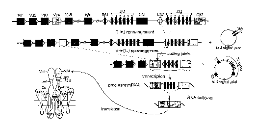
Figure 1. Schematic diagram of sequential rearrangement steps, transcription,
and translation of the TCRB gene. In this example first a D[32 to J(32.3
rearrangement occurs, followed by V(34 to D(3~-J(32.3 rearrangement, resulting
in the
formation of a V(34-D[32-J[32.3 coding joint. The rearranged TCRB gene is
transcribed
into precursor mRNA, spliced into mature mRNA, and finally translated into a
TCR(3
protein chain. The two extrachromosomal TCR excision circles (TRECs) that are
formed
during this recombination process are indicated as well; they contain the D-J
signal joint
and V-D signal joint, respectively.
Figure 2. Schematic diagram of heteroduplex analysis and GeneScanning of
PCR products, obtained from rearranged Ig and TCR genes. A. Rearranged Ig
and TCR genes (IGH in the example) show heterogeneous functional regions with
respect
to size and nucleotide composition. Germline nucleotides of V, D, and J gene
segments
are given in large capitals and randomly inserted nucleotides in small
capitals. The
functional region heterogeneity is employed in heteroduplex analysis (size and
composition) and GeneScanning (size only) to discriminate between products
derived
from monoclonal and polyclonal lymphoid cell populations. B. In heteroduplex
analysis,
PCR products are heat-denatured (5 min, 94°C) and subsequently rapidly
cooled (1 hour,
4°C) to induce duplex (homo- or heteroduplex) formation. In cell
samples consisting of
clonal lymphoid cells, the PCR products of rearranged IGH genes give rise to
homoduplexes after denaturation and renaturation, whereas in samples which
contain
polyclonal lymphoid cell populations the single-strand PCR fragments will
mainly form
heteroduplexes, which result in a background smear of slowly migrating
fragments upon
electrophoresis. C. In GeneScanning fluorochrome-labeled PCR products of
rearranged
IGH genes are denatured prior to high-resolution fiagment analysis of the
resulting
single-stranded fragments. Monoclonal cell samples will give rise to PCR
products of
identical size (single peak), whereas in polyclonal samples many different
IGHPCR
products will be formed, which show a characteristic Gaussian size
distribution.
Figure 3. PCR analysis of IGH (VH-JH) rearrangements. A. Schematic diagram of
IGH gene complex on chromosome band 14q32.3 (adapted from ImMunoGeneTics
database). "fi'°' Only rearrangeable non-polymorphic VH gene segments
are included in
blue (functional VH), or in gray (rearrangeable pseudogenes). Recently
discovered
(generally truncated) VH pseudogenes are not indicated. B. Schematic diagram
of IGH
CA 02501863 2005-04-08
WO 2004/033728 PCT/NL2003/000690
VH-JH rearrangement with three sets of VH primers and one JH consensus primer,
combined in three multiplex tubes. The relative position of the VH and JH
primers is
given according to their most 5' nucleotide upstream (-) or downstream (+) of
the
involved RSS. The VH gene segment used as representative VH family member for
primer design is indicated in parentheses. C, D, and E. Heteroduplex analysis
and
GeneScanning of the same polyclonal and monoclonal cell populations, showing
the
typical heteroduplex smears and homoduplex bands (left panels) and the typical
polyclonal Gaussian curves and monoclonal peaks (right panels). The
approximate
distribution of the polyclonal Gaussian curves is indicated in nt.
Figure 4. PCR analysis of IGH (DH-JH) rearrangements. A. Schematic diagram of
IGH (DH-JH) rearrangement with seven DH family primers and one JH consensus
primer, divided over two tubes (IGH tubes D and E). The DH7 (7-27) primer was
separated from the other six DH primers, because the DH7 and JH consensus
primer will
give a germline PCR product of 211 nt. The relative position of the DH and JH
primers is
given according to their most 5' nucleotide upstream (-) or downstream (+) of
the
involved RSS. The DH gene segment used as representative DH family member for
pririzer design is indicated in parentheses. B and C. Heteroduplex analysis
(left panels)
- and GeneScanning (right panels) of the same polyclonal and monoclonal cell
populations.
The approximate distribution of the polyclonal and monoclonal peaks is
indicated. The
potential background band/peak in tube D is indicated with an asterisk and is
located
outside the expected range of DH-JH rearrangements. The germline DH7-JH band
of tube
E is also indicated with an asterisk.
Figure 5. PCR analysis of IGK gene rearrangements. A~ Schematic diagram of the
IGKgene complex on chromosome band 2p11.2 (adapted from ImMunoGeneTics
database).46.~' Only rearrangeable non-polymorphic V~: gene segments are
indicated in
blue (functional VK) or in grey (nonfunctional VK). The cluster of inverted Vx
gene
segments (coded with the letter D) is located 800 kb upstream of the non-
inverted Vo
gene segments. These upstream VK gene segments are presented as a mirrored
image to
their corresponding non-inverted counterparts. B. Schematic diagrams of VK-Joe
rearrangement and the two types of Kde rearrangements (Vk-Kde and intron RSS-
Kde).
The relative position of the VK, JK, Kde and intron RSS (INTR) primers is
given
according to their most 5' nucleotide upstream (-) or downstream (+) of the
involved RSS.
The VK gene segment used as representative member of the Vxl, Vtc2, and V~e3
families
CA 02501863 2005-04-08
WO 2004/033728 PCT/NL2003/000690
are indicated in parentheses. VK4, VK5, and V~c7 are single-member Vx
families. The
primers are divided over two tubes: tube A with Vac and Jx primers and tube B
.with VK,
intron RSS, and Kde primers. C and D. Heteroduplex analysis and GeneScanning
of the
same polyclonal and monoclonal cell populations, showing the typical
heteroduplex
smears and homoduplex bands (left panels) and the typical Gaussian curves and
monoclonal peaks (right panels). The approximate distribution of the
polyclonal
Gaussian curves is indicated in nt.
Figure 6. PCR analysis of IGL gene rearrangements. A. Schematic diagram of IGL
gene complex on chromosome band 22q11.2 (adapted from ImMunoGenetics
database)."°,
~' Only rearrangeable non-polymorphic V7~ gene segments are included in blue
(functional
V7~) or in grey (nonfunctional V?~) B. Schematic diagram of V7~-J~,
rearrangement with
two V7~ family primers and one J7~ consensus primer. Only two V7~ primers were
designed
for V~,1 plus V~,2 and for V~,3, because these three V7~ families cover
approximately 70%
1~ of rearrangeable V~, gene segments and because approximately 90% of all IGL
gene
rearrangements involve V~,1, V7~2, or V~,3 gene segments. ~° Although
five of the seven J7~
gene segments can rearrange, only a single J7~ consensus primer was designed
for J~,1,
J7~2, and J7~3, because 98% of all IGL gene rearrangements involve one of
these three
gene segments. ~9 The relative position of the V~, and J7~ primers is given
according to
their most 5' nucleotide upstream (-) or downstream (+) of the involved RSS.
C.
Heteroduplex analysis and GeneScanning of the same polyclonal and monoclonal
cell
populations, showing the typical heteroduplex smears and homoduplex bands
(left panel)
and the polyclonal Gaussian curves and monoclonal peaks (right panel). The
approximate position of the polyclonal Gaussian curves is indicated in nt.
Figure 7. PCR analysis of TCRB gene rears angements. A. Schematic diagram of
the human TCRB locus. The gene segment designation is according to Arden et
al. 5° with
the designation according to Rowen et al. 5' and Lefranc et al. ~647 in
parentheses. The
figure is adapted from the international TmMunoGeneTics database. 46,47 Only
the
rearrangeable non-polymorphic V(3 gene segments are depicted in blue
(functional V(3), in
half blue/half gray (potential functional, but no protein expression found)
and in grey
(non-functional V(3). B. Schematic diagram of V[3-J(3 and D(3-J(3
rearrangements. The 23
V(3 primers, 13 J(3 primers and two D(3 primers are combined in three tubes:
tube A with
23 V(3 primers and nine J(3 primers, tube B with 23 V~3 primers and four J~3
primers, and
tube C with two DJ3 primers and 13 J(3 primers. The 23 V(3 primers and the 13
J(3
CA 02501863 2005-04-08
WO 2004/033728 PCT/NL2003/000690
primers are aligned in order to obtain comparably sized PCR products (see
panels C and
D). The Vii primers cover approximately 90% of all V~3 gene segments. The
relative
position of the V(3, D~3, and J(3 primers is given according to their most 5'
nucleotide
upstream (-) or downstream (+) of the involved RSS. C, D, and E. Heteroduplex
analysis
and GeneScanning of the same polyclonal and monoclonal cell populations,
showing the
typical heteroduplex smears and homoduplex bands (left panels) and the typical
polyclonal Gaussian curves and monoclonal peaks (right panels). The
approximate
distribution of the polyclonal Gaussian curves is indicated in nt.
Figure 8. PCR analysis of TCRG gene rearrangements. A. Schematic diagram of
the human TCRG locus on chromosome band 7p14. Only the rearrangeable Vy gene
segments are depicted in blue (functional Vy) or in gray (non-functional Vy).
For the Jy
gene segments, both nomenclatures are used. 4fi,47,52 B. Schematic diagram of
TCRG Vy-Jy
rearrangement with four Vy primers and two Jy primers, which are divided over
two
tubes. The relative position of the Vy and Jy primers is indicated according
to their most
5' nucleotide upstream (-) or downstream (+) of the involved RSS. C and D.
Heteroduplex analysis and GeneScanning of the same polyclonal and monoclonal
cell
populations, showing the typical heteroduplex smears and homoduplex bands
(left
panels) and the typical polyclonal Gaussian curves and monoclonal peaks (right
panels).
The approximate distribution of the polyclonal Gaussian curves is indicated in
nt.
Figure 9. PCR analysis of TCRD gene rearrangements. A. Schematic diagram of
human TCRD locus on chromosome band 14q11.~. The six "classical" Vd gene
segments
are indicated in blue, scattered between the Va gene segments in black. Since
V84, V~5,
and V36 are also recognized as Va gene segments, their Va gene code is given
in
parenthesis. B. Schematic diagram of V8-J8, D82-J8, D8~-Db3, and V8-D33
rearrangements, showing the positioning of six V8, four J8, and two DS
primers, all
combined in a single tube. The relative position of the V8, Db, and J8 primers
is
indicated according to their most 5" nucleotide upstream (-) or downstream (+)
of the
involved RSS. C. Heteroduplex analysis (left panel) and GeneScanning (right
panel) of
the same polyclonal and monoclonal cell populations. The polyclonal cell
populations
show a vague smear in heteroduplex analysis and a complex and broad peak
pattern in
GeneScanning. The monoclonal bands and peaks are clearly visible. The
approximate
position of the PCR products of the different types of rearrangements in
GeneScanning is
indicated.
CA 02501863 2005-04-08
WO 2004/033728 PCT/NL2003/000690
Figure 10. Detection of BCLI-IGH rearrangements. A. Schematic diagram of the
CCNDI gene and the BCLI breakpoint region MTC on chromosome band 11q13 as well
as the JH gene segment on chromosome band 14q32. For the primer design in the
BCLI-
MTC region an artificial BCLI-MTClJH4 functional sequence was composed (as
partially
reported for JVM253): the first 50-nucleotides as reported by Williamss~ were
linked to
nucleotide 1-439 from MTC-sequence present at NCBI (accession-number S'7'7049
55); the
N-region "GCCC" of JVM253 was added followed by nucleotide 1921-3182
representing
the JH4-JH6 genomic region (accession-number J00256). B. Agarose gel
electrophoresis
of a series of BCLI-IGH PCR products from different MCL patients and the
positive
control cell line JVM2. The PCR products differ is size, indicating different
positions of
the BCLI-MTC breakpoints. The larger bands of lower density represent PCR
products
that extend to the next downstream germline JH gene segment.
Figure 11. PCR detection of BCL2-IGH rearrangements. A. Schematic diagram of
the BCL2 gene on chromosome band 18q21. The majority of the BCL2 breakpoints
cluster in three regions: MBR, 3' MBR, and mcr. Consequently, multiplex
primers have
been designed to cover the potential breakpoints in these three regions: two
MBR
primers, four 3' MBR primers, and three mcr primers. The relative position of
the BCL
primers is indicated according to their most 5' nucleotide upstream (-) or
downstream (+)
to the 3' end of BCL2 axon 3 (according to NCBI accession no. AF32519451),
except for
two BCL2-mcr primers; their position is indicated downstream of the first
nucleotide of
the AF2'15873 sequence. B, C, and D. Agarose gel electrophoresis of PCR
products from
different FCL patients and several positive control cell lines (DoHH2, K231,
OZ, and
SC1). Panel B and D contain the same samples and show complementarity in
positivity,
illustrating that tube C (mcr tube) has added value. The PCR products differ
in size,
related to different position of the BCL2 breakpoints. The larger bands of
lower density
in the same lanes represent PCR products that extend to the next downstream
germline
JH gene segment or to the next upstream BCL2 primer.
Figure 12. Control gene PCR for assessment of amplifiability and integrity of
DNA samples. A. Schematic diagram of five control genes axons and the five
primer
sets for obtaining PCR products of 600 bp, 400 bp, 300 bp, 200 bp, and 100 bp.
The
relative position of the control gene primers is given according to their most
5' nucleotide
downstream of the 5' splice site of the involved control gene axon. B. Control
gene PCR
CA 02501863 2005-04-08
WO 2004/033728 PCT/NL2003/000690
products of six DNA samples, separated in a 6% polyacrylamide gel. Two control
samples
contained high molecular weight DNA (outer lanes) and four DNA samples were
obtained from paraffin-embedded tissue samples, showing reduced amplifiability
(e.g.
GBS-4 50 ng versus GBS-4 500 ng) or reduced integrity of the DNA (PT-4).
Figure 13. Multicolor GeneScanning for supporting the rapid and easy
identification of TCR gene rearrangements. A. Two-color analysis of TCRB tube
A
with differential labeling of J(31 primers (TET-labeled; green) and J(32
primers (FAM
labeled; blue). The top panel nicely shows the two polyclonal J(31 and J(32
rearrangement
patterns (c.f. Figure 7C), whereas the other two panels show clonal J(32
rearrangements.
. B. Two-color analysis of TCRG tube A with differential labeling of the
Jyl.3/2.3 primer
(FAM-labeled; blue) and the Jyl.1/2.1 (TET-labeled; green). The top panel
nicely shows
the polyclonal rearrangement patterns (c.f. Figure 8C), whereas the other two
panels
show clonal Jyl.3/2.3 and clonal Jyl.1/2.1 rearrangements, respectively. C.
Three-color
analysis of TCRD gene rearrangements with differential labeling of Jb primers
(FAM-
labeled; blue), D82 primer (HEX-labeled; green) and Db3 primer (NED-labeled;
black).
Within the complex rearrangement patterns of the TCRD tube (Figure 9C), the
three-
color analysis allows direct detection of V8-J8 rearrangements (blue peaks),
D82-J8
rearrangements (blue and green peaks, not fully comigrating because of
differences in
migration speed of the two fluochromosomes), V82-D83 rearrangement (black
peaks),
and D82-D83 rearrangement (comigrating green and black peaks).
CA 02501863 2005-04-08
WO 2004/033728 PCT/NL2003/000690
MATERIALS AND METHODS
Selection of PC,R, targets: aiming for complementarity
It was decided to aim for the availability of at least one PCR-detectable
clonality target
in each lymphoid malignancy. In mature B-cell malignancies this aim might be
hampered by the occurrence of somatic hyper mutations in Tg genes, which are
particularly found in follicular and post-follicular B-cell malignancies.
Therefore it was
decided to include PCR targets that have some degree of complementarity.
Several rationales were used for target selection:
- IGH genes: not only complete VH-JH rearrangements but also incomplete DH-JH
rearrangements were included as PCR targets, because DH-JH rearrangements are
probably not affected by somatic hypermutations;
- IGK and IGL genes: both Ig light chain genes were included as PCR targets,
because
this increases the chance of finding a PCR-detectable Ig gene rearrangement in
each
mature B-cell malignancy;
- IGK genes: not only VK-Jo rearrangements were included, but also
rearrangements
of the kappa deleting element (Kde), because they occur on one or both alleles
in
(virtually) all Ig7~+ B-cell malignancies and in one third of IgK+ B-cell
malignancies
and because Kde rearrangements are probably not affected by somatic
hypermutation;
- TCRB genes: both complete V~3-J(3 and incomplete D~3-J(3 rearrangements,
because
complete and incomplete TRCB gene rearrangements occur in all mature TCRa(3+ T-
cell malignancies and also in many TCRYB+ T-cell malignancies;
- TCRG genes: -this classical PCR clonality target is useful in all T-cell W
alignancies of
the TCRy~ and the TCRa(3 lineage.
- TCRD genes: this is a potentially useful target in immature T-cell
malignancies as
well as in TCR~yB+T-cell malignancies;
- TCRA gene: this gene was not included as PCR target, because of its high
degree of
complexity with ~50 V and 61 J gene segments. Furthermore, all T-cell
malignancies
with TCRA gene rearrangements contain TCRB gene rearrangements and generally
also have TCRG gene rearrangements;
- functional gene segments: most suspect lymphoproliferations concern mature
lymphocytes, which consequently have functional Ig or TCR gene rearrangements.
CA 02501863 2005-04-08
WO 2004/033728 PCT/NL2003/000690
Therefore PCR primer design aimed at inclusion of (virtually) all functional
Ig/TCR
gene segments.
- well-defined- chromosome aberrations: t(11;14) with BCLI-IGH and t(14;18)
with
BCL2-IGH were included as additional targets, because these two aberrations
are
PCR-detectable at relatively high frequencies in lymphomas i.e. in 30% of
mantle cell
lymphoma (MCL) and in 60 to 70% of follicular cell lymphomas (FCL),
respectively.
Primer design for multiplex PCR
Precise detection of all V, D, and J gene segments in rearranged Ig and TCR
genes would
require many different primers (Table 2). For some gene complexes this might
be
possible (e.g. 2'CRG and TCRD), but for other loci in practice this is
impossible because
of the high number of different gene segments. To solve this problem, family
primers can
be designed, which recognize most or all gene segments of a particular family
(Table 2).
Alternatively, consensus primers can be made, which recognize conserved
sequences that
occur in many or all involved gene segments.
The design of family primers and consensus primers balances between a limited
number of primers and maximal homology with all relevant gene segments. In
this
study, we aimed at maximal homology with all relevant gene segments
(particularly
functional gene segments) in order to prevent suboptimal primer annealing,
which might
cause false-negative results. Furthermore, we aimed at the design of specific
family
primers without cross-annealing to other families
In order to limit the number of PCR tubes per locus, multiplexing of PCR
primers
became important for practical reasons. Consequently, special guidelines were
developed
to ensure maximal possibilities for designing primers useful in multiplex PCR
tubes. For
this purpose dr. W. Rychlick (Molecular Biology Insights, Cascade; CO, USA)
provided
his specially-adapted OLIGO 6.2 software program and supported the development
of
the guidelines for optimal primer design.
The general guidelines for primer design were as follows:
- the position of the primers should be chosen in such a way that the size of
the PCR
products would preferably be <300 by (preferably 100 to 300 bp) in order to be
able to
use paraffin-embedded material;
- a minimal distance to the functional region of preferaby >10-15 by should be
taken
into acount (in order to avoid false-negativity due to impossibility of the 3'
end of the
CA 02501863 2005-04-08
WO 2004/033728 PCT/NL2003/000690
primer to anneal to the rearranged target because of nucleotide deletion from
the
germline sequence);
- primers preferably should not be too long (e.g. <25 nucleotides).
The following parameters were used for primer design with the OLIGO 6.2
program:
- search for primers should be performed with ntode~°a.te stringency;
- primer efficiency (PE) value should preferably be 400 (and >630, if the
primer is to
be used as consensus primer for other gene segments as well);
- the most stable 3' dimes of upper/upper, lower/lower, or upper/lower primers
should
not exceed -4 Kcal (moderate search strategy); the most stable dimes overall
being
less important;
- in view of multiplex PCR, the following guidelines were taken into account:
a common primer would have to be designed in the most consensus region (i.e.
high
PE in consensus search), whereas individual primers (family or member) have to
be
designed in the least consensus region (i.e. low PE value of that primer for
gene
segments that should not be covered) to avoid cross-annealing to other gene
segments
and thereby multiple (unwanted) PCR products.
PCR protocol
A standardised PCR protocol was developed based on pre-existing experience
from earlier European collaborative studies. After initial testing and
approval, the
protocol was accepted as summarized in Table 3.
Techniques for analysis of PCR products obtained from Ig/ TCR gene
rearrangements
The PCR products obtained from Ig and TCR gene rearrangements have to be
analysed for discrimination between monoclonal lymphoid cells with identical
functional
regions and polyclonal lymphoid cells with highly diverse functional regions.
Based on the combined experience of the participating laboratories, two
techniques were selected: heteroduplex (HD) analysis and Gene Scanning (GS)
analysis.
HD analysis uses double-stranded PCR products and takes advantage of the
length and
composition of the functional regions, whereas in GS single-stranded PCR
products are
separated in a high resolution gel or polymer according to their length only
(Figure 2).
CA 02501863 2005-04-08
WO 2004/033728 PCT/NL2003/000690
Heter°oduplex ar2alysis of PCR products
PCR products obtained with unlabeled primers are denatured at high temperature
(~95°C for 5 min), followed by rapid random renaturation at low
temperature (preferably
at 4°C for 1 hour). This enforced duplex formation results in many
different
heteroduplexes with different migration speed in case of polyclonal
lymphoproliferations,
but resulting in homoduplexes with identical r apid migration in case of
monoclonal
lymphoproliferations. Electrophoresis of the homoduplexes in a 6%
polyacrylamide gel
results in a single band of predictable size, whereas the heteroduplexes form
a smear at
a higher position (Figure 2). The heteroduplex technique is rapid, simple and
cheap (see
Table 4 for technical details) and has a detection limit of ~ 5 %,~0, 41 The
detection limit is
influenced by the frequency of polyclonal lymphocytes, because the formation
of many
heteroduplexes will also consume a part of the monoclonal PCR products.4'
1~ Genescar2ni,ng analysis of PCR products
The PCR primers for GeneScanning analysis need to be labeled with a
fluorochrome to
allow detection of the PCR products with automated sequencing equipment
(Figure 2).
The fluorochrome labeled single-strand (denatured) PCR products are size-
separated in a denaturing polyacrylamide sequencing gel or capillary
sequencing
polymer and detected via automated scanning with a laser (see Table 5 for
technical
details). This results in a Gausian distribution of multiple peaks,
representing many
different PCR products in case of polyclonal lymphoproliferations, but gives a
single peak
consisting of one type of PCR product in case of a fully monoclonal
lymphoproliferation
(Figure 2).
GeneScanning is rapid and relatively simple, but needs expensive equipment.
GeneScanning is generally more sensitive than heteroduplex analysis and can
reach
sensitivities of 0.5 to 1% of clonal lymphoid cells.
Control genes and paraffin-embedded tissues
In several European countries, fresh tissue material is not easily available
for
molecular diagnostics such as PCR-based clonality studies. Therefore one of
the aims of
the present study was to develop a strategy for PCR-based clonality studies in
paraffin-
embedded tissues.
To control for the quality and amplifiability of DNA from paraffin-embedded
material, a special multiplex control gene PCR was developed, resulting in a
ladder of
CA 02501863 2005-04-08
WO 2004/033728 PCT/NL2003/000690
five fragments (100 bp, 200 bp, 300 bp, 400 bp, and 600 bp). From 45 of the
above
described 90 cases sufficient paraffin-embedded tissue was available for DNA
extraction.
These DNA samples were tested in parallel to the freshly-obtained DNA samples,
using
the Control Gene multiplex tube as well as the IglTCRlBCLIlBCL2 multiplex
tubes for
clonality diagnostics (see Example 10).
EXAMPLE 1. Complete IGH gene rearrangements: VH-JH
Background
The functional rearrangement of the IGH gene, first DH to JH and subsequently
V
to DFI-JH, is followed by antibody expression, the hallmark of mature B-cells.
The IGH
gene is located on chromosome 14q32.3 in an area covering approximately 1250
kilobases. 46 to 52 functional VH segments (depending on the individual
haplotype) have
been identified, which can be grouped according to their homology in six or
seven VH
subgroups. In addition approximately 30 non-functional VH segments have been
described. Furthermore, 27 DH segments and functional six JH segments have
been
consistently found (Table 2 and Figure 3A).56
The VH segments contain three framework (FR) and two complementarity
determining regions (CDR) (Figure 3B). The FRs are characterized by their
similarity
among the various VH segments whereas the CDRs are highly different even
within the
same VH family. Furthermore, the CDRs represent the preferred target sequences
for
somatic hypermutations in the course of the germinal center reaction, which
increase the
variability within those regions. Although the FRs are usually less affected
by somatic
mutations, nucleotide substitutions may also occur within these regions,
especially in B-
cells under a heavy mutational process.
The highly variable V-D-JH regions can be amplified by PCR to detect clonal B-
cell populations indicative of the presence of a malignant B-cell disorder.
Clonal B-cells
can be discriminated from polyclonal B-cells (i.e. normal or reactive lymphoid
tissues)
based on the identical size and composition of the clonal PCR products as
compared to
the many different polyclonal PCR products with a size range of approximately
60 bp,
arranged in a Gaussian distribution. PCR-based str ategies for detection of
clonal B-cell
populations in histological sections and cell suspensions have already been
established
in the early nineties. However, the initial PCR protocols used single VH
consensus
primers which were able to bind to one of the three framework regions, mainly
FR3.
Such consensus primers were not suitable to amplify all VH segments with the
same
efficiency leading to non-detectability of a significant number of clonal
rearrangements.
CA 02501863 2005-04-08
WO 2004/033728 PCT/NL2003/000690
In addition, somatic mutations introduced in the course of the germinal center
reaction
are not restricted to the CDRs, but can also occur in FRs, thereby preventing
primer
annealing and consequently leading to absence of clonal PCR products despite
the
presence of a neoplastic B-cell population. This is especially true for
follicular
lymphomas, diffuse large B-cell lymphomas, and multiple myelomas which usually
contain high numbers of somatic mutations.
To further increase the detection rate of the IGII PCR, several attempts have
been made to design family-specific primers to overcome the limitations of
consensus
primers. However, these family-specific primers are largely based on the
sequences of
the previous consensus primers. Although these PCR strategies have helped to
improve
the detection rate, there is still a need of primer systems which are less
sensitive to
somatic hypermutations, thus allowing amplification of (virtually) all
possible V-D-JH
rearrangements.
Primer design
According to the guidelines of the invention, three sets of VH primers were
designed with the help of the OLIGO-6.2 program corresponding to the three VH
frame
work regions (FR1, FR2 and FR3) (Figure 3B). Each set of primers consisted of
six or
seven oligonucleotides capable to anneal to their corresponding VH segments
(VHi to
VH7) with no mismatches for most VH segments and one or at most two mismatches
for
some rare VH segments. The design was such that mismatches would be located at
the
very 5'-end of the primer. These VH primer sets were used in conjunction with
a single
JH consensus primer, designed to anneal to the most homologous 3'-end of the
six JH
segments, approximately 35 by downstream of the JH RSS. This ensures that all
JH
segments are detectable with the same binding efficiency-and that the primer
binding
will not easily be affected by extensive nucleotide deletion in the course of
the
rearrangement process. In addition, there was no cross-annealing between the
VH
primers and the JH primer as evaluated by the OLIGO-6.2 program.
The JH primer was also designed to be used for amplification of other PCR
targets, such as incomplete DH-JH rearrangements as well as t(11;14) (BCL.T-
IGI~ and
t(14;18) (BCL2-IGI~. This allows the detection of different PCR products by GS
analysis
employing the same labeled JH primer.
CA 02501863 2005-04-08
WO 2004/033728 PCT/NL2003/000690
Results of initial testing phase
The initial testing of the newly designed VH-JH PCR was done by separate
application of each VH primer together with the JH primer in an individual
PCR. For
this purpose, DNA extracted from B-cell lines as well as well-defined clonal
patient
samples was used. Furthermore, clonal rearrangements were tested for
sensitivity by
serial dilution in DNA extracted from reactive tollslls. Clonal control
samples were not
available for each possible IGHrearrangement, but all major VH segments and
several
rarely rearranged VH segments have been included in the initial testing phase.
All primer pairs worked with high efficiency and sensitivity. The expected
clonal
VH rearrangements were detectable and the sensitivity was at least 1% (10-2).
There was
no background within the expected size range and the amplification of
tonsillar DNA
gave the expected ~Gaussian distribution curve. (Figure 3C, D, and E)
Based on these results we started the next phase of the initial primer testing
by
combining the VH primers into three sets, each specific for one of the three
framework
regions, which were used together with the common JH primer (Figure 3B). The
results
were the same as those obtained with single primer pairs, but with a slightly
lower
sensitivity. In addition, no nonspecific products were amplified within the
expected size
range, with the exception of a 340 by PCR product which appeared in the FRl
multiplex
PCR. This PCR product was generated irrespective of the source of the DNA
(lymphoid
and non-lymphoid) used for PCR, whereas no PCR product was obtained when no
DNA
template was applied. Furthermore, this amplicon was only detectable in
heteroduplex
analyis, not in GeneScanning. This indicates that the fluorescent labeled JH
primer was
not involved in the generation of this PCR product. Sequence analysis of this
PCR
product disclosed a VH4 fragment amplified by the FRl VH4 primer in
conjunction with
the FR1 VH2 primer which apparently acted as a downstream primer by binding to
the
intronic VH4 sequence. This problem could be solved by designing a new FR1 VH2
primer which was located 25 by upstream to the previous primer binding site.
Results of general testing phase
The approved IGH PCR was applied to the 90 Southern blot defined DNA samples,
which were derived from well-characterized cases. Six of the 11 laboratories
involved in
the general testing phase performed GS analysis of the PCR products and five
performed
HD analysis. In addition several polyclonal as well as monoclonal samples
(cell line
DNA) were included as controls. 45 of these cases displayed dominant PCR
products
after GS analysis and 40 cases after HD detection, indicating the presence of
a
CA 02501863 2005-04-08
WO 2004/033728 PCT/NL2003/000690
monoclonal B-cell population. The clonal rearrangements were detectable with
all three
FR primer sets in 33 of the 45 clonal cases (GS) and in the remaining 12 with
one or two
of the three FR primer sets. It was concluded that most negative results were
caused by
somatic hypermutations in the primer binding site, preventing primer annealing
and
thus amplification.
The comparison of the VH-JH PCR results with the Southern blot results
revealed
a high degree of concordance. 85°/ (46 out of 55) and 76% (42 out of
55) of the samples
with rearranged VH genes by Southern blot analysis showed a dominant
amplification
product by GS analysis and HD analysis, respectively. Vice versa, all but two
samples
harboring germline VH genes by Southern blot displayed a polyclonal pattern by
GS and
HD analysis.
Conclusion
In conclusion, the three multiplex PCRs for detection of clonal VH-JH
rearrangements
provide a new and reliable assay to identify clonal B-cell proliferations. The
combined
use of standardized primers in the three. different FRs helps to decrease the
rate of false-
negative results due to somatic hypermutation in primer binding sites of the
involved VH
gene segments. EXAMPLE 2. Incomplete IGH gene rearrangements: DH-JH
Background
The formation of complete V-D-J rearrangements in the IGH locus on
chromosome 14q32.3 is a sequential process that occurs in two steps: VH
coupling is
generally preceded by an initial rearrangement between DH and JH gene segments
in
early precursor-B cells (reviewed by 5'). In addition to the many distinct VH
gene
segments and the six functional JH gene segments (see Example 1), the human
TGH
locus also contains 27 DH gene segments.se Based on sequence homology, the 27
DH
segments can be grouped into seven families: DH1 (formerly known as D1V1), DH2
(DLR),
DH3 (DXP), DH4 (DA), DH5 (DK), DH6 (DID, and DH'7 (Df9,152); all families
comprise at
least four members, except for the seventh which consists of the single DH7-27
segment
just upstream of the JH region (Figure 3A).58.5~
Recombination between any of the DH and JH segments will result in the
formation of incomplete DH-JH joints, which can easily be detected in bone
marrow-
derived CD10+ / CD19- precursor B-cells 6°.6' and hence also in a
subset (20-25 %) of
precursor B-cell acute lymphoblastic leukemias, which show an immature
genotype.62
Sequencing revealed a predominance of DH2 (DH2-2), DH3 (DH3-9) , and DH7-27
gene
CA 02501863 2005-04-08
WO 2004/033728 PCT/NL2003/000690
segments in precursor B-ALL, comprising 36%, 33%, and 19% of all identified
segments,
respectively. 62
However, also in mature B-cell malignancies incomplete DH-JH rearrangements
have been reported.s'.6' Moreover, even in a subset of IgH-negative multiple
myelomas,
which can be considered as the most mature type of B-lineage malignancy, DH-JH
joints
were observed.s° These DH-JH rearrangements were derived from the non-
coding second
allele and involved segments from DH1 to DH4 families.fi" Based on the
description of DH-
JH joints in precursor-B-ALL and multiple myelomas, it is assumed that
incomplete DH-
JH rearrangements are also present in other types of B-cell leukemias and
lymphomas.
ZO In immature T-cell malignancies DH-JH couplings have been identified as
cross-lineage
rearrangements;3" interestingly, these almost exclusively occurred in the more
immature
non-TCRa(3+ T-ALL subset and mainly involved the more downstream DH6-19 and
DH7-
27 segments. The latter segment is frequently (up to 40%) used in fetal B
cells but rarely
in adult B cells.65, 66 Human adult precursor and mature B cells mainly seem
to use DH2
and DH3 family segments, as evidenced from sequences of complete VH-DH-JH
rearrangements,sfi
Although the exact frequencies of incomplete DH-JH couplings in different
types
of mature B-cell malignancies are largely unknown, it is clear that they will
at least be
lower than those of VH-JH joinings. Nevertheless, DH-JH rearrangements might
still
represent an important complementary target for PCR-based clonality
assessment. This
presumed contribution of DH-JH rearrangements as PCR target is based on the
assumption that incomplete rearrangements in the IGH locus will not to contain
somatic
hypermutations, because transcription starting from the promoters in the V
gene
segments does not occur, which is regarded as an essential prerequisite for
somatic
hypermutation to take .place.fi'~ 6g Especially in those types of B-lineage
proliferations in
which somatic hypermutations are frequent, PCR analysis of a possible DH-JH
recombination product might therefore be relevant, and sometimes even the only
possibility to detect the B-cell clone.
Primer design
Based on the high degree of homology within each DH family, seven family-
specific DH primers were designed (Figure 4) in combination with the consensus
JH
primer that is also used for detection of VH-JH rearrangements (see Example 1)
and
t(11;14) (BCLT-IGI~ and t(14;18) (BCL2-IGI~ (Examples 8 and 9). Primers were
designed such that cross-annealing to other DH family segments would be
minimal or
CA 02501863 2005-04-08
WO 2004/033728 PCT/NL2003/000690
preferably absent, resulting in distinct positions for the various family
primers relative
to the RSS elements (Figure 4). The expected PCR product sizes of DH-JH
joints. range
from 110-130 by (for DH7-JH joinings) to 395-415 by (for DH3-JH
rearrangements). Of
note, due to the position of the DH7-27 segment close to the segments in the
JH region,
PCR products of 211 by (and also 419,1031,1404,1804, and 2420 by in case of
primer
annealing to downstream JH gene segments) will be amplified from non-
rearranged
alleles and will be detected as a ladder of germline bands in virtually every
sample.
Results of initial testing phase
For initial testing of the individual DH primers, high tumor load precursor B-
ALL
or T-ALL samples with well-defined clonal DH-JH rearrangements were used.
Under
standard PCR conditions using 1.5 mM MgCla and AmpliTaq Gold buffer, all seven
primer combinations appeared to detect the clonal DH-JH targets with product
lengths
within the expected size ranges. Cross-annealing of the DH primers to
rearranged gene
segments from other DH families was only very weak or not observed at all.
Furthermore, also in healthy control tonsillar or MNC DNA PCR products of the
correct
size ranges were observed. Nonspecific annealing of the primers was not
observed for
virtually all primers sets, using non-template specific control DNA; only in
case of the
DH2 / JH primer set a (sometimes faint) 340-350 by product was observed in
HeLa DNA.
Further sequencing revealed that this nonspecific product was due to false
priming of
the DH2 primer to a DNA sequence upstream of the JH4 segment. However, as the
size
of this nonspecific product was so different from the sizes of any of the true
DH-JH PCR
products, it was decided not to design a new DH2 primer. In fact, the
nonspecific 350 by
band can be employed as an internal marker for successful DNA amplification
and hence
the -quality of the template DNA, being hardly or only faintly visible when-
enough clonal
or polyclonal DH-JH template is available (e.g. in tonsillar DNA or DNA from
particular
leukemic samples), but being especially strong in samples containing low
numbers of
lymphoid cells with DH-JH rearrangements.
Serial dilutions of DNA from the clonal reference samples into tonsillar DNA
generally resulted in sensitivities of 5% or lower (0.5-1% in case of the DH6-
JH
rearrangement) using HD analysis; sensitivities in GS analysis were generally
1-2
dilution steps better, i.e. 1% or lower. The clonal DH7-JH target could only
be detected
with a sensitivity of ~10%, which is most probably caused by primer
consumption in PCR
amplicons involving the non-rearranged germline DH7 and JH gene segments.
CA 02501863 2005-04-08
WO 2004/033728 PCT/NL2003/000690
Although the initial multiplex strategy, as suggested from the OLIGO 6.2-
assisted primer design, was to divide the various DH primers over two tubes,
it was
decided after testing various multiplex approaches to combine all primers into
one
multiplex tube (tube D of IGH clonality assay), except for the DH7 primer,
which was
included in a separate tube (tube E of IGH clonality assay). The reason to
exclude the
DH7 primer was the complicated germline pattern, due to easy amplification of
alleles
with non-rearranged DH7 segments. Using this two-tube multiplex approach, all
clonal
reference samples were still detectable. Under multiplex conditions the
detection limits
for these various clonal targets were logically less optimal as compared to
the single
assays, ranging from ~5% (DH3, DH4, and DH6) to ~10% (DH2, and DH5). For the
DH1
clonal reference sample that was available, a sensitivity of ~20% was
observed; at a later
stage the DH1-JH rearrangement of cell line KCA was found to be detectable
down to
10% in the multiplex assay. As tube E only contains the DH7 primer, the 10%
sensitivity
for this tube was the same as mentioned before. The same multiplex analysis
performed
with 500 ng instead of 100 ng DNA of the serial dilutions, resulted in
slightly better
sensitivities. The use of serial dilutions in MNC DNA instead of tonsillar DNA
did not
clearly affect detection limits of the assays for DH-JH recombinations.
Results of general testing phase
Following initial testing in the three laboratories involved in primer design,
the
developed IGH DH-JH multiplex PCR assay was further evaluated using the 90
Southern
blot-defined samples. Every sample was analyzed in parallel in four
laboratories by HD
analysis and in five laboratories by GS analysis; in another two laboratories
all samples
were analyzed by both techniques. All together a total of six HD and seven GS
analysis
results were obtained per sample per tube. Despite concordant results (> 80%
of
laboratories with identical results) in the vast majority of samples, nine
showed inter-
laboratory discordancies in tube D. Further analysis revealed that these could
be
explained by either the presence of a small clone with weak clonal products,
or to large
size products 0390 and larger). In a few cases the products were so large,
that only after
sequencing it became clear that they concerned true but extended DH-JH
rearrangements, either from upstream DH (e.g. DH6-25-DH1-26-JH in NL-12) or
from
downstream JH gene segments (e.g. DH6-25-JH4-JH5 in PT-14). In all three cases
(NL-
17, mycosis fungoides; FR-1, B-CLL; FR-5, FCL) in which clonal products were
found
using tube E, the results were completely concordant between laboratories.
CA 02501863 2005-04-08
WO 2004/033728 PCT/NL2003/000690
When evaluating results from HD and GS analysis, it appeared that these were
comparable, although in general the number of laboratories showing identical
results
was slightly higher upon HD as compared to GS analysis (Figure 4B and C).
Direct comparison of DH-JH multiplex PCR results with SB data is virtually
impossible, as hybridization with a single probe (IGHJ6) in the JH region does
not allow
discrimination between VH-JH and DH-JH rearrangements. In three samples it was
clear
that detection of clonal products of the combined VH-JH and DH-JH assays did
not fit
with configuration of the IGH locus in SB analysis. Remarkably, no clonal DH-
JH PCR
products were observed in the pre-follicular B-cell malignancies. In contrast,
11/16 B-
CLL samples and 12/25 (post-)follicular B-cell malignancy samples did contain
clonally
rearranged DH-JH PCR products. In three of the eighteen T-cell malignancy
cases clonal
DH-JH rearrangements were seen; these concerned T-LBL (ES-9) and mycosis
fungoides
(NL-17) cases with SB-detected IGH rearrangements, and a case of T-NHL/EATL
(PT-4)
without SB-detected IGH rearrangements, probably because of the low tumor load
of
<15%. All 15 reactive cases only showed polyclonal DH-JH PCR products, in
accordance
with SB results. In category D with difficult diagnoses, three samples (PT-12,
GBS-10,
and GBN-8) showed clonal TGHDH-JH PCR products, which was in line with SB data
as
well as IGI~PCR data in two of three cases; in another two samples (PT-6 and
GBS-9),
both T-cell rich B-NHL cases, clonal DH-JH products were found in addition to
clonal
IGK and/or IGL products, but without evidence for clonality from SB analysis,
which
might best be explained by the small size of the B-cell clone in these
samples.
In order to determine the additional value of DH-JH PCR analysis, the results
were compared to those of VH-JH PCR analysis. In five (NL-4, PT-14, GBN-2, FR-
7, NL-
12) B-cell malignancies clonal DH-JH PCR products were found, whereas only
polyclonal
VH-JH PCR products were observed. -
Conclusion
In conclusion, based on the initial and general testing phases, DH-JH PCR
analysis appears to be of added value for clonality assessment. Although HD
analysis
results might be interpreted slightly more easily, there is no clear
preference for either
HD or GS analysis as they are both suitable for analyzing amplified PCR
products. A
potential difficulty in DH-JH PCR analysis is the relatively large size range
of expected
PCR products, due to scattered primer positions and to extended amplifications
from
upstream DH or downstream JH gene segments, implying that long runs are
recommended for GS analysis. Finally, the remarkable position of the DH7-27
gene
CA 02501863 2005-04-08
WO 2004/033728 PCT/NL2003/000690
segment in the IGH Iocus causes a ladder of germline amplification products in
tube E,
with clonal products being easily recognizable as much smaller bands / peaks.
CA 02501863 2005-04-08
WO 2004/033728 PCT/NL2003/000690
EXAMPLE 3. IGK gene rearrangements: VK-Jx, Vac-Kde / intronRSS-Kde
Background
The human IGKlight chain locus (on chromosome 2p11.2) contains many distinct
Vo gene segments, grouped into seven VK gene families, as well as five JK gene
segments
upstream of the CK region.Originally, the Vo gene segments were designated
according
to the nomenclature as described by Zachau et al.s° An alternative
nomenclature groups
the VK gene segments in seven families arid is used in the ImMunoGeneTics
database. '°
Here we follow the latter nomenclature. The VKl, V~e2, and VK3 families are
multi-
member families including both functional and pseudo gene segments, whereas
the other
families only contain a single (VK4, V~cS, VK7) or a few segments
(VK6).'° Remarkably, all
VK gene segments are dispersed over two large duplicated clusters, one
immediately
upstream and in the same orientation as the Jo segments, and the other more
distal and
in an inverted orientation (Figure 5A)." The latter implies that so-called
inversion
rearrangements are required to form VK-JK joints involving VK genes of the
distal
cluster. In addition to the Vtc and JK segments, there are other elements in
the IGKlocus
that can be involved in recombination. The kappa deleting element (Kde),
approximately
24 kb downstream of the Jtc-Co region, can rearrange to VK gene segments (VK-
Kde), but
also to an isolated RSS in the JK-CK intron (intronRSS-Kde).Z"~'2 Both types
of
rearrangements lead to functional inactivation of the IGK allele, through
deletion of
either the CK exon (intronRSS-Kde rearrangement) or the entire JK-CK area (V~e-
Kde
rearrangement).
As human IGK recombination starts in precursor B-cells in the bone marrow, IGK
rearrangements can also be detected in precursor B-cell acute leukemias (30-
45% of
alleles, depending on age). Although Vo-JK joinings are present, these IGK
rearrangements mainly concern recombinations involving Kde (25-35% of
alleles). In
childhood precursor B-ALL VK-Kde recombination predominates over intron-Kde,
whereas in adult ALL the deletions exclusively concern Vx-Kde
couplings.z'~"~'~ In chronic
B-cell leukemias IGKrearrangements are even more frequent, being detectable
on'75%
(Igo+ cases) or even 95% (Ig~,+ cases) of all IGK alleles. By definition,
functional VK-Jx
rearrangements are found on at least one allele in Ig~c+ B-cell leukemias; the
non-coding
second allele is either in germline configuration, or inactivated through VK-
Kde (8% of
alleles) or intronRSS-Kde (8% of alleles) recombination. Kde rearrangements
are
frequent in Ig~,+ B-cell leukemias (~85% of alleles), with a slight
predominance of
intronRSS-Kde recombinations over Vac-Kde rearrangements. This implies that
virtually
all Ig7~+ leukemias contain a Kde rearrangement, while potentially functional
VK-Jtc
CA 02501863 2005-04-08
WO 2004/033728 PCT/NL2003/000690
couplings are relatively rare.2'~'S Several studies have shown that Vx gene
segment usage
is almost identical between various normal and malignant B-cell populations
and largely
reflects the number of available gene segments within each family. Both in Vx-
Jx as well
as in Vx-Kde rearrangements, Vx gene segments from the first four families
(Vxl to Vx4)
predominate. Vx2 gene usage appeared to be higher in precursor B-ALL than in
more
mature B-cell lymphoproliferations or normal B cells. Remarkably, the distal
inverted
Vx cluster was rarely used in Vx-Jx rearrangements, whereas Vx pseudogene
segments
were never involved, also not in Ig7~~ cases.'6 Little is known about Jx gene
segment
usage, but sparse data show that Jxl, Jx2, and Jx4 are the most frequently
used Jx gene
segments.'S
Vx-Jx rearrangements can be important complementary PCR target for those
types of B-cell proliferations in which somatic hyper mutations may hamper
amplification of the VH-JH target, but recombinations involving Kde are
probably even
more valuable. Deletion of intervening sequences in the Jx-Cx intron results
in the
removal of the IGKenhancer, which is thought to be essential for the somatic
hypermutation process to occur. Rearrangements involving Kde are therefore
assumed to
be free of somatic hypermutations, and hence should be amplified rather
easily.
Primer design
Using OLIGO 6.2 software, six family-specific Vx primers were designed to
recognize the various Vx gene segments of the seven Vx families; the Vx6
family gene
segments were covered by the Vxl family primer (Figure 5B). In case of the
relatively
large V7cl, Vx2, and Vx3 families only the functional Vx gene segments were
taken into
consideration, as the less homologous pseudo gene segments complicated optimal
primer
design too much. The family-specific Vo primers were designed to be used in
combination
with either a set of two Jx primers (Jx1-4, covering the first four Jx
segments and Jx5
covering the fifth) or a Kde primer (Figure 5B). For analysis of Kde
rearrangements an
additional forward primer recognizing a sequence upstream of the intronRSS was
made.
Tn order to show minimal cross-annealing to other Vx family segments and still
be useful
in multiplex reactions, the various primers could not be designed at similar
positions
relative to RSS elements (Figure 5B). The expected PCR product sizes of Vx-Jx
joints
range from 115-135 by (for Vx7-Jx joints) to 280-300 by (Vx2-Jx
rearrangements). For
the Kde rearrangements, product size ranges are from 195-215 by (Vx7-Kde) to
~360-
380 by (Vx2-Kde), whereas the intronRSS-Kde products are 275-295 bp.
CA 02501863 2005-04-08
WO 2004/033728 PCT/NL2003/000690
Results of initial testing phase
For initial testing of the individual primers, several cell lines and patient
samples
with precisely defined clonal VK-JK, or VK-Kde / intronRSS-Kde rearrangements
were
used. The patient samples with VK-JK joints mostly concerned chronic B-cell
leukemias,
which were additionally selected on basis of a high tumor load for easy and
sensitive
detection of the involved rearrangement. Unfortunately, clonal reference
samples were
not available for all VK-JK targets; especially the more rare types of
rearrangements
involving VKS, VK7 and/or JK5 were not represented in the series of reference
samples.
For these targets and also for the targets for which clonal reference samples
were
available, healthy control tonsillar or MNC DNA samples were employed, in
which PCR
products of the correct expected sizes were indeed observed. The only
exception was the
V~c7 / JK5 primer combination; most probably Vo7-J~c5 joinings are so rare in
normal B
cells, that these PCR products were hardly or not detectable in tonsils.
Rearranged
products within the expected size ranges could be detected in all clonal
reference
samples, under standard PCR conditions using 1.5 mM MgCh and either ABI Gold
Buffer or ABI Buffer II. However, in a few cases weak amplification of
particular VK-Jo
rearrangements was observed with other Vo family / Jo primer sets, due to
slight cross-
annealing of the Vo3 primer to a few Vo1 gene segments. Furthermore, in a few
of the
clonal reference samples clear additional clonal PCR products were seen with
other Vac /
~0 Jo or even Vo /Kde and intronRSS / Kde primer sets; in most samples this
could be
explained by the complete configuration of the two IGK alleles. This
occurrence of
multiple clonal PCR products illustrates the complexity of IGKrearrangement
patterns
in a given cell sample, mainly caused by the potential occurrence of two
clonal
rearrangements on one allele (Vo-JK and intron RSS-Kde). This complexity does
not
~5 hamper but support the discrimination between polyclonality and
monoclonality.
No nonspecific annealing of the primers was observed for any of the VK-JK and
Vo-Kde / intron RSS-Kde primer sets, when using HeLa DNA as a non-template
specific
control. Serial dilutions of DNA from the clonal reference samples into
tonsillar DNA
generally resulted in sensitivities of 5-10 % for Vo-Jo rearrangements and 1-
10 % for VK-
30 Kde rearrangements, using HD analysis. In general, the sensitivities in GS
analysis
were approximately one dilution step better. The only slightly problematic
target was
the intronRSS-Kde target that could only be detected down to the 10% serial
dilution in
the employed patient sample. This is probably caused by the fact that
intronRSS-Kde
rearrangements are abundant in DNA from both Ig~c+ and Ig7~+ tonsillar B
cells, which
35 were used in the dilution experiments.
CA 02501863 2005-04-08
WO 2004/033728 PCT/NL2003/000690
The multiplex strategy that was chosen after testing several approaches
consisted
of two different multiplex PCR reaction tubes. In the VK-JK tube (tube A) all
VK primers
were combined with both JK primers, whereas tube B contained all VK primers
plus the
intronRSS primer in combination with the Kde reverse primer (Figure 5B). All
beforementioned clonal reference samples were detectable using this two-tube
multiplex
approach. Of note is the observation that in non-clonal tonsil samples a
predominant,
seemingly clonal band of ~ 150 by was detected using the Vo-JK multiplex tube
A
analysis. The presence of this product, which is seen in HD analysis but
especially in GS
analysis, can be explained by the limited heterogeneity of VK-JK functional
regions
leading to a high frequency of products of an average size of 150 bp.
Furthermore, in
some samples a sometimes weak 404 by nonspecific band was observed in tube B.
Although sensitivities were on average slightly better in other multiplex
approaches in
which the VK primers were further subdivided over multiple tubes, the
feasibility of
having only two tubes to analyze all relevant IGKrearrangements, finally was
the most
important argument for choosing the two-tube multiplex strategy as given in
Figure 5B.
Detection limits for the various clonal targets in the two-tube multiplex
approach were
~10% for most of the clonal VK-JK rearrangements (Vol-J~c4, VK2-JK4, VK3-JK4)
derived
from informative samples with a high tumor load; sever al of the Vie-Kde
targets were
detectable with a still reasonable sensitivity of ~10%, but a few other
samples containing
Vo2-Kde, VK5-Kde, and also intronRSS-Kde targets showed detection limits above
10%.
Even the use of 500 ng serially diluted DNA instead of 100 ng hardly resulted
in better
sensitivities, whereas serial dilutions in MNC DNA did not affect the
detection limits
either. Nevertheless, detection limits of serial dilutions of reference DNA in
water were
all in the order of 0.5-1 %, which shows that the chosen multiplex IGKPCR
assay as
such is good. It is important to note that potential clonal cell populations
in lymph nodes
or peripheral blood in practice will have to be detected within a background
of polyclonal
cells, which can hamper sensitive clonality detection, especially in samples
with a
relatively high background of polyclonal B-cells.
Results of general testing phase
Following initial testing in the four laboratories involved in primer design,
the developed
IGKmultiplex PCR assay was further evaluated using 90 Southern blot-defined
samples. Every sample was analyzed in parallel via HD (five laboratories) and
GS (two
laboratories) analysis; in another four laboratories all samples were analyzed
by both
techniques. Taken together, eight HD and five GS analysis results were
available per
CA 02501863 2005-04-08
WO 2004/033728 PCT/NL2003/000690
sample per tube. In the vast majority of samples >80% of laboratories produced
identical
results, i.e. either clonal bands l peaks or polyclonal smears / curves in one
or both tubes.
However, in nine (~10%) samples discordancies were found between laboratories,
which
remained after repetitive analysis of these samples. More detailed analysis
revealed that
in at least six cases the approximately 150 and 200 by sizes of the clonal
products in
tube A could not easily be discriminated from polyclonal products of roughly
the same
size. This is an inherent difficulty in especially Vo-Jo analysis, which is
caused by the
relatively limited functional heterogeneity of these rearrangements. In two
samples the
results from tube B were however so clear in all laboratories with both
techniques that
in fact no discrepancy prevailed. In one sample (ES-8) a large product of
around 500 by
appeared to be the reason for discrepant inter-laboratory results; further
sequencing
revealed that amplification starting from the downstream JK segment caused
production
of an extended VKl-Jo3-JK4 PCR product.
When evaluating results from HD and GS analysis, it appeared that these were
rather comparable, although in general the number of laboratories showing
identical
results was slightly higher upon HD as compared to GS analysis (Figure 5C and
D).
Remarkably, in one sample (GBS-4) HD analysis revealed a clear product in both
tubes,
whereas GS analysis only showed polyclonality. Cloning of the HD product
showed a
peculiar V~3-Vo5 PCR product, which was not observed in any other sample; the
VK-Vo
configuration of this product explained why it was not detected with labeled
Jrc primers
in GS analysis.
Comparison of PCR results with SB data revealed no SB-PCR discrepancies in
the pre-follicular B-cell malignancies and B-CLL samples; in line with the
presence of
rearranged IGKbands in SB analysis, all samples contained clonal IGKPCR
products.
In contrast, in the 25 (post-)follicular B-cell malignancy samples clonal IGK
PCR
products were missed in four DLCL cases (ES-5, PT-13, PT-14, FR-7) and one PC
leukemia (NL-19) with both techniques and in another DLCL case (GBS-4, see
above)
with GS analysis only. In all cases this was most probably caused by somatic
hypermutation. Interestingly, in one FCL case (NL-4), a clonal PCR product was
found,
whereas SB analysis revealed a germline band in case of the IGK genes and weak
clonal
bands upon IGH analysis. In all 18 T-cell malignancy cases and all 15 reactive
cases
(category C) polyclonal IGK PCR products were found in accordance with SB
results,
except for one peripheral T-NHL case (FR-10). Next to the clonal TCR and IGK
products
this sample also showed clonal IGH and IGL PCR products, but no clonal ~Ig
rearrangements in SB analysis, probably reflecting the presence of a small
additional B-
CA 02501863 2005-04-08
WO 2004/033728 PCT/NL2003/000690
cell clone in this sample. Finally, in the category with difficult diagnoses
(D), two
samples (GBS-10 and GBN-8) showed clonal IGKPCR products, in line with SB
data;
however, in another two samples (PT-6 and GBS-9), both T-cell rich B-NHL
cases, clonal
IGK PCR products were found as well as clonal IGH and/or IGL products, but
without
evidence for clonality from SB analysis. Also this discrepancy can probably be
explained
by the small size of the B-cell clone in these two patient samples.
To determine the additional value of analyzing the IGKlocus, we compared the
results of IGKPCR analysis to those of IGH PCR analysis. In five (ES-2, NL-4,
PT-8,
GBN-2, ES-8) of the nine samples in which no clonal VH-JH PCR products were
found,
clonal products were readily observed in IGK analysis. When taking into
account both
VH-JH and DH-JH analysis, IGK PCR analysis was still complementary to IGH PCR
analysis in three of these cases in detecting clonal Ig PCR products.
Conclusion
In conclusion, based on the initial and general testing phases as well as
preliminary evidence from use of these multiplex assays in pathologically well-
defined
series of lymphoproliferations, PCR analysis of the IGKlocus has clear
(additional) value
for clonality detection. Nevertheless, care should be taken with
interpretation of
seemingly clonal bands in especially tube A, due to the inherent restricted
IGK
~0 functional heterogeneity. As this problem is especially apparent in GS
analysis, HD
analysis is slightly preferred over GS analysis, although it should be marked
that in
some cases GS analysis may facilitate proper interpretation of results.
Another potential
pitfall is the relatively large size range of expected rearranged IGKproducts,
due to
scattered primer positions, and to extended amplifications from downstream JK
gene
segments. This implies that long runs are recommended for GS analysis.
Finally, the
inherent complexity of multiple rearrangements in the IGKlocus (Vx-JK and Kde
rearrangements on the same allele), together with a low level of cross-
annealing of Vo
primers, may occasionally result in patterns with multiple bands or peaks,
resembling
oligoclonality. However, with these considerations in mind, the two-tube TGK
multiplex
PCR system can be valuable in PCR-based clonality diagnostics.
EXAMPLE 4. IGL gene rearrangements
CA 02501863 2005-04-08
WO 2004/033728 PCT/NL2003/000690
Background
IGL gene rearrangements are present in 5 to 10% of IgK+ B-cell malignancies
and
in all Ig~,+ B-cell malignancies. 'S Therefore V7~-J~, rearrangements
potentially represent
an attractive extra PCR target for clonality studies to compensate for false-
negative IGH
VH-JH PCR results, mainly caused by somatic mutations. The IGL locus spans 1Mb
on
chromosome 22q11.2."~'° There are 73-74 V~, genes over 900 kb, among
which 30-33 are
functional (Figure 6A). Upon sequence homology, the V~, genes can be grouped
in 11
families and three clans. Members of the same family tend to be clustered on
the
chromosome. The J~, and C~, genes are organized in tandem with a J~, segment
preceding
a C~, gene. Typically there are 7 J-C7~ gene segments, of which J-C~.1, J-
C~,2, J-C~,3, and
J-C~,7 are functional and encode the four Ig~, isotypes (Figure
6A).B°~e' There is however a
polymorphic variation in the number of J-C7~ gene segments, since some
individual may
carry up to 11 of them, due to an amplification of the C7~2-C~,3
region.°z 83
Several studies have shown that the IGL gene repertoire of both normal and
malignant B cells is biased."8~4°,84,°5 Thus over 90% of V7~
genes used by normal B cells
belong to the V~,1, V~,2 and V~,3 families, which comprise 60% of the
functional genes.
Moreover, three genes (2-14, 1-40, 2-8) account for about half of the
expressed repertoire.
While normal B cells use J-C7~1, J-C7~2 and J-C~,3 gene segments in roughly
equivalent
- proportions, neoplastic B cells tend to use predominantly J-C7~2 and J-C7~3
gene
segments.~° In both normal and malignant B cells the J-C7~7 is used
very rarely (1%).
This latter finding was however challenged by a single-cell study of normal
cells which
found that more than half of the rearrangements employed the J-C7~7 gene
segments.°° In
contrast to the mouse, there is some functional diversity due to exonuclease
activity and
N nucleotide addition in human IGL gene rearrangements.e2,°5-e' It is
however much less
extensive than that of the IGH locus, and a number of rearrangements result
from the
directly coupling of germline V~, and J7~ gene segments. Nevertheless, the IGL
locus
might represent an alternative complementary locus to IGH for B-cell clonality
studies.
Primer design
Considering the biased V~, repertoire, we chose to amplify only rearrangements
which used the V7~1, V~.2 and V~,3 gene segments. A single consensus primer
recognizing
both V?~1 and V7~2 gene segments, as well as a V7~3 primer, were designed in
regions of
high homology between members of the same family (Figure 6B). Initial
experiments
showed that they worked as well in multiplex as separately. In fact, cross
annealing of
CA 02501863 2005-04-08
WO 2004/033728 PCT/NL2003/000690
V7~3 primer hybridizing to some V7~1 or V~,2 genes (or vice versa) could be
observed when
V7~ primers were used separately; it was not seen however in multiplex PCR. .
A single consensus primer was designed for the J7~1, J7~2 and J~,3 gene
segments
and has one mismatch in its central portion compared to each of the germline
sequences.
In preliminary experiments it was found to give rather better results than a
combination
of perfectly matched J7~1 and J~,2-J~,3 primers. Since a single study reported
the frequent
usage of the J~,7 gene in normal B cells,efi we also designed a J~,7 specific
primer. When
tested on various polyclonal B cell samples, we could hardly detect any signal
in HD
analysis, in contrast to amplifications performed on the same samples using
the J7~1,
J~,2-J~,3 or the J~, consensus primers. Similarly, we could not detect any
rearrangement
with this primer when analyzing a collection of monoclonal B-cell tumors.
Based on these
results as well as the other reports in the literature ~~, we concluded that
the non-
confirmed high frequency of J~,7 rearrangements (in a single study) Bshad been
caused by
a technical pitfall and consequently, we decided not to include the J~,7
primer. The PCR
assay for the detection of IGL gene rearrangements in clonality study
therefore consists
of a single tube containing three primers (Figure 6B). This single tube was
expected to
detect the vast majority of the rearrangements.
Results of initial testing phase
Initial testing on a set of monoclonal and polyclonal samples showed they
could
very well be differentiated upon HD analysis of PCR products on 10%
polyacrylamide gel
electrophoresis (Figure 6C). Clonal IGL rearrangements were seen in the
homoduplex
region, with one or sometimes two weaker bands in the heteroduplex region,
while
polyclonal rearrangements appeared as a smear in the heteroduplex region
(Figure 6C).
Nonspecific bands were not observed. It should be noted that because of the
limited size
of the functional region, it is extremely difficult to distinguish polyclonal
from
monoclonal rearrangements by running a simple polyacrylamide gel without
performing
a heteroduplex formation. Along this line, analysis of PCR products by GS
proved to be
less straightforward (Figure 6C). While monoclonal rearrangements were clearly
identified, the polyclonal rearrangement pattern had an oligoclonal aspect due
to the
limited functional diversity. The interpretation was more difficult,
particularly to
distinguish polyclonal cases from those with a minor clonal B-cell population
in a
background of polyclonal B-cells. We therefore recommend HD analysis as the
method of
choice to analyze IGL gene rearrangements.
CA 02501863 2005-04-08
WO 2004/033728 PCT/NL2003/000690
The sensitivity of the assay, performed on several cases, proved to be about
5%
(2.5% - 10%) when dilution of tumor DNA was done in PB-MNC and about 10% (5% -
20%) when diluted in lymph node DNA.
Results of general testing phase
The single-tube IGL PCR assay was evaluated on the series of 90 Southern blot
defined lymphoid proliferations. This testing was done by nine laboratories,
four with
HD analysis only, one with GS analysis only, and four using both techniques.
Clonal IGL
gene rearrangements were detected in 19 cases. In 15 of them more than 70%
concordance was obtained within the nine laboratories. In four cases less than
'l0%
concordancy was obtained, which could be explained by minor clonal IGL gene
rearrangements iri three of them (ES-12, GB-10, and FR-10). This discordancy
in the
fourth case (PT-11) remains unexplained, particularly because no TGL gene
rearrangements were detected by Southern blotting. As concluded from the
initial
testing, interpretation of GS analysis was more difficult than HD analysis,
especially in
the case of minor clonal populations. Of these 19 clonal IGL gene cases, 17
were B-cell
proliferations (16 mature and one precursor B-cell). One case (ES12)
corresponded to
Hodgkin's disease and another (FR-10) to a T-NHL. Both had only a minor clonal
IGL
gene rearrangement, and FR-10 also displayed a clonal TGK gene rearrangement.
Comparison with Southern blot data showed some discrepancies. Six cases with
clonal IGL gene rearrangements by PCR appeared as polyclonal by Southern blot
analysis. Three of them (PT-6, ES-12, FR-10) concerned minor clonal
populations which
may have been below the sensitivity level of the Southern blot technique. In
the three
other cases (NL-19, ES-1, PT-11) a clonally rearranged band may have been
missed by
the fairly complex rearrangement pattern of the IGL locus on Southern
blot.26,49
Conversely the PCR assay failed to detect clonal rearrangements which were
seen by
Southern blot analysis in two cases (GBS-6, FR-5). However these were
follicular
lymphomas in which a high degree of somatic hypermutations may have prevented
annealing of the IGL gene primers.
Conclusion
In conclusion, a single-tube PCR assay for the detection of IGL gene
rearrangements
containing only three primers (Figure 6B) allows to detect the vast majority
of IGL gene
rearrangements (V~,1, V~,2, and V3 gene rearrangements). Heteroduplex analysis
is the
preferred analytic method, though GeneScan analysis can be used, but maximal
caution
CA 02501863 2005-04-08
WO 2004/033728 PCT/NL2003/000690
is recommended to avoid overinterpretation of clonality due to the limited
functional
diversity.
EXAMPLE 5: TCRB gene rearrangements: V(3-J(3, D(3-J(3
Back round
Molecular analysis of the TCRB genes is an important tool for assessment of
clonality in suspect T-cell proliferations. TCRB gene rearrangements occur not
only in
almost all mature T-cell malignancies but also in about 80% of the CD3
negative T-cell
acute lymphoblastic leukemias (T-ALL) and 95% of the CD3 positive T-ALL.28
TCRB
rearrangements are not restricted to T-lineage malignancies as about one third
of
precursor-B-ALL harbor rearranged TCRB genes.°° Their frequency
is much lower (0 to
7%) in mature B cell proliferations.Z'
The human TCRB locus is located on the long arm of chromosome 7, at band 7q34
and spans a region of 685 kb. In contrast to the TCRG and TCRD loci the V
region gene
cluster of the TCRB locus is far more complex (Figure 7A).' It contains about
65 V(3 gene
elements for which two different nomenclatures are used: the one summarized by
Arden
et al.s° follows the gene designation of Wei et al.ee and groups the
V(3 genes into 34
families. The alternative nomenclature proposed by Rowen et al.$' subdivides
30 V[i gene
subgroups and was later adopted by IMGT, the international ImMunoGeneTics
database
http://im~t.cines.fr (initiator and coordinator: Marie-Paule Lefranc,
Montpellier,
France). [Lefranc, 2003 #212;Lefranc, 2003 #219] The largest families, V~35,
V(36, V(38 and
V[313 (Arden nomenclature) reach a size of seven, nine, five and eight
members,
respectively. Twelve V(3 families contain only a single member. In general,
the families
are clearly demarcated from each other.s° In this report we follow the
Arden
nomenclature. 5°
39-47 of the V[3 gene elements are qualified as functional and belong to 23
families. '7-9 of the nonfunctional V(3 elements have an open reading frame
but contain
alterations in the splice sites, recombination signals andlor regulatory
elements. 10-16
are classified as pseudogenes. In addition, a cluster of six non-functional
orphan V(3
genes have been reported that are localized at the short arm of chromosome 9
(9p21).e9~°°
They are not detected in transcripts.5°.5,
All but one V[3 genes are located upstream of two D(3-J(3-C[i clusters. Figure
7A
illustrates that both C(3 gene segments (C(31 and C(32) are preceded by a D[3
gene (D(31
and D(32) and a J~3 cluster which comprises six (J(31.1 to J(31.6) and seven
(J[i2.1 to J~32.7)
CA 02501863 2005-04-08
WO 2004/033728 PCT/NL2003/000690
functional J(3 segments. J(3 region loci are classified into two families
according to their
genomic localization, not to sequence similarity.5l. ea. °'
Due to the large germline encoded repertoire, the combinatorial diversity of
TCRB gene rearrangements is extensive compared to the TCRG and TCRD
rearrangements. The primary repertoire of the TCR(3 molecules is further
extended by
an addition of an average of 3.6 (V-D junction) and 4.6 (D-J junction)
nucleotides and
deletion of an average of 3.6 (V), 3.8 (5'of D), 3.7 (3'of D) and 4.1 (J)
nucleotides.s' The
complete hypervariable region resulting from the junction of the V, D and J
segments
comprises characteristically nine or ten codons. Size variation is limited, as
7 to 12
residues account for more than 80% of all functional rearrangements in
contrast to the
broad length repertoire of the IGH CDR3 region.9z
During early T-cell development the rearrangement of the TCRB gene consists of
two consecutive steps: D(3 to J(3 rearrangement and V(3 to D-J(3 rearrangement
with an
interval of one to two days between these two processes.~3 The D(31 gene
segment may
join either J(31 or J(32 gene segments but the D(32 gene segment generally
joins only J(32
gene segments because of its position in the TCRB gene locus.Z°~S' Due
to the presence of
two consecutive TCRB D-J clusters, it is also possible that two rearrangements
are
detectable on one allele: an incomplete TCRB D(32-J(32 rearrangement in
addition to a
complete or incomplete rearrangement in the TCRB D~31-J~i1 region.'
In TCRB gene rearrangements, a non-random distribution of gene segment usage
is seen. In healthy individuals, some V(3 families predominate in the
peripheral blood T-
cell repertoire (e.g V(31-V(35), while others are only rarely used (e.g.
V(311, V(316, V(318,
V~323). Mean values of the V(3 repertoire seem to be stable during aging,
although the
standard deviation increase in the elderly.'3~ 9" Also in the human thymus
some V(3 gene
segments dominate: the most prevalent seven V~3 genes (V[33-1, V(34-l, V(35-1,
V(36-7,
V~37-2, V(38-2, V~313-2) cover nearly half of the entire functional TCRB
repertoire.95 The
representation of J segments is also far from even. The J(32 family is used
more
frequently than the J(31 family (72% vs. 28% of TCRB rearrangements).96 In
particular,
the proportion of J(32.1 is higher than expected (24%) followed by J(32.2
(11%) and J(32.3
and J~i2.5 (10% each).95
TCRB gene rearrangement patterns differ between categories of T cell
malignancies. Complete TCRB V(3-J(31 rearrangements and incompletely
rearranged
alleles in the TCRB D(3-J(32 cluster are seen more frequently in TCRa~i+ T-ALL
as
compared to CD3- T-ALL and TCR~yB~ T-ALL.ze In the total group of T-ALL the
TCRB D(3-
J(31 region is relatively frequently involved in rearrangements in contrast to
cross-
CA 02501863 2005-04-08
WO 2004/033728 PCT/NL2003/000690
lineage TCRB gene rearrangements in precursor-B-ALL which exclusively involve
the
TCRB D(3-J(32 region.3~,'3
The development of monoclonal antibodies against most V(3 domains has helped
to identify V(3 family expansions.'3 However, TCR gene rearrangement analysis
is
essential for clonality assessment in T cell lymphoproliferative disorders. As
the
restricted germline encoded repertoire of the TCRG and TCRD loci facilitates
DNA
based PCR approaches, various PCR methods have been established for the
detection of
TCRG and TCRD gene rearrangements.9'-99 Nevertheless, the limited functional
diversity
of TCRG rearrangements leads to a high background amplification of similar
rearrangements in normal T cells (Example 6). The TCRD gene on the other hand
is
deleted in most mature T cell malignancies.2' Therefore DNA based TCRB PCR
techniques are needed for clonality assessment. In addition, TCRB
rearrangements are
of great interest for follow-up studies of lymphoproliferative disorders,
because the
extensive combinatorial repertoire of TCRB rearrangements and the large
hypervariable
region enables a highly specific detection of clinically occult residual tumor
cells.
However, the extensive germline encoded repertoire renders PCR assays more
difficult.
Some published PCR approaches use the time consuming procedure of multiple
tube
approaches with a panel of family- or subfamily-specific primers.~s~'~~ Usage
of highly
degenerated consensus primers limits the number of detectable rearrangements
that are
theoretically covered by the primers because there is no single common
sequence of
sufficient identity to allow a reliable amplification of all possible
rearrangements."2~'~,.,~z
Some published assays use a nested PCR requiring an additional PCR
reaction.~2~'~Z
Other assays focus on analysis of the TCRB V(3-D[3-J~3-C(3 transcripts to
limit the number
of primers needed.'~~'~~,'~3 However, a major drawback of these mRNA based
approaches is
~5 - the need for fresh or frozen material and a reverse transcription step
before the PCR
amplification.
We tried to overcome these limitations by creating a completely new and
convenient DNA based TCRB PCR. We designed multiple V(3 and J[3 primers,
covering
all functional V(3 and J(3 gene segments and being suitable for combination in
multiplex
PCR reactions. In addition the assay is applicable for HD and GS analysis and
also
detects the incomplete TCRB D(3-J(3 rearrangements with the same set of J(3
primers. In
order to avoid problems due to cross priming we decided to design all V(3 and
J(3 primers
at the same conserved region of each gene segment.
CA 02501863 2005-04-08
WO 2004/033728 PCT/NL2003/000690
Primer design
Initially a total of 23 V(3, 2 D[3 (D(31 and D(32) and 13 J[3 (J(31.1 to 1.6
and J~i2.l to
2.7) primers were designed with all the V(3 and J(3 primers positioned in the
same
conserved region of each V(3 and J(3 gene segment so that the effects of cross-
annealing in
a multiplex reaction could be neglected. In addition, rare polyclonal TCRB V-J
rearrangements would not be mistaken for a clonal rearrangement even if they
do not
produce a fully polyclonal Gaussian peak pattern, because PCR products of all
possible
rearrangements are situated in the same size range.
For primer design, the rearrangeable pseudogenes or open reading frame genes
with alterations in splicing sites, recombination signals and/or regulatory
elements or
changes of conserved amino acids were taken into consideration whenever
possible.
However, the main objective was to cover all functional V[3 genes. The priming
efficiency
of each V(3 primer was checked for every V(3 gene using OLIGO G.2 software.
This led to
primers that were not strictly V(3 family specific and some of which covered
V[3 gene
segments of more than one family (Figure 7B). Since the 13 J(3 primers
annealed to the
same segment of each J(3 gene primer, dimerization made it necessary to split
the J
primers into two tubes. Initially, it was planned to use the primers in four
sets of
multiplex reactions as follows: all 23 V[3 primers in combination with the six
J(31 family
primers (240-285 bp), all 23 V(3 primers with the seven J~32 family primers
(240-285 bp),
D[31 (280-320 bp) with the six J(31 primers, and D(31 (280-320 bp) plus D(32
(170-210 bp)
with the seven J(32 family primers.
Results of initial testing phase
Initial monoplex testing of each possible primer combination was done using
samples
with known monoclonal TCRB rearrangements and polyclonal controls. PCR
products of
the expected size range were generated with differences in product intensity
and signal
profile for polyclonal samples depending on the frequency of usage of distinct
V(3 and J(3
gene segments. However, when the primers were combined in a multiplex reaction
some
J(32 rearrangements in particular were missed and nonspecific products in the
tubes B
and D were observed. In addition cross-priming between the J(31 and J(32
primers
resulted in interpretation problems. As a consequence the J(32 primers had to
be
redesigned and the primer combinations in the different tubes had to be
rearranged: J(3
primers J(32.2, 2.6 and 2.7 were slightly modified and added to tube A. The
localization of
the primers J(32.1, 2.3, 2.4 and 2.5 was shifted by 4 by downstream to avoid
primer
dimerization and cross priming with the remaining J(3 primers. Only
nonspecific bands
CA 02501863 2005-04-08
WO 2004/033728 PCT/NL2003/000690
with varying intensity outside the expected size range persisted in tube B
(bands<150
bp, 221 bp) and tube C (128 bp, 337 bp) using specific template DNA. However,
because
all nonspecific amplification products were outside the size ranges of the
TCRB specific
products, they did not affect interpretation and were considered not to be a
problem.
However, using nonspecific template controls one additional faint 273 by
aspecific peak
in tube A was visible by GS analysis. This product is completely suppressed
when the
DNA contains enough clonal or polyclonal TCRB rearrangements but can appear in
samples comprising low numbers of lymphoid cells. In the initial testing phase
relatively
faint V-D-J PCR products were generated. Thus we optimized PCR conditions for
complete V-D-J rearrangements by increasing MgCh concentration and the amount
of
Taq polymerase. Also usage of highly purified primers and application of ABI
Buffer II
instead of ABI Gold Buffer turned out to be very important. For detection of
the
incomplete D[3-J(3 rearrangements, it was finally possible to mix all J(3
primers into one
tube without loss of sensitivity or information. Consequently, the total
number of
multiplex reactions could be reduced to three tubes.
The finally approved primer set is (Figure 7B):
tube A: 23 V(3 primers and 9 J(3 primers: J(31.1-1.6, 2.2, 2.6 and 2.7
tube B: 23 V(3 primers and 4 J(3 primers: J(32.1, 2.3, 2.4 and 2.5
tube C: D[31, D[32 and all 13 J(3 primers.
As tubes A and C contain J(31 and J(32 primers, differential labeling of J(31
and J(32
primers with different dyes (TET for J[31.1-1.6 and FAM for J(32.1-2.7
primers) allows GS
discrimination ofJ(31 or J(32 usage in tube A and C reactions (see Figure
13A).
Sensitivity testing was performed via dilution experiments with various cell
lines
and patient samples with clonally rearranged TCRB genes in MNC. Single PCR
dilution
experiments generally reached sensitivity levels of at least 0.1% to 1%. As
expected, the
sensitivity decreased in multiplex testing,-probably due to an increase of
background
amplification. Especially in GS analysis this background hampered
interpretation due to
the relative small length variation of the TCRB PCR products. Nevertheless, in
40 of 46
positive controls tested a sensitivity of at least 1% to 10% was reached using
heteroduplex or GeneScanning (Table 6).
CA 02501863 2005-04-08
WO 2004/033728 PCT/NL2003/000690
Results of general testing phase
Eleven groups participated in the analysis of DNA from a series of 90
Southern.blot-
defined malignant and reactive lymphoproliferative disorders using the TCRB
multiplex
protocol. Every sample was analysed by HD in two laboratories and in six
laboratories
using GS analysis. Another three laboratories used both techniques for PCR
analysis
(Figure 7C, D, and E). This testing phase as well as experience from use of
these TCRB
PCR assays raised some general issues about the protocol that were in part
already
described in the initial testing phase: 1. The limited length variation of the
TCRB PCR
products may hamper GS detection of clonal signals within a polyclonal
background. 2.
Only bands/peaks within the expected size range represent clonal TCRB gene
rearrangements. Especially for tube A a nonspecific control DNA must be
included to
define the aspecific 273 by peak that may occur in situations without
competition. 3. It is
extremely important to use highly purified primers and ABI Buffer II (and not
ABI Gold
Buffer) for good PCR results as well as the recommended PCR product
preparation
protocol for HD analysis. Of the 90 Southern blot-defined cases submitted, 29
were SB
positive for monoclonal TCRB rearrangements. 25 of these clonal rearrangements
(86%)
were also detectable by the TCRB PCR. 23 rearrangements were disclosed by GS
and
HD analysis, two additional cases only by HD. One of the GS negative HD
positive cases
(FR-9) was interpreted as monoclonal on GS analysis by four of the nine
laboratories
involved in the general testing phase (Figure 7C). However, due to a
significant
polyclonal background, interpretation of the GS patterns was difficult in this
particular
case. The other GS negative HD positive case displayed an atypical PCR product
in tube
C with a size of about 400 by (Figure 7E). The PCR product was clearly visible
in agarose
gels and HD analysis but not by GS. After DNA sequencing of this fragment a
TCRB
D~il-D(32 amplification product was identified explaining the-unlabelled PCR
product.
Four SB positive cases (NL-15, NL-16, GBN-2 and FR-6) were neither detected by
GS
nor by HD analysis all of them with an underlying B lymphoid malignancy.
Possible
explanations for this failure are atypical rearrangements (e.g. incomplete V(3-
D(3
rearrangements),ze.lo4 sequence variations of the rearranged V[3 gene
segments5' or a lack
of sensitivity for particular rearrangements.
62 of the samples were considered to be polyclonal by SB. For 61 (98%) of
these cases
PCR results were concordant with at least one method of analysis, for 57 (92%)
cases
results were concordant using both methods. The one SB negative sample (ES-14)
found
to be monoclonal by HD and GS analysis showed an incomplete D(32-
rearrangement. For
CA 02501863 2005-04-08
WO 2004/033728 PCT/NL2003/000690
four samples non-uniform results were obtained: one sample was considered to
be clonal
by GS but only by 50% of the labs analyzing the PCR products by HD (GBS-4).
Three
samples were found to produce weak clonal signals only by HD analysis (ES-6,
GBS-9
and DE-2). TCRB rearranged subclones being too small to be detected by SB
analysis
may only be seen by the more sensitive PCR methodology. In B cell malignancies
the
detected rearrangements may also represent clonal or oligoclonal expansions of
residual
T cells.'~S In this case these weak clonal PCR products should not be regarded
as evidence
of a clonal T cell disorder. This stresses the importance of the
interpretation of the PCR
results in context with other diagnostic tests and the clinical picture of the
patients.
Optimal PCR assessment of TCRB rearrangements is obtained by the combined use
of
HD and GS analysis. Sensitivity may differ between the two detection methods
as a
function of clonal PCR product size compared to the polyclonal size
distribution: on the
one hand HD analysis disperses the polyclonal background from the clonal
products and
on the other hand PCR products outside the main size range allow a more
sensitive GS
detection. Also the risk of false-positive results is reduced in the combined
use of HD and
GS analysis. Furthermore, HD analysis allows detection of some additional
atypical
TCRB D(31-D(32 rearrangements that cannot be detected by GS analysis of the
PCR
product as no labeled primer is involved in amplification. However, GS
analysis is in
general the more informative method for samples with a high tumor load because
the
exact size of the monoclonal PCR product is indicated, which may be used for
monitoring
purposes and differentially labeled J(3 primers provide additional information
about J(3
gene usage.
Conclusion
In conclusion, the three-tube TCRB multiplex PCR system provides a new and-
convenient assay for clonality assessment in suspect T-cell proliferations
with an
unprecedentedly high clonality detection rate.
CA 02501863 2005-04-08
WO 2004/033728 PCT/NL2003/000690
EXAMPLE 6: TCRG gene rearrangements
Back round
TCRG gene rearrangements have long been used for DNA PCR detection of
lymphoid clonality and represent the "prototype" of restricted repertoire
targets. It is a
preferential target for clonality analyses since it is rearranged at an early
stage of T
lymphoid development, probably just after TCRD,'°° in both
TCRa~i and TCRyB lineage
precursors. It is rearranged in greater than 90% of T-ALL, T-LCrL and T-PLL,
in 50-75%
of peripheral T-NHL and mycosis fungoides but not in true NK cell
proliferations. It is
also rearranged in a major part of B lineage ALLs, but much less so in B-
NHL.'~3°,'3
Unlike several other Ig/TCR loci, the complete genomic structure has been
known for
many years. It contains a limited number of Vy and Jy segments. Amplification
of all
major Vy-Jp combinations is possible with limited number of four Vy and three
J~y
primers.
The human TCRG locus on chromosome 7p14 contains 14 Vy segments, only ten
of which have been shown to undergo rearrangement (Figure 8A). The expressed
Vy
repertoire includes only six Vy genes (Vy2, Vy3, Vy4, VyS, Vy8 and Vy9) but
rearrangement also occurs with the yVy7, yVylO, yyVyll
segments.'°'~'°8 Rearrangement
of yVyB (also known as Vyl2) '°' is so exceptional that it is rarely
used in diagnostic PCR
strategies. Rearranging V~y segments can be subdivided into those belonging to
the VyI
family (Vyfl: Vy2, Vy3, Vy4, V~5, yrVy7 and VyB; overall homology > 90% and
highest
between Vy2 and Vy4 and between V~y3 and Vy5) and the single member Vy9,
~rV~lO,
y~Vyl1 families. The TCRG locus contains five J~y segments: Jyl.1 (JyPl),
Jyl.2 (JyP),
JY1.3 (Jy1), JY2.1 (JyP2), Jy2.3 (Jy2), of which Jyl.3 and Jy2.3 are highly
homologous, as
are Jyl.1 and Jy2.l.'°°
Whilst the restricted TCRG germline repertoire facilitates PCR amplification,
the
limited functional diversity of TCRG rearrangements complicates distinction
between
clonal and polyclonal PCR products. The TCRG locus does not contain D segments
and
demonstrates relatively limited nucleotide additions. TCRG V-J functional
length
therefore varies by 20-30 bp, compared to approximately 60 by for IGH and
TCRD. The
capacity to distinguish clonal from polyclonal TCRG rearrangements depends on
the
complexity of the polyclonal repertoire. In general, minor clonal populations
using
frequent Vy-Jy rearrangements such as VyfI-Jyl.3/2.3 are at risk of being lost
amidst the
polyclonal repertoire, whereas rare combinations will be detected with greater
sensitivity. However, occasional polyclonal T lymphocytes demonstrating rare
Vy-Jy
rearrangements may be mistaken for a clonal rearrangement, due to absence of a
CA 02501863 2005-04-08
WO 2004/033728 PCT/NL2003/000690
polyclonal background for that type of rearrangement. A further possible
source of false
positivity results from the presence of TCRyB expressing T lymphocytes
demonstrating
"canonical" TCRG rearrangements, which do not demonstrate N nucleotide
additions.
The most commonly recognized human canonical TCRG rearrangement involves the
Vy9-
Jyl.2 segments and occurs in approximately 1 % of blood T-
lymphocytes."°'"' It is
therefore extremely important to analyze TCRG PCR products using high
resolution
electrophoretic techniques or to separate PCR products on criteria other than
purely on
size, in order to reduce the risk of false positive results. It is also
important to be aware
of the profile of canonical rearrangements and the situations in which they
most
commonly occur. Canonical Vy9-Jyl.2 rearrangements, for example, are found
predominantly in peripheral blood and increase in frequency with age, since
they result
from accumulation of TCR~ys+ T-lymphocytes.'°
Unlike TCRD, TCRG is not deleted in TCRa.(3 lineage cells. Since TCRG
rearrangements occur in both TCRa(3 and TCRyB lineage precursors, their
identification
cannot be used for determination of the type of T cell lineage. Similarly,
TCRG
rearrangements occur in 60% of B lineage ALLs,3° implying that they can
not be used for
assessment of B vs. T cell lineage in immature proliferations. However, they
occur much
less frequently in mature B lymphoproliferative disorders, including the
majority of B-
NHL,' and might therefore be used, in combination with clinical and
immunophenotypic
data, to determine lineage involvement in mature lymphoproliferative
disorders.
The limited germline repertoire allows determination of Vy and J~y segment
utilization, either by Southern blot or PCR analysis. Identification of Vy and
Jy usage is
not of purely academic interest, since specific amplification is required for
MRI)
analysis. "2
We undertook to develop a minimal number of multiplex TCRG strategies which
would maintain optimal sensitivity and informativity, minimize the risk of
false positive
results and allow simple Vy and Jy identification, including by HD analysis or
monofluorescent GS strategies. We chose to include Vy primers detecting all
rearranging
segments other than yVyB (yVyl2), given its rarity. Tn order to reduce the
risk of falsely
identifying canonical rearrangements as clonal products, we excluded the Jyl.2
primer,
since it is r arely involved in lymphoid neoplasms and is usually, although
not always,
associated with a TCRG rearrangement on the other allele."3
CA 02501863 2005-04-08
WO 2004/033728 PCT/NL2003/000690
Primer design
We initially developed 3Vy and 2 Jy primers, to be used in two multiplex
reactions, as
follows: one tube with Jyl.3/2.3 with Vy9 specific (160-190 bp), VyfI
consensus (200-230
bp) and VylO/11 consensus (220-250 bp) and a second tube with Jyl.ll2.l with
Vy9
specific (190-220 bp), Vyfl consensus (230-260 bp) and VylO/11 consensus (250-
280bp). Vy
usage was to be identified by PCR product size by HD analysis. No distinction
between
Jyl.3 and Jy2.3 or Jyl.1 and Jy2.1 was attempted.
Results of initial testing phase
While all Vy-Jy combinations gave the expected profiles on single PCR
amplification, multiplex amplification led to competition of larger PCR
products, with
preferential amplification of smaller fragments, and failure to detect some
Vyfl and
VylO/11 rearrangements. This was further complicated by significant primer
dimes
formation between the VylO/11 consensus and the Vyfl primers. Competition
between
differently sized fragments and primer dimes formation both led to
unsatisfactory
sensitivity and informativity and this strategy was therefore abandoned.
We reasoned that competition would be minimized by separating the most
frequently used Vy primers (Vyfl and Vy9) and combining them with VylO and
Vyll
specific primers, respectively. The latter rearrangements are rarely used and
therefore
minimize competition for the predominant repertoires. The VylO/11 consensus
primer
was therefore replaced by two specific Vy primers which generated smaller PCR
products
(Figure 8B). By mixing Jyl.3/2.3 and Jyl.1/2.1 it was possible to maintain a
two-tube
multiplex which allows approximate identification on the basis of product size
of Vy
usage by HD analysis and of both Jy and Vy usage by GS analysis.
The approved set of multiplex TCRG PCR tubes with four Vy and two Jy primers
includes (Figure 8B):
Tube A: Vyfl + VylO + Jyl.1/2.1+ Jyl.3/2.3
Tube B: Vy9 + Vyl1 + Jyl.l/2.1+ Jyl.3/2.3
The position and the sequence of the primers are shown in Figure 8B. These
primers
gave satisfactory amplification in both single and multiplex PCR formats and
allowed
detection of virtually all known Vy-Jy combinations. The competition of larger
PCR
fragments was no longer seen, although it cannot be excluded that some
competition of
Vy9 or VyfI rearrangements may occur if these are present in a minority
population.
CA 02501863 2005-04-08
WO 2004/033728 PCT/NL2003/000690
Sensitivity of detection varied between 1% and 10°/, as a function of
the complexity of
the polyclonal repertoire and the position of the clonal rearrangement
relative .to the
polyclonal Gaussian peak."~ Interpretation of yrVyl l rearrangements can be
difficult,
since the normal~repertoire is extremely restricted and since these primitive
rearrangements are often present in subclones.
Since the V~y4 segment is approximately 40 by longer than the other Vyfl
members and
Vy4 rearrangements are relatively common in both physiological and
pathological
lymphoid cells, the polyclonal repertoire can be skewed towards larger sized
fragments,
and clonal Vy4-Jyl.3/2.3 rearrangements could theoretically be mistaken for
Vyff-
Jyl.l/2.1 rearrangements. The proximity of the different repertoires also
makes Vy and
Jy identification much more reliable if differently labeled Jy primers are
used. For
example, the use of a TET-labeled Jyl.l/2.1 and a FAM labeled Jyl.3/2.3 was
tested in a
single center and was shown to give satisfactory results (Figure 13B). It is,
however,
possible to estimate Vy and Jy usage following GS analysis on the basis of
size alone
(Figure 8C and D).
Results of general testing phase
Given the limited germline TCRG repertoire and the restricted functional
diversity, reactive T lymphocytes which have undergone TCRG rearrangements
using a
single Vy and Jy segment with variable CDR3 sequences which are of uniform
length,
will migrate as an apparent clonal population by GS analysis. HD formation
will
disperse these rearrangements more easily and will therefore prevent their
erroneous
interpretation as evidence of lymphoid clonality. In contrast, GS analysis
provides
improved resolution and sensitivity compared to HID analysis: For these
reasons, optimal
assessment of TCRG rearrangements requires both HD and GS analysis. If this is
not
possible, HD analysis alone is probably preferable, since it might be
associated with a
risk of false-negative results, whereas GS analysis alone will increase the
risk of false-
positive results.
Of the 18 TCRG rearrangements detected by Southern blotting in the 90 cases ,
16 were also detected by PCR. The minor Vyf1-Jyl.3/2.3 rearrangement detected
by
Southern in the NL-1 oligoclonal case, was only detected by PCR in a
proportion of
laboratories performing GS analysis. A major Vy9-Jyl.3/2.3 rearrangement
detected in
GBS-6 was found to be polyclonal by both HD and GS in all laboratories and, as
such,
probably represents a false-negative result.
CA 02501863 2005-04-08
WO 2004/033728 PCT/NL2003/000690
Comparison of allele identification showed that, for all alleles identified by
Southern blotting, PCR Vy and Jy identification on the basis of size gave
concordant
results. Seven rearrangements were detected by Southern blotting but precise
allele
identification was not possible. Six of these were due to Jyl.l/2.1 usage,
suggesting that
PCR allows preferential detection of this type of rearrangement.
Seventy two samples were considered to be polyclonal by Southern. Sixteen
(22%)
of these demonstrated a total of 24 rearrangements by TCRG PCR. Of these, 13
(81%)
were B lymphoid proliferations. Sixteen of the 24 clonal rearrangements were
minor,
with 15 only being detected by GS in the majority of laboratories. It is worth
noting that,
of these minor rearrangements, nine (39%) involved the ~rVylO segment and
eight (33%)
Vy9. ~rVyl1 rearrangements were not detected. No yVylO rearrangements were
detected
by Southern blot analysis. PCR therefore allowed more sensitive detection of
minor
clonal y1Vy10 rearrangements, particularly by GS analysis. It is likely that
these
rearrangements represent residual, predominantly TCRcc[3 lineage, T
lymphocytes with a
restricted repertoire, which may or may not be related to the underlying B
lymphoid
malignancy. These minor peaks should obviously not be interpreted as evidence
of a
clonal T cell disorder. They emphasize the importance of understanding the
nature of
TCRG rearrangements before using this locus as a PCR target in the lymphoid
clonality
diagnostic setting. Consequently, it is also extremely important to interpret
TCRG gene
results within their clinical context.
Conclusion
In conclusion, the two TCRG multiplex tubes allow detection of the vast
majority of
clonal TCRG rearrangements. The potential risk of false positive results due
to over-
interpretation of minor clonal peaks-can be minimized by the combined use of
heteroduplex analysis and GeneScanning and by interpreting results within
their
clinical context, particularly when the apparent clonality involved the ~VyIO
and ~Vyll
segments. The relative merits of TCRG compared to TCRB analysis for the
detection of
clonal T lymphoproliferative disorders should be studied prospectively. They
are likely to
represent complementary strategies.
CA 02501863 2005-04-08
WO 2004/033728 PCT/NL2003/000690
EXAMPLE 7. TCRD gene rearrangements: V8-Db-J8, D8-D8, Vs-D8, and D8-J8
Background
The human TCRD gene locus is located on chromosome 14q11.2 between the Va
and Ja gene segments. The major part of the TCRD locus (D8-J8-C8) is Ranked by
TCRD-deleting elements, ~rJa and BREC such that rearrangement of the deleting
elements to each other or rearrangement of Va to Ja gene segments causes
deletion of
the intermediate TCRD gene locus (Figure 9A). The germline encoded TCRD locus
consists of 8V8, 4J8, and 3D8 gene segments, of which at least five of the
eight V8 gene
segments can also rearrange to Ja gene segments."5 Other Va gene segments may
also
be utilized in TCRD gene rearrangements in rare cases. The WHO-IUIS
nomenclature"6
for TCR gene segments uses a different numbering system for those V genes used
mainly
or exclusively in TCRB chains from those which can be used in either TCRa or
TCRB
chains. Thus TCRDVZOISI (V81), TCRDV102S1 (V82) and TCRDTj103S1 (V83) are used
alrr~ost exclusively in TCRD rearrangements, whereas TCRADV6S1 (V84),
TCRADV21S1 (VS5) and TCRADV17S1 (V86) can be used in either TCRB or a chains.
TCRADV28S1 (V87) and TCRADV14S1 (V88) are used extremely rarely in TCRD
r ear rangements.
The germline-encoded repertoire of the TCRycS+ T cells is small compared to
the
TCRa(3+ T cells and the combinatorial repertoire is even more limited due to
preferential
recombination in peripheral blood and thymocyte TCRyb+ T cells. At birth, the
repertoire
of cord blood TCRys~ T cells is broad, with no apparent restriction or
preferred
expression of particular Vy/V8 combinations. During childhood, however, the
peripheral
blood TCRyB+ T cell repertoire is strikingly shaped so that Vy9/V82 cells
clearly dominate
in adults."' Studies have shown that V81 and V82 repertoires become restricted
with age
leading to the appearance of oligoclonal V81~ and V82+ cells in blood and
intestine."e
TCRyB+ T cells are evenly distributed throughout human lymphoid tissues but
there is
preferential expression of particular V8 segments in specified anatomical
localizations.
Notably, most intraepithelial TCRyB T cells occurring in the small intestine
and in the
colon express VS1. Similarly, V81 is expressed by normal spleen TCRyB+ T
cells, but
TCRys~ T cells in the skin express the V&2 gene.
Although the small number of V, D and J gene segments available for
recombination limits the potential combinatorial diversity, the CDR3 or
functional
diversity is extensive due to the addition of N regions, P regions and random
deletion of
nucleotides by recombinases. This diversity is also extended by the
recombination of up
to three D8 segments and therefore up to four N-regions within the rearranged
TCRD
CA 02501863 2005-04-08
WO 2004/033728 PCT/NL2003/000690
locus. This limited germline diversity encoded at the TCRD locus in
conjunction with
extensive functional diversity results in a useful target for PCR analysis and
TCRD
recombination events have been used most extensively as clonal markers in both
T and
B cell acute lymphoblastic leukemia (ALL)."9~'z° The TCRD locus is the
first of all TCR
loci to rearrange during T cell ontogeny. The first event is a D82-D53
rearrangement,
followed by a V82-(D81-D82)-D~3 rearrangement, and finally V8-DS-J8
rearrangement.
Immature rearrangements (Vb2-DS3 or DSc-D83) occur in 70% of precursor B-ALL
(and
are therefore non lineage restricted)3° while there is a predominance
of mature
rearrangements comprising incomplete D82-J81 and complete VS1, V82, V83 to J81
found in T-ALL.23.'2, Thus specific primer sets can be used to identify
different types of
complete and incomplete rearrangements corresponding to different types of
ALL.'2z
TCRyB+ T-ALL form a relatively small subgroup of ALL, representing 10-15% of
T-ALL but still only constitute 2% of all ALL. V81-J81 rearrangements
predominate in
TCRyb+ T ALL; interestingly V81 is never found in combination with J~ segments
other
than Jsl.'S~2° Other recombinations occur in less than ~5% of alleles.
Furthermore, V81
J81-C8 chains are almost always disulfide linked to either VyI or VyII gene
families
recombined to Jy2.3-Cy2. Such gene usage is consistent with the immature
thymic origin
of these leukemic cells.
Most T cell lymphomas express TCRa.(3 while the minority express TCRyB and
~0 comprise of several distinct entities. Peripheral T cell lymphomas (PTCL)
expressing
TCRyB comprise 8-13% of all PTCL and V81-J81 as well as other V8 to J~1
recombinations have been documented.'23, 124 gepatosplenic yb T-cell lymphoma
is derived
from splenic TCRyB T cells which normally express V81. It is an uncommon
entity that
exhibits distinctive clinicopathologic features and gene usage analysis has
indicated
clonal V81-JS1 rearrangements associated with these lymphomas.'25 Furthermore,
the
rare type of cutaneous TCRys+ T cell lymphomas express VS2 and therefore
appear to
represent a clonal expansion of TCRyB+ T cells which normally reside in the
skin.'zs Other
clonal TCRyB proliferations include CD3+ TCR~yB+ large granular lymphocyte
(LGL)
proliferations which comprise about 5% of all CD3+ LGL and often show V81-J81
rearrangements.'2'
The development of monoclonal antibodies towards framework regions of TCRy3
and more recently to specific V8 gene segments has helped identify TCRyB+ T
cell
populations by flow cytometric analysis,'S but PCR clonality studies are still
required to
identify whether these populations represent clonal or polyclonal
expansions.'28
CA 02501863 2005-04-08
WO 2004/033728 PCT/NL2003/000690
Primer design
The TCRD gene segments, consisting of eight Vb, four J8 and three D8 gene
segments, show little or no homology to each other and so segment-specific
primers were
designed which would not cross-anneal with other gene segments. Usage of V87
and V88
gene segments was considered too rare to justify inclusion of primers for
these segments
and so, following the general guidelines according to the invention for primer
design, a
total of 16 primers were designed: 6 V8, 4 J8 and 5' and 3' of the 3 D8 gene
segments
(Figure 9B). All primers were designed for multiplex together in any
combination, but
originally it was planned to have one tube (A) with all V and all J primers
which would
amplify all the complete V(D)J rearrangements and a second tube (B) with V82,
D52-5',
D83-3' and J81 primers to amplify the major partial rearrangements (V82-D83,
D82-D83
and D82-J81). Together these tubes should amplify 95% of known rearrangements.
The
other primers (Db1-5', D83-5', D81-3' and D82-3') could be used to amplify
other D8-J8,
V8-D8 or D8-Db rearrangements, but were always intended to be optional.
Results of initial testing phase
All primer pair combinations were tested using polyclonal DNA (tonsil and
MNC).
Most gave products of the expected size, but some (D51-5', D81-3' and D82-3')
gave no
visible product in combination with any other primer. Rearrangements involving
these
primer regions are likely to be extremely rare and so these, and D83-5', were
excluded
from subsequent testing. Clonal cases for the six main rearrangements (V81-
J81, VS2-
J81, V83-J81, D8~-D83, V82-DS3 and D82-J81) were tested initially in monoplex
PCR and
then in multiplex tubes A and B (see above). Serial dilutions of clonal DNA in
polyclonal
DNA (tonsil or MNC) showed detection sensitivities of at least 5% in all
cases. However,
in clonal cases with biallelic rearrangements, which were clearly detected in
single PCR
reactions, the second, usually larger, allele often failed to amplify on
multiplexing. In
addition, it was found, using a different set of clonal cases that several of
the V82-J81
rearrangements failed to amplify. A polymorphic site was subsequently
identified at the
position of the original V82 primer;'2~ the frequency of this polymorphism in
the general
population unknown, and so this primer was redesigned to a new region of the
VS2 gene
segment, retested and found to amplify all cases. The problem with the failure
to amplify
the second allele was overcome by increasing the MgCl~ concentration from 1.5
mM to
2.0 mM.
We also tested the possibility of combining the two tubes into a single
multiplex
reaction. Twelve clonal cases were tested, which had a total of 21 gene
rearrangements
CA 02501863 2005-04-08
WO 2004/033728 PCT/NL2003/000690
between them. A single multiplex tube containing 12 primers (6 V8, 4 JS, D82-
5' and
D83-3') was used with ABI Gold buffer and 2.0 mM MgClz to amplify all the
cases. All
gene rearrangements were indeed detected with a sensitivity of 0.5-10% by HD
analysis
when diluted in polyclonal MNC DNA (Table 7). The only problem with combining
all
TCRD primers in a single tube was the appearance of a nonspecific band at
about 90 by
in all amplifications, which was not present when the two separate multiplex
tubes were
used. Since the band was outside the size range of the TCRD products and did
not
interfere with interpretation, it was not considered to be a problem.
Results of general testing phase
The testing of the 90 Southern blot-defined samples in ten laboratories raised
some general issues about the TCRD protocol:
Interpretation of some GS results was difficult. Because of the large size
range of
products for the TCRD locus, there is no classical Gaussian distribution for
polyclonal
samples (see Figure 9C) and this, coupled with the low usage of TCRD in many
samples
meant that in some cases it was hard to determine whether a sample was
polyclonal or
clonal. The same problem did not arise with HD analysis and so the
recommendation is
that GS should only be used for TCRD with extreme care and awareness of the
potential
problems.
The 90 by nonspecific band was quite intense in soiree laboratories, but less
so in others.
It appeared to be weaker when using Buffer II rather than Gold buffer
(confirmed by
subsequent testing) and is also sensitive to MgCh concentration, becoming more
intense
as MgCh concentration increases. This product has now been sequenced and found
to be
an unrelated gene utilizing the DS2 and J~3 primers.
The results of the general testing of the 90 Southern blot defined samples
showed
that the overall concordance of all the PCR groups doing the testing was very
high (95%).
Of the 90 cases, six were Southern blot positive for TCRD clonal
rearrangements, five of
which were found to be clonal by PCR. The remaining case (DE-Z0, a T-ALL with
high
tumor load) was found to be polyclonal by all labs. Of the 84 Southern blot
negative
cases, 75 were found to be polyclonal by PCR, four were found to be clonal and
the
remaining five cases showed discordance between the GS and HD results. Of the
clonal
cases, two (DE-2 and GBS-9) were T-rich B-NHLs with presumably low tumor load
and
so the results may reflect the increased sensitivity of PCR over Southern
blotting. The
other two clonal cases (GBS-15 and ES-7) had high tumor load. Of the five
cases, which
showed discrepancy between the GS and HD results, one (NL-1) was a difficult
CA 02501863 2005-04-08
WO 2004/033728 PCT/NL2003/000690
oligoclonal case, which caused problems for several other loci. The remaining
four were
found to be polyclonal by HD and clonal by GS. In three of the cases (NL-13,
NL-15 and
NL-18) this may reflect the greater sensitivity of GS over HD analysis, but
the
remaining case (PT-1, a reactive lymph node) may be attributed to
"pseudoclonality" on
GS analysis because of the limited repertoire of TCRD usage in some samples.
Conclusion
In conclusion, the recommended protocol for detection of TCRD gene
rearrangements is a single tube assay containing 12 primers for detection of
all major
V8(D)J8, V8-D8, D8-DS and D5-J8 rearrangements using Buffer II and 2.0 mM MgCh
to
ensure maximum specificity and detection. The preferred analysis method is HD,
but GS
may be used with care if consideration is given to the problems of
pseudoclonality caused
by the limited usage of TCRD in some samples. However, the use of multi-color
GeneScanning (see Figure 13C) can be helpful in rapid recognition of the
different types
of complete and incomplete TCRD gene rearrangements in the different types of
ALL.
With these limitations in mind, TCRD can nevertheless be a valuable target for
the more
immature T-cell leukemias as well as TCRyB~ T-cell proliferations.
CA 02501863 2005-04-08
WO 2004/033728 PCT/NL2003/000690
EXAMPLE 8. t(11;14) with BCLI- IGH rearrangement
Background
The t(11;14)(q13;q32) is characteristic for mantle cell lymphoma (MCL) because
this cytogenetic reciprocal translocation was observed in 60-70% of MCL cases
and only
sporadically in other B-cell NHL.,3° The breakpoint region was
originally cloned by
Tsujimoto et al (1983) and referred to as the BCLI-region.,3, However in only
few cases
with a cytogenetic t(11;14) a genomic breakpoint in the BCLI-region was
identified.
Using fiber and interphase FISH with probes covering the approximately 750 kb
11q13-
BCLI region, in almost all MCL (33 out of 34) a breakpoint was observed and
all
breakpoints were confined to a region of 360 kb 5' of the cyclin D1
gene.,3~~,33 In nearly
half of MCL cases (41%) the breakpoints were clustered within an 85 by region
that was
referred to as the major translocation cluster region, BCL1-
MTC.,°°,134,,35 In most if not all
cases of MCL the break at the IGH locus located at 14q32 involves the JH genes
juxtaposing the IGH E~, enhancer to chromosome 11q13 sequences and
consequently
resulting in transcriptional activation of the cyclin D1 gene.,36 Cyclin D1
together with
CDK4 phosphorylates (and inactivates) pRB and allows for progression through
the G1
phase of the cell cycle. Because cyclin D 1 is silent in B-lymphocytes and B-
cell NHL
other than MCL, and the presence of this translocation correlates well with
cyclin D1
expression, this gene is considered to be the biological relevant target in
MCL.,3° Both
expression of cyclin D1 and/or the presence of t(11;14)(q13;q32) is used as an
additional
. tool in the differential diagnosis of NHL.Z The gold standard detection
strategy for the
presence of the t(11;14) that will identify almost all breakpoints is
interphase FISH
using breakpoint-flanking probes in fresh or frozen materia1,33 as well as in
archival
specimens.,°' However, a PCR based detection strategy for the t(11;14)
might be useful
for e.g: residual disease monitoring. Many groups have developed PCR based
assays to
detect the BCL1/JH-breakpoints, in general using a consensus JH-primer in
combination
with primers in the BCLI-MTC region that were all located in a region of 392
bp.5"~55
Breaks within the BCLI-MTC region can occur upto 2 kb downstream of the MTC
region, but the majority of breakpoints are tightly clustered within an 85 by
segment,
immediately downstream of the reported most 3'-primer ("primer B" in
5°,,34). Because
breaks in this BCLI-MTC-region account for only part of the breakpoints in the
11q13-
BCLI region in MCL cases (41%), the PCR based strategy for t(11;14) seriously
impairs
the diagnostic capability with an high rate of false-negative results as
compared to
FISH.
CA 02501863 2005-04-08
WO 2004/033728 PCT/NL2003/000690
The t(11;14)(q13;q32) has also been reported to be observed in other B-cell
proliferative diseases such as multiple myeloma (20%), SLVL (30%), B-PLL (33%)
and B-
CLL (8%).,3°,,38,,39 One reason for the presence of the t(11;14) in B-
CLL in some studies
might be due to the incorrect classification of B-CLL.'3° In myeloma
the breakpoints are
quite different from those in MCL because (i) the frequency is much lower;
(ii) most
breaks involve switch-class recombination sites; and (iii) although all tested
cases are
located in the same 360 kb BCLI-region there seems to be no preferential
clustering
within the BCLI-MTC region. On the other hand, in all cases with a break the
cyclin D1
gene is activated. Of note, in a subgroup of multiple myelomas with a IGH-
switch-break
myeou, an additional region in the 11q13-BCLI region, is involved.'3a
Primer design
Based on the location of the reported most-far 5'-breakpoint and available
nucleotide sequences from the BCLI-MTC region (GenBank accession number
577049),
we designed a single BCLI primer (5'-GGATAAAGGCGAGGAGCATAA-3') in the 472-by
region 5' of this breakpoint by using the primer design program OL1G06.2
relative to the
consensus JH primer.
Results of initial testing phase
Using the consensus JH-primer in combination with the single BCLI-MTC-primer
on a small series of MCL (n=5) previously identified as positive with an in-
house
BCL1/JH-PCR using a similar consensus JH18-primer (18 nt) and 5'-
GCACTGTCTGGATGCACCGC-3' as BCLI-MTC-primer, we initially compared both
assays in parallel. Tn contrast to the analysis of Ig/TCR gene rearrangements
via GS
and/or HD analysis, the BCLI-JH PCR products (as for BCL2-JH products) are
identified
via agarose gel electrophoresis using ethidium bromide staining only. The
results on the
five positive arid two negative samples were identical except that the PCR
products were
significantly weaker. To evaluate whether we could increase the sensitivity of
the PCR,
we determined the effect of different concentrations of MgCl~a and primers,
and different
temperatures in a Stratagene-Robocycler PCR-machine (all other PCR were done
on
ABI-480 or ABI-9700). Most intriguing was the variation due to small changes
in MgCh
concentration. At 2.0 mM a weak nonspecific product of 550 by became apparent
whereas at 2.5 mM and higher this nonspecific product was very prominent in
all DNAs
including non-template DNA controls. At lower concentrations (less than 1.5
mM) no
nonspecific fragments were observed but the expected specific products were
very weak.
CA 02501863 2005-04-08
WO 2004/033728 PCT/NL2003/000690
Hybridizations with a BCL1-MTC-internal oligo-probe (5'-ACCGAATATGCAGTGCAGC-
3') did not show hybridization to this 550 by product. PCRs with each of the
primers
separately revealed that the 550 by product could be generated by using the JH-
consensus primer only. In some MCL cases, in addition to the PCR-products
ranging
from 150-350 by (Figure 10B), larger specific PCR-products might be apparent
due to
annealing of the consensus JH-primer to downstream JH5 and JH6 segments as
described for BCL2/JH.'~° From the initial testing phase the most
optimal PCR-conditions
for the BCLI-MTC/JH-PCR were: annealing temperature of 60~C, 2.0 mM MgCl2 and
10
pmol of each primer (for 35 PCR-cycles in the ABI 9700).
To evaluate the specificity of the PCR on a larger series of cases, the BCLI-
MTC/JH-PCR was performed in three laboratories on DNA from in total 25 cases
MCL
that were all previously identified as positive with in-house BCLIlJH-PCR, and
from 18
negative controls. None of the negative cases revealed a PCR-product whereas
22 of 25
positive cases showed products of the expected size. In the three cases that
did not reveal
a product on agarose-gel, a product was detected with GS suggesting that the
sensitivity
is lower when compared to in-house PCR.
The sensitivity of the PCR was evaluated by amplifying DNA dilutions of a MCL
in normal tonsillar DNA. A sensitivity between 10-3 and 10-4 was observed on
agarose gel
using the developed PCR-primers. An in-house PCR performed in parallel on the
same
samples was at least lOx snore sensitive. Hybridizations with the in-house
BCLI-MTC-
oligo-probe revealed a 10-100x higher sensitivity of both PCRs. Dilutions with
DNA of an
established cell line JVM2 (available through DSMZ; http://www.dsmz.de) with
an
BCL1-MTC/JH4-breakpoint5° is used as our standard positive control. As
a negative
control normal tonsillar tissue or peripheral blood cells might be used, but
almost any
non-MCL B-cell NHL should be suitable because of the very low frequency of
this
aberration.'3°
Results of general testing phase
To evaluate inter-laboratory variations for the detection of breakpoints at
the
BCLI-MTC region, ten groups participated in the analysis of DNA from a series
of 90
histologically defined malignant and reactive lymphoproli.ferations using the
BCLI-
MTC/JH-PCR protocol. All cases were defined for their status at the Ig and TCR
loci
using Southern hybridization techniques. Of the 90 cases, seven were
histologically
characterized as MCL. All seven MCL cases were shown to have a clonal IGH
rearrangement by Southern hybridization. Assessment of rearrangements within
the
CA 02501863 2005-04-08
WO 2004/033728 PCT/NL2003/000690
BCLI-MTC-region at chromosome 11q13 by either Southern hybridization or FISH
was
not performed in all cases. In six of the seven MCL cases the PCR-product
was.identified
in all ten laboratories. In MCL case NL-15 in six of the laboratories the
expected 1.8 kb
PCR product was identified. This particular case carries an exceptional
breakpoint with
an uncommon large PCR-product (normally ranging from 150 to 350 bp) and
represents
the 3'-most-far detectable BCLI-MTC-breakpoint to our knowledge. In two of six
labs the
PCR product was observed but initially considered as nonspecific because of
its
uncommon size. In ES-4, characterized histologically as MCL in none of the ten
labs a
PCR-product could be detected suggesting that this case carries a breakpoint
outside the
BCLI-MTC. It should be stressed that the MCL cases submitted to this series
for the
general testing phase were selected and thus are expected to carry breaks at
the BCLI-
MTC region at an~higher incidence than normal. Importantly, except for one
single case
(FR-1), in all 83 other non-MCL cases including 16 cases that were
histologically
characterized as B-CLL, no BCLI- MTC/JH-PCR product was detected in any
laboratory.
In case FR-1 histologically characterized as B-CLL, in three of the ten labs a
product was
identified indicating that the number of cells with this break is low. The IGH
status
determined by Southern blot analysis revealed that this sample was composed of
90%
clonal B-cells in good agreement with the histological examination. PCR-based
B-cell
clonality analyses for TGH and IGK (sensitivity of approximately 1%) revealed
a single
clone and Southern blot analysis for IGK showed a single major IGK
rearrangement
only . In addition, Northern blot analysis for expression of cyclin D1 did not
show
overexpression. All these data suggested that the very small number (less than
1%)
t(11;14)-positive cells represent either (i) a subclone derived from the B-
CLL, (ii) an
independent second B-malignancy or (iii) normal B-cells as described for
t(14;18)-positive
B-cells in normal individuals.'4° However, with the -available data of
this patient at
present we can not discriminate between these three alternatives. In summary,
the
analysis by the ten laboratories illustrates the high specificity of the BCLI-
MTC/JH-
PCR strategy.
To evaluate the presence of possible false-negative cases due to the relative
low
sensitivity of the PCR, in one laboratory the previously described in-house
PCR (with
about 10-fold higher sensitivity) was performed on DNA of all 90 cases and the
PCR
products of both assays were also hybridized with an internal-BCLI-MTC oligo-
probe
that increases the sensitivity another 10-100-fold. This analysis revealed no
PCR
products in other cases.
CA 02501863 2005-04-08
WO 2004/033728 PCT/NL2003/000690
Conclusion
We conclude that also the sensitivity of the BCLI-MTC/JH PCR (between 10-3 and
10-4) is sufficiently high for the detection of the BCLr-MTC /JH-breakpoint in
diagnostic
material. The results of this approach are very encouraging and suggest that
the
definition of common approaches and reaction conditions can minimize erroneous
results. However, it should be remembered that maximally about 50% of the
t(11;14)
breakpoints in MCL will be detected and that for diagnosis additional
detection tools are
recommended.
EXAMPLE 9. t(14;18) with BCL2-IGH rearrangement
Background
The t(14;18) is one of the best characterized recurrent cytogenetic
abnormalities
in peripheral B cell lymphoproliferative disease.'~' It is detectable in up to
90% of
follicular lymphomas and 20% of large cell B-cell lymphomas depending upon the
diagnostic test used.'~Z As a consequence of the translocation the BCL2 gene
from 18q32
is placed under the control of the strong enhancers of the IGHlocus resulting
in
deregulation of its normal pattern of expression."3.1A4 BCL2 is located on the
outer
mitochondrial membrane and its normal function is to antagonize apoptosis and
when
deregulated it is intimately involved in the pathogenesis of the tumor.'"5-'~e
As a
consequence of this role in pathogenesis the t(14;18) provides an ideal target
for both
diagnosis and molecular monitoring of residual disease.
The IGH locus is located at 14q32.3 with the VH regions lying telomeric and
the
DH, JH and constant regions placed more centromeric. The transcriptional
orientation is
from telomere to ceiitroinere with enhancers located 5' of the V regions and
between each
of the constant regions. The most common form of the translocation involves
the process
of VDJ recombination and one of the six germline JH regions is closely opposed
to BCL2.
Most PCR based detection strategies have utilized a consensus JH primer that
will detect
the majority of translocations."9~'S° In contrast to the IGHlocus, the
pattern of breaks in
BCL2 is more complicated. BCL2 is located on chromosome 18q21 and is
orientated 5' to
3' from centromere to telomere. The majority of breakpoints fall within the
150 by MBR
located in the 3' untranslated region of exon 3.'S' As a consequence of the
translocation,
the S~, enhances located 3' of the JH regions is placed in close proximity to
the BCL2 gene
leading to its deregulation. As more translocations have been investigated it
has become
apparent that there are a number of other breakpoint regions which must be
taken into
CA 02501863 2005-04-08
WO 2004/033728 PCT/NL2003/000690
account for an efficient PCR detection strategy. Positioned 4 kb downstream of
the MBR
is a further breakpoint region, the 3'MBR subcluster, encompassing a region
of.3.8 kb.'Sz
The mcr is located 20 kb 3' of the MBR and covers a region of 500 bp.'S'
However, though
analogous to the MBR, the mcr is more extensive than was initially envisaged
and a
region 10 kb upstream of the mcr, the 5' mcr subcluster, has been
described.'S~,,55 In
addition to these classical breakpoints a number of variant translocations are
described
where the breaks occur 5' of BCL2.'S° These are, however, rare and thus
can not be taken
into account using a PCR based detection strategy.
There is no single gold standard detection strategy for the t(14;18) and a
ZO combination of cytogenetics and Southern blotting have been generally
used.'S'~'S8
Interphase FISH detection strategies offer an applicable alternative that have
the
potential to pick up more translocations.'S° Tn contr ast DNA based
fiber FISH has been
very informative for defining variant translocations but is unsuitable for
routine
application.'°° For molecular diagnostic laboratories PCR based
detection strategies offer
rapid results, are generally applicable and can be used for residual disease
monitoring.
However, the primers commonly used have been derived on an ad laoc basis and
have not
been designed to take into account recent information on the molecular anatomy
of the
breakpoints. As a consequence when compared to gold standard approaches, PCR
based
techniques only detect up to 60% of translocations which seriously impairs the
diagnostic
capability of PCR. Compounding this high percentage of false negative results
is the
problem of false positive results arising from contamination from other
samples and
previously amplified PCR products.
Primer design
We initially evaluated a two tube multiplex system, one tube designed to
detect
breakpoints within the MBR and a second tube used to identify breakpoints
outside this
region. The MBR strategy contained three primers MBR1, MBR2 and the consensus
JH
primer. The second multiplex reaction contained five primers, MCR1, MCR2,
5'mcr, 3'
MBR1 and the consensus JH (Figure 11A) and was designed to detect breakpoints
within
the mcr, 5'mcr and 3' MBR regions.
Results of initial testing phase
The evaluation of these primers was performed in three laboratories on DNA
derived from a total of 124 cases of follicular lymphoma known to carry a
t(14;18). 109
cases (88%) were identified with an BCL2-IGH fusion, 83/124 (67%) were
positive using
CA 02501863 2005-04-08
WO 2004/033728 PCT/NL2003/000690
the MBR multiplex and 26/124 (21%) were positive using the non-MBR multiplex
strategy. In 15/124 (12%) cases there was no amplifiable PCR product. Further
examination of the cases identified with the non-MBR multiplex showed that 11
(9%)
had a breakpoint within the mcr, five cases (4%) within the 5'mcr and 10/124
(8%) within
the 3'MBR.
To further investigate the value of this set of primers for the detection of
breakpoints within the 5'mcr and 3'MBR sub-cluster regions a series of 32
cases of
t(14;18) positive follicular lymphomas known to be germline at the MBR and mcr
by
Southern hybridization were analyzed in one laboratory. Five of the cases had
breakpoints within the 5'mcr (260-490bp) and were amplified using both the
5'mcr
primer in isolation and with the multiplex reaction. None of the remainder of
cases
showed a positive result. Of the series of 32 cases, nine were already known
to have
breakpoints within the 3'MBR region and the multiplex approach was able to
detect 5/9
of these cases.
In order to improve the sensitivity of the assay within this region we
designed
three further primers that spanned the 3'MBR sub-cluster region; 3'MBR2,
3'MBR3 and
3'MBR4 and combined them with 3'MBR1 and the consensus JH in an additional
multiplex reaction; 3'MBR multiplex (Figure 11). This new approach confirmed
that
eight of the 32 cases were positive but missed the ninth case. The primers
were then
used individually and in this experiment 11 of the 32 cases were positive. The
breakpoints were distributed as follows; 2/11 cases had a breakpoint present
between
primer 3'MBR1 and 3'MBR2, 3/11 cases between primers 3'MBR2 and 3'MBR3, 2/11
cases between primers 3'MBR3 and 3'MBR4 and the remaining four cases amplified
using primer 3'MBR4 and were distributed 200-1000bp 3' of this primer. In this
series of
cases there were three false negative results using the 3'MBR multiplex. One
of the
cases was a true false negative where the break occurred in the middle of the
3'MBR, in
proximity to an Alu repeat sequence. The translocation was detected using the
3'MBR3
primer when used in isolation and a product of 450 by was generated suggesting
a
reduced sensitivity of the multiplex. The remaining two false negative cases
generated
products larger than 1000bp with the 3'MBR4 primer, placing them in the far
3'MBR not
fully covered by this approach. Further improvement in the sensitivity of the
3'MBR
assay has been achieved following the general testing phase of this study.
Substituting
primer 3'MBR3 with a new downstream primer 5'-GGTGACAGAGCAA.AACATGAACA-
3' (see Figure 11A) significantly improved both the sensitivity and
specificity of the
3'MBR assay.
CA 02501863 2005-04-08
WO 2004/033728 PCT/NL2003/000690
Based on this, the 3'MBR multiplex was incorporated into our diagnostic
strategy.
Analysis of the Southern blot defined cases was therefore carried out using
the three
tube multiplex system presented in Figure 11A.
Results of general testing phase
Inter-laboratory variations feature significantly in diagnostic PCR
strategies. To
evaluate this, 11 groups participated in an extensive external quality control
exercise.
DNA was extracted from a series of 90 histologically defined malignant and
reactive
lymphoproliferations were analyzed using the t(14;18) multiplex protocol
(Figure 11B, C,
and D). All cases were defined for their status at the Ig and TCR loci using
Southern
hybridization techniques. I~aryotypic confirmation of the t(14;18) was not
available on
this series. We therefore adopted an approach requiring greater than 70%
concordance
between members of the network for acceptance of the t(14;18). Of the 90
cases, 11 were
characterized histologically as follicular lymphoma. All 11 cases were shown
to have a
clonal IGH rearrangement by Southern hybridization. Assessment of
rearrangements
within the BCL2 gene was also performed by Southern hybridization using
specific
probes to the MBR, mcr and 3'MBR in 10/11 cases. 4/10 cases showed a
rearrangement
within the MBR that was concordant with the PCR result. A single case, GBS-7,
shown
to be mcr multiplex positive, gave an inconclusive SB result with the mcr
probe.
Immunophenotypically this case demonstrated two distinct clonal populations,
representing approximately 5°/ and 15% of the original diagnostic
material. The
discrepancy between the two techniques in this case probably represents the
reduced
sensitivity of SB compared with PCR. There was no evidence of a 3'MBR
rearrangement
in any of the remaining cases by SB.
Of the six SB negative FCL cases, a single case; ES-7, showed a t(14;18)-using
the
MBR multiplex. 5/11 FCL cases showed no evidence of a t(14;18) by either SB or
PCR. A
t(14;18) was detected in two further cases by PCR; FR-6, a case of DLBCL
showed an
MBR breakpoint and was identified by all 11 laboratories, this finding is
compatible with
previous studies that have detected a t(14;18) in 20-40% of DLBCL
cases.,6,,,62 TJsing the
3'MBR multiplex, 10/11 laboratories reported a positive result for sample ES-
12, this
was a case of Hodgkin's disease which contained very few B cells. It is
difficult to explain
this result in the absence of an IGH rearrangement by Southern blotting.
Contamination
or incorrect labeling of the sample at source is the most likely explanation.
Overall there was excellent concordance throughout the network, although small
numbers of both false positive and false negative results were encountered.
Overall 12
CA 02501863 2005-04-08
WO 2004/033728 PCT/NL2003/000690
67
false positive results were identified, representing less than 0.4% (12/3036)
of the total
number of analyses. These were reported by five laboratories and involved six
of the
samples. The majority of the false positives (9/12) were found in three cases.
Five false
negative results, representing a 6% (5/88) failure rate, were reported by
three
laboratories, ES-7 was not detected by two laboratories, three further groups
within the
network commented that this case had shown weak amplification signals with the
MBR
multiplex. The remaining three false negative cases were reported in isolation
by
individual laboratories. The results of diagnoses using this approach are very
encouraging and suggest that the definition of common approaches and reaction
conditions can minimize erroneous results.
Conclusion
In conclusion, we have designed and evaluated a robust three-tube multiplex
PCR in
order to maximize the detection of the t(14;18). This strategy is capable of
amplifying
across the breakpoint region in the majority of cases of FCL with a
cytogenetically
defined translocation. Although the sensitivity of this strategy is lower than
conventional single round or nested PCR approaches, it is still perfectly
acceptable for
diagnostic procedures. The widespread adoption of standardized reagents and
methodologies has helped to minimize inaccurate results within this large
multi-center
network. However, it is noteworthy from the general testing phase of this
study that it is
impossible to detect a t(14;18) in all cases. This is certainly influenced by
additional
molecular mechanisms capable of deregulating the BCL2 gene. X63,164
CA 02501863 2005-04-08
WO 2004/033728 PCT/NL2003/000690
68
E~A.MPLE 10: Use of DNA extracted from paraffin-embedded tissue biopsies and
development of corztrol gene primer set
Back round
Freshlfrozen tissue is considered to be the ideal sample type for extraction
of
DNA for use in PCR-based clonality analysis. However, fresh/frozen material is
not
always available to diagnostic laboratories and in many laboratories
throughout Europe,
paraffin-embedded tissue samples constitute the majority of diagnostic
biopsies
submitted for analysis. DNA extracted from paraffin-embedded material is often
of poor
quality and so PCR protocols need to be evaluated for use with these sample
types before
they can be widely used in diagnostic laboratories.
The integrity of DNA extracted from paraffin-embedded samples and its
amplification by PCR are affected by a number of factors such as thickness of
tissue,
fixative type, fixative time, length of storage before analysis, DNA
extraction procedures
and the co-extraction of PCR inhibitors.'65."2 Ten percent neutral buffered
formalin (ZO%
NBF) is the most commonly used fixative, although laboratories also use a
number of
other fixatives, including unbuffered formalin and Bouins. The use of 10% NBF
permits
the amplification of DNA fragments of a wide range of sizes whereas Bouins
appears to
be the least amenable for use in PCR analysis.'6'~'se.171,,~3 The integrity of
DNA fragments
extracted from paraffin-embedded samples also depends on the length of time
the blocks
have been stored with the best results usually obtained from blocks less than
2 years old,
while blocks over 15 years old tend to yield very degraded fragments."4
Primer design
Initially, five pairs of control gene PCR primers were designed to amplify
products of exactly 100, 200; 400, 600 and 1,000 by in order to assess-tFie
quality of DNA
submitted for analysis. The target genes were selected on the basis of having
large exons
with open reading frames to reduce the risk of selecting polymorphic regions
and the
primers were designed for multiplex usage in the standardized protocols. The
following
target genes were selected: human thromboxane synthase gene (TBXASI, Exon 9;
GenBank Accession No D34621), human recombination activating gene (RAGI, Exon
2;
GenBank Accession No M29474), human promyelocytic leukemia zinc finger gene
(PL2F',
Exon 1; GenBank Accession No AF060568), and human AF4 gene (Exon 3; GenBank
Accession No 283679, and Exon 11; GenBank Accession No 283687).
CA 02501863 2005-04-08
WO 2004/033728 PCT/NL2003/000690
69
Results of initial testin~phase
The primer pairs were tested in separate reactions and subsequently in
multiple
reactions using high molecular weight DNA. Due to the large size range of the
products
(100 to 1,000 bp), it was necessary to vary the ratio of primer concentrations
to obtain
bands of equal intensities in the multiplex reactions. However, it proved
extremely
difficult to be able to amplify all the bands reproducibly and it was decided
that the
1,000 by product was probably unnecessary, since all the PCR protocols
according to the
invention give products of less than 600 bp. It was therefore decided to
exclude the 1,000
by product in order to improve the reproducibility of the assay. By increasing
the MgClz
concentration to 2 mM and adding the primers in a 1:1:1:2 ratio, it was
possible to
reproducibly amplify four bands (100, 200, 400 and 600 bp) of equal intensity
from high
molecular weight DNA samples. However, for DNA extracted from paraffin blocks,
it was
thought that an extra size marker at 300 by would be extremely informative and
that
the 600 by marker might not be necessary. Using the gene sequence for the
1,000 by
marker (PLZ~, primers were redesigned to generate a 300 by product. These were
tested successfully both in monoplex reactions and in multiplex reactions
combining the
100, 200, 300, 400 and 600 by primers (see Figure 12A).
Thus two primer sets are available for assessing the quality of DNA for
amplification: The 100, 200, 300 and 400 by primers used at 2.5 pmol each can
be used
for assessing DNA from paraffin-embedded tissues. The addition of the 600 by
primers
at 5 pmol allows this set to be used to check the quality of any DNA sample
for use with
the primers and protocols according to the invention. Both primer sets can be
used with
ABI Buffer II and 2.0 mM MgCl2 under standardized amplification conditions.
Products
can be analyzed on 6% PAGE or 2% agarose (see Figure 12B).
Results of general testing phase
Forty five paraffin-embedded biopsies were collected corresponding to 30 of
the B-
cell malignancies, eight of the T-cell malignancies and seven of the reactive
lymphoproliferations submitted as fresh/frozen tissue samples. The age of the
paraffin
blocks as well as the methods of fixation and embedding of the samples varied
between
National Networks. The ES samples were submitted as pre-cut sections, NL-14,
15 and
16 were submitted as DNA samples and the remaining biopsies were submitted as
paraffin blocks. Five sections (10 gm each) were cut from the paraffin blocks
and DNA
was extracted using the QIAamp DNA Mini Kit (QIAGEN) following the
manufacturer's
protocol for isolation of genomic DNA from paraffin-embedded tissue. This
method of
CA 02501863 2005-04-08
WO 2004/033728 PCT/NL2003/000690
DNA extraction was chosen since the kit can be used to rapidly extract good
quality DNA
from blood, fresh/frozen tissue and paraffin-embedded tissue and thus enables
.the
parallel processing of a variety of sample types with assured quality control.
Numerous
protocols for extraction of DNA from paraffin-embedded tissue for PCR analysis
have
5 been published. "'~ "z~ "5-"' Many of these aim to reduce DNA degradation
and co-
extraction of PCR inhibitors, but many of these methods require prolonged
extraction
procedures and can be unsuitable for use in the routine diagnostic laboratory.
166,178,179
DNA sample concentration and integrity were estimated by spectrophotometry
and by comparison of sample DNA with known standards on agarose gel
electrophoresis.
10 DNA samples (100 ng) were then analyzed for integrity and amplifiability
using the
control gene PCR primers (100-400 bp) and assessed for clonality at all'
target loci using
the PCR protocols.
In the control gene PCR reaction of 24145 cases the amplified products were at
least 300 bp, whereas in the remaining 21 samples the amplified products were
200 by
15 or less. No clear correlation between the quality of the DNA and the age of
the block or
fixation method could be demonstrated. Therefore it is likely that a
combination of
factors is responsible for the DNA quality in these samples.
The DNA samples were evaluated for clonality using the 18 multiplex PCR
reactions and were analyzed by both HD and GS. The number of paraffin samples
20 showing clonality and translocations at the nine target loci were compared
with the
corresponding fresh/frozen sample data. In samples with control gene PCR
products of
up to 200 bp, the overall detection of clonality at the nine target loci was
9155 (16%). Of
the 46 missed rearrangements, 45 could be explained by the fact that the
expected cl~nal
PCR products had a molecular weight higher than the maximum size amplified by
the
25 sample in the control gene PCR. The remaining sample (PT-9) amplified to
100 by in the
control gene PCR but the expected 81 by TCRG clonal product was not detected.
In
samples with control gene PCR products of at least 300 bp, the overall
detection of
clonality at the nine target loci was 42/55 (76°/). Of the 13 missed
rearrangements, five
could again be explained by the fact that the expected clonal PCR products
were larger
30 than the maximum size amplified by the sample in the control gene PCR. The
remaining
eight missed rearrangements could not be explained directly by the quality of
the DNA.
One false positive clonal result (GBN-9; IGL) was detected in a reactive lymph
node
which may represent pseudoclonality.
PCR inhibitors are known to be present in DNA extracted from paraffin samples.
35 Dilution of the DNA sample may reduce the concentration of these inhibitors
to levels
CA 02501863 2005-04-08
WO 2004/033728 PCT/NL2003/000690
71
that allow successful amplification to occur. To investigate the effect of
diluting DNA
samples on the efficiency of amplification, four different concentrations of
DNA.were
tested in the control gene PCR reaction: 5, 50, 100 and 500 ng. We observed
that dilution
of the DNA samples has a significant effect on the size of the PCR products in
the control
gene PCR. Overall, 24/45 cases (53%) showed an increased efficiency of
amplification
when diluted from 100 ng to 50 ng. The optimal DNA concentration appears to be
between 50 to 100 ng whereas the use of 500 ng appears to inhibit the
amplification of
large products (300 by or above). Although the use of 5 ng of DNA gives
acceptable
results with the control gene PCR, this can lead to false positivity in PCR-
based clonality
assays due to the low representation of total lymphoid cell DNA. 180,181 More
importantly,
5 ng of DNA has no advantage over a dilution to 50 ng of DNA.
To assess whether the use of 50 ng of DNA would also increase the detection of
clonality, all the samples were retested at the IGH V-J locus using this DNA
concentration. The number of clonal rearrangements detected in the three IGH V-
J tubes
using 100 ng of DNA was 12, compared with 23 using the corresponding
fresh/frozen
samples. The overall detection of clonality at this locus increased to 1'l out
of 23 when 50
ng of DNA was used, with an additional 9 FR1, 6 FR2 and 4 FR3 clonal products
being
detected. Thus dilution of the DNA can increase the detection of clonal
products,
presumably because of dilution of PCR inhibitors. Logically, dilution of DNA
is only
likely to improve both control gene PCR results and the detection of
clonality, if PCR
inhibitors are present, not if the DNA sample is highly degraded. Therefore it
is
recommended that at least two dilutions of DNA are tested using the control
gene PCR
and that the dilution that gives the better result is used in subsequent
clonality analysis.
Nine clonal rearrangements remained undetected after initial analysis, which
could not be explained by DNA quality (TCRG in PT-9 and NL-11; TCRB in GBS-4;
TCRD in NL-15; IGKin GBN-4, NL-4 and NL-5; IGH V-JH in GBS-6 and GBS-8). These
samples were retested using 50 ng of DNA, but only one sample (GBSB; IGH)
showed
improved detection, suggesting that other, unknown, factors can prevent
amplification of
specific targets in a small number of cases. However, it should be noted that
for seven of
these samples (NL-11, GBS-4, NL-15, GBN-4, NL-5, GBS-6 & GBS-8) clonal
products
were detected in at least one other locus. This demonstrates that testing for
clonality at
multiple target loci increases the likelihood of detecting clonal lymphocyte
populations.
CA 02501863 2005-04-08
WO 2004/033728 PCT/NL2003/000690
72
Conclusion
In conclusion, the protocols as provided herein work well with DNA extracted
from paraffin-embedded material provided that the DNA can amplify products of
300 by
or more in the control gene PCR. Two concentrations of DNA are preferably
tested in the
control gene PCR and the more 'amplifiable' concentration should be used in
further
testing, although with the proviso that concentrations of DNA less than 20 ng
may
contribute to the detection of pseudoclonality due to the low representation
of target
lymphoid DNA. ,8~,,8, pverall the data show that assessment of DNA quality
using the
control gene PCR provides a good indication of the suitability of the DNA for
clonality
analysis using the protocols provided. It is also important to note that the
control gene
PCR will give no indication of the amount of lymphoid cell DNA present in the
sample
and therefore good quality DNA may still produce negative results for
clonality analysis.
To ensure monoclonal results are reproducible (and to avoid potential
pseudoclonality),
all,clonality assays, particularly using paraffin-extracted DNA, are
preferably performed
Z5 in duplicate and analyzed by HD and GS, wherever possible.
CA 02501863 2005-04-08
WO 2004/033728 PCT/NL2003/000690
73
REFERENCES
1. Van Dongen JJM and Wolvers-Tettero ILM. Analysis of immunoglobulin and T
cell receptor genes. Part II: Possibilities and limitations in the diagnosis
and
management of lymphoproliferative diseases and related disorders. Clin, Chirn
Acta 1991; 198: 93-174.
2. Jaffe ES, Harris NL, Stein H, Vardiman JW, eds. World Health ~rganizatio>2
classification of tumoicrs. Pathology and genetics of tumours of
haematopoietic
and lymphoid tissues. 2001, IARC Press: Lyon.
3. Tonegawa S. Somatic generation of antibody diversity. Nature 1983; 302: 575-
581.
4. Davis MM and Bjorkman PJ. T-cell antigen receptor genes and T-cell
recognition.
Nature 1988; 334: 395-402.
5. Van Dongen JJM, Szczepanski T, Adriaansen HJ, Imnzurzobiology of leuhernia,
in
Leul~enaia, E.S. Henderson, T.A. Lister, and M.F. Greaves, Editors. 2002, WB
Saunders Company: Philadelphia. p. 85-129.
6. Szczepanski T, Pongers-Willemse MJ, Langerak AW, van Dongen JJM. Unusual
immunoglobulin and T-cell receptor gene rearrangement patterns in acute
lymphoblastic leukemias. Cm°~° Top Mici°obiol, Inzmunol
1999; 246: 205-215.
7. , Kuppers R, Klein U, Hansmann ML, Rajewsky K. Cellular origin of human B-
cell
lymphomas. NEngl JMed 1999; 341: 1520-1529.
8. Smith BR, Weinberg DS, Robert NJ, Towle M, Luther E, Pinkus GS, Ault KA.
Circulating monoclonal B lymphocytes in non-Hodgkin's lymphoma. N Erzgl J
Med 1984; 311: 1476-1481.
9. Letwin BW, Wallace PK, Muirhead KA, Hensler GL, Kashatus WH, Horan PK.
An improved clonal excess assay using flow cytometry and B-cell gating. Blood
1990; 75: 1178-1185.
10. Fukushima PI, Nguyen PK, O'Grady P, Stetler-Stevenson M. Flow cytometric
analysis of kappa and lambda light chain expression in evaluation of specimens
for B-cell neoplasia. Cytornetr y 1996; 26: 243-252.
11. McCoy JP, Jr., Overton WR, Schroeder K, Blumstein L, Donaldson MH.
Immunophenotypic analysis of the T cell receptor V beta repertoire in CD4+ and
CD8+ lymphocytes from normal peripheral blood. Cytometry 1996; 26: 148-153.
12. Van Dongen JJM, van den Beemd MWM, Schellekens M, Wolvers-Tettero ILM,
Langerak AW, Groeneveld K. Analysis of malignant T cells with the V13 antibody
panel. Immur2ologist 1996; 4: 37-40.
13. Van den Beemd MWM, Boor PPC, Van Lochem EG, Hop WCJ, Langerak AW,
Wolvers-Tettero ILM, Hooijkaas H, Van Dongen JJM. Flow ctometric analysis of
the V13 repertoire in healthy controls. Cytoraiet~ y 2000; 40: 336-345.
14. Lima M, Almeida J, Santos AH, dos Anjos Teixeira M, Alguero MC, ~ueiros
ML,
Balanzategui A, Justica B, Gonzalez M, San Miguel JF, Orfao A.
Immunophenotypic analysis of the TCR-Vbeta repertoire in 98 persistent
expansions of CD3(+)/TCR-alphabeta(+) large granular lymphocytes: utility in
assessing clonality and insights into the pathogenesis of the disease. Am ~l
PathoL
2001; 159: 1861-1868.
15. Langerak AW, Wolvers-Tettero ILM, van den Beemd MWM, van Wering ER,
Ludwig W-D, Hahlen K, Necker A, van Dongen JJM. Immunophenotypic and
immunogenotypic characteristics of TCRgd + T cell acute lymphoblastic
leukemia.
Leul~emia, 1999; 13: 206-214.
16. Langerak AW, van Den Beemd R, Wolvers-Tettero ILM, Boor PP, van Lochem
EG, Hooijkaas H, van Dongen JJM. Molecular and flow cytometric analysis of the
CA 02501863 2005-04-08
WO 2004/033728 PCT/NL2003/000690
74
Vbeta repertoire for clonality assessment in mature TCRalphabeta T-cell
proliferations. Blood 2001; 98: 165-173.
17. Semenzato G, Zambello R, Starkebaum G, Oshimi K, Loughran TP, Jr. The
lymphoproliferative disease of granular lymphocytes: updated criteria for
diagnosis. Blood 1997; 89: 256-260.
18. Triebel F, Faure F, Graziani M, Jitsukawa S, Lefranc MP, Hercend T. A
unique
V-J-C-rearranged gene encodes a gamma protein expressed on the majority of
CD3+ T cell receptor-alpha/beta- circulating lymphocytes. ~JExp Med 1988; 167:
694-G99.
19. Breit TM, Wolvers-Tettero IL, van Dongen JJ. Unique selection determinant
in
polyclonal V delta 2-J delta 1 functional regions of human peripheral gamma
delta T lymphocytes. Jlrnnzurzol 1994; 152: 2860-2864.
20. Breit TM, Wolvers-Tettero ILM, Hahlen K, Van Wering ER, Van Dongen JJM.
Limited combinatorial repertoire of gd T-cell receptors expressed by T-cell
acute
lymphoblastic leukemias. Leuhernia 1991; 5: 116-124.
21. Van Dongen JJM and Wolvers-Tettero ILM. Analysis of immunoglobulin and T
cell receptor genes. Part I: Basic and technical aspects. Clin Chim Acta 1991;
198:
1-91.
22. Beishuizen A, Verhoeven MA, Mol EJ, Breit TM, Wolvers-Tettero ILM, van
Dongen JJM. Detection of immunoglobulin heavy-chain gene rearrangements by
Southern blot analysis: recommendations for optimal results. Leuhenzia 1993;
7:
2045-2053.
23. Breit TM, Wolvers-Tettero ILM, Beishuizen A, Verhoeven M-AJ, van Wering
ER,
van Dongen JJM. Southern blot patterns, frequencies and functional diversity
of
T-cell receptor d gene rearrangements in acute lymphoblastic leukemia. Blood
1993; 82: 3063-3074.
24. Beishuizen A, Verhoeven MA, Mol EJ, van Dongen JJM. Detection of
immunoglobulin kappa light-chain gene rearrangement patterns by Southern blot
analysis. Leuhenzia 1994; 8: 2228-2236.
25. Tumkaya T, Comans-Bitter WM, Verhoeven MA, van Dongen JJM. Southern blot
detection of immunoglobulin lambda light chain gene rearrangements for
clonality studies. Leukemia 1995; 9: 2127-2132.
26. Tumkaya T, Beishuizen A, Wolvers-Tettero ILM, van Dongen JJM.
Identification
of immunoglobulin lambda isotype gene rearrangements by Southern blot
analysis. Leukemia 1996; 10: 1834-1839.
27. Moreau EJ, Langerak AW, van Gastel -Mol EJ, Wolvers-Tettero ILM, Zhan M,
Zhou -Q, Koop BF, van Dongen JJM. Easy detection of all T cell receptor gamma
(TCRG) gene rearrangements by Southern blot analysis: recommendations for
optimal results. Leuhenzia 1999; 13: 1620-1626.
28. Langerak AW, Wolvers-Tettero ILM, van Dongen JJM. Detection of T cell
receptor beta (TCRB) gene rearrangement patterns in T cell malignancies by
Southern blot analysis. Leuhenzia 1999; 13: 965-974.
29. Hara J, Benedict SH, Mak TW, Gelfand EW. T cell receptor alpha-chain gene
rearrangements in B-precursor leukemia are in contrast to the findings in T
cell
acute lymphoblastic leukemia. Comparative study of T cell receptor gene
rearrangement in childhood leukemia. J Clin Irzuest 1987; 80: 1770-1777.
30. Szczepanski T, Beishuizen A, Pongers-Willemse MJ, Hahlen K, van Wering ER,
Wijkhuijs JM, Tibbe GJM, De Bruijn MAC, van Dongen JJM. Cross-lineage T-cell
receptor gene rearrangements occur in more than ninety percent of childhood
precursor-B-acute lymphoblastic leukemias: alternative PCR targets for
detection
of minimal residual disease. Leuhenzia 1999; 13: 196-205.
CA 02501863 2005-04-08
WO 2004/033728 PCT/NL2003/000690
31. Szczepanski T, Langerak AW, van Dongen JJ, van Krieken JH. Lymphoma with
mufti-gene rearrangement on the level of immunoglobulin heavy chain, light
chain, and T-cell receptor beta chain. Ar~z JHenzatol 1998; 59: 99-100.
32. Przybylski G, Oettle H, Ludwig WD, Siegert W, Schmidt CA. Molecular
5 characterization of illegitimate TCR delta gene rearrangements in acute
myeloid
leukaemia. B~° JHaematol 1994; 87: 301-307.
33. Boeckx N, Willemse MJ, Szczepanski T, van Der Velden VHJ, Langerak AW,
Vandekerckhove P, van Dongen JJM. Fusion gene transcripts and Ig/TCR gene
rearrangements are complementary but infrequent targets for PCR-based
10 detection of minimal residual disease in acute myeloid leukemia. Leukemia
2002;
16: 368-375.
34. Szczepanski T, Pongers-Willemse MJ, Langerak AW, Harts WA, Wijkhuijs JM,
van Wering ER, van Dongen JJM. Ig heavy chain gene rearrangements in T-cell
acute lymphoblastic leukemia exhibit predominant DH6-19 and DH7-27 gene
15 usage, can result in complete V-D-J rearrangements, and are rare in T-cell
receptor ab lineage. Blood 1999; 93: 4079-4085.
35. Kluin-Nelemans HC, Kester MG, van deCorput L, Boor PP, Landegent JE, van
Dongen JJ, Willemze R, Falkenburg JH. Correction of abnormal T-cell receptor
repertoire during interferon-alpha therapy in patients with hairy cell
leukemia.
20 Blood 1998; 91: 4224-4231.
36: Sarzotti M, Patel DD, Li X, Ozaki DA, Cao S, Langdon S, Parrott RE, Coyne
K,
Buckley RH. T cell repertoire development in humans with SLID after
nonablative allogeneic marrow transplantation. Jlnzmunol 2003; 170: 2711-2718.
37. Mariani S, Coscia M, Even J, Peola S, Foglietta M, Boccadoro M, Sbaiz L,
25 Restagno G, Pileri A, Massaia M. Severe and long-lasting disruption of T-
cell
receptor diversity in human myeloma after high-dose chemotherapy and
autologous peripheral blood progenitor cell infusion. Br J Haematol 2001; 113:
1051-1059.
38. Davis TH, Yockey CE, Balli SP. Detection of clonal immunoglobulin gene
30 rearrangements by polymerase chain reaction amplification and single-strand
conformational polymorphism analysis. A~az JPathol 1993; 142: 1841-1847.
39. Bourguin A, Tung R, Galili N, Sklar J. Rapid, nonradioactive detection of
clonal
T-cell receptor gene rearrangements in lymphoid neoplasms. Proc Natl Acad Sci
U S A 1990; 87: 8536-8540.
35 40. Bottaro M, Berti E, Biondi A, Migone N, Crosti L. Heteroduplex analysis
of T-cell
receptor gamma gene rearrangements for diagnosis and monitoring of cutaneous
T-cell lymphomas. Blood 1994; 83: 3271-3278.
41. Langerak AW, Szczepanski T, van der Burg M, Wolvers-Tettero ILM, van
Dongen
JJM. Heteroduplex PCR analysis of rearranged T cell receptor genes for
clonality
40 assessment in suspect T cell proliferations. Leukemia 1997; 11: 2192-2199.
42. Kneba M, Bolz I, Linke B, Hiddemann W. Analysis of rearranged T-cell
receptor
beta-chain genes by polymerase chain reaction (PCR) DNA sequencing and
automated high resolution PCR fragment analysis. Blood 1995; 86: 3930-3937.
43. Linke B, Bolz I, Fayyazi A, von Hofen M, Pott C, Bertram J, Hiddemann W,
45 Kneba M. Automated high resolution PCR fragment analysis for identification
of
clonally rearranged immunoglobulin heavy chain genes. Leukemia 1997; 11:
1055-1062.
44. Szczepanski T, Flohr T, van der Velden VH, Bartram CR, van Dongen JJ.
Molecular monitoring of residual disease using antigen receptor genes in
50 childhood acute lymphoblastic leukaemia. Best, Pract Res Clin Haematol
2002; 15:
37_57.
CA 02501863 2005-04-08
WO 2004/033728 PCT/NL2003/000690
76
45. Willemse MJ, Seriu T, Hettinger K, d'Aniello E, Hop WC, Panzer-Grumayer
ER,
Biondi A, Schrappe M, Kamps WA, Masera G, Gadner H, Riehm H, Bartram CR,
van Dongen JJ. Detection of minimal residual disease identifies differences in
treatment response between T-ALL and precursor B-ALL. Blood 2002; 99: 4386-
4393.
46. Lefranc MP. IMGT, the international ImMunoGeneTics database. Nucleic Acids
Res 2003; 31: 307-310.
47. Lefranc MP. IMGT databases, web resources and tools for immunoglobulin and
T
cell receptor sequence analysis, http://im~t.cines.fr. Leuhenaaa 2003; 17: 260-
266.
48. Ignatovich O, Tomlinson TM, Jones PT, Winter G. The creation of diversity
in the
human immunoglobulin V(lambda) repertoire. JMoI Baol 1997; 268: 69-77.
49. Tumkaya T, van der Burg M, Garcia Sanz R, Gonzalez Diaz M, Langerak AW,
San Miguel JF, van Dongen JJM. Immunoglobulin lambda isotype gene
rearrangements in B-cell malignancies. Leukemia 2001; 15: 121-127.
50. Arden B, Clark SP, Kabelitz D, Mak TW. Human T-cell receptor variable gene
segment families. Immur2ogenetacs 1995; 42: 455-500.
51. Rowen L, Koop BF, Hood L. The complete 685-kilobase DNA sequence of the
human beta T cell receptor locus. Scier2ce 1996; 272: 1755-1762.
52. Quertermous T, Strauss WM, Van Dongen JJ, Seidman JG. Human T cell gamma
chain joining regions and T cell development. ~Ilwrnunol 1987; 138: 2687-2690.
53. ~ Rabbitts P, Douglas J, Fischer P, Nacheva E, Karpas A, Catovsky D, Melo
J, Baer
R, Stinson M, Rabbitts T. Chromosome abnormalities at 11q13 in B cell tumours.
Oncogene 1988; 3: 99-103.
54. Williams ME, Swerdlow SH, Meeker TC. Chromosome t(11;14)(q13;q32)
breakpoints in centrocytic lymphoma are highly localized at the bcl-1 major
translocation cluster. Leukemia 1993; 7: 1437-1440.
55. Segal GH, Masih AS, Fox AC, Jorgensen T, Scott M, Braylan RC. CD5-
expressing
B-cell non-Hodgkin's lymphomas with bcl-1 gene rearrangement have a relatively
homogeneous immunophenotype and are associated with an overall poor
prognosis. Blood 1995; 85: 1570-1579.
56. Matsuda F, Ishii K, Bourvagnet P, Kuma K, Hayashida H, Miyata T, Honjo T.
The complete nucleotide sequence of the human immunoglobulin heavy chain
variable region locus. JExp Med 1998; 188: 2151-2162.
57. Ghia P, ten Boekel E, Rolink AG, Melchers F. B-cell development: a
comparison
between mouse and man. Inan aunol Today 1998; 19: 480-485.
58. Corbett SJ, Tomlinson IM, Sonnhammer ELL, Buck D, Winter G. Sequence of
the
human immunoglobulin diversity (D) segment locus: a systerriatic analysis
provides no evidence for the use of DIR segments, inverted D segments, "minor"
D
segments or D-D recombination. JMoI Biol 1997; 270: 587-597.
59. Ichihara Y, Matsuoka H, Kurosawa Y. Organization of human immunoglobulin
heavy chain diversity gene loci. EMBO J 1988; 7: 4141-4150.
60. Bertrand FE, III, Billips LG, Burrows PD, Gartland GL, Kubagawa H,
Schroeder
HW, Jr. Ig D(H) gene segment transcription and rearrangement before surface
expression of the pan-B-cell marker CD19 in normal human bone marrow. Blood
1997; 90: 736-744.
61. Ghia P, ten Boekel E, Sanz E, de la Hera A, Rolink A, Melchers F. Ordering
of
human bone marrow B lymphocyte precursors by single-cell polymerase chain
reaction analyses of the rearrangement status of the immunoglobulin H and L
chain gene loci. J Exp Med 1996; 184: 2217-2229.
62. Szczepanski T, Willemse MJ, van Wering ER, Weerden JF, Kamps WA, van
Dongen JJM. Precursor-B-ALL with DH-JH gene rearrangements have an
CA 02501863 2005-04-08
WO 2004/033728 PCT/NL2003/000690
77
immature immunogenotype with a high frequency of oligoclonality and
hyperdiploidy of chromosome 14. Leuhenzia 2001; 15: 1415-1423.
63. Davi F, Faili A, Gritti C, Blanc C, Laurent C, Sutton L, Schmitt C, Merle-
Beral H.
Early onset of immunoglobulin heavy chain gene rearrangements in normal
human bone marrow CD34+ cells. Blood 1997; 90: 4014-4021.
64. Szczepanski T, van 't Veer MB, Wolvers-Tettero ILM, Langerak AW, van
Dongen
JJM. Molecular features responsible for the absence of immunoglobulin heavy
chain protein synthesis in an IgH(-) subgroup of multiple myeloma. Blood 2000;
96: 1087-1093.
65. Schroeder HW, Jr. and Wang JY. Preferential utilization of conserved
immunoglobulin heavy chain variable gene segments during human fetal life.
Proc Natl Acad Sci U S A 1990; 87: 6146-6150.
66. Raaphorst FM, Raman CS, Tami J, Fischbach M, Sanz I. Human Ig heavy chain
CDR3 regions in adult bone marrow pre-B cells display an adult phenotype of
diversity: evidence for structural selection of DH amino acid sequences. Znt
Inznzurzol 1997; 9: 1503-1515.
6 ~. Lebecque SG and Gearhart PJ. Boundaries of somatic mutation in rearranged
immunoglobulin genes: 5' boundary is near the promoter, and 3' boundary is
approximately 1 kb from V(D)J gene. JExp Med 1990; 172: 1717-1727.
68. Fukita Y, Jacobs H, Rajewsky K. Somatic hypermutation in the heavy chain
locus
correlates with transcription. Inzrrzunity 1998; 9: 105-114.
69. Zachau HG. 2'he TnLnzurzologist 1996; 4: 49-54.
70. Schable KF and Zachau HG. The variable genes of the human immunoglobulin
kappa locus. Biol Chenz Hoppe Seyler 1993; 374: 1001-1022.
71. Weichhold GM, Ohnheiser R, Zachau HG. The human immunoglobulin kappa
locus consists of two copies that are organized in opposite polarity.
Gerzornics
1993; 16: 503-511.
72. Siminovitch KA, Bakhshi A, Goldman P, Korsmeyer SJ. A uniform deleting
element mediates the loss of kappa genes in human B cells. Nature 1985; 316:
260-262.
73. Szczepanski T, Langerak AW, Wolvers-Tettero ILM, Ossenkoppele GJ, Verhoef
G,
Stul M, Petersen EJ, de Bruijn MAC, van't Veer MB, van Dongen JJM.
Immunoglobulin and T cell receptor gene rearrangement patterns in acute
lymphoblastic leukemia are less mature in adults than in children:
implications
for selection of PCR targets for detection of minimal residual disease.
Leukemia
1998; 12: losl-los8.
74. Van der Velden VHJ, Willemse MJ, van der Schoot CE, van Wering ER, van
Dongen JJM. Immunoglobulin kappa deleting element rearrangements in
precursor-B acute lymphoblastic leukemia are stable targets for detection of
minimal residual disease by real-time quantitative PCR. Leul~enzia 2002; 16:
928-
936.
75. van der Burg M, Tumkaya T, Boerma M, de Bruin-Versteeg S, Langerak AW, van
Dongen JJM. Ordered recombination of immunoglobulin light chain genes occurs
at the IGK locus but seems less strict at the IGL locus. Blood 2001; 97: 1001-
1008.
76. Cannell PK, Amlot P, Attard M, Hoffbrand AV, Foroni L. Variable kappa gene
rearrangement in lymphoproliferative disorders: an analysis of V kappa gene
usage, VJ joining and somatic mutation. Leulzenzia 1994; 8: 1139-1145.
77. Frippiat JP, Williams SC, Tomlinson IM, Cook GP, Cherif D, Le Paslier D,
Collins JE, Dunham I, Winter G, Lefranc MP. Organization of the human
immunoglobulin lambda light-chain locus on chromosome 22q11.2. Hunz Mol
Genet 1995; 4: 983-991.
CA 02501863 2005-04-08
WO 2004/033728 PCT/NL2003/000690
78
78. Williams SC, Frippiat JP, Tomlinson IM, Ignatovich O, Lefranc MP, Winter
G.
Sequence and evolution of the human germline V lambda repertoire. JMoI Biol
1996; 264: 220-232.
79. Kawasaki K, Minoshima S, Nakato E, Shibuya K, Shintani A, Schmeits JL,
Wang
J, Shimizu N. One-megabase sequence analysis of the human immunoglobulin
lambda gene locus. Genonae Res 1997; 7: 250-261.
80. Hieter PA, Korsmeyer SJ, Waldmann TA, Leder P. Human immunoglobulin
kappa light-chain genes are deleted or rearranged in lambda-producing B cells.
Natur°e 1981; 290: 368-372.
81. Vasicek TJ and Leder P. Structure and expression of the human
immunoglobulin
lambda genes. J Exp Med 1990; 172: 609-620.
82. Taub RA, Hollis GF, Hieter PA, Korsmeyer S, Waldmann TA, Leder P. Variable
amplification of immunoglobulin lambda light-chain genes in human populations.
Nature 1983; 304: 172-174.
83. van der Burg M, Barendregt BH, van Gastel-Mol EJ, Tumkaya T, Langerak AW,
van Dongen JJ. Unraveling of the polymorphic C lambda 2-C lambda 3
amplification and the Ke+Oz- polymorphism in the human Ig lambda locus. ~I
Inzmunol 2002; 169: 271-276.
84. Bridges SL, Jr. Frequent N addition and clonal relatedness among
immunoglobulin lambda light chains expressed in rheumatoid arthritis synovia
and PBL, and the influence of V lambda gene segment utilization on CDR3
length. Mol Med 1998; 4: 525-553.
85. Kiyoi H, Naito K, Ohno R, Saito H, Naoe T. Characterization of the
immunoglobulin light chain variable region gene expressed in multiple myeloma.
Leulzemia 1998; 12: 601-609.
86. Farner NL, Dorner T, Lipsky PE. Molecular mechanisms and selection
influence
the generation of the human V lambda J lambda repertoire. Jlmmurzol 1999;
162: 2137-2145.
87. Ignatovich O, Tomlinson IM, Popov AV, Bruggemann M, Winter G. Dominance of
intrinsic genetic factors in shaping the human immunoglobulin Vlambda
repertoire. JMol Biol 1999; 294: 457-465.
88. Wei S, Charmley P, Robinson MA, Concannon P. The extent of the human
germline T-cell receptor V beta gene segment repertoire. Irremur2ogenetics
1994;
40: 27-36.
89. Charmley P, Wei S, Concannon P. Polymorphisms in the TCRB-V2 gene segments
localize the Tcrb orphon genes to human chromosome 9p21. Irnnzun~gerzetacs
1993; 38: 283-286. _
90. Robinson MA, Mitchell MP, Wei S, Day CE, Zhao TM, Concannon P.
Organization
of human T-cell receptor beta-chain genes: clusters of V beta genes are
present on
chromosomes 7 and 9. Proc Natl Acad Scv U S A 1993; 90: 2433-2437.
91. Toyonaga B, Yoshikai Y, Vadasz V, Chin B, Mak TW. Organization and
sequences
of the diversity, joining, and constant region genes of the human T-cell
receptor
beta chain. Pr°oc Natl Acad Sci U S A 1985; 82: 8624-8628.
92. Liu D, Callahan JP, Dau PC. Intrafamily fragment analysis of the T cell
receptor
beta chain CDR3 region. Jlmmunol Methods 1995; 187: 139-150.
93. Tsuda S, Rieke S, Hashimoto Y, Nakauchi H, Takahama Y. Il-7 supports D-J
but
not V-DJ rearrangement of TCR-beta gene in fetal liver progenitor cells. J
Inzn2urvol 1996; 156: 3233-3242.
94. Weidmann E, Whiteside TL, Giorda R, Herberman RB, Trucco M. The T-cell
receptor V beta gene usage in tumor-infiltrating lymphocytes and blood of
patients with hepatocellular carcinoma. Cancer Res 1992; 52: 5913-5920.
CA 02501863 2005-04-08
WO 2004/033728 PCT/NL2003/000690
79
95. Jores R and Meo T. Few V gene segments dominate the T cell receptor beta-
chain
repertoire of the human thymus. J Irrznaunol 1993; 151: 6110-6122.
96. Rosenberg WM, Moss PA, Bell JI. Variation in human T cell receptor V beta
and
J beta repertoire: analysis using anchor polymerase chain reaction. Eur J
Immunol 1992; 22: 541-549.
97. Pongers-Willemse MJ, Seriu T, Stolz F, d'Aniello E, Gameiro P, Pisa P,
Gonzalez
M, Bartram CR, Panzer-Grumayer ER, Biondi A, San Miguel JF, van Dongen
JJM. Primers and protocols for standardized MRD detection in ALL using
immunoglobulin and T cell receptor gene rearrangements and TALI deletions as
PCR targets. Report of the BIOMED-1 Concerted Action: Investigation of minimal
residual disease in acute leukemia. Leulaen zi,a 1999; 13: 110-118.
98. Hansen-Hagge TE, Yokota S, Bartram CR. Detection of minimal residual
disease
in acute lymphoblastic leukemia by in vitro amplification of rearranged T-cell
receptor delta chain sequences. Blood 1989; 74: 1762-1767.
99. Cave H, Guidal C, Rohrlich P, Delfau MH, Broyart A, Lescoeur B, Rahimy C,
Fenneteau O, Monplaisir N, d'Auriol L, Elion J, Vilmer E, Grandchamp B.
Prospective-monitoring and quantitation of residual blasts in childhood acute
lymphoblastic leukemia by polymerase chain reaction study of delta and gamma
T-cell receptor genes. Blood 1994; 83: 1892-1902.
100. Gorski J, Yassai M, Zhu X, Kissela B, Kissella B, Keever C, Flomenberg N.
Circulating T cell repertoire complexity in normal individuals and bone marrow
recipients analyzed by CDR3 size spectratyping. Correlation with immune
status.
J Iran iunol 1994; 152: 5109-1519.
101. McCarthy KP, Sloane JP, Kabarowski JH, Matutes E, Wiedemann LM. The rapid
detection of clonal T-cell proliferations in patients with lymphoid disorders.
Am J
Pa.thol 1991; 138: 821-828.
102. Assaf C, Hummel M, Dippel E, Goerdt S, Muller HH, Anagnostopoulos I,
Orfanos
CE, Stein H. High detection rate of T-cell receptor beta chain rearrangements
in
T-cell lymphoproliferations by family specific polymerase chain reaction in
combination with the GeneScan technique and DNA sequencing. Blood 2000; 96:
640-646.
103. O'Shea U, Wyatt JI, Howdle PD. Analysis of T cell receptor beta chain
CDR3 size
using RNA extracted from formalin fixed paraffin wax embedded tissue. J Clin
Pa,thol 1997; 50: 811-814.
104. Duby AD and Seidman JG. Abnormal recombination products result from
aberrant DNA rearrangement of the human T-cell antigen receptor beta-chain
- gene. Proc Natl Acad Sci U S A 1986; 83: 4890-4894.
105. Alatrakchi N, Farace F, Fr au E, Carde P, Munek JN, Triebel F. T-cell
clonal
expansion in patients with B-cell lymphoproliferative disorders. Jlnzmunother
1998; 21: 363-370.
106. Blom B, Verschuren MC, Heemskerk MH, Bakker AQ, van Gastel-Mol EJ,
Wolvers-Tettero IL, van Dongen JJM, Spits H. TCR gene rearrangements and
expression of the pre-T cell receptor complex during human T-cell
differentiation.
Blood 1999; 93: 3033-3043.
107. Chen Z, Font MP, Loiseau P, Bories JC, Degos L, Lefranc MP, Sigaux F. The
human T-cell V gamma gene locus: cloning of new segments and study of V
gamma rearrangements in neoplastic T and B cells. Blood 1988; 72: 776-783.
108. Zhang XM, Tonnelle C, Lefranc MP, Huck S. T cell receptor gamma cDNA in
human fetal liver and thymus: variable regions of gamma chains are restricted
to
V gamma I or V9, due to the absence of splicing of the V10 and Vll leader
intron.
Eur° Jlnzrnunol 1994; 24: 571-578.
CA 02501863 2005-04-08
WO 2004/033728 PCT/NL2003/000690
109. Huck S and Lefranc MP. Rearrangements to the JP1, JP and JP2 segments in
the
human T-cell rearranging gamma gene (TRG gamma) locus. F'EBS Lett 1987;
224: 291-296.
110. Delfau MH, Hance AJ, Lecossier D, Vilmer E, Grandchamp B. Restricted
5 diversity of V gamma 9-JP rearrangements in unstimulated human gamma/delta
T lymphocytes. Ezzr Jlmrnunol 1992; 22: 2437-2443.
111. Porcelli S, Brenner MB, Band H. Biology of the human gamma delta T-cell
receptor. Inzmunol Rev 1991; 120: 137-183.
112. Van der Velden VHJ, Wijkhuijs JM, Jacobs DCH, van Wering ER, van Dongen
10 JJM. T cell receptor gamma gene rearrangements as targets for detection of
minimal residual disease in acute lymphoblastic leukemia by real-time
quantitative PCR analysis. Leuhenzia 2002; 16: 1372-1380.
113. Szczepanski T, Langerak AW, Willemse MJ, Wolvers-Tettero ILM, van Wering
ER, van Dongen JJM. T cell receptor gamma (TCRG) gene rearrangements in T
15 cell acute lymphoblastic leukemia reflect "end-stage" recombinations:
implications for minimal residual disease monitoring. Lezchenzia 2000; 14:
1208-
1214.
114. Delabesse E, Burtin ML, Millien C, Madonik A, Arnulf B, Beldjord K,
Valensi F,
Macintyre EA. Rapid, multifluorescent TCRG Vgamma and Jgamma typing:
20 application to T cell acute lymphoblastic leukemia and to the detection of
minor
clonal populations. Leuhen 2ia 2000; 14: 1143-1152.
115. Verschuren MC, Wolvers-Tettero IL, Breit TM, van Dongen JJ. T-cell
receptor V
delta-J alpha rearrangements in human thymocytes: the role of V delta-J alpha
rearrangements in T-cell receptor-delta gene deletion. Inznzunology 1998; 93:
208-
25 212.
116. Nomenclature for T-cell receptor (TCR) gene segments of the immune
system.
WHO-IUIS Nomenclature Sub-Committee on TCR Designation. Inznzunogenetics
1995; 42: 451-453.
117. Kabelitz D, Wesch D, Hinz T. Gamma delta T cells, their T cell receptor
usage
30 and role in human diseases. Springer Senzin Irnnzunopathol 1999; 21: 55-75.
118. Shen J, Andrews DM, Pandolfi F, Boyle LA, Kersten CM, Blatman RN, Kurnick
JT. Oligoclonality of Vdeltal and Vdelta2 cells in human peripheral blood
mononuclear cells: TCR selection is not altered by stimulation with gram-
negative bacteria. J Imnzunol 1998; 160: 3048-3055.
35 119. Breit TM, Wolvers-Tettero ILM, Hahlen K, Van Wering ER, Van Dongen
JJM.
Extensive functional diversity of gd T-cell receptors expressed by T-cell
acute
lymphoblastic leukemias: implications for the detection of minimal residual
disease. Leukemia 1991; 5: 1076-1086.
120. Langlands K, Eden OB, Micallef Eynaud P, Parker AC, Anthony RS. Direct
40 sequence analysis of TCR V delta 2-D delta 3 rearrangements in common acute
lymphoblastic leukaemia and application to detection of minimal residual
disease.
Br° ~T Haernatol 1993; 84: 648-655. ..
121. Schneider M, Panzer S, Stolz F, Fischer S, Gadner H, Panzer-Grumayer ER.
Crosslineage TCR delta rearrangements occur shortly after the DJ joinings of
the
45 IgH genes in childhood precursor B ALL and display age-specific
characteristics.
Br J Haenzatol 1997; 99: 115-121.
122. Hettinger K, Fischer S, Panzer S, Panzer-Grumayer ER. Multiplex PCR for
TCR
delta rearrangements: a rapid and specific approach for the detection and
identification of immature and mature rearrangements in ALL. Br JHaematol
50 1998; 102: 1050-1054.
CA 02501863 2005-04-08
WO 2004/033728 PCT/NL2003/000690
81
123. Theodorou I, Raphael M, Bigorgne C, Fourcade C, Lahet C, Cochet G,
Lefranc
MP, Gaulard P, Farcet JP. Recombination pattern of the TCR gamma locus in
human peripheral T-cell lymphomas. J Pathol 1994; 174: 233-242.
124. Kanavaros P, Farcet JP, Gaulard P, Haioun C, Divine M, Le Couedic JP,
Lefranc
MP, Reye~s F. Recombinative events of the T cell antigen receptor delta gene
in
peripheral T cell lymphomas. J Clin Tnvest 1991; 87: 666-672.
125. Przybylski GK, Wu H, Macon WR, Finan J, Leonard DG, Felgar RE, DiGiuseppe
JA, Nowell PC, Swerdlow SH, Kadin ME, Wasik MA, Salhany KE. Hepatosplenic
and subcutaneous panniculitis-like gamma/delta T cell lymphomas are derived
from different Vdelta subsets of gamma/delta T lymphocytes. JMol Daagn 2000;
2: 11-19.
126. Kadin ME. Cutaneous gamma delta T-cell lymphomas--how and why should they
be recognized? Arch Dermatol 2000; 136: 1052-1054.
127. Hodges E, Quin C, Farrell AM, Christmas S, Sewell HF, Doherty M, Powell
RJ,
Smith JL. Arthropathy, leucopenia and recurrent infection associated with a
TcR
gamma delta population. B~° J Rheumatol 1995; 34: 978-983.
128. Van Oostveen JW, Breit TM, de Wolf JT, Brandt RM, Smit JW, van Dongen
JJM,
Borst J, Melief CJ. Polyclonal expansion of T-cell receptor-gd+ T lymphocytes
associated with neutropenia and thrombocytopenia. Leulzemia 1992; 6: 410-418.
129. Triebel F, Faure F, Mami-Chouaib F, Jitsukawa S, Griscelli A, Genevee C,
Roman-Roman S, Hercend T. A novel human V delta gene expressed
predominantly in the Ti gamma A fraction of gamma/delta+ peripheral
lymphocytes. Eu~° J Imnaunol 1988; 18: 2021-2027.
130. De Boer CJ, van Krieken JH, Schuuring E, Kluin PM. Bcl-1/cyclin D1 in
malignant lymphoma. Ann Oncol 1997; 8: 109-117.
131. Tsujimoto Y, Yunis J, Onorato-Showe L, Erilison J, Nowell PC, Croce CM.
Molecular cloning of the chromosomal breakpoint of B-cell lymphomas and
leukemias with the t(11;14) chromosome translocation. Science 1984; 224: 1403-
1406.
132. Vaandrager JW, Kleiverda JK, Schuuring E, Kluin-Nelemans JC, Raap AK,
Kluin PM. Cytogenetics on released DNA fibers. V~erh Dtsch Ges Pathol 1997;
81:
306-311.
133. Vaandrager JW, Schuuring E, Zwikstra E, de Boer CJ, Kleiverda KK, van
Krieken JH, Kluin-Nelemans HC, van Ommen GJ, Raap AK, Kluin PM. Direct
visualization of dispersed 11q13 chromosomal translocations in mantle cell
lymphoma by multicolor DNA fiber fluorescence in situ hybridization. Blood
1996;
88:1177-1182. - _ _
134. Pott C, Tiemann M, Linke B, Ott MM, von Hofen M, Bolz I, Hiddemann W,
Parwaresch R, Kneba M. Structure of Bcl-1 and IgH-CDR3 rearrangements as
clonal markers in mantle cell lymphomas. Leul~enaia, 1998; 12: 1630-1637.
135. Luthra R, Hai S, Pugh WC. Polymerase chain reaction detection of the
t(11;14)
translocation involving the bcl-1 major translocation cluster in mantle cell
lymphoma. Daagrz Mol Pathol 1995; 4: 4-7.
136. de Boer CJ, Schuuring E, Dreef E, Peters G, Bartek J, Kluin PM, van
Krieken
JH. Cyclin D1 protein analysis in the diagnosis of mantle cell lymphoma. Blood
1995; 86: 2715-2723.
137. Haralambieva E, Kleiverda K, Mason DY, Schuuring E, Kluin PM. Detection
of
three common translocation breakpoints in non-Hodgkin's lymphomas by
fluorescence in situ hybridization on routine paraffin-embedded tissue
sections. J
Pathol 2002; 198: 163-170.
138. Janssen JW, Vaandrager JW, Heuser T, Jauch A, Kluin PM, Geelen E,
Bergsagel
PL, Kuehl WM, Drexler HG, Otsuki T, Bartram CR, Schuuring E. Concurrent
CA 02501863 2005-04-08
WO 2004/033728 PCT/NL2003/000690
82
activation of a novel putative transforming gene, myeov, and cyclin D1 in a
subset
of multiple myeloma cell lines with t(11;14)(q13;q32). Blood 2000; 95: 2691-
2698.
139. Troussard X, Mauvieux L, Radford-Weiss I, Rack K, Valensi F, Garand R,
Vekemans M, Flandrin G, Macintyre EA. Genetic analysis of splenic lymphoma
with villous lymphocytes: a troupe Francais d'Hematologie Cellulaire (GFHC)
study. Br ~J HaerrZatol 1998; 101: 712-721.
140. Limpens J, Stad R, Vos C, de Vlaam C, de Jong D, van Ommen GJ, Schuuring
E,
Kluin PM. Lymphoma-associated translocation t(14;18) in blood B cells of
normal
individuals. Blood 1995; 85: 2528-2536.
141. Fukuhara S, Rowley JD, Variakojis D, Golomb HM. Chromosome abnormalities
in poorly differentiated lymphocytic lymphoma. Cance~° Res 1979; 39:
3119-3128.
142. Weiss LM, Warnke RA, Sklar J, Cleary ML. Molecular analysis of the
t(14;18)
chromosomal translocation in malignant lymphomas. N Engl J Med 1987; 317:
1185-1189.
143. Bakhshi A, Jensen JP, Goldman P, Weight JJ, McBride OW, Epstein AL,
Korsmeyer SJ. Cloning the chromosomal breakpoint of t(14;18) human
lymphomas: clustering around JH on chromosome 14 and near a transcriptional
unit on 18. Cell 1985; 41: 899-906.
144. Cleary ML and Sklar J. Nucleotide sequence of a t(14;18) chromosomal
breakpoint in follicular lymphoma and demonstration of a breakpoint-cluster
region near a transcriptionally active locus on chromosome 18. Proc Natl Acad
Sei
U S A 1985; 82: 7439-7443.
145. Korsmeyer SJ. BCL-2 gene family and the regulation of programmed cell
death.
Cancer Res 1999; 59: 1693s-1700s.
146. Lithgow T, van Driel R, Bertr am JF, Strasser A. The protein product of
the
oncogene bcl-2 is a component of the nuclear envelope, the endoplasmic
reticulum, and the outer mitochondrial membrane. Cell Growt7z Duffer 1994; 5:
411-417.
147. Woodland RT, Schmidt MR, Korsmeyer SJ, Gravel KA. Regulation of B cell
survival in xid mice by the proto-oncogene bcl-2. ~I Trnmunol 1996; 156: 2143-
2154.
148. Hsu SY, Lai RJ, Finegold M, Hsueh AJ. Targeted overexpression of Bcl-2 in
ovaries of transgenic mice leads to decreased follicle apoptosis, enhanced
folliculogenesis, and increased germ cell tumorigenesis. Endocrinology 1996;
137:
4837-4843.
149. Lee MS, Chang KS, Cabanillas F, Freireich EJ, Trujillo JM, Stass SA.
Detection
of minimal residual cells carrying the t(14;18) by DNA sequence amplification.
Seience 1987; 237: 175-178.
150. Crescenzi M, Seto M, Herzig GP, Weiss PD, Griffith RC, Korsmeyer SJ.
Thermostable DNA polymerase chain amplification of t(14;18) chromosome
breakpoints and detection of minimal residual disease. Proc Natl Acad Sei U S
A
1988; 85: 4869-4873.
151. Lee MS. Molecular aspects of chromosomal translocation t(14;18). Senzan
Hematol
1993; 30: 29'7-305.
152. Buchonnet G, Lenain P, Ruminy P, Lepretre S, Stamatoullas A, Parmentier
F,
Jardin F, Duval C, Tilly H, Bastard C. Characterisation of BCL2-JH
rearrangements in follicular lymphoma: PCR detection of 3' BCL2 breakpoints
and evidence of a new cluster. Leul~emia 2000; 14: 1563-1569.
153. Cleary ML, Galili N, Sklar J. Detection of a second t(14;18) breakpoint
cluster
region in human follicular lymphomas. ~l Exp Hed 1986; 164: 315-320.
154. Akasaka T, Akasaka H, Yonetani N, Ohno H, Yamabe H, Fukuhara S, Okuma M.
Refinement of the BCL2/immunoglobulin heavy chain fusion gene in
CA 02501863 2005-04-08
WO 2004/033728 PCT/NL2003/000690
83
t(14;18)(q32;q21) by polymerise chain reaction amplification for long targets.
Genes Chr°omosornes Cancer- 1998; 21: 17-29.
155. Willis TG, Jadayel DM, Coignet LJ, Abdul-Rauf M, Treleaven JG, Catovsky
D,
Dyer MJ. Rapid molecular cloning of rearrangements of the IGHJ locus using
long-distance inverse polymerise chain reaction. Blood 1997; 90: 2456-2464.
156. Yabumoto K, Akasaka T, Muramatsu M, Kadowaki N, Hayashi T, Ohno H,
Fukuhara S, Okuma M. Rearrangement of the 5' cluster region of the BCL2 gene
in lymphoid neoplasm: a summary of nine cases. Leulaemaa 1996; 10: 970-977.
157. Pezzella F, Ralfkiaer E, Gatter KC, Mason DY. The 14;18 translocation in
European cases of follicular lymphoma: comparison of Southern blotting and the
polymerise chain reaction. Br J Haema,t~l, 1990; 76: 58-64.
158. Turner GE, Ross FM, Krajewski AS. Detection of t(14;18) in British
follicular
lymphoma using cytogenetics, Southern blotting and the polymerise chain
reaction. Br° ~T Haematol 1995; 89: 223-225.
159. Vaandrager JW, Schuuring E, Raap T, Philippo K, Kleiver da K, Kluin P.
Interphase FISH detection of BCL2 rearrangement in follicular lymphoma using
breakpoint-flanking probes. Genes Chi°omosomes Cancer° 2000; 27:
85-94.
160. Vaandrager JW, Schuuring E, Kluin-Nelemans HC, Dyer MJ, Raap AK, Kluin
PM. DNA fiber fluorescence in situ hybridization analysis of immunoglobulin
class switching in B-cell neoplasia: aberrant CH gene rearrangements in
follicle
center-cell lymphoma. Blood 1998; 92: 2871-2878.
161. Jacobson JO, Wilkes BM, Kwaiatkowski DJ, Medeiros LJ, Aisenberg AC,
Harris
NL. bcl-2 rearrangements in de novo diffuse large cell lymphoma. Association
with distinctive clinical features. Cancer 1993; 72: 231-236.
162. Hill ME, MacLennan KA, Cunningham DC, Vaughan Hudson B, Burke M, Clarke
P, Di Stefano F, Anderson L, Vaughan Hudson G, Mason D, Selby P, Linch DC.
Prognostic significance of BCL-2 expression and bcl-2 major breakpoint region
rearrangement in diffuse large cell non-Hodgkin's lymphoma: a British National
Lymphoma Investigation Study. Blood 1996; 88: 1046-1051.
163. Vaandrager JW, Schuuring E, Philippo K, Kluin PM. V(D)J recombinase-
mediated transposition of the BCL2 gene to the IGH locus in follicular
lymphoma.
Blood 2000; 96: 1947-1952.
164. Fenton JA, Vaandrager JW, Aarts WM, Bende RJ, Heering K, van Dijk M,
Morgan G, van Noesel CJ, Schuuring E, Kluin PM. Follicular lymphoma with a
novel t(14;18) breakpoint involving the immunoglobulin heavy chain switch mu
region indicates an origin from germinal center B cells. Blood 2002; 99: 716-
718.
165. Alaibac M, Filotico R, Giannella C, Paradiso A, Labriola A, Marzullo F.
The effect
of fixation type on DNA extracted from paraffin-embedded tissue for PCR
studies
in dermatopathology. Der°rnatology 1997; 195: 105-107.
166. An SF and Fleming KA. Removal of inhibitor (s) of the polymerise chain
reaction
from formalin fixed, paraffin wax embedded tissues. J Clin Pathol 1991; 44:
924-
927.
167. Camilleri-Broet S, Devez F, Tissier F, Ducruit V, Le Tourneau A, Diebold
J,
Audouin J, Molina T. Quality control and sensitivity of polymerise chain
reaction
techniques for the assessment of immunoglobulin heavy chain gene
rearrangements from fixed- and paraffin-embedded samples. Anr2 Daagn Pathol
2000; 4: 71-76.
168. Greer CE, Peterson SL, Kiviat NB, Manos MM. PCR amplification from
paraffin-
embedded tissues. Effects of fixative and fixation time. Am J Clan Pathol
1991;
95:117-124.
CA 02501863 2005-04-08
WO 2004/033728 PCT/NL2003/000690
84
169. Legrand B, Mazancourt P, Durigon M, Khalifat V, Crainic K. DNA genotyping
of
unbuffered formalin fixed paraffin embedded tissues. Fog°ensic Sci Int
2002; 125:
205-211.
170. Lo YM, Mehal WZ, Fleming KA. In vitro amplification of hepatitis B virus
sequences from liver tumour DNA and from paraffin wax embedded tissues using
the polymerase chain reaction. J Clin Pathol 1989; 42: 840-846.
171. Longy M, Duboue B, Soubeyran P, Moynet D. Method for the purification of
tissue
DNA suitable for PCR after fixation with Bouin's fluid. Uses and limitations
in
microsatellite typing. Diagn Mol Pathol 1997; 6: 167-173.
172. Sato Y, Sugie R, Tsuchiya B, Kameya T, Natori M, Mukai K. Comparison of
the
DNA extraction methods for polymerase chain reaction amplification from
formalin-fixed and paraffin-embedded tissues. Diagn Mol Pathol 2001; 10: 265-
271.
173. Tbakhi A, Totos G, Pettay JD, Myles J, Tubbs RR. The effect of fixation
on
detection of B-cell clonality by polymerase chain reaction. Mod Pathol 1999;
12:
272-278.
174. Goelz SE, Hamilton SR, Vogelstein B. Purification of DNA from
formaldehyde
fixed and paraffin embedded human tissue. Biochenz Biophys Res Common 1985;
130: 118-126.
175. Chan PK, Chan DP, To KF, Yu MY, Cheung JL, Cheng AF. Evaluation of
extraction methods from paraffin wax embedded tissues for PCR amplification of
human and viral DNA. J Clin Patlaol 2001; 54: 401-403.
176. Coombs NJ, Gough AC, Primrose JN. Optimisation of DNA and RNA extraction
from archival formalin-fixed tissue. Nucleic Acads Res 1999; 27: e12.
177. Wickham CL, Boyce M, doyner MV, Sarsfield P, Wilkins BS, Jones DB, Ellard
S.
Amplification of PCR products in excess of 600 base pairs using DNA extracted
from decalcified, paraffin wax embedded bone marrow trephine biopsies. Mol
Pathol 2000; 53: 19-23.
178. Cawkwell L and Quirke P. Direct multiplex aiuplification of DNA from a
formalin
fixed, paraffin wax embedded tissue section. Mol Pathol 2000; 53: 51-52.
179. Diaz-Cano SJ and Brady SP. DNA extraction from formalin-fixed, paraffin-
embedded tissues: protein digestion as a limiting step for retrieval of high-
quality
DNA. Daagn Mol Pathol 1997; 6: 342-346.
180. Hoeve MA, Krol AD, Philippo K, Derksen PW, Veenendaal RA, Schuuring E,
Kluin PM, van Krieken JH. Limitations of clonality analysis of B cell
proliferations using CDR3 polymerase chain reaction. Mol Pathol 2000; 53: 194-
200.
181. Zhou XG, Sandvej K, Gregersen N, Hamilton-Dutoit SJ. Detection of clonal
B
cells in microdissected reactive lymphoproliferations: possible diagnostic
pitfalls
in PCR analysis of immunoglobulin heavy chain gene rearrangement. Mol Pathol
1999; 52: 104-110.
CA 02501863 2005-04-08
WO 2004/033728 PCT/NL2003/000690
Table 1. B, T, and NK lineage of lymphoid malignanciesa
Lineage ALL Chronic Non-Hodgkin lymphomas Mul-
lymphocy
childhood adult tic leuke-nodal extra- skin tiple
mias
nodal mye-
loma
B 82 - 86% 75 95 - 95 - 97% 90 - 95% 30 100%
- 97/a -
80%
40%
T 14-18% 20-25%3-5/a 3-5% 5-10% 60- 0%
70/
NK < 1% < 1r ~. - < 2% < 2% < o~
2~ 2i
5 a. See Van Dongen et al. 1991 ~, Jaffe et ai. 2001 L, and Van Dongen et al.
2002
Table 2. Estimated number of non-polymorphic human V, D, and J gene
segments that can potentially be involved in Ig or TCR gene
10 rearrangementsa
Gene segment IGH IGK IGL TCRA TCRB TCRG TCRD
V segments
- functional 44 (7) 43 (7) 38 (10) 46 (32) 47 (23) 6 (4) 8
(family) 66 (7)u 76 (7) 56 (11) 54 (32) 67 (30) 9 (4) 8
- rearrangeable
(family)
D segments
- rearrangeable 27(7) - - - 2 - 3
(family)
J segments
- functional 6° 5~1 4 53 13 5 4
- rearrangeable 6° 5a 5e 61 13 5 4
a. Only non-polymorphic gene segments with a suitable RSS are included in this
table.
15 b. This estimation does not include the recently discovered (generally
truncated) VH
pseudogenes, which are clustered in three clans
c. The six JH gene segments are highly homologous over a stretch of ~20
nucleotides,
which is sufficient for the design of a consensus primer.
CA 02501863 2005-04-08
WO 2004/033728 PCT/NL2003/000690
86
d. The Jx segments have a high homology, which allows the design of 2 to 3 Jx
consensus primers.
e. Five of the seven J7~ gene segments have a suitable RSS.
CA 02501863 2005-04-08
WO 2004/033728 PCT/NL2003/000690
87
Table 3. Standardized PCR protocol
Reaction co~xditions.
~ buffer: ABI Buffer II or ABI Gold Buffer
~ 50 ~,l final volume
~ 100 ng DNA
~ 10 pmol of each primer (unlabeled or 6-FAM labeled)
(irrespective of total numbers of primers in each multiplex PCR tube)
~ dNTP: 200 p,M final concentration
~ MgCh: 1.5 mM final concentration (to be optimized per target)
~ Taq enzyme: lU in most tubes; 2U in tubes with many primers (>15)
1.5
Cycliiag coraditio~as
pre-activation 7 min. at 95C
annealing temperature: 60C
cycling times: "classical" "newer"
, PCR equipr~aentPCR equiprrzent
-denaturation 45 sec. 30 sec.
-annealing >_45 sec. >_30 sec.
-extension 1.30 min. >_30 sec.
-formal extension >_10 min. >_10 min.
number of cycles: 35
hold 15C (or room temperature)
a. AmpliTaq Gold (Applied Biosystems, Foster City, CA) was used in combination
with
1x ABI Buffer II or 1x ABI Gold Buffer (Applied Biosystems), depending on the
target.
CA 02501863 2005-04-08
WO 2004/033728 PCT/NL2003/000690
88
Table 4. Standardized protocol for heteroduplex analysis of PCR, products
PCR p~°oduct prepay°ataon
~ tube with 10-20 ~,1 of PCR product
~ denaturation of PCR product: 5 min. at 95°C
~ re-annealing of PCR product: 60 min. at 4°C
Electrophoresis conditions (non-commercial polyacrylamide gels)
~ gel: 6% non-denaturing polyacrylamide ~(acrylamide: bisacrylamide 29:1)
~ buffer: 0.5 x TBE
~ loading buffer: 5 ~,1 ice-cold non-denaturing bromophenol blue loading
buffer
~ electrophoresis: typically 2-3 hours at 110 V or overnight at 40-50 Va
Electrophoresis conditions (commercial polyac~ ylamide gels)
~ gel: non-denaturing polyacrylamide (e.g. BioRad Precast Gel System or
Amersham
Pharmacia Biotech Gene Gel Excel Kit)
~ buffer: 1 x TBE
~ loading buffer: ice-cold non-denaturing bromophenol blue loading buffer
~ , electrophoresis: 1,5 hours at 100 V
Visualization
~ staining: 5-10 min. in 0.5 ~.g/ml EtBr in HBO
~ destaining / washing: 2x 5-10 min. in HBO
~ visualization: ITV illumination
~ alternative: silver staining using Amersham Pharmacia Biotech DNA Silver
stain kit
a. Voltage and electrophoresis time depend on PCR amplicon sizes, thickness of
polyacrylamide gel, and type of PCR equipment, and should be adapted
accordingly.
CA 02501863 2005-04-08
WO 2004/033728 PCT/NL2003/000690
89
Table 5. Standardized protocol for GeneScanning of PCR products
A. Gel-based sequencers
PCR product preparation
1. PCR product dilution: initially 1:10 in formamide or H20 (can be altered if
fluorescent signal is outside optimal range; see electrophoresis conditions)
2. sample volume: 2 ~,1 diluted PCR product
3. loading buffer volume: 0.5 ~1 blue dextran loading buffer + 0.5 ~.1 TAMRA
internal
standard + 2 E.~l deionized formamide
4. denaturation of PCR product: 2 min. at 95°C or higher temperature
5. cooling of PCR product at 4°C
Electrophoresis conditions
6. gel: 5% denaturing polyacrylamide
7. buffer: 1 x TBE
8. electrophoresis: 2-3.5 hoursa (see Table 25)
9. optimal fluorescent signal intensity:
- 600-4,000 fluorescent units (373 platforms)
- 400-7,000 fluorescent units (377 platforms)
B. Capillary sequencers (to be optimized per sequencer)
PCR pr°oduct pr°epar°ation
1. 1 ~,1 PCR product (volume of PCR product or sampling times can be altered
if
fluorescent signal is outside optimal range; see electrophoresis conditions)
2. sample volume: 1 yl PCR product + 9.5 ~,1 (Hi-Di) formamide + 0.5 ~1 ROX-
400
heteroduplex analysis internal standard
3. denaturation of PCR product: 2 min. at 95°C or higher temperature
4. cooling of PCR product at 4°C for an hour
Electr°ophoresis conditions
5. gel: 3100 POP4 polymer
6. buffer: 1x 3100 buffer with EDTA
7. electrophoresis: 45 minutesb
8. optimal fluorescent signal intensity:
- up. to 10, 000 fluorescent units
-a Electrophoresis time depends on amplicon sizes and on employed platform.
b For 36 cm capillary; time taken depends on capillary used.
CA 02501863 2005-04-08
WO 2004/033728 PCT/NL2003/000690
Table 6. Sensitivity of detection of clonal TCI~B rearrangements
TC.RB Involved
primer
tube pair Size Sensitivity
of of detection
Clonal PCR multiplex
V J Control productsingle PCRa PCR
tube V[32 J(31.2patient 261 1-5e 5%
A nt
V~2 J[31.3patient 267 5% 5%
nt
V(32 J(31.6patient 267 <5%
nt
V(37 J(32.2patient 254 10%
nt
V~38a J~il.2Jurkat 267 0.1/ 0.5 - 1
nt
V~38a J(32.7patient 264 10%
nt
V(310 J(32.7PEER 263 20%
nt
V(33/12a/13a/1
5 J(31.6patient 278 <5% 5%
nt
V[33/12a/13a/1
5 J(32.7patient 286 10%
nt
RPMI-
V(317 J~32.78402 260 10%
nt
V(317 J(31.1patient 260 1% 10%
nt
V(318 J(31.2DND41 261 1% 10%
nt
V(322 J[31.1patient 265 0.1/ 10%
nt
V(38b/23 J(31.2H9 257 0.1% 0.5%
nt
RPMI-
V(324 J(31.58402 264 0.5% 10%
nt
tube V~i2 J(32.1Molt-4 267 5% 5%
B nt
V(31/5 J(32.1patient 266 5/ 1-5%
nt
V[36a/11 J/32.1patient 265 1% 5/
nt
V(36a/11 J[32.5patient 258 5%
nt
V~i7 J(32.3PEER 271 <5%
nt
V(38a J~i2.1patient 293 0.1/ 1%
nt
V(33/12a/13a/1
5 J(32.1patient 258 5% 10%
nt
Va3/12a/13a/1
5 J(32.3patient 258 <5%
nt
V(316 J(32.1patient 258 0.5% 10%
nt
V(317 J(32.5CML-T1 270 0.1 - 1% 1%
nt
V~i21 J(32.3patient 282 0.5% <10%
nt
tube D[31 J(31.1patient 304 0.10% <5%
C nt
D(31 J~il.2patient 306 5% 5%
nt
D(31 J~il.4patient 310 5-10%
nt
D~il J(31.6patient 320 20%
nt
D[31 J(32.1patient 309 5% 20%
nt
D(31 J(32.7patient 30 r <5%
nt
D(31 J(32.5patient 310 <1%
nt
D~i2 J(31.4patient 182 <1%
nt
D(32 J~32.1patient 185 1% <5%
nt
CA 02501863 2005-04-08
WO 2004/033728 PCT/NL2003/000690
91
TCRB Involved primer
tube pair Size of Sensitivity of detection
Clonal PCR multiplex
V J Control product single PCRa PCR
D(32 J(32.5 patient 7.91 nt 5%
a. The dilution expe~°ament for assessing the serzsitavity of the
single PCR was not
perforrned in each case.
CA 02501863 2005-04-08
WO 2004/033728 PCT/NL2003/000690
92
Table 7. Sensitivity of detection of clonal TCRD gene rearrangements
TCRD Clonal control Sensitivity of
rearrangement sample detection by
(approximate size) heteroduplex
V81-J81 patient (200 nt) 5%
patient (190 nt) 1-5%
patient (200 nt) 5%
V82-J81 patient (200 nt) 5%
patient (220 nt) 5%
patient (210 nt) 5%
V82-J83 patient (220 nt) 5%
V83-J81 patient (270 nt) 5%
V86-J82 Loucy (210 nt) 0.5%
patient (210 nt) 10
D82-J81 Loucy (150 nt) 0.2%
. patient (160 nt) 0.5%
patient (135 nt) 0.5%
D82-J83 patient (150 nt) 5%
D82-D83 NALM-16(170 nt) 1%
patient (200 nt) 1%
patient (190 nt) 0.5/
patient (170 nt) 0.5%
V82-D83 REH (240 nt) 5-10%
NALM-16 (230 nt) 1-5%
patient (250 nt) 5%
Table 8. Concordance between multiplex PCR results and Southern blot (SB)
analysis results (PCR/SB) on Ig/TCR gene rearrangements per
(sub)category of included frozen samples
to
Diagnosis IGHa IGK IGL TCRB TCRG TCRD
pre-follicularCb: 8/8 C: 8/8 C: 4/4 C: 2/4b C: 0!0 C: 0/0
(n-8) Pb: 0/0 P: 0/0 P: 4/4 P: 4/4 P: 8/8 P: 8/8e
B-CLL (n=16) C: C: 16/16C: 5/5 C: 1/1 C: 0/0 C: 2/2
15/16 P: 0/0 P: 9/11 P: 15/15P: P:
P: 0/0 16/16 14/14
(post-)follicularC: C: C: 3/5 C: 2/4 C: 0/1 C: 0/0
(n=25) 22/25b 19/24 P: P: P: P:
P: 0/0 P: 0/1 19/20 21/2ld~e22/24 24/25e
All B-cell C: C: C: C: 4/8 C: 0/1 C: 2/2
malignancies 45/49 43/48 12/14 P: 41/41P: P:
(n=49) P: 0/0 P: 0/1 P: 46/48 46/47
32/35
T-cell C: 2/2 C: 0/0 C: Ol0 C: 17/17C: C: 2/3
malignancies P: P: P: P: 1/1 15/16b P:
(n=18) 15116 17/18 17/18 P: 1/2 14/15e
Reactive samplesC: Ol0 C: 0/0 C: 0/0 C: Ol0 C: 0/0 C: O/0
' ~ I I ' I
(n-15) P: P: P: P: 14/15P: P:
CA 02501863 2005-04-08
WO 2004/033728 PCT/NL2003/000690
93
15/15 15/15 15/15 15/15 15/15
Miscellaneous C: 3/3 C: 2/2 C: 0/0 C: 3l3 C: 1/1 C: 1/1
(n=8) P: 3/5 P: 4/6 P: 6/8 P: 5/5d~dP: 6/7 P: 5/7
All samples C: C: C: C: 25/29 C: C: 5lG
(n=90) ' 50/54 45/50 12/14 P: 60/61 16/18 P:
P: P: P: P: 80/84
33/36 36/40 70/76 68/72
a. Includes both VH-JH and DH-JH PCR analysis
b. C, clonal rearrangements; P, polyclonal rearrangements
c. In one sample clonality in GeneScanning only
d. In one sample clonality in heteroduplex analysis only
e. In one sample polyclonality in GeneScanning only
f. In one sample polyclonality in heteroduplex analysis only
CA 02501863 2005-04-08
WO 2004/033728 PCT/NL2003/000690
94
Table 9. Complementarity of different Ig multiplex PCR targets for clonality
detection in Southern blot-defined B-cell malignancies '
Multiplex Diagnosisa
PCR
tubes
Pre-germinal B-CLL (past-)germinalall B-cell
center B (n=16) center B malignancies
(n=g) (n=25 (n=49)
-
IGH VH-JH 8/Sb (100 14/16 (88 15/25b (60 37/49 (76
%) %) l) %)
FRl
IGH VH-JH 8/8 (100 %) 15/16 (94 14/25b (56 3'7149 ('76
%) %) %)
FR2
IGH VH-JH 8/8 (100 %) 14/16 (88 11125 (44 33/49 (67
%) %) %)
FR3
IGH VH-JH 8/8 (100 %) 15!16 (94 1'7/25 (68 40!49 (82
%) %) %)
3FR
IGH DH-Jx 0/8 (0 %) 11/16 (69 11/25 (44 22/49 (45
%) %) %)
IGH VH-JH 8/8 (100 %) 15/16 (94 22/25 (88 45/49 (92
%) %) %)
+ IGH DH-JH
IGK 8/8 (100 %) 16/16 (100 21/254 (84 45/49 (92
%) %) %)
IGL 4/8 (50 %) 7/16e (44 4125f (16 15/49 (31
%) %) J)
IGH VH-JH 8/8 (100 %) 16/16 (100 21/25 (84 45/49 (92
%) %) %)
+ IGK
IGH VH-JH 8l8 (100 %) 15/16 (94 17/25 (68 40149 (82
-. %) %) %)
+ IGL
IGH VH-JH 8/8 (100 %) 16/16 (100 24125 (96 48149 (98
%) %) %)
+ IGH DH-JH
+ IGK
IGH VH-JH 8l8 (100 I) 16/16 (100 24/25 (96 48/49 (98
I) %) %)
+ IGH DH-JH
+ IGK
+ IGL
a. All samples have clonal gene rearrangements in at least the IGH locus as
determined
by Southern blat analysis
b. Two-cases showed clonal products in GeneScanning, but polyclonal products
in
heteroduplex analysis
c. One case shaved clonal products in GeneScanning, but polyclonal products in
heteroduplex analysis
d. Including case 25-NL-4 with weak clonal IGH but polyclonal IGK gene
rearrangements in Southern blot analysis
e. Including cases 11-NL-19 and 12-ES-1 with clonal IGH+ IGKbut polyclonal IGL
gene
rearrangements in Southern blot analysis
20
CA 02501863 2005-04-08
WO 2004/033728 PCT/NL2003/000690
Table 10. Conditions and control samples for multiplex PCR, analysis of
Ig / TCR gene rearrangements and translocations t(11;14) and
t(14;18)
MultiplexTubes PCR conditions Positive
controls
(examples)
PCR
Buffer TaqGold MgCh polyclonmonoclonala
(U) (mM) al
IGH VH- A / Gold 1 1.5 tonsil A: NALM-G;
B /
/
JH C II SU-DHL-5;
SU-DHL-6
B: NALM-G;
SU-DHL-5;
SU-DHL-6
C: NALM-G;
SU-DHL-5;
SU-DHL-6
IGH DH- D / Gold 1 1.5 tonsil D: KCA; ROS 15
E
JH E: HSB-2, HPB-
ALL
IGK A / Gold 1 1.5 tonsil A: KCA; ROS 15
B /
II B: ROS15, 380
IGL A Gold 1 2.5 tonsil A: CLL-1; EB-4B;
/
II KCA
TCRB A / II 2 (A,B)u3.0 (A,B)PB- A: RPMI-8402;
B
/
C 1 (C) 1.5 (C) MNC~ Jurkat; PEER;
DND-41
B: PEER; CML-T1,
MOLT-3
C: Jurkat
TCRG A / II 1 1.5 PB- A: MOLT-3; RPMI-
B
MNC 8402; Jurkat;
PEER
B: Jurkat; PEER
TCRD A II 1 2.0 PB- A: PEER, REH
MNC
BCLZ- A II 1 2.0 NA~ A: JVM 2
IGH
BCL2- A / II 1 1.5 NA~ A: DoHH2; SU-
B
/
IGH C DHL-6
B: K231d
CA 02501863 2005-04-08
WO 2004/033728 PCT/NL2003/000690
96
C: OZ; SCId;. SU-
DHL-16
a. Most clonal cell line controls can be obtained via the Deutsche Sammlung
von
Mikroorganismen and Zellkulturen GmbH; contact person: dr. H.G. Drexler
(address:
Department of Human and Animal Cell Cultures, Mascheroder Weg 1B, 38124
Braunschweig, Germany). 1~~, ies
b. In most multiplex tubes only 1 U TaqGold is needed, but 2 U TaqGold are
needed in
TCRB tubes A and B because they contain >15 different primers.
c. Abbreviations: PB-MNC, mononuclear cells from peripheral blood; NA, not
applicable.
d. The t(14;18) positive cell lines K231, OZ, and SC1 were kindly provided by
prof.
Martin Dyer, University of Leicester, Leicester, GB.
CA 02501863 2005-04-08
WO 2004/033728 PCT/NL2003/000690
97
Table 11. Size ranges, non-specific bands, and detection method in multiplex
PCR analysis of Ig/TCR gene rearrangements and chromosome
aberrations t(11;14) and t(14;18)
Multipl Size range (bp) Nonspecific Preferred GeneScan
ex PCR bands (bp) method of running
analysis time:
gel/capilla
ry
IGH Tube A: 310-360 Tube A: ~85 GeneScanning 3 - 3.5
h /
VH-JH Tube B: 250-295 Tube B: - and heteroduplex45 min
Tube C: 100-170 Tube C: - analysis equally
suitable
IGH Tube D: 110-290 Tube D: 350aheteroduplex 3 - 3.5
h /
DH-JH (DH1/2/4/5/6-JH) analysis slightly45 min
+390-420 (DH3- Tube E: 211bpreferred over
JH) GeneScanning
Tube E: 100-130 (variation
of
product sizes
hampers
GeneScannin
)
IGK Tube A: 120-160 Tube A: - heteroduplex 3 - 3.5
h /
(VKlf/6/VK7-JK) analysis slightly45 min
+190-210 (VK3f- preferred over
JK) +260-300 GeneScanning
(VK2f/VK4/VK5- Tube B: 404 (small junction
JK) size + variation
of
Tube B: 210-250 product sizes
VKlf/6/VK7-Kde hampers
+270-300 GeneScanning)
(V~e3f/intron-
Kde) +350-390
(VK2f/VK4/VK5-
Kde)
IGL Tube A: 140-165 Tube A: - heteroduplex 2 h / 45
min
_ _ analysis clearly
_
preferred over
GeneScanning
(small junction
size hampers
GeneScanning)
TCRB Tube A: 240-285 Tube A: heteroduplex 2 h / 45
min
Tube B: 240-285 (273) analysis slightly
Tube C: 170-210 Tube B: preferred over
(D[32)
+ 285-325 (D(31) <150, 2214 GeneScanning
Tube C: 128,(limited
3374 repertoire,
particularly
in
case of yrVylO
and
1 V 11 usage)
CA 02501863 2005-04-08
WO 2004/033728 PCT/NL2003/000690
98
TCRG Tube A: 145-255 Tube A: - GeneScanning 2 h / 45
min
Tube B: 80-220 Tube B: - and heteroduplex
analysis equally
suitable
TCRD Tube A: 120-280 Tube A: ~90 heteroduplex 2 h / 45
min
analysis clearly
preferred over
GeneScanning
(low amount
of
template +
variation of
product sizes
hampers
GeneScannin
)
BCLr- Tube A: 150-2000 Tube A: 550 agarose NAe
IGH
(weak)
BCL2- Tube A: variable Tube A: - agarose NA~
IGH Tube B: variable Tube B: -
Tube C: variable Tube C: -
a. ' The nonspecific 350 by band is the result of cross-annealing of the DH2
primer to
a sequence in the region upstream of JH4. In GeneScanning this nonspecific
band does
not comigrate with D-J products (see Figure 5B).
b. The 21l by PCR product represents the smallest background band derived from
the germline DH7-JHl region. When the PCR amplification is very efficient,
also longer
PCR products might be obtained because of primer annealing to downstream JH
gene
rearrangements; e.g. 419 by (DH7-JH2), 1031 by (DH7-JH3), etc.
c.The 273 by band (mainly visible by GeneScanning) is particularly seen in
samples with
low numbers of contaminating lymphoid cells.
d. Intensity of non-specific band depends on primer quality.
e. NA, not applicable