Note: Descriptions are shown in the official language in which they were submitted.
CA 02502093 2005-03-22
-1-
BIOPSY DEVICE
[0001] This applications cross references and incorporates by reference the
following application
filed on even date herewith: "Method of Forming a Biopsy Device" in the names
of
Tsonton et al, Serial No. (Attorney Docket END-5294).
[0002] Field of the Invention
(0003] The present invention is related generally to biopsy devices, and more
particularly, to an
improved biopsy device for acquiring a tissue sample.
[0004) Backeround of the Invention
[0005] The diagnosis and treatment of patients with cancerous tumors, pre-
malignant conditions,
and other disorders has long been an area of intense investigation. Non-
invasive methods
fox examining tissue include palpation, thermography, PET, SPECT, Nuclear
imaging, X-
ray, MRI, CT, and ultrasound imaging. When the physician suspects that tissue
may
contain cancerous cells, a biopsy may be done either in an open procedure or
in a
percutaneous procedure. For an open procedure, a scalpel is used by the
surgeon to
create a large incision in the tissue in order to provide direct viewing and
accxss to the
tissue mass of interest. Removal of the entire mass (excisional biopsy) or a
part of the
mass (incisional biopsy) is done. For a percutaneous biopsy, a needle-like
instrument is
used through a very small incision to access the tissue mass of interest and
to obtain a
tissue sample for later examination and analysis.
[0006] The advantages of the percutaneous method as compared to the open
method are
significant: less recovery time for the patient, less pain, less surgical
time, lower cost, less
risk of injury to adjacent bodily tissues such as nerves, and less
disfigurement of the
patient's anatomy. Use of the percutaneous method in combination with
artificial
CA 02502093 2005-03-22
imaging devices such as X-ray and ultrasound has resulted in highly reliable
diagnoses
and treatments.
(0007] Generally there are two ways to percutaneously obtain a portion of
tissue from within the
body, by aspiration or by core sampling. Aspiration of the tissue through a
fine needle
requires the tissue to be fragmented into small enough pieces to be withdrawn
in a fluid
medium. The method is less intrusive than other known sampling techniques, but
one
can only examine cells in the liquid (cytology) and not the cells and the
structure
(pathology). In core sampling, a core or fragment of tissue is obtained for
histologic
examination, genetic tests, which may be done via a frozen or paraffin
section. The type
of biopsy used depends mainly on various factors present in the patient, and
no single
procedure is ideal for all cases. However, core biopsies seem to be more
widely used by
physicians.
[0008] The following patent documents are incorporated herein by reference for
the purpose of
illustrating biopsy devices and methods: US Patent 5,526,822 issued June 18,
1996; US
5,895,401 issued April 20, 1999; US Patent 6,086,544 issued July 11, 2000; US
Patent
6,620,111 issued Sept. 16, 2003; US Patent 6,626,849 issued September 30,
2003; US
Patent 6,638,235 issued Oct 28, 2003; US Patent Application 200310109803
published
June 12, 2003; US Patent Application 2003/0199753 published Oct 23, 2003; US
Patent
Application 2003!0199754 published Oct. 23, 2003; US Patent Application
200310199785 published Oct. 23, 2003; and US Serial Number 08/825,899 filed on
April
2, 1997.
[0009] In making and using biopsy devices for use with magnetic resonance
imaging (MRI)
machines, it is desirable to avoid distortion of the image provided by the MRI
machine,
yet still be able to accurately position the needle with respect to a desirred
location in a
tissue mass.
CA 02502093 2005-03-22
-3-
[0010] Summar~r of the Invention
[0011] The present invention recognizes the desirability of providing a biopsy
device, which is
compatible for use with MRI devices, while maintaining strength and stiffness
characteristics of a biopsy device, which are useful in providing for accurate
placement of
a biopsy needle at a target tissue site. The present invention also recognizes
the
desirability of providing a method for making an MRI compatible biopsy device
while
maintaining the strength, stiffness, and/or other advantageous characteristics
of the
biopsy device.
[0012] In one embodiment, the present invention provides a biopsy device
suitable for use with
an MRI device. The biopsy device includes a needle having a proximal segment
and a
distal segment. The distal needle segment includes a tissue receiving port,
and the distal
needle segment is formed of a first material, which does not interfere with
MRI imaging
of the portion of the needle associated with the tissue r~;eiving port, such
as the of
the tissue receiving port. The proximal needle segment is formed at least of a
second
material different from the first material. The second material can be
selected such that
the proximal needle segment provides the needle with strength and/or stiffness
suitabk to
provide accurate placement of the tissue receiving port of the needle, with
respect to a
tissue mass to be sampled.
[0013) In one embodiment, the first material can be non-metallic and non-
magnetic, and the
second material can be a non-magnetic metal. The second material can be spaced
at least
about 0.5 inch from a proximal edge of the tissue receiving port.
[0014] The needle can further comprise a distal piercing tip. The distal
piercing tip can be
formed of a material which does not interfere with MRI imaging of the portion
of the
probe body associated with the tissue receiving port, such as the edges of the
tissue
receiving port. The distal piercing tip can be formed of a non-metallic
material, such as a
ceramic or glass material.
CA 02502093 2005-03-22
-4-
[0015) Brief Description of the Drawings
[0016] The novel features of the invention are set forth with particularity in
the appended claims.
The invention itself, however, both as to organization and methods of
operation, together
with further objects and advantages thereof, may best be understood by
reference to the
following description, taken in conjunction with the accompanying drawings in
which:
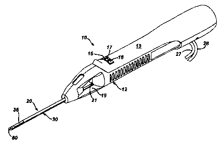
(001'n FIGURE 1 is an isometric view of a hand held vacuum assisted biopsy
device constructed
in accordance with US Patent 6,628,849.
[0018] FIGURE 2 is an isometric view of the elongated needle of the hand held
vacuum assisted
biopsy device of figure 1.
[0019] FIGURE 3 is an isometric view of the right body member of the elongated
needle of the
hand held vacuum assisted biopsy device of figure 1. A cutter tube liner is
illustrated in
assembly with the elongated needle.
(0020] FIGURE 4 is an exploded isometric view of the separated left body
member and right
body member of the elongated needle of the hand held vacuum assisted biopsy
device of
figure 1.
[00Z1J FIGURE 5 is an exploded isometric view of the two member ne~le tip on
the elongated
needle of the hand held vacuum assisted biopsy device of figure 1 as viewed
from the
proximal side thereof.
[002] FIGURE 6 is an exploded isometric view of the two member ne~le tip of
the elongated
needle of the hand held vacuum assisted biopsy device of figure 1 as viewed
from the
distal end thereof.
(OOZ3] FIGURE 7 is an isometric view of a biopsy device according to one
embodiment of the
present invention.
[004] FIGURE 8 is an alternate isometric view of the biopsy device of Figure
7.
CA 02502093 2005-03-22
-5-
[0025] FIGURE 9 is a schematic cross sectional illustration of the biopsy
device of Figure 7.
[0026] Figure 10 is an isometric illustration of a composite needle according
to an embodiment
of the present invention and having a mounting flange molded to a proximal
needle
portion.
(0027] Figure 11 is an isometric illustration of the needle of Figure 10 with
a vacuum manifold
component attached to the mounting flange.
{002$] Figure 12 is a schematic cross-sectional illustration of a mold
assembly which can be
used to form a biopsy device according to the present invention.
[0029) Detailed Description of the Invention
[0030) Figures 1-6 illustrate a biopsy device according to US Patent
6,626,849. Figures 7-12
illustrate embodiments of a biopsy device and a mold for making a biopsy
device
according to the present invention.
[0031] Figure 1 shows a hand-held vacuum assisted biopsy device 10 comprising
a noodle
assembly 20 and a holster 15, as described in US Patent 6,626,849. Needle
assembly 20
is detachably connected to holster 15. Together they constitute a lightweight,
ergonomically shaped, hand manipulatable portion referred to as handpiece 12.
Since
handpiece 12 is manipulated by the operator's hand rather than by an
electromechanical
arm, the operator may steer the handpiece 12 with great freedom towards the
tissue mass
of interest. The surgeon has tactile feedback while doing so and can thus,
ascertain to a
significant degree, the density and hardness of tissue being encountered. In
addition,
handpiece 12 may be held approximately parallel to the chest wall of a patient
for
obtaining tissue portions closer to the chest wall than may be obtained when
using an
instrument mounted to an electromechanical arm.
CA 02502093 2005-03-22
-d-
[0032] The device includes a means for obtaining a tissue sample. Holster 15
includes a forward
button 16 which may be used to move cutter 21 (shown in Figure 1 ) distally
though cutter
lumen 32 and sever tissue collected in port 36. Holster 15 further includes a
reverse
button 17 which may be used to move cutter 21 proximally thmugh cutter lumen
32 and
thereby moving the tissue sample in port 36 to a tissue collection surface 19.
A vacuum
button 18 on holster 15 is used to open or close first and second vacuum
lines, 27 and 28,
for activating a vacuum lumen 34 so as to cause tissue to become disposed
within port
36.
[0033) Referring now to Figure 2 there is shown an isometric view of the
needle assembly 20 of
the hand held vacuum assisted biopsy device 10 of figtu~e 1. Needle assembly
20
includes an elongated needle 30 having a distal end 31, a proximal end 33 and
a
longitudinal axis therebetween. Needle assembly 20 has a ne~lle tip 60 at its
distal end
for penetrating the soft tissue of a surgical patient. Elongated needle 30
comprises a
cutter lumen 32 and a vacuum chamber lumen 34.
[0034] At the distal end of the elongated needle 30 is a needle tip 60, which
is sharpen~l and is
preferably made from an MRI compatible resin such as Ultem or Vectra. Needle
tip 60 is
designed to penetrate soft tissue, such as the breast of a female surgical
patient, In this
embodiment, needle tip 60 is a three-sided pyramidal shaped point, although
the ne~le
tip 60 configuration may also have other shapes.
[0035] Referring now to Figure 3, elongated needle 30 can be made from a
thermoplastic
material such as Vectra A130 or B130 liquid crystal polymer, although other
MRT
compatible resins may be available from Ticona of Summit, NJ. Elongated needle
30
includes a cutter lumen 32 which houses the cutter 21 (shown in Figure 1).
Adjacent the
distal end 31 of the cutter lumen 32 is a port 35 for receiving the tissue
that is extracted
from a surgical patient by the cutter 21. Joined alongside the cutter lumen 32
is a vacuum
chamber lumen 34. The vacuum chamber lumen 34 receives vacuum from the second
vacuum line 28 which is connected the vacuum chamber lumen 34 on the elongated
CA 02502093 2005-03-22
-7-
needle 30 by the vacuum manifold 26 which is located at the proximal end 33 of
elongated needle 30. Also located at the proximal end of the elongated needle
30 is a
flange 38, which allows the elongated needle 30 and needle assembly 20 to
interlock with
the handpiece 12 on the hand-held vacuum assisted biopsy device 10. The liner
22,
discussed below, can be made from a MRI compatible material, such as a
polypropylene
such as Prolene available from Ethicon, Inc., Somerville NJ, or a material
known as
Radel-5000, available from British Petroleum, London UK.
[0036] Referring to Figure 4, the needle 30 of Figures 1-4 can be formed from
a left body
member 40 and a right body member 50 on either side of the longitudinal axis.
The
edges of the halves 40 and 50 are gated for easy part filling, and the edges
are stepped
with ridges that allow the two halves 40 and 50 to attach together with ease.
Preferably
needle 30 is molded from a thermoplastic, such as Vectra A130 or Vectra B130
liquid
crystal polymer. Other glass fiber reinforced resins known to those skilled in
the art
could also be used. Preferably the pmbe is made from a polymer material having
the
combination of high stiffness, low viscosity, and low mold shrink rate, such
as LCP
resins.
[0037] During assembly of the elongated needle 30, the left body member 40 and
right body
member 50 of the elongated needle 30 can be pushed together. Ore the left body
member 40 and the right body member 50 are pressed together, a thin-walled
slecve of
high strength tubing is slipped over the elongated needle and is shrink fitted
into place.
The shrink tubing holds the left body member 40 and the right body member SO
together
for easier handling prior to adhesive curing. In addition, the shrink tubing
makes the
exterior of the elongated needle 30 smoother for reduced insertion forces.
[0038] Referring back to Figure 3, there is shown the right body member 50 of
the elongated
needle 30, separated fmm the left body member 40, which has been omitted from
this
figure for clarity. The right body member 50 has upper and lower ends
comprising
alternating male and female portions or members, 42 and 52, which alternate
and are
CA 02502093 2005-03-22
-8-
arranged axially along the length of the right body member 50 of the elongate
needle 30.
In addition to the male and female members, 42 and 52, there is an upper
female distal
member 54 and a lower male distal member 46, both of which are located at he
distal end
of the right body member 50. The upper female distal member 54 is located just
below
the distal end of the cutter lumen 32 and above the distal end of the vacuum
chamber
lumen 34. At the proximal end of the right body member SO are three female
receivers
56 which surround the vacuum manifold 26 at the proximal end of the right body
member
50.
(0039] Still referring to figure 3, needle 20 includes a cutter tube liner 22,
which helps keep
adhesive out of the lumen to provide a smooth surface thereon. Liner 22
generally abuts
in the inner surface of cutter 20 along lumen 32. The distal end 31 of liner
22 is proximal
to port 36 but otherwise is disposed along the length of lumen 32. The cutter
tube liner
22 is formed from a thin-walled extrusion of a low-friction, abrasion-
resistant plastic,
such as polypropylene, polyetherimide or polyethersulfone. The cutter tube
liner 22
provides a smooth, low-friction, abrasion-resistant surface for the cutter 21.
[0040] Referring again to Figure 4 there is shown an exploded isometric view
of the elongate
needle 30 of the hand held vacuum assisted biopsy device 10 of figure 1. Both
the left
body member 40 and the right body member 50 of the elongated needle 30 are
shown.
The male features 42 which are arranged axially on the left body member 40,
mate to the
female features 52 which are arrange axially on the right body member 50. The
male
features 42 arranged axially on the right body member 50 mates to the female
features 52
which are arranged axially on the left body member 40.
[0041) In addition to male and female members, 42 and 52, which are arranged
axially and mate,
the left body half 40 and right body member 50 have additional features that
mate at both
the proximal and the distal ends. At the proximal end of the right body member
50 are
three female receivers 56 which surround the vacuum manifold 26. At the
proximal end
of the left body member 40 are three male bosses 46 which surround the vacuum
CA 02502093 2005-03-22
-9-
manifold 36 and correspond to the three female receivers S6 on the right body
member
50. When the left body member 40 and the right body member 50 are pushed
together,
the three female receivers 56 on the proximal end of the left body member 40.
The
proximal end of the elongated needle 30 is thus, retained by the three female
receivers 56
and three male bosses 46, which mate at the proximal end of the elongated
needle 30.
[0042] The needle tip 60 at the distal end of the elongated needle 30 is
retained by the upper
female distal part 54 and the upper male distal portion 44 and the lower
female distal
portion 55 on the left body member 40. The upper male distal portion 44 is
located
above the cutter lumen 32 at the distal end on the left body member 40, and
the lower
female distal part 55 is located below the cutter lumen 32 and above the
vacuum chamber
lumen 34 at the distal end of the left body member 40. On the right body SO is
an upper
female distal part 54 and a lower male distal portion 45, which correspond to
the upper
male distal portion 44 and the lower female distal part 55 on the left body
member 40.
The upper female distal part 54 is located above the cutter lumen 32 at the
distal end of
the right body member 50, and the lower male distal portion 45 is located
below the
cutter lumen 32 and above the vacuum chamber lumen 34 at the distal end of the
right
body member 50.
[0043] Still referring to figure 4, the right body member 50 and left body
member 40 can be
configured so that when the member 50 and the member 40 are joined, the
combined
members provide the interlumen vacuum holes 23, which are located below the
tissue
receiving port 36 on the distal end of the elongated needle 30. The interlumen
vacuum
holes 23 can be in the form of six cylindrically shaped holes which are open
to port 36.
Vacuum communicated from vacuum lumen 34 through holes 23 can be used to draw
tissue into the cutter lumen 32. Cutter 21 can have a sharpened distal end
adapted to cut
tissue, and can rotationally driven as it is advanced distally past tissue
port 36, thereby
severing tissue drawn into cutter lumen 32. The cutter 21 can then be
retracted and the
severed tissue sample deposited at collection surface 19 (Figure 1) by
retracting
proximally.
CA 02502093 2005-03-22
-10-
[0044] Still referring to figure 4, the male and female members, 42 and 52,
which mate and are
located on the left body member 40 and the right body member 50 have a number
of
distinct advantages. The male and female members, 42 and 52, on the left body
member
40 and right body member 50 orient the left body member 40 and right body
member ~0
during assembly of the elongated needle 30.
(0045] The male and female members, 42 and 52, which mate, are also key
factors in increasing
both the strength and lateral bending stiffness of the elongated needle 30.
When the
needle 30 is subjected to a lateral bending moment, nearly all of the material
being loaded
axially is the high-strength, high stiffness body material. Only the small
amount of
adhesive that is used to fill the axial clearances between the male and female
members,
42 and 52, which mate, is of a lower stiffness. A conventional bonded joint
would result
in the bond line being loaded in a manner similar to that used for adhesive
peel strength
testing, which is the most severe type of loading for an adhesive joint. In
contrast to this,
the male female members, 42 and 52, which mate, would create lateral bond
surfaces
along the elongated needle 30. This substantially increases the bond line
length of the
elongated needle 30. Because of significant portions of the bond line being
loaded in
shear, the strength and lateral stiffness of the elongated needle 30 is
increased. This is
improved over a singly piece molded cylinder in that with the bond line loadai
in shear,
the elongated needle 30 will be able to sustain bending moments of its joints
rather than
at its base, which decreases the possibility of breakage.
[0046] Figure 5 shows and exploded isometric view of the needle tip 60 of the
elongated needle
30 of the hand held vacuum assisted biopsy device 10 of figure 1 as viewed
from the
proximal side thereof. The needle tip 60 has two halves; a composite tip
member 70, and
a composite hub member 80. Both the composite tip member 70 and the composite
hub
member 80 are preferably molded from a magnetic Resonance Imaging (IViRI)
compatible resin such as Ultem or V~tra ceramic or other ll~iRI compatible
materials
known to those skilled in the art is sharp. The composite tip member 70 has a
three-sided
pyramidal shaped point, but may also have other shapes. The composite tip
member 70
CA 02502093 2005-03-22
-11-
has a hollow cavity 74 and protruding connectors ?6. The two protruding
connectors 76
are inserted into the two receiving holes 82 on the composite hub member 80
when the
composite hub member 80 is pushed into the composite tip member 70 during
assembly.
Cavity preferably contains a capsule 90 made from a material which will leave
and MRI
artifact. Having a capsule 90 made from and MRI artifact leaving material is
necessary
because since the elongated needle 30 is made of an MRI compatible resin, the
elongated
needle 30 does not show up on an MRI scan. Therefore, it is difficult for a
physician to
discern the orientation of the elongated needle 30 during and MRI scan MRI
artifact
leaving material 90 solves the aforementioned problems in that a needle tip 60
leaves a
small, but not troublesome artifact on an MRI scan. This small, artifact
indicates the
orientation of the elongated needle 30 relative to the sight of biopsy, and
where the tissue
receiving bowl begins during and MRI scan. The MRI artifact leaving material
90 that is
preferred is a capsule of Gadolinium. However, there are other materials that
could be
put into the hollow cavity74 of the composite tip member 70 that would leave
and
acceptable MRI artifact. These include, but not limited to: liquid Gadolinium,
Titanium
Wire, Aluminum, Copper, Brass Iron, and Bronze.
[0047] Figure 6 shows an exploded isometric view of the needle tip 60 of the
elongated needle
30 of the hand held vacuum assisted biopsy device 10 of figure 1 as viewed
from the
distal end thereof. This figure clearly illustrated the components on the
composite hub
member 80. On the distal end of the composite hub member 80 is a male part 84,
which
pushes the MRI artifact leaving material 80 down into the hollow cavity 74 on
the
composite tip member 70. Also located on the distal end of the composite hub
member
80 is a knock out boss 8b, which pushes a collected breast tissue sample into
the end of
the cutter tube 21 the hand held vacuum assisted biopsy device 10 during a
breast biopsy.
The two receiving holes 82 on the composite hub member 80 receive the two
protruding
connectors 76 on the composite tip member 70 when the composite tip member 70
and
composite hub member 80 are pushed together. The reception of the two
protruding
connectors 76 on the composite tip member 70 by the two receiving holes 82 on
the
CA 02502093 2005-03-22
-12-
composite hub member 80 locks the composite tip member 70 and the composite
hub
member 80 together, and seals the NIRI artifact leaving material 90 in the
hollow cavity
74 in between the composite tip member 70 and composite hub member 80.
[0048] In using the hand member vacuum assisted biopsy device 10, as shown in
figure 1, for a
breast biopsy in an MRI environment, physician will first positions outside of
the MRI
magnet, the patient is moved into the MRI magnet and imaging of the breast is
performed. During imaging of the breast, serial slices of the breast are
examined, and a
contrast agent is administered to highlight suspicious areas of breast tissue.
At this time,
the location of the suspicious breast tissue is determined relative to the
compression grid.
[0049] After the location of the suspicious breast tissue is determined, the
patient is moved
outside the magnet. Local anesthesia is administered to the patient and the
probe 20 is
inserted into the area of suspicious breast tissue.
[0050] After the probe is inserted into the suspicious area of breast tissue,
the patient is moved
back into the MRI magnet and a set of images of the breast are taken. The sets
of images
confirm that the probe 20 is adjacent to the suspicious breast tissue, the
patient is moved
outside of the MRI magnet and the hand held vacuum assisted biopsy device 10
of figure
1 is then inserted into the sleeve, replacing the obturator.
[0051] After the hand held vacuum assisted biopsy device 10 of figurc 1 is
inserted through the
sleeve; multiple tissue samples are taken. 1n taking multiple tissue samples,
the needle
tip 60 as the distal end of the elongated needle 30 on the hand hehd vacuum
assisted
biopsy 10, of figure 1, penetrates the breast in the area that is adjacent of
the suspicious
breast tissue. Prior to, and during penetration by the needle tip 60, the
cutter 21 is fully
forward, and is advanced forward through the cutter lumen 32 by pressing the
forward
button 16 on the holster 15 of the vacuum assisted biopsy device 10 of figure
1.
[0052] Once the elongated needle 30 is positioned in the area adjacent to the
suspicious breast
tissue, vacuum suction is applied to he vacuum chamber lumen 34. The vacuum
suction
is applied by pressing the vacuum button 18 on the holster 15 of the hand held
vacuum
CA 02502093 2005-03-22
-13-
assisted biopsy device 10 of figure 1. Pressing the vacuum button 18 on the
holster 1S
opens the second vacuum line 28, which transports vacuum suction through the
handpiece 12 of the hand held vacuum assisted biopsy device 10 and into the
vacuum
chamber lumen 34 on the elongated needle 30. The second vacuum line 28 runs
through
the handpiece 12 of the hand held vacuum assisted biopsy device 10 and into
the
elongated needle 30 through the vacuum manifold 24 at he proximal end of the
elongated
needle 30. The vacuum suction that is applied to the vacuum chamber lumen
travels
from the proximal, of the distal end of the vacuum chamber lumen 34, below the
interlumen vacuum holes 23. The interlumen vacuum holes 23 receive suction
from the
vacuum chamber lumen 34.
[0053) The suction from the interlumen vacuum holes 23 actively pulls breast
tissue thmugh the
port 36 and into the cutter lumen 32 on the elongated needle 30. After the
breast the
tissue is pulled into the elongat~l needle 30 through the port 36, the cutter
21 begins to
rotate and advances through the breast tissue until a sample has been
obtained. Afttr the
breast tissue sample has been obtained, the elongated needle 30 is rotated to
position the
port 36 toward a different clockwise position in preparation for obtaining the
next tissue
sample. After the elongated 30 is rotated, the cutter 21 is withdrawn
backwards within the
cutter lumen 32 on the elongated needle 30 and the breast tissue sample is
carried back to
a knock-out boss 86, which pushed the collected breast tissue sample out into
a tissue
collection surface 19 on the handheld vacuum assisted biopsy device 10. Vacuum
suction is then reapplied to the vacuum chamber lumen 34 from the second
vacuum line
28, and the aforementioned process is repeated continuously until the
elongated needle 30
has been rotated clockwise once around the entire clock.
[0034) After multiple breast tissue samples have been obtained from the
patient, the patient is
moved back into the MRI magnet. Once in the MRI magnet, a set of images of the
breast
are taken in order to confinm that the suspicious breast tissue has been
removed. The
artifact in the probe tip is a useful point of reference to confirm after the
biopsy site is
marked, the breast biopsy in an MRI environment is complete.
CA 02502093 2005-03-22
- 14-
(0055) Referring now to Figures 7-9, an improved needle assembly 120 for use
with a biopsy
device is illustrated. The needle assembly 120 can be used with a handheld
device such
as a handpiece 12 of the type shown in Figure 1. Alternatively, the needle
assembly 120
can be used with a biopsy device which is mounted on a platform, table, or
other suitable
support.
(0056) Needle assembly 120 can include an elongated needle 130 and a mounting
component
200. Mounting component 200 can be used to support the needle assembly 120 on
a
biopsy handpiece, a biopsy device base or platform, or other mounting surface
for
supporting a biopsy device.
[003'1) The elongated needle 130 can include a distal needle segment 160 and a
proximal needle
segment 140. The distal needle segment 160 can comprise a tissue receiving
port 136
formed therein. The distal needle segment can be formed of a first material
that does not
interfere with MRI imaging of a portion of the distal needle segment
associated with the
tissue receiving port 136. The first material can be used to form the edges
136A, B, C,
and D of the port 136, and the first material can extend proximally from edge
1368 and
.
distally from edgy 136C. The distal needle segment 160 can include interlumen
vacuum
holes 123 for use in drawing tissue into the port 136, the holes 123
illustrated in Figures
7, 8, and 9.
[0058) By the phrase "not interfere with MRI imaging" it is meant
substantially no distortion of
the imaged area by MRI artifact such as "blooming" due to metallic pies or
components, and substantially no local distortion of the magnetic field caused
by a mass
material, such that the tissue receiving port 136 can be identified using MRI
imaging.
[0059) The proximal needle segment 140 is disposed proximally of the tissue
receiving port 136,
and extends proximally of the distal needle segment 160. The proximal needle
segment
140 is formed at least in part of a second material different from the first
material.
{0060) A distal tissue piercing tip 190 can be disposed at the distal end of
the needle assembly
120, such as by attachment to the distal end of the distal needle segment 160.
The distal
CA 02502093 2005-03-22
-15-
tissue piercing tip 190 is disposed distal of the tissue receiving port 136.
The distal tissue
piercing tip 190 can be formed of a material that does not interfere with MRI
imaging of
the tissue receiving port 136. In one embodiment, the piercing tip 190 can be
formed of a
material different from the first material and the second material. For
instance, piercing
tip 190 can comprise a flat blade formed of a suitable material such as a
glass or ceramic.
(0061 ] The distal needle segment can be formed of a first material which is
non-metallic and
non-magnetic. In one embodiment, the first material can be selected from
materials
including, but limited to, plastics, thermoplastics, thermoresins, and
polymers. For
instance, the distal needle segment can be formed, at least in part, of a
liquid crystal
polymer or a glass reinforced polymer. One suitable material is a glass
reinforced liquid
crystal polymer such as VECTRA A130 available from Ticona Coip. In one
embodiment, the first material can have a melt flow index of at least about 10
grams/minute, more particularly at least about 15 grams/minute. Without being
limited
by theory, such a mold flow index is thought to be beneficial for molding
relatively long,
thin-walled cross-sections.
[0062] The proximal needle portion 140 can be formed of a second material
which is a non
magnetic metal. Suitable materials from which the proximal needle portion 140
can be
formed include, but are not limited to, aluminum, aluminum alloys, stainless
steel,
titanium, titanium alloys, and combinations thereof. In one particular
embodiment, the
proximal needle portion 140 can be formed of titanium, and the distal needle
portion 160
can be injection molded over the titanium proximal needle portion 140, as
described more
fully below. The piercing tip 190 can be formed of a material sel~ted from
ceramics
and glasses. In one embodiment, the tip 190 can be formed, at least in part,
of a ceramic
comprising alumina or zirconia. The piercing tip 190 can also be formed of a
natural or
synthetic gemstone, such as a natural or synthetic ruby or sapphire.
CA 02502093 2005-03-22
-16-
[0063) Referring to the cross-sectional illustration of Figure 9, the distal
needle segment 160 can
include an upper cutter lumen 162 and a lower vacuum lumen 164, with
interlumen
vacuum holes 123 providing flow communication between the lumen 162 and the
lumen
164. The proximal needle segment 140 can include an upper cutter lumen 142 and
a
lower vacuum lumen 144. Cutter lumen 142 and cutter lumen 162, together, form
a
continuous, smooth, uninterrupted lumen for receiving a rotating and
reciprocating cutter,
such as the cutter 21 described above with respect to Figures 1-6. Vacuum
lumen 144
and vacuum lumen 164, together, form a continuous, uninterrupted lumen for
conveying
vacuum from a vacuum source (not shown) to the interlumen vacuum holes 123.
[0064) Still referring to Figure 9, the distal needle portion 160 can also
include fluid passages
166. Fluid passages 166 can extend from an outside surface of the distal
needle portion
160, such as the bottom surface, and can communicate with the vacuum lumen
164. In
Figure 9, the fluid passages 166 are generally cylindrically shaped holes
positioned
generally opposite and below the vacuum holes 123, and the passages 166 extend
generally downward from the lumen 164 to extend through the exterior bottom
surface of
the distal needle portion 160, opposite the tissue port 136. Alternatively,
the holes 166
can also be positioned to extend from the vacuum lumen 164 at various
circumferential
positions around the distal needle portion. Without being limited by theory,
the fluid
holes 166 can be used to aid in providing suction and irrigation at the biopsy
site. For
instance, fluid holes 166 can be used to deliver an anesthetic substance,
other
medications, to irrigate the biopsy site, or provide suction at the opposite
end of the
needle from the tissue receiving port 136.
[OOb3) By way of example, the proximal needle portion 140 can be formed of
thin wall titanium
tubing, and the distal needle portion 160 can be a liquid crystal polymer
molded over an
end of the proximal needle portion 140, so that a proximal portion of the
distal ne~le
CA 02502093 2005-03-22
-17-
portion 160 overlaps the distal portion of the proximal needle portion 140.
For example,
the proximal needle portion 140 can be formed by welding or otherwise joining
two
pieces of thin walled titanium tubing, such as upper tube portion 146 and
lower tube
portion 148, to form the upper lumen segment 142 and lower lumen segment 144.
The
distal needle portion 160 can then be molded over the proximal needle portion
140. In
Figure 9, the piercing tip 190 is illustrated with an anchoring hole 192.
Anchoring hole
192 can aid in attaching piercing tip 190 to the end distal needle portion 160
when the
distal needle portion 160 is formed by molding (i.e. the molten molding
material flows
into hole 192 and when solidified, serves to fix piercing tip 190 at the
distal end of the
distal needle portion 160.
[0066] Still referring to Figure 9, the distal most portion of the proximal
needle portion 140 is
preferably spaced a distance L of at least about 0.5 inch from the proximal
edge 136B of
the port 136. In particular, the distal end of tube portion 146 is spaced a
distance L from
the proximal edge 136B, as shown in Figure 9. In one embodiment, the distance
L can be
between about 0.5 inch and about 2.5 inches, and more particularly between
about 0.5
and about 1.5 inches. Without being limited by theory, providing such a
spacing can
reduce interference with MRI imaging of the portion of the needle surrounding
the tissue
receiving port 136 by the metal of proximal needle portion 140, while
maintaining the
strength and stiffness of the needle assembly 120.
[0067] Figure 10 illustrates needle 130 having a component 200 comprising a
mounti~ flange
338 attached adjacent a proximal end of the needle 130. Component 200 with
flange 338
can be molded onto the proximal needle portion 140, either before or after the
distal
needle portion 160 is molded onto the proximal needle portion 140. In one
embodiment,
the flange 338 can be molded onto a metallic proximal needle portion 140
first, and the
distal facing surface of flange 338 can be used as a reference surf~e/locating
surface in a
CA 02502093 2005-03-22
-18-
subsequent molding operation in which the distal needle portion 160 is molded
onto the
proximal needle portion. Figure 11 shows a vacuum manifold 326 attached to the
mounting flange 338, such as by gluing, welding, or press fit. The needle, as
shown in
Figure 11, can be used in the device of Figure l, as a replacement needle for
the needle
assembly shown in Figure 2.
(0068] Figure 12 illustrates a mold configuration that can be used to form a
needle assembly
130. As described above, a mounting component 200 can be first molded onto a
metallic
proximal needle component, such as a metallic needle shaft 1140. A surface of
the
mounting component 200 can then serve to locate other features to molded in
the distal
needle portion 160.
(0069] Refernng to Figure 12, a mold assembly 2000 comprising a first mold
half 2010 and a
second mold half 2012 is provided. Mold halves 2010 and 2012 separate along
mold
split line 2016. A metallic needle shaft 1140 (corresponding to proximal
needle portion
140) with a mounting component 200 molded thereto is provided. The needle
shaft 1140
can include an upper lumen and a lower lumen corresponding to a portion of the
cutter
lumen and the vacuum lumen in the completed biopsy device. The previously
molded
component 200 has one or more surfaces that can be used to locate features to
be molded
in the mold assembly 2000.
[0070] The shaft 1140 is supported by core support shafts 1144 and 1148. Core
support shafts
1144 and 1148 are supported by and extend from a support block 1142. Core
support
shaft 1144 extends distally from support block 1142 and extends into and
through the
upper lumen of needle shaft 1140. Core support shaft 1148 extends distally
from support
block 1142 and extends into and through the lower lumen of needle shaft 1140.
The core
CA 02502093 2005-03-22
-19-
support shafts 1144 and 1148 extend through the needle shaft 1140 and extend
beyond
the distal end of the needle shaft 1140. The core support shafts 1144 and 1148
serve to
form the upper and lower lumens in the molded, non metallic distal needle
portion of the
needle assembly (molten mold material flows around the core support shafts to
form the
distal needle portion 160). The core support shafts can be forms of any
suitable
metallic or non-metallic material. In one embodiment, the core support shafts
comprise
stainless steel, though other metals may be employed.
[0071] The needle shaft 1140, support block 1142, and core support shafts 1144
and 1148 are
inserted into the mold assembly 2000. A metal blade 1190 is supported in the
mold
assembly by a blade support 1192, such as a "puck" of a suitable material. A
suitable
material from which the puck can be formed is a liquid crystal polymer
material, such as
Vectra brand liquid crystal polymer available from Ticona Corp. The blade 1190
can be
in the form of a flat metallic blade with a generally triangular shaped tip
and having a
hole near the base. The triangular shaped tip can be held in puck, such as by
embedding
the tip in the high temperature plastic material of the puck. The blade 1190
serves to
form the piercing tip 190 of the finished needle 130. The hole in the blade
1190 is
provided so that molten molding material can flow into the hole and surround
the portion
of the blade 1190 that is not embedded in the puck.
[0072] Core support pins 1244 and 1248 are provided in association with the
mold halves 2010
and 2012. As the mold halves 2010 and 2012 are closed about the needle shaft
1140 and
core support shafts 1144 and 1148, the core support pins 1244 and 1248 are
positioned to
engage with core support shafts. Core support pins 1248 engage the core shaft
1148 and
help support the core shaft 1148 at its distal end. The ends of the core
support pins 1248
can extend into recesses in core shaft 1148. The core support pins 1248 also
take up
space when molten material is provided to the mold 2000, so as to form the
fluid holes
166 in the bottom surface of the vacuum lumen (holes 166 shown in Figure 9).
The core
support pins 1244 extend through core support shaft 1144 and engage the top of
core
support shaft 1148. Each of the core support pins 1244 serve to form one of
the
CA 02502093 2005-03-22
_20_
interlumen vacuum holes 123 (shown in Figure 9) when molten material is
solidified
around the core support pins 1244.
[0073] Once the mold halves 2010 and 2012 are closed, molten plastic is
injected into one or
more cavities formed by the mold halves. The mold halves 2010 and 2012 can
comprise
multiple segments for forming different portions of the needle. For instance,
mold
segments 2010A and 2012A contact needle shaft 1140 without providing a cavity,
so that
no molten material flows over the proximal end of needle shaft 1140. Mold
segments
2010B and 2012B are sized and shaped to provide a cavity 2023 about the distal
portion
of needle shaft 1140, and a mold cavity 2025 about the portions of the core
support shafts
1144 and 1148 extending from the needle shaft 1140. Molten material flowing
into the
cavity 2023 and the cavity 2025, on solidifying, forms the portion of the
distal needle
segment 160 which is positioned proximal of the tissue receiving port 136 of
the finished
needle 130.
[0074] Mold segment 2010C is sized and shaped to form the tissue receiving
port 136 in the
upper portion of the distal needle portion 160, while mold segment 2012C is
sized and
shaped to form the bottom portion of the distal needle portion 160 below the
tissue
receiving port 136. Mold segments 2010D and 2012D, together with the puck
1192, are
used to form the distal most part of distal needle portion 160 that is between
tissue
receiving port 136 and the piercing tip 190. Molten material flowing amund the
blade
1190 and through the hole in the blade serves to entrap the blade 1190 in the
distal end of
distal needle portion 160. Accordingly, the piercing tip 190 is entrapped in
the distal end
of molded distal needle portion 160.
[0075] In the embodiment described, the distal needle portion 160 is formed by
injection
molding the distal needle portion about the proximal needle portion. The
molding step is
"insert molding" in the sense that the proximal needle portion forms a part of
the
supporting structure as part of the molding process as well as a functional
part of the
finished needle assembly. Alternatively, the distal needle portion can be
formed
CA 02502093 2005-03-22
-21-
separately, and then attached by any suitable means, such as by adhesive, to
the proximal
needle portion 140. In yet another embodiment, the distal needle portion can
be formed
in symmetric half portions, similar to those shown in Figures 3 and 4, with
the half
portions then fastened together and then attached by any suitable fastening
means to the
proximal needle portion. Without being limited by theory, it is believed that
molding the
distal needle portion about the proximal needle portion provide a smooth,
uninterrupted
transition between the portion of the cutter lumen associated with the
proximal needle
portion and the portion of the cutter lumen associated with the distal needle
portion, so
that there is a smooth lumen surface at the interface to permit smooth
translation of the
cutter through the entire length of the cutter lumen. Accordingly, there is no
lip, seam, or
other restriction at the lumen juncture that would otherwise require an
additional
machining or processing step for removal. Prior to placing the metallic
proximal needle
portion in the mold, the outer surface of the proximal needle portion 140 can
be
roughened or otherwise textured, such as by bead blasting or knurling, to
enhance
attachment of the distal needle portion to the proximal needle portion.
[0076] While preferred embodiments of the present invention have been shown
and
described herein, it will be obvious to those skilled in the art that such
embodiments are
provided by way of example only. Numerous variations, changes, and
substitutions will
now occur to those skilled in the art without departing from the present
invention.
Additionally, each component or element can be described in terms of a means
for
performing the component's function. Accordingly, it is intended that the
invention be
limited only by the spirit and scope of the appended claims.