Note: Descriptions are shown in the official language in which they were submitted.
CA 02504613 2005-05-02
WO 2004/045363 PCT/IL2003/000940
METHOD AND SYSTEM FOR MOUNTING AN MPS SENSOR ON A
CATHETER
s FIELD OF THE DISCLOSED TECHNIQUE
The disclosed technique relates to medical devices in general,
and to methods and systems for determining the position and orientation
of a catheter, in particular.
io BACKGROUND OF THE DISCLOSED TECHNIQUE
Medical operations on human or animal lumens, such as the
vascular system, ureter, urethra, brain vessels, coronary vessels, lumens
of the liver, kidney, lung, digestive system, and the like, can be performed
by employing a medical catheter. Such medical operations include dilating
15 a lumen by a balloon or a stent, implanting a stent, delivering a
pharmaceutical substance to the lumen, performing coronary bypass,
removing plaque from the intima of a blood vessel, implanting a graft, and
the like. Such a medical catheter includes a lumen intervention element,
such as a balloon, stent, balloon expanding stent, substance delivery
2o element, tissue severing element, and the like, at the distal end thereof.
In some cases, the medical catheter also includes a radiopaque
material at the distal end, which serves as a marker for the location of the
distal end. In order to perform the medical operation, usually a guiding
catheter is initially inserted in the lumen. Sometimes an auxiliary,
25 large-diameter guidewire is inserted prior to the guiding catheter for
aiding
it to enable manipulation of the guiding catheter. Next, the large-diameter
guidewire is pulled out, another guidewire, with of smaller diameter, is
inserted in the guiding catheter and the small-diameter guidewire is
advanced to the desired location within the lumen, by manipulating the tip
30 of the small-diameter guidewire from outside the body of the patient. The
CA 02504613 2005-05-02
WO 2004/045363 PCT/IL2003/000940
proximal end of the small-diameter guidewire is inserted into the distal end
of the medical catheter and the medical catheter is advanced to the
desired location, by passing the medical catheter over the guidewire inside
the guiding catheter. The physician determines the position of the distal
s end of the medical catheter, by viewing an image of the marker in an
imaging device, such as fluoroscope, X-ray table, and the like. When the
physician assures that the lumen intervention element is .located at the
desired location, the physician performs the medical task on the lumen.
US Patent No. 6,233,476 issued to Strommer et al., assigned to
io the present assignee, and entitled "Medical Positioning System", is
directed to a medical positioning system (MPS) for determining the
position and orientation of a medical device within a living tissue. The MPS
includes a 3D electromagnetic field (EMF) generator, a main sensor, an
auxiliary sensor, a sensor interface, a position and orientation processor, a
15 superimposing processor, an image interface, a 3D image database and a
display unit.
The position and orientation processor is connected to the 3D
EMF generator, the sensor interface and to the superimposing processor.
The auxiliary sensor and the main sensor are connected to the sensor
~o interface. The image interface is connected to the superimposing
processor and to the 3D image database. The display unit is connected to
the superimposing processor. The main sensor is located at the tip of the
medical device. The auxiliary sensor is located in the vicinity of the
inspected tissue of the patient.
25 The 3D image database includes a plurality of predetected
images of the inspected tissue of the patient. The auxiliary sensor
compensates for the movement of the patient. The 3D EMF generator
includes a plurality of electromagnetic coils that produce electromagnetic
fields in different directions and in different magnitudes. Each of the main
ao sensor and the auxiliary sensor includes three electromagnetic coils. Each
of the electromagnetic coils of the main sensor and the auxiliary sensor
-2
CA 02504613 2005-05-02
WO 2004/045363 PCT/IL2003/000940
detects an electromagnetic field in a different direction. Each of the main
sensor and the auxiliary sensor produces a signal in response to the
electromagnetic field generated by the 3D EMF generator, corresponding
to the position and orientation of the main sensor and the auxiliary sensor,
respectively.
' The position and orientation processor receives the signal from
the main sensor through the sensor interface and the position and
orientation processor determines the position and orientation of the main
sensor according to this signal. The superimposing processor retrieves a
io predetected image of the inspected tissue from the 3D image database,
through the image interface. The superimposing processor superimposes
a representation of the tip of the medical device on the retrieved image
and produces a video signal. The representation of the tip of the medical
device corresponds to the position and orientation of the tip of the medical
device relative to the inspected tissue. The display unit produces a video
image according to the video signal. US Patent No. 5,646,525 issued to
Gilboa and entitled "Three Dimensional Tracking System Employing a
Rotating Field", provides a description of three dimensional tracking
system employed by the MPS for determining position and orientation.
2o US Patent No. 6,179,811 issued to Fugoso, et al., and entitled
"Imbedded Marker and Flexible Guide Wire Shaft", is directed to a balloon
catheter which includes a marker band imbedded into a guidewire shaft of
the balloon catheter. The balloon catheter includes a balloon, a shaft, a
manifold, a guidewire shaft and a plurality of marker bands. The guidewire
~s shaft is located within the shaft. The proximal end of the balloon is
affixed
to a distal end of the shaft and the distal end of the balloon is bonded to a
distal end of the guidewire shaft. The manifold is located at a proximal end
of the shaft. The marker bands are imbedded into the guidewire shaft at a
region of the guidewire shaft below the balloon. The marker bands can be
so viewed by fluoroscope equipment.
-3-
CA 02504613 2005-05-02
WO 2004/045363 PCT/IL2003/000940
US Patent No. 5,928,248 issued to Acker and entitled "Guided
Deployment of Stents", is directed to an apparatus for applying a stent in a
tubular structure of a patient. The apparatus includes a catheter, a hub, a
pressure control device, a balloon, a stent, a probe field transducer, a
plurality of external field transducers, a field transmitting and receiving
device, a computer, an input device and a cathode ray tube. The catheter
includes a bore. The hub is affixed to a proximal end of the catheter. The
balloon is mounted on a distal end of the catheter. The pressure control
device is connected to the balloon through the hub and the bore. The stent
io is made of a shape memory alloy and is located on the balloon.
The probe field transducer is located within the catheter, at a
distal end thereof. The external field transducers are located outside of the
patient (e.g., connected to the patient-supporting bed). The field
transmitting and receiving device is connected to the external field
transducers, the probe field transducer and to the computer. The computer
is connected to the cathode ray tube and to the input device.
A user calibrates the field transmitting and receiving device in an
external field of reference, by employing the external field transducers.
The field transmitting and receiving device together with the computer,
2o determine the position and orientation of the probe field transducer in the
external field of reference. The user views the position and orientation of a
representation of the stent which is located within a tubular structure of the
patient, on the cathode ray tube. When the user determines that the distal
end is located at the desired location within the tubular structure, the user
expands the stent by operating the pressure control device and inflating
the balloon, thereby positioning the stent at the desired location.
US Patent No. 5,897,529 issued to Ponzi and entitled "Steerable
Deflectable Catheter Having Improved Flexibility", is directed to a system
for mapping a heart chamber and creating channels in the heart tissue.
so The system includes a catheter, a computer, a monitor and a pad
containing coils. The catheter includes a catheter body, a control handle,
-4
CA 02504613 2005-05-02
WO 2004/045363 PCT/IL2003/000940
an optical fiber, a puller wire, a compression coil, a tip electrode, a ring
electrode, temperature sensing means, an electromagnetic sensor and a
circuit board. The control handle is attached to a proximal end of the
catheter body. A distal end of each of the optical fiber, the puller wire and
s the compression coil, is located at a distal end of the catheter body. A
proximal end of each of the optical fiber, the puller wire and the
compression coil, is located at a proximal end of the catheter body.
The tip electrode, the ring electrode and the temperature means
are located at the distal end of the catheter body. The circuit board is
io located within the control handle. The circuit board is attached to the
electromagnetic sensor and to the computer. The computer is connected
to the monitor and to the coils. The circuit board prevents the system from
being used twice, according to a signal received from the electromagnetic
sensor. The compression coil provides flexibility to the catheter body.
15 The coils are located under the patient and generate a magnetic
field. The electromagnetic sensor generates a signal in response to the
generated magnetic field and the computer determines the position of the
electromagnetic sensor and thus the distal end of the catheter body, by
processing the signal. The tip electrode and the ring electrode monitor the
2o strength of the electrical signals at a selected location. The temperature
sensing means monitor the temperature of the tip electrode.
The tip electrode and the ring electrode allow the user to map
the heart chamber. The user simultaneously maps the contours of the
heart chamber, the electrical activity of the heart and the displacement of
25 the catheter body, thereby identifying the location of an ischemic tissue.
The user then creates channels in the ischemic tissue, via the optical fiber.
US Patent No. 5,830,222 issued to Makower and entitled
"Device, System and Method for Interstitial Transvascular Intervention", is
directed to a method for gaining percutaneous access to a diseased
~o vessel through an adjacent intact vessel. Using this method, it is possible
to bypass the diseased vessel, such as a coronary artery, through the
-5
CA 02504613 2005-05-02
WO 2004/045363 PCT/IL2003/000940
intact vessel, such as a cardiac vein. The diseased vessel may include an
occlusion that restricts the flow. A guide-catheter is advanced through the
vena cava into the coronary sinus, within the right atrium of the heart. A
transvascular interstitial surgery (TVIS) guide catheter is inserted through
s the guide-catheter and advanced through the cardiac vein over a first
guidewire, to a desired location adjacent the coronary artery.
The TVIS guide-catheter includes a balloon, a TVIS probe and
either or both of active orientation detection means and passive orientation
detection means. The TVIS probe is a rigid wire, antenna, light guide or
io energy guide capable of being inserted in tissue. The passive orientation
detection means allow radiographic, fluoroscopic, magnetic or
sonographic detection of position and orientation of the TVIS probe. The
active orientation detection means is a transmitter. A second guidewire is
inserted into the coronary artery adjacent the cardiac vein, wherein the
is second guidewire includes a small receiver to receive a signal emitted by
the active orientation detection means. The second guidewire further
includes a wire bundle which is capable to return the signal detected by
the receiver, to an operator, thereby enabling the operator to determine
the position and location of the TVIS probe.
2o When the orientation of the TVIS guide-catheter is assured, the
balloon is inflated against the wall of the cardiac vein, in order to block
the
flow, stabilize the TVIS guide-catheter within the cardiac vein and dilate
the passageway. The TVIS probe, is then advanced through the wall of the
cardiac vein into the coronary artery, thereby bypassing the diseased
2s section of the coronary artery.
US Patent No. 5,489,271 issued to Andersen and entitled
"Convertible Catheter", is directed to a percutaneous transluminal
coronary angioplasty (PTCA) device, which can be used in either the rapid
exchange mode or over-the-wire mode. The device includes a catheter
ao shaft and a hub assembly. The hub assembly is bonded to a proximal end
of the catheter shaft and the balloon is bonded to a distal end of the
-6-
CA 02504613 2005-05-02
WO 2004/045363 PCT/IL2003/000940
catheter shaft. The hub assembly includes a handle. The catheter shaft
includes a guide element, a guidewire lumen, a balloon inflation lumen,
and a third lumen in which a nitinol wire permanently resides.
In the rapid exchange mode, a first guidewire extends through
s the distal end of the guidewire lumen and exits from the catheter shaft,
through a side port located distal of the guide element. In this mode, a
stylet is located within the guidewire lumen, wherein the distal end of the
stylet is proximal to the guide element and the proximal end of the stylet is
bonded to the handle. In over-the-wire mode, the guide element is raised
io into general alignment with the wall of the catheter shaft and the stylet
and
the first guidewire are replaced by a second guidewire. The second
guidewire extends through the guidewire lumen, from the proximal end of
the device to the distal end thereof.
US Patent No. 6,035,856 issued to LaFontaine et al., and
1s entitled "Percutaneous Bypass with Branching Vessel", is directed to a
method for performing a bypass on a first occlusion of a branching vessel
of the aorta. A coronary artery which includes the first occlusion, and a
branching vessel branch out of the aorta. A standard guide-catheter is
advanced through the aorta up to the ostium of the branching vessel. An
20 occlusion forming device is advanced through the guide-catheter into the
branching vessel, to produce a second occlusion in the branching vessel.
The occlusion device includes an elongate portion and a heated balloon.
The occlusion forming device is removed from the aorta through
the guide-catheter and a cutting device is advanced through the
25 guide-catheter proximal to the second occlusion, The cutting device
includes an elongate member, a steerable guidewire, a proximal occlusion
balloon, a distal balloon, a stent, a cutting blade, a first piece of magnetic
material and a transmitter. The cutting blade is located distal to the distal
balloon, the first piece of the magnetic material is located between the
so cutting blade and the distal balloon and the transmitter is located within
the
CA 02504613 2005-05-02
WO 2004/045363 PCT/IL2003/000940
distal balloon. The distal balloon is located within the stent. The
transmitter
emits radio frequency signals.
The wall of the branching vessel is cut by employing the cutting
blade. The distal balloon is kept in the expanded position, in order to
s occlude the branching vessel after the branching vessel has been cut. The
severed end of the branching vessel is steered toward a region of the
coronary artery distal to the first occlusion, by maneuvering the steerable
guidewire or by manipulating the first piece of the magnetic material by a
second piece of magnetic material, wherein the second piece of magnetic
1o material is located outside the body of the patient.
The true position and the relative position of the transmitter and
thus the position of the severed end of the branching vessel, is determined
by employing a triangulation and coordinate mapping system. The
triangulation and coordinate mapping system includes three reference
15 electrodes which are located outside the body of the patient. Two of the
reference electrodes are located on opposite sides of the heart and the
third is located on the back. The three reference electrodes are used to
triangulate on the transmitter.
When the severed end of the branching vessel is properly
2o positioned, an aperture is formed in the coronary artery distal to the
first
occlusion, by employing the cutting blade. The severed end of the
branching vessel is inserted into the coronary artery through the aperture
and the stent is expanded by inflating the distal balloon, thereby attaching
the severed end of the branching vessel to the lumen of the coronary
2s artery.
_$_
CA 02504613 2005-05-02
WO 2004/045363 PCT/IL2003/000940
SUMMARY OF THE DISCLOSED TECHNIQUE
It is an object of the disclosed technique to provide a novel
method and system for mounting an MPS sensor on a catheter, which
overcomes the disadvantages of the prior art.
In accordance with the disclosed technique, there is thus
provided a catheter for performing a medical operation on an organic
lumen. The catheter includes an elongated member, a medical operational
element located at a distal end of the elongated member, an
electromagnetic field detector located at the distal end, and a wiring for
1o coupling the electromagnetic field detector with a medical positioning
system. The medical positioning system determines the position and
orientation of the distal end.
In accordance with another aspect of the disclosed technique,
there is thus provided a position and orientation determination system.
The position and orientation system includes a guiding catheter, and a
guiding catheter electromagnetic field detector located at a guiding
catheter distal end of the guiding catheter. The guiding catheter
electromagnetic field detector is coupled with the medical positioning
system. The medical positioning system determines the position and
orientation of the guiding catheter distal end, relative to a reference
coordinate system.
In accordance with a further aspect of the disclosed technique,
there is thus provided a method for performing a medical operation on an
organic lumen. The method includes the procedures of advancing a
medical catheter to a desired location within the organic lumen, and
coupling an electromagnetic field detector located at a distal end of the
medical catheter, with a medical positioning system, by a wiring.
The method further includes the procedures of generating an
electromagnetic field by the medical positioning system, detecting the
so generated electromagnetic field by the electromagnetic field detector, and
transmitting a signal respective of the detected electromagnetic field, to
_g_
CA 02504613 2005-05-02
WO 2004/045363 PCT/IL2003/000940
the medical positioning system, via the wiring. The method further includes
the procedures of determining the position and orientation of the medical
catheter distal end, by the medical positioning system, according to the
transmitted signal, and performing the medical operation, by activating a
s medical operational element located at the medical catheter distal end.
In accordance with another aspect of the disclosed technique,
there is thus provided a position and orientation determination method.
The method includes the procedures of coupling an electromagnetic field
detector located at a distal end of a guiding catheter, with a medical
1o positioning system, and generating an electromagnetic field by the medical
positioning system.
The method further includes the procedures of detecting the
generated electromagnetic field, by the electromagnetic field detector, and
transmitting a signal respective of the detected electromagnetic field, by
15 the electromagnetic field detector. The -method further includes the
procedure of determining the position and orientation of the guiding
catheter distal end relative to a reference coordinate system, by the
medical positioning system, according to the transmitted signal.
-10-
CA 02504613 2005-05-02
WO 2004/045363 PCT/IL2003/000940
BRIEF DESCRIPTION OF THE DRAWINGS
The disclosed technique will be understood and appreciated
more fully from the following detailed description taken in conjunction with
the drawings in which:
s Figure 1 A is a schematic illustration of a system for determining
the position and orientation of an activation site of a medical operational
element of a medical catheter of the over-the-wire type, constructed and
operative in accordance with an embodiment of the disclosed technique;
Figure 1 B is a schematic perspective illustration of a distal end
io of the medical catheter of Figure 1A;
Figure 1 C is a schematic illustration of a longitudinal cross
section of the distal end of one example of the medical catheter of Figure
1 A;
Figure 1 D is a schematic illustration of a longitudinal cross
1s section of the distal end of another example of the medical catheter of
Figure 1 A;
Figure 2 is a schematic illustration of a system for determining
the position and orientation of an activation site of a medical operational
element of a medical catheter, constructed and operative in accordance
2o with another embodiment of the disclosed technique;
Figure 3 is a schematic illustration of a longitudinal cross section
of a distal end of a medical catheter, constructed and operative in
accordance with a further embodiment of the disclosed technique;
Figure 4 is a schematic illustration of a lateral cross section of
25 the wiring of a system for determining position and orientation, such as
shown in Figure 1 A, in a twisted pair formation, constructed and operative
in accordance with another embodiment of the disclosed technique;
Figure 5 is a schematic illustration of a lateral cross section of
the wiring of a system for determining position and orientation, such as
3o shown in Figure 1 A, in a coaxial formation, constructed and operative in
accordance with a further embodiment of the disclosed technique;
-11-
CA 02504613 2005-05-02
WO 2004/045363 PCT/IL2003/000940
Figure 6 is a schematic illustration of a lateral cross section of
the wiring of a system for determining position and orientation, such as
shown in Figure 1 A, in a triaxial formation, constructed and operative in
accordance with another embodiment of the disclosed technique;
Figure 7A is a schematic illustration of a longitudinal cross
section of the distal end of the medical catheter of a system for
determining position and orientation, such as shown in Figure 1A,
constructed and operative in accordance with a further embodiment of the
disclosed technique;
io Figure 7B is a lateral cross section of the medical catheter of
Figure 7A;
Figure 8 is a schematic illustration of a lateral cross section of
the distal end of the medical catheter of a system for determining position
and orientation, such as shown in Figure 1 A, constructed and operative in
1s accordance with another embodiment of the disclosed technique;
Figure 9 is a schematic illustration of a longitudinal cross section
of the distal end of the medical catheter of a system for determining
position and orientation, such as shown in Figure 1 A, constructed and
operative in accordance with a further embodiment of the disclosed
2o technique;
Figure 10 is a schematic illustration of a longitudinal cross
section of the distal end of the medical catheter of a system for
determining position and orientation, such as shown in Figure 1 A,
constructed and operative in accordance with another embodiment of the
25 disclosed technique;
Figure 11 is a schematic illustration of a longitudinal cross
section of the distal end of a medical catheter of the rapid-exchange type,
constructed and operative in accordance with a further embodiment of the
disclosed technique;
so Figure 12 is a schematic illustration of a longitudinal cross
section of the distal end of a medical catheter of the rapid-exchange type,
-12
CA 02504613 2005-05-02
WO 2004/045363 PCT/IL2003/000940
constructed and operative in accordance with another embodiment of the
disclosed technique;
Figure 13 is a schematic illustration of a system for determining
the relative positions and orientations of a plurality of medical catheters,
constructed and operative in accordance with a further embodiment of the
disclosed technique;
Figure 14 is a schematic illustration of a system for determining
the position and orientation of a guiding catheter, constructed and
operative in accordance with another embodiment of the disclosed
io technique; and
Figure 15 is a schematic illustration of a method for operating
the system of Figure 1 A, operative in accordance with a further
embodiment of the disclosed technique.
-13-
CA 02504613 2005-05-02
WO 2004/045363 PCT/IL2003/000940
DETAILED DESCRIPTION OF THE EMBODIMENTS
The disclosed technique overcomes the disadvantages of the
prior art by providing a medical catheter which includes a medical
operational element, and an electromagnetic field detector located in
s proximity of the activation site of the medical operational element. The
activation site of the medical operational element is located at a distal end
of the medical catheter. The electromagnetic field detector is coupled with
a medical positioning system by a wiring. The wiring may be constructed
to improve the pushability and trackability of the medical catheter (i.e., the
io possibility of the medical catheter to follow the path within a human or
animal lumen, when pushed through the lumen). A transmitter of the
medical positioning system generates an electromagnetic field and the
electromagnetic field detector detects the generated electromagnetic field.
The electromagnetic field detector sends a signal respective of the
15 detected electromagnetic field to the medical positioning system and the
medical positioning system determines the position and orientation of the
electromagnetic field detector, and hence the activation site, according to
the received signal. It is noted that the term "lumen" refers to an organic
tubular structure of the human patient or the operated animal. This lumen
2o is different than the "guidewire lumen" which is a channel in the medical
catheter used for passing a guidewire there through.
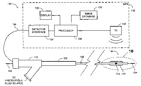
Reference is now made to Figures 1 A, 1 B, 1 C and 1 D. Figure
1 A is a schematic illustration of a system for determining the position and
orientation of an activation site of a medical operational element of a
25 medical catheter of the over-the-wire type, generally referenced 100,
constructed and operative in accordance with an embodiment of the
disclosed technique. Figure 1 B is a schematic perspective illustration of a
distal end 144 of the medical catheter of Figure 1 A. Figure 1 C is a
schematic illustration of a longitudinal cross section of the distal end of
so one example of the medical catheter of Figure 1 A. Figure 1 D is a
-14-
CA 02504613 2005-05-02
WO 2004/045363 PCT/IL2003/000940
schematic illustration of a longitudinal cross section of the distal end of
another example of the medical catheter of Figure 1 A.
System 100 includes a medical catheter 102, a guidewire 104
and a medical positioning system (MPS) 106. Medical catheter 102
includes an elongated member 108, a manifold 110, a medical operational
element 112 and an electromagnetic field detector 114. The medical
operational element can include a lumen intervention element, a lumen
diagnostic element, a lumen imaging element, and the like. Elongated
member 108 is made of a substantially flexible material, such as poly ether
io ether ketone (PEEK), polyethylene (PE), nylon, polyurethane, polyvinyl
chloride (PVC), polyethylene terephthalate (PET), Pebax~, polyimide,
metal (either solid or coiled), such as nitinol, stainless steel, hypotube
(i.e.,
an ultra low diameter and ultra thin walled tube), and the like. Elongated
member 108 has a substantially circular cross section and includes a
guidewire lumen 116. Manifold 110 is located at a proximal end of medical
catheter 102 and medical operational element 112 is located at a distal
end of medical catheter 102.
Medical operational element 112 is an element for performing
medical operations in the lumen, such as modifying the characteristics of
2o the lumen, or diagnosing the lumen, such as obtaining an image of the
lumen. The characteristics of the lumen can be modified by performing a
medical procedure thereon, such as percutaneous transluminal coronary
angioplasty (PTCA), percutaneous transluminal angioplasty (PTA),
vascularizing the lumen, severing a portion of the lumen or a plaque there
within (e.g., atherectomy), providing a suture to the lumen, increasing the
inner diameter of the lumen (e.g., by a balloon, a self expanding stent, a
stent made of a shape memory alloy (SMA), or a balloon expanding stent)
and maintaining the increased diameter by implanting a stent.
Medical operational element 112 can be further used to deliver
so substances to the lumen. For example, medical operational element 112
can be used to deliver a pharmaceutical substance to a selected site
-15-
CA 02504613 2005-05-02
WO 2004/045363 PCT/IL2003/000940
within the lumen, such as for inhibiting angiogenesis of cancerous cells,
inhibiting metastasis, stimulating local hormonal activity of tissue cells and
stimulating healing following a trauma. Medical operational element 112
can be further used for killing selected cells (either cancerous or
s non-cancerous) at the activation site of medical operational element 112
or in the vicinity thereof, by irradiating the cells with a radioactive
substance, electric current, laser, or subjecting the cells to a cryogenic
fluid, and the like. Medical operational element 112 can be further include,
or be used for deployment of, a device within the lumen. Such a device
io can be for example, a valve (e.g., mitral valve, sphincter), suturing
device,
implant, biological marker, radiopaque marker, substance delivery device,
imaging device, diagnostic device, miniature camera, infrared camera,
optical coherence tomography (OCT), magnetic resonance imaging (MRI),
ultrasound, sensor, such as pressure sensor, temperature sensor, pH
is sensor, and the like. The sensor can be in form of a passive ultrasonic
transducer, which transmits signals bearing the value of the detected
parameter (pressure, temperature, pH etc.), in response to an ultrasonic
wave directed from an external source toward the sensor. Medical
operational element 112 can also be used to perform a valvuloplasty
20 operation (i.e., repair of an organic or an artificial valve). The lumen
can be
a portion of the vascular system, ureter, urethra, brain vessels, coronary
vessels, vas deferens, lumens of the liver, kidney, lung (e.g., trachea and
bronchus), digestive system, gal bladder, prostate gland, urogenital
system, and the like. The lumen can be in the body of a human being as
2s well as an animal.
Medical operational element 112 can be an expansion unit such
as a balloon, stent, balloon expanding stent, an ablation unit such as laser,
cryogenic fluid unit, electric impulse unit, cutting balloon, rotational
atherectomy unit (i.e., rotablator), directional atherectomy unit,
3o transluminal extraction unit, a substance delivery unit such as coated
stent, drug delivery balloon, brachytherapy unit, and the like.
-16-
CA 02504613 2005-05-02
WO 2004/045363 PCT/IL2003/000940
The balloon expanding stent unit includes a stent which is
located around a balloon. When the balloon is inflated, the stent expands.
The cutting balloon unit includes a balloon having a plurality of blades on
the periphery thereof, along the longitudinal axis of the elongated member.
s The cryogenic fluid unit includes a fluid delivery lumen through which a
fluid at a substantially low temperature is delivered to a desired site of the
lumen. The electric impulse unit includes two electrical conductors. An
electrical arc generated at the tip of the electrical conductors ablates the
desired site of the lumen.
io The rotablator includes a diamond coated tip which is coupled
with an external motor via a flexible shaft. The flexible shaft rotates the
diamond coated tip at a substantially high speed, wherein the diamond
coated tip grinds calcified plaque which is formed on the inner wall of the
lumen. The ground material enters the circulation.
15 The directional atherectomy unit includes a cutter and a balloon.
The cutter is coupled with an external motor via a flexible shaft. The
balloon pushes the cutter toward the sidewall opposite to the balloon,
thereby allowing the cutter to cut the calcified plaque. The calcified
particles are pumped out through the medical catheter. The transluminal
2o extraction unit includes a cutter which is coupled with an external motor
via
a flexible shaft. The motor rotates the cutter, wherein the cutter cuts the
calcified plaque and the calcified particles are pumped out through the
medical catheter.
The coated stent is coated with a pharmaceutical substance,
25 wherein the substance is released into a desired region of the lumen,
when the coated stent is installed in the lumen. The drug delivery balloon
is a balloon which is coupled to a source of a pharmaceutical substance,
via a drug delivery lumen. The pharmaceutical substance exits the balloon
through a plurality of micropores. The brachytherapy unit includes a
so substance delivery lumen, through which radioactive palettes are delivered
to a desired site within the lumen. The radioactive palettes remain at the
-17
CA 02504613 2005-05-02
WO 2004/045363 PCT/IL2003/000940
desired site for a prescribed time and then are scavenged out through the
substance delivery lumen. Thus, a prescribed dose of radiation is
delivered to the desired site of the lumen.
In the example set forth in Figure 1 A, medical catheter 102 is a
balloon type catheter. Hence, medical operational element 112 includes a
tube portion 118 and a balloon portion 120. Each of tube portion 118 and
balloon portion 120 is made of a substantially thin and flexible material,
such as polyamide (e.g., nylon), and the like. Balloon portion 120 can be
made either of a compliant material, semi-complaint material, or a
io non-compliant material. A compliant balloon continuously expands as
higher pressures are applied thereto. A non-compliant balloon expands up
to a predetermined diameter which is designed therein, and ceases to
expand above this predetermined diameter, even if the applied pressure
continues to rise. The expansion rate of a semi-compliant balloon drops as
the pressure rises. Balloon portion 120 is located at a distal end of tube
portion 118. A proximal end of tube portion 118 is coupled with a
pressurized fluid source (not shown), via manifold 110 and a
circumferential fluid lumen 140. Circumferential fluid lumen 140 runs along
the entire length of medical catheter 102. The pressurized fluid source can
2o be an ampoule such as a syringe, and the like, which contains a
biocompatible fluid. The pressurized fluid source can be provided with a
sensor to detect a property of the fluid, such as pressure, temperature, pH,
and the like.
A distal end 122 of balloon portion 120 is coupled with an outer
2s wall (not shown) of elongated member 108, by methods known in the art,
such as by an adhesive, ultrasonic welding, heat bonding, by applying
infrared radiation, radio frequency (RF) radiation, laser, ultraviolet (UV)
radiation, and the like. The circumference of balloon portion 120 is larger
than that of tube portion 118. In an uninflated state, balloon portion 120
so folds around elongated member 108. When fluid flows under pressure
from the pressurized fluid source into tube portion 118, balloon portion 120
-18
CA 02504613 2005-05-02
WO 2004/045363 PCT/IL2003/000940
unfolds and expands. When the pressure fluid source is unpressurized or
the fluid is withdrawn from tube portion 118, the interstitial fluid in the
lumen forces balloon portion 120 to fold around elongated member 108.
Electromagnetic field detector 114 is an electric conductor
formed into a coil. Electromagnetic field detector 114 is embedded within
elongated member 108, such that guidewire lumen 116 passes through
the winding of electromagnetic field detector 114. Alternatively, the
electromagnetic field detector can be sufficiently small to be entirely
embedded within a lateral portion of the wall of the elongated member,
io adjacent to guidewire lumen 116. In the example set forth in Figures 1A,
1 B and 1 C, electromagnetic field detector 114 is embedded within
elongated member 108, in such a location that when balloon portion 120
expands, balloon portion 120 encompasses electromagnetic field detector
114. However, it is noted that electromagnetic field detector 114 can be
located either distal or proximal to balloon portion 120. Furthermore,
electromagnetic field detector 114 can be made of a radiopaque material
or coated with such a material, thereby being detectable by an imaging
device, such as radiographic, fluoroscopic, magnetic, sonographic device,
and the like.
2o With reference to Figure 1A, MPS 106 includes a detector
interface 124, a processor 126, a display 130, an image database 132 and
a transmitter 134. MPS 106 is located outside the body of a patient (not
shown). Processor 126 is coupled with detector interface 124, display 130,
image database 132 and with transmitter 134. Image database 132
includes a plurality of images of a lumen (not shown) of the patient,
wherein each image is associated with a set of position and orientation
coordinates, in a reference coordinate system.
Two ends (not shown) of electromagnetic field detector 114 are
coupled with two distal ends (not shown) of a wiring 136, via a flexible
0o printed circuit board (PCB) 138. However, it is noted that the two ends of
electromagnetic field detector 114 can be coupled with the two distal ends
-19
CA 02504613 2005-05-02
WO 2004/045363 PCT/IL2003/000940
of wiring 136, directly, (e.g., by soldering or conductive adhesion) in which
case flexible PCB 138 can be disposed of. Proximal ends (not shown) of
wiring 136 are coupled with detector interface 124. For a more elaborate
description of an MPS, confer US Patent No. 6,233,476 mentioned above.
s Wiring 136 is made of an electric conductor, such as copper,
gold, silver, and the like. Wiring 136 is spirally embedded within elongated
member 108, such that guidewire lumen 116 is surrounded by wiring 136.
It is noted that the term "spiral" includes, inter alia, helical forms. The
pitch
of wiring 136 is referenced by P1. Wiring 136 is spirally embedded within
1o elongated member 108 at pitch P1, in a section (not shown) of elongated
member 108 which starts from the two distal ends of electromagnetic field
detector 114 and ends at manifold 110. Alternatively, wiring 136 is spirally
embedded within the section of elongated member 108, at a plurality of
different pitches. Further alternatively, a portion of wiring 136 proximal to
1s electromagnetic field detector 114 is spirally embedded within elongated
member 108 at pitch P1, and the rest of wiring 136 is embedded within
elongated member 108 along a substantially straight line. Alternatively, at
least one portion of wiring 136 is spirally embedded within elongated
member 108 and at least another portion of wiring 136 is embedded within
2o elongated member 108, along a substantially straight line. With reference
to Figure 1 D, a wiring 142 is embedded within elongated member 108,
along a substantially straight line.
According to one aspect of the invention the spiral winding of
wiring 136 modifies certain mechanical properties of elongated member
25 108, such as improving the pushability and trackability of medical catheter
102 within the lumen of the patient (i.e., reducing the tendency of medical
catheter 102 to buckle when pushed within the lumen and increasing the
ability of the medical catheter to follow the vessel path), increasing the
elasticity of elongated member 108 (i.e., increasing the tendency of
so elongated member 108 to return to the original shape, after being
deformed), increasing the modulus of elasticity of elongated member 108
-20
CA 02504613 2005-05-02
WO 2004/045363 PCT/IL2003/000940
(i.e., increasing the mechanical stress in either compression or tension,
which is required to deform elongated member 108 by a certain amount),
increasing the coefficient of rigidity of elongated member 108 (i.e.,
increasing the mechanical shear stress which is required to twist
s elongated member 108 by a certain angle), affecting the flexibility or
resilience of elongated member 108, and the like. It is further noted that
the mechanical properties of wiring 136, also modifies the mechanical
properties of elongated member 108. Wiring 136 can be coated with a
coating that provides electrical insulation, or electrical shielding, as well
as
1o mechanical protection to wiring 136.
Following is a description of operation of system 100. Initially,
the user (usually a physician) inserts a guiding catheter (not shown) into
the lumen, such that a distal end of the guiding catheter reaches a desired
location within the lumen. The physician can view an image of the guiding
1s catheter by employing an imaging device, such as radiographic,
fluoroscopic, magnetic, sonographic device, and the like. The physician
inserts guidewire 104 in the guiding catheter and maneuvers a distal end
(not shown) of guidewire 104 past the guiding catheter through the lumen,
by observing an image of guidewire 104 in an imaging device, such as
2o radiographic, fluoroscopic, magnetic, sonographic device, and the like
Guidewire 104 is a "small-diameter" guidewire, referred to in the
Background of the Disclosed Technique, hereinabove. The physician, then
inserts a proximal end (not shown) of guidewire 104 in the distal end of
medical catheter 102, and passes medical catheter 102 over guidewire
25 104, into the lumen, such that the proximal end of guidewire 104 usually
exits a proximal end (not shown) of medical catheter 102. This mode of
operation is known in the art as "over-the-wire". Alternatively, no guidewire
is employed in the procedure, in which case the physician passes the
medical catheter out through the distal end of the guiding catheter, until
so the distal end of the medical catheter reaches a selected location within
-21-
CA 02504613 2005-05-02
WO 2004/045363 PCT/IL2003/000940
the lumen. Transmitter 134 produces a rotating magnetic and electric field
of fixed strength, orientation and frequency.
Electromagnetic field detector 114 produces a signal according
to the position and orientation thereof, relative to transmitter 134 and
electromagnetic field detector 114 provides this signal to detector interface
124, via wiring 136. Processor 126 receives the signal via detector
interface 124 and processor 126 determines the position and orientation of
electromagnetic field detector 114 relative to the reference coordinate
system, according to the received signal.
io Processor 126 retrieves an image of the lumen from image
database 132 and superimposes a representation of medical operational
element 112 on the retrieved image, according to the determined position
and orientation. Processor 126 produces a video signal respective of the
superimposed image to display 130 and display 130 produces the
representation of medical operational element 112, superimposed on the
image of the lumen. When the physician is assured that medical
operational element 112 is located at the desired site within the lumen, by
viewing the superimposed image on display 130, the physician can
commence the medical operation on the lumen.
2o Various electronic devices which are present in the operation
room, may emit electromagnetic radiation which may interfere with the
signal which the electromagnetic field detector transmits to the MPS, via
the wiring. In this case, necessary hardware or software has to be
incorporated with the system, in order to reduce the effect of these
interfering signals.
For example, at least a portion of the electromagnetic field
detector can be covered with a shielding of such thickness and material
(e.g., a conductive foil, a wire mesh), to selectively cancel out these
interfering signals, while allowing the signal from the transmitter of the
so MPS, to reach the electromagnetic field detector. This electrical shielding
-22-
CA 02504613 2005-05-02
WO 2004/045363 PCT/IL2003/000940
of the wiring acts as a Faraday cage within a predetermined range of
frequencies.
Reference is now to Figure 2, which is a schematic illustration of
a system for determining the position and' orientation of an activation site
s of a medical operational element of a medical catheter, generally
referenced 146, constructed and operative in accordance with another
embodiment of the disclosed technique. System 146 includes a medical
catheter 148 and an MPS 150. Figure 2 illustrates the distal portion of
medical catheter 148, which is typically about 20 cm long.
io Medical catheter 148 includes an elongated member 152, a
medical operational element 154, an electromagnetic field detector 156, a
wiring 158 and a transmitter 160. Elongated member 152 includes a
guidewire lumen 162. MPS 150 includes a processor 164, a transmitter
166, an image database 168, a display 170, a detector interface 172 and a
15 receiver 174.
Wiring 158 is similar to wiring 136 (Figure 1 B), as described
herein above and is embedded within elongated member 152. Medical
operational element 154 and electromagnetic field detector 156 are
located at a distal end 176 of medical catheter 148. Electromagnetic field
2o detector 156 is embedded within elongated member 152, and
encompasses guidewire lumen 162. Transmitter 160 is embedded within
elongated member 152 and located proximal to distal end 176.
Alternatively, the transmitter can be located at a manifold similar to
manifold 110 (Figure 1 A) or anywhere along elongated member 152 or
25 external thereto. One end of wiring 158 is coupled with electromagnetic
field detector 156 and the other end thereof is coupled with transmitter
160. The length of wiring 158 is much shorter than that of elongated
member 152, such that wiring 158 occupies a relatively short section of the
distal portion of elongated member 152 (usually about 20 cm).
3o Processor 164 is coupled with transmitter 166, image database
168, display 170 and with detector interface 172. Receiver 174 is coupled
-23
CA 02504613 2005-05-02
WO 2004/045363 PCT/IL2003/000940
with detector interface 172. Transmitter 166 transmits an electromagnetic
wave which is received by electromagnetic field detector 156 and
electromagnetic field detector 156 sends a signal respective of the position
and orientation of distal end 176 to transmitter 160, via wiring 158.
s Transmitter 160 transmits this signal to receiver 174 and processor 164
determines the position and orientation of distal end 176, according to a
signal received from detector interface 172.
It is noted that wiring 158 modifies the mechanical properties of
the distal portion of elongated member 152, as described herein above in
1o connection with Figure 1 B, such as pushability and trackability.
Alternatively, the electromagnetic field detector can be located external to
the elongated member (as described herein below in connection with
Figure 3). Further alternatively, the wiring can be wound around the
elongated member. Further alternatively, the transmitter can be located
15 external to the elongated member. It is further noted that medical catheter
148 can be of over-the-wire type, as well as rapid exchange type.
Reference is now made to Figure 3, which is a schematic
illustration of a longitudinal cross section of a distal end of a medical
catheter, generally referenced 180, constructed and operative in
2o accordance with a further embodiment of the disclosed technique. Medical
catheter 180 includes an elongated member 182, a medical operational
element 184, an electromagnetic field detector 186, a marker 188 and a
wiring 190. A guidewire 192 can pass through a guidewire lumen 194
within elongated member 182.
25 Elongated member 182 and medical operational element 184
are similar to elongated member 108 (Figure 1 A) and medical operational
element 112, respectively. Electromagnetic field detector 186 is made of a
conductor which is wound around an outer wall 196 of elongated member
182, at an activation site of medical operational element 184, such as a
so balloon portion 198. Marker 188 is made of a radiopaque material, such as
platinum, iridium, gold, tungsten, stainless steel, silver, composite
-24
CA 02504613 2005-05-02
WO 2004/045363 PCT/IL2003/000940
material, and the like, which can be detected by an imaging device, such
as radiographic, fluoroscopic, magnetic, sonographic device, and the like.
Marker 188 is embedded within elongated member 182 at the activation
site of medical operational element 184, such as balloon portion 198.
Alternatively, marker 188 is located on outer wall 196 (i.e., outer wall 196
is coated with marker 188).
Wiring 190 is wound around outer wall 196 at a pitch P2. For this
purpose, spiral grooves (not shown) can be formed on outer wall 196, by a
laser, mechanical engraving, chemical etching, molding;, injection;, and
io extrusion, and the like, and wiring 190 is then placed in the spiral
grooves.
Electromagnetic field detector 186 and wiring 190 are coated with a
protective coating, in order to provide electrical insulation and mechanical
protection to electromagnetic field detector 186 and to wiring 190 and
mechanically couple electromagnetic field detector 186 and wiring 190 to
outer wall 196. Alternatively, electromagnetic field detector 186 and wiring
190 are enclosed by a heat-shrinkable material. Two ends (not shown) of
electromagnetic field detector 186 are coupled with two distal ends (not
shown) of wiring 190. Two proximal ends (not shown) of wiring 190 are
coupled with an MPS similar to MPS 106 (Figure 1 A). Further alternatively,
2o the wiring is coupled to the outer wall of the elongated member, along a
substantially straight line (not shown). Alternatively, the wiring is wound
around the outer wall of the elongated member, at either a constant pitch
or a variable pitch along the length of the elongated member. Further
alternatively, at least one portion of the wiring is substantially straight
and
at least another portion is spiral.
Reference is now made to Figure 4, which is a schematic
illustration of a lateral cross section of the wiring of a system for
determining position and orientation, such as shown in Figure 1 A, in a
twisted pair formation, generally referenced 200, constructed and
so operative in accordance with another embodiment of the disclosed
technique. It is noted that the cross sectional proportions of the different
-25
CA 02504613 2005-05-02
WO 2004/045363 PCT/IL2003/000940
elements in Figure 4 and all other Figures accompanying this disclosure
are not intended to illustrate the actual dimensions or proportions and are
exaggerated for the sake of clarity. Wiring 200 includes electrical
conductors 202 and 204, electrical insulations 206, 208 and 212 and an
s electrical shielding 214. Electrical shielding 214 is a shielding layer
similar
to the shielding of the electromagnetic field detector described above, and
provides electrical shielding to electrical conductors 202 and 204.
Alternatively, electrical shielding 214 can be a fluid layer which blocks
electromagnetic waves in predetermined frequency ranges. Further
io alternatively, a circumferential fluid lumen similar to circumferential
fluid
lumen 140 (Figure 1 B), can function as an electrical shielding for the
wiring, or an electromagnetic field detector similar to electrical field
detector 114. Further alternatively, each of electrical conductors 202 and
204 can be hollow, wherein the hollow space is filled with a fluid. This fluid
15 can be employed for transmitting signals or for other medical intervention
purposes.
Electrical conductors 202 and 204 are enclosed within electrical
insulations 206 and 208, respectively. Distal ends (not shown) of electrical
conductors 202 and 204 are coupled with two ends (not shown) of an
2o electromagnetic field detector (not shown), similar to electromagnetic
field
detector 114 (Figure 1 C). Proximal ends (not shown) of electrical
conductors 202 and 204 are coupled with an MPS (not shown) similar to
MPS 106 (Figure 1A). Electrical conductors 202 and 204 together with
electrical insulations 206 and 208, are twisted together between the
2s coupling to the electromagnetic field detector and the coupling to the MPS.
Thus, electrical conductors 202 and 204 together with electrical insulations
206 and 208, form a twisted pair (not shown). Electrical shielding 214
encloses electrical conductors 202 and 204 and electrical insulations 206
and 208. Electrical insulation 212 encloses electrical conductors 202 and
so 204, electrical insulations 206 and 208 and electrical shielding 214.
-26-
CA 02504613 2005-05-02
WO 2004/045363 PCT/IL2003/000940
Reference is now made to Figure 5, which is a schematic
illustration of a lateral cross section of the wiring of a system for
determining position and orientation, such as shown in Figure 1 A, in a
coaxial formation, generally referenced 240, constructed and operative in
s accordance with a further embodiment of the disclosed technique. Wiring
240 includes electrical conductors 242 and 244 and electrical insulations
246 and 248. Electrical insulation 246 encloses electrical conductor 242.
Electrical conductor 244 has a substantially annular cross section and
thus, encompasses electrical conductor 242 and electrical insulation 246.
1o Electrical insulation 248 encompasses electrical conductors 242 and 244
and electrical insulation 246. Distal ends (not shown) of electrical
conductors 242 and 244 are coupled with two ends (not shown) of an
electromagnetic field detector (not shown), similar to electromagnetic field
detector 114 (Figure 1 C). Proximal ends (not shown) of electrical
is conductors 242 and 244 are coupled with an MPS (not shown) similar to
MPS 106 (Figure 1A). Thus, electrical conductors 242 and 244 together
with electrical insulations 246 and 248, form a coaxial cable.
Reference is now made to Figure 6, which is a schematic
illustration of a lateral cross section of the wiring of a system for
2o determining position and orientation, such as shown in Figure 1 A, in a
triaxial formation, generally referenced 270, constructed and operative in
accordance with another embodiment of the disclosed technique. Wiring
270 includes electrical conductors 272 and 274, electrical shielding 276
and electrical insulations 278, 280 and 282. Electrical shielding 276 is
2s made of a conductive material, which operates as a Faraday cage and
provides electrical shielding to electrical conductors 272 and 274.
Electrical insulation 278 encompasses electrical conductor 272.
Electrical conductor 274 has a substantially annular cross section and
thus, encompasses electrical conductor 272 and electrical insulation 278.
so Electrical insulation 280 encompasses electrical conductors 272 and 274
and electrical insulation 278.
-27-
CA 02504613 2005-05-02
WO 2004/045363 PCT/IL2003/000940
Electrical shielding 276 encompasses electrical conductors 272
and 274 and electrical insulations 278 and 280. Electrical insulation 282
encompasses electrical conductors 272 and 274, electrical insulations 278
and 280 and electrical shielding 276. Distal ends (not shown) of electrical
s conductors 272 and 274 are coupled with two ends (not shown) of an
electromagnetic field detector (not shown), similar to electromagnetic field
detector 114 (Figure 1 C). Proximal ends (not shown) of electrical
conductors 272 and 274 are coupled with an MPS (not shown) similar to
MPS 106 (Figure 1A). Thus, electrical conductors 272 and 274 together
1o with electrical insulations 278, 280 and 282 and electrical shielding 276,
form a triaxial cable.
Reference is now made to Figures 7A and 7B. Figure 7A is a
schematic illustration of a longitudinal cross section of the distal end of
the
medical catheter of a system for determining position and orientation, such
~s as shown in Figure 1A, generally referenced 300, constructed and
operative in accordance with a further embodiment of the disclosed
technique. Figure 7B is a lateral cross section of the medical catheter of
Figure 7A.
Medical catheter 300 includes an elongated member 302, an
2o electromagnetic field detector 304, electrical conductors 306 and 308 and
a medical operational element 310. Elongated member 302 and
electromagnetic field detector 304 are similar to elongated member 108
(Figure 1 A) and electromagnetic field detector 114 (Figure 1 C),
respectively. In the example set forth in Figure 7A, medical catheter 300 is
25 a balloon-stent type catheter. Therefore, medical operational element 310
includes a tube portion 312, a balloon portion 314 and a stent 316. Tube
portion 312 and balloon portion 314 are similar to tube portion 118 (Figure
1 C) and balloon portion 120, respectively. Tube portion 312 is coupled
with a pressurized fluid source (not shown), via a fluid lumen 318. A
so guidewire 320 can be passed through a guidewire lumen 322 within
elongated member 302.
-28-
CA 02504613 2005-05-02
WO 2004/045363 PCT/IL2003/000940
Electromagnetic field detector 304 is embedded within elongated
member 302, in a manner similar to the one described herein above in
connection with electromagnetic field detector 114 (Figure 1 C). Distal ends
(not shown) of electrical conductors 306 and 308 are coupled with two
ends (not shown) of electromagnetic field detector 304. Proximal ends (not
shown) of electrical conductors 306 and 308 are coupled with an MPS (not
shown), similar to MPS 106 (Figure 1A).
Each of electrical conductors 306 and 308 can be encompassed
within an electrical insulation (not shown). Alternatively, each of electrical
1o conductors 306 and 308 can be encompassed within an electrical
shielding (not shown). Further alternatively, an electrical shielding can
encompass each of electrical conductors 306 and 308 and the respective
electrical insulation. Alternatively, an electrical insulation can encompass
each of electrical conductors 306 and 308 and the respective electrical
shielding.
Electrical conductors 306 and 308 are substantially located on
the same diametrical line of elongated member 302 and equally spaced
from the center of elongated member 302. In other words, electrical
conductor 306 is embedded within elongated member 302 along a first
2o path and electrical conductor 308 is embedded within elongated member
302 along a second path. These first and second paths substantially lie on
a plane, whereby the plane substantially passes through the longitudinal
axis of elongated member 302. It is noted that electrical conductors 306
and 308 can modify the mechanical properties of elongated member 302,
as described herein above in connection with wiring 136 (Figure 1 C).
Stent 316 is an expandable type of stent as known in the art,
such as a wire mesh, a cylinder which includes a longitudinal cut, and the
like. A fluid flowing from the pressurized fluid source to tube portion 312,
causes balloon portion 314 to expand and the expansion of balloon portion
314 causes stent 316 to expand.
-29-
CA 02504613 2005-05-02
WO 2004/045363 PCT/IL2003/000940
Reference is now made to Figure 8, which is a schematic
illustration of a lateral cross section of the distal end of the medical
catheter of a system for determining position and orientation, such as
shown in Figure 1 A, generally referenced 350, constructed and operative
s in accordance with another embodiment of the disclosed technique.
Medical catheter 350 includes an elongated member 352, electrical
conductors 354 and 356 and a support element 358. A guidewire 360
passes through a guidewire lumen 362 within elongated member 352.
Each of electrical conductors 354 and 356 is similar to electrical
1o conductors 306 and 308, as described herein above in connection with
Figure 7A. Support element 358 can be made of a material whose physical
properties are substantially similar to those of either one of electrical
conductors 354 or 356, but support element 358 can be made of other
materials or have other properties. Electrical conductors 354 and 356 and
15 support element 358 are located equally apart on a circle (not shown),
which is substantially concentric with the longitudinal axis of elongated
member 352 (i.e., on radial lines whose angle there between is
approximately 120 degrees).
In this manner, electrical conductors 354 and 356 and support
2o element 358, modify the mechanical properties of elongated member 352,
as described herein above in connection with wiring 136 (Figure 1 C).
Analogously, any number of electrical conductors and support elements
can be distributed in the lateral cross section of the elongated member,
according to the desired mechanical properties of the elongated member.
25 Reference is now made to Figure 9, which is a schematic
illustration of a longitudinal cross section of the distal end of the medical
catheter of a system for determining position and orientation, such as
shown in Figure 1 A, generally referenced 410, constructed and operative
in accordance with a further embodiment of the disclosed technique.
3o Medical catheter 410 includes an elongated member 412, an
electromagnetic field detector 414, a PCB 416, a wiring 418 and a medical
-30
CA 02504613 2005-05-02
WO 2004/045363 PCT/IL2003/000940
operational element 420. Medical catheter 410 is a stent type catheter.
Hence, medical operational element 420 includes a stent 422 and a sleeve
424. A guidewire 426 can pass within a guidewire lumen 428, within
elongated member 412.
s Elongated member 412, electromagnetic field detector 414, PCB
416 and wiring 418 are similar to elongated member 108 (Figure 1 A),
electromagnetic field detector 114, PCB 138 and wiring 136, respectively,
as described herein above in connection with Figure 1 C. Electromagnetic
field detector 414, PCB 416 and wiring 418 are embedded within
io elongated member 412, in a manner similar to one described herein above
in connection with Figure 1 C. Distal ends (not shown) of wiring 418 are
coupled with two ends (not shown) of electromagnetic field detector 414,
via PCB 416. Proximal ends (not shown) of wiring 418 are coupled with an
MPS similar to MPS 106 (Figure 1A).
15 Stent 422 is a spring type stent (i.e., self expandable stent) as
known in the art, which tends to expand, if no restraint is imposed thereon.
During assembly of medical operational element 420 on elongated
member 412, stent 422 is passed over an outer wall 430 of elongated
member 412 together with restraining sleeve 424, such that sleeve 424
2o keeps stent 422 in a compressed state. In order to activate medical
operational element 420, sleeve 424 is pulled in a direction designated by
arrows 432, wherein stent 422 expands and leaves outer wall 430.
Alternatively, stent 422 is made of a shape memory alloy (SMA),
such as nickel-titanium (nitinol), and the like, and sleeve 424 is disposed
25 Of. The SMA stent is constructed such that when the metallurgical
structure of the SMA stent changes from a first phase (e.g., Martensite) to
a second phase (e.g., Austenite), the SMA stent expands.
Reference is now made to Figure 10, which is a schematic
illustration of a longitudinal cross section of the distal end of the medical
ao catheter of a system for determining position and orientation, such as
shown in Figure 1 A, generally referenced 450, constructed and operative
-31
CA 02504613 2005-05-02
WO 2004/045363 PCT/IL2003/000940
in accordance with another embodiment of the disclosed technique.
Medical catheter 450 includes an elongated member 452, an
electromagnetic field detector 454, a wiring 456 and an optical fiber 458.
Elongated member 452 and electromagnetic field detector 454 are similar
s to elongated member 108 (Figure 1 A) and electromagnetic field detector
114 (Figure 1 C), respectively, as described herein above. Wiring 456 is
similar to either wiring 136 (Figure 1 C) or wiring 142 (Figure 1 D), as
described herein above. Electromagnetic field detector 454 and wiring 456
are embedded within elongated member 452, in a manner similar to the
io one described herein above in connection with Figure 1 C. A guidewire 460
can pass through a guidewire lumen 462, within elongated member 452.
Optical fiber 458 is embedded within elongated member 452. A
distal end 464 of optical fiber 458 is located at a distal end 466 of
elongated member 452. Distal end 464 can point either toward the front of
15 distal end 466, or toward a side (not shown) of distal end 466. A proximal
end (not shown) of optical fiber 458 is coupled to a laser (not shown).
When the laser is activated, optical fiber 458 ablates a tissue (not shown),
which is located in the vicinity of distal end 466.
Reference is now made to Figure 11, which is a schematic
2o illustration of a longitudinal cross section of the distal end of a medical
catheter of the rapid-exchange type, generally referenced 490, constructed
and operative in accordance with a further embodiment of the disclosed
technique. Rapid-exchange catheter is also known in the art as Single
Operator Exchange (SOE). Medical catheter 490 includes an elongated
25 member 492, an electromagnetic field detector 494, a wiring 496 and a
medical operational element 498. Medical catheter 490 is a balloon type
catheter. Therefore, medical operational element 498 includes a tube
portion 500 and a balloon portion 502.
Wiring 496 is similar to either wiring 136 (Figure 1 C) or wiring
30 142 (Figure 1 D), as described herein above. Electromagnetic field detector
494 is made of an electrical conductor (not shown), wound around a core
-32-
CA 02504613 2005-05-02
WO 2004/045363 PCT/IL2003/000940
504. Core 504 is made of a material whose permeability is substantially
greater than that of the air. Hence, core 504 can be made of a
ferromagnetic material (e.g., ferrite, iron, Mu-metal, superalloy, soft
ferrite),
and the like, as well as a paramagnetic material. Electromagnetic field
s detector 494 is embedded within elongated member 492. Wiring 496 is
embedded within elongated member 492 in a manner similar to the one
described herein above in connection with Figure 1 C. Distal ends (not
shown) of wiring 496 are coupled with two ends (not shown) of
electromagnetic field detector 494. Proximal ends (not shown) of wiring
io 496 are coupled with an MPS, similar to MPS 106 (Figure 1A). A distal end
506 of balloon portion 502 is coupled with a distal end 508 of elongated
member 492, in a manner similar to the one described herein above, in
connection with Figure 1 C.
Elongated member 492 includes a guidewire lumen 510, whose
15 entrance 512 is located at distal end 508 and whose exit 514 is located at
a side portion 516 of elongated member 492. Side portion 516 is located
at a proximal end 518 of balloon portion 502. Electromagnetic field
detector 494 is located proximal to exit 514 (i.e., adjacent to proximal end
518). A concentric fluid lumen 520 formed between tube portion 500 and
2o an outer wall 522 of elongated member 492, is coupled with a pressurized
fluid source similar to the one described herein above, in connection with
Figure 1 A.
A region of tube portion 500 in the vicinity of side portion 516 is
coupled with side portion 516, in order to prevent fluid communication
25 between guidewire lumen 510 and concentric fluid lumen 520. Tube
portion 500 is perforated at side portion 516, in order to keep exit 514
open. In order to guide medical catheter 490 over a guidewire 524, the
physician enters a proximal end 526 of guidewire 524 through entrance
512, until proximal end 526 of guidewire 524 passes through guidewire
30 lumen 510 and exits guidewire lumen 510 at exit 514. This mode of
operation is known in the art as "rapid-exchange".
-33-
CA 02504613 2005-05-02
WO 2004/045363 PCT/IL2003/000940
It is noted that since a portion of elongated member 492
proximal to exit 514 is solid, it is possible to incorporate core 504 with
electromagnetic field detector 494. Furthermore, since core 504 is made of
a ferromagnetic material, electromagnetic field detector 494 is more
sensitive to the electromagnetic field generated by a transmitter similar to
transmitter 134 (Figure 1 A), than an electromagnetic field detector similar
to electromagnetic field detector 114 (Figure 1 C).
Reference is now made to Figure 12, which is a schematic
illustration of a longitudinal cross section of the distal end of a medical
io catheter of the rapid-exchange type, generally referenced 550, constructed
and operative in accordance with another embodiment of the disclosed
technique. Medical catheter 550 includes an elongated member 552, an
electromagnetic field detector 554, a wiring 556 and a medical operational
element 558. Medical catheter 550 is a balloon type catheter. Therefore,
is medical operational element 558 includes a tube portion 560 and a balloon
portion 562. Medical operational element 558 is similar to medical
operational element 498 (Figure 11 ), as described herein above. Medical
operational element 558 is constructed in a manner similar to the one
described herein above in connection with Figure 11.
2o Elongated member 552 includes a guidewire lumen 564, whose
entrance 566 is located at a distal end 568 of elongated member 552. An
exit 570 of guidewire lumen 564 is located at a side portion 572 of
elongated member 552. Side portion 572 is located at a proximal end 574
of balloon portion 562.
25 Guidewire lumen 564 is similar to guidewire lumen 510 (Figure
11 ), as described herein above. Electromagnetic field detector 554 is
similar to electromagnetic field detector 114 (Figure 1 C), as described
herein above. Wiring 556 is similar to either wiring 136 (Figure 1 C) or
wiring 142 (Figure 1 D), as described herein above. Distal ends (not
so shown) of wiring 556 are coupled with two ends (not shown) of
-34-
CA 02504613 2005-05-02
WO 2004/045363 PCT/IL2003/000940
electromagnetic field detector 554. Proximal ends (not shown) of wiring
556 are coupled with an MPS, similar to MPS 106 (Figure 1A).
Electromagnetic field detector 554 is embedded within elongated
member 552 as described herein above in connection with Figure 1 C,
such that guidewire lumen 564 passes through the winding of
electromagnetic field detector 554. Electromagnetic field detector 554 is
embedded in such a location within elongated member 552, that when
balloon portion 562 expands, balloon portion 562 encompasses
electromagnetic field detector 554.
io The physician enters a proximal end 576 of a guidewire 578 into
guidewire lumen 564 through entrance 566, passes guidewire 578 through
guidewire lumen 564 and pushes guidewire lumen 564 out through exit
570. Medical catheter 550 operates in rapid-exchange mode, while
electromagnetic field detector 554 is located such that balloon portion 562
encompasses electromagnetic field detector 554, when balloon portion
562 expands. Thus, medical catheter 550 allows the MPS to determine the
location of medical operational element 558, more accurately than that of
medical catheter 490 (Figure 11 ).
Alternatively, the electromagnetic field detector is wound around
2o an outer wall 580 of elongated member 552. Further alternatively, the
electromagnetic field detector is located proximal to exit 570, while the
electromagnetic field detector is either embedded within the elongated
member or is wound around the outer wall of the elongated member.
Reference is now made to Figure 13, which is a schematic
illustration of a system for determining the relative positions and
orientations of a plurality of medical catheters, generally referenced 600,
constructed and operative in accordance with a further embodiment of the
disclosed technique. System 600 includes a plurality of medical catheters
602 and 604, a plurality of guidewires 606 and 608 and an MPS 610. Each
so of medical catheters 602 and 604 is similar to either medical catheter 102
(Figure 1A), medical catheter 490 (Figure 11) or medical catheter 550
-35
CA 02504613 2005-05-02
WO 2004/045363 PCT/IL2003/000940
(Figure 12), as described herein above. MPS 610 is similar to MPS 106
(Figure 1 A), as described herein above.
Medical catheter 602 includes a medical operational element
612 and an electromagnetic field detector 614. Medical catheter 604
s includes a medical operational element 616 and an electromagnetic field
detector 618. Each of medical operational elements 612 and 616 is similar
to medical operational element 112 (Figure 1 A), as described herein
above. If a guidewire lumen (not shown) within an elongated member (not
shown) of each of medical catheter 602 and 604, is similar to guidewire
lumen 116 (Figure 1 C), then each of electromagnetic field detectors 614
and 618 is similar to electromagnetic field detector 114 (Figure 1 C) or
electromagnetic field detector 186 (Figure 3), as described herein above. If
the guidewire lumen within the elongated member of each of medical
catheter 602 and 604, is similar to guidewire lumen 510 (Figure 11 ), then
1s each of electromagnetic field detectors 614 and 618 is similar to
electromagnetic field detector 494, as described herein above.
Electromagnetic field detectors 614 and 618 are coupled to MPS
610, via wirings 620 and 622, respectively. Each of wirings 620 and 622 is
similar to wiring 136 (Figure 1 C), as described herein above. Medical
2o catheters 602 and 604 are passed over guidewires 606 and 608,
respectively, into lumens 624 and 626, respectively, of a patient (not
shown). Electromagnetic field detectors 614 and 618 detect the
electromagnetic field generated by a transmitter (not shown) of MPS 610
and provide MPS 610 respective signals, via wirings 620 and 622,
2s respectively. MPS 610 determines the position and orientation of medical
operational element 612 relative to medical operational element 616,
according to the signals received from electromagnetic field detectors 614
and 618.
Reference is now made to Figure 14, which is a schematic
so illustration of a system for determining the position and orientation of a
guiding catheter, generally referenced 650, constructed and operative in
-36
CA 02504613 2005-05-02
WO 2004/045363 PCT/IL2003/000940
accordance with another embodiment of the disclosed technique. System
650 includes a guiding catheter 652, a medical catheter 654 and an MPS
656. Guiding catheter 652 includes an electromagnetic field detector 658.
Medical catheter 654 includes an elongated member 660, an
electromagnetic field detector 662 and a medical operational element 664.
Elongated member 660 includes a guidewire lumen 666.
Guidewire lumen 666 is either similar to guidewire lumen 116
(Figure 1 C) or guidewire lumen 510 (Figure 11 ), as described herein
above. Electromagnetic field detector 662 is similar to either
io electromagnetic field detector 114 (Figure 1 C), electromagnetic field
detector 186 (Figure 3), or electromagnetic field detector 494 (Figure 11 ),
according to the type of guidewire lumen 666. Medical operational element
664 is similar to medical operational element 112 (Figure 1 C), as
described herein above. Electromagnetic field detector 662 is located at
1s an activation site (not shown) of medical operational element 664.
Electromagnetic field detector 658 is made of an electric
conductor wound around an outer wall 668 of guiding catheter 652.
Alternatively, the electromagnetic field detector is located within a wall of
the guiding catheter. Electromagnetic field detector 658 is coupled with
2o MPS 656 via a wiring 670. Wiring 670 is wound around outer wall 668.
Alternatively, wiring 670 lies in a substantially straight line on outer wall
668. Electromagnetic field detector 658 and wiring 670 are coated with a
protective layer, such as an adhesive, and the lilee. The protective layer
provides mechanical and electrical protection to electromagnetic field
25 detector 658 and to wiring 670. The protective layer is coated with a
lubricant to facilitate the travel of guiding catheter 652 within a lumen (not
shown) of a patient (not shown).
Electromagnetic field detector 662 is coupled with MPS 656, via
a wiring 672. Wiring 672 is similar to either wiring 136 (Figure 1 C) or
wiring
30 142 (Figure 1 D), as described herein above. Medical catheter 654 is
-37-
CA 02504613 2005-05-02
WO 2004/045363 PCT/IL2003/000940
located within guiding catheter 652. A guidewire 674 passes through
guidewire lumen 666.
Electromagnetic field detector 658 detects an electromagnetic
field generated by a transmitter (not shown) of MPS 656 and provides a
respective signal to MPS 656, via wiring 670. Electromagnetic field
detector 662 detects an electromagnetic field generated by the transmitter
and provides a respective signal to MPS 656, via wiring 672. MPS 656
determines the position and orientation of electromagnetic field detector
658 in a reference coordinate system, according to the signal received
1o from electromagnetic field detector 658. If electromagnetic field detector
658 is located at a distal end 676 of guiding catheter 652, then MPS 656
determines the position and orientation of distal end 676 in the reference
coordinate system. Alternatively, MPS 656 determines the position and
orientation of electromagnetic field detector 662 (i.e., the activation site
of
medical operational element 664), relative to electromagnetic field detector
658 (i.e., distal end 676), according to signals received from
electromagnetic field detectors 658 and 662.
Reference is now made to Figure 15, which is a schematic
illustration of a method for operating the system of Figure 1 A, operative in
2o accordance with a further embodiment of the disclosed technique. In
procedure 700, a medical catheter is advanced to the desired location
within an organic lumen.
Prior to procedure 700, a guiding catheter can be advanced to
an approximate location proximal to a desired location within an organic
lumen. Additionally, the guidewire can be advanced within the guiding
catheter to the desired location, past a guiding catheter distal end. With
reference to Figure 1 A, in case a guidewire was previously inserted
through the guiding catheter, the physician inserts the proximal end of
guidewire 104 into distal end 144 of medical catheter 102 and advances
3o medical catheter 102 to the desired location within a lumen of a patient,
over guidewire 104. At this stage, the physician can view an image of
-38
CA 02504613 2005-05-02
WO 2004/045363 PCT/IL2003/000940
guidewire 104 on an imaging device, such as radiographic, fluoroscopic,
magnetic, sonographic device, and the like. In case no guidewire was
previously employed, the physician advances the medical catheter to the
desired location, through the guiding catheter. Optionally, with reference to
s Figure 14, electromagnetic field detector 658 is mounted at distal end 676
of guiding catheter 652, thereby allowing detection of the position and
orientation of guiding catheter 652 without employing X-ray or fluoroscopy,
and with the precision of MPS.
In procedure 702, an electromagnetic field detector located at a
io medical catheter distal end, is coupled with an MPS by a wiring.
According to a preferable embodiment, the wiring affects the mechanical
properties of the medical catheter, such as the pushability and trackability
of the medical catheter through the organic lumen (when the medical
catheter extends beyond the guiding catheter distal end). With reference
15 to Figures 1 A and 1 C, electromagnetic field detector 114 which is located
at distal end 114 of medical catheter 102, is coupled with MPS 106, via
wiring 136. Since wiring 136 is spirally embedded within elongated
member 108, the mechanical properties of elongated member 108, such
as pushability and trackability of elongated member 108 through the
20 lumen, are modified. It is noted that wiring 136 can be embedded within
elongated member 108 in a substantially straight line, or a combination of
spiral and straight sections, wherein wiring 136 still modifies the
mechanical properties of elongated member 108.
In procedure 704, an electromagnetic field is generated by the
25 MPS. With reference to Figure 1A, transmitter 134 generates an
electromagnetic field. In procedure 706, the generated electromagnetic
field is detected by the electromagnetic field detector.
In procedure 708, a signal respective of the detected
electromagnetic field is transmitted to the MPS, via the wiring. With
so reference to Figure 1 A, electromagnetic field detector 114 detects the
electromagnetic field generated by transmitter 134 and electromagnetic
-39
CA 02504613 2005-05-02
WO 2004/045363 PCT/IL2003/000940
field detector 114 transmits a signal respective of the detected
electromagnetic field, to detector interface 124, via wiring 136.
In procedure 710, the position and orientation of the medical
catheter distal end is determined by the MPS, according to the transmitted
s signal. With reference to Figure 1A, processor 126 receives from detector
interface 124, the signal which was transmitted to detector interface 124
by electromagnetic field detector 114 and processor 126 determines the
position and orientation of distal end 144 of medical catheter 102,
according to the transmitted signal.
1o In procedure 712, a medical operation is performed by activating
a medical operational element located at the medical catheter distal end.
With reference to Figures 1 A and 1 B, the physician inflates balloon portion
120 by introducing a fluid from a pressurized fluid source, into tube portion
118.
15 It will be appreciated by persons skilled in the art that the
disclosed technique is not limited to what has been particularly shown and
described hereinabove. Rather the scope of the disclosed technique is
defined only by the claims, which follow.
-40-