Note: Descriptions are shown in the official language in which they were submitted.
CA 02505042 2005-05-04
Translation for MElssNert, BOLTS & PAKTN6R: M/SME-012-DE
Apparatus for the dynamic stabilization of bones or bone
fragments, in particular spinal vertebrae
DESCRIPTION
The present invention relates to an apparatus for the dynamic
stabilization of bones or bone fragments, in particular spinal
vertebrae, with at least one longitudinal support that can be
fixed to the vertebrae.
The main indications for dynamic fixation, in particular when
performed from the posterior aspect, are age- and/or disease-
induced degeneration of structures in the spinal column as well
as inflammation and/or injuries in the region of the
intervertebral disk, the ligament apparatus, the facet joints
and/or the subchondral bone.
Posterior dynamic fixation systems have the function of
modifying the movement pattern in the affected spinal-column
segment in such a way that the pains produced by chemical
stimulation (nucleus material in contact with neural structures)
and/or by mechanical stimulation (hypermobility) disappear,
while the metabolism of the structures is preserved or restored.
Clinical experience with existing posterior dynamic fixation
systems, such as are described for example in EP 0 669 109 B1
and in the manual entitled "Fixateur externe" (authors: B.G.
Weber and F. Magerl, Springer-Verlag 1985, pp. 290-366), shows
that a posterior dynamic fixation system is advantageous in
being flexible with respect to bending and stiff with respect to
CA 02505042 2005-05-04
Translation for MEISSNER, BoLre & PARTNER: M/SME-012-DE
- 2 -
compression (buckling), shear and rotation. Thus such a system
must be dimensioned so as to allow maximal deformation under
flexion and also to resist the greatest possible force regarding
the aspects of buckling, shear and rotation. These conditions
are in themselves contradictory, and in order to reconcile them
the longitudinal supports are advantageously constructed of a
biocompatible high-performance plastic material. Because such
materials have an E modulus much lower than those of titanium
and steel, these longitudinal supports can be made relatively
thick in comparison to the metals steel and titanium in general
clinical use, without any loss of flexibility; this is
beneficial regarding their resistance to shear forces and
buckling, as follows:
o critical load for buckling: Pkr = const. * E
o critical shear force: Qkr = eonst. * i~X
o critical bending: akr = eonst. * am$x * 1/E * 1/~
The above formulas show how the material properties, the E
modulus and the diameter can be modified in order to be able to
fulfill the various criteria regarding deformation and
resistance.
The problem encountered when biocompatible high-performance
plastic is used for the longitudinal supports is that such
structures, in contrast to metallic longitudinal supports, can
be permanently bent in situ only with considerable difficulty,
e.g. by heating.
It is particularly important for longitudinal supports to be
bendable in the case of posterior stabilization by way of
pedicle screws, because when these are screwed into the vertebra
by way of the pedicle, they often turn out to be incorrectly
aligned on account of the anatomical situation. In order
nevertheless to connect the longitudinal supports to the pedicle
screws with the least possible tension, the shape of the
supports must be adjusted to the position and orientation of the
CA 02505042 2005-05-04
Translation for MEISSNER, BOLTS & PARTNER: M/SME-012-DE
- 3 -
pedicle screws in situ. In the case of polyaxial pedicle screws
the necessity of bending can be limited to one plane, whereas
with monoaxial pedicle screws the longitudinal supports must be
bent three-dimensionally.
Another embodiment of a dynamic fixation system is proposed in
EP 0 690 701 B1. This system comprises a connecting rod that can
be fixed at its ends to two adjacent vertebrae and that
comprises a curved middle section, so that it is flexible within
certain limits. In other respects the shape of this connecting
rod cannot be altered.
The document WO 01/45576 A1 also proposes a dynamic
stabilization system incorporating a longitudinal support, which
comprises two metallic end sections that can be fixed within
complementary receiption openings in the heads of two adjacent
pedicle screws. Between the two end sections is disposed a
linking element that is flexible in the long direction and
preferably is made of flexible material. Both of the end
sections of the longitudinal support are rigid. In addition to
this linking element it is proposed that an elastic band be
disposed between two pedicle screws, which extends parallel to
the elastic linking element.
In this embodiment, again, the longitudinal extent of the
linking element is prespecified by the manufacturer, and hence
cannot be altered. Finally, reference should be made to the
construction according to FR 2 799 949, which is characterized
by the construction of the longitudinal support as a spring
element, for example in the form of a leaf spring curved into a
meander shape.
In the construction according to WO 98/22033 Al the longitudinal
support also comprises a spring element that retains the shape
predetermined by the manufacturer.
CA 02505042 2005-05-04
Translation for MEtsSNER, BoLTE & PARTNER: M/SME-012-DE
- 4 -
Accordingly, one of the objectives of the present invention is
to create an apparatus for the dynamic stabilization of bones or
bone fragments, in particular vertebrae, with at least one
longitudinal support that can be fixed to the vertebrae and can
effortlessly be adapted to the most diverse situations for
implantation, with no impairment of the dynamics.
This objective is achieved by the characterizing features given
in Claim 1, preferred structural details of which are described
in the subordinate claims. The basic idea of the present
invention is thus that the at least one longitudinal support,
which for example is fixed between two adjacent pedicle screws,
is so constructed that by applying a predetermined bending
force, it can be deformed plastically from a first shape state
"A" into a second, alternative shape state "B", the bending
force needed for this purpose being distinctly greater than the
peak forces that occur in vivo. While remaining in each of the
stable shape states, however, the longitudinal support should be
flexibly bendable within the limits imposed by the mechanical
interaction between fixation system and vertebral-column
segment, which define a so-called "elastic flexion range".
It should be noted at this juncture that the apparatus in
accordance with the invention is fundamentally also suitable for
anterior implantation, when it is desired to shift the center of
rotation of the affected spinal-column segment toward the
anterior.
An especially advantageous embodiment of the apparatus in
accordance with the invention solves the problem of bending
longitudinal supports made of a biocompatible high-performance
plastic, in that a metal rod is disposed centrally in the
support. The metal rod must on one hand be so thin that its
critical bending angle is larger than or equal to the maximal
bending angle through which the stabilized vertebrae will bend
when connected to the dynamic fixation system, while on the
CA 02505042 2005-05-04
Translation for MEISSNER, BOLTS & PARTNER: M/SME-012-DE
_ 5 _
other hand being thick enough that the longitudinal support
retains the shape into which it was bent in situ.
To obtain a particular bending elasticity it is conceivable for
the central metal rod to be coated with several layers, each of
which is distinguished from the others by having a modulus of
elasticity related in a very special way to those of the other
layers.
DE 93 08 770 Ul describes a plastic rod with a metal core. This
plastic rod serves as a trial rod or template that can be used
to adapt the shape of the longitudinal support optimally to the
position and orientation of the pedicle screws. For this purpose
it must be possible to adjust the shape of the trial rod by hand
in situ, in the patient. Accordingly, the trial rod is made of a
soft plastic (e.g., silicone) and a metal rod that can easily be
plastically deformed (e.g., of pure aluminum). If the trial rod
has the same outside diameter as the longitudinal support, the
trial rod exactly reproduces the shape that is necessary for a
stress-free seating of the support in the pedicle screws.
The present invention is distinguished from the teaching
according to DE 93 08 770 U1 on the basis of the condition,
specified above, that
a) the at least one longitudinal support can be deformed
plastically from a first shape state "A" into a second,
alternative shape state "B" by applying a predetermined
bending force, the bending force needed for this purpose
being distinctly greater than the peak forces that occur
in vivo, and
b) the at least one longitudinal support is, however,
flexible while in each of the stable shape states,
specifically within the limits imposed by the mechanical
interaction between fixation system and vertebral-column
segment, which define a so-called "elastic flexion range".
CA 02505042 2005-05-04
Translation for MEISSNER, BOLTS & PARTNER: M/SME-012-DE
- 6 -
Preferably the bending elasticity of the longitudinal support in
accordance with the invention is specified such that when fixed
at one end, the support can be elastically deflected through an
angle of 5° to 12°, in particular about 8°, while
remaining in a
dimensionally stable state.
In order to initiate the above-mentioned pain alleviation and
healing processes, the at least one longitudinal support must be
so configured that it is as stiff as possible with respect to
the compression and shear forces encountered in vivo, and that
the construction consisting of longitudinal support plus
anchoring means is substantially torsion-proof.
The longitudinal support in accordance with the invention can
a) be shaped like a flat band or strip, or
b) have a cross section that is rotationally symmetric,
circular, polygonal or elliptical, and that may remain
constant over the entire length of the longitudinal
support or else can vary according to a mathematically
describable rule and/or change in a stepwise manner.
Furthermore, care should be taken that the longitudinal support
is dimensioned such that in the above-mentioned "elastic flexion
range" its surface tension is always below the dynamic fracture
limit. This applies in particular also to the individual
components of a longitudinal support that consists of a core
enclosed in a covering layer or layers.
When the at least one longitudinal support made of biocompatible
plastic is so designed that it has the same geometry as the
metallic longitudinal supports normally used for fusions, then
the dynamic fixation system can at any time be converted to a
fusion-inducing fixation system, inasmuch as the dynamic
longitudinal support can be replaced by a metallic (and
CA 02505042 2005-05-04
Translation for MEISSNER, BoLTE & PARTNER: M/SME-012-DE
correspondingly stiff) longitudinal support with no need to
exchange the pedicle screws, and conversely.
It is also intended to make available a dynamic stabilization
system based on the following fundamental considerations:
In the present case the aim is to develop a dynamic pedicle-
screw system that can be inserted posteriorly and does not cause
pathologically altered spinal-column segments to become fused,
but rather is specifically designed to support the function of
the affected structures.
As mentioned at the outset, primary indications for a dynamic
system are diseases, inflammations and/or injuries in the region
of the intervertebral disc, the ligament apparatus, the facet
joints and/or the subchondral bone. In these situations it is
important to modify the load pattern in the affected region in
such a way that the pathological state at least does not become
worse. The ideal would be healing, although in the case of
degenerative diseases, at least, this is unlikely to be
possible.
However, the aim of the dynamic system to be developed is not
only to preserve the pathological state or even to bring about
healing, but also to combine with the affected structures so as
to form a unit that supports the structures' metabolism.
As soon as a pedicle-screw system is put into place from the
posterior aspect, the center of rotation of the affected
movement segment is shifted posteriorly out of the
intervertebral disk, however flexible the support system may be.
A backward shift of the center of rotation as far as the region
of the posterior facet joints can have the following effects,
depending on the pathology:
1. Pain source ~~posterior facet joints":
CA 02505042 2005-05-04
Translation for MEISSNER, BOLTE & PARTNER: M/SME-012-DE
_ g
Depending on the position of the posteriorly shifted center
of rotation relative to the posterior facet joints and on
the axial compressibility of the system, the movement in the
joints is more or less drastically reduced. This creates the
prerequisite for a degeneratively altered joint to be able
to recover, inasmuch as the missing hyaline joint cartilages
are, at least theoretically, replaced by fibrous cartilages
(the Passive Motion Principle of Salter). However, the
prerequisite for recovery is that the system can be inserted
without stress.
2. Pain source "posterior annulus" of the intervertebral disk,
lordosis and disk height preserved:
In the posterior annulus fissures can appear because of
traumatic developments or degenerative modifications. These
fissures often start on the nuclear side and progressively
penetrate toward the outer, innervated edge of the annulus.
With Magnetic Resonance Imaging (MRI) it is possible to
identify pockets of fluid in the region of such fissures.
These so-called "hot spots" can be an indication of an
inflammatory process in the region of the posterior annulus.
Inflammations can occur, for instance, in the region where
granulation tissue is growing in from the exterior and/or
where nerve endings, which can also come from the interior,
encounter nuclear material being pressed through fissures in
the annulus (physiological pain). This inflammatory process
is promoted in the long term by the continuously maintained
flow of nuclear material. Theoretically, however, an
inflammation is not absolutely necessary to produce pains;
instead, the mechanical pressure exerted by a pocket of
fluid on afferent nerve endings can in itself cause pain. A
suitable stabilization can stop the inflammatory process and
even induce healing. In this regard the following
considerations are relevant:
Because of the posterior displacement of the center of
CA 02505042 2005-05-04
Translation for MEISSN6R, BOLTS ~c PARTNER: M/SME-012-DE
- 9 -
rotation of the spinal segment, its range of movement in
both flexion and extension is drastically reduced, and the
axial force acting on the intervertebral disk is uniformly
distributed over the whole intervertebral disk. As a result,
during "global" flexion/extension of the patient the nuclear
material is no longer being squeezed back and forth; that
is, less of the nuclear material that triggers the
inflammatory process is pressed through fissures in the
posterior annulus and toward the site of inflammation. This
is the situation required for the inflammation to become
healed so that a repair process can begin.
3. Problem of "primary disk hernia":
In the case of disk hernia there is a connection between the
nucleus and the vicinity of the annulus. Therefore nuclear
material can continuously flow through annular fissures.
During nucleotomy the material that has emerged is removed
along with material taken from the nucleus, the latter in
order to avoid secondary disk hernia. In this process, the
lesion in the posterior annulus is enlarged by the surgery.
Here, again, a posterior shift of the center of rotation of
the spinal segment reduces the subsequent flow of nuclear
material. The disk hernia cannot continue to increase, and
emerging material that had not already been surgically
removed becomes encapsulated and is resorbed by the body. A
repair process can take place at the posterior annulus.
Thus for cases of primary disk hernia a dynamic system at
least theoretically offers the advantage that the surgical
intervention can be minimized (there is no need for opening
of the epidural space or for additional damage to the
annulus). Thus optimal conditions can be created for healing
of the disk and restoration of its function.
CA 02505042 2005-05-04
Translation for MEISSNER, BOLTS & PARTNER: M/SME-012-DE
- 10 -
4. Pain source "posterior annulus of the intervertebral disk"
(collapsed disk):
The pain in the posterior annulus can be caused by
delamination of the annulus. Delamination of the posterior
annulus occurs when the nucleus becomes dehydrated and the
disk collapses correspondingly. Shifting the center of
rotation to a more posterior position, in the region behind
the posterior facet joints, reduces the pressure in the
region of the posterior annulus, which inhibits further
delamination of the posterior annulus. This creates the
prerequisites for the annulus to heal or form a cicatrix -
assuming, of course, that the annulus has the a suitable
healing potential.
5. Pain source "cover plate/subchondral bone":
MRI makes it possible to observe changes in the fluid
balance within the subchondral bones of the vertebrae. In
particular, it is also possible to detect a sclerotic change
in the bony cover plate indicating that the nutrient supply
to the intervertebral disk has encountered a bottleneck or
been completely interrupted. A sclerotic alteration of the
cover plate can hardly be reversed: the degenerative
"devastation" of the disk is preprogrammed.
It is also conceivable for the fluid content to be
increased. For this there are two explanations:
a) inflammation in the subchondral region, which causes
inflammatory pain.
b) accumulation because the connecting channels in the bony
cover plate of the vertebra have become "blocked" (owing
to sclerotic alterations, etc.).
CA 02505042 2005-05-04
Translation for MEISSNER, BOLTE & PARTNER: M/SME-012-DE
- 11 -
The firstly mentioned, inflammation can be alleviated by
suitable means insofar as the affected tissue is not
permanently damaged.
In the latter case, at least in theory the increased
pressure in the subchondral bone resulting from the blockage
can cause mechanical stimulation of the afferent nerve
endings (mechanical pain). Measures taken to reduce the
pressure in the subchondral region can at least reduce the
mechanical pain, if not make it vanish altogether. In this
case, however, the cause of the problem is very difficult to
eliminate.
The posterior shifting of the center of rotation in the
region behind the posterior facet joints reduces the load
not only on the intervertebral disk, but also on the
underlying subchondral bone. Thus with a suitable dynamic
fixation the prerequisites for alleviation of pain are
created and even for healing, in the case of inflammation in
the region of the subchondral bone.
6. Pain source ~~nerve root":
Mechanical pressure on the nerve root produces an
insensitivity radiating into the lower extremities as well
as muscle weakness, but not pain. Pains (sciatica, etc.)
arise only when inflammation-inducing nuclear material
emerges through fissures in the posterior annulus and
presses on the nerve roots.
Here, again, a posterior shift of the center of rotation of
the spinal segment reduces the flow of nuclear material that
stimulates the inflammatory process. This creates the
prerequisites for the inflammation to heal, so that a repair
process can to some extent be initiated at the posterior
annulus. It is even conceivable for a disk hernia to be
reversed if no new nuclear material flows out.
CA 02505042 2005-05-04
Translation for MEISSNER, BOLTE & PARTNER: M/SME-012-DE
- 12 -
7. Problem of "spinal-column fracture":
In the case of spinal-column fracture the structures
usually affected are the vertebra situated cranially in the
relevant segment and the associated intervertebral disk.
With good blood perfusion, the present-day fixation
techniques as described above allow healing of the bone
tissue in the vertebra today without a problem. Healing of
the disk, in contrast, follows other rules because of the
inadequate blood flow and takes significantly longer. If
after ca. 6 months a stiff posterior fixation is converted
to a flexible posterior fixation, this relieves the load on
the disk and permits certain movement components. Depending
on the degree of load relief and the remaining extent of
movement, the prerequisites for healing of the disk are
satisfied - assuming that the supply to the disk from the
subchondral region of the adjacent vertebral is not
disturbed (for example, by callus formation in the region
of the subchondral bone).
The posterior shift of the center of rotation of the associated
spinal segment brought about by a posteriorly inserted dynamic
system reduces the load on the traumatized intervertebral disk,
as has been described above, and furthermore allows an axial
deformation that is important for the nutrition of the disk.
In the light of the preceding considerations, it is also the
goal of the present invention, by moving the center of rotation
of an affected spinal segment to a more posterior position, to
immobilize the posterior annulus of the affected intervertebral
disk, with the consequence that posterior emergence of nuclear
material is correspondingly reduced while an amount of axial
deformation that is important for the nutrition of the disk
simultaneously remains possible; this is done in such a way that
pressure is largely homogeneously exerted on the disk and the
associated cover plates. Accordingly, it is also an objective to
make available a sufficiently dynamic stabilization system,
CA 02505042 2005-05-04
Translation for MEISSNER, BOLTE & PARTNER: M/SME-012-DE
- 13 -
through which the center of rotation of the affected spinal
segment is shifted posteriorly in a predetermined manner.
The system in accordance with the invention should thus also be
distinguished on one hand by an extremely elegant construction
and surgical technique as well as the advantages of a dynamic
system, and on the other hand by offering the possibility of
optimally determining the posterior center of rotation of a
prespecified spinal-column segment.
This objective is achieved in accordance with the invention by
the features given in Claim 13, both independently of the
considerations underlying Claims 1 to 12 and also, in
particular, in combination therewith.
That is, from a medical viewpoint it can certainly be
advantageous for the bone-anchoring means, such as pedicle
screws, to comprise openings or slots to receive the
longitudinal support that can be positioned at an axial distance
from the opposed distal end that is variable, in particular
adjustable, so that the longitudinal support itself can be
positioned at a correspondingly variable distance from the
vertebra. As a result, for example, the posterior center of
rotation can be adjusted to suit the individual. The simplest
embodiment of these considerations consists in having a supply
of pedicle screws with screw heads of different heights, in
which the slots to receive the longitudinal support are formed.
An alternative design comprises screw heads that can be moved
into different axial positions on the shaft of the pedicle
screw; in this case, for example, the screw heads can be screwed
onto the screw shafts and individually fixed at different
heights by means of locknuts.
It is also conceivable to make available pedicle screws with
separate screw heads that can be stuck and/or rust onto the
threaded shaft and that have openings of different lengths to
receive the longitudinal support. In this case it should be kept
CA 02505042 2005-05-04
Translation for MEISSNER, BOLTS & PARTNER: M/SME-012-DE
- 14 -
in mind that after a pedicle screw has been put into place by
the surgeon, it need not subsequently be set lower or higher
(with the danger of loosening) to ensure that the longitudinal
support will be disposed at the prescribed distance from the
vertebra. All that is needed is to exchange the screw head, or
to alter its height.
In the following an exemplary embodiment of a stabilization
system in accordance with the invention is explained in greater
detail with reference to the attached drawings, wherein
Fig. 1 shows a spinal segment comprising four vertebrae with
posterior stabilization of this segment as seen from
posterior;
Fig. 2 shows the arrangement according to Fig. 1 in side view
along line 2-2 in Fig. 1; and
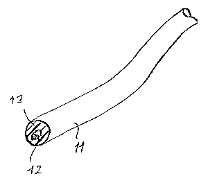
Fig. 3 shows a longitudinal support constructed in accordance
with the invention in the shape of a round rod, partly
in section, partly in perspective, and at an enlarged
scale.
In Figures 1 and 2 is shown part of a spinal column, wherein the
individual vertebrae are identified by the reference letters
"V". The spinal column is identified by the letter "S".
The individual vertebrae "V" are stabilized posteriorly, for
which purpose pedicle screws are screwed from the back into four
vertebrae "V". Each of the screw heads comprises receiption
openings or slots to receive a rod-shaped longitudinal support
11. The longitudinal support 11, as Fig. 3 also shows
particularly well, is constructed in the shape of a round rod
and is fixed in place by clamping in the heads of the pedicle
screws 10. In this way a spinal segment comprising four
vertebrae "V" can be stabilized. The longitudinal support or
supports 11 is/are so designed as to be plastically deformable
CA 02505042 2005-05-04
Translation for MEISSNER, BOLTE & PARTNER: M/SME-012-DE
- 15 -
by application of a prespecified bending force, so that they are
changed from a first stable shape state into a second,
alternative stable shape state as shown in Figures 1 and 2.
While in this implantation state, however, the longitudinal
supports 11 are intended to be flexible within prespecified
limits, as was presented in the introductory section. This
achieves a dynamic stabilization of a predetermined spinal
segment, with all the advantages explained above.
Specifically, in the embodiment presented here the longitudinal
support 11 is provided with a core 12 made of metal, in
particular titanium or a titanium alloy, encased in a human-
tissue-compatible plastic 13. The plastic deformability of the
longitudinal support 11 is ensured primarily by the metallic
core 12, whereas the flexibility in the deformed state is
determined primarily by the plastic casing 13. The above-
mentioned bending elasticity of the longitudinal support 11 is
indicated in Fig. 2 by a double-headed arrow 14. It is
determinded such, that, when the longitudinal support 11 is
clamped at one end, it can be elastically deflected by an angle
of 5° to 12°, in particular about 8° (double-headed arrow
14),
while remaining in a dimensionally stable state.
It should also be mentioned at this juncture that the apparatus
described here can comprise connecting means for the
longitudinal support, which can be used to connect at least two
support sections together. The support-connecting means can, for
example, comprise two oppositely situated receiption openings or
slots, into each of which one end section of a longitudinal
support can be inserted and fixed by a clamping screw or the
like.
The support-connecting means can be made either rigid or,
preferably, flexible with regard to bending. They allow supports
to be implanted one segment at a time, and permit extremely
individual stabilization of a section of the spinal column.
CA 02505042 2005-05-04
Translation for MEISSNER, BoLTE & PARTNER: M/SME-012-DE
- 16 -
In can also be seen in Figs. 1 and 2 that the stabilization of a
spinal-column section by means of the apparatus in accordance
with the invention is always carried out in such a way that
flexibility is available only in the context of flexion and
extension. Thus pressure on the cover plate and intervertebral
disk is considerably reduced, with no impairment of the axial
deformation of the disk, which is important for its nutrition.
The longitudinal support thus described must of course also be
designed such that it can be permanently deformed with a
prespecified force, which is greater than the peak forces
encountered anatomically, i.e. in vivo. This deformation is
carried out apart from the implantation, and preferably should
be possible without the need for special accessory devices. The
deformation is carried out "on site" by the surgeon.
In both the long direction of the longitudinal support and also
the transverse direction, the support should be stable, i.e.
unyielding, with respect to the anatomically customary shear
forces. Furthermore, it is very often desirable for the
longitudinal support to be stable with respect to torsion, in
order to ensure that the affected vertebral segment extends, as
a rule approximately horizontally, substantially only around a
posteriorly shifted center of rotation. As already mentioned
above, the longitudinal support can be constructed as a flat
band or strip. In the embodiment described here, longitudinal
supports in the shape of a round rod are implanted.
With regard to the bending elasticity of the longitudinal
support in accordance with the invention it should also be
mentioned that the angular range cited above refers to a length
of the support 11 that corresponds to the spacing of two
adjacent vertebrae, i.e. to a distance of about 2-6 cm, in
particular about 4-5 cm.
In other respects, regarding preferred embodiments, reference is
made to those according to Claims 16-18, which state for example
CA 02505042 2005-05-04
Translation for M6lssIVEK, BoL're & PARTNER: M/SME-012-DE
- 17 -
that the core can be shaped as a flat band or strip, with a
width that is the same as or smaller than the corresponding
dimension of the longitudinal support. This configuration is
naturally primarily appropriate for supports that have a band-
s like shape.
The with and/or height of the band-like core can vary
continuously or stepwise along the length of the longitudinal
support, at least over one longitudinal section thereof.
Regarding a rotationally symmetrical core, reference is made to
Claim 17.
In particular, it is fundamentally also conceivable for the
diameter of the core to become continuously larger or smaller,
at least in sections, so that the core acquires the form of a
wedge or cone. A stepwise change in the core diameter is also
conceivable, wherein in this last case the transitions in the
regions of a step are preferably rounded in order to reduce or
completely avoid stresses associated with steps.
Alternatively, it is also conceivable to form a groove in the
region of a stepwise transition, in order to reduce stresses.
All the features disclosed in the application documents are
claimed as essential to the invention insofar as they are new to
the state of the art individually or in combination.
CA 02505042 2005-05-04
Translation for MEISSNER, BOLTE & PARTNER: M/SME-012-DE
- 18 -
List of reference numerals
Pedicle screw
11 Longitudinal support
12 Core
5 13 Plastic casing
14 Double-headed arrow
Stabilization system
S Spinal column
V Vertebra