Note: Descriptions are shown in the official language in which they were submitted.
CA 02506625 2005-05-18
WO 2004/045544 PCT/US2003/036792
AUTOCRINE GROWTH FACTOR RECEPTOR ANTIBODIES AND
METHODS
[0001 J This application claims priority to provisional application, serial
number 60/427,220 filed November 19, 2002, the entirety of which is
incorporated by reference herein.
BACKGROUND OP THE INVENTION
[0002) PC-cell-derived growth factor ("PCDGF") is an 88-kDa
glycoprotein autocrine growth factor expressed in a tightly regulated fashion
in
normal cells but overexpressed and unregulated in tumorigenic cells.
Inhibition
of PCDGF expression or activity inhibits the growth of tumorigenic cells.
PCDGF is composed of a 68-KDa protein core and a 20-KDa carbohydrate
moiety. PCDGF belongs to a novel family of double cysteine rich polypeptides
and was originally isolated from the culture medium of the highly tumorigenic
mouse teratoma-derived cell line PC. PCDGF has been shown to be
overexpressed in mouse and human tumors including liver, kidney, breast, bone,
bone marrow, testes, brain, ovary, skin, and lung.
[0003] Amino-acid and cDNA sequencing indicated that PCDGF is
identical to the precursor of epithelins/granulins first purified as 6 KDa
double
cysteine-rich polypeptides from rat kidney or human granulocyte extracts. The
granulinjepithelin precursor was previously thought to be inactive. fee U.S.
Patent Number 5,416,192. However, Serrero et al. demonstrated that PCDGF
is a highly active, tumorigenic protein associated with a variety of tumor
cell
types. ee U.S. Patent Number 6,309,826. The degree of overexpression of
PCDGF positively correlates with the degree of tumorigenicity of cells. Ice.
[0004) PCDGF is a growth modulator for a variety of cell lines, including
fibroblasts, PC cells, and mammary epithelial cells. Comparison of the
expression of PCDGF in the highly tumorigenic PC cells and in parent 1246
CA 02506625 2005-05-18
WO 2004/045544 PCT/US2003/036792
cells demonstrated that PCDGF expression was very low in the non-tumorigenic
cells and was overexpressed in the highly tumorigenic cells. The same result
was
observed in human breast carcinomas where PCDGF expression was very low in
non-tumorigenic mammary epithelial cells and increased in breast carcinoma
cells.
[0005] PCDGF antagonists (e.g., anti-PCDGF antibodies and PCDGF
antisense nucleic acids) inhibit or interfere with the activity of PCDGF and
with
the growth of tumorigenic cells. Zhang, H., and G. Serrero, 1998, PNAS 95,
no. 24:14202; Lu, R., and G. Serrero, 2000, PNAS 97, no. 8:3993. In both
teratoma-derived cells and breast cancer cells, PCDGF activity was inhibited
by
treating the cells with an anti-PCDGF neutralizing antibody or by transfecting
the cells with an antisense PCDGF cDNA. Treatment of cells with PCDGF
antagonists in teratoma cells or breast carcinoma cells completely inhibited
cell
proliferation and tumorigenesis in vivo. Id.
[0006] Antibodies are specialized proteins capable of binding a target
molecule with great specificity. Originally identified as naturally produced
protein products of the animal immune system, antibodies function as a primary
arm of the immune system by binding to and facilitating clearance of micro-
organisms and other foreign substances from the body. Antibodies, also known
as immunoglobulins (Ig), are generally formed from two "light" chains and two
"heavy" chains. The carboxy terminus of the chains forms the constant or Fc
region of the antibody while the amino terminus forms the variable or antigen-
binding domain. There are at least five isotype categories of antibodies: IgG,
IgE, IgA, IgM, and IgD. Each Ig isotype interacts with different effector
cells
resulting in different biological activities. For example, IgG marks foreign
antigens for clearance by white blood cells (e.g., T cells) while IgE, located
on
the surface of mast cells, triggers allergic responses to particular antigens.
2
CA 02506625 2005-05-18
WO 2004/045544 PCT/US2003/036792
[0007] Animal models for generating antibodies are usefizl for making
large quantities of antibodies directed to a desired antigen. However, human
immune responses against "foreign" antibodies limit the therapeutic usefulness
of
antibodies developed in animals. "Humanized" antibodies substitute the
complementarity determining regions ("CDRs" ) of animal antibodies with
human CDRs resulting in a greatly reduced immune response in humans to the
"foreign" antibody. See, ~,.g,., Winter (British Application Number
GB2188538A).
[0008] An anti-idiotypic antibody ( "anti-IdAb") is an antibody that
recognizes unique determinants generally associated with the antigen-binding
site of an antibody. An anti-IdAb can be prepared by immunizing an animal of
the same species and genetic type (e.g., mouse strain) as the source of the
mAb
with the mAb to which an anti-IdAb is being prepared. The immunized animal
will recognize and respond to the idiotypic determinants of the immunizing
antibody by producing antibody to these idiotypic determinants (the anti-
IdAb).
The anti-IdAb may also be used as an immunogen to produce an immune
response in yet another animal, producing a so-called anti-anti-IdAb. The anti-
anti-IdAb may be epitopically identical to the original mAb which induced the
anti-IdAb. Thus, antibodies directed to the idiotypic determinants of an mAb
can be used to generate antibodies of identical specificity to the original
mAb.
[0009] U.S. Patent Number 6,309,826 refers to the existence of PCDGF
receptors on cell surfaces of several cell lines, including the mammary
epithelial
cell line C57MG, the 1246 and PC cell lines, and the mink lung epithelial cell
line CCL64. In these studies, the PCDGF receptor was detected by binding
labeled PCDGF to the PCDGF receptor and detecting the presence of PCDGF
bound to its receptor on the cell surface. What is needed are PCDGF receptor
antibodies and methods for binding to cell surfaces to interfere with the
activity
of the PCDGF receptor and the tumor promoting activity of PCDGF.
3
CA 02506625 2005-05-18
WO 2004/045544 PCT/US2003/036792
BRIEP SUMMARY OP THE INVENTION
[0010] The present invention provides antitumor compositions capable of
binding to the surface of a cell expressing the PCDGF receptor and interfering
with the binding of PCDGF to the PCDGF receptor. We have discovered
antibodies capable of binding to the PCDGF receptor to inhibit the biological
activity of PCDGF including, but not limited to, tumor cell proliferation
induced
by PCDGF. Anti-PCDGF receptor antibodies and/or antibody fragments can
be made by immunizing an animal with an anti-PCDGF antibody. The resulting
anti-PCDGF receptor antibodies or antibody fragments can be used to reduce
the proliferation of tumor cells in vit~~o and in vivo.
[0011] The invention provides, in one embodiment, antitumor
compositions comprising an antibody or antibody fragment capable of binding to
the surface of a cell expressing the PCDGF receptor and interfering with the
binding of PCDGF to the PCDGF receptor. Another embodiment of the
invention provides a composition comprising an anti-PCDGF receptor antibody
attached to a cytotoxic molecule for delivering the cytotoxic molecule to
cells
expressing the PCDGF receptor. The antibody-cytotoxic molecule composition
can be used to kill cells expressing the PCDGF receptor. Further embodiments
of the invention provide methods for inhibiting tumor cell proliferation by
contacting a tumorigenic cell with an effective amount of anti-PCDGF receptor
antibody.
[0012] Additional embodiments and advantages of the present invention
will be set forth in part in the description that follows, and in part will be
obvious
from the description, or may be learned through the practice of the invention.
The objects and advantages of the invention will be attained by means of the
instrumentalities and combinations particularly pointed out in the appended
claims.
4
CA 02506625 2005-05-18
WO 2004/045544 PCT/US2003/036792
BRIEP DESCRIPTION OF THE DRAWINGS
[0013] FIG. 1 shows that anti-PCDGF receptor antibodies bind to the
surface of MCF-7 human breast cancer cells. Antibody 6G8, an IgM antibody,
exhibited the strongest binding to the cell surface (lanes 1 and 2).
[0014] FIG. 2 shows MCF-7 cells immunostained with anti-PCDGF
receptor antibody 6G8.
[0015] FIG. 3 shows that anti-PCDGF receptor antibodies 6G8, 4H11,
and 5A8 do not react with PCDGF protein in an ELISA assay (Lanes 2, 3, and
6).
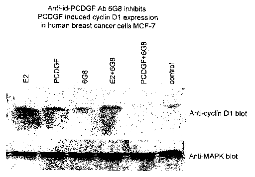
[0016] FIGS. 4 and 5 demonstrate that anti-PCDGF receptor antibodies
block PCDGF-induced cyclin D1 expression.
[0017] FIG. 6 shows that anti-PCDGF receptor antibodies block
PCDGF-induced phosphorylation of MAPIC.
[0018] FIG. 7 shows immunostaining of breast cancer and normal breast
tissue with anti-PCDGF receptor antibody 6G8. Anti-PCDGF receptor
antibody 6G8 strongly stains breast cancer tissue, but does not significantly
stain
normal tissue.
[0019] FIG. 8 shows the inhibiting effect of anti-PCDGF receptor
antibodies on the proliferation of MCF-7 cells. Anti-PCDGF receptor antibodies
6G8 and 4H11 inhibited MCF-7 cell proliferation by up to about 60% (columns
2and3).
DETAILED DESCRIPTION OP THE INVENTION
[0020] PCDGF is a highly tumorigenic autocrine growth factor and
causative agent for a wide variety of tumors. As described in U.S. Patent
Number 6,309,826, incorporated by reference herein in its entirety,
overexpression of PCDGF leads to uncontrolled cell growth and increased
CA 02506625 2005-05-18
WO 2004/045544 PCT/US2003/036792
tumorigenesis. The degree of PCDGF overexpression directly correlates with the
degree of cellular tumorigenicity. Cells overexpressing PCDGF do not require
external signals to maintain uncontrolled cell growth. Loss of regulated cell
growth, such as a loss in responsiveness to insulin and/or estrogen, leads to
increased malignancy and excessive unregulated cell growth. Development of
methods and compositions that interfere with the tumorigenic activity of
PCDGF is therefore of great interest for the treatment of cancer.
[0021] PCDGF antagonists, such as anti-PCDGF antibodies, interfere
with the biological activity of PCDGF (e.g., tumorigenic activity) by binding
PCDGF directly and preventing PCDGF from transmitting cell growth signals to
a target cell (e.g., breast cancer cell). An anti-PCDGF antibody may bind the
active site of PCDGF (e.g., the PCDGF receptor binding site) and prevent
PCDGF from binding to its receptor. Alternatively, anti-PCDGF antibodies may
bind to a site on PCDGF other than the active site, alter the conformation of
the
active site, and thus render PCDGF incapable of binding to its receptor. Anti-
PCDGF antibodies include PCDGF neutralizing antibodies. "Neutralizing"
antibodies have the ability to inhibit or block the normal biological activity
of
PCDGF, including PCDGF's ability to stimulate cell proliferation, increase
cell
survival, block apoptosis, or induce tumor growth in animals and in humans.
[0022] We have found that another useful target for interfering with the
biological activity of PCDGF is the PCDGF receptor. The term "receptor"
refers to a protein capable of transmitting signals from one ligand to another
ligand. Generally, receptors are transmembrane proteins having extracellular,
transmembrane, and intracellular domains. For example, a ligand, such as a
growth factor, can bind to its receptor's extracellular domain, resulting in a
receptor having an altered conformation. The intracellular domain, having an
altered conformation, is now capable of binding to an intracellular molecule,
which transmits the signal to another molecule in another cellular location
(e.g.,
a nuclear transcription factor). Interfering with the biological activity of a
6
CA 02506625 2005-05-18
WO 2004/045544 PCT/US2003/036792
receptor breaks the signal chain from the extracellular ligand to the
intracellular
ligand. Alternatively, molecules that target the receptor can be designed to
inactivate or kill a cell expressing a particular receptor (e.g., cytotoxic
molecules
linked to anti-receptor antibodies).
[0023] We first demonstrated the existence of the PCDGF receptor in
U.S. Patent 6,309,826 using the mink lung epithelial cell line CCL64.
Scatchard analysis of binding of lzsl-PCDGF revealed the presence of two
classes
of cell surface receptors: a high affinity class with a Kd of 4.3 +- 1.5 X 10-
" M
and 560 +- 170 sites/cell, and a low affinity class of receptors with a Kd of
3.9 +-
1.9 X 10⁻⁹ M and 17,000 +- 5900 sites/cell. Cross-linking studies and
autoradiographic analysis revealed the presence of one major cross-linked band
with a molecular weight of 190-195 KDa, corresponding to a molecular weight
for the unbound receptor of about 110 KDa for the major band. We have
shown that PCDGF receptors belong to the tyrosine kinase family of receptors.
Upon binding of PCDGF to the cell surface, the PCDGF receptor is activated by
phosphorylation on tyrosine residues, resulting in phosphorylation of several
signaling molecules, including IRS-l, SHC, and Grb2, and leading to activation
of MAP kinase ERK-2.
[0024] The term antibody herein includes but is not limited to human and
non-human polyclonal antibodies, human and non-human monoclonal
antibodies (mAbs), chimeric antibodies, anti-idiotypic antibodies (anti-IdAb),
neutralizing antibodies, non-neutralizing antibodies, and humanized
antibodies.
Polyclonal antibodies are heterogeneous populations of antibody molecules
derived either from sera of animals immunized with an antigen or from chicken
eggs. Monoclonal antibodies ( "mAbs" ) are substantially homogeneous
populations of antibodies to specific antigens. mAbs may be obtained by any
suitable method. Such antibodies may be of any immunological class including
IgG, IgM, IgE, IgA, IgD and any subclass thereof. The hybridoma producing
human and'non-human antibodies to the PCDGF receptor may be cultivated in
7
CA 02506625 2005-05-18
WO 2004/045544 PCT/US2003/036792
vitro or in vivo. For production of a large amount of mAbs, in vivo is the
presently preferred method of production. Briefly, cells from the individual
hybridomas can be injected intraperitoneally into pristane primed Balb/c mice
or
Nude mice to produce ascites fluid containing high concentrations of the
desired
mAbs. mAbs may be purified from such ascites fluids or from culture
supernatants using various chromatography methods known in the art.
[0025] Human monoclonal Ab to human PCDGF receptor can be
prepared by immunizing transgenic mice expressing human immunoglobulin
genes. Hybridomas produced by using lymphocytes from these transgenic
animals will produce human immunoglobulin instead of mouse immunoglobulin.
Since most monoclonal antibodies are derived from murine and other non-
human sources, their clinical e~cacy may be relatively limited due to the
immunogenicity of rodent mAbs administered to humans, weak recruitment of
effector function, and rapid clearance from serum. To circumvent these
problems, the antigen-binding properties of murine antibodies can be conferred
to human antibodies through a process called humanization. A humanized
antibody contains the amino-acid sequences for at least 6 complementarity-
determining regions (CDRs) of the parent murine mAb which are grafted onto a
human antibody framework. The low content of non-human sequences in
humanized antibodies (around 5%) has proven effective in both reducing the
immunogenicity and prolonging the serum half life in humans. Methods of
using monovalent phage display and combinatorial library strategy for
humanization of monoclonal antibodies are known in the art. Humanized
antibodies and human antibodies developed with transgenic animals as described
above are of therapeutic use for diseases including cancer.
[0026] Chimeric antibodies have different portions from different animal
species. For example, a chimeric antibody might have a variable region from a
murine mAb and a human immunoglobulin constant region. Chimeric
8
CA 02506625 2005-05-18
WO 2004/045544 PCT/US2003/036792
antibodies and methods for their production are also known to those skilled in
the art.
[0027] Hybridoma supernatants may be screened for the presence of
antibody specific for PCDGF receptor by any number of immunoassays including
dot blots and standard immunoassays (EIA or ELISA) which are well known in
the art. Once a supernatant has been identified as having an antibody of
interest,
it may be further screened by Western blotting to identify the size of the
antigen
to which the antibody binds. One of ordinary skill in the art may prepare and
screen such hybridomas without undue experimentation in order to obtain a
desired polyclonal or mAb given the teachings herein.
[0028] In a preferred embodiment of the invention, anti-PCDGF receptor
antibodies can be made by immunizing animals with PCDGF, permitting the
animals to produce anti-ID PCDGF antibodies, and utilizing spleen cells from
the immunized mice to produce hybridoma cell lines capable of secreting anti-
PCDGF receptor antibodies. See ~, U.S. Patent Number 5,144,010 hereby
incorporated by reference in its entirety. For example, anti-PCDGF receptor
antibodies can be made by (a) injecting animals (e.g., mice, rabbits) with an
effective amount of PCDGF to elicit an immune response; (b) periodically
collecting blood samples from the animals; (c) screening the blood samples to
determine if the animals are making antibodies directed to PCDGF and/or
antibodies to the PCDGF receptor; (d) making hybridoma cells (e.g., fusing
spleen cells from the animals with myeloma cells) after the animals cease
making
anti-PCDGF antibodies and continue to make anti-PCDGF receptor antibodies;
(e) purifying the anti-PCDGF receptor antibodies produced by the selected
hybridoma cell lines.
[0029) Initially, the animals respond by producing anti-PCDGF
antibodies. About 15 to 30 days after the appearance of high-titered anti-
PCDGF IgG, the animals produce antibodies to the anti-PCDGF antibodies and
9
CA 02506625 2005-05-18
WO 2004/045544 PCT/US2003/036792
stop or reduce the production of anti-PCDGF antibodies. For example, an
animal (e.g., mouse, rat, rabbit) can be injected with about 10 to about 100
ug
of purified PCDGF (e.g., recombinant PCDGF). The animals can be injected
with PCDGF about three to five times over the course of about 30 to 60 days.
The titer of anti-Id PCDGF antibodies can be monitored by screening blood
samples (e.g., tail bleeds) against cells expressing a high level of PCDGF
receptors (e.g., C57MG, 1246, PC-cells, CCL64). About 15 to 30 days after
high titer anti-PCDGF antibodies are formed, anti-Id antibodies will be
produced by the animals. Spleen cells from immunized mice can be used to
produce hybridomas secreting anti-PCDGF receptor antibodies. Anti-PCDGF
receptor antibodies can be purified from the hybridoma cells by several well-
established methods including, but not limited to, purification using Protein
A
or Protein G affinity chromatography columns.
[0030] Alternatively, anti-PCDGF antibodies may be used to induce
human and non-human anti-IdAbs in suitable animals. Animals can be
immunized with anti-PCDGF antibodies and will produce antibodies to the anti-
PCDGF antibodies. Anti-PCDGF antibodies that can be used to make anti-
idiotypic anti-PCDGF receptor antibodies include 6B3, 6B2, 6C12, 5B4, 5G6,
4D1, 3F8, 3F5, 3F4, 3G2, and 2A5. Hybridoma cell lines producing these anti-
PCDGF antibodies have been deposited with the American Type Culture
Collection (ATCC), 10801 University Blvd., Manassas, VA 20110-2209, and
have the following designations: 6B3 hybridoma cell line (ATCC Accession
Number PTA-5262), 6B2 hybridoma cell line (ATCC Accession Number PTA-
5261), 6C12 hybridoma cell line (ATCC Accession Number PTA-5597), 5B4
hybridoma cell line (ATCC Accession Number PTA-5260), 5G6 hybridoma cell
line (ATCC Accession Number PTA-5595), 4D1 hybridoma cell line (ATCC
Accession Number PTA-5593), 3F8 hybridoma cell line (ATCC Accession
Number PTA-5591), 3F5 hybridoma cell line (ATCC Accession Number PTA-
5259), 3F4 hybridoma cell line (ATCC Accession Number PTA-5590), 3G2
CA 02506625 2005-05-18
WO 2004/045544 PCT/US2003/036792
hybridoma cell line (ATCC Accession Number PTA-5592), and 2A5 hybridoma
cell line (ATCC Accession Number PTA-5589). Each of the above referenced
hybridoma cells lines can be induced to express the designated anti-PCDGF
antibody by maintaining the cell line in DMEM plus 10% FBS until the desired
quantities of anti-PCDGF antibodies are secreted into the cell culture medium.
Anti-PCDGF antibodies can be purified from the cell culture medium by well
established methods such as purification using Protein A or Protein G affinity
chromatography columns. Such anti-PCDGF antibodies can be used, for
example, to detect the presence of PCDGF, diagnose tumorigenicity, and/or
inhibit tumor cell proliferation as described in U.S. Patent 6,309,826.
[0031] Cells overexpressing the PCDGF receptor can also be used as
antigens to induce human and non-human anti-PCDGF antibodies in animals.
Animals can be immunized with whole cells or cell fractions (e.g., membranes)
overexpressing the PCDGF receptor (e.g., C57MG, 1246, PC-cells, CCL64
cells). For example, 1 to 10 million cells can be injected into suitable
animals.
The titer of anti-PCDGF antibodies can be monitored by screening blood
samples (e.g., tail bleeds) against cells expressing a high level of PCDGF
receptors (e.g., C57MG, 1246, PC-cells, CCL64). Spleen cells from immunized
mice can be used to produce hybridomas secreting anti-PCDGF receptor
antibodies. The culture supernatants from the hybridomas can be screened
against cancer cells by enzyme-linked immunoabsorbance assay (ELISA). Anti-
PCDGF receptor antibodies will compete with purified PCDGF for cell surface
binding to cells overexpressing the PCDGF receptor. The selected anti-PCDGF
receptor antibodies can be purified from the hybridoma cells by several well-
established methods including, but not limited to, purification using Protein
A
or Protein G affinity chromatography columns.
[0032] The term antibody is also meant to include both intact molecules
as well as fragments thereof such as, for example, Fab and F(ab')2, which are
capable of binding to the antigen. Fab and F(ab')2 fragments lack the Fc
11
CA 02506625 2005-05-18
WO 2004/045544 PCT/US2003/036792
fragment of intact antibody, clear more rapidly from the circulation, and may
have less non-specific tissue binding than an intact antibody. Such fragments
are
typically produced by proteolytic cleavage, using enzymes such as papain (to
generate Fab fragments) and pepsin (to generate F(ab')2 fragments).
[0033) One embodiment of the present invention provides antitumor
compositions which include an antibody or antibody fragment capable of binding
to the surface of a cell expressing the PCDGF receptor and interfering with
the
binding of PCDGF to the PCDGF receptor. The anti-PCDGF receptor
antibody may be an anti-idiotypic antibody. Anti-PCDGF receptor antibodies
6G8, 4H1, 2C1, 5A8, 2F8, and 2B12 are examples of anti-idiotypic antibodies
made by immunizing mice with PCDGF. Hybridoma cell lines producing these
anti-PCDGF receptor antibodies have been deposited with the American Type
Culture Collection (ATCC) and have the following designations: 6G8 (ATCC
Accession Number PTA-5263) and 5A8 (ATCC Accession Number PTA-5594).
The above referenced hybridoma cells line can be induced by maintaining the
cell
line in DMEM plus 10% FBS until the desired amount of anti-PCDGF receptor
antibody is secreted from the cells into the cell culture medium. Anti-PCDGF
receptor antibody can be purified from the cell culture medium by well
established methods such as, but not limited to affinity chromotography using
Protein A or Protein G columns. Other anti-idiotypic antibodies can be
prepared
as described above.
[0034) As used herein, the term "binding" refers to specific or non-
specific interactions between one molecule and another molecule. Examples of
"binding" include, but are not limited to, the direct interaction between the
antigen binding site of an antibody and the antigenic determinant of its
antigen
or to non-specific associations between molecules (e.g., co-localization,
electrostatic interactions, and phase interactions).
12
CA 02506625 2005-05-18
WO 2004/045544 PCT/US2003/036792
[0035] The term "contacting" means providing anti-PCDGF receptor
antibody to the environment of a cell whereby the anti-PCDGF receptor
antibody is capable of binding to a cell having a PCDGF receptor on its
surface.
For example, injecting anti-PCDGF receptor antibody directly into a tumor such
that the antibody diffuses into the tumor is considered "contacting" the cells
with the anti-PCDGF receptor antibody whether the anti-PCDGF receptor
antibody actually binds to its receptor on the surface of the cell or not.
Administration of anti-PCDGF receptor antibody and compositions
[0036] Anti-PCDGF receptor antibodies can be provided to cells both in
vitro and i~ vivo. For in vitro applications, anti-PCDGF receptor antibodies
can
be added to cell culture medium at concentrations typically ranging from 0.01
ng to about 500 ug/ml and preferably from about 10 ng to about 100 pg/ml.
Antibody may be administered alone or in conjunction with other therapeutics
directed to the same disease. Cells can also be transfected with DNA or RNA
encoding anti-PCDGF receptor antibodies or antibody fragments or vectors
containing such DNA or RNA sequences. Transfected cells can be induced to
make anti-PCDGF receptor antibodies or antibody fragments using any suitable
technique (e.g., inducible promoter, and multiple plasmid copies).
[0037] Anti-PCDGF receptor antibody compositions can also be
administered to cells using ex vivo techniques. Tumorigenic or normal cells
can
be removed from a subject (e.g., human, dog, cow, goat, mouse, rat, rabbit,
horse, or chicken) and grown in culture. The cells can be transfected with DNA
or RNA encoding anti-PCDGF receptor antibodies and induced to produce anti-
PCDGF receptor antibodies. The transfected cells can then be re-introduced
into the subject and produce anti-PCDGF receptor antibodies or antibody
fragments in order to inhibit the activity of PCDGF and reduce tumor cell
proliferation.
13
CA 02506625 2005-05-18
WO 2004/045544 PCT/US2003/036792
[0038] For i~c vivo applications, anti-PCDGF receptor antibody
compositions can be provided to a subject by a variety of administration
routes
and dosage forms. A subject, preferably a human subject, suffering from
disease
associated with increased PCDGF expression is treated with an anti-PCDGF
receptor antibody or fragment. Alternatively, a subject's cells are
transfected with
a polynucleotide encoding an anti-PCDGF receptor antibody or fragment. A
typical regimen comprises administration of an effective amount of the anti-
PCDGF receptor antibody over a period of one or several weeks and including
between about one and six months. The antibody of the present invention may
be administered by any means that achieves its intended purpose. For example,
administration may be by various routes including but not limited to
subcutaneous, intravenous, intradermal, intramuscular, intraperitoneal and
oral.
Parenteral administration can be by bolus injection or by gradual perfusion
over
time. Preparations for parenteral administration include sterile aqueous or
non-
aqueous solutions, suspensions and emulsions, which may contain auxiliary
agents or excipients known in the art. Pharmaceutical compositions such as
tablets and capsules can also be prepared according to routine methods. It is
understood that the dosage will be dependent upon the age, sex and weight of
the recipient, kind of concurrent treatment, if any, frequency of treatment
and
the nature of the effect desired. The ranges of effective doses provided below
are
not intended to limit the invention and merely represent illustrative dose
ranges.
However the most preferred dosage will be tailored to the individual subject
as is
understood and determinable by one of ordinary skill in the art given the
teachings herein. The total dose required for each treatment may be
administered by multiple doses or in a single dose. Effective amounts of
antibody are typically from about 0.01 ng to about 500 ug/ml and preferably
from about 10 ng to about 100 ~ag/ml. Antibody may be administered alone or
in conjunction with other therapeutics directed to the same disease.
14
CA 02506625 2005-05-18
WO 2004/045544 PCT/US2003/036792
[0039] As shown in FIGS. 1 and 2, anti-PCDGF receptor antibodies bind
to the surface of MCF-7 breast cancer cells. In an immunostaining assay,
purified anti-PCDGF receptor antibodies designated as 6G8, 2F8, 5A8, lEl,
2C1, and 3B3 were added to MCF-7 cells seeded in 96 well plates, incubated
with a secondary antibody linked to a horseradish peroxidase (HRP). The
results
show the anti-PCDGF receptor antibodies bind strongly to the surface of the
MCF-7 cells (FIG. 1 ). FIG. 2 shows that anti-PCDGF receptor antibodies stain
the cell surface of MCF-7 cells.
[0040] As shown in FIG. 3, anti-PCDGF receptor antibodies 6G8, 4H1,
2C1, 5A8, 2F8, and 2B12 do not bind to purified PCDGF in an ELISA (enzyme
linked immunoabsorption assay). However, anti-PCDGF antibody 6B3 binds to
PCDGF. Thus, anti-PCDGF receptor antibodies do not bind to PCDGF.
Anti-PCDGP receptor antibodies block P DGF induced biological
o-
functions
[0041] In one embodiment, PCDGF receptor antibodies specifically block
PCDGF-induced cyclin D1 expression. As shown in FIG. 4, both PCDGF and
estradiol stimulate cyclin D1 expression in MCF-7 cells. Anti-PCDGF receptor
antibody 6G8 specifically blocks PCDGF-induced cyclin D1 expression but not
estradiol-induced cyclin Dl expression. Compare lanes 4 and 5 of FIG. 4. FIG.
shows that anti-PCDGF receptor antibody 5B4 specifically blocks PCDGF-
induced cyclin D1 expression in MCF-7 cells. Anti-PCDGF receptor antibodies
also block PCDGF-induced phosphorylation of MAPK. As shown in FIG. 6,
Western blot analysis using anti-phospho-p44/p42 antibody shows that PCDGF
induces phosphorylation of MAPK (lane 2). The addition of anti-PCDGF
receptor antibody (e.g., 6G8) blocks PCDGF-induced phosphorylation of
MAPK (lane 4). Anti-PCDGF antibody alone does not induce phosphorylation
of MAPK.
CA 02506625 2005-05-18
WO 2004/045544 PCT/US2003/036792
[0042] While not wishing to be bound by theory, when anti-PCDGF
receptor antibodies do not bind PCDGF (FIGS. 1 and 2), and bind to the cell
surface to block PCDGF-induced biological activity (e.g., inducement of cyclin
D1 and phosphorylation of MAPK), we believe that such anti-PCDGF receptor
antibodies are specifically binding the PCDGF receptor.
[0043] Anti-PCDGF receptor antibodies are capable of binding to
tumorigenic cells but not to normal cells. As shown in FIG. 7, anti-PCDGF
receptor antibodies bind strongly to breast cancer tissue (panels B and C) but
not
to normal tissue (panel A) in an immunostaining protocol using 10
micrograms/ml of 6G8 anti-PCDGF receptor antibody. Thus, anti-PCDGF
receptor antibodies can also be used to diagnose tumorigenicity by comparing
the level of PCDGF receptor in a tissue sample or biopsy to the level of PCDGF
receptor in normal tissue. Elevated levels of PCDGF receptor indicate the
cells
are tumorigenic.
[0044] In accordance with a preferred embodiment of the invention, anti-
PCDGF receptor antibodies are utilized to reduce the proliferation of
tumorigenic cells. As shown in FIG. 8, anti-PCDGF receptor antibodies have
been shown to reduce proliferation of human breast cancer cells by up to about
60% (6G8). Anti-PCDGF receptor antibodies are used in accordance with
another embodiment of the invention to reduce the proliferation of tumorigenic
cells by contacting the cells with an effective amount of anti-PCDGF receptor
antibody. An effective amount of anti-PCDGF receptor antibody typically ranges
from about 0.01 ng to about 500 ug/ml and preferably from about 10 ng to
about 100 ug/ml. The "tumorigenic cells" includes, but are not limited to,
cells
derived from blood, cerebrospinal fluid, serum, plasma, urine, nipple
aspirate,
liver, kidney, breast, bone, bone marrow, testes, brain, ovary, skin,
prostate,
colon rectum, stomach, cervix, endometrial, pancreas, nasopharynx, neural and
lung.
16
CA 02506625 2005-05-18
WO 2004/045544 PCT/US2003/036792
[0045] It is to be understood that application of the teachings of the
present invention to a specific problem or environment will be within the
capability of one having ordinary skill in the art in light of the teachings
contained herein. The present invention is more fully illustrated by the
following
non-limiting examples.
EXAMPLE 1
Cell Surface Staining Of PCDGP Receptor Antibody On Human Breast
Cancer MCF-7 Cell Line
[0046] 1x10⁵ / well MCF-7 cells were seeded in 96 well plates and
incubated at 37°C, in a 5% COZ incubator overnight. Purified anti-id-
PCDGF
mAbs 6G8, 2F8, lEl, 2C1 and 3B2 were diluted in DMEM, 5% FBS medium at
50 pg/ml. 200 ul/well of each monoclonal antibody (mAb) was added in
duplicate wells and incubated at room temperature for 1 hour. Cells were then
washed three times in PBS and incubated with a goat anti-mouse IgG or IgM
(for 6G8 only) conjugated to horseradish peroxidase ("HRP") at 1:2000
dilution for 1 hour. After an additional three washes in PBS, a TMB microwell
component peroxidase substrate was added to each well. The plates were read in
a plate reader set at a wavelength of 620 nanometers. Anti-PCDGF receptor
antibodies stained the surface of the MCF-7 cells (FIG. 1 ).
17
CA 02506625 2005-05-18
WO 2004/045544 PCT/US2003/036792
Immunostaining of MCP-7 Cells With Anti-PCDGF Receptor Antibody
[0047] Fixed MCF-7 cells were blocked with 3% BSA/PBS and incubated
with 10 lag/ml of anti-PCDGF receptor antibody 6G8 in 3% BSA/PBS for 1
hour at 25°C. After three washes, the cells were incubated with goat
anti-mouse
IgM conjugated with HRP for 1 hour at 25°C. After an additional
wash, the
cells were incubated with HRP substrate. MCF-7 cells were stained specifically
with 6G8 (FIG. 2).
PCDGF Receptor Antibodies Stain Breast Cancer Tissue But Not Normal
Tissue
[0048] Tissue sections from a normal breast (low left panel) and breast
cancers (right up and low panels) were stained with 10 pg/ml of 6G8 mAb using
immunohistochemistry techniques described above. No significant staining was
observed with human normal breast tissue. However, anti-PCDGF receptor
antibody 6G8 stained the cancer tissues specifically as shown in FIG. 7.
EXAMPLE 2
PCDGP Receptor Antibodies Which Do Not Bind To PCDGP
[0049] Purified PCDGF protein was diluted in PBS and coated onto 96
well ELISA plates at a concentration of 100 ng/well and 50 ng/well. The
treated plates were incubated overnight at 4°C. After washing 3 times
with PBS,
the plate was blocked with 5% non-fat milk PBS for 1 hour at room temperature.
2ug/ml purified or 1:10 diluted BioRx fluids of PCDGF receptor mAb was
added to each well and incubated at room temperature for 1 hour. The plate
was washed 3 times with PBS and incubated with HRP conjugated goat anti-
mouse IgG secondary antibody for another hour. After an additional three
18
CA 02506625 2005-05-18
WO 2004/045544 PCT/US2003/036792
washes in PBS, a TMB microwell 1 component peroxidase substrate was added
to each well. The plates were read in a plate reader set at a wavelength of
620
nanometers. As shown in FIG. 3, the anti-PCDGF receptor antibodies did not
bind PCDGF.
EXAMPLE 3
PCDGP Receptor Antibody 6G8 inhibits PCDGP induced cyclin Dl
expression in human breast cancer MCP-7 cells
[0050] 2x10⁵/ml MCF-7 cells were plated in 6 well plates in DME/F12
medium plus 5% FBS overnight. The cell culture medium was replaced with
phenol-red free DMEM/F12 supplemented with 5°/ charcoal-stripped FBS
and
synchronized by treatment with luM Tamoxifen for 48 hours. The cell culture
medium was then replaced with serum free, phenol-red free DME/F12 and
treated with either 10-9M estradiol (E2), 200ng/ml PCDGF or 6G8 (50pg/ml)
alone or with E2 or PCDGF for 5 hours. After treatment, cells were lysed with
1RIPA buffer plus protease inhibitors. 60 ug of whole cell lysates were
separated
by 10% SDS-PAGE gel, and proteins were electrotransferred onto nitro-cellulose
membranes. Western blot detection of cyclin D1 expression was performed
using anti-cyclin D1/2 clone 5D4 monoclonal antibody (FIG. 4). As shown in
FIG. 4, either PCDGF or E2 induced the expression of cyclin D1 expression in
MCF-7 cells. Anti-PCDGF receptor antibody alone has no significant effect on
cyclin D1 expression. However, addition of 6G8 inhibits cyclin D1 expression
induced by either E2 or PCDGF.
EXAMPLE 4
The Effect Of Anti-PCDGP Mab 5B4 On The Expression Of Cyclin D1
[0051] 2x10⁵/ml MCF-7 cells were plated in 6 well plates in DME/F12
medium plus 5% FBS overnight. The cell culture medium was replaced with
19
CA 02506625 2005-05-18
WO 2004/045544 PCT/US2003/036792
phenol-red free DMEM/F12 supplemented with 5% charcoal-stripped FBS and
synchronized by treatment with luM Tamoxifen for 48 hours. The cell culture
medium was then replaced with serum free, phenol-red free DME/F12 and
treated with 200ng/ml PCDGF or 5B4 (100ug/ml) and 5B4 with PCDGF for
hours. After treatment, cells were lysed with RIPA buil'er plus protein
inhibitors. 60 pg of whole cell lysates were separated by 10% SDS-PAGE gels,
and the proteins were electrotransferred onto vitro-cellulose membranes.
Western blot detection of cyclin D1 expression was performed using anti-cyclin
D1/2 clone 5D4 monoclonal antibody. anti-PCDGF antibodies block PCDGF-
induced cyclin D1 expression (FIG. 5).
EXAMPLE 5
The Effect Of PCDGF Receptor Antibody 6G8 On Phosphorylation Of
MAPK
[0052] 2x10⁵/ml MCF-7 cells were plated in 6 well plates in DME/F12
medium plus 5% FBS overnight. The cell culture medium was replaced with
phenol-red free DMEM/F12 supplemented with 5% charcoal-stripped FBS and
cultured for 1 day. The cell culture medium was then replaced with serum free,
phenol-red free medium for another 1 day. After treatment with PCDGF
(200ng/ml), 6G8 (50pg/ml) or PCDGF with 6G8 for ten minutes, the cells
were lysed with 1ZIPA buffer plus protein inhibitors. 60 ~g of whole cell
lysates
were separated by 10% SDS-PAGE gels, and the proteins were electrotransferred
onto vitro-cellulose membranes. Western blot detection of phospho-MAPK
expression was performed using anti-phospho-p44/42 MAPK
(Thr202/Thr204) E10 monoclonal antibody (FIG. 6). In the control lane, no
phosphorylated MAPK was detected. PCDGF stimulated the phosphorylation of
MAPK, a key cell signal protein in cells. 6G8 antibody alone has no effect.
However, the addition of 6G8 blocks PCDGF-induced phosphorylation of
MAPK.
CA 02506625 2005-05-18
WO 2004/045544 PCT/US2003/036792
EXAMPLE 6
The Effect Of PCDGF Receptor Antibodies On The Proliferation Of
Human Breast Cancer Cells
[0053) MCF-7 cells were plated in 24-well plates at 105 cells per well in
DME/F12 medium plus 5°/ FBS. Two days later, the medium was
replaced with
phenol-red free DMEM/F12 supplemented with 5% charcoal-stripped FBS. After
another 24 hours of incubation, the medium was replaced with serum free,
phenol-
red free DME/F12 medium. 200 ng/ml PCDGF or 50 ug/ml 6G8, 4Hllor 50
ug/ml non-immune IgG was added into the wells in triplicate. ;H-thymidine was
added 24 hours later and after 5 hours labeling, cells were lysed and
radioactivity was
counted by a liquid scintillation counter. Anti-PCDGF receptor antibodies
significantly reduced the proliferation of MCF-7 cells as shown in FIG. 8.
EXAMPLE 7
Binding of PCDGF And PCDGP Antibodies To Cancer Cells
[0054] Various tumor cell lines were tested to determine the percentage of
cells that bound to either PCDGF or PCDGF receptor antibodies. Cells were
suspended in PBS containing 2 mM EDTA and 0.5% Bovine serum albumin and
incubated with either purified PCDGF or PCDGF receptor antibody 6G8 for 1 hour
at room temperature. Purified PCDGF was labeled with biotin. PCDGF receptor
antibody 6G8 was labeled with biotin. As shown in Table 1 below, PCDGF
receptor antibody 6G8 was able to significantly bind to a high percentage of
cells in
a variety of cell lines including MCF-7 (breast cancer), 04EM (breast cancer),
HL-
60 (human acute promyelocytic leukemia), ARP-1 (human multiple myeloma), and
RPMI8226 (human multiple myeloma). The results indicate the presence of
PCDGF receptor on the surface of the cells. In addition, the same cells that
bound
biotinylated 6G8 were able to bind biotinylated PCDGF.
21
CA 02506625 2005-05-18
WO 2004/045544 PCT/US2003/036792
Table 1
Cell types PCDGP binding % total 6G8 binding % total
cells cells
MCF-7 3~5/ 20-25/
04EM 50-60% 80~-90%
HL-60 40-50% 70-80%
A431 1-2 3-5
ARP-1 3-8% 15%
RPMI8226 2-4 10-15
U937 0 0
Jurkat 0 0
[0055] Cell surface binding with GP88 receptor antibody 6G8 was carried
out as follows. Tumor cells were suspended in 2mM EDTA-0.5% BSA-PBS (buffer
1) at a concentration of 5x10~cells/500u1 (at least 2x10⁶ cells in 200u1
buffer 1).
6G8 5-40ug/ml of biotinylated 6G8 antibody in buffer 1 was added to the cell
suspension and incubated at room temperature for 1 hour. The cells were washed
twice with buffer. Strepavidin-HRP was added at a dilution of 1:5000 in buffer
1
( 5 x 10⁶cells /500u1) and incubated for 1 hour. The cells were washed from 1
to 3
times in buffer 1 and substrate (DMPDA+4-CN) was added (5x10⁶cells/500 ul).
The cells in substrate were incubated at room temperature for one hour and
washed
in buffer 1 once. The cell suspension was dropped onto a slide and examined
with a
light microscope to determine the percentage of cells that were bound to 6G8
antibody.
[0056] Cell surface binding with PCDGF was carried out as follows.
Tumor cells were resuspended in PBS 0.5°/ BSA (buffer 2) at a
concentration of
5x10⁶cells/500u1. Biotinylated PCDGF (2ug/1x10'cells/ml of buffer 2) was
added and the resulting mixture was incubated at room temperature for 1 hour.
The cell were washed with buffer 2 twice followed by the addition of
streptavidin-
HRP at a dilution of 1:5000 in buffer 2 (5x10⁶cells/500u1) and incubated at
room
22
CA 02506625 2005-05-18
WO 2004/045544 PCT/US2003/036792
temperature for 30 minutes. Fresh substrate (DMPDA+4-CN) was prepared.
The cells were washed twice with buffer 2 and added to the substrate
(5x10⁶cells/500u1). The cells were incubated with the substrate at room
temperature for 15 minutes. The cells were washed once with buffer 2 and
resuspended in buffer 2. The resulting cell suspension was dropped onto a
slide
and examined under a light microscope to determine the percentage of cells
bound to PCDGF.
23