Note: Descriptions are shown in the official language in which they were submitted.
CA 02512891 2005-07-07
WO 2004/067087 PCT/EP2004/000724
APPARATUS OF ELECTRO-STIMULATION AND RELATIVE DATA SUPPORT
The invention relates to an apparatus and a method of electro-
stimulation and a data support that can be read by processing
means. On the data support data are recorded that are required
for the operation of the apparatus and the actuation of the
method.
The apparatus and the method of electro-stimulation according
t~ the invention are particularly suitable for carrying out
bioactive neuro-stimulation and for modulation of cytokines,
growth factors and of enzymatic cellular metabolism.
Clinical data show that more than half of the population of
western countries suffers from vascular pathologies, and in
particular pathologies affecting the cardiovascular system.
Alterations of the vascular walls frequently occur that are
caused by degenerative pathologies such as arteriosclerosis
which, together with thrombosis, is one of the most frequent
causes of obstruction of the peripheral arteries and of those
that affect the myocardium and the brain.
Arteriosclerosis manifests itself in a particularly aggressive
and premature manner in diabetic patients, who make up about
of the European population and a similar percentage of the
population in Italy. This pathology is accompanied by long-
term complications that gravely disable the patient that are
25 due t~ the degeneration of the larger blo~d vessels (macro-
angiopathy), of the smaller blood vessels (micro-angiopathy)
and of the peripheral and vegetative nervous system
(near~pathy). Peripheral macro-ane~iopathy in diabetic patients
produces anal~gous symptoms t~ th~se ~bserved in non-diabetic
30 patients~ however, this manifests itself prematurely, with
greater frequency and deteriorates rather rapidly.
For the above explained reasons, the vascular pathol~gies
causes in diabetic patients a mortality rate twice the
mortality rate in non-diabetic patients, and make long
CONFIRMATION COPY
CA 02512891 2005-07-07
WO 2004/067087 PCT/EP2004/000724
2
hospitalisations necessary, with remarkable economic and
social consequences.
Furthermore, in diabetic patients arteriosclerosis is
responsible for a majority of the amputations of the lower
limbs (50-700), which such patients undergo 5 times more
frequently than non-diabetic patients. The occlusion of small
and medium-calibre distal arteries below the knee causes
gangrene to develop. Furthermore, diabetic patients suffer
more frequently than non-diabetic patients from claudicatio
intermittens due to ischemia of the muscles in the calves, the
thigh or the gluteus.
Substances have recently been discovered and described in the
literature that are produced by endothelium cells and cause
new blood vessels to be formed (angiogenesis) and
vasodilatation, such as, for example, the Fibroblast Growth
Factor (FGF), Neuronal Growth Factor (NGF), Epithelial Growth
Factor (EGF), Vascular Endothelial Growth Factor (VEGF) and
Angiopoietin-2.
To promote angiogenesis, VEGF and other angiogenic factors,
such as FGF, can be injected directly into the vascular bed
affected by ischemia and/or occlusion.
But the direct injection of VEGF or other angiogenic factors
has many drawbacks, which are mainly due to the difficulty of
release to all the cells affected. In fact, less than 2% of
the VEGF injected is effectively involved in neo-angiogenesis~
furthermore, the method is potentially toxic.
Experiments conducted by r~anno et al. have shown that when
continuous electrical stimulation was applied for 5 days to
isolated animal muscles by means of pulses having a width of
0.3 ms, a frequency of 50 H~ and an intensity of 0.1 V, an
increase in the production of VEGF was observed and neo-
angiogenesis was promoted through an increase in the number of
capillaries and of the blood flow.
CA 02512891 2005-07-07
WO 2004/067087 PCT/EP2004/000724
3
Although said experiments seem to suggest that electric
stimulation of the muscles has beneficial effects on the
circulation they do not teach how to apply electric
stimulation to humans.
In addition, they require treatment lasting several days,
which could cause the patient discomfort because of its
excessive length.
Furthermore, it is known to use laser transmyocardial
rivascularisation to reduce the pain caused by angina~ this
determines an increase in the level of VEGF in the myocardium
and in the endothelium cells of capillaries and arterioles
(Lee, SH, Wolf PL, Escudero R, N Eng. J. Med. 2000~ 342, 626-
33). However, laser transmyocardial rivascularisation is an
invasive technique that achieves limited results.
US 2002/0010492 describes an electro-stimulation device for
the controlled production of angiogenic growth factors,
through which device the level of VEGF can be increased in
vitro by 30-40o through continuous electro-stimulation lasting
at least 8 hours.
However, even in this case, long periods of treatment are
required that cause significant discomfort to the patient.
WO 02/09809 discloses an apparatus for treating vascular,
muscular or tendinous pathologies by means of which a series
of pulses having a width from 10 to 40 us and an intensity
from 100 to 170 ~,A is applied to the patient. In this way, an
increase in the production of VEGF can be obtained, with
consequent vasodilatation and neo-angiogenesis.
An object of the invention is to improve the condition of
patients affected by vascular pathologies, and more in
particular of diabetic patients suffering from said
pathologies.
A further object of the invention is to stimulate the
production of large quantities of substances that promote the
formation of new blood vessels and the dilatation of existing
CA 02512891 2005-07-07
WO 2004/067087 PCT/EP2004/000724
4
ones, in particular VEGF, with relatively short treatment
time, i.e, without subjecting the patient to exhausting
treatment lasting several hours.
In particular, it is desired to induce production of VEGF or
of other growth factors in quantities that are substantially
greater than those obtained by means of the apparatus
described in WO 02109809.
In a first aspect of the invention, there is provided an
electro-stimulation apparatus, comprising electric-pulse
l0 generating means arranged to generate pulses having preset
values of typical parameters, applying means arranged to apply
a sequence of said pulses to an organism, said sequence
comprising an initial pulse and a final pulse, characterised
in that, it further comprises variation means arranged to
perform a substantial variation of at least one typical
parameter at a moment comprised between said initial pulse and
said final pulse.
In a second aspect of the invention, there is provided a
method of electro-stimulating an organism, comprising
generating a sequence of electric pulses having preset values
of typical parameters, said sequence comprising an initial
pulse and a final pulse, and applying said sequence to said
organism, characterised in that, said generating comprises
considerably varying at least one typical parameter at a
moment comprised between said initial pulse and said final
pulse.
In a third aspect of the invention, there is provided a
support readable by data processing means, containing a
plurality of data with preset values of typical parameters,
said data being intended to originate a sequence of electric
pulses to be applied to an organism by means of electro-
stimulation techniques, said sequence comprising an initial
pulse and a final pulse, characterised in that, a substantial
variation of at least one typical parameter is provided in
CA 02512891 2005-07-07
WO 2004/067087 PCT/EP2004/000724
said sequence at a moment comprised between said initial pulse
and said final pulse.
In one embodiment, the parameter that is considerably varied
is the frequency of the pulses.
5 In a further embodiment, the parameter that is considerably
varied undergoes a decrease in its value.
This decrease can be of an order of magnitude.
As will described in detail below, experimental data have
shown that owing to the invention and particularly owing to
the substantial variation occurring in one of the typical
parameters in the sequence of electric pulses, it is possible
t~ obtain a relaxing effect on the muscle fibres, an
activating effect on the vessels and on the neuroreceptors and
a release of growth factors. It is furthermore possible to
obtain an anti-inflammatory effect and to inhibit the
cytokines that cause the inflammation. Finally, the invention
enables stimulation of the small neurological afferent fibres
and better interaction with the motor system to be obtained.
As the good effects that have been noted are linked t~ the
substantial variation of a typical parameter that occurs in an
almost instantaneous manner, it is no longer necessary to
subject the patient to treatment lasting several hours,
because a session of only a few minutes enables said
improvements to be observed.
Furthermore, the electric pulses can be applied
transcutaneously, i.e. by using a technique that is not
invasive and does not cause to the patient particular
discomfort.
In order that the invention may be clearly and completely
disclosed, reference will now be made, by way of examples that
do not limit the scope of the invention, to the accompanying
drawings, in which:
Figure 1 is a table disclosing the sub-phases of a stimulation
sequence with relaxing effect;
CA 02512891 2005-07-07
WO 2004/067087 PCT/EP2004/000724
6
Figure 2 is a table disclosing the sub-phases of a stimulation
sequence with anti-inflammatory effect;
Figure 3 is a table disclosing the sub-phases of a stimulation
sequence for activating the microcirculatory system;
Figure 4 shows the variation of the operational parameters
during the sequence for activating the microcirculatory system
shown in Figure 3;
Figure 5 is a table disclosing the levels of VEGF found in
patients subjected to electro-stimulation treatment according
to the invention;
Figure 6 shows the values of VEGF detected during experimental
stimulation of a distal gone of the leg;
Figure 7 shows a detail of Figure 6;
Figure 8 shows the sub-phases of the first part of a
neuromuscular stimulation sequence of the hypotonic muscle;
Figure 9 shows the sub-phases of the second part of a
sequence, the first part of which is shown in Figure 8.
An apparatus for electro-stimulation comprises one or more
generators of electric pulses that can be controlled by a
control device provided with a microprocessor. The control
device can modulate the frequency and/or the width and/or the
intensity of the electric pulses according to preset
sequences. The electric pulses can be sub-threshold, i.e.
maintained below values that could cause contraction of the
muscle or a sensation of pain in the patient.
The apparatus further comprises applying means for applying
the electric pulses to an organism, for example a human or a
laboratory animal. The applying means may comprise electrodes
provided with a highly conductive surface that are positioned
directly on the skin of the patient to transcutaneously
transmit the pulses.
The parameters that distinguish the pulses are defined on the
basis of the rheobasis and/or of the chronaxy of the
stimulated neuro-muscular tissue, or in general on the basis
CA 02512891 2005-07-07
WO 2004/067087 PCT/EP2004/000724
7
of the bioreaction. Rheobasis is intended as the minimum
current intensity required to excite a tissue, whereas
chronaxy is the minimum duration that an electric pulse having
twice the intensity of the rheobasis must have to generate a
stimulation.
Bioreaction is defined as the time that elapses between a
trailing edge of an applied pulse and the leading edge of the
following pulse, i.e. the biological reaction time available
to a preset tissue before the application of the following
pulse.
Variation means is furthermore provided arranged to vary the
typical parameters of the applied pulses, namely the frequency
and/or the width and/or the intensity.
In a first embodiment, the pulses generated by the apparatus
according to the invention have a width from 1 to 90 ~,s and a
frequency from 0.1 Hz to 1 kHz. Their peak voltage is above 50
V and may vary up to 300 V.
In a second embodiment, the pulses have a width between 1 and
49 ~,s, a frequency from 0.1 Hz to 100 Hz and a peak voltage up
to 200 V.
In a third embodiment, the width of the pulses varies from 1
to 40 ~,s, the frequency varies from 0.1 Hz to 100 Hz and peak
voltage reaches a maximum of 300 V.
The electro-stimulation apparatus is configured in such a way
as to apply a sequence of stimuli comprising a preset
succession of sub-sequences. Each sub-sequence is the result
of the modulation of frequency, width and intensity according
to a protocol that depends on the biochemical effect that is
desired to have on the cell: and on the tissues.
For example, to obtain a relaxing effect on the muscular
fibres, a sequence of sub-threshold pulses is applied which
stimulates the muscle with a gradually increasing frequency,
until a condition of tetany is reached in which the muscle
reaches a spasm situation. Frequency is thereafter sharply
CA 02512891 2005-07-07
WO 2004/067087 PCT/EP2004/000724
0
reduced to the value of 1 Hz, so as to create a traumatic
event and cause muscular relaxation.
One example of said sequence is shown in Figure 1, and
comprises 27 sub-phases according to the indicated parameters.
In the first sub-phase, pulse trains are sent to the patient
for a time interval having a duration of 20 seconds. In this
period, the frequency has a value of one pulse per second (1
Hz), each pulse having a width of 10 microseconds. During the
second sub-phase, which lasts 5 seconds, the pulse trains
applied to the patient have a pulse frequency of 1 Hz, and
each pulse has a duration of 20 microseconds. The frequency of
the pulses of each sub-phase is then gradually increased until
the sub-phase 13 is reached, in which the frequency reaches a
value of 29 Hz with a pulse width. of 40 microseconds. In the
following sub-phase there is a sudden decrease in the
frequency of the pulses, which drops by an order of magnitude
from 29 Hz to 1 Hz, and in the pulse width, which decreases
from 40 microseconds to 10 microseconds. After this sudden
decrease, the frequency and the pulse width are increased
again in a gradual manner, until they reach a final value of
respectively 39 Hz and 40 microseconds.
Experimental results have shown that the sudden decrease in
the pulse frequency applied to the muscle allows the muscle to
relax. To reinforce the positive effects of the decrease in
frequency, it is possible to repeat the sequence in Figure 1
several times, in which case the frequency discontinuity
occurs a greater number of times.
On the other hand, in order to obtain an effective action on
the blood vessels and an anti-inflammatory effecto substances
have to be released such as the growth factors promoting
neoangiogenesis and producing cytokines, that are able to
produce an anti-inflammatory effect. At the same time, the
formation of other cytokines such as TNF-oc, interleukin-6,
CA 02512891 2005-07-07
WO 2004/067087 PCT/EP2004/000724
9
interferone-a and cortisole, that are responsible for the
inflammatory state, has to be inhibited.
In order to do this without stimulating the tissue for an
excessively long time and with a marginal release of energy,
sequences of pulses are applied to the patient in which the
frequency is rapidly increased until the required value is
reached. This value varies according to the substance to be
released, produced or inhibited.
The inventor thinks that the electrical field applied by the
electro-stimulation apparatus creates a series of vibrations
by pulse polarisation and depolarisation of the cells and of
the molecules. Such vibrations induce resonance conditions in
sub-structures of the cells of the connective tissue, and in
particular in the sub-structures of the endothelium cells, of
the muscles, of the dermis and of the epidermis, for example
the cell membrane, mitochondria, and/or the immunological
molecules or complexes. This causes specific enzymes,
cytokines and growth factors to be released into the
interstitial spaces and therefore into the circulating blood.
Depending on the different model of resonance induced in the
cellular sub-structures, a release or transcription of
different molecules is obtained. Therefore, by appropriately
varying the frequency of the pulses applied, it is possible to
reach the typical resonance frequency corresponding to the
type of molecule that one wishes to release or inhibit.
One example of a sequence of pulses to apply in order to
obtain an anti-inflammatory effect, operating according to the
mechanism above-described, is set out in Figure 2.
If it is rather desired to activate the miceocirculatory
system, a sequence of the type shown in Figure 3 can be
applied. The variations of the typical parameters of the pulse
for this latter sequence are shown in Figure 4.
As can be noted, the sequence shown in Figure 3 comprises an
initial sub-sequence that is substantially analogous to the
CA 02512891 2005-07-07
WO 2004/067087 PCT/EP2004/000724
initial part of the sequence shown in Figure 1 and that aims
to obtain a relaxing effect. Subsequently, during sub-phase 13
the frequency is sharply reduced to the value of 1 Hz and
subsequently increased up to 11 Hz. After this, the frequency
5 is kept constant for a few seconds in order to cause an
effective vaso-action on the blood vessels. Then, from sub-
phase 38, the value of the frequency is increased by 10 Hz at
each sub-phase, until the value of 41 Hz is reached, around
which value it has been experimentally established that the
10 greatest release of VEGF is obtained. Said frequency
reasonably seems to be the resonance frequency of VEGF.
In order to obtain an even higher level of VEGF in the blood,
the sequence shown in Figure 3 can be repeated several times a
day.
By repeating the same sequence several times in succession, a
surprising synergic effect was observed, inasmuch as it was
seen that the obtained result was greater than the sum of the
results that could logically be expected by applying two
sequences independently of each other.
This seems to be due to the sudden reduction in the frequency
of the pulses applied, i.e. the sharp transition from a
relatively high frequency value to the initial value of 1 Hz,
which introduces a discontinuity in the applied pulses. This
results in a significant energy variation. By repeating the
sub-sequence several times, an effect analogous to the so-
called "water hammer~~ occurring in hydraulics takes place, by
means of which the stimulation by sub-threshold electric
pulses enables creak chemical bonds to be broken and large
quantities of the desired molecules to be released or
transformed, without inducing a significant transfer of energy
to the tissue.
In one embodiment, the variation in the applied frequency is
greater than 20 Hz. In another embodiment, the variation in
CA 02512891 2005-07-07
WO 2004/067087 PCT/EP2004/000724
11
the applied frequency is greater than 40 Hz. In a further
embodiment, such variation may be greater than 60 Hz.
The above-formulated hypothesis was experimentally tested by
stimulating a lower limb of 10 diabetic patients, of 10 non
diabetic patients and of 10 healthy subjects whose behaviour
was observed for control purposes. The pulses were applied to
the peripheral distal part of the leg.
The stimulation sequence applied to all the individuals taking
part in the experiment comprised two consecutive sub-sequences
aimed to obtaining muscular relaxation, followed by two sub
sequences of activation of the microcirculatory system, in the
manner described above. Stimulation was thereafter applied for
a period of 10 minutes at a constant frequency of 100 Hz and
with a constant pulse. width of 40 microseconds.
Blood samples from the systemic circulation were taken of the
individuals taking part in the experiment (samples were taken
from the brachial vein) 10 and 5 minutes before stimulation,
and 0, 1, 2, 3, 4, 5, 7, 10, 20 and 40 minutes after the
beginning of stimulation. The results obtained are shown in
Figures 5, 6 and 7.
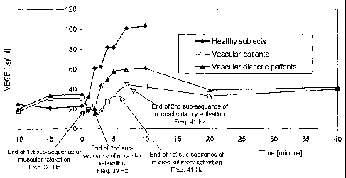
In particular, Figure 5 shows the average VEGF values measured
in the blood samples taken from the different patients at the
times indicated. The values at -10 and -5 minutes refer to the
period preceding stimulation, the values at 0, 1 and 2 minutes
were recorded during the sub-sequences of muscular relaxation,
the values at 3, 4, 5 and 7 minutes were~recorded during the
sub-sequences of activation of the microcirculatory system.
The values at 10, 20 and 40 minutes were rec~rded dozing the
final sub-sequence at a constant frequency and width. The
recorded VEGF pattern is set ~ut graphically in Figures 6 and
7.
As can be noted, at the end of every sub-sequence a sudden
increase in the measured VEGF values occurred. The healthy
subjects showed increases in VEGF that were up to 5 times
CA 02512891 2005-07-07
WO 2004/067087 PCT/EP2004/000724
12
greater than their base value, whereas in diabetic patients
the VEGF value increased by up to 3 times more than the
initial value.
It was furthermore noted that if electro-stimulation was not
applied in an appropriate manner, VEGF did not increase. This
was shown in the last phase, in which the frequency and the
width of the pulses were kept constant and in both. diabetic
patients and in non-diabetic patients VEGF tended to decrease
returning to the base values within 10 minutes.
Only when the stimulation frequency was appropriately modified
in such a manner as to reach the typical resonance frequency
of the cells that produce VEGF, and then suddenly decreased to
create a traumatic event, an effective and consistent increase
in the growth factor, occurred, through a mechanism that in
certain respects is analogous to the one that determines the
so-called "water hammer~~.
The detected increases in VEGF, as shown in Figures 5, 6 and
7, appear to be particularly significant if one considers that
they were measured in the blood samples taken from the
brachial veins of the subjects examined, whereas electro-
stimulation was carried out in the distal peripheral part of
the leg. This means that the VEGF that had been produced in
the stimulated zone, rapidly spread throughout the organism,
thereby determining a considerable increase in the average
value of VEGF current in the patient's blood at the systemic
level.
Therefore the increase in VEGF from the value of 21 pg/ml
recorded after 2 minutes of electro-stimulation, to the value
of 60 pg/ml measured after 7 minutes of electro-stimulation in
the blood taken from the brachial veins of the diabetic
patients is indicative of a much more considerable increase in
VEGF in the stimulated zone that is affected by the occlusion
of the blood vessels. This results, in the stimulated zone, in
a substantial benefit to the patient deriving from the
CA 02512891 2005-07-07
WO 2004/067087 PCT/EP2004/000724
13
formation of new blood vessels and from the dilatation of
existing ones.
Lastly, it has been proposed to use a sequence like the one
shown in Figures 8 and 9 to stimulate small afferent
neurological fibres and their interaction with the motor
units. The data shown in Figures 8 and 9 actually constitute a
single sequence, which has been set out on two separate sheets
for the sake of clarity.
As can be noted, this last sequence is a combination of a
modified sub-sequence of muscular relaxation, followed by a
vasoactive sub-sequence. A sub-sequence activating the small
ne w~us fibres is then provided until a pulse frequency of 220
Hz is reached. This produces a gradual increase in
prioreception and in peripheral sensitivity im patients
affected by paraplegia, tetraplegia or hemiplegia, secondary
lesions to the brain, traumas to the head or to the spine, or
apoplectic stroke.
According to an embodiment of the invention, the pulse width
can also be varied and in particular it can be increased from
the current value until a preset maximum value is reached.
This maximum value can be of about 90-100 ~.s.
The increase in pulse width is equal to a percentage of the
current pulse width value, for example 200, 250, 330 or 50% of
the current value. Experimental tests have shown that the best
results are obtained if percentage increases of 200 of the
current pulse width value are chosen.
Hetween an increase in pulse width and the subsequent
increase, a time interval occurs having a duration which can
be randoml~r varied between a minimum value and a ma~~imum
value. In particular, the minimum value of this duration can
be of about 15 seconds, whereas the maximum value can be of
about 60 seconds.
When the preset maximum pulse width is reached, the pulse
width is suddenly decreased to its initial value.
CA 02512891 2005-07-07
WO 2004/067087 PCT/EP2004/000724
14
This variation of the pulse width can be repeated several
times. It can in particular be applied when the pulse
frequency is kept constant, for example when, after applying
to the patient the sequences previously disclosed with
reference to the drawings, stimulation is applied for some
minutes at a constant frequency.
By varying the pulse width, adaptation phenomena are avoided
in the patient, which means that the patient does not get used
to the applied pulses, which might decrease the therapy
efficiency.