Note: Descriptions are shown in the official language in which they were submitted.
CA 02513769 2005-07-19
WO 2004/066946 PCT/US2004/002261
METHOD FOR EVALUATING THE EFFICACY OF
CERTAIN CANCER TREATMENTS
This application claims priority from U.S. Provisional
Application 60/442,902, filed January 28, 2003.
Field of the Lnvention
[0001] This invention relates to a method for evaluating
the efficacy of cancer therapies that act through or result
in the induction of apoptosis. More specifically, this
invention relates to such a method which involves detecting
the l7kDa subunit of cleaved Caspase 3 as a marker c~f
apoptosis.
Description of the Background Art
[0002] One highly productive focus of research in recent
years in the development of systemic gene therapy delivery
systems has been on developing non-viral, pharmaceutical
formulations of genes for in vivo human therapy,
particularly cationic liposome-mediated gene transfer
systems (Massing U, et al., Int. J. Clin. Pharmacol. Ther.
35:87-90 (1997)). Features of cationic liposomes that make
them versatile and attractive for DNA delivery include:
simplicity of preparation; the ability to complex large
amounts of DNA; versatility in use with any type and size of
DNA or RNA; the ability to transfect many different types of
cells, including non-dividing cells; and lack of
immunogenicity or biohazardous activity (Felgner PL, et al.,
Ann. NY Acad. Sci. (1995) 772:126-139; Lewis JG, et al.,
1
CA 02513769 2005-07-19
WO 2004/066946 PCT/US2004/002261
Proc. Natl. Acad. Sci. USA (1996) 93;3176-3181). More
importantly from the perspective of human cancer therapy,
cationic liposomes have been proven to be safe and efficient
for .in vivo gene delivery (Aoki, K. et al., Cancer Res.
55:3810-3816 (1997) Thierry, A.R., Proc. Natl. Acad. Sci.
USA 92:9742-9746 (1997)).
[0003] The transfection efficiency of cationic liposomes
can be dramatically increased when they bear a ligand
recognized by a cell surface receptor. Receptor-mediated
endocytosis represents a highly efficient internalization
pathway present in eukaryotic cells (Cristiano, R.J., et
al., Cancer Gene Ther. 3:49-57 (1996), Cheng, P.W., Hum.
Gene Ther. 7:275-282 (1996)). The presence of a ligand on a
liposome facilitates the entry of DNA into cells through
initial binding of the ligand by its receptor on the cell
surface followed by internalization of the bound complex. A
variety of ligands have been examined for their
liposome-targeting ability, including transferrin and folate
(Lee, R.J., et al., J. Biol. Chem. 271:8481-8487 (1996)).
Folate receptor (FR) and Transferrin receptor (TfR) levels
are elevated in various types of cancer cells including, but
not limited to, prostate, breast, pancreatic, head and neck,
bladder, brain, ovarian, skin, lung, and liver cancers.
Elevated TfR and FR levels also correlate with the
aggressive or proliferative ability of tumor cells (Elliot,
R.L., et al., Ann. NY Acad. Sci. 698:159-166 (1993)).
Therefore, TfR and FR levels are considered to be useful as
prognostic tumor markers, and they are potential targets for
drug delivery in the therapy of malignant cells (Miyamoto,
T., et al., Int. J. Oral Maxillofac. Surg. 23:430-433
(1994); Thorstensen, K., et al., Scand. J. Clin. .Lab.
Invest. Suppl. 215:113-120 (1993)).
2
CA 02513769 2005-07-19
WO 2004/066946 PCT/US2004/002261
[0004] In addition to ligands that are recognized by
receptors on tumor cells, specific antibodies also can be
attached to the liposome surface (Allen, T.M., et al.,
Stealth Ziposomes, pp. 233-244 (1995)), enabling them to be
directed to specific tumor surface antigens (including but
not limited to receptors) (Allen, T.M., Biochim. Biophys.
Acta 1237:99-108 (1995)). These "immunoliposomes,"
especially the sterically stabilized immunoliposomes, can
deliver therapeutic drugs to a specific target cell
population (Allen, T.M. , et al. , Stealth .Liposomes, pp.
233-244 (1995)). Park, et al. (Park, J.W., et al., Proc.
Natl. Acaa'. Sci. USA 92:1327-1331 (1995)) found that
anti-HER-2 monoclonal antibody (Mab) Fab fragments
conjugated to liposomes could bind specifically to HER-2
overexpressing breast cancer cell line SK-BR-3. The
immunoliposomes were found to be internalized efficiently by
receptor-mediated endocytosis via the coated pit pathway and
also possibly by membrane fusion. Moreover, the anchoring
of anti-HER-2 Fab fragments enhanced their inhibitory
effects. Doxorubicin-loaded anti-HER-2 immunoliposomes also
showed significant and specific cytotoxicity against target
cells in vitro and in vivo (Park, J.W., et al., Proc. Natl.
Acad. Sci. USA 92:1327-1331 (1995)). In addition, Suzuki et
al., Br. J. Cancer 76:83-89 (1997), used an anti-transferrin
receptor monoclonal antibody conjugated immunoliposome to
deliver doxorubicin more effectively in human leukemia cells
in vitro. Huwyler et al., Proc. Natl. Acad. Sci. USA
93:14164-14169 (1996), used an anti-TfR monoclonal antibody
immunoliposome to deliver daunomycin to rat glioma (RT2)
cells in vivo. This PEGylated immunoliposome resulted in a
lower concentration of the drug in normal tissues and
organs. These studies demonstrated the utility of
immunoliposomes for tumor-targeting drug delivery.
3
CA 02513769 2005-07-19
WO 2004/066946 PCT/US2004/002261
[0005] Progress in biotechnology has allowed the
derivation of specific recognition domains from Mab (Poon,
R.Y., Biotechnology International; International
Developments in the Biotechnology Industry, pp. 113-128
(1997)). The recombination of the variable regions of heavy
and light chains and their integration into a single
polypeptide provides the possibility of employing
single-chain antibody derivatives (designated scFv) for
targeting purposes. Retroviral vectors engineered to
display scFv directed against carcinoembryonic antigen,
HER-2, CD34, melanoma associated antigen and transferrin
receptor have been developed (Jiang, A., et al., J. TTirol.
72 : 10148-10156, ( 1998 ) ; Konishi, H. , et al . , Hum. Gene Ther.
9: 235-248 (1994 ) ; Martin, F. , et al. , Hum. Gene Ther.
9:737-746 (1998)). These scFv directed viruses have been
shown to target, bind to and infect specifically the cell
types expressing the particular antigen. Moreover, at least
in the case of the carcinoembryonic antigen, scFv was shown
to have the same cellular specificity as the parental
antibody (Nicholson, I.C., Mol. Immunol. 34:1157-1165
(1997)).
[0006] A variety of immunoliposomes are capable of tumor-
targeted, systemic delivery of nucleic acids for use in
human gene therapy. The antibody- or antibody fragment-
targeted immunoliposome complexes can be made via chemical
conjugation of the antibody or antibody fragment to the
liposome complex or by a simple and efficient non-chemical
conjugation method. The TfRscFv can be chemically
conjugated to lipoplex using various methods (PCT
application publication No. WO 00/50008, incorporated herein
by reference) and can efficiently transfect human prostate
tumor cells in vitro and in vivo. Alternatively, the
antibody or single chain protein is bound to the liposome
and the antibody- or scFv-liposome-therapeutic or diagnostic
4
CA 02513769 2005-07-19
WO 2004/066946 PCT/US2004/002261
agent complex is formed by simple mixing of the antibody or
scFv, liposome and ligand in a defined ratio and order.
[0007] The targeted liposomes can carry a variety of
therapeutic molecules to target cells. Therapeutic agents
are attached to the liposome surface. Such agents include
chemotherapeutic agents, high molecular weight DNA molecules
(genes), plasmid DNA molecules, and small oligonucleotides.
[0008] To date, more than 600 gene therapy clinical
trials have been approved worldwide, and this number will
only increase. Currently, however, there is a dearth of
reliable, minimally invasive means of determining if a
particular gene therapy is reaching, and affecting, its
intended target. This issue is of particular importance as
research moves toward the development of systemic gene
therapy delivery systems that can affect metastatic disease.
Since repeated tumor biopsies are not practicable, it is
imperative to develop new approaches that employ less
invasive methodologies.
[0009] Many cancer agents, including, but not limited to,
chemotherapeutic agents, radiation, and tumor suppressor
genes, such as RB and p53, work by inducing apoptosis.
Apoptosis (also called programmed cell death) is a highly
regulated physiological process that plays a central role in
tissue patterning during development and in maintaining
homeostasis in adult cells/tissue (Horvitz, H.R., Cancer
Res. 59:1701-1706 (1999); Jacobsen, M.D. and Weil, M., Cell,
88:407-454 (1997)). Defects in the apoptotic machinery are
a hallmark of cancer (Hanahan, D., and Weinberg, R., Cell,
100:57-70 (2000)).
[0010] It is an object of this invention to provide a
simple and reliable and less-invasive method for evaluating
the efficacy of a therapeutic agent in the body of a mammal.
More particularly, it is an object of the present invention
CA 02513769 2005-07-19
WO 2004/066946 PCT/US2004/002261
to provide such a method when the therapeutic agent acts to
stimulate apoptosis.
Summary of the Invention
[0011] In accordance with this invention, a method is
provided for evaluating the efficacy in the body of a mammal
of a therapeutic agent which acts to stimulate apoptosis.
The method comprises:
obtaining a sample of a body tissue in which tumor
cells are found or a body fluid from a mammal to be treated
with said therapeutic agent, wherein said tissue or fluid
can contain a 17 kDa fragment of caspase 3, said fragment
obtained by specific cleavage of caspase 3 in vivo;
assaying said sample to determine the amount of said 17
kDa fragment of caspase 3 present;
administering said therapeutic agent to said mammal;
obtaining a second sample of said body tissue or body
fluid from said mammal; and
assaying said second sample to determine the amount of
said 17 kDa fragment of caspase 3 present;
wherein an increase in the amount of said 17 kDa
fragment measured in said second sample over the amount
measured in said first sample correlates with apoptosis
stimulation by and efficacy of said therapeutic agent.
Brief Description of the Figures
[0012] Fig. 1 shows the 17 kDa cleaved caspase 3 subunit
from mouse plasma purified through P30 and P6 columns.
[0013] Fig. 2 shows the expression of exogenous wtp53 and
17 kDa caspase 3 subunit expression in Panc-1 xenografts
following i.v. injection of TfRscFv-LipA-p53.
[0014] Fig. 3 shows the expression of the 17 kDa caspase
3 subunit in Panc-1 xenografts over time following i.v.
injection of a complex of folate-LipA-p53. In the Figure,
6
CA 02513769 2005-07-19
WO 2004/066946 PCT/US2004/002261
UT = untreated animal used as~ a control; Fpp53 = folate-
Liposome A-p53 complex; and Fpuec = folate-Liposome A
complex carrying an empty vector.
[0015] Fig. 4 shows the presence of the 17 kDa protein in
blood cell pellets extracted from DU145 tumor-bearing mice
following treatment with a complex of transferrin-liposome
A-p53 and cisplatin (CDDP).
[0016] Fig. 5 shows the presence of the 17 kDa subunit of
caspase 3 in serum from mice with or without PANC-1
xenograft tumors following treatment with a combination of a
complex comprising transferrin-liposome A-p53 and cisplatin.
[0017] Fig. 6 shows the presence of the 17 kDa subunit of
caspase 3 in PANC-1 cells following treatment with a complex
of TfRscFv-liposome A-antisense HER-2 in comparison to such
cells treated with a complex of TfRscFv-liposome A-
scrambled HER-2.
[0018] Fig. 7 shows the presence of the 17 kDa subunit of
caspase 3 in PANG-1 cells following treatment with a
combination of the TfRscFv-liposome A-antisense HER-2
complex and Gemzar~ in comparison to untreated cells and to
treatment with either Gemzar~ alone, the TfRscFv-liposome A-
AS HER-2 complex alone, or the combination of TfRscFv-
liposome A-scrambled HER-2 complex and Gemzar~.
[0019] Fig. 8 shows the presence of the 17 kDa subunit of
caspase 3 in plasma from mice bearing PANC-1 xenograft tumor
following i.v. administration of a combination of a complex
of TfRscFv-liposome A-antisense HER-2 and Gemzar~ in
comparison to an untreated animal or to treatment with
either Gemzar~ alone, the TfRscFv-liposome A-AS HER-2
complex alone or the combination of a complex of TfRscFv-
liposome A-scrambled HER-2 and Gemzar~.
[0020] Fig. 9A and 9B show in vitro down-modulation of
protein expression in apoptotic pathways by TfRscFv-liposome
7
CA 02513769 2005-07-19
WO 2004/066946 PCT/US2004/002261
A-antisense HER-2 alone or in combination with Gemzar0 eight
hours post-transfection of PANG-1 and COL0357 cells,
respectively. Both types of cells showed clear evidence of
the presence of the 17 kDa subunit of caspase 3. These
results are contrasted to the results in untreated cells and
in cells which were treated with either Gemzar~ alone or a
combination of Gemzar~ and TfRsvFv-liposome A-scrambled HER-
2.
[0021] Fig. 10 shows in vitro down-modulation of protein
expression in apoptotic pathways by TfRscFv-liposome A-
antisense HER-2 alone or in combination with Gemzar~ sixteen
hours post-transfection of PANC-1 cells. Controls as in
Figures 9A and 9B.
[0022] Fig. 11 shows the localization of the antisense
HER-~ effect in tumor cells following i.v. delivery of
TfRscFv-LipA-antisense HER-2 complex alone or in combination
with Gemzar~ into nude mice bearing subcutaneous PANG-1
xenograft tumors. The arrow showing the presence of the 17
kDa subunit points to the middle band in the tumor that is
not present in either the liver or lung cell samples.
[0023] Fig. 12 is a graft showing the in visro effect of
the combination of TfRscFv-liposome A-antisense HER-2 and
Gemzar~ treatment on PANG-1 xenograft tumors in comparison
to untreated tumors or tumors treated with Gemzar~ alone,
the complex alone or a combination of Gemzar~ and a complex
of TfRscFv-liposome A-scrambled HER-2.
[0024] Fig. 13 shows the presence of the 17 kDa subunit
of caspase 3 in mouse plasma following systemic treatment
with the RB94 tumor suppressor gene.
[0025] Fig. 14 shows the presence of the 17 kDa subunit
of caspase 3 in serum of human breast cancer patients after
chemotherapy.
8
CA 02513769 2005-07-19
WO 2004/066946 PCT/US2004/002261
Detailed Description of the Invention
[0026] The mechanism of apoptosis is remarkably conserved
throughout evolution and is controlled by a family of
cysteine proteases. These enzymes cleave after an asparate
residue in their specific substrate, thus mediating many of
the typical biochemical and morphological changes that
characterize apoptotic cells. While it is known to one well
versed in the field that identification of activated
caspases can be used as an indicator or biochemical marker
of apoptosis, their use up to now has been only in in vitro
cell culture or lysate or in intact live cells.
[0027] There are at least 14 different mammalian members
of the caspase family. These enzymes are constitutively
expressed in most cell types as inactive precursors
(zymogens) that undergo proteolytic activation in response
to proapoptotic signals (see Kohler et al., J. Immuno
Methods 265:97-110 (2002)). The large proenzyme is cleaved
at specific internal sequences separating the large and
small subunits which then form a heterodimer (Jacobsen, M.D.
and Weill, M; Cell 88:307-354 (1997); Cryns, V. and Yuan, M.
J., Gene Dev. 12:1551-1570 (1998); and Nunez,~G., et al.,
Oncogene 17:3237-3245 (1998)). The active caspase is
composed of two such heterodimers (Nicholson, D.W. and
Thornberry, N., trands biochem. Sci. 22:299-306 (1997)).
[0028] The caspases involved in apoptosis generally are
divided into two categories, the initiator caspases
(caspases 2, 8, 9 and 10) and the effector caspases
(caspases 3, 6 and 7). The former group autoactivate, then
proceed to activate the effector caspases. It is these
activated effector caspases that cleave a spectrum of
cellular targets ultimately leading to cell death. This
sequential activation of initiator to effector caspases has
lead to the idea of a caspase cascade. For example, binding
9
CA 02513769 2005-07-19
WO 2004/066946 PCT/US2004/002261
of tumor necrosis factor (TNF) or fas ligand to its receptor
leads to the assembly of the "death-inducing signaling
complex" which recruits initiator pro-caspase 8 resulting in
its activation. Active caspase 8 cleaves and activates pro-
caspase 3 giving rise to the proteolytic cascade.
[0029] In healthy cells, the caspases exist in
mitochondria and cytosol as their inactive proenzymes
(Mancini, M., et al., J. Cell Biol. 140: 1485-1495 (1998)).
Apoptotic signals are transduced along two major pathways:
an intrinsic pathway associated with the mitochondria and an
extrinsic pathway mediated by death receptors of the tumor
necrosis factor receptor superfamily. This cascade can be
triggered by a number of different types of stimuli
(Mathiasen and Jaattela, Trends in Molecular Medicine 8:212-
20 (2002)). Agents that damage DNA, such as irradiation and
chemotherapeutic agents, activate p53, which can stimulate
both pathways of apoptosis. Importantly, caspase 3
activation is required for the execution of both pathways.
Thus, caspase 3-induced proteolysis has been shown to be a
critical event in virtually all cellular apoptotic pathways.
All of the current data suggests that defects in apoptosis
are a prerequisite of cancer (Jaattela, M., Exp. Cell
Research 248:30-43 (1999); Evans, G. and Vousden,K., Nature
411:342-348 (2001). Cell growth signals induced by
unregulated activity of oncoproteins, such as HER-2, or
inactivation of tumor suppressor proteins, such as p53,
should trigger caspase activation and increase apoptosis.
However, human tumors contain mutations in pro-apoptotic
genes (leading to their inactivation) (e. g. p53) and/or have
increased expression/activity of anti-apoptotic proteins
(Mathiason and Jaattela TRENDS in Mol. Med. 8:212-220
(2002))(e.g. HER-2), resulting in a reduction of or
inability of a tumor cell's ability to respond to
therapeutic modalities.
CA 02513769 2005-07-19
WO 2004/066946 PCT/US2004/002261
[0030] It now has been found that detection of the 17 kDa
subunit of caspase 3 in tumor cells and/or a body fluid
provides a means of verifying the efficacy of therapy by
showing that the apoptotic pathway is functioning following
administration of a therapeutic agent that acts to stimulate
apoptosis. Thus, by measuring the level of the 17 kDa
subunit in a sample of a body fluid or tumor cell-containing
body tissue from a patient prior to initiation of treatment
with such a therapeutic agent, and comparing that level to
the level of the 17 kDa subunit in a second sample of the
body fluid or tumor cell-containing body tissue from the
patient following treatment with the therapeutic agent, one
can determine whether the therapeutic agent has stimulated
apoptosis.
[0031] The method of this invention can be used to both
qualitatively measure the existence of apoptosis and to
evaluate the extent of apoptosis. Thus, the method can be
used in a dose response study to compare and evaluate the
relative effectiveness of different therapies. The more
effective a particular therapy is, the higher the level of
apoptosis and, therefore, the greater the amount of the 17
kDa subunit that will be produced. In accordance with this
invention, apoptosis as a result of the action by a
therapeutic agent or combination of therapeutic agents will
be found to have occurred if the amount of the 17 kDa
subunit of caspase 3 in the tumor cells or body fluid is
found to be at least about 1.5 - 2 times above any
background level (i.e., of the amount of the subunit
measured in a sample of the tumor cells or body fluid from
the same host prior to the administration of the therapeutic
agent(s)). A highly efficacious therapeutic regimen can
result in 17 kDa levels at least about 3 to 4 times that of
any background level. If the tissue or body fluid sample
obtained prior to administration of the therapeutic agent
11
CA 02513769 2005-07-19
WO 2004/066946 PCT/US2004/002261
shows no presence of the 17 kDa subunit, then any amount of
the subunit detected in the second sample, obtained post-
administration of the therapeutic agent, is viewed as a
result of the action of the agent inducing apoptosis.
[0032] Typically, measurement of the amount of the 17 kDa
subunit in a tumor sample or body fluid sample can be
carried out from about 30 minutes to about 5 days following
administration of the therapeutic agent, depending upon the
nature of the agent, and preferably from about ~ hours to
about 72 hours post-administration. The administration of a
therapeutic agent comprising a HER-2 antisense
oligonucleotide, for example, results in apoptosis
relatively rapidly, whereas a therapeutic agent comprising a
wtp53 gene takes longer to be effective. If the treatment
is a multi-dose treatment spread over a number of days or
weeks, one can determine the amount of the subunit 30
minutes - 5 days following the initial treatment, after each
treatment or following the last of the treatments. If the
treatment is~effective, the amount of apoptosis and,
therefore, the amount of the 17 Da subunit produced will
keep increasing over time.
[0033] As noted above, amounts of the 17 kDa subunit can
be measured in either tumor cells or a body fluid. The body
fluid can comprise blood or a component thereof, such as
serum or plasma, or saliva. The preferred body fluid is
blood or a component thereof.
[0034] This method of evaluating the efficacy of a
particular therapy is effective with any therapeutic agent
or modality which acts to stimulate apoptosis. Such agents
include irradiating or radiotherapeutic agents,
chemotherapeutic agents and tumor suppressor genes such as
p53, RB 94 and RB or oligonuceotides, such as antisense HER-
2 or a combination thereof, such as the administration of a
tumor suppressor gene in combination with radiation or
12
CA 02513769 2005-07-19
WO 2004/066946 PCT/US2004/002261
chemotherapy. Preferred agents include a DNA molecule
encoding a wild type p53 molecule, an RB or RB94 molecule,
an apoptin molecule and a HER-2 antisense oligonucleotide.
[0035 In one embodiment, the therapeutic agent comprises
a gene therapy and is administered via a viral vector, or,
more preferably, as part of a cationic liposome-ligand
complex, as described above. Such complexes are described
in detail in U.S. patent applications Serial Numbers
09/601,444; 09/914,046 and 10/113,927 incorporated herein by
reference in their entireties. These complexes are targeted
to a site of interest, typically to a cancer cell, such as a
cancer cell expressing a transferrin receptor. The
targeting agent is the ligand, such as transferrin or folate
or an antibody or antibody fragment, which binds to a
receptor of interest on the target cells. A preferred
antibody fragment is a single chain Fv fragment (scFv).
Such a fragment contains the complete antibody binding site
for the epitope of interest recognized by the intact
antibody and is formed by connecting the component VH and VL
variable domains from the heavy and light chains,
respectively, with an appropriately designed linker peptide
which bridges the C-terminus of one variable region and N-
terminus of the other, ordered as either VH-linker-VL or VL-
linker-VH. The therapeutic complexes can be administered
intratumoraly, intraperitonealy, intramuscularly, orally or
systemically, preferably intravenously.
[0036 It now has been found that there is an association
between exogenous expression of certain therapeutic agentsl,
such as wtp53, RB94 or antisense HER-2 and changes in
angiogenic and apoptotic factors in vivo. The tumor's
response correlates to the administration of the therapeutic
agent, and these changes show that the factors can serve as
useful molecular markers for the effectiveness of the ,
therapy in treating cancer. When tumor and tissue samples
13
CA 02513769 2005-07-19
WO 2004/066946 PCT/US2004/002261
are obtained, the extent of apoptosis can be determined
using various known method of analysis, such as a Western
blot assay, the TUNNEL assay or the AnnexinV Staining
(TREVIGEN TACS~) apoptosis detection kit. The presence of
the 17 kDa cleaved caspase 3 subunit can be assessed by
Western analysis in tissues and in blood samples.
Correlation of changes with the presence of the therapeutic
agent, such as exogenous wtp53 expression, in the tumor and
tumor response supports the use of the caspase 3 subunit as
a marker of tumor response.
[0037] In specific preferred therapeutic treatments, the
therapeutic composition comprises either a nucleic acid
encoding p53, RB or RB94 or an antisense (AS) HER-2
oligonucleotide. It is known that the apoptotic pathway is
induced by p53 RB or RB94, and it now has been found that it
also is induced by AS HER-2 treatment. It has been shown
that through its interaction with the P13K/Akt pathway, HER-
2 oan affect apoptosis, and down-modulation of HER-2
following administration of an antisense HER-2 oligo induces
caspase 3 cleavage. One example of a ligand-liposome-
antisense HER-2 complex is a TfRscFv-lipA-AS HER-2 complex,
wherein TfRscFv stands for a single chain Fv fragment of a
monoclonal antibody which binds to the transferrin receptor
and ZipA represents a cationic liposome comprising a 1:1
ratio of dioleoyltrimethylammonium phosphate (DOTAP) and
dioleoylphosphatidylethanolamine (DOPE). This and similar
complexes are described in detail in U.S. Patent Application
S.N. 09/914,046, incorporated herein by reference. Examples
of ligand-cationic liposome-p53 complexes are described in
detail in U.S. Patent Application Serial Number 09/601,444,
incorporated herein by reference, and include DOTAP:DOPE,
DOTAP:cholesterol, DOTAP:DOPE:cholesterol,
dimethyldioctadecylammonium bromide (DDAB):DOPE,
DDAB:cholesterol or DDAB:DOPE:cholesterol. As illustrated
14
CA 02513769 2005-07-19
WO 2004/066946 PCT/US2004/002261
in the Examples below, there is a clear correlation between
treatment with wtp53, RB94 or AS HER-2 and the presence of
the 17 kDa subunit as a marker of apoptosis.
[0038] The level of the 17 kDa subunit of caspase 3 can
be measured by obtaining either samples of the tumor or of
the patient's blood both before and after treatment with the
selected therapeutic composition. It has been found that
this subunit is not detectable in blood cell pellets or in
tumor cells from untreated tumor-bearing subjects or from
normal cells from tumor-bearing subjects but is detectable
in both the blood and in tumor cells following treatment
with therapeutic agents which induce apoptosis. As noted
above, a measurement of the 17 kDa subunit which is at least
about 1.5-2 times the background amount is indicative of
apoptosis resulting from the action of the therapeutic agent
administered.
[0039] Expression of the 17 kDa subunit of caspase 3 can
be determined using a commercially available antibody to the
fragment, such as one from Cell Signaling Technology,
Beverly, MA, by Western analysis.
[0040] By measuring the levels of the 17 kDa fragment,
one can evaluate and establish the efficacy of a therapy of
interest. For example, as described in detail in the
examples below, the effects of treatment with a combination
of TfRscFv-liposome-AS HER-2 and the chemotherapeutic agent
Gemzar~ (gemcitabine) on induction of the 17 kDa fragment in
mice bearing human pancreatic cancer xenograft tumors who
had received multiple i.v. treatments of the antibody
fragment-liposome complex carrying either antisense HER-2 or
a scrambled HER-2 oligonucleotide plus multiple treatments
of Gemzar~ were determined. Animals receiving either
Gemzar~ alone or the antibody fragment-liposome-antisense
HER-2 oligonucleotide complex alone were used as controls.
Western analysis of serum samples clearly indicated a
CA 02513769 2005-07-19
WO 2004/066946 PCT/US2004/002261
synergistic induction of the 17 kDa subunit in animals
treated with the antisense-containing liposome complex plus
Gemzar~ in comparison to treatment with either therapy
alone. This strong induction was not evident in mice
receiving the scrambled oligo-containing liposome complex
plus Gemzar~. This and the other studies described
in detail in the Examples demonstrate that the 17 kDa
subunit can be used as a non-invasive pharmacodynamic marker
for therapeutic efficacy.
EXAM PL~E S
EXAMPLE 1
Method of Isolating Protein from Cell Culture and Tissue
[0041] A. To detect the presence of the 17 kDa active
caspase 3 fragment in cell cultures the following procedure
was used in subsequent Examples to isolate total protein
from both living and dead floating cells. The medium from
the cell culture vessel was removed and reserved.
Procedure:
[0042] Wash the cells in the vessel (e. g. 75 cm2 flask)
with 10 ml cold PBS. Combine the PBS with reserved medium.
Add 5 ml PBS to each flask and scrape the cells with a
rubber policeman. Add the cells to the media/PBS solution.
Add 5 ml PBS to each flask and wash. Check under the
microscope to determine amount of cells remaining.
If necessary, add 5 ml PBS and scrape again. Add to the
previous solution.
[0043] Centrifuge at 200-300 x g for 7 minutes at 4°C.
[0044] Remove supernatant from the tube, leaving cell
pellet~intact.
[0045] Resuspend the cell pellet in 1 ml of PBS and
transfer to a 1.5 ml microcentrifuge tube.
[0046] Centrifuge again, as above.
16
CA 02513769 2005-07-19
WO 2004/066946 PCT/US2004/002261
[0047] Remove PBS, leaving cell pellet intact.
[0048] To lyse the cells, resuspend the cell pellet in 150
ul - 200 ~a.l RIPA buffer (with freshly prepared inhibitors)
per 3X10⁶ cells. RIPA buffer is 1X PBS, 1o NP40, 0.5o sodium
deoxycholate, 0.1o SDS (this may be made in large volumes).
Add inhibitors at time of use from the following stock
solutions:
a) 10 mg/ml PMSF in isopropanol (use at 10 ~.~.1/ml) .
b) Aprotinin (Sigma catalog # A6279, use at 30 p.l/ml).
c) 100 mM sodium orthovanadate in frozen aliquots (use
at 10 ~.tl /ml ) .
[0049] Incubate the cell suspension on ice for 20 minutes,
vortex every 5-7 minutes (avoid generating bubbles in the
solution).
[0050] Pass 5-10 times through a 21~ gauge needle. Add
freshly prepared PMSF and incubate on ice for an additional
20 minutes.
[0051] Centrifuge at 13,OOOxg for 10 minutes at 4°C.
[0052] Transfer supernatant (cell lysate) to a 1.5 ml
microcentrifuge tube before the pellet dissociates.
[0053] Aliquot 30-50 ~.l cell lysate per tube and freeze at
-70° to -80°C. Make one 5-10 ~tl aliquot for use in
determining protein concentration using the Pierce Micro BCA
protein assay kit, with BSA as the standard, according to
the manufacturer's protocol.
[0054] B. The following procedure was used in the
experiments of subsequent Examples to isolate total protein
from tumor or any other animal organ/tissue. After
euthanasia the tissues were rapidly dissected from the
animal, rinsed in excess cold PBS 1-3 times and minced while
kept on ice using clean, sterile instruments. The minced
tissue was placed in one or more sterile pre-weighed
17
CA 02513769 2005-07-19
WO 2004/066946 PCT/US2004/002261
polypropylene tubes and immediately flash frozen in liquid
nitrogen by immersion. T,he frozen tissue was kept at -70°C
to -80°C until ready to isolate protein.
Procedure:
[0055] Place tissue on dry ice, and place a clean,
sterile Bessman tissue pulverizer into liquid nitrogen for a
few minutes before use.
[0056] Place tissue in cold pulverizer and crush by
hammering to a very fine powder.
[0057] Quickly harvest the pulverized tissue (powder)
using a clean, dry, cold spatula, and place in a
microcentrifuge tube on dry ioe. Rapidly weigh tube and
tissue to get approximate weight of tissue.
[0058] To lyse tissue, add 600 ~.a.1 RIPA buffer (with
freshly added inhibitors) for every 100 ~.zg tumor tissue, or
1 ml of RIPA buffer for every 100 ~tg of. normal tissue.
Vortex to mix and follow above procedure for cell culture.
EXAMPLE 2
Preparation of Plasma for Analysis
of Caspase 3 l7kDa Fragment in Blood
[0059] In the procedures set forth in subsequent
Examples, either of two methods were used to collect blood
for preparation of plasma to be used for assessment of the
caspase 3 17 kDa fragment.
[0060] In one preferred embodiment whole blood was taken
from an animal or a human in standard heparinized 3 ml tubes
(Vacutainer~, CAT#366387, Becton Dickson VACUTAINER~
Systems, Franklin Lakes, NJ) containing 45 USP units of
Sodium Heparin, mixed well and placed on ice. For small
blood volumes 30 ~tl of 1 x PBS was added to the 3 ml
VACUTAINER~ tube to dissolve the Heparin and 1/25 to 1/50
ratio of Heparin/ Blood volume desired was placed in a
18
CA 02513769 2005-07-19
WO 2004/066946 PCT/US2004/002261
sterile microcentrifuge tube. To this tube 50-100 ~.a.l of
fresh blood was added, mixed well and placed on ice. The
blood/Heparin mixture was centrifuged at 1000 x g at 4°C for
minutes (large volumes were transferred from the
VACUTAINER~ tube to a sterile microcentrifuge tube prior to
centrifugation). After centrifugation the plasma was removed
and placed into a separate sterile microcentrifuge tube. The
plasma could be aliquoted and frozen at -70° - -80°C.
(0061] In another preferred embodiment, whole blood was
collected in heparinized tubes and plasma obtained as above.
To remove other blood components that might interfere with
detection of the 17 kDa fraction the plasma could be
purified using the commercially available "Micro Bio-Spin"CW
Chromotography Columns (Bio-Rad Laboratories, Hercules CA).
Either the P6 column (in Tris) or the P30 column (in Tris)
could be used. However, in the preferred embodiment P6 (in
Tris) was used. The plasma was purified according to the
manufacturer's protocol except that in one embodiment before
Step 2 (centrifuging the column to remove the remaining
packing buffer) the column was washed once by gravity with 1
ml of 10 mM Tris-HCl buffer pH=7.4-8.0 without sodium azide.
The 17 kDa protein was in the flow through. Figure 1 shows
the 17 kDa cleaved caspase 3 fragment purified in this
manner from P30 and P6 columns. The positive control was
unpurified mouse plasma spiked with protein lysate from
PANC-1 cells treated in vitro with gemcitabine which induces
apoptosis. The negative controls were void volume proteins,
mainly albumin, from a P30 column using gravity flow rather
than centrifugation.
[0062] As an alternative to plasma, serum could also be
,isolated from blood. In this case no Heparin was used.
Instead, the whole blood was allowed to coagulate in a non-
Heparinized tube at room temperature for 30 minutes to 1
hour then the samples were centrifuged at 0.1 x g for 10
19
CA 02513769 2005-07-19
WO 2004/066946 PCT/US2004/002261
minutes and the serum removed. Serum could also be stored at
-70° to -80°C and also can be purified by the P6 or P30
Microspin columns as described above.
EXAMPLE 3
Western Analysis of Proteins for
Caspase 3 17 kDa Expression
[0063] Western analysis of proteins from blood or tissue
was performed as follows:
_Electrophoresis
[0064] Dilute the total protein cell lysate (20-60 fag
total protein) with an equal volume of RIPA buffer at 4°C.
Mix well.
[0065] Mix this lysate solution with 1/6 volume of 6x
electrophoresis sample buffer [6x Electrophoresis sample
buffer (for discontinuous systems): 7ml of 4x Tris-HC1, pH
6.8, 3.0 ml glycerol, 1 g SDS, 0.93 g DTT, 1.2 mg
bromophenol blue, add H~0 to 10m1 (if needed). Store in 0.5
ml aliquots at -70°C.
[0066] Boil for 5 minutes, then pulse at 13,000 xg for 5
seconds at room temperature.
[0067] Load immediately onto a Criterion precast 4-200
gel from Bio-Rad Laboratories (Hercules, CA) or any
appropriate gel such as a 13o polyacrylamide gel, a 4-20o
polyacrylamide/SDS gel. Run gel at 100 V, but not higher
than 30 MA, until the dyefront appears at the bottom of the
gel.
[0068] Alternatively, after column chromatography on Mini
Bio-Spin P-30 or P-6 columns, up to 10 ~.a.1 of human serum
will be separated on NuPAGE~ Bis-Tris Electrophoresis
Systems using 4-120 or 10-20o precast gels and MES SDS
CA 02513769 2005-07-19
WO 2004/066946 PCT/US2004/002261
running buffer (Invitrogen, Life Technologies, Carlsbad, CA)
according to the manufacturer's protocol.
sample preparation:
[0069] 10 ~a.l of human serum (after P-30 or P-6
purification) is diluted to lx sample buffer with 4 x sample
buffer (Invitrogen) and the same solution the post-column
sample is in to a total volume of 24 ~.a.1 (for 1 mm combs) and
36 ~,tl for 1.5 mm combs). NuPAGE~ Reducing Agent (0.5 M DTT
in stabilized liquid form) is added to 10o of the final
sample volume just prior to heating the solution at 65-75°C
(preferably 70 °C) for 5-15 minutes, preferably 10 minutes,
and loading the sample onto the NuPAGE~ gel. MES SDS
Running buffer is used and the gel is run at 100-200
constant voltage and 70-125 m A per gel.
[0070] Transfer, immunoblotting and detection are
performed as described above.
sample buffer
NuPAGE~ LDS sample buffer
(4X) 10 ml
glycerol 4.OOg
Tris base 0.682 g
Tris HCl 0.666 g
LDS 0.800 g
EDTA 0.006 g
Serva Blue 6250 0.75 ml of 1o solution
Phenol Red 0.25 ml of 1o solution
ultrapure water to 10 ml
1X buffer should be pH 8.5
no acid or base should be used to adjust pH.
LDS= lithium dodecyl sulfate
Reducing conditions:
dilute 20X NuPAGE~ SDS Running Buffer (MES) to prepare 1X
NuPAGE~ SDS Running Buffer as follows:
Mix thoroughly:
21
CA 02513769 2005-07-19
WO 2004/066946 PCT/US2004/002261
NuPAGE~ SDS Running Buffer (MES) (20X) 50 ml
ultrapure water 950 ml
Set aside 800 ml of the 1X NuPAGE~ SDS Running Buffer for
use in the lower (outer) buffer chamber. Approximately 600
ml of 1X Running Buffer will fill the lower buffer chamber.
Immediately prior to the run, prepare the remaining 200 ml
for the upper (inner) buffer chamber by adding 500 dal of the
NuPAGE~ antioxidant per 200 ml lX~NuPAGE~ SDS Running
Buffer. Mix thoroughly.
MES SDS Running Buffer:
(20X) 500 ml
MES . 97 . 6 g ( L OOM)
2-(N-morpholino)'ethane sulfonic acid
Tris Base 60.6 g (1.00M)
SDS 10.0 g (69.3 mM)
EDTA 3.0 g (20.5 mM)
ultrapure water to 500 ml
1X buffer should be pH 7.3. No acid or base should be used
to adjust the pH.
EXAMPLE 4
Immunoblottinc~ for Detection
of Caspase 3 17 kDa Protein
from Blood or Tissue
Immunoblottina
[0071] Block non-specific protein binding by soaking the
membrane in Blotto A [Blotto A (for general use): 50 (w/v)
powdered milk, TBS, 0.050 Tween-20.] for 1'hour. If the
entire Western cannot be completed in one day, the membrane
should be soaked overnight covered in TBS (without Tween-
20), at 4°C.
[0072] Inculcate the membrane in primary antibody. The
primary antibody used is a rabbit polyclonal antibody
against cleaved caspase 3 (Asp 175) from Cell Signaling
22
CA 02513769 2005-07-19
WO 2004/066946 PCT/US2004/002261
Technology (Beverly, MA) (cat# 966115) at a dilution of
1:500 to 1:2000, preferably 1:1000 in 50 (w/v) powdered milk
in TBST at a volume of 5-50 ml solution/50 cm2 membrane,
preferably 10-30 ml/50 cm2 preferably overnight at 4°C with
gentle rocking. Alternatively, the primary antibody, at the
same dilution, can be incubated at room temperature (20-
27°C) for 2-3 hours with gentle rocking.
[0073] Wash membrane with TBST, three times for 10
minutes each wash with gentle rocking.
[0074] Incubate with Horseradish Peroxidase (HRP)
conjugated goat anti-rabbit, anti-mouse, or anti-rat IgG
(secondary antibody), diluted 1:1000 to 1:10,000, preferably
1:2000 in Blotto A, for 30 minutes with gentle rocking.
[0075] Wash with TBST three times with gentle rocking for
15 minutes each wash, followed by one wash in TBS for 15
minutes.
[0076] Detection is performed using Amersham ECL reagents
according to manufacturer specifications.
EXAMPLE 5
Detection of Cleaved Caspase 3~17kDa Proteins
in Pancreatic Tumors and Liver
[0077] As one therapeutic agent, a TfRscFv-LipA-p53
complex, described in detail in U.S. Patent Application S.N.
091914,046, incorporated herein by reference, was used.
Athymic nude mice carrying human pancreatic cancer (PANC-1)
subcutaneous xenograft tumors were i.v. injected with
TfRscFv-LipA-p53 three times over a 24 hour period. For each
injection the complex carried 40 ~.tg of p53 plasmid DNA in a
total volume of 800 ~.a.l/mouse. Sixty hours after the last
injection the animals were euthanized, the tumor and liver
excised and protein isolated for Western analysis as in
Examples 1, 3 and 4. The protein from the tumor and liver
of an untreated animal also was included as a control. The
23
CA 02513769 2005-07-19
WO 2004/066946 PCT/US2004/002261
same membrane was subsequently probed with a commercial
antibody specific~for Actin to assess equal loading.
[0078] Figure 2 shows the expression of exogenous wtp53
primarily in the PANG-1 tumors of mice that had been i.v.
injected with the TfRscFv-LipA-p53 complex. The identical
Western blot also was used to probe for the presence of the
l7kDa fragment. As is shown in the bottom lane of Figure 3,
there was a substantial increase in the presence of this
l7kDa marker protein in the tumor but only low levels
evident in the liver after treatment with the TfRscFv-LipA-
p53 complex, indicating that restoration of wtp53 function
resulted in an induction of apoptosis particularly in the
tumor. There was a clear correlation between expression of
the exogenous wild-type (wt) p53 and the expression of the
l7kDa cleaved caspase 3 fragment. The upper band of this
panel represents a l9kDa precursor of the l7kDa subunit.
[0079] Ligand-liposomes carrying wtp3, directed by other
targeting moieties, showed the same effect. Another study
was done using folate as the targeting ligand for the
liposome-p53 complex in PANG-1 tumors. Athymic nude mice
carrying human PANC-1 subcutaneous xenograft tumors were
i.v. injected 3 times within 24 hours with LipA-p53 targeted
by a folate ligand. For each injection 20 ~.g of plasmid
DNA/mouse, either carrying the wtp53 DNA, or as empty
vector, was included in the complex (total volume of 300
~a.g/mouse/injection). 42 and 66 hours later the animals were
euthanized; the tumor excised and protein isolated for
Western analysis as in examples 1, 3 and 4. The protein
from the tumor of an untreated animal also was included as a
control. The same membrane was subsequently probed~with a
commercial antibody specific for the l7kDa subunit of
Caspase 3, with an Actin for equal loading. No expression
of the l7kDa fragment was evident in tumors of animals
injected with the complex carrying empty vector in place of
24
CA 02513769 2005-07-19
WO 2004/066946 PCT/US2004/002261
p53, indicating that this is a p53 directed phenomenon
(Figure 3).
EXAMPLE 6
Detection of 17 kDa FracLment of
Cleaved Caspase 3 in Blood Cells
[0080] It was~envisioned that if the 17 kDa fragment is
detectable in blood, it could be used as a non-invasive
means of establishing the efficacy of wtp53 gene therapy.
To evaluate this, blood was taken from mice systemically
treated with Tf-ZipA-p53 which has Tf as the targeting
ligand. Tf-LipA-p53 (100p.g p53) was i.v. administered once
to athymic nude mice bearing Du145 xenograft tumors over 100
mm3. The mice also received one injection at 5mg/kg of the
chemotherapeutic agent cisplatin (CDDP) (i.p.). Sixty hours
later the mice were euthanized and approximately 1 ml. of
blood collected in heparinized tubes. The cells were
separated by centrifugation, protein isolated from the cell
pellets as in Example 1 and run on a 13o polyacrylamide gel
as in Example 3. The l7kDa cleaved active subunit of
caspase 3 was identified by Western analysis using an anti-
l7kDa specific antibody (Cell Signaling) as described in
Example 4.
[0081] Results in Figure 4 show the presence of the l7kDa
protein in blood cell pellets extracted from DU145 tumor
bearing mice 60 hours after systemic treatment with Tf-LipA-
p53. However, this subunit..was not detectable in blood cell
pellets from the untreated tumor bearing animals.
Therefore, this indicates that there is a clear correlation
between the presence of exogenous wtp53 and the presence of
this marker of apoptosis detectable in blood through
relatively non-invasive means.
CA 02513769 2005-07-19
WO 2004/066946 PCT/US2004/002261
EXAMPLE 7
Demonstration that Ex~aression of the 17 kDa
Fracrment of Cleaved Caspase 3 is Related
to Tumor Restaonse to Therapy
[0082] The results in Example 6 show the presence of the
17 kDa protein in blood cell pellets extracted from DU145
tumor bearing mice 60 hours after systemic treatment with
Tf-LipA-p53 plus CDDP. Normal lymphocytes are sensitive to
p53-induced apoptosis. Therefore, an evaluation was made to
determine whether the appearance of the l7kDa fragment in
blood is truly tumor related. The same treatment given to
the mice bearing DU145 tumors described in Example 6 was
repeated in mice with or without PANG-1 subcutaneous
xenograft tumors. Moreover, in this experiment serum was
used to try and avoid complications due to the presence of
blood cells. Since serum is being used it is not possible
to use a housekeeping gene to assess equal loading, but
equal volumes were loaded/lane. The serum was isolated from
1 ml of whole blood without use of heparin as described in
Example 2 and Western Analysis was performed as described in
Examples 3 and 4. As shown in Figure 5, the 17 kDa fragment
was strongly expressed in the tumor-bearing, and only the
tumor-bearing, animals. Thus, the presence of this band is
clearly related to tumor response to the wtp53/CDDP therapy.
EXAMPLE 8
Detection of the l7kDa Fragment of Caspase 3 as an Indicator
of Apoptosis: After Treatment with AS-HER-2 ODN in TTit.ro
[0083] In a further specific embodiment of this
invention, it was determined that the cleaved l7kDa subunit
can be detected in tumor and/or blood as a means of
verifying the efficacy of therapy with antisense (AS) HER-2,
i.e. that the apoptotic pathway is induced by AS HER-2
treatment. (The antisense Her-2 oligonucleotide used is that
26
CA 02513769 2005-07-19
WO 2004/066946 PCT/US2004/002261
described in U.S. Patent 6,027,892 and U.S. Application S.N.
09,716,320, incorporated by reference herein in their
entireties. It has been shown that through its interaction
with the P13K/Akt pathway HER-2 can affect apoptosis. Thus,
it was expected that down-modulation of HER-2 via the
TfRscFv-LipA-AS HER-2 complex would induce caspase 3
cleavage. The commercially available (Cell Signaling
Technology) antibody to the l7kDa fragment was used to
detect its expression by Western analysis as in Examples 3
and 4. Protein lysates were obtained using the procedure
described in Example 1. 24 hours post-transfection, protein
from cells treated with the TfRscFv-lipA complex carrying
either the AS HER-2 or the scrambled (SC) HER-2 ODN was
isolated as described in Example 1. The scrambled HER-2 ODN
has the same nucleotide composition as the antisense
molecule but in random order. In one embodiment the AS-HER-
2 ODN is a 15 nucleotide piece of DNA having homology near
the initiation codon to the sense strand of the gene coding
for human HER-2 gene. 1.2 x 10⁶ PANG-1 cells were seeded in
a T75 flask and transfected 24 hours later with the TfRscFv-
LipA complex containing 0.5 ~.zM of AS HER-2 or scrambled (SC)
ODN. 24 hours later, protein was isolated for Western
analysis as described in Example 1. 40~.~.g were loaded/lane
of a 4-20o gradient polyacrylamide/SDS gel. After transfer
to nitrocellulose membrane, the blot was probed with a
commercial Ab specific for the 17 kDa fragment of caspase 3,
and Actin for equal loading as in Examples 3 and 4. The
band above the 17 kDa band represents a 19 kDa precursor of
the 17 kDa subunit. As shown in Figure 6, there was a clear
induction of the caspase 3 l7kDa fragment, demonstrating a
stimulation of the apoptotic pathway after TfRscFv-LipA-AS
HER-2 treatment. This band was not evident in either the
untreated or SC HER-2 ODN treated cells, indicating a clear
AS HER-2 specific effect.
27
CA 02513769 2005-07-19
WO 2004/066946 PCT/US2004/002261
[0084] Also evaluated in Vitro was the induction of the
17 kDa fragment by the combination of the TfRscFv-LipA-AS
HER-2 plus the chemotherapeutic agent Gemzar~ (gemcitabine).
As shown in Figure 7, treatment (as above) of PANG-1 cells
for 9 hours with 0.8 ~M Gemzar~ did not result in the
expression of this fragment above background levels. In
contrast, treatment with TfRscFv-LipA-AS HER-2 (at 1 ~ZM
ODN) plus Gemzar~ (0.8 ~.a.M) induced a strong 17 kDa band
which was not present in cells treated with the complex
carrying the same amount of SC ODN in combination with the
same amount of Gemzar~. This indicates that this was not a
non-specific ODN or Gemzar~ effect. Actin levels showed
equal protein loaded per lane.
EXAMPLE 9
Detection of the 17 kDa Fragment of Cleaved
Cas~ase 3 In Vivo after Treatment with
AS HER-2: An In jTivo Pharmacodynamic Marker
[0085] The 17 kDa protein also can be used as a non-
invasive in vivo pharmacodynamic marker for establishing the
efficacy of AS HER-2 therapy. The effects of combination
treatment (TfRscFv-LipA-AS HER-2 plus Gemzar~) on induction
of the 17 kDa fragment in mice bearing PANC-1 xenograft
tumors that had received multiple (a total of 19) i.v.
treatments of TfRscFv-LipA, carrying either AS HER-2 or SC
ODN (9 mg/kg) plus 11 i.p. injections of Gemzar~ (60 mg/kg)
were determined. Animals receiving either ~Gemzar~ or the
TfRscFv-LipA AS HER-2 complex alone were used as controls.
[0086] Plasma was isolated from 1 ml of blood from each
animal as described above in Example 2. 30 ~.a.1 of each
plasma sample were run on a 4-20% gradient
polyacrylamide/SDS gel. The 17 kDa cleaved active subunit of
caspase 3 was identified by Western analysis as described in
28
CA 02513769 2005-07-19
WO 2004/066946 PCT/US2004/002261
Examples 3 and 4. Western analysis of plasma samples clearly
indicated a synergistic induction of the 17 kDa fragment in
animals treated with TfRscFv-LipA AS HER-2 plus Gemzar~
compared to treatment with either therapy by itself (Figure
8). This strong induction was not evident in the mice
receiving SC ODN (TfRscFv-LipA-SC ODN) plus Gemzar~. There
thus.is a clear correlation between treatment and effect of
either wtp53 or AS_HER-2 and the presence of this marker of
apoptosis. These studies demonstrate that this protein can
be used as a non-invasive pharmacodynamic marker for
therapeutic efficacy.
EXAMPLE 10
Expression of the 17 kDa Fractment of Cleaved
Caspase 3 in Relation to AS HER-2 in Vitro
Down-Modulation of Signal Transduction Pathways
[0087 Treatment of Pancreatic Cancer (PanCa) with the
tumor targeting TfRscFv-LipA-AS HER-2 complex can down-
regulate HER-2 expression (even when not overexpressed),
thus negatively affecting cell growth/survival and
positively enhancing apoptotic pathways leading to increased
tumor cell killing. To demonstrate that HER-2 down-
regulation via the TfRscFv-liposome complex can affect down-
stream cell signaling pathways the ability of this complex
to affect components of the PI3K/AKT pathway and apoptosis
in PanCa cell lines PANC-1 and C0L0357 was assessed by
Western analysis. These two cell lines were chosen because
they have different levels of HER-2 expression; COL0357
expresses significantly higher HER-2 levels than PANC-1.
The phosphorothioate sequence specific AS HER-2,
complementary to the initiation codon region (5'-TCC ATG GTG
CTC ACT-3'), and the control, non-sequence specific SC (5'-
CTA GCC ATG CTT GTC-3') ODNs were synthesized and purified
by reverse phase HPLC by Ransom Hill Biosciences (Ramona,
29
CA 02513769 2005-07-19
WO 2004/066946 PCT/US2004/002261
CA). Screening of both the AS and SC sequences against the
GenBank Database indicated that the AS ODN had homology only
to HER-2, while there was no homology between the SC ODN and
any sequence in the database. PANC-1 or COLO 357 cells were
seeded/in a six well plate and transfected 24 hours later
with the TfRscFv-LipA complex carrying 1~.~M (for PANG-1) or
0.5~.tM (for COLO 357) AS HER-2 or SC HER-2 ODN (negative
control). The cells were transfected with either oligo alone
or, to look for a synergistic effect, in combination with
gemcitabine (Gemzar~). At the indicated times, the cells
were harvested, lysed in RIPA buffer, protein determined,
run (60 ug total protein/lane) on a 4-20o gradient
polyacrylamide/SDS g,el and transferred to nitrocellulose for
Western analysis as described in Examples 1, 3 and 4. To
detect HER-protein expression the membranes were probed with
the anti-human HER-2/Neu (C-18) rabbit polyclonal Ab (Santa
Cruz Biotechnology) and the signal detected by ECL
(Amersham). Change in protein expression as compared to
untreated cells was also ascertained for total and/or
phosphorylated Akt (Ser 473), a central component in the
PI3K pathway (using an anti-Human polyolonal Ab, Cell
Signaling Technology),phosphorylated BAD.(Ser 136), an
important factor in regulation of apoptosis (using an anti-
human rabbit polyclonal antibody, Cell Signaling
Technology), as well as cleaved caspase 3 (Asp 175) (using
the rabbit polyclonal antibody, specific for the 17 kDa
subunit, Cell Signaling Technology) and PARP/cleaved PARP
(poly ADP ribopolymerase, another marker of apoptosis)using
an anti-human rabbit polyclonal antibody, (Cell Signaling
Technology) both downstream indicators of apoptosis.
[0088 Figures 9A and 9B show the effect of transfection
of TfRscFv-LipA-AS HER-2, alone or in combination with
Gemzar~, eight hours post-transfection. The half-life of
the HER-2 protein has been reported to be between 10 and 25
CA 02513769 2005-07-19
WO 2004/066946 PCT/US2004/002261
hours (Bae et. al Experimental and Molecular Medicine 33:15-
19 (2001)). Thus, as expected, no changes in HER-2 protein
levels by Western analysis were detected at this early time.
Howevery -this time point was chosen in an effort to detect
early antisense specific effects or any synergistic effect
of the combination of AS HER-2 plus Gemzar~. In the PANG-1
cells (Fig 10A), there was some effect on the
phosphorylated, active form of AKT by the combination of AS
HER-2 and Gemzar~. However, a clear synergistic down-
modulation by AS HER-2 plus Gemzar~ was evident on the
expression of pBAD even at this early time. More
significantly, the cleaved forms of caspase 3 (appearance of
the l7kDa protein) and PARP, both indicative of the
induction of apoptosis, appeared only in the cells treated
with the AS HER-2 ODN, primarily in the combination therapy
(but faintly with the single therapy as well) and not in
those cells treated with Gemzar~ alone or with SC ODN plus
Gemzar~, indicating that these effects are AS HER-2
specific. COZO357 cells were also examined for changes in
protein expression 8 hrs post-transfection. _ As observed
with PANC-1, at this point in time there was virtually no
change in HER-2 expression and only minimal down modulation
of pAKT. However, here also both cleaved caspase 3 (l7kDa
subunit) and cleaved PARP are clearly evident in both the
cells treated with AS HER-2 alone and in combination with
Gemzar~. This, taken together with the fact that there is
little or no evidence of these bands in the cells treated
with Gemzar~ only or the combination of SC ODN plus Gemzar~,
again demonstrate that this is an AS specific effect.
[0089] Since at 8 hours the phosphorylated active form of
AKT (pAKT) showed only minimal effect of TfRscFv-ZipA-AS
HER-2 treatment, PANC-1 cells also were examined 16 hours
post-transfection (Figure 10). As this time point was still
less than the reported half-life of the HER-2 protein, there
31
CA 02513769 2005-07-19
WO 2004/066946 PCT/US2004/002261
was no HER-2 down-modulation evident, as expected. However,
significant down modulation of pAKT was observed in the
cells treated with AS HER-2, both alone and in combination
with Gemzar~, that was not evident in the controls. pBAD is
even further down-modulated at 16 hrs as compared to ~ hrs,
its protein expression almost totally eliminated. A Gemzar~
effect also now was observed. The cleaved forms of caspase
3 (17 kDa fragment) and PARP were evident not only in the
antisense treated cells but also in those treated with
Gemzar~ alone or with SC ODN plus drug. However, with
respect to PARP, there was still a significant difference
between the cells treated with AS HER-2 ODN and the
controls. In both AS HER-~ single and combination treatment
the overall level of PARP (cleaved and uncleaved) was much
less than that observed with Gemzar~ or SC ODN plus Gemzar~,
presumably due to earlier onset and continued degradation as
a result of AS HER-2 treatment. It should also be noted
that for both AKT and BAD the inactive, unphosphorylated
forms of these proteins were unaffected by AS HER-2
treatment, supporting the idea that the observed down-
modulation is pathway specific and not a result of general
non-specific cytotoxicity of the treatment.
EXAMPLE 11
Detection of the Tumor Specific
Localization of the 17 kDa Fraament of
Cleaved Caspase 3 in TTivo after Systemic
Treatment of PanCa Tumors by AS HER-2
[0090] It has been shown that through its interaction
with.the PI3K/Akt pathway HER-2 can inhibit apoptosis.
Thus, down-modulation of HER-2 via the TfRscFv-LipA-AS HER-2
complex should induce caspase 3 cleavage.
[0091] PANC-1 tumors were induced by implantation of <lmm3
tumor sections from serially passaged PANC-1 xenograft
32
CA 02513769 2005-07-19
WO 2004/066946 PCT/US2004/002261
tumors into 4-6 week old female nude mice. When the tumors
reached approximately 100-200 mm3 the TfRscFv-LipA-AS HER-2
complex was i.v. injected into the tail vein daily for six
days. The dose of ODN (AS or SC) administered per mouse was
mg/kg/injection. For comparison to standard therapy, a
separate animal received chemotherapeutic agent Gemzar~
(i.p.) only (60 mg/kg/injection) every other day to a total
of three injections. In addition, one mouse received the
combination of TfRscFv-LipA-AS HER-2 and Gemzar~ at the
above dose and schedule, and, as a control, one received the
combination of complex carrying SC 0DN and Gemzar~ at the
above dose and schedule. All mice were sacrificed 24 hours
after the last injection and tumor, liver and lung were
harvested as in Example 1. To assess tumor specific
targeting in this model HER-2 and the 17 kDa cleaved caspase
3 fragment expression in the tissues was examined by Western
Blot analysis Examples 3 and 4. The effeot of TfRscFv-LipA-
AS HER-2 on induction of the l7kDa fragment in tumor and
tissue samples also screened for HER-2 levels was examined.
Western analysis as described in Examples 3 and 4, clearly
shows induction of the 17 kDa fragment in the tumor from
animals treated with TfRscFv-LipA AS HER-2. alone or plus
Gemzar~ (Figure ll). This strong induction was not evident
in the mice receiving SC ODN (TfRscFv-LipA-SC ODN) plus
Gemzar~ or Gemzar~ alone. More importantly, this l7kDa
cleaved caspase 3 band was not evident in any of the liver
or lung samples. These studies demonstrate that after
intravenous administration, the complex could preferentially
target and deliver the AS HER-2 ODN to tumor. In addition,
the expression of the 17 kDa was evident only where the
therapeutic molecule was expressed.
33
CA 02513769 2005-07-19
WO 2004/066946 PCT/US2004/002261
EXAMPLE 12
In Tlivo Chemosensitization of PanCa
Xenoaraft Tumors by- TfRscFv-LipA-AS-HER-2
[0092] The in vitro studies described above indicated
that treatment of PanCa cells with the TfRscFv-LipA-AS HER-2
complex could increase their response to Gemzar~. For this
gene therapy delivery system to be clinically relevant for
human cancers, e.g., PanCa, the increased sensitization
observed in vitro must translate to an in vivo model. The
efficacy of the TfRscFv-LipA-AS HER-2 in treating PanCa in
vivo was assessed using the subcutaneous PANC-1 xenograft
mouse model. Athymic nude mice (5-9 mice/group with two
tumors/mouse) bearing subcutaneous xenograft tumors of ~50
mm3 were treated three times per week with the TfRscFv-LipA-
AS HER-2 complex containing ODN at 9mg/kg/injection. As
controls one group of animals received Gemzar~ alone, the
TfRscFv-LipA-AS HER-2 alone, or the combination of Gemzar~
and the complex carrying the SC ODN. Gemzar~ was given I.P.
twice weekly at 60 mg/kg. The animals received a total of
18 i.v, injections of complex and 12 of Gemzar~. As shown
in Figure 12, Gemzar~ alone had only minimal effect on tumor
growth, while AS HER-2 only was ineffective. The groups
receiving Gemzar~ alone or control SC ODN plus Gemzar~ are
not statistically different, indicating that any growth
inhibition by TfRscFv-LipA-SC ODN plus Gemzar~ is strictly a
drug effect. However, tumor growth was substantially
inhibited in the mice that received the combination of
TfRscFv-LipA-AS HER-2 and Gemzar0. The differences between
the group receiving the combination therapy and Gemzar~
alone or TfRscFv-LipA-AS HER-2 alone are highly
statistically significant (p < 0.001 by student's t-test).
Thus, i.v. administration of the complex carrying AS HER-2,
in combination with Gemzar~, is efficacious against PanCa.
34
CA 02513769 2005-07-19
WO 2004/066946 PCT/US2004/002261
[0093] The weights of the animals also were monitored as
an indicator of toxicity. No weight loss occurred and there
was no significant difference between any of the treatment
groups. Thus, it appears that the TfRscFv-LipA-AS HER-2 has
no major non-specific cytotoxicity. Therefore, this study
clearly demonstrates that the systemically delivered, tumor-
targeted liposome AS HER-2 complex can sensitize PanCa
tumors to chemotherapeutic agents by inducing apoptosis as
demonstrated by expression of the 17 kDa cleaved caspase 3
fragment, resulting in a more effective treatment modality.
EXAMPLE 13
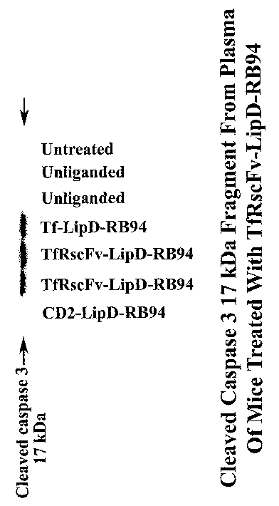
Induction of the 17 kDa Cleaved Caspase
3 Fracrment by Tumor Suppressor RB94-Detection
in Mouse Plasma
[0094] In vivo treatment with a different tumor
suppressor gene, RB94 also has been shown to induce
expression of the 17 kDa fragment of cleaved caspase 3, an
indicator of apoptosis. The retinoblastoma gene RB is a
tumor suppressor that encodes a nuclear phosphoprotein of
928 amino acids. The normal function of this 110-kDa protein
is to repress DNA transcription and prevent cell division,
thus inhibiting cell growth. (Li et.al., Cancer Research
62:4637-44, 2002 Xu et.al., PNAS 91:9837-41, 1994)
[0095] Gene replacement therapy using wild-type RB in
multiple types of human cancers could suppress or reduce
their tumorigenicity in vitro and in vivo. RB94 is a
truncated version of RB, lacking the 112 amino acids
residues at the NH2-terminal of the full length protein with
even greater efficacy than full length RB in suppressing
tumor growth. The RB94 protein was found to remain
hypophosphorylated longer than full length RB. Since it is
the un- or hypophosphorylated form that is responsible for
repression of cellular proliferation, this likely accounts
CA 02513769 2005-07-19
WO 2004/066946 PCT/US2004/002261
for the increased potency of RB94. It has also been
suggested that this N-terminal truncated RB protein also
could contribute to the cellular control of apoptosis/
survival (Tomei, L.D. in Apoptosis: the Molecular Basis of
Cell Death, pp 279-316, 1991). Thus, delivery and expression
of RB94 to tumor cells in,vivo could result in induction of
apoptosis. Detection of the 17 kDa fragment of cleaved
caspase 3 in the plasma of tumor-bearing mice treated with
RB94 would be indicative of ongoing apoptosis.
[0096] Female nude mice bearing subcutaneous xenograft
tumors of human bladder carcinoma cell line HTB-9 were i.v.
injected three times within 24 hours,with a complex (800
~.tl/injection) carrying the RB94 gene (40
~tg/mouse/injection). The complex also consisted of liposome
D (1:1 DOTAP:cholesterol) and as a ligand, either Tf itself
or the TfRscFv molecule. As controls, other mice were i.v.
injected with the complex without targeting ligand, or with
a non-tumor specific molecule (CDR) as the ligand. None of
these were expected to go to or affect the tumor. Sixteen
hours after the last injection the animals were sacrificed,
blood taken as plasma isolated as described in Example 2.
Western analysis of the expression of the 17 kDa fragment of
cleaved caspase 3 was performed as described in Examples 3
and 4. Forty ~.tg of protein were run per lane of a 4-200
polyacrylamide/SDS gel. As shown in Figure 13, the 17 kDa
cleaved caspase 3 protein was only evident in the plasma
from the mice receiving the RB94 complex that could target
and affect the tumors. Thus, the non-invasive detection in
plasma of the 17 kDa cleaved caspase 3 fragment, an
indicator of apoptosis, can serve as a general
pharmacodynamic marker of gene therapy. The method is
broadly applicable, not simply for p53.
36
CA 02513769 2005-07-19
WO 2004/066946 PCT/US2004/002261
EXAMPLE 14
_Detection of 17 kDa Cleaved Caspase 3 Fraament~in
Serum from Human Breast Cancer Patients After Thera~y
[0097 To establish that the results observed in the
animal model can be applied to humans and that the
expression of the 17 kDa cleaved caspase 3 fragment can be
used to non-invasively assess therapeutic effect, a matched
set of serum samples was obtained from two human patients
who had been treated for breast cancer using conventional
chemotherapy. These serum samples were obtained before
(pre-) and after (post-) treatment. The serum was purified
using the P6 (in Tris) Micro-Bio-Spin~ Chromotography
Columns. (Bio-Rad Laboratories, Hercules, CA)(Example 2).
The flow-through from the columns was diluted at a ratio of
serum to RIPA buffer of from 0.1:1, to 10:1, preferably at
1:1. Equal volumes (1 to 100 ~.al) were run on a 4-200
polyacrylamide/SDS gel, transferred and probed for
expression of the 17 kDa cleaved caspase 3 fragment as
described in Examples 3 and 4. As shown in Figure 14, the 17
kDa cleaved caspase 3 fragment is not evident in the serum
from either a control (non-cancer bearing) human subject or
the patients pre-treatment. This band is clearly present,
however, post-standard chemotherapy. Thus, as shown in the
animal model, the expression of the 17 kDa cleaved caspase 3
fragment, an indicator of apoptosis, does correlate with
cancer therapies (gene, antisense, and chemotherapy) in
human patients. Thus, for any therapy that induces
apoptosis, including radiation therapy, analysis of blood
(as serum or plasma) for the 17 kDa cleaved caspase 3
fragment, as described in the Examples contained in this
application, can be a relatively non-invasive method to
monitor the effectiveness of the therapy. In human cancer
patients it is envisioned that blood (1 ml to 3 ml) can be
37
CA 02513769 2005-07-19
WO 2004/066946 PCT/US2004/002261
drawn in heparinized tubes and centrifuged at 300 to 1000 x
g, at 4° to 27°C for 3 to 10 minutes to obtain plasma. This
plasma can be run directly (as described in Examples 3 and
4) or further purified by centrifugation of a 20-75 dal
aliquot of the sample through a P6 or P30 Micro Bio-Spin~
Chromatography Column (preferably P6) at 300 to 2000 x g
(preferably 1000 x g) for 1 to 10 minutes (preferably 4
minutes) at 4° to 27°C (preferably -18-24°C, most
preferably
20°C). The flow-through is diluted with RIPA buffer at a
ratio of plasma to RIPA of 0.1:1 to 10:1, preferably 1:1
before electrophoresis on a 4-20o polyacrylamide/SDS gel,
transferred to any nylon or nitrocellulose solid support
membrane, preferably Protran~ (S+S), with a pore size of 0.1
to 0.45 Vim, preferably 0.22 dam. Detection is performed
using a polyclonal or monoclonal anti-caspase 3 antibody
that detects the 17 kDa fragment, preferably only the 17 kDa
fragment, by radioactive or non-radioactive means,
preferably non-radioactive, preferably non-colorimetric,
preferably via chemiluminescence, preferably enhanced
chemiluminescence such as found in the ECL Western Blotting
detection reagents and analysis system (Amersham
Biosciences, Piscataway, NJ), with exposure to
autoradiography film including, but not limited to Hyperfilm
ECL, for times ranging from 30 seconds to 24 hours,
preferably 1 minute to 18 hours.
[0098] In another embodiment, serum, isolated from 1-3 ml
of whole blood by collection in non-heparinized tubes and
allowed to coagulate for 5-90 minutes, preferably 30-60
minutes, at 18-24°C, can be used to detect the presence of
the l7kDa fragment. The clotted sample is then centrifuged
at 0.01 to 1000 x g, preferably at 0.05 to 0.1 x g, most
preferably at 0.1 x g for 0.5 to 30 minutes, preferably for
5-15 minutes, most preferably for 10 minutes and the serum
removed. The serum can be analyzed directly as described in
38
CA 02513769 2005-07-19
WO 2004/066946 PCT/US2004/002261
Examples 3 and 4, or purified through the same columns and
analyzed by Western blot analysis in the same manner as
described above for plasma.
39