Note: Descriptions are shown in the official language in which they were submitted.
CA 02515203 2011-08-29
IMMUNOCOMPROMISED RODENTS AS DUAL COLOR TUMOR MODELS
Technical Field
[0002] This application relates to the production and use of transgenic
immtmocompromised rodents including athymic nude mice that visibly express
fluorescent
protein in multiple tissues while maintaining their immunocompromised state.
The rodents
may be used for whole body optical imaging of cells and tissues, including the
visualization
of tumors and metastases present in said rodents, in particular, tumors
provided fluorescent
proteins of alternative emission spectra.
Background Art
100031 Fluorescent proteins that emit light in the presence of stimulating
radiation in
the absence of substrate have been used as research tools for many years..
The. best .known
and initially used such protein is the green fluorescent protein (GFP)
isolated .from
Aequorea victoria, but a large number Of such proteins have been isolated from
other
sources or obtained synthetically which display a wide variety of emission
maxima so that
the historical term GFP has been used to describe proteins that appear in a
full spectrum of
visible color, including red, blue, and yellow. See, .e.g., Delagrave, S., et
al.,
BloTechnology (1995) 13:151-154; Heim, R., et al., Proc. Natl. Acad.. Sci. USA
(1994)
91:12501-12504. A number of organisms have been successfully modified to
express such
fluorescent proteins. These include Caenorhabditis elegans (Chalfie. M., et
at, Science
(1994) 263:802-805), Drosophila melanogaster (Wang, S., et al., Nature (1994)
369:400-403), zebrafish (Peters, KG., et at, Dev. Biol. (1995) 171:252-257;
Amsterdam, A., et al., Dev. Biol. (1995) 171:123-129), Dictyostelium and
Arabidopsis
thalicn7a (Sheen, J., et at., Plant .1. (1995) 8:777-784; Hu, W., FEBS Lett.
(1995) 369:331-334).
CA 02515203 2005-08-04
WO 2004/072098 PCT/US2004/003636
[0004] Okabe, et al., FEBS Lett. (1997) 407:313-319, have inserted the wild-
type GFP
into pCAGGS (containing the chicken beta-actin promoter and cytomegalovirus
enhancer,
beta-actin intron and bovine globin polyadenylation signal ¨ Niwa, H., Gene
(1991)
108:193-199) and produced transgenic mouse lines (Ikawa, M., FEBS Lett. (1995)
375:125-128; and Ikawa, M., Dev. Growth Dill (1995) 37:455-459). Although a
bright
green light emission was observed in the muscle and pancreas of more than 20
of these
transgenic mouse lines, GFP expression was not ubiquitous and light emission
was not
visible to the naked eye in other tissues. However, when a modified form
(EGFP) was used
in this expression system, the transgenic mice express the EGFP transgene in
the entire
body, from pre-implantation embryo to adult stages.
[0005] Wild-type eggs fertilized with green male sperm were not green at the 2-
cell
stage but subsequently became green after subsequent stages of embryogenesis.
Newborns
were green fluorescent. The blood vessels were classified as 'bright' in the
EGFP-bearing
lines. The hair of these animals was not green. Transgenic mice were uniformly
green
with the exception of hair and red blood cells. The brain, liver, kidney,
adrenal gland and
testis, lung, muscle, heart, intestine, and adipose tissue, thymus, spleen and
testicular cells
fluoresced green when irradiated with blue excitation light.
[0006] The transgenic mouse lines were normal despite a significant amount of
EGFP
expression; EGFP therefore is non-toxic.
[0007] One embodiment of the immunocompromised rodents exhibiting fluorescence
described in the invention is reported in Yang, M., et al., PrOC. Natl. Acad.
Sci. USA (2003)
100:14259-14262 (November 25th issue).
[0008] Citation of documents herein is not intended as an admission that any
is
pertinent prior art. All statements as to the date or representation as to the
contents of
documents is based on the information available to the applicant and does not
constitute
any admission as to the correctness of the dates or contents of the documents.
Disclosure of the Invention
[0009] For reasons that are not clear, transgenic immunocompromised rodents,
such as
athymic nu/nu mice are rare. The present invention provides for the production
of
fluorescent proteins in such immunocompromised rodents. The resulting rodents
are useful
for the imaging of cells and tissues in vivo, in particular as recipients of
transplanted cells
or tissues, such as tumors, which can be observed against the background of
GFP
2
CA 02515203 2011-08-29
expressing cells and tissues ;of theõtransgenic rodents. Thus, the
transplanted:cells or tissues
express visible indicator, such as, but nothmited to, another >fluorescent,
protein. The
transplanted cells or tissues may be observed by contrast against the
background of-GFP
= expressing cells and tissues of the transgenic rodents of the invention.
The transplanted
cells or tissues may comprise tumor cells or cells that are otherwise
cancerous such that
their growth properties and/or spread may be monitored.
[0010] The transgenic immunocompromised rodents may also be used to screen for
the
effect of various agents on interactions between the host rodent tissue and
the transplanted
cell or tissue. Examples of such agents include drugs or candidate drugs to
modulate host-
transplant interactions or modulate growth and spread of transplanted cells or
tissue.
[0011] The rodents may also be used as a source of GFP expressing cells or
tissues for
further study and/or transplantation into another animal or embryo.
Optionally, such
transplantation is serial in nature, and may be used to study aging as one
embodiment. The
transplanted cells may.include.embryonic E,11.d adult stem cells.
[0011A] Various embodiments of this invention provide a method to assay the
effects
of a drug on tumor-host interactions comprising: a) contacting a rodent with
said drug
wherein said rodent is an immunocompromised transgenic rodent which is
heterozygous
for expression of a first fluorescent protein in all tissues except hair and
erythrocytes-and
maintains an immunocompromised phenotype, wherein said transgenic rodent is
further
modified to contain a tumor that expresses a second fluorescent protein that
emits a
wavelength different from that of the first fluorescent protein, and b)
imaging tumor-host
cell interactions by observing emissions of said first and second fluorescent
proteins, and
c) comparing the resulting images to a rodent not contacted with said drug.
The rodent
may be prepared by a process which comprises first, crossing a rodent that
expresses said
first fluorescent protein by virtue of derivation from a fertilized egg
provided with a
transgenic expression system comprising a nucleotide sequence encoding said
first
fluorescent protein operatively linked to a promoter that effects said
expression in all said
tissues, which rodent is not immunocompromised with a rodent that does not
express the
fluorescent protein and is immunocompromised to produce Fl offspring, second,
crossing
those Fl offspring that express said first fluorescent protein in all tissues
except hair and
erythrocytes to obtain F2 offspring that express said first fluorescent
protein and are
immunocompromised; third, crossing those F2 offspring that express said first
fluorescent
3
CA 02515203 2011-08-29
protein in all tissues except hair and erythrocytes and are immunocompromised
with a rodent
that does not express said first fluorescent protein and is immunocompromised
to obtain F3
offspring that are heterozygous for said expression of said first fluorescent
protein and are
immunocompromised; modifying said F3 offspring to contain a tumor that
expresses a second
fluorescent protein that emits a wavelength different from that of the first
fluorescent protein,
thus producing said rodent.
100121 In one embodiment, the GFP expressing rodents are athymic nu/nit mice
obtained by first crossing a GFP expressing mouse with nu/nu mice. The Fl
generation is
then collected and crossed with each other to produce progeny including GFP
expressing
nu/nu mice. Male and female GFP expressing nu/nu mice from the Fl x Fl cross
are then
used to produce GFP expressing MOM mice progeny. These resultant mice may be
crossed
with nu/nu non-GFP expressing 17U/1711 mice to maintain GFP expressing nu/nu
mice.
[0013] Similar strategies are used to obtain other immunocompromised rodents,
such as
immunocompromised rats where immunocompromised strains are crossed with normal
strains that have been modified to express fluorescent protein.
[0014] Thus, in one aspect, the invention is directed an immunocompromised
transgenic rodent that expresses a first fluorescent protein in essentially
all tissues while
maintaining its immunocompromised phenotype: In a further embodiment, the
invention is
directed to this rodent transplanted with beterologous tissue, said tissue
modified to express
a second fluorescent protein with a different emission spectrum from the first
fluorescent
protein. In another aspect, the invention is directed to a transgenic rodent
that expresses a-
gene encoding a first fluorescent protein in essentially all tissues and which
has been
transplanted by heterologous tissue that expresses 'a second fluorescent
protein having a
different emission spectrum from said first fluorescent protein.
3a
CA 02515203 2005-08-04
WO 2004/072098 PCT/US2004/003636
[0015] In still another aspect, the invention is directed to a method to assay
the effects
of a drug on tumor host interactions by contacting a rodent that expresses a
first fluorescent
protein and which harbors tumor cells expressing a second fluorescent protein
of a different
color comprising contacting said rodent with a drug or protocol and observing
the effects
on the tumor cells contained in the host.
Brief Description of the Drawings
[0016] Figure 1 is a whole body image of orthotopically growing human colon
cancer
after implantation into an immunocompromised mouse. The tumor expresses red
fluorescent protein, while the host exhibits whole body expression of
fluorescent protein
that emits green light.
[0017] Figures 2A-2D show real-time interaction of macrophages in the
immunocompromised host that are labeled with green fluorescent protein wherein
the
cancer cells are labeled with red fluorescence. The figure shows initial
contact
(Figure 2A), engulfment (2B), the cancer cell engulfed in the macrophage (2C),
and the
cancer cell digested by the macrophage (2D).
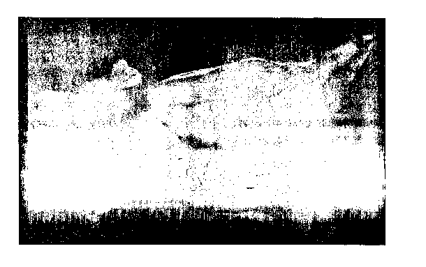
[0018] Figure 3 shows the transgenic mouse of the invention in contrast to a
non-
transgenic nude mouse.
Modes of Carrying Out the Invention
[0019] The invention relates to GFP expressing immunocompromised rodents such
as
athymic nu/nu mice as well as to methods for the preparation and use thereof.
The rodents
express GFP in essentially all tissues, preferably at levels such that light
emission is visible
to the naked eye. The rodents are otherwise immunocompromised ¨ e.g., athymic
nu/nu
mice, and may be used, for example, as hosts to accept transplants of human
tumor tissue
or other xenografts.
[0020] Using similar techniques to those described herein, other
immunocompromised
rodents, such as rats, expressing GFP can be obtained.
[0021] The term "GFP" may be used for convenience as an acronym not only for
fluorescent proteins that appear green, but in general for fluorescent
proteins of any color
that are capable of emitting light in response to incident exciting radiation.
It will be clear
from the context whether GFP is used in the generic sense or is used to
designate a protein
that actually admits green fluorescent light.
4
CA 02515203 2005-08-04
WO 2004/072098 PCT/US2004/003636
[0022] As used herein, "GFP" refers to a fluorescent protein of whatever
wavelength
emitted as well as "enhanced" forms of GFP and the Aequorea victoria green
fluorescent
protein. The description of transplanted cells or tissues labeled with a
visible indicator,
such as fluorescent dyes and as generally known in the art, is selected such
that a different
color fluorescent protein is used in the transplanted cells or tissues in
comparison to the
fluorescent protein expressed in the host. As a non-limiting example, if green
GFP is
expressed in the transgenic rodents of the invention, then the transplanted
cells or tissues
may express red fluorescent protein (RFP).
[0023] Moreover, and in the case of transplanting multiple cells or tissues,
labeling
with different colors is provided by the instant invention to permit the cells
or tissues to be
visualized and/or followed simultaneously. Non-limiting examples of other
fluorescent
colors include yellow, blue, and far-red. The expression of other fluorescent
indicators
may optionally be specific to individual cell types, genes or processes. Non-
limiting
examples of how to provide such specificity include by operably linking
sequences
encoding the fluorescent indicators to be under the regulatory control of a
promoter that is
cell specific, a promoter that is responsive to particular activation events,
a promoter that
regulates the expression of a particular gene of interest, and a promoter that
regulates the
expression of a gene product involved in a cell process of interest.
[0024] The immunocompromised rodents may also be used as a source of cells
and/or
tissues that express GFP. Non-limiting examples of such tissues include an
embryo or
embryo tissues; stem cells, and cells or tissues of the brain, liver, kidney,
adrenal gland,
testis (including testicular cells), lung, muscle, heart, intestine, ovary and
spleen as well as
adipose tissue.
[0025] The transplantation of tissues modified to contain fluorescent protein
with a
different emission spectrum from that of the host can be practiced to a
limited extent with
immunocompetent subjects as well. In order to practice this aspect of the
invention in
immunocompetent subjects, the transplanted tissue must be syngeneic or the
observations
must be limited to short term exploration of an immune response or other
response,
including rejection of the transplant.
[0026] Turning again to the transgenic, GFP-expressing, immunocompromised
rodents
of the invention, these may be used to visualize gene expression in the manner
taught by
Yang, et al. ("Visualizing Gene Expression by Whole-Body Fluorescence
Imaging." Proc.
Natl. Acad. Sci. USA (2000) 97:12278-12282), which describes visualization, by
CA 02515203 2005-08-04
WO 2004/072098 PCT/US2004/003636
noninvasive techniques, transgene expression in intact animals. That system
permits rapid
visualization of transgene expression in major organs of intact live mice
which is simple,
rapid, and eminently affordable. Against the background of the GFP transgenic,
a
fluorescent protein of different color is expressed in the cells such as those
of brain, liver,
pancreas, prostate, and bone, and its fluorescence is encoded in whole-body
optical images.
As non-limiting examples, higher-magnification imaging may be performed with a
trans-
illuminated epifluorescence dissecting microscope while low-magnification
imaging may
be performed atop a fluorescence light box and directly viewed with a
thermoelectrically
cooled color charge-coupled device camera, or using simpler LED-based devices.
[0027] The fluorescent transgenic rodents may be provided with the expression
system
to be tested by directly injecting expressible vector-borne nucleic acid
encoding fluorescent
. protein (such as, but not limited to 8 x 10¹⁰ plaque-forming units/ml of
adenoviral
expression system encoding fluorescent protein in 20-100 p.1 PBS and 10%
glycerol) into a
tissue such as the brain, liver, pancreas, prostate, or bone marrow. Within 5-
8 h after
injection, the fluorescence of the expressed GFP in tissues like the brain
becomes visible,
and whole-body images are recorded at video rates. The GFP fluorescence
continues to
increase for at least 12 h and remains detectable in tissues like the liver
for up to 4 months.
Real-time recordings may be made without requiring either exogenous contrast
agents
(where the vector encoded GFP is not the same as the GFP expressed in rodent
tissues),
radioactive substrates, or long processing times. This method requires only
that the
expressed coding sequence or promoter to be tested be fused or operatively
linked to the
vector borne GFP to allow the study of the therapeutic and diagnostic
potential of suitably
tagged genes in relatively opaque organisms or, as here, in fluorescent
rodents.
[0028] In another aspect, transgenic rodents may also be imaged and used in
the
manner taught by Yang, et al. ("Whole-Body Optical Imaging of Green
Fluorescent
Protein-Expressing Tumors and Metastases," PrOC. Natl. Acad. Sci. USA (2000)
97:1206-1211) which describes a whole-body optical imaging system. Such a
system
affords unprecedented continuous visual monitoring of cell growth and spread,
including
that exhibited by transplanted (and optionally cancerous) cells or tissues, in
this case
labeled with expressed GFP of a different hue within intact animals.
[0029] In preferred embodiments of the invention, the fluorescent rodents
contain
transplanted fluorescent tumors growing and metastasizing in the live rodents.
Non-
6
CA 02515203 2005-08-04
WO 2004/072098 PCT/US2004/003636
limiting examples of such tumors include human and rodent tumors that stably
express very
high levels of a GFP as described by Yang, et al. (supra). Tumors that express
a GFP other
than the GFP expressed in the rodent host are used. As indicated above, the
immunocompromised fluorescent rodents of the invention are preferred for this
method
resulting in dual labeling of the host that can be maintained over long
periods of time;
however, for short-teun studies or studies using syngeneic tumors, such as
rodent tumors
transplanted into rodents, even immunocompetent transgenic fluorescent rodents
may be
used.
[0030] As a non-limiting example, B16FO-GFP mouse melanoma cells are injected
into
the tail vein or portal vein of 6-week-old fluorescent immunocompromised
rodents.
Whole-body optical images are used to show metastatic lesions such as those
that may
develop in the brain, liver, and bone. The B16F0-GFP cells are readily
visualized to
provide real time, quantitative measurement of tumor growth in each of these
organs.
[0031] In another non-limiting example, AC3488-GFP human colon cancer may be
surgically implanted orthotopically. Whole-body optical images may be used to
show, in
real time, growth of the primary colon tumor and its metastatic lesions in the
liver and
skeleton.
[0032] The invention also provides for methods of using the fluorescent,
immunocompromised rodents to identify, or screen for, modulators of cancer
growth. In
one embodiment, the methods may be used to identify inhibition by potential
chemotherapeutic agents. Thus, the model of tumor progression in the
fluorescent
immunocompromised rodents of the invention is established by implantation,
preferably
orthoµtopically, of cancer cells or intact portions of a tumor that expresses
a fluorescent
protein with an emission of different wavelength from that of the background
provided by
the transgenic host. Once the model is established, the candidate
chemotherapeutic agents
or protocols are administered to the host and the effect on tumor progression
and metastasis
is directly observed.
[0033] Inununocompromised fluorescent rodents may also be used to image
angiogenesis in the manner taught by Yang, et al. ("Whole-Body and Intravital
Optical
Imaging of Angiogenesis in Orthotopically Implanted Tumors," PrOC. Natl, Acad.
Sci. USA
(2001) 98:2616-2621). The instant invention thus also provides for methods of
assaying
for tumor-induced vascularization. These methods are an adaptation of the
orthotopic
implantation model for angiogenesis measurement by using tumors labeled with a
GFP for
7
CA 02515203 2005-08-04
WO 2004/072098 PCT/US2004/003636
grafting into fluorescent rodents: The use of a GFP emitting a different color
from that of
the host GFP-expressing capillaries of the host to be clearly visible against
the tumor
fluorescence as examined either intravitally or by whole-body luminence in
real time. This
is preferably practiced with human tumors to permit intravital images of
orthotopically
implanted human pancreatic tumors to show angiogenic capillaries at both
primary and
metastatic sites, in the immunocompromised labeled hosts of the invention. A
quantitative
time course of angiogenesis may be determined for an orthotopically growing
human
prostate tumor periodically imaged intravitally in a single rodent over a 19-
day period.
[0034] Whole-body optical imaging of tumor angiogenesis may be demonstrated,
for
example, by injecting fluorescent Lewis lung carcinoma cells into the
subcutaneous site of
the footpad. The footpad is relatively transparent, with comparatively few
resident blood
vessels, and thus allows quantitative imaging of tumor angiogenesis (such as
by increases
in capillary density) in the intact animal.
[0035] In an alternative embodiment, the GFP expressing human breast tumor MDA-
MB-435 may be orthotopically transplanted to the fat pad which is then imaged
to detect
changes, particularly increases, in blood vessel density linearly over an
extended period,
such as up to or beyond 20-week period. Such powerful and clinically relevant
angiogenesis nude mouse models may also be used for real-time in vivo
evaluation of
agents inhibiting or promoting tumor angiogenesis in physiological
microenvironments, as
described above, by observing the effect of the agent on angiogenesis.
[0036] In yet another aspect, the rodents of the invention may be used in a
manner
analogous to that described by Yang, et al. ("Direct External Imaging of
Nascent Cancer,
Tumor Progression, Angiogenesis, and Metastasis on Internal Organs in the
Fluorescent
Orthotopic Model," Proc. Natl. Acad. Sci. USA (2002) 99:3824-3829) to overcome
limits
on the sensitivity of external imaging due to light scattering by intervening
tissue, most
especially skin. The invention thus provides for opening a skin-flap in the
light path to
markedly reduce signal attenuation and increase detection sensitivity many-
fold. The
observable depth of tissue is greatly increased and many tumors that were
previously
hidden become clearly observable.
[0037] The skin flap can be reversibly opened and closed. Typically, after
anesthetizing the animal, an arc-shaped incision is made in the skin and
subcutaneous
connective tissue is separated to free the skin flap. The flap can be closed
by suturing. The
8
CA 02515203 2005-08-04
WO 2004/072098 PCT/US2004/003636
invention thus provides for observations made on the internal organs of a
tumor model
system.
[0038] The ability to observe, directly through the opened skin flap, the
labeled tumor
cells greatly enhances the sensitivity and resolution of the model system of
the invention.
The model can be used simply to monitor the progress of the condition or can
be used as a
means to evaluate potential therapeutics, as well as to evaluate effects which
may result in
more negative outcomes than no treatment at all. In this instance, a compound
and/or
protocol is supplied to test animals and compared to controls where the
compound and/or
protocol are not present. Enhancement of tumor progression, angiogenesis
and/or
metastasis in the presence of these experimental conditions indicates that the
compound
and/or protocol is deleterious to the subject; similarly, inhibition of any of
these features
identifies the compound and/or protocol as a potential therapeutic. .
[0039] In one embodiment, single tumor cells, expressing fluorescent protein,
are
seeded on the brain image through a scalp skin-flap. Lung tumor microfoci
representing a
few cells are viewed through a skin-flap over the chest wall, while
contralateral
micrometastases are imaged through the corresponding skin-flap. Pancreatic
tumors and
their angiogenic microvessels are imaged by means of a peritoneal wall skin-
flap. A skin-
flap over the liver allows imaging of physiologically relevant micrometastases
originating
in an orthotopically implanted tumor. Single tumor cells on the liver arising
from
intraportal injection are also detectable. Cells or tissues expressing two
different GFP's,
such as host tissues versus transplanted tissues or two transplanted tissues,
may also be
visualized by the use of a skin flap. Particularly preferred is the use of a
lower-abdominal
skin-flap to visualize tissues of the prostate or the surrounding area.
[0040] Methods for providing cells or tissues for transplant with a GFP are
known.
Tumor cells may be provided with an expression system for one or more
fluorescent
proteins using standard methods. The cells may be transduced in vitro and
grown into
tumors in vitro or in vivo and the resulting tumors transplanted in to the
model subject. The
cells may be injected or may be transplanted surgically. Surgical orthotopic
transplantation
is preferred when a model of tumor progression is desired. However, other
methods of
providing the model with modified tumor cells that stably express the
fluorescent protein
may also be used. In addition, rodents of the invention that bear an
endogenous tumor or
introduced tumor may be provided with a viral vector, particularly a
retroviral vector, for
expression of a GFP protein of emission different from that of the host by
infecting the
9
CA 02515203 2005-08-04
WO 2004/072098 PCT/US2004/003636
tumor already present in the animal. This is especially relevant with respect
to models for
tumor-susceptible mammals such as the "oncomouse" described in U.S. patent
4,736,866.
This vector is preferably introduced locally and directly to the already
present tumor.
[0041] Generally, any model of tumor progression, angiogenesis, and/or
metastasis
which relies for observation on the emission of fluorescence may be used with
fluorescent
rodents and methods of the invention.
[0042] In many instances, a single color is used to observe the metastasis of
a single
tumor. However, the method of the invention includes simultaneous observation
of two or
more tumors each labeled with a different color of fluorescent protein. By
utilizing this
method, not only is it possible to obtain multiple observations of multiple
tumor
progressions, the effects or interferences of each tumor on the other can be
observed
directly.
[0043] The methods of the invention have a number of advantages. First,
enhanced
sensitivity permits observation of only a single or two transplanted cells
against a
background of fluorescent host cells and tissues. Second, angiogenesis is
directly
observable, which is extremely important in evaluating therapeutic efficacy of
proposed
compounds and protocols. Third, it is possible to observe multiple
transplanted tissues
(such as tumors) simultaneously. This is especially important because of the
phenomenon
of interference between disparate tissues or tumors. Since multiple different
colors can be
used, the interaction of separate tissues or tumors can be observed directly.
Fourth,
because it is possible to make observations over substantial periods of time
in the
inummocompromised fluorescent rodents, distinctions can be made between cells
that are
actively proliferating and dormant cells. Thus, the presence of dormant cells
can be
determined by the method of the invention.
[0044] In a further aspect, the fluorescent hosts may be injected with
detectable,
preferably visible, compounds for generating images against the GFP background
of the
nude mouse tissues. As a non-limiting example, a visible indicator, such as a
dye, may be
injected into the bloodstream of a mouse of the invention to visualize all or
part of the
circulatory system. This is particularly appropriate where the red blood cells
of the rodent
do not express visible GFP, leaving the bloodstream without detectable light
emissions.
[0045] The following examples are provided to illustrate the invention and are
not
limiting.
CA 02515203 2005-08-04
WO 2004/072098 PCT/US2004/003636
Example 1
Production of GFP expressing nude mice
[0046] C-57 B6-GFP mice (Okabe, M., et al., FEBS Lett (1997) 467:313-319) were
crossed with 1711/1111 mice. The C-57/B6-GFP mice were obtained from the
Research
Institute for Microbial Diseases at Osaka University. These mice expressed A.
victoria
GFP under control of the chicken beta-actin promoter and CMV enhancer. All
tissues
except erythrocytes and hair fluoresced green under excitation light.
[0047] The F 1 generation was collected and crossed with each other. Two rare
GFP
7111/1111 mice, one male and one female, were obtained. The GFP nu/nu male and
GFP nu/nu
female were crossed with each other and produced a litter of 6 GFP nu/nu mice.
[0048] A male GFP nu/nu mouse was crossed with a female nu/nu non-GFP mouse.
Nine nude nu/nu offspring were produced. All expressed fluorescence in the
tissues
generally. See Figure 3, which contrasts non-GFP nu/nu to GFP 1214/7711 mice.
Example 2
Preparation of Tumors With Red Fluorescent Protein
[0049] Red fluorescent protein (RFP), DsRed2 (Clontech) was inserted into
pLNCX2
(Clontech) at the EglI and NotI sites.
[0050] Saturating amounts of the resulting vector, pLNCX2-DsRed2 were
incubated for
18 h with a precipitated mixture of DOTAP reagent (Boehringer Mannheim) and
PT67
packaging cells at 70% confluence. PT67 cells are an NIH 3T3-derived packaging
cell line
expressing the 10 Al viral envelope, and were cultured in DMEM (Irvine
Scientific)
supplemented with 10% heat-inactivated FBS. Fresh medium was replenished at
this time,
and cells were examined by fluorescence microscopy 48 h posttransfection. For
selection
of brightly fluorescing cells producing high-titer retroviral supernatants,
the RFP-
expressing packaging cells were cultured in the presence of 500-2,000m/m1 G418
increased in a stepwise manner (Life Technologies, Grand Island, NY) for 7
days.
[0051] Desired tumor cell lines at 20% confluence were incubated with a 1:1
precipitated mixture of retroviral supernatants of PT67 cells and RPMI 1640 or
other
culture media (GIBCO) containing 10% PBS (Gemini Biological Products) for 72
h. Fresh
medium was replenished at this time. Tumor cells were harvested with
trypsin/EDTA and
subcultured at a ratio of 1:15 into selective medium, which contained 50
jig/m1 G418. To
11
CA 02515203 2005-08-04
WO 2004/072098 PCT/US2004/003636
select brightly fluorescent cells, the level of G418 was increased to 800
p.g/m1 in a stepwise
manner. Clones expressing RFP were isolated with cloning cylinders (Bel-Art
Products)
by trypsin/EDTA and were amplified and transferred by conventional culture
methods in
the absence of selective agent.
[0052] The tumor cells which were thus obtained as red fluorescent cells
included:
rodent B16F0 melanoma cells;
mouse MMT060562 mammary tumor cells;
mouse Dunning prostate carcinoma cells;
human PC-3 prostate carcinoma cells; and
human HCT-116 colon cancer cells.
Example 3
=
Dual-Labeled Model of Tumor Progression
[0053] HCT-116-RFP ¨ i.e, human colon cancer cells labeled with red
fluorescent
protein, were harvested by trypsinization, washed three times with cold serum-
free medium
and re-suspended with serum-free RPMI medium 1640. The cells were injected
within
40 minutes of harvesting into 6-week-old transgenic female GFP nude mice, as
prepared in
Example 1, by exposure of the colon through a lower-left abdominal incision.
106 HCT-116-RFP cells in 50 pl were injected under the serosa of the
descending colon
using a 25 pi syringe. The incision in the abdominal wall was closed with a 6-
0 surgical
suture in one layer. The animals were kept under ketamine anesthesia during
surgery.
[0054] Whole-body imaging was performed in a fluorescent light box illuminated
by
fiber-optic lighting at 470 nm (Lightools Research, Encinitas, CA). Emitted
fluorescence
was collected through a long-pass filter GG475 (Chroma Technology,
Brattleboro, VT) on
a Hamamatsu C5810 three-chip cooled color CCD camera (Hamamatsu Photonics,
Bridgewater, NJ.) High-resolution images of 1,024/724 pixels were captured
directly on an
IBM PC, Images were processed for contrast and brightness and analyzed with
the use of
Image Pro Plus 3.1 software (Media Cybernetics, Silver Spring, MD).
[0055] Figure 1 shows a whole-body image of the orthotopically growing
HCT-116-RFP human colon cancer 10 weeks after the implantation. The image was
acquired in a fluorescent light box with a CCD camera. As shown, this system
readily
distinguishes tumor from the host.
12
CA 02515203 2005-08-04
WO 2004/072098 PCT/US2004/003636
Example 4
Interaction of Macrophages with Prostate Tumor
[0056] PC-3-RFP,i.e., human prostate cancer cells labeled with red
fluorescent protein
were harvested by trypsinization, washed 3 times with cold serum-containing
medium, and
kept on ice. Within 40 minutes, 106 cells in 30 ill were injected into bladder
and prostate of
the immunocompromised mice obtained in Example 1 as follows: Bladder and
prostate
were exposed after a lower midline abdominal incision; after injection, the
incision was
closed with a 6-0 surgical suture, and the animals kept under isoflurane
anesthesia.
[0057] For observation, fresh tissue was cut into approximately 1 mm3 pieces
and
pressed on slides for fluorescence microscopy. In the microscopic
visualization, an
Olympus BH 2-RFCA fluorescence microscope equipped with a mercury 100-W lamp
power supply was used to visualize both GFP and RFP fluorescence at the 'same
time.
Excitation light was produced through a D425/60 bind pass filter, 470 DCXR
dichroic
mirror. Emitted fluorescence light was collected through a long pass filter
GG475 (Chroma
Technology). High-resolution images of 1,024/724 pixels were captured by a
Hamamatsu
C5810 three-chip cooled color CCR camera (Hamamatsu Photonics) and directly
stored on
an IBM PC. Images were processed for contrast and brightness and analyzed with
the use
of Image Pro Plus 4.0 software (Media Cybernetics).
[0058] The interaction of macrophages, which fluoresce green by virtue of the
transgenic nude mouse, are shown interacting with the tumor cells in Figure 2.
This picture
was taken 35 days after implantation. Panel A shows host GFP macrophage
contacting
RFP cancer cells; Panel B shows the GFP macrophage engulfing the RFP cancer
cell;
Panel C shows an RFP cancer cell engulfed by the GFP macrophage and Panel D
shows the
ultimate digestion of the RFP cancer cell by macrophage.
Example 5
Studies in Fluorescent Immunocompetent Mice
[0059] Short term studies using dual-color imaging can be conducted on
immunocompetent subjects when syngeneic transplants are employed. In the
murine model
described by Okabe, C57/B6-GFP mice were produced. Studies were conducted with
106
RFP expressing mouse B16F0 melanoma cells, 106 RFP-expressing mouse MMT060562
mammary tumor cells and 106 RFP-expressing Dunning (rat) prostate cancer
cells. Using
13
CA 02515203 2005-08-04
WO 2004/072098 PCT/US2004/003636
these techniques, angiogenesis in live tumor tissue could be observed 3 weeks
after
injection of Bl6F10-RFP melanoma cells and interaction of host dendritic cells
and tumor
cells in fresh tumor tissue was observed as well. Lymphocyte infiltration was
observed
with the breast cancer model
[00601 Thus, dual-color images of early events in tumor angiogenesis could be
observed as well as interactions of the immune system with the transplanted
tumor.
14