Note: Descriptions are shown in the official language in which they were submitted.
CA 02515916 2005-08-12
WO 2004/076635 PCT/US2004/005453
TITLE OF THE INVENTION
THERAPEUTIC APPLICATIONS OF THROMBOMODULIN GENE
VIA VIRAL AND NON-VIRAL VECTORS
This application claims priority from U.S. Provisional Application Serial
No. 60/449,408 filed February 25, 2003. The entirety of that provisional
application is incorporated herein by reference.
Field of the Invention
The present invention is directed to methods and compositions of the
treatment of thrombotic diseases and, in particular, to the treatment of
atherosclerotic cardiovascular disease, pulmonary hypertension, acute
inflammatory diseases, end-stage renal failure disease, and Ahheimer disease
by
modulating expression of the thrombomodulin gene.
BACI~GROU1~TD OF THE INVENTION
Thrombomodulin (TM) is an integral membrane glycoprotein expressed on
the surface of endothelial cells. It is a high affinity thr~mbin receptor that
converts
thrombin into a protein C activator. Activated protein C then functions as an
anticoagulant by inactivating two regulatory proteins of the clotting system,
namely factors Va and VI[Ila. The latter two proteins are essential for the
function
of two of the coagulation proteases, namely factors IXa and Xa. TM, thus,
plays an
active role in blood clot formation ifa vivo and can function as a direct or
indirect
anticoagulant.
TM is a single chain protein composed of 5 distinct domains. A short
cytoplasmic domain containing a free cysteine is located at the COOH-terminal
end
and is joined by a membrane spanning region to an o-glycosylation rich domain.
The latter is followed by an epidermal growth factor (EGF) homology region and
the NH2-terminal hydrophobic domain. The EGF homology region contains 6
EGF lilee domains and contains the binding sites for both thrombin and protein
C.
CA 02515916 2005-08-12
WO 2004/076635 PCT/US2004/005453
TM is also prevalent in other cell types includes keratinocytes, osteoblasts,
macrophages. In these cells/tissues, TM is involved in the differentiation and
inflammation. Abnormal TM function is also associated with many diseases. For
example, abnormal TM in the endothelial cells contribute to myocardial
infarction
(MI), stroke and the development of atherosclerotic plaque. In other diseases,
natural TM is missing, deficient or simply cleaved into soluble form.
Therefore,
modulation of ih vivo TM expression is desirable in these clinical scenarios.
SUMMARY OF THE INVENTION
The present invention provides a method for treating a thrombotic disease
in a mannnal comprising administering to the mammal a therapeutically
effective
amount of a pharmaceutical composition comprising a viral or a non-viral
vector,
wherein the viral or non-viral vector comprises an isolated nucleotide
sequence
encoding thrombomodulin and its variant. The present invention also provides a
method for treating a thrombotic disease in a mammal comprising administering
to
the mannnal an effective amount of thrombomodulin-producing cells, wherein
said
thrombomodulin-producing cells are generated by introducing an isolated
polynucleotide encoding an amino acid sequence of thrombomodulin or its
variant
into cultured cells.
The vector- or cell-mediated ira viv~ TI~ gene expression may used for the
treatment of any thrombomodulin-related diseases, such as atherosclerotic
cardiovascular disease, pulmonary hypertension, acute inflammatory diseases,
end-
stage renal failure disease, or Alzheimer disease. The present invention
further
provides a vector carrying an isolated polynucleotide in which the vector is
introduced into a mammal to reduce the TM activity or TM gene expression ira
vivo.
BRIEF DESCRIPTION OF THE FIGURES
Figure 1 is a schematic drawing of an embodiment of the baclcbone shuttle
vector of the present invention.
Figure 2 is the DNA sequence (SEQ ID NO: 1) of the gutless backbone
shuttle vector.
_2_
CA 02515916 2005-08-12
WO 2004/076635 PCT/US2004/005453
Figure 3 is the full length amino acid sequence (SEQ ID N0:2) of human
thrombomodulin.
Figure 4 is the full length DNA sequence (SEQ ID N0:3) encoding human
thrombomodulin.
Figure 5 is the DNA sequence (SEQ ID N0:4) of the expression cassette
encoding human thrombomodulin.
Figure 6 is the DNA sequence (SEQ ID NO:S) of the CMV promoter of the
expression cassette encoding the human thrombomodulin.
Figure 7 is the cDNA (SEQ ID N0:6) of the human thrombomodulin gene.
DETAILED DESCRIPTION OF THE INVENTION
The primary objective of the present invention is to provide methods and
compositions for treating diseases or conditions relating to the TM
expression.
One aspect of the invention relates to the treatment for diseases or
conditions
associated with reduced TM expression or loss of TM activity. These diseases
may
be treated by expressing a therapeutically effective amount of the TM protein
iaa
viv~ uS111g a viral or a non-viral vector. Another aspect of the invention
relates to
the treatment for diseases associated with enhanced TM expression. Under these
conditions, TM gene express or TM activity may be inhibited by the in viv~
expression of a TM inhibitouy polynucleotide using a gene expression vector.
~0 The practice of the present invention will employ, unless other wise
indicated, conventional methods of histology, virology, microbiology,
immunology, and molecular biology within the skill of the art. such techniques
are
explained fully in the literature. All publications, patents and patent
applications
cited herein, whether supra or infra, are hereby incorporated by reference in
their
entirety.
Definitions
In describing the present invention, the following teens will be employed,
and are intended to be defined as indicated below.
"Gene transfer" or "gene delivery" refers to methods or systems for reliably
introducing a particular nucleotide sequence (e.g., DNA) into targeted cells.
The
-3-
CA 02515916 2005-08-12
WO 2004/076635 PCT/US2004/005453
introduced nucleotide sequences may persist in vivo in episomal forms or
integrate
into the genome of the target cells. Gene transfer provides a unique approach
for
the treatment of acquired and inherited diseases, and a number of systems have
been developed in the art for gene transfer into mammalian cells. See, e.g.,
U.S.
Pat. No.5,399,346.
As used herein, the term "therapeutically effective amount" refers to a level
of transfection which brings about at least partially a desired therapeutic or
prophylactic effect in an organ or tissue infected by the method of the
present
invention. The transfection with a therapeutically effective amount of the
vector
carrying genetic material of interest can then result in the modification of
the
cellular activities, e.g., a change in phenotype, in an organ or a tissue that
has been
infected by the method of the present invention. In a preferred embodiment,
the
transfection with an effective amount of the vector carrying genetic material
of
interest results in modulation of cellular activity in a sigxlificant number
of cells of
an infected organ or a tissue.
A gene transfer "vector" refers to any agent, such as a plasmid, phage,
transposon, cosmid, chromosome, liposome, DNA-viral conjugates, RNA/DNA
oligonucleotides, virus, bacteria, etc., which is capable of transferring gene
' sequences into cells. Thus, the term includes cloning and expression
vehicles
including "naked" expression vectors, as well as viral and non-viral vectors.
A
vector may be targeted to specific cells by linking a target molecule to the
vector.
A targeting molecule is any agent that is specific for a cell or tissue type
of interest,
including for example, a ligand, antibody, sugar, receptor, or other binding
molecule. The invention is also intended to include such other forms of
vectors
which serve equivalent functions and which become lcnown in the art
subsequently
hereto.
The term "expression control element" or "regulatory element" refers
collectively to promoter sequences, polyadenylation signals, transcription
termination sequences, upstream regulatory domains, origins of replication,
internal
ribosome entry sites ("IRES"), enhancers, and the like, which collectively
provide
-4-
CA 02515916 2005-08-12
WO 2004/076635 PCT/US2004/005453
for the replication, transcription and translation of a coding sequence in a
recipient
cell. Not all of these control sequences need always be present so long as the
selected coding sequence is capable of being replicated, transcribed and
translated
in an appropriate host cell.
The term "promoter" is used herein in its ordinary sense to refer to a, DNA
regulatory sequence that are sufficient for RNA polymerase recognition,
binding
and transcription initiation. Additionally, a promoter includes sequences that
modulate the recognition, binding and transcription initiation activity of RNA
polymerase. Such sequences may be cis acting or may be responsive to trans
acting
factors. Depending upon the nature of the regulation, promoters may be
constitutive or regulated. Examples of promoters are SP6, T4, T7, SV40 early
promoter, cytomegalovirus (CMV) promoter, mouse mammary tumor virus
(MMTV) steroid-inducible promoter, Moloney marine leukemia virus (MMLV)
promoter, phosphoglycerate kinase (P(aI~) promoter, muscle creatine kinase
(MCI~) promoter, myosin promoter, a-actin promoter and the like.
The term "transduction" denotes the delivery of a DNA molecule to a
recipient cell either ira vivo or i~z vitro, via a replication-defective viral
vector, such
as via a recombinant AAV virus.
"~perably linked" refers to an arrangement of elements wherein the
components so described are configured so as to perform their usual function.
Thus, control elements operably linl~ed to a coding sequence are capable of
effecting the expression of the coding sequence. The control elements need not
be
contiguous with the coding sequence, so long as the function to direct the
expression thereof. Thus, for example, intervening untranslated yet
transcribed
sequences can be present between a promoter sequence and the coding sequence
and the promoter sequence can still be considered "operably lined" to the
coding
sequence.
The term "native thrombomodulin" refers to both the natural protein and
soluble peptides having the same characteristic biological activity of
membrane-
-5-
CA 02515916 2005-08-12
WO 2004/076635 PCT/US2004/005453
bound or detergent solubilized (natural) thrombomodulin. These soluble
peptides
are also referred to as "wild-type" or "non-mutant" analog peptides.
Biological
activity is the ability to act as a receptor for thrombin, increase the
activation of
protein G, or other biological activity associated with native thrombomodulin.
Oxidation resistant TM analogs are these soluble peptides that in addition to
being
soluble contain a specific artificially induced mutation in their amino acid
sequence.
"Thrombotic disease" refers to a pathogenic condition in a mammal
characterized by the formation of one or more thrombi that are or can be
detrimental to the health of the mammal. Examples of the thrombotic diseases
include, but are not limited to, atherosclerotic cardiovascular disease,
pulmonary
hypertension, acute inflammatory disease, end-stage renal failure disease,
Alzheimer disease, acute coronary syndrome, myocardial infarction, unstable
angina, refractory angina, occlusive coronary thrombus occurring post-
thrombolytic therapy or post-coronary angioplasty, a thro111botlcally mediated
cerebrovascular syndrome, embolic strolce, thrombotic strobe, transient
ischemic
attaclcs, venous thrombosis, deep venous thrombosis, pulmonary embolus,
coagulopathy, disseminated intravascular coagulation, thrombotic
thrombocytopenic purpura, thromboangiitis obliterans, thrombotic disease
?0 associated with heparin-induced thrombocytopenia, thrombotic complications
associated with extracorporeal circulation, thrombotic complications
associated
with instuumentation such as cardiac or other intravascular catheterization,
intra-
aortic balloon pump, coronary stmt or cardiac valve.
The term "thrombomodulin variant" is a polypeptide that differs from a
native thrombomodulin polypeptide in one or more substitutions, deletions,
additions and/or insertions, such that the bioactivity of the native
thrombomodulin
polypeptide is not substantially diminished or enhanced. In other words, the
bioactivity of a thrombomodulin variant may be enhanced or diminished by, less
than 50%, and preferably less than 20%, relative to the native protein.
Preferred
variants include those in which one or more portions, such as an N-terminal
leader
-6-
CA 02515916 2005-08-12
WO 2004/076635 PCT/US2004/005453
sequence or transmembrane domain, have been removed. Other preferred variants
include variants in which a small portion (e.g., 1-30 amino acids, preferably
5-15
amino acids) has been removed from the - and/or C-terminal of the mature
protein.
Preferably, a thrombomodulin variant contains conservative substitutions.
A "conservative substitution" is one in which an amino acid is substituted for
another amino acid that has similar properties, such that one skilled in the
art of
peptide chemistry would expect the secondary structure and hydropathic nature
of
the polypeptide to be substantially unchanged. Amino acid substitutions may
generally be made on the basis of similarity in polarity, charge, solubility,
hydrophobicity, hydrophilicity and/or the amphipathic nature of the residues.
For
example, negatively charged amino acids include aspartic acid and glutamic
acid;
positively charged amino acids include lysine and arginine; and amino acids
with
uncharged polar head groups having similar hydrophilicity values include
leucine,
isoleucine and valine; glycine and alanine; asparagine and glutamine; and
satins,
threonine, phenylalanine and tyrosine. A variant may also, or alternatively,
contain
nonconservative changes. In a preferred embodiment, variant polypeptides
differ
from a native sequence by substitution, deletion or addition of five amino
acids or
fewer. Variants may also (or alternatively) be modified by, for example, the
deletion or addition of amino acids that have minimal influence on the
bioactivity,
secondary structure and hydropathic nature of the polypeptide.
Thrombomodulin variants preferably exhibit at least about 70%, more
preferably at least about 90% and most preferably at least about 95°/~
sequence
homology to the original thrombomodulin polypeptide.
A thrombomodulin variant also include a thrombomodulin polypeptides
that is modified from the original thrombomodulin polypeptides by either
natural
processes, such as posttranslational processing, or by chemical modification
techniques which are well known in the art. Such modifications are well
described
in basic texts and in more detailed monographs, as well as in a voluminous
research
literature. Modifications can occur anywhere in a polypeptide, including the
peptide backbone, the amino acid side-chains and the amino or carboxyl
termini. It
CA 02515916 2005-08-12
WO 2004/076635 PCT/US2004/005453
will be appreciated that the same type of modification may be present in the
same
or varying degrees at several sites in a given polypeptide. Also, a given
polypeptide may contain many types of modifications. Polypeptides may be
branched, for example, as a result of ubiquitination, and they may be cyclic,
with or
without branching. Cyclic, branched, and branched cyclic polypeptides may
result
from posttranslation natural processes or may be made by synthetic methods.
Modifications include acetylation, acylation, ADP-ribosylation, amidation,
covalent attachment of flavin, covalent attachment of a heme moiety, covalent
attachment of a nucleotide or nucleotide derivative, covalent attachment of a
lipid
or lipid derivative, covalent attachment of phosphotidylinositol, cross-
linking,
cyclization, disulfide bond formation, demethylati~n, formation of covalent
cross
links, formation of cysteine, formation of pyroglutasnate, formulation,
garrrnna-
carboxylation, glycosylation, GPI anchor formation, hydroxylation, iodination,
methylation, myristoylation, oxidation, pegylation, proteolytic processing,
phosphorylation, prenylation, racemization, selenoylation, sulfation, transfer-
I2NA
mediated addition of amino acids to proteins such as arginylation, and
ubiquitination.
The present invention also relates to fragments of thrombomodulin. A
fragment of thrombomodulin may comprise 5 to 575 consecutive amino acids of
thrombomodulin, preferably comprise 20 to 575 consecutive amino acids of
thrombomodulin, more preferably comprise 100 to 575 consecutive amino acids of
thrombomodulin, and most preferably comprise 200 to 575 consecutive amino
acids of thrombomodulin.
drz viva thrombomodulin gene Iransfer
The amino acid sequence of human thrombomodulin (SEQ ID N~: 2) and
the DNA sequence encoding human thrombomodulin (SEQ ID N0:3) have been
reported (Suzuki et al., EMBO J. 6:1891-1897, [1987]). Somatic gene transfer
techniques offer a new approach to replace a defective thrombomodulin gene or
to
modulate i~a vivo thrombomodulin gene expression. A preferred approach for
introducing genetic material encoding a gene product into an organ or a tissue
is by
_g_
CA 02515916 2005-08-12
WO 2004/076635 PCT/US2004/005453
use of a gene transfer vector. Commonly used gene transfer vectors include
viral
vectors and non-viral vectors. In the case of a viral vector, the genetic
material
encoding thrombomodulin or a thrombomodulin variant is inserted into the viral
genome (or a partial viral genome) using molecular cloning techniques well
known
in the art. The regulatory elements directing the expression of the
thrombomodulin
or thrombomodulin variant can be included with the genetic material inserted
into
the viral genome (i.e., operably linked to the gene inserted into the viral
genome) or
can be provided by the viral genome itself, for example, a retrovirus long
terminal
repeat (LTR) or an Adeno-associated virus (AAV) inverted terminal repeat
(ISR).
Transfection of cells with a viral vector has the advantage that molecules
encoded
within the viral vector, e.g., by a cDNA contained in the viral vector, are
expressed
efficiently in cells which have taken up viral vector nucleic acid and viral
vector
systems can be used ira vivo. Different viral vectors are described separately
in the
subsections below.
1. Aclera~viYUS vec~~i~s: The genome of an adenovirus can be manipulated
such that it encodes and expresses a gene product of interest but is
inactivated in
terms of its ability to replicate in a nonnal lyric viral life cycle (Curie,
Anna N Y
Acac~ ~'ci 886:158-171, [1991]). Suitable adenoviral vectors derived from the
adenovirus strain Ad type 5 d1324~ or other strains of adenoviuus (e.g., Ad2,
Ad3,
Ad7 etc.) are well known to those skilled in the art. Recombinant adenovirus
es are
advantageous in that they do not require dividing cells to be effective gene
delivery
vehicles and can be used to infect a wide variety of cell types, including
airway
epithelium, endothelial cells and muscle cells. Additionally, introduced
adenoviral
DNA (and foreign DNA contained therein) is not integrated into the genome of a
host cell but remains episomal, thereby avoiding potential problems that can
occur
as a result of insertional mutagenesis in situations where introduced DNA
becomes
integrated into the host genome (e.g., retroviral DNA). Moreover, the carrying
capacity of the adenoviral genome for foreign DNA is large (up to 8 kilobases)
relative to other gene delivery vectors (Haj-Ahmand et al., J. Yiy~ol. 57:267-
273,
[1986]). Most replication-defective adenoviral vectors currently in use are
deleted
-9-
CA 02515916 2005-08-12
WO 2004/076635 PCT/US2004/005453
for all or parts of the viral El and E3 genes but retain as much as 80% of the
adenoviral genetic material.
Adenovirus vectors have been successfully tested in a number of animal
models (Ragot et al., Nature 361:647-650, [1993]; Howell et al., Husn Gene
Thef°
9:629-634, [1998]). Nonetheless, the toxicity and imrnunogenicity remain major
hurdles to overcome before the adenoviral vectors can be safely used in
humans.
Adenoviral vectors deleted of all viral coding regions (gutless adenoviral
vectors) are also described by Kochanek et al., and Chamberlain et al., (U.S.
Pat.
No. 5,985,846 and U.S. Pat. No. 6,083,750). A new viral backbone shuttle
vector
was also developed for the construction of gutless adenoviral vectors (IJ.S.
Patent
Application Serial No. 10/725,013, the entirety of which is incorporated
herein by
reference).
The viral backbone shuttle vector may contain a left and a right inverted
terminal repeats of adenovirus, an encapsidation signal (~e) of adenovirus, a
pER322 replication origin, a kanamycin resistance gene, and a stuffer
sequence,
which is the hypoxanthine phosphoribosyltransferase (HPRT) intro fragment with
an approximately 10 Kb. (Figure 1).
The "inverted terminal repeats (ITRs) of adenovirus" are short elements
located at the 5' and 3' ternzini of the linear Ad genorne~ respectively and
are
required for replication of the viral I~NA. The left ITR is located between 1-
130 by
in the Ad genome (also refereed to as 0-0.5 mu). The right ITR is located from
about 3,7500 by to the end of the genome (also referred to as 99.5-100 mu).
The
two ITRs are inverted repeats of each other. For clarity, the left ITR or 5'
end is
used to define the 5' and 3' ends of the ITRs. The 5' end of the left ITR is
located at
the extreme 5' end of the linear adenoviral genome; picturing the left ITR as
an
arrow extending from the 5' end of the genome, the tail of the 5' ITR is
located at
mu 0 and the head of the left ITR is located at about 0.5 mu (further the tail
of the
left ITR is referred to as the 5' end of the left ITR and the head of the left
ITR is
referred to as the 3' end of the left ITR). The tail of the right or 3' ITR is
located at
mu 100 and the head of the right ITR is located at about mu 99.5; the head of
the
-10-
CA 02515916 2005-08-12
WO 2004/076635 PCT/US2004/005453
right ITR is referred to as the 5' end of the right ITR and the tail of the
right ITR is
referred to as the 3' end of the right ITR. In the linear Ad genome, the ITRs
face
each other with the head of each ITR pointing inward toward the bulk of the
genome. When arranged in a "tail to tail orientation" the tails of each ITR
(which
comprise the 5' end of the left ITR and the 3' end of the right ITR) are
located in
proximity to one another while the heads of each ITR are separated and face
outward.
The "encapsidation signal of adenovirus" or "adenovirus packaging
sequence" refers to the yr sequence which comprises five (AI-AV) packaging
signals and is required for encapsidation of the mature linear genome; the
packaging signals are located from about 194 to 35~ by in the Ad genome (about
0.5-1.0 mu).
The viral backbone shuttle vector may contain multiple restriction
endonuclease sites for the insertion of a foreign DNA sequence of interest.
The
foreign DNA sequence of interest typically comprises cDNA or genomic fragments
that are of interest to transfer into mammalian cells. Foreign DNA sequence of
interest may include any naturally occurnng or synthetic DNA sequence. The
foreign DNA may be identical in sequence to naturally-occurring DNA or may be
mutated relative to the naturally occurring sequence. The foreign DNA need not
be
?0 characterized as to sequence or function.
The size of foreign DNA that may be included in the shuttle vector will
depend upon the size of the rest of the vector. If necessary, the HPRT introns
may
be removed to adapt large size foreign DNA fragment. The total size of foreign
DNA may vary from llcb to 351cb.
The foreign DNA may encode protein, or contain regulatory sites, including
but not limited to, transcription factor binding sites, promoters, eWancers,
silencers, ribosome binding sequences, recombination sites, origins of
replication,
sequences which regulate RNA stability and polyadenylation signals. The
promoters used may vary in their nature, origin and properties. The choice of
promoter depends in fact on the desired use and on the gene of interest, in
-11-
CA 02515916 2005-08-12
WO 2004/076635 PCT/US2004/005453
particular. Thus, the promoter may be constitutive or regulated, strong or
weak,
ubiquitous or tissue/cell-specific, or even specific of physiological or
pathophysiological states (activity dependent on the state of cell
differentiation or
the step in the cell cycle). The promoter may be of eukaryotic, prokaryotic,
viral,
animal, plant, artificial or human, etc., origin. Specific examples of
promoters are
the promoters of the genes PGK, TK, GH, a-EF1, APO, CMV, etc. or artificial
promoters, such as those for p53, E2F or CAMP.
2. Adefao-associated viruses (AA V) vectors: AAV is a naturally occurnng
defective virus that requires another virus, such as an adenovirus or a herpes
virus,
as a helper virus for efficient replication and a productive life cycle
(Muzyczka et
al., Curr. T~pics ih Micr~. arad Inayrtun~l. 158:97-129, [1992]). AAV vector
is the
only viral vector system that is based on a non-pathogenic and replication
defective
virus. It is also one of the few viruses that may integrate its DNA into non-
dividing cells, and exhibits a high frequency of stable integration (Flotte et
al.,
Arn. .I Respif°. Cell. M~l. ~i~l. 7:349-356, [1992]; Samulski et al.,
.J:: Tirol.
63:3822-3828, [1989]). Vectors containing as little as 300 base pairs of AAV
DNA can be packages.
AAV vectors have been successfully used to establish efficient and long-
term gene expression iaa viv~ in a variety of tissues without significant
immune
response or toxicity (Niao et al., ~: fir°~l. 70:8098-108, [1996];
Kessler et al., Proc
Natl Acad Sci USA 93, 14082-7, [1996]; Xiao et al., .I Tlirol72:10222-6,
[1989]).
Unlike other viral vectors, AAV readily bypasses extracellular barriers due to
its
small viral particle size (2Q nM) that facilitates efficient transduction of
muscle
myofibers of various maturity (Pruchnic et al., Huns Gene Ther, 11:521-36,
[2000]). However, a major obstacle for AAV vectors is the limited paclcaging
size
that only allows for genes smaller than 4.7 kb (Song et al., Proc Natl Acad
Sci
USA 95:14384-8, [1998]; Kay et al., Nat Geraet 24:257-261, [2000]), therefore
precludes such large gene as dystroplun with a CANA of 14 kb.
3. Herpes simplex virus (HSV) vectors: The main feature of an HSV vector
is that it has very large packaging capacity, is usually replication
defective, and
-12-
CA 02515916 2005-08-12
WO 2004/076635 PCT/US2004/005453
does not integrate into the host genome. HSV infects cells of the nervous
system
(Fink et al., Ayafau Rev NeuYOSCi 19:265-287, [1996]). The virus contains more
than
80 genes, one of which (IE3) can be replaced to create the vector. The
generation
of HSV vectors with deletions in many of the immediate early gene products has
resulted in vectors with reduced toxicity and antigenicity, as well as
prolonged
expression in vivo. However, these modifications also result in a lower virus
yield.
Construction of HSV vectors is described in U.S. Pat. No. 5,661,033.
4. Retr~ovi~us vector°s: Defective retroviruses are well characterized
for use
in gene transfer for gene therapy purposes (Miller BZ~od 76:271-278, [1990]).
The
members of the family Retroviridae are characterized by the presence of
reverse
transcriptase in their virions. There are several genera included within this
family,
including Cistemavirus A, Oncovirus A, Oncovirus B, Oncovirus C, Oncovirus D,
Lentivirus, and Spumavirus.
A recombinant retrovirus can be constructed having a nucleic acid encoding
a gene product of interest inseuted into the retroviral genome. Additionally,
portions of the retroviral genome can be removed to render the retrovirus
replication defective. The replication defective retrovirus is then packaged
into
virions which can be used to infect a target cell through the use of a helper
virus by
standard techniques. Protocols for producing recombinant retrovinases and for
infecting cells ira vitf~~ or rya. vivo with such viruses can be found in
"Current
Protocols in Molecular Biology, Ausubel, et al., (eds.) Cireene Publishing
Associates, (1989), Sections 9.10-9.14" and other standard laboratory manuals.
Examples of suitable retroviruses include pLJ, PZIP, pWE and pEM which are
well
known to those skilled in the art. Examples of suitable packaging virus cell
lines
include psi.Crip, psi.Cre, psi.2 and psi.Am. Retroviruses have been used to
introduce a variety of genes into many different cell types, including
epithelial
cells, endothelial cells, lymphocytes, myoblasts, hepatocytes, hematopoietic
stem
cells, in vitro, and/or in vivo (U. S. Pat. No. 4,868,116; U.S. Pat. No.
5,449,614
and U.S. Pat. No. 6,207,455). Retroviral vectors require target cell division
in
order to be integrated into the host genome to stable introduce nucleic acid
into the
-13-
CA 02515916 2005-08-12
WO 2004/076635 PCT/US2004/005453
cell. Thus, it may be necessary to stimulate replication of the target cell.
Successful transduction of hematopoietic stem or progenitor cells with
retroviral
vectors in an ex vivo setting have been reported. However, Recombinant
retroviral
vectors can only accorninodate about 8 kb to 10 kb of foreign DNA, and this
packaging capacity limits its use.
5. Lerativirus vectors: Lentivirus also belong to the retrovirus family, but
they can infect both dividing and non-dividing cells. The best-known
lentivirus is
the human immunodeficiency virus (HIV), which has been disabled and developed
as a vector for in vivo gene delivery. Like the simple retroviruses, HIV has
the
three gag, pol ahd env genes, but it also carnes genes for six accessory
proteins
ten-ned tat, ~~ev, vpr, vpu, fief af~d vif. Using the retrovirus vectors as a
model,
lentivirus vectors have been made, with the transgene enclosed between the
LTRs
and a packaging sequence (Naldu et al., Scieface 272:263=267, [1996]). Some of
the accessory proteins can be eliminated without affecting production of the
vector
or efficiency of transfection.
When lentiviral vectors are injected into rodent brain, liver, muscle, or
pancreatic islet cells, they give sustained expression for over six months.
Little is
known about the possible immune problems associated with lentiviral vectors.
Furthermore, there seems to be no potent antibody response. A major concern
about lentiviral vector is its safety in human applications. However, recent
development in producing the third generation lentiviral vectors with more
deletion
in viral genes and improved safety may allow for the general application of
lentiviral vectors to if2 vivo gene therapy.
~ther viral vector systems that may have application in the subj ect
invention have been derived from vaccinia virus (Chen et al., J.
Inafnuv~other~ 24:46-
57, [2001]), and several RNA viruses. The plus-strand RNA viridae, such as
polio
(Bledsoe et al., IVat Biotechfaol. 18:964-9, [2000]), hepatitis A (Romano G.
Stefra
Cells; 18:19-39, [2000]), and sindbis virus (Wahlfors et al., Geyae Ther 7:472-
80,
[2000]) are being developed for high-level gene expression, following either
viral
infection or delivery of nucleic acids using a non-viral system. These viruses
-14-
CA 02515916 2005-08-12
WO 2004/076635 PCT/US2004/005453
express a replicase protein that can specifically replicate the viral RNA. By
inserting a transgene in place of the viral capsid gene(s), it is possible to
generate a
chimeric RNA that replicates autonomously in the cell and expresses a high
level
of protein from the plus-coding strand of RNA. These viral vectors are well
suited
for immunization strategies in which high, transient gene expression is needed
to
induce an immune response to the transduced cells.
In addition to the viral gene transfer vectors, powerful non-viral gene
transfer vectors have also become available for clinical application in the
past
several years (Ropert et al., Braz JMed Biol Res. 32:163-9, [ 1999]; Lee et
al., CYit
Rev Ther-17f°ug Carriey~ Syst 14:173-206, [1997]). These vectors rely
on normal
mechanisms used by mammalian cells for the uptake and intracellular transport
of
macromolecules to deliver genetic materials into cells. Commonly used non-
vector
include cationic and other liposomes.
Liposomes are fomnulated based on the requirement of the delivery system
in a particular application. The characteristics of liposomes, such as size
and
composition, can be modified during the preparation of the liposomes.
Typically, liposomes are prepared by dissolving one or more lipids in an
organic solvent. The solvent is evaporated under controlled conditions
resulting in
a uniform, thin lipid layer of lipid mix in the evaporating flask. Phosphate
buffered
saline or water is added to the dried lipid mix layer in the evaporating flask
and is
sonicated briefly to form a liposome suspension. The preparation is
dehydrated,
rehydrated and stored at 4.°C.
The lipids may be natural, synthetic or semisynthetic (i.e., modified
natural). Lipids useful in the invention include, and are not limited to,
fatty acids,
lysolipids, oils (including safflower, soybean and peanut oil),
phosphatidylcholine
with both saturated and unsaturated lipids. The lipids also include cationic
lipids
and synthetic cationic lipids. The lipids may also include derivatized lipids,
including common natural lipids derivatized to contain one or more basic
functional groups. Additionally lipid moieties capable of polymerization may
be
used as coatings for the liposomes. Examples of these include, but are not
limited
-15-
CA 02515916 2005-08-12
WO 2004/076635 PCT/US2004/005453
to, alkenyl and alkynyl moieties, such as oleyl and linoleyl groups,
diacetylene,
acryloyl and methacryloyli groups with or without polar groups to enhance
water
solubility.
Other non-viral vectors include DNA-viral conjugates, RNA/DNA
oligonucleotides and naked DNA molecules. Physical procedures, such as
hydrodynamics-based and electroporation-based procedures, have been used to
improve gene transfer efficiency of some non-viral vectors (Zhang et al., Gene
Ther 7:1344-9, [2000]; Yamasluta et al., Cancef° Res. 61:1005 -12,
[2001]).
Recently, it was also reported that intraperitoneal injection of a [i-
galactosidase
fused to the protein transduction domain from the human immunodeficiency viuus
TAT protein resulted in delivery of the fusion protein to all tissues in mice
(Schwarze et al., Science, 3:1569-1572, [1999]).
In vitro expression of thrombomodulin or a thrombomodulin variant may
also be achieved with traditional transfection methods such as calcium
phosphate
precipitation, DEAF-dextron transfection, and electroporation.
Another aspect of the invention pertains to the expression of
thrombomodulin or a thrombomodulin variant using a regulatable expression
system. Systems to regulate expression of therapeutic genes have been
developed
and incorporated into the current viral and non-viral gene delivery vectorse
These
systems are briefly described below:
Tet-~nl~ff system. The Tet-system is based on two regulatory elements
derived from the tetracycline-resistance operon of the E. c~li Tn 10
transposon: the
tet repressor protein (TetR) and the Tet operator DNA sequence (tetO) to which
TetR binds. The system consists of two components, a "regulator" and a
"reporter"
plasmid. The "regulator" plasmid encodes a hybrid protein containing a mutated
Tet repression (tetr) fused to the VP 16 activation domain of herpes simplex
virus.
The "reporter" plasmid contains a tet-responsive element (TRE), which controls
the
"reporter" gene of choice. The tetr-VP16 fusion protein can only bind to the
TRE,
therefore activate the transcription of the "reporter" gene, in the presence
of
tetracycline. The system has been incorporated into a number of viral vectors
-16-
CA 02515916 2005-08-12
WO 2004/076635 PCT/US2004/005453
including retrovirus, adenovirus and AAV (Gossen and Bujard, Proc. Natl. Acad.
Sci. USA 89:5547-5551, [1992]; Gossen et al., Science 268:1766-1769, [1995];
Kistner et al., Proc. Natl. Acad. Sci. USA. 93:10933-10938, [1996]).
Ecd~sorae system. The Ecdysone system is based on the molting induction
system fouaid in D~osoplaila, but modified for inducible expression in
mammalian
cells. The system uses an analog of the drosophila steroid hormone ecdysone,
muristerone A, to activate expression of the gene of interest via a
heterodimeric
nuclear receptor. Expression levels have been reported to exceed 200-fold over
basal levels with no effect on mammalian cell physiology (No et al., P~oc.
Natl.
Acad. Sci. USA 93:3346-3351, [1996]).
Ps°og~estea°orae-system. The progesterone receptor is normally
stimulated to
bind to a specific DNA sequence and to activate transcription through an
interaction with its hormone ligand. Conversely, the progesterone antagonist
mifepuistone (RU486) is able to block hormone-induced nuclear transport and
subsequent DNA binding. A mutant form of the progesterone receptor that can be
stimulated to bind through an interaction with RU486 has been generated. To
generate a specific, regulatable transcription factor, the RU486-binding
domain of
the progesterone receptor has been fused to the DNA-binding domain of the
yeast
transcription factor GAL4~ and the transactivation domain of the I~SV protein
VP16. The chimeric factor is inactive in the absence of RU486. The addition of
hormone, however, induces a conformational change in the chimeric protein, and
this change allows binding to a GAL4-binding site and the activation of
transcription from promoters containing the GAL4-binding site (Wang et al.,
Pooc.
Natl. Acad. Sci. USA 93:8180-8184, [1994]; Wang et al., Nat. Eioteclz 15:239-
243, [1997]).
Rapamycin-system. Immunosuppressive agents, such as FK506 and
rapamycin, act by binding to specific cellular proteins and facilitating their
dimerization. For example, the binding of rapamycin to FK506-binding protein
(FKBP) results in its heterodimerization with another rapamycin binding
protein
FRAP, which can be reversed by removal of the drug. The ability to bring two
-17-
CA 02515916 2005-08-12
WO 2004/076635 PCT/US2004/005453
proteins together by addition of a drug potentiates the regulation of a number
of
biological processes, including transcription. A chimeric DNA-binding domain
has been fused to the FKBP, which enables binding of the fusion protein to a
specific DNA-binding sequence. A transcriptional activation domain also has
been
used to FRAP. When these two fusion proteins are co-expressed in the same
cell, a
fully functional transcription factor can be formed by heterodimerization
mediated
by addition of rapamycin. The dimerized chimeric transcription factor can then
bind to a synthetic promoter sequence containing copies of the synthetic DNA-
binding sequence. This system has been successfully integrated into adenoidal
and
AAV vectors. Long-term regulatable gene expression has been aclueved in both
mice and baboons (Magari et al., J. Clip. IfZVest. 100: 2865-2872, [1997]; Ye
et al.,
Scieh.ce 283:88-91, [1999]).
Another aspect of the invention pertains to isolated polynucleotide
molecules., which may be used to reduce or to eliminate a thrombomodulin or a
thrombomodulin variant. ~ne method of reducing or eliminating T1~I gene
expression is to introduce an antisense TTeiI construct into a mammal.
An "antisense" polynucleotide comprises a nucleotide sequence, which is
complementary to a "sense" polynucleotide encoding a protein, e.g.,
complementary to the coding strand of a double-stranded cDI~TA molecule or
complementary to an mI~NA sequence. Accordingly, an antisense polynucleotide
can hydrogen bond to a sense polynucleotide. The antisense polynucleotide can
be
complementary to an entire coding strand of a gene of the invention or to only
a
portion thereof. In one embodiment, an antisense polynucleotide molecule is
antisense to a "coding region" of the coding strand of a nucleotide sequence
of the
invention. The term "coding region" includes the region of the nucleotide
sequence
comprising codons, which are translated into amino acid. In another
embodiment,
the antisense polynucleotide molecule is antisense to a "noncoding region" of
the
coding strand of a nucleotide sequence of the invention.
Antisense polynucleotides of the present invention can be designed
-18-
CA 02515916 2005-08-12
WO 2004/076635 PCT/US2004/005453
according to the rules of Watson and Crick base pairing. The antisense
polynucleotide molecule can be complementary to the entire coding region of an
mRNA corresponding to a gene of the invention, but more preferably is an
oligonucleotide, which is antisense to only a portion of the coding or
noncoding
region. An antisense oligonucleotide can be, for example, about 5, 10, 15, 20,
25,
30, 35, 40, 45 or 50 nucleotides in length. An antisense polynucleotide of the
invention can be constructed using chemical synthesis and enzymatic ligation
reactions using procedures known in the axt. For example, an antisense
polynucleotide (e.g., an antisense oligonucleotide) can be chemically
synthesized
using naturally occurring nucleotides or variously modified nucleotides
designed to
increase the biological stability of the molecules or to increase the physical
stability
of the duplex formed between the antisense and sense polynucleotides, e.g.,
phosphorothioate derivatives and acridine substituted nucleotides can be used.
Examples of modified nucleotides which can be used to generate the antisense
polynucleotide include 5-fluorouracil, 5-bromouracil, 5-chlorouracil, 5-
iodouracil,
hypoxanthine, xantine, 4-acetylcytosine, 5-(carboxyhydroxyhnethyl) uracil, 5-
carboxymethylaminomethyl-2-thiouridine, 5-carboxymethylaminomethyluracil,
dihydrouracil, beta-D-galactosylqueosine, inosine, N6-isopentenyladenine, 1-
methylguanine, 1-methylinosine, 2,2-dimethylguanine, 2-methyladenine, 2-
methylguanine, 3-methylcytosine, 5-methylcytosine, N6-adenine, 7-
methylguanine, 5-methylaminomethyluracil, 5-methoxyaminomethyl2-thiouracil,
beta-D-mannosylqueosine, 5'-methoxycarbox3nnethyluracil, 5-methoxyuracil, 2-
methylthio-N6-isopentenyladen4exine, unacil-5-oxyacetic acid (v),
wybutoxosine,
pseudouracil, queosine, 2-thiocytosine, 5-methyl-2-thin-aracil, 2-thiouracil,
4-
thiouracil, 5-methyluracil, uracil-5-oxyacetic acid methylester, uracil-5-
oxyacetic
acid (v), 5-methyl-2-thiouracil, 3-(3-amino-3-N-2-carboxypropyl) uracil,
(acp3)w,
and 2,6-diaminopurine. Alternatively, the antisense polynucleotide can be
produced biologically using an expression Vector into which a polynucleotide
has
been subcloned in an antisense orientation (i.e., RNA transcribed from the
inserted
-19-
CA 02515916 2005-08-12
WO 2004/076635 PCT/US2004/005453
polynucleotide will be of an antisense orientation to a target polynucleotide
of
interest, described further in the following subsection).
The antisense polynucleotide molecules of the present invention are
typically administered to a mammal or generated ih situ such that they
hybridize
S with or bind to cellular mRNA and/or genomic DNA encoding a thrombomodulin
or a thrombomodulin variant to thereby inhibit expression of the protein,
e.g., by
inhibiting transcription and/or translation. The hybridization can be by
conventional nucleotide complementarity to form a stable duplex, or, for
example,
in the cases of an antisense polynucleotide molecule which binds to DNA
duplexes,
through specific interactions in the major groove of the double helix. An
example
of a route of administration of antisense polynucleotide molecules of the
invention
is direct injection at a tissue site (e.g., intestine or blood).
Alternatively, antisense
polynucleotide molecules can be modified to target selected cells and then
administered systemically. The antisense polynucleotide molecules can also be
delivered to cells using the vectors described herein. To achieve sufficient
intracellular concentrations of the antisense molecules, vector constructs it
which
the antisense polynucleotide molecule is placed under the control of a strong
promoter are preferred.
E~~pression of the TI~I gene can also be inhibited using RhTA interferon ce
("RNA,"). I~NAi is a phenomenon of the introduction of double-stranded RNA
(dsRNA) into certain organisms and cell types causes degradation of the
homologous mRNA.
RNAi was first discovered in the nematode G'czeyao~°laabelitis
ele~aras, and it
has since been found to operate in a wide range of organisms. In recent years,
hNAi has becomes an endogenous, efficient, and potent gene-specific silencing
technique that uses double-stranded RNAs (dsRNA) to mark a particular
transcript
for degradation isa vivo. RNA; technology is disclosed, for example, in U.S.
Patent
No. 5,919,619 and PCT Publication Nos. W099/14346 and WO01/29058.
Briefly, dsRNAs 21-25 nucleotides long, called short interfering RNAs
(siRNA), are introduced into the cell. SiRNAs may also be produced
-20-
CA 02515916 2005-08-12
WO 2004/076635 PCT/US2004/005453
endogenously by degradation of long dsRNA molecules by an RNAse III-related
nuclease called Dicer. Once formed, the siRNAs assemble with protein
components into an RNA-induced silencing complex (RISC). An ATP-generated
unwinding of the siRNA activates the RISC, which in turn targets the
homologous
mRNA transcript by Watson-Crick base-pairing and cleaves the mRNA. This
sequence specific degradation of mRNA results in gene silencing.
Gene transfer vectors can be delivered to a mammal by, for example,
intravenous administration, intraportal administration, intrabiliary
administration,
infra-arterial administration, direct injection into the liver parenchyma (see
U.S.
Patent 6,328,958), by intramusclular injection (see U.S. Patent 6,335,011), by
inhalation (see U.S. Patent 6,344,194), by perfusion (U.S. Patent 6,342,214)
or by
stereotactic injection (see e.g., Chen et al., Pf°oc. Ncztl. Acad. Sci.
USA 91:3054-
3057, [1994]). The pharmaceutical preparation of the gene therapy vector can
include the gene therapy vector in an acceptable diluent, or can comprise a
slow
release matrix in which the geiae delivery vehicle is imbedded. Alternatively,
where the complete gene delivery vector can be produced intact from
recombinant
cells, e.g., retroviral vectors, the pharmaceutical preparation can include
one or
more cells which produce the gene delivery system.
The invention is further directed to pharmaceutical compositions
compuising a gene transfer vector described hereinabove and a pharmaceutically
acceptable carrier.
As used herein the language "pharmaceutically acceptable carrier" is
intended to include any and all solvents, solubilizers, fillers, stabilizers,
binders,
absorbents, bases, buffering agents, lubricants, controlled release vehicles,
diluents,
emulsifying agents, humectants, lubricants, dispersion media, coatings,
antibacterial or antifungal agents, isotonic and absorption delaying agents,
Ind the
like, compatible with pharmaceutical administration. The use of such media and
agents for pharmaceutically active substances is well-known in the art. [See
e.g.,
A.H. Kibbe Handbook of Pharmaceutical Excipients, 3rd ed. Pharmaceutical Press
London, UK (2000)]. Except insofar as any conventional media or agent is
-21-
CA 02515916 2005-08-12
WO 2004/076635 PCT/US2004/005453
incompatible with the active compound, use thereof in the compositions is
contemplated. Supplementary agents can also be incorporated into the
compositions.
The invention includes methods for preparing pharmaceutical compositions
for modulating the expression or activity of thrombomodulin. Such methods
comprise formulating a pharmaceutically acceptable carrier with a gene
transfer
vector capable of modulating expression or activity of thrombomodulin. Such
compositions can further include additional active agents. Thus, the invention
further includes methods for preparing a pharmaceutical composition by
formulating a pharmaceutically acceptable carrier with a gene transfer vector
capable of modulating expression or activity of thrombomodulin and one or more
additional bioactive agents.
A pharmaceutical composition of the invention is formulated to be
compatible with its intended route of administration. Examples of routes of
administration include paa-enteral, e.g., intravenous, intradermal,
subcutaneous, oral
(e.g., inhalation), transderlnal (topical), transmucosal, and rectal
administration.
Solutions or suspensions used for parenteral, intradermal, or subcutaneous
application can include the following components: a sterile diluent such as
water
for injection, saline solution, fixed oils, polyethylene glycols, glycerine;
propylene
glycol or other synthetic solvents; antibacterial agents such as ben~yl
alcohol or
methyl parabens; antioxidants such as ascorbic acid or sodium bisulfate;
chelating
agents such as ethylene-diarninetetracetic acid; buffers such as acetates,
citrates or
phosphates and agents for the adjustment of tonicity such as sodium chloride
or
dextrose. pH can be adjusted with acids or bases, such as hydrochloric acid or
'?5 sodium hydroxide. The parenteral preparation can be enclosed in ampoules,
disposable syringes or multiple dose vials made of glass or plastic.
Pharmaceutical compositions suitable for injectable use include sterile
aqueous solutions (where water soluble) or dispersions and sterile powders for
the
extemporaneous preparation of sterile injectable solutions or dispersion. For
intravenous administration, suitable carriers include physiological saline,
CA 02515916 2005-08-12
WO 2004/076635 PCT/US2004/005453
bacteriostatic water, Cremophor ELTM (BASF, Parsippany, NJ) or phosphate
buffered saline (PBS). In all cases, the injectable composition should be
sterile and
should be fluid to the extent that easy syringability exists. It must be
stable under
the conditions of manufacture and storage and must be preserved against the
S contaminating action of microorganisms such as bacteria and fungi. The
carrier
can be a solvent or dispersion medium containing, for example, water, ethanol,
polyol (for example, glycerol, propylene glycol, and liquid polyethylene
glycol,
and the like), and suitable mixtures thereof. The proper fluidity can be
maintained,
for example, by the use of a coating such as lecithin, by the maintenance of
the
requited particle size in the case of dispersion and by the use of
surfactants.
Prevention of the action of microorganisms can be achieved by various
antibacterial and antifungal agents, for example, parabens, chlorobutanol,
phenol,
ascorbic acid, thimerosal, and the like. In many cases, it will be preferable
to
include isotonic agents, for example, sugars, polyalcohols such as mannitol,
sorbitol, sodium chloride in the composition. Prolonged absorption of the
injectable compositions can be brought about by including in the composition
an
agent which delays absorption, for example, aluminum monostearate and gelatin.
Sterile injectable solutions can be prepared by incorporating the active
ingredient (e.g.9 a viral or non viral vector) in the required amount in an
appropriate
solvent with one or a combinati~n of ingredients enumerated above, as
required,
followed by filtered sterilization. Generally, dispersions are prepared by
incorporating the active ingredient into a sterile vehicle which contains a
basic
dispersion medium and the required other ingredients from those enumerated
above. In the case of sterile powders far the preparation of sterile
injectable
solutions, the preferred methods of preparation are vacuum drying and fieeze
drying which yields a powder of the active ingredient plus any additional
desired
ingredient from a previously sterile-filtered solution thereof.
Oral compositions generally include an inert diluent or an edible carrier.
They can be enclosed in gelatin capsules or compressed into tablets. For the
purpose of oral therapeutic administration, the active ingredient can be
-23-
CA 02515916 2005-08-12
WO 2004/076635 PCT/US2004/005453
incorporated with excipients and used in the form of tablets, troclies, or
capsules.
Pharmaceutically compatible binding agents, and/or adjuvant materials can be
included as part of the composition. The tablets, pills, capsules; troches and
the
like can contain any of the following ingredients, or compounds of a similar
nature:
a~binder such as microcrystalline cellulose, gum tragacanth or gelatin; an
excipient
such as starch or lactose, a disintegrating agent such as alginic acid,
Primogel, or
corn starch; a lubricant such as magnesium stearate or Stertes; a glidant such
as
colloidal silicon dioxide; a sweetening agent such as sucrose or saccharin; or
a
flavoring agent such as peppermint, methyl salicylate, or orange flavoring.
For administration by inhalation, the compounds are delivered in the form
of an aerosol spray from pressured container or dispenser which contains a
suitable
propellant, e.g., a gas such as carbon dioxide, or a nebulizer.
Systemic administration can also be by transmucosal or transdermal meals.
For transmucosal or transdermal administration, penetrans appropriate to the
barrier to be permeated are used in the formulation. Such penetrans are
generally
known in the art, and include, for example, for transmucosal administration,
detergents, bile salts, and fusidic acid derivatives. Transmucosal
administration
can be accomplished through the use of nasal sprays or suppositories. For
transdermal administration, the bioactive ingredient are fonnulated into
ointanents,
salves, gels, or creams as generally known in the art.
The composition can also be prepared in the form of suppositories (e.g.,
with conventional suppository bases such as cocoa butter and other glycerides)
or
retention enemas for rectal delivery.
It is especially advantageous to formulate oral or parenteral compositions in
dosage unit form for ease of administration and uniformity of dosage. Dosage
unit
form as used herein includes physically discrete units suited as unitary
dosages for
a mammal to be treated; each unit containing a predetermined quantity of
active
compound calculated to produce the desired therapeutic effect in association
with
the required pharmaceutical Garner. The specification for the dosage unit
forms of
the invention are dictated by and directly dependent on the unique
characteristics of
-24-
CA 02515916 2005-08-12
WO 2004/076635 PCT/US2004/005453
the active ingredient and the particular therapeutic effect to be achieved,
and the
limitations inherent in the art of compounding such an active ingredient for
the
treatment of individuals.
Toxicity and therapeutic efficacy of such ingredient can be determined by
standard pharmaceutical procedures in cell cultures or experimental animals,
e.g.,
for determining the LD50 (the dose lethal to 50% of the population) and the
ED50
(the dose therapeutically effective in 50% of the population). The dose ratio
between toxic and therapeutic effects is the therapeutic index and it can be
expressed as the ratio LD50/ED50. Compounds which exhibit large therapeutic
indices are preferred. While compounds that exhibit toxic side effects may be
used, care should be taken to design a delivery system that targets such
compounds
to the site of affected tissue in order to minimize potential damage to
uninfected
cells and, thereby, reduce side effects.
The data obtained from the cell culture assays and animal studies can be
used in formulating a range of dosage for use in humans. The dosage of such
compounds lies preferably within a range of circulating concentrations that
include
the ED50 with little or no toxicity. The dosage may vary within this range
depending upon the dosage form employed and the route of administration
utilized.
p°or any compound used in the method of the invention, the
therapeutically
effective dose can be estimated initially from cell culture assays. A dose may
be
formulated in animal models to achieve a circulating plasma concentration
arrange
that includes the IC50 (i.e., the concentration of the test compound which
achieves
a half maximal inhibition of symptoms) as detennined in cell culture. Such
information can be used to more accurately determine useful doses in humans.
Levels in plasma may be measured, for example, by high performance liquid
chromatography. The pharmaceutical compositions can be included in a
container,
pack, or dispenser together with instructions for administration.
In one embodiment, the in vivo expression of thrombomodulin or a
thrombomodulin variant is used for the treatment of atherosclerotic
cardiovascular
disease (CVD). Though venous grafts can be used for bypass surgeries, the
veins
-25-
CA 02515916 2005-08-12
WO 2004/076635 PCT/US2004/005453
eventually, become occluded by thrombosis resulting the recurrence of the
diseases. TM gene delivery can be used in coronary artery bypass grafting,
percutaneous transluminal coronary angioplasty, peripheral artery angioplasty
or
thrombectomy, intravascular stenting and vascular graft prostheses to block
thrombosis. TM gene delivery can be also used for the reduction of non-intima
formation, for the prevention of atherosclerosis; for the prevention of
myocardial
infarction and for the inhibition of Fbrinolysis in hemophilic plasma. TM gene
transfer at the site of thrombus; formation is potent approach to reverse
these
vascular diseases.
In another embodiment, the in vivo expression of thrombomodulin or a
thrombomodulin variant is used for the treatment of pulmonary hypertension.
Reduction of TM levels cause altered homeostasis in pulmonary hypertension.
Therefore, in vivo TM expression can be used to correct this disease state.
In another embodiment, the in viv~ expression of thrombomodulin, or a
thrombomodulin variant is used for the treatment of end-stage renal failure
disease.
(ESRI~). ESRI? patients often exhibit decreased antithrombotic activity due to
low
TM levels. In such patients, enhanced is2 vivo TM gene expression can be
potentially very useful.
In yet another en ~bodiment, the ifa viv~ expression of thrombomodulin or a
thrombomodulin variant is used for the treatment of acute inflannnatory
diseases
such as Sepsis. In sepsis, liver participates in host defense and tissue
repair
through hepatic cross talk that controls coagulation and inflammatory
processes. In
the absence of this control, it can lead to bacterial spill over, enhanced
procoagulant and inflarmnatory process. This can result in multiple organ
failure
and death. TM can be used to hock septic shock induced by variety of bacterial
and other infections.
In yet another embodiment, the ifz vivo expression of thrombomodulin or a
thrombomodulin variant is used for the treatment of Alzheimer's disease (AD).
Studies have shown that vascular risk factors are also involved in early
Alzheimer's
-26-
CA 02515916 2005-08-12
WO 2004/076635 PCT/US2004/005453
disease. Thus TM gene transfer can be also useful in reversing, inhibiting AD
progression.
In another embodiment, the ih vivo expression of thrombomodulin
inhibitory polynucleotide is used for the treatment of the diseases/conditions
relating to an overexpression of thrombomodulin.
The present invention is further illustrated by the following examples which
should not be construed as limiting. The contents of all references, patents
and
published patent applications cited throughout this application, as well as
the
Figures and Tables are incorporated herein by reference.
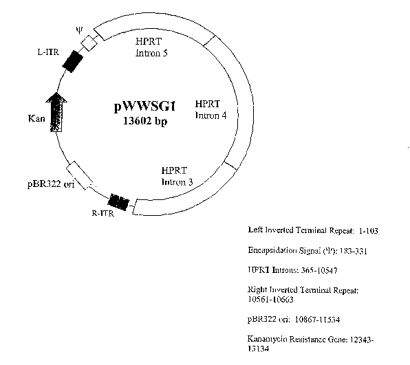
E~~AMPLE 1. Construction of gutless viral backbone shuttle vector
An embodiment of a gutless viral backbone shuttle vector pShuttle is shown
in Figure 1. The shuttle vector pShuttle has a total length of 13602 by (SEQ
ID
NO:1). Sequence portion containing R-ITR, PBR322 ori, Iran, L-ITR, and
encapsidation signal was obtained from the p~ldEasy system from Stratagene.
1~t
by 3667 of the original pShuttle sequence, there is a BamHI site just beyond
the R-
ITR. PCR primers were designed to include the BamHI site and then was to
create
an EcoRI site at the end of the R-ITR. The R-ITR was PCR replicated and then
digested with BainHI and EcoRI to create sticky ends. The viral backbone was
then cut with both B~.mHI and EcoRI. The BamIII cut the backbone at by 3667
and there was also an EcoRI site inside the MCS at by 377. The backbone
portion
of the plasmid was then gel purified and the PCR replicated R-ITR was recloned
into position. This essentially puts the L-ITR, encapsidation signal, MCS, and
R-
ITR all in close proximity to each other.
Insertion of the HPRT introns was a two step cloning process. First, the
viral backbone was digested with EcoRI and XbaI, both enzyme sites are in the
MCS. The HPRT source was also digested with EcoRI and XbaI yielding a 7477
by fragment that was cloned into the EcoRI/XbaI digested viral backbone. Then
the HPRT source was digested with only XbaI yielding a 2715 by fragment. One
of the XbaI sites in this cut is the same XbaI site that was cut from the
EcoRI/XbaI
_27_
CA 02515916 2005-08-12
WO 2004/076635 PCT/US2004/005453
double digest in step 1. The viral backbone was cut with only XbaI and the
2715
by fragment was inserted.
Overall, from the HPRT source, the HPRT stuffer sequence is inserted into
the viral backbone in reverse orientation, hence intron 5, then 4, then 3. The
2715
by fragment was inserted and checked to follow the original source sequence.
EXAMPLE 2 Construction and preparation of gutless viral shuttle vector
(a) Construction and preparation of gutless viral shuttle vector carrying
human
thrombomodulin (hTM) gene
The insertion of hTM gene into the gutless adenovirus backbone first
required the creation of a CMV-hTM expression cassette.
The intermediate vector used was pcDNA3.1/Zeo(+) (Invitrogen). A CMV
promoter is available commercially and a CMV promoter was cloned into the
multiple cloning site (MCS) at the Xbal/EcoRV restriction enzyme site
locations.
The CMV from ps5 was removed using XbaI/EcoRV. pcDNA3.1/Zeo(+) uses
1 S cleaved inside the MCS using both XbaI and EcoRV as well. The CMV promoter
was then ligated. Due to the location of the enzyme sites in the MCS, the CMV
promoter (Figure 6, SEQ ID NO:S) was inserted in a backwards orientation
relative
to the pcDNA3.1/Zeo(+) plasmid. The TM cDNA (Figure 7, SEQ ID N0:6) was
obtained from Dr. Sadler (Dittman et al., ~a~chen~2s~r~y, 26(14):4350-4.357
[1987])
which the sequence was also submitted to ATCC and to (senDank. The TM gene
was removed from the plasmid using EcoRI and inserted into pcDNA3.1/Zeo(+),
also in the reverse orientation to pcDNA3.1/Zeo(+) downstream of the inseuted
CMV promoter. To remove the cassette, PmeI enzyme was used to cut both ends
of the MCS. The gutless adenovirus backbone was linearized using SmaI which is
at by 381 of the backbone. The two were ligated together in the foa-~wards
orientation with respect to the gutless virus backbone. Sequence of the
expression
cassette (from PmeI site to PmeI site, SEQ ID N0:4) is shown in Figure 5.
(b). Construction and preparation of gutless viral shuttle vector carrying
LacZ gene
The insertion of LacZ also required creation of an intermediate vector to
create the expression cassette. pcDNA3.1/Zeo(+) was again used. First, a
portion
_28_
CA 02515916 2005-08-12
WO 2004/076635 PCT/US2004/005453
of the vector from the end of the MCS, restriction enzyme site Apal, to the
beginning of the SV40 poly A, restriction site Nael, was removed and the
vector
relegated to itself. Then the LacZ gene was inserted into the vector MCS using
NotI/Xbal. The expression cassette, containing CMV promoter, LacZ gene, and
SV40 poly A, was removed using Nrul/Sall retraction enzymes and blunt-end
cloned into the gutless adenovirus at the Smal restriction enzyme site.
ENAMI'LE 3. Preparation of gutless adenovirus
The helper virus was an E1/E3 deleted adenovirus in which a special flp
recognition sequence site (FRS) flanks the encapsidation signal. Helper
adenovirus
need to be grown in 293 cells.
293 cell line has long ago been engineered to express E1 and E3 genes of
adenovirus. These two genes are necessary for viral reproduction. The flp gene
is
similar in function to Cre-Lox. The flp gene will recognize the FRS, cleave at
that
location, and then relegate the DNA. Its basic function is to promote
recombination between different pieces of DNA with the FRS, but in this case,
it
will cleave out the encapsidation signal thereby not allowing helper-viral DNA
to
be packaged. [Beauchamp et al., Moleculaf° Tlae~apy, 3(5):09-S15
(2001);
ZJmana et al., Nature Bi~teclanolo~y, 19:52-5~5 (2001)].
293-flp cells were transfected with the backbone DNA using
Lipofectamine. ifJhile performing the transfection, helper virus were used to
infect
the 293-flp cells. The helper virus inserted its own DNA into the 293-flp
cells.
The flp protein expressed in the cells cleaved the encapsidation signal
thereby not
allowing the helper virus DNA to package. Consequently, the gutless adenoviuus
backbone DNA was packaged into the adenoviral proteins expressed from the
helper virus DNA and formed gutless adenovirus (gutless Ad hTM or gutless Ad
lacZ). The gutless viruses contain the hTM or lacZ expression cassette but
could
nor replicate in normal cells due to the E1/E3 deletions.
The virus were produced by the following procedure:
(a) Virus Reproduction
_2g_
CA 02515916 2005-08-12
WO 2004/076635 PCT/US2004/005453
Seed 293 cells in l5cm dishes and grow in 10% FBS until approximately
70% confluent. Viral media was made as follows: 2 ml of FBS-free IMEM
containing antibiotic, antimycotic; adjust pfu per cell of purified virus
until reached
the final concentration of media as 1 ~,1 virus in 2 ml IMEM (viral Conc. 1 x
10'0
pfu/ml)/ each 15 cm dish. For Example: 30 Dishes = 60 ml IMEM + 30 ~1 virus
Old media was Aspirated from dishes, and 2 ml viral media was added per dish.
Dishes were rocked at 37°C for 1.5 - 2 hours, and lBmL 10% media was
added per
dish and incubated according to time course.
Cells were harvested by pipeting and collocating in 50 ml tubes at
4°C, and
cells were centrifuge at 4°C, 2000 rpm for 5 min. Save 10 ml of
supernatant from
one of the tubes into a separate tube. The supernatant was removed from all of
the
tubes. Take SmL supernatant from the saved tube and resuspend all the pellets
to
one tube. All of the tubes were re-wash with the remaining SxnL of supernatant
to
collect any leftover sample, and the pellet was store at -80°C.
(b) Virus Collection
Sample tubes) were frozen/thawed 5 times to lyse the cells, and the virus
were released using dry ice and incubated at 37°C water bath for 15
minutes until
each to obtain crude viral lysate (CVL). The CVL was collected in two 2059
Falcon Tubes and centrifuged using Sor~all HS4. at 7000 rpm, 4°C for S
minutes
and the supernatant was recovered.
To purify the virus, ultra-clear SW41 (Beckman) tubes were prepared by
soalcing in Ultra Pure Water, then 70% ETOH. Cotton swabs (one swab for each
tube) was used to completely dry out the tube, and two tubes were used per
sample.
Preparation of the first gradient: 2.5 ml CsCI - Density 1.25, and 2.5 ml
CsCI - Density 1.40. Place the 1.25 density CsCI into the Beckman. tubes
first.
Underlay slowly the high density, 1.40 CsCl using a sterile pasteur pipette,
and
overlay an equal amount (in ml) of CVL, about 4.25 ml/tube. Samples were
centrifuged in a SW41 rotor with speed: 35,000 rpm at 20°C for 1 hour
and with
acceleration: 1 and deceleration: 4. The lower opalescent band was collected
using
1 or 3 ml syringe with green cap needles.
-3 0-
CA 02515916 2005-08-12
WO 2004/076635 PCT/US2004/005453
Preparation of second gradient: CsCI was prepared to density 1.33. Two
fresh ultra-clear tubes were placed 8 ml of CsCI and overlay the band just
recovered after the first spin. (To equilibrate the tubes, measure before the
volume
of the recovered band and divide equally in the 2 tubes). Samples were
centrifuged
at the conditions above for 18 hours. The opalescent band was recovered and
collected in a sterile eppendorf tube. (From this moment, keep the tube always
on
ice). Samples were dialyze with dialysis buffer: (1) 10X Dialysis Buffer: 100
mM
Tris - pH 7.4, 10 mM MgClz; (2) 1X Dialysis Buffer (2 Liters): 400 ml
Glycero1,200 ml l OX Dialysis Buffer 140 ml, and Ultra Pure Water. The
dialyzed
samples were immediately stored at -70°C.
Alternative, the virus can be purified using column chromatography. Such
method has been described, for example, by Sakhuja et al and Green et al
[Sakhuja
et al., Hufraara Gefae TheYapy, 14:243-254 (2003); Green et al., Humafa Gene
Tla~y-apy, 13:1921-1934, (2002)]. Purification kit for adenovirus using column
chromatography is also commercially available, e.g., the ViraI~it from
Virapur,
LLC (San Diego, CA). Briefly, the infected cell will be harvested and lysed by
several freeze-and-thaw cycles. The cell debris will be precipitated by
centrifugation. The supernatant will be collected and clarified by passing
through a
0.4.5 micron clarification filter. The clarified supernatant will be treated
with
DNase and then applied to a purification filter by centrifugation. After two
or more
washes, the virus will be eluted from the purification filter. The protein
concentration of the eluant will be determined using a Biol~ad protein
estimation
kit and the following formula will be used to convert protein concentration to
titer:
[12.956 + 224.15 (~g/ml)] x 10g.
EXAMPLE 4. Expression of hTM iT2 vitY~ using; gutless Ad hTM
When enough gutless Ad hTM has been produced, experiments will be
performed to demonstrate the viable expression of hTM ifz vitYO with gutless
Ad
hTM in cultured human cells such as HUVEC cells. Briefly, cells at 80-90%
confluency will be infected with Ad hTM at various multiplicity of infection
(MOI)
in F12I~ medium without any supplements for 30 min at 37°C. The medium
will
-31-
CA 02515916 2005-08-12
WO 2004/076635 PCT/US2004/005453
then be removed and fresh growth medium will be added. The cells may
optionally
be washed with saline before the addition of the fresh medium. The cells will
be
incubated at 37°C for 48-72 hours and analyzed for hTM expression. RT-
PCR will
be performed post infection using hTM specialized primers to detect for
thrombomodulin mRNA. A hTM ELISA assay will be performed to determine
hTM secretion in the culture medium. Western blots will also be performed to .
detect hTM protein expression in the virus infected cells.
As a control, the same cells will be infected with the gutless adenovirus
expressing (3-galactosidase (gutless Ad LacZ). The infected cells will
subsequently
be stained with X-gal using the (3-galactosidase reporter gene staining kit
from
Sigma (Saint Louis, Missouri). Briefly, cells will be rinsed with PBS, fixed
for 10
min at room temperature with the fixation solution, and then stained at
37°C for
0.5-2 hours with the staining solution. The lacZ positive cells will be
counted
under a microscope. This will demonstrate the viability of the gutless
adenovirus
1 S backbone itself.
EXAMPLE 5. Iya vivo expression of hTM
Ira vivo expression of hTM will be achieved by administering a
therapeutically effective amount of the gutless Ad hTM into a rodent. The
virus
may administered intravascularly, subcutaneously, or intramuscularly. As is
well-
known to one skilled in the art, the dose and route of viral administration
may vary
based on the disease to be treated and the severity of the disease.
EXAMPLE 6. Ex vivo expression of hTM
Ex vivo expression of hTM will be achieved by introducing the hTM gene
into cultured cells, such as human umbilical vein endothelium cells (HUVEC)
cells, with viral or non-viral vectors. The hTM-expressing cells will then be
implanted in a patient to provide a local supply of hTM. As is well-known to
one
skilled in the art, the dose and site of cell implantation may vary based on
the
disease to be treated and the severity of the disease. Similarly, hTM gene may
be
introduced into sections of blood vessels in. vitro and the vessels will then
be
implanted in a patient.
-32-
CA 02515916 2005-08-12
WO 2004/076635 PCT/US2004/005453
EXAMPLE 7 E~ression of hTM ih vitro using calcium phosphate precipitation
The hTM cDNA will be cloned into an expression vector under the control
of a constitutive promoter, such as a CMV promoter. The resulting plasmid,
pCMVhTM, will be transfected in vitro into HCTVEC cells using calcium
phosphate precipitation. The transfection may be performed with the Calcium
Phosphate Transfection I~it from Invitrogen (Palo Alto, California). Briefly,
cells will be placed in 100 mm or 60 mm dishes at the required density.
Incubate
overnight at 37°C in a humidified COZ incubator. 3-4 hours prior to
transfection,
the media will be changed on the dishes. A transfection mixture will be
prepared
by slowly add solution A dropwise to solution B wlule bubbling air through
solution B with a pipette. The transfection mixture will be incubated at room
temperature for 30 minutes, and will then be add dropwise to the media to the
cells
in either a 60 mm or 100 mm dish. The cells will be incubated with the
transfection mixture at 37°C for 30 min, washed with 1X Phosphate
Buffered
Saline (PBS), and incubated with fresh media at 37°C for 48-72 hours.
Optionally,
a glycerol or DMSO shock may be carried out as described in the instruction
manual to improve transfection efficiency. Expression of the hTM will be
detected
by analysing the tissue culture medimn using the IMIJBIND~ thrombomodulin
ELISA kit from American Diagnostics Inc (Greenwich, Connecticut).
Alternatively, the cells will be harvested 4.8-72 hours post transfection.
V~estern
blots will be performed to confirm the hTM protein expression in the
transfected
cells.
EXAMPLE 8. Expression of hTM in vitf°o using Lipofectin
transfection
HCTVEC cells will be transfected in vitro using a plasmid carrying the hTM
gene and the Lipofectin reagent from Invitrogen. Briefly, HUVEC cells will be
seeded in a six-well or 35-mm tissue culture plate in 2 ml of the appropriate
growth
medium supplemented with serum. The cells will be incubated at 37°C in
a COz
incubator until the cells are 40-60% confluent. On the day of transfection,
the
following solutions will be prepared in 12 x 75 mm sterile tubes:
-33-
CA 02515916 2005-08-12
WO 2004/076635 PCT/US2004/005453
Solution A: For each transfection, dilute 1-2 ~g of DNA into 100 ~,1 serum-
free medium.
Solution B: For each transfection, dilute 2-20 ~1 of Lipofectin~ Reagent
into 100 ~,1 serum-free medium, and allow to stand at room temperature for
30-45 min.
The two solutions will then be mixed gently and incubate at room temperature
for
10-15 min. The cells will be washed once with 2 ml of senun-free medium. For
each transfection, 0.8 ml serum-free medium will be added to each tube
containing
the Lipofectin~ Reagent-DNA complexes. The mixture will be added dropwise to
the cells. The cells will be incubated for 30 min at 37°C in a COZ
incubator. The
DNA-containing medium will then be replaced with 2 ml of normal growth
medium containing serum. The cells will be incubated at 37°C in a COZ
incubator
for a total of 48-72 hours. Expression of the hTM will be detected by
analyzing the
tissue culture medium at various time points post transfection using the
IMUBIND° thrombomodulin ELISA kit from American Diagnostics Inc.
Western
blots will also be performed to confirm the hTM protein expression in the
transfected cells.
EXAMPLE 9 Expression of hTM irZ vita°~ using NeoFhectinT"'
transfection
Tlae plasmid pCM~'hTM v~ill be transfected in ~ritro into IIIT~EC cells
using the NeoPhectin'~' reagent from NeoPharm (Lake Forest, IL). Briefly,
appropriate number of cells will be incubated in 100 ~,1 of culture medium
containing 10% fetal bovine serum in a COZ incubator at 37°C until the
cells are
80-90% confluent. For each transfection per well, 1-7 ~1 of IVeoPhectin~"'
will be
mixed with 25 ~,1 serum-free medium in a sterile tube and incubate at room
temperature for 5 min. DNA (plasmid) in 25 ~,1 serum-free medium will be
combined with NeoPhectin~''' dilution to a total volume of 50 ~1 in a sterile
tube and
incubated for 30 min to form lipid-nucleic complexes. The lipid-nucleic
complexes will be added to each well containing cells and medium and incubated
with the cells for 30 min at 37°C in a COz incubator. The medium will
be replaced
with 10% FBS media. Expression of the hTM will be detected by analyzing the
-34-
CA 02515916 2005-08-12
WO 2004/076635 PCT/US2004/005453
tissue culture medium at 24-72 hours post transfection using the IMUBIND~
thrombomodulin ELISA kit from American Diagnostic Inc. Alternatively, the
cells
will be harvested 48-72 hours post transfection. Westenz blots will be
performed to
confirm the hTM protein expression in the transfected cells.
EXAMPLE 10. Delivery hTM ira vitro using micro emulsions
HUVEC cells will be transfected ira vitro using a plasmid carrying the hTM
gene and micro emulsions. Preparation of micro emulsions has been described,
for
example, by Yi et al. [Yi et al., PlaarnZaceutical Research, 17:314-320,
(2000)], Cui
et al. [Cui et al., Biocohjugate Chem. 13:1319-1327, (2002)], and in US Patent
Nos. 5,061,688 and 5,438,041. Briefly, an emulsion/DNA complex will be
prepared, added to cultured HUVEC cells, and incubated with the cells for 30
min.
The medium will be replaced with 10% FBS media. Expression of the hTM will
be detected by analyzing the tissue culture medium at 24-72 hours post
transfection
using the IMUBIND~ thrombomodulin ELISA kit from American Diagnostic Inc.
Alternatively, the cells will be harvested 48-72 hours post transfection.
Western
blots will be performed to confirm the hTM protein expression in the
transfected
cells.
EXAMPLE 11. Thrombomodulin ELISA
The amount of hTM in tlae tissue culture medibun (iaa vitro e~~pression) or in
the plasma (ira vivo expression) will be determined using II~~IIJBT~TD~
Thrombomodulin ELISA kit from American Diagnostics Inc. (Greenwich,
Comlecticut). Briefly, samples will be diluted in Sample Buffer. 200 ~,1 of
the
thrombomodulin standard, diluted reference plasma or diluted plasma sample
will
be added to the micro-test wells and incubated for 1 hour at room temperature.
The
wells will be washed 4 times with the Wash Buffer. 200 ~1 of Detection
Antibody
will be added to each well and incubated for 30 minutes at room temperature.
The wells will be washed 4 times with the Wash Buffer. 200 ~,l of Substrate
solution will be added to each well and incubated for 20 minutes at room
temperature. A blue color will be developed, and the enzymatic reaction will
be
stopped by adding 100 ~1 of 0.5 HZS04. The solution color will turn yellow.
The
-35-
CA 02515916 2005-08-12
WO 2004/076635 PCT/US2004/005453
absorbances will be read on a micro-test plate reader at a wavelength of 450
nM
within 30 minutes. The background average of the blanks will be deducted from
the standards and sample readings.
The above description is for the purpose of teaching the person of ordinary
skill in the art how to practice the present invention, and it is not intended
to detail
all those obvious modifications and variations of it which will become
apparent to
the skilled worker upon reading the description. It is intended, however, that
all
such obvious modifications and variations be included within the scope of the
present invention, which is defined by the following claims. The claims are
intended to cover the claimed components and steps in any sequence which is
effective to meet the objectives there intended, unless the context
specifically
indicates the contrary.
-36-