Note: Descriptions are shown in the official language in which they were submitted.
CA 02516188 2011-05-09
DESCRIPTION
CHITOSAN-DERIVATIVES FOR GENE DELIVERY AND EXPRESSION
Background of the Invention
An elegant approach to in vivo gene expression involves the use of plasmid
DNAs, pDNAs, which have a number of advantages, including ease of use and
preparation, stability and heat resistance, and unlimited size. Plasmids do
not replicate in
mammalian hosts and do not integrate into host genomes; yet they can persist
in host cells
and express the cloned gene for a period of weeks to months. A major drawback
of the
pDNA approach is that gene transfer is inefficient under physiologically
relevant
conditions, especially in slow and non-dividing cells, such as epithelial
cells. There is a
need for the development of safer and more effective delivery vehicles, both
for antigens
and genes. The gene delivery systems should offer the freedom to manipulate
the
complex stoichiometry, surface charge density, and hydrophobicity needed for
interaction
with the cellular lipid components.
Cationic polymers and cationic phospholipids are the two major types of non-
viral
gene delivery vectors currently being investigated. Due to their permanent
cationic
charge, both types interact electrostatically with negatively charged DNA and
form
complexes (lipo- or polyplexes). Despite the ease of fabrication of the
lipoplexes, their
low transfection efficiency and toxicity limits their success. However,
polyplexes
involving cationic polymers are more stable than cationic lipids (De Smedt,
S.C. et al.
Phann. Res., 2000, 17:113-126). Nevertheless, the transfection efficiency is
relatively
CA 02516188 2005-08-15
WO 2004/074314 PCT/US2004/004262
2
lower than that of viral vectors. The precise mechanism for gene transfection
mediated
by cationic liposomes is still unclear. However, fusion of endosomal and
liposomal
membranes or destabilization of the endosomal membrane by cationic liposomes
may
trigger cytosolic delivery of DNA (Koltover, T. et al. Science, 1998, 281:78-
81).
Cationic polymers have been used to condense and deliver DNA both in vitro and
in vivo. Several cationic polymers have been investigated that lead to higher
transfection
efficiencies (De Smedt, S.C. et al. Pharm. Res., 2000, 17:113-126; Garnett,
M.C. Crit.
Rev. Ther. Drug Carrier Syst., 1999, 16:147-207). They form polyelectrolyte
complexes
with plasmid DNA in which the DNA becomes better protected against nuclease
degradation (Minagawa, K. et al. FEBS Lett., 1991, 295:67-69). They show
structural
variability and versatility including the possibility of covalent binding of
the targeting
moieties for gene expression mediated through specific receptors (De Smedt,
S.C. et al.
Pharm. Res., 2000, 17:113-126). Cationic liposomes form a complex with anionic
DNA
molecules and are thought to deliver DNA by endocytosis (Wrobell, D. et al.
Biochem.Biophys.Acta, 1995, 1235:296-304). Polymeric gene carriers might have
some
advantages over liposome systems: (i) relatively small size and narrow
distribution; (ii)
high stability against nucleases; and (iii) easy control of the hydrophilicity
of the complex
by copolymerization (Kabanov, A.V. Pharm.Sci. Tech. Today, 1999, 2:265-372).
The best characterized chitin-based copolymer, chitosan, is a biodegradable
and
biocompatible natural biopolymer that increases nasal absorption of the drug
without any
adverse effects (Thanou, M . et al. Biomaterials 2002, 23:153-9; Kim, Y.H. et
al.
Bioconjug Chem, 2001, 12:932-8; Singla, A.K. et al. JPharm Pharmacol, 2001,
53:1047-
67; Brooking, J, et al. J Drug Target, 2001, 9:267-79; Kotze, A.F. et al. J
Pharm Sci,
1999, 88:253-7; van der Lubben, I.M. et al. Eur JPharm Sci, 2001, 14:201-7). A
major
stumbling block in in vivo gene expression systems has been the lack of
efficient
transfection in vivo, and the improvements have been empirical.
Chitosan, a natural, biocompatible cationic polysaccharide prepared from
crustacean shells, has shown much potential as a vehicle for gene delivery.
Chitosan has
many beneficial effects, including immunostimulatory activity (Nishimura, K.
et al.
Vaccine, 1984, 2:93-9), anticoagulant activity (Otterlei, M. et al. Vaccine,
1994, 12:825-
32), wound-healing properties (Muzzarelli, R. et al. Biomaterials, 1988,
10:598-603), and
CA 02516188 2005-08-15
WO 2004/074314 PCT/US2004/004262
3
anti-microbial properties (Pappineau, A.M. et al. Food Biotechnol, 1991, 5:45-
47).
Additionally, chitosan is non-toxic, non-hemolytic, weakly immunogenic, slowly
biodegradable, and nuclease resistant; and it has been used in controlled drug
delivery
(Erbacher, P. et al. Pharm Res, 1998, 15:1332-9; Richardson, S.C. et al. Int J
Pharm,
1999, 178:231-43). Chitosan increases transcellular and paracellular transport
across the
mucosal epithelium and thus may facilitate mucosal gene delivery and the
immune
responsiveness of the mucosa and bronchus-associated lymphoid tissue.
Therefore,
chitosan appears to possess the attributes for an ideal gene delivery agent
required for
therapies such as lung disease therapy.
IFN-y, a pleiotropic cytokine, promotes T-helper type-1 (Thl) responses, which
downregulate the T112-like immune responses that are hallmarks of allergic
diseases,
including asthma (Mosman, T.R. et al. Ann Rev Immunol, 1989, 7:145-173;
Umetsu, D.T.
et al. J Allergy Clin Immunol, 1997, 100:1-6). Administration of recombinant
IFN-y
reverses established airway disease and inflammation in murine models
(Flaishon, L. et
al. Jlmmunol, 2002, 168:3707-11; Yoshida, M. et al. Am JRespir Crit Care Med,
2002,
166:451-6). Application of IFN-y for treatment of asthma has been limited
because of the
short half-life of IFN-y in vivo and the potentially severe adverse effects
associated with
high dose administration (Murray, H. Intensive Care Med, 1997, 22(Suppl
4):S456-61).
IFN-y gene transfer inhibits both antigen- and Th2-induced pulmonary
eosinophilia and
airway hyperreactivity (Li, X.M. et al. J Immunol, 1996, 157:3216-9; Dow, S.W.
et al.
Hum Gene Ther, 1999, 10:1905-14). However, those results are not directly
applicable to
humans because of the methods used in the investigations, such as the
intratracheal
administration or injection of DNA with lipofectamine. Moreover, the direct
effects of
these cytokine plasmids as therapeutics for allergic asthma have not been
addressed. A
major drawback of the pDNA approach is that gene transfer is inefficient under
physiologically permissible conditions, especially in non-dividing cells such
as epithelial
cells.
The protective role of IFN-y gene transfer in a mouse model for respiratory
syncytial virus infection (U.S. Patent No. 6,489,306 (Mohapatra et al., issued
December
3, 2002); Kumar, M. Vaccine, 1999, 18:558-567) and the role of IFN-y as a
genetic
adjuvant in the immunotherapy of grass-allergic asthma (Kumar, M. et al.
JAllergy Clin
CA 02516188 2005-08-15
WO 2004/074314 PCT/US2004/004262
4
Immunol, 2001, 108:402-408) has previously been reported. IFN-y is considered
to be a
prime candidate for asthma therapy because of its capacity to decrease: (i) IL-
13-induced
goblet cell hyperplasia and eosinophilia by upregulation of the IL-13Ra2 decoy
receptor,
which diminishes IL-13 signaling (Ford, J.G. et al. J Immunol, 2001, 167:1769-
1777;
Daines, M.O. and Hershey, G.K. J Biol Chem 2002, 277(12):10387-10393); (ii)
LTC4
production in murine and human macrophages (Boraschi, D. et al. J lininunol,
1987,
138:4341-4346; Thivierge, M. et al. J Immunol, 2001, 167:2855-2860), in human
peripheral blood lymphocytes after wasp venom immunotherapy (Pierkes, M. et
al. J
Allergy Clin Immunol, 1999, 103:326-332), and in leukocytes of pollinosis
patients
(Krasnowska, M. et al. Arch Immunol Ther Exp (Warsz), 2000, 48:287-292); and
(iii)
TGF-0 and procollagen-I and -III, which cause fibrosis and airway remodeling
(Gurujeyalakshmi, G. et al. Exp Lung Res, 1995, 21:791-808; Minshall, E. et
al. Am J
Respir Cell Mol Biol, 1997, 17:326-333).
This disclosure demonstrates that the gene transfer efficiency can be
significantly
increased using a novel improved formulation of hybrid nanoparticles, referred
to as
Chlipids. Further, therapy with chitosan-IFN-gamma gene-nanoparticles carrying
(CIN)
constitutes a novel non-viral approach to mucosal gene transfer for asthma.
C1N therapy
significantly inhibits the production of IL-4, IL-5, ovalbumin (OVA)-specific
serum IgE,
airway inflammation, and hyperreactivity in a BALB/c mouse model of allergic
asthma.
Brief Summary of the Invention
The present invention pertains to gene delivery systems using chitosan, or
derivatives thereof. In one aspect, the present invention provides particles
comprising
chitosan, or a derivative thereof, useful as delivery vehicles for
polynucleotides,
compositions comprising such particles and a pharmaceutically acceptable
carrier, and
methods for delivering and expressing polynucleotides to hosts in vitro or in
vivo using
such particles. Optionally, the particles of the invention further comprise a
lipid
component and are referred to herein interchangeably as "chliposomes" or
"chlipids" or
"chitosan-lipid nanoparticles" or "CLNs". The invention further includes
methods for
producing particles of the subject invention.
CA 02516188 2011-05-09
The present further provides a method for enhancing interferon-gamma
expression
to regulate the production of cytokines secreted by T-helper type 2 (Th2)
cells within a
subject by administering an effective amount of a particle of the subject
invention to the
subject, wherein the particle comprises a polynucleotide encoding interferon-
gamma.
5
Brief Description of the Drawings
For a fuller understanding of the nature and objects of the invention,
reference
should be made to the following detailed description, taken in connection with
the
accompanying drawings, in which:
Figures 1A - 1C show optimization protocols of combining chitosan and lipids
for gene transfer. Figure 1A shows the DNA recovery from pelleted chlipids.
Figure 1B
shows the optimal lipid concentration. Figure 1C shows the optimal serum
concentration.
Figures 2A-2C show electron micrographs of nanoparticles. Figure 2A shows
chitosan at 14,000X magnification. Figure 2B shows lipid-DNA at 7,000X
magnification. Figure 2C shows chitosan + (lipid-DNA) at 44,000X
magnification.
Figures 3A-3C show distribution and quantification of transfection of the GFP
gene lung cells. The green fluorescence seen in the lung section suggests that
the
epithelial cells are predominantly transfected by chitosan-lipid nanoparticle
(CLN)
(Figure 3A). The cells from the BAL fluid showed that monocytes are also
transfected
and express GFP (Figure 3B). In Figure 3C, "1" is chitosan, "2" is lipofectin,
"3" is
CLNs, and "4" is DNA alone. The quantification of EGFP-positive BAL cells
showed
TM
that while chitosan and LIPOFECTIN showed a similar transfection efficiency
(20%) in
vivo, CLN showed significantly higher (30%, P<0.05) transfection efficiency,
as shown in
Figure 3C.
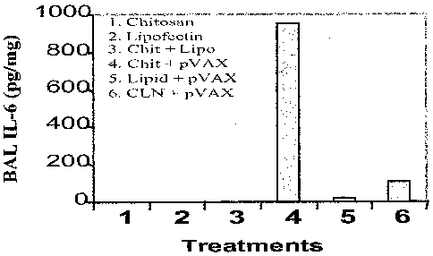
Figure 4 shows quantification of IL-6 in bronchioalveolar fluid (BAL)
following
intranasal administration of nanoparticle. Quantification of IL-6 showed that
CLN-DNA
nanoparticles induced significantly decreased IL-6 levels compared to chitosan-
pVAX
complexes.
Figures 5C-5C show that chitosan particles target lung epithelial cells and
monocytes. BALB/c mice were administered with chitosan particles containing
pVAX-
GFP. After 24 hours, mice were sacrificed and their lungs were fixed and
sectioned by
CA 02516188 2005-08-15
WO 2004/074314 PCT/US2004/004262
6
cryotome. Sections (15 microns) were thaw-mounted to slides and sections were
viewed
for green fluorescent protein under a microscope and photographed ("Lung";
Figure 5A).
BAL cells were fixed after cytospin on a slide and visualized under a
fluorescent
microscope to identify GFP expressing cells ("BAL"; Figure 5B). Figure SC is a
graph
showing that chitosan IFN-gamma-pDNA nanoparticle (CIN) administration induces
IFN-y production in the lung over a period of 10 days. Lung homogenates were
prepared
from mice after 1, 2, 4, 6, 8, or 10 days of treatment with CIN (25 g/mouse)
or chitosan
alone, and IFN-y levels were determined by ELISA (n=3).
Figures 6A-6F show prevention of airway hyperresponsiveness (AHR). Figure
6A shows a schematic prophylaxis protocol. Mice were challenged with
methacholine on
day 22 to measure airway responsiveness (Figure 6B). The values are mean
enhanced
pause (PENH) expressed as percent of baseline f SEM (*P<0.05, **P<0.01). On
day 24,
BAL was performed and differential cell count was obtained (Figure 6C). On day
24,
lungs were removed, sectioned, and the sections stained with hematoxylin/eosin
("PBS,
phosphate-buffered saline control; "N-DNA", naked DNA without chitosan; "CIN",
chitosan-DNA complex), as shown in Figures 6D, 6E, and 6F. Differential cell
counts
and examination of tissue sections were performed by different persons in a
blinded
fashion. Representative results are shown.
Figures 7A-7C show that CIN alters production of cytokines and IgE. On day 23
of the prophylactic procedure (see schematic of Figure 6A), spleens ere
removed and
single-cell suspensions of splenocytes were prepared. Cells were cultured for
48 hours
with ovalbumin (OVA) and the levels of secreted IFN-y and IL-5 (Figure 7A) and
IL-4
(Figure 7B) were measured. Total serum IgE was measured on day 23 (Figure 7C).
Values are means SEM (*p<0.05, **p<0.01).
Figures 8A-8D show reversal of established AHR and eosinophilia. Figure 8A
shows a schematic of the therapeutic protocol. Mice were sensitized (i.p.) and
challenged
(i.n.) with OVA and treated with CIN as described. AHR was measured 24 hours
after
the last challenge (n=4). CIN-treated mice exhibited reduced AHR compared to
the
controls (Figure 8B). Data are mean enhanced pause (PENH) expressed as percent
of
baseline SEM (*p<0.05). On day 31, BAL was performed and eosinophils in BAL
fluid
were counted (**p<0.01). Figure 8C shows that CIN therapy decreases
eosinophils. On
CA 02516188 2005-08-15
WO 2004/074314 PCT/US2004/004262
7
day 23, spleens were removed and single-cell suspensions of splenocytes were
prepared.
Cells were cultured for 48 hours in the presence of OVA and cell supernatants
were
analyzed for IFN-y, IL-4, and IL-5. Mice receiving CIN showed more production
of IFN-
y and less IL-4 and IL-5 compared to the chitosan-only control (Figure 8D).
Data are
means SEM (*p<0.05).
Figures 9A-9D show that CIN treatment induces apoptosis of goblet cells.
BALB/c mice (n=3) were sensitized and challenged with OVA as in Figures 8A,
and then
treated with intranasal C1N therapy. Mice were sacrificed at 0, 3, 6, or 12
hours after CIN
treatment and lungs were removed, sectioned and stained with hematoxylin/eosin
(Figures 9A-9D, respectively).
Figures 1OA-10D show that CIN treatment induced apoptosis of goblet cells.
BALB/c mice (n=3) were sensitized and challenged with OVA as in Figures 8A,
and then
treated with intranasal CIN therapy. Mice were sacrificed at 0, 3, 6, or 12
hours after CIN
treatment and lungs were removed, sectioned, and analyzed for apoptosis by
TUNEL
(terminal dUTP nick end labeling) assay (Figures 1OA-10D, respectively).
Figures 11A-11C show a final set of lung sections from Figure 10B (6-hour time
point) stained for the goblet cell-specific Muc5a (Figure 11 C), and for
apoptosis by the
TUNEL assay (Figure 1 1B). Figure 11A shows staining of nuclei with
diamidinophenylindole (DAPI).
Figures 12A-12C show that C1N therapy involves the STAT4 pathway. OVA-
sensitized BALB/c wild-type (WT) and STAT 4"/- knockout mice (n=4) were given
CIN
therapy intranasally and challenged with OVA. AHR in response to methacholine
was
measured one day after the last challenge (Figure 12A). The values are means f
SEM
(*p<0.05). Mice were sacrificed the day following AHR measurement and their
lungs
were removed, paraffin-embedded and stained with hematoxylin/eosin (Figures
12B and
12C).
Detailed Disclosure of the Invention
The present invention provides particles comprising chitosan, or a derivative
thereof; and a polynucleotide. Preferably, the particle further comprises a
control
sequence operably-linked to the polynucleotide, which is capable of causing
expression of
CA 02516188 2005-08-15
WO 2004/074314 PCT/US2004/004262
8
the polynucleotide within a host in vitro or in vivo. The present invention
further
provides compositions comprising a particle of the present invention and a
pharmaceutically acceptable carrier.
Optionally, the particle of the present invention comprises a lipid that is
complexed with the chitosan and the polynucleotide component of the particle.
Since
efficient gene expression in vivo requires both complex formation for cell
uptake and
prevention of nucleotide degradation and complex dissociation for
transcription by RNA
polymerase, the present inventor hypothesized that a combination of both
chitosan and
liposomes may lead to increased gene delivery and expression in vivo.
Therefore, the
present inventor has developed methods that combine these two different
carrier systems
to develop a novel gene delivery system designated "chliposomes" that exhibits
a
significant increase in gene DNA transfection and gene expression (also
referred to herein
as "chlipids" and used interchangeably). Preferably, the components of the
chlipid are
oriented such that the polynucleotide is surrounded by a lipid monolayer, with
polynucleotide-lipid inverted cylindrical micelles arranged in a hexagonal
lattice.
The present invention further includes a method for producing the particles of
the
invention by mixing (e.g., complexing) a polynucleotide and chitosan or a
chitosan
derivative, to form a particle comprising a binary complex of the
polynucleotide and the
chitosan or chitosan derivative. Optionally, the method further comprises
mixing
(complexing) a lipid with the polynucleotide and chitosan or chitosan
derivative to form a
particle (chlipid) comprising a multiplex of the polynucleotide, chitosan or
chitosan
derivative, and the lipid. Typically, the particles of the present invention
range in size
from the nanometer range (e.g., less than one micrometer; nanoparticles) to
the
micrometer size range (e.g., about one micrometer or larger).
The type of reaction vessel or vessels utilized for producing the particles of
the
present invention, or their sizes, are not critical. Any vessel or substrate
capable of
holding or supporting the reactants so as to allow the reaction to take place
can be used.
It should be understood that, unless expressly indicated to the contrary, the
terms
"adding", "contacting", "mixing", "reacting", "combining" and grammatical
variations
thereof, are used interchangeably to refer to the mixture of reactants of the
method of the
present invention (e.g., polynucleotide or non-polynucleotide agent, chitosan
or chitosan
CA 02516188 2005-08-15
WO 2004/074314 PCT/US2004/004262
9
derivative, lipid, and so forth), and the reciprocal mixture of those
reactants, one with the
other (i.e., vice-versa), in any order.
It will be readily apparent to those of ordinary skill in the art that a
number of
general parameters can influence the efficiency of transfection or
polynucleotide delivery.
These include, for example, the concentration of polynucleotide to be
delivered, the
concentration of chitosan or chitosan derivative, and the concentration of
lipid (for
chlipids of the present invention). For in vitro delivery, the number of cells
transfected,
the medium employed for delivery, the length of time the cells are incubated
with the
particles of the invention, and the relative amount of particles can influence
delivery
efficiency. For example, a 1:5 ratio of polynucleotide to lipid, 1:5 ratio of
polynucleotide
to chitosan, and 20% serum is suitable. These parameters can be optimized for
particular
cell types and conditions. Such optimization can be routinely conducted by one
of
ordinary skill in the art employing the guidance provided herein and knowledge
generally
available to those skilled in the art. It will also be apparent to those of
ordinary skill in
the art that alternative methods, reagents, procedures and techniques other
than those
specifically detailed herein can be employed or readily adapted to produce the
particles
and compositions of the invention. Such alternative methods, reagents,
procedures and
techniques are within the spirit and scope of this invention.
In another aspect, the present invention provides a method for delivery and
expression of a polynucleotide within a host or subject by administering a
particle of the
present invention to the host or subject. Optionally, the polynucleotide
encodes a
polypeptide. The polypeptide encoded by the polynucleotide of the particle can
be a
hormone, receptor, enzyme, or other desired polypeptide. For example, the
polypeptide
can comprise a cytokine, such as interferon-gamma. The polypeptide may serve a
therapeutic and/or diagnostic purpose, for example. In other embodiments, the
polynucleotide does not encode a polypeptide. The polynucleotide may comprise
interfering RNA, for example.
In another aspect, the present invention provides a method for enhancing
interferon-gamma expression to regulate the production of cytokines secreted
by T-helper
type 2 (Th2) cells within a subject by administering an effective amount of a
particle to
the subject, wherein the particle comprises chitosan, or a derivative thereof,
and a
CA 02516188 2005-08-15
WO 2004/074314 PCT/US2004/004262
polynucleotide encoding interferon-gamma. Preferably, the particle is
administered to the
respiratory tract of the subject. In one embodiment, the subject is suffering
from asthma.
In another embodiment, the subject is not suffering from asthma. Preferably,
the particle
administered to the subject is a chlipid of the present invention.
5 The method of the subject invention for enhancing interferon-gamma
expression
to regulate the production of cytokines secreted by Th2 cells (such as IL-4
and/or IL-5)
within a subject preferably results in inhibition of airway inflammation and
airway
hyperresponsiveness (AHR), the hallmarks of allergic asthma, when administered
to the
subject.
10 The term "chitosan", as used herein, will be understood by those skilled in
the art
to include all derivatives of chitin, or poly-N-aceryl-D-glucosamine
(including all
polyglucosamine and oligomers of glucosamine materials of different molecular
weights),
in which the greater proportion of the N-acetyl groups have been removed
through
hydrolysis. Generally, chitosans are a family of cationic, binary hetero-
polysaccharides
composed of (1- 4)-linked 2-acetamido-2-deoxy-/13-D-glucose (G1cNAc, A-unit)
and 2-
amino-2-deoxy-O-D-glucose, (G1cN; D-unit) (Varum K.M. et al., Carbohydr. Res.,
1991,
217:19-27; Sannan T. et al., Macromol. Chem., 1776, 177:3589-3600).
Preferably, the
chitosan has a positive charge. Chitosan, chitosan derivatives or salts (e.g.,
nitrate,
phosphate, sulphate, hydrochloride, glutamate, lactate or acetate salts) of
chitosan may be
used and are included within the meaning of the term "chitosin". As used
herein, the term
"chitosan derivatives" are intended to include ester, ether or other
derivatives formed by
bonding of acyl and/or alkyl groups with OH groups, but not the NH2 groups, of
chitosan.
Examples are O-alkyl ethers of chitosan and 0-acyl esters of chitosan.
Modified
chitosans, particularly those conjugated to polyethylene glycol, are included
in this
definition. Low and medium viscosity chitosans (for example CL113, G210 and
CL110)
may be obtained from various sources, including PRONOVA Biopolymer, Ltd. (UK);
SEIGAGAKU America Inc. (Maryland, USA); MERON (India) Pvt, Ltd. (India);
VANSON Ltd. (Virginia, USA); and AMS Biotechnology Ltd. (UK). Suitable
derivatives include those which are disclosed in Roberts, Chitin Chemistry,
MacMillan
Press Ltd., London (1992). Optimization of structural variables such as the
charge
CA 02516188 2011-05-09
11
density and molecular weight of the chitosan for efficiency of polynucleotide
delivery and
expression is contemplated and encompassed by the present invention.
The chitosan (or chitosan derivative or salt) used preferably has a molecular
weight of 4,000 Dalton or more, preferably in the range 25,000 to 2,000,000
Dalton, and
most preferably about 50,000 to 300,000 Dalton. Chitosans of different low
molecular
weights can be prepared by enzymatic degradation of chitosan using chitosanase
or by the
addition of nitrous acid. Both procedures are well known to those skilled in
the art and
are described in various publications (Li et al., Plant Physiol. Biochenz.,
1995, 33: 599-
603; Allan and Peyron, Carbohydrate Research, 1995, 277:257-272; Damard and
Cartier,
Int. J. Biol. Macroinol., 1989, 11: 297-302). Preferably, the chitosan is
water-soluble and
may be produced from chitin by deacetylation to a degree of greater than 40%,
preferably
between 50% and 98%, and more preferably between 70% and 90%.
The lipid utilized for the particles, compositions, and methods of the present
invention is preferably a phospholipid or cationic lipid. Cationic lipids are
amphipathic
molecules, containing hydrophobic moieties such as cholesterol or alkyl side
chains and a
cationic group, such as an amine. Phospholipids are amphipathic molecules
containing a
phosphate group and fatty acid side chains. Phospholipids can have an overall
negative
charge, positive charge, or neutral charge, depending on various substituents
present on
the side chains. Typical phospholipid hydrophilic groups include phosphatidyl
choline,
phosphatidylglycerol, and phosphatidyl ethanolamine moieties. Typical
hydrophobic
groups include a variety of saturated and unsaturated fatty acid moieties. The
lipids used
in the present invention include cationic lipids that form a complex with the
genetic
material (e.g., polynucleotide), which is generally polyanionic, and the
chitosan or
chitosan derivative. The lipid may also bind to polyanionic proteoglycans
present on the
surface of cells. The cationic lipids can be phospholipids or lipids without
phosphate
groups.
A variety of suitable cationic lipids are known in the art, such as those
disclosed in
International Publication No. WO 95/02698.
Exemplified structures of cationic lipids useful
in the particles of the present invention are provided in Table 1 of
International
Publication No. WO 95/02698. Generally, any cationic lipid, either monovalent
or
CA 02516188 2011-05-09
12
polyvalent, can be used in the particles, compositions and methods of the
present
invention. Polyvalent cationic lipids are generally preferred. Cationic lipids
include
saturated and unsaturated allyl and alicyclic ethers and esters of amines,
amides or
derivatives thereof. Straight-chain and branched alkyl and alkene groups of
cationic
lipids can contain from 1 to about 25 carbon atoms. Preferred straight-chain
or branched
alkyl or alkene groups have six or more carbon atoms. Alicyclic groups can
contain from
about 6 to 30 carbon atoms. Preferred alicyclic groups include cholesterol and
other
steroid groups. Cationic lipids can be prepared with a variety of counterions
(anions)
including among others: chloride, bromide, iodide, fluoride, acetate,
trifluoroacetate,
sulfate, nitrite, and nitrate.
Transfection efficiency can be increased by using a lysophosphatide in
particle
formation. Preferred lysophosphatides include lysophosphatidylcholines such as
I-
oleoyllysophosphatidylcholine and lysophosphatidylethanolamines. Well known
lysophosphatides which may be used include DOTMA (dioleyloxypropyl
TM
trimethylammonium chloride/DOPE (i.e., LIPOFECTIN, GIBCO/BRL, Gaithersburg,
Md:.), DOSPA, (dioleyloxy sperminecarboxamidoethyl dimethylpropanaminium
TM TM
trifuoroacetate)IDOPE (i.e., LIPOFECTAMINE), LIPOFECTAMINE 2000, and DOGS
TM
(dioctadecylamidospermine) (i.e., TRANSFECTAM), and are all commercially
available.
Additional suitable cationic lipids structurally related to DOTMA are
described in U.S.
Patent No. 4,897,355.
TM
TRANSFECTAM belongs to a group of cationic lipids called lipopolamines (also
referred to as second-generation cationic lipids) that differ from the other
lipids used in
gene transfer mostly by their spermine head group. The polycationic spermine
head
group promotes the formation of lipoplexes with better-defined structures
(e.g., 50 to 100
nm) (Remy J.S. et al., "Gene Transfer with Lipospermines and
Polyethylenimines", Adv.
Drug Deliv. Rev., 1998, 30:85-95).
Another useful group of cationic lipids related to DOTMA and DOTAP are
commonly called DORI-ethers or DORI-esters, such as (DL-1-O-oleyl-2-oleyl-3-
dimethylaminopropyl-j3-hydroxyethylammonium or DL-1-oleyl-2-O oleyl-3-dimethyl-
aminopropyl-0-hydroxyethylammonium). DORI lipids differ from DOTMA and DOTAP
in that one of the methyl groups of the trimethylammonium group is replaced
with a
CA 02516188 2011-05-09
13
hydroxyethyl group. The oleoyl groups of DORI lipids can be replaced with
other alkyl
or alkene groups, such as palmitoyl or stearoyl groups. The hydroxyl group of
the DORI-
type lipids can be used as a site for further functionalization, for example
for
esterification to amines, like carboxyspermine. Additional cationic lipids
which can be
employed in the particles, compositions, and methods of the present invention
include
those described in International Publication No. WO 91/15501.
Cationic sterol derivatives, like 3 (3 [N-(N',N'-
dimethylaminoeth-ane)carbamoyl] cholesterol (DC-Chol) in which cholesterol is
linked
to a trialkyammonium group, can also be employed in the present invention. DC-
Chol is
reported to provide more efficient transfection and lower toxicity than DOTMA-
containing liposomes for some cell lines. DC-Chol polyamine variants such as
those
described in International Publication No. WO 97/45442 may also be used.
Polycationic
lipids containing carboxyspermine are also useful in the delivery vectors or
complexes of
this invention. EP-A-304111 describes carboxyspermine containing cationic
lipids
including 5-carboxyspermylglycine dioctadecyl-amide (DOGS), as referenced
above, and
dipalmitoylphosphatidylethanolamine 5-carboxyspermylamide (DPPES). Additional
cationic lipids can be obtained by replacing the octadecyl and palmitoyl
groups of DOGS
and DPPES, respectively, with other alkyl or alkene groups. Cationic lipids
can
optionally be combined with non-cationic co-lipids, preferably neutral lipids,
to form the
chlipids of the invention. One or more amphiphilic compounds can optionally be
incorporated in order to modify the particle's surface property.
Suitable cationic lipids include esters of the Rosenthal Inhibitor (RI) (DL-
2,3-
distearoyloxypropyl(dimethyl)-13-hydroxyethylammoniumbromide), as described in
U.S.
Patent No. 5,264,618.
These derivatives can be prepared, for example, by acyl and alkyl substitution
of
3-dimethylaminopropane diol, followed by quatemization of the amino group.
Analogous phospholipids can be similarly prepared.
The particles of the present invention can be targeted through various means.
The
size of the particle provides one means for targeting to cells or tissues. For
example,
relatively small particles efficiently target ischemic tissue and tumor
tissue, as described
CA 02516188 2011-05-09
14
in U.S. Patent No. 5,527,538, and U.S. Patent Nos. 5,019,369, 5,435,989 and
5,441,745.
The particles of the invention can be targeted according to the mode of
administration. For example, lung tissue can be targeted by intranasal
administration,
cervical cells can be targeted by intravaginal administration, and prostate
tumors can be
targeted by intrarectal administration. Skin cancer can be targeted by topical
administration. Depending on location, tumors can be targeted by injection
into the tumor
mass.
Further, particles of the invention can be targeted by incorporating a ligand
such
as an antibody, a receptor, or other compound known to target particles such
as liposomes
or other vesicles to various sites. The ligands can be attached to cationic
lipids used to
form the particles of the present invention, or to a neutral lipid such as
cholesterol used to
stabilize the particle. Ligands that are specific for one or more specific
cellular receptor
sites are attached to a particle to form a delivery vehicle that can be
targeted with a high
15- degree of specificity to a target cell population of interest.
Suitable ligands for use in the present invention include, but are not limited
to,
sugars, proteins such as antibodies, hormones, lectins, major
histocompatibility complex
(MHC), and oligonucleotides that bind to or interact with a specific site. An
important
criteria for selecting an appropriate ligand is that the ligand is specific
and is suitably
bound to the surface of the particles in a manner which preserves the
specificity. For
example, the ligand can be covalently linked to the lipids used to prepare the
particles.
Alternatively, the ligand can be covalently bound to cholesterol or another
neutral lipid,
where the ligand-modified cholesterol is used to stabilize the lipid monolayer
or bilayer.
IFN-,y is a 14-18 kDalton 143 amino acid glycosylated protein that is a potent
multifunctional cytokine. As used herein, "interferon-gamma", "IFN-gamma",
"interferon-J', and "IFN-'y refer to IFN-y protein, biologically active
fragments of IFN--y,
and biologically active homologs of "interferon-gamma" and "IFN-'/', such as
mammalian homologs. These terms include IFN-'y-like molecules. An "IFN-like
molecule" refers to polypeptides exhibiting IFN-'y--like activity when the
polynucleotide
encoding the polypeptide is expressed, as can be determined in vitro or in
vivo. For
purposes of the subject invention, IFN-y-like activity refer to those
polypeptides having
CA 02516188 2011-05-09
one or more of the functions of the native IFN-y cytokine, such as those
disclosed herein.
Fragments and homologs of IFN-y retaining one or more of the functions of the
native
IFN-y cytokine, such as those disclosed herein, is included within the meaning
of the term
"IFN-''. In addition, the term includes a nucleotide sequence which through
the
5 degeneracy of the genetic code encodes a similar peptide gene product as IFN-
y and has
the IFN-y activity described herein. For example, a homolog of "interferon-
gamma" and
"IFN-1' includes a nucleotide sequence which contains a "silent" codon
substitution (e.g.,
substitution of one codon encoding an amino acid for another codon encoding
the same
amino acid) or an amino acid sequence which contains a "silent" amino acid
substitution
10 (e.g., substitution of one acidic amino acid for another acidic amino
acid). An
exemplified nucleotide sequence encodes human IFN-y (Accession No: NM 000639,
NCBI database).
The polynucleotides are administered and dosed in accordance with good medical
practice, taking into account the clinical condition of the individual
patient, the site and
15 method of administration, scheduling of administration, patient age, sex,
body weight,
and other factors known to medical practitioners. The therapeutically or
pharmaceutically
"effective amount" for purposes herein is thus determined by such
considerations as are
known in the art. A therapeutically or pharmaceutically effective amount of
nucleic acid
molecule (such as an IFN-y-encoding polynucleotide) is that amount necessary
to provide
an effective amount of the polynucleotide, or the corresponding polypeptide(s)
when
expressed in vivo. An effective amount of an agent, such as a polynucleotide
or non-
polynucleotide agent, or particles comprising such polynucleotide or non-
polynucleotide
agents, can be an amount sufficient to prevent, treat, reduce and/or
ameliorate the
symptoms and/or underlying causes of any pathologic condition, such as a
disease or
other disorder. In some instances, an "effective amount" is sufficient to
eliminate the
symptoms of the pathologic condition and, perhaps, overcome the condition
itself. In the
context of the present invention, the terms "treat" and "therapy" and the like
refer to
alleviate, slow the progression, prophylaxis, attenuation, or cure of existing
condition.
The term "prevent", as used herein, refers to putting off, delaying, slowing,
inhibiting, or
otherwise stopping, reducing, or ameliorating the onset of such conditions.
CA 02516188 2005-08-15
WO 2004/074314 PCT/US2004/004262
16
In the method of the invention for enhancing interferon-gamma expression, the
amount of the polypeptide (IFN-y) is preferably effective to achieve
regulation of one or
more cytokines secreted by Th2 cells, such as interleukin-4 (IL-4). The amount
of IFN-y
may be sufficient to achieve inhibition of (Th2)-associated airway
inflammation and
airway hyperresponsiveness when administered to a subject. In accordance with
the
present invention, a suitable single dose size is a dose that is capable of
preventing or
alleviating (reducing or eliminating) a symptom in a patient when administered
one or
more times over a suitable time period. One of skill in the art can readily
determine
appropriate single dose sizes for systemic administration based on the size of
a mammal
and the route of administration.
Mammalian species which benefit from the disclosed particles, compositions,
and
methods include, and are not limited to, apes, chimpanzees, orangutans,
humans,
monkeys; domesticated animals (e.g., pets) such as dogs, cats, guinea pigs,
hamsters,
Vietnamese pot-bellied pigs, rabbits, and ferrets; domesticated farm animals
such as
cows, buffalo, bison, horses, donkey, swine, sheep, and goats; exotic animals
typically
found in zoos, such as bear, lions, tigers, panthers, elephants, hippopotamus,
rhinoceros,
giraffes, antelopes, sloth, gazelles, zebras, wildebeests, prairie dogs, koala
bears,
kangaroo, opossums, raccoons, pandas, hyena, seals, sea lions, elephant seals,
otters,
porpoises, dolphins, and whales.
As used herein, the term "patient", "subject", and "host" are used herein
interchangeably and intended to include such human and non-human mammalian
species
and cells of those species. For example, the term "host" includes one or more
host cells,
which may be prokaryotic (such as bacterial cells) or eukaryotic cells (such
as human or
non-human mammalian cells), and may be in an in vivo or in vitro state. In
those cases
wherein the polynucleotide utilized is a naturally occurring nucleic acid
sequence, the
polynucleotide encoding the polypeptide product can be administered to
subjects of the
same species or different species from which the nucleic acid sequence
naturally exists,
for example.
The particles of the present invention (and compositions containing them) can
be
administered to a subject by any route that results in delivery and/or
expression of the
genetic material (e.g., polynucleotides) or delivery of other non-
polynucleotide agents
CA 02516188 2011-05-09
17
,carried by the particles. For example, the particles can be administered
intravenously
(I.V.), intramuscularly (I.M.), subcutaneously (S.C.), intradermally (I.D.),
orally,
intranasally, etc.
Examples of intranasal administration can be by means of a spray, drops,
powder
or gel and also described in U.S. Patent No. 6,489,306.
One embodiment of the present invention is the administration
of the invention as a nasal spray. Alternate embodiments include
administration through
any oral or mucosal routes such as oral, sublingual, intravaginal or intraanal
administration, and even eye drops. However, other means of drug
administrations such
as subcutaneous, intravenous, and transdermal are well within the scope of the
present
invention.
The term "polynucleotide", as used herein, refers to a polymeric form of
nucleotides of any length, either ribonucleotides or deoxyribonucleotides.
This term
refers only to the primary structure of the molecule. Thus, the term includes
double-
stranded and single-stranded DNA, as well as double-stranded and single-
stranded RNA.
Thus, the term includes DNA, RNA, or DNA-DNA, DNA-RNA, or RNA-RNA hybrids,
or protein nucleic acids (PNAs) formed by conjugating bases to an amino acid
backgone.
It also includes modifications, such as by methylation and/or by capping, and
unmodified
forms of the polynucleotide. The nucleotides may be synthetic, or naturally
derived, and
may contain genes, portions of genes, or other useful polynucleotides. In one
embodiment, the polynucleotide comprises DNA containing all or part of the
coding
sequence for a polypeptide, or a complementary sequence thereof, such as
interferon
gamma. An encoded polypeptide may be intracellular, i.e., retained in the
cytoplasm,
nucleus, or in an organelle, or may be secreted by the cell. For secretion,
the natural
signal sequence present in a polypeptide may be retained. When the polypeptide
or
peptide is a fragment of a protein, a signal sequence may be provided so that,
upon
secretion and processing at the processing site, the desired protein will have
the natural
sequence. Specific examples of coding sequences of interest for use in
accordance with
the present invention include the polypeptide-coding sequences disclosed
herein. The
polynucleotides may also contain, optionally, one or more expressible marker
genes for
CA 02516188 2005-08-15
WO 2004/074314 PCT/US2004/004262
18
expression as an indication of successful transfection and expression of the
nucleic acid
sequences contained therein.
The polynucleotides may also be oligonucleotides, such as antisense
oligonucleotides, chimeric DNA-RNA polymers, ribozymes, as well as modified
versions
of these nucleic acids wherein the modification may be in the base, the sugar
moiety, the
phosphate linkage, or any combination thereof.
Antisense oligonucleotides of the particles of the invention may be
constructed to
inhibit expression of a target gene. An antisense sequence can be wholly or
partially
complementary to a target nucleic acid, and can be DNA, or its RNA
counterpart.
Antisense nucleic acids can be produced by standard techniques (see, for
example,
Shewmaker et al., U.S. Patent No. 5,107,065, issued April 21, 1992). Antisense
oligonucleotides may comprise a sequence complementary to a portion of a
protein
coding sequence. A portion, for example a sequence of 16 nucleotides, may be
sufficient
to inhibit expression of the protein. An antisense nucleic acid sequence or
oligonucleotide complementary to 5' or 3' untranslated regions, or overlapping
the
translation initiation codons (5' untranslated and translated regions), of
target genes, or
genes encoding a functional equivalent can also be effective. Accordingly,
antisense
nucleic acids or oligonucleotides can be used to inhibit the expression of the
gene
encoded by the sense strand or the mRNA transcribed from the sense strand. In
addition,
antisense nucleic acids and oligonucleotides can be constructed to bind to
duplex nucleic
acids either in the genes or the DNA:RNA complexes of transcription, to form
stable
triple helix-containing or triplex nucleic acids to inhibit transcription
and/or expression of
a gene (Frank-Kamenetskii, M. D. and Mirkin, S. M., 1995, Ann. Rev. Biochem.
64:65-
95). Such oligonucleotides can be constructed using the base-pairing rules of
triple helix
formation and the nucleotide sequences of the target genes.
According to the present invention, an isolated nucleic acid molecule or
nucleic
acid sequence is a nucleic acid molecule or sequence that has been removed
from its
natural milieu. As such, "isolated" does not necessarily reflect the extent to
which the
nucleic acid molecule has been purified.
The terms "polypeptide" and "protein" are used interchangeably herein and
indicate a molecular chain of amino acids of any length linked through peptide
bonds.
CA 02516188 2005-08-15
WO 2004/074314 PCT/US2004/004262
19
Thus, peptides, oligopeptides, and proteins are included within the definition
of
polypeptide. The terms include post-translational modifications of the
polypeptide, for
example, glycosylations, acetylations, phosphorylations and the like. In
addition, protein
fragments, analogs, mutated or variant proteins, fusion proteins and the like
are included
within the meaning of polypeptide.
The particles of the present invention are useful as vectors for the delivery
of
polynucleotides to hosts in vitro or in vivo. The term "vector" is used to
refer to any
molecule (e.g., nucleic acid or plasmid) usable to transfer a polynucleotide,
such as
coding sequence information (e.g., nucleic acid sequence encoding a protein or
other
polypeptide), to a host cell. A vector typically includes a replicon in which
another
polynucleotide segment is attached, such as to bring about the replication
and/or
expression of the attached segment. The term includes expression vectors,
cloning
vectors, and the like. Thus, the term includes gene expression vectors capable
of
delivery/transfer of exogenous nucleic acid sequences into a host cell. The
term
, "expression vector" refers to a vector that is suitable for use in a host
cell (e.g., a subject's
cell, tissue culture cell, cells of a cell line, etc.) and contains nucleic
acid sequences which
direct and/or control the expression of exogenous nucleic acid sequences.
Expression
includes, but is not limited 'to, processes such as transcription,
translation, and RNA
splicing, if introns are present. Nucleic acid sequences can be modified
according to
methods known in the art to provide optimal codon usage for expression in a
particular
expression system. The vector of the present invention may include elements to
control
targeting, expression and transcription of the nucleic acid sequence in a cell
selective
manner as is known in the art. The vector can include a control sequence, such
as a
promoter for controlling transcription of the exogenous material and can be
either a
constitutive or inducible promoter to allow selective transcription. The
expression vector
can also include a selection gene.
A "coding sequence" is a polynucleotide sequence that is transcribed into mRNA
and/or translated into a polypeptide. The boundaries of the coding sequence
are
determined by a translation start codon at the 5'-terminus and a translation
stop codon at
the 3'-terminus. A coding sequence can include, but is not limited to, mRNA,
cDNA, and
recombinant polynucleotide sequences. Variants or analogs may be prepared by
the
CA 02516188 2005-08-15
WO 2004/074314 PCT/US2004/004262
deletion of a portion of the coding sequence, by insertion of a sequence,
and/or by
substitution of one or more nucleotides within the sequence. For example, the
particles of
the present invention may be used to deliver coding sequences for interferon
gamma, or
variants or analogs thereof. Techniques for modifying nucleotide sequences,
such as site-
5 directed mutagenesis, are well known to those skilled in the art (See, e.g.,
Sambrook et
al., Molecular Cloning: A Laboratory Manual, Second Edition, 1989; DNA
Cloning,
Vols. I and II, D.N. Glover ed., 1985). Optionally, the polynucleotides used
in the
particles of the present invention, and composition and methods of the
invention that
utilize such particles, can include non-coding sequences.
10 The term "operably-linked" is used herein to refer to an arrangement of
flanking
control sequences wherein the flanking sequences so described are configured
or
assembled so as to perform their usual function. Thus, a flanking control
sequence
operably-linked to a coding sequence may be capable of effecting the
replication,
transcription and/or translation of the coding sequence under conditions
compatible with
15 the control sequences. For example, a coding sequence is operably-linked to
a promoter
when the promoter is capable of directing transcription of that coding
sequence. A
flanking sequence need not be contiguous with the coding sequence, so long as
it
functions correctly. Thus, for example, intervening untranslated yet
transcribed
sequences can be present between a promoter sequence and the coding sequence,
and the
20 promoter sequence can still be considered "operably-linked" to the coding
sequence.
Each nucleotide sequence coding for a polypeptide will typically have its own
operably-
linked promoter sequence. The promoter can be a constitutive promoter, or an
inducible
promoter to allow selective transcription. Optionally, the promoter can be a
cell-specific
or tissue-specific promoter. Promoters can be chosen based on the cell-type or
tissue-type
that is targeted for delivery or treatment, for example.
The terms "transfection" and "transformation" are used interchangeably herein
to
refer to the insertion of an exogenous polynucleotide into a host,
irrespective of the
method used for the insertion, the molecular form of the polynucleotide that
is inserted, or
the nature of the host (e.g., prokaryotic or eukaryotic). The insertion of a
polynucleotide
per se and the insertion of a plasmid or vector comprised of the exogenous
polynucleotide
are included. The exogenous polynucleotide may be directly transcribed and
translated
CA 02516188 2005-08-15
WO 2004/074314 PCT/US2004/004262
21
by the host or host cell, maintained as a nonintegrated vector, for example, a
plasmid, or
alternatively, may be stably integrated into the host genome. The terms
"administration"
and "treatment" are used herein interchangeably to refer to transfection of
hosts in vitro or
in vivo, using nanoparticles of the present invention.
The term "wild-type" (WT), as used herein, refers to the typical, most common
or
conventional form as it occurs in nature.
Thus, the present invention includes methods of gene therapy whereby
polynucleotides encoding the desired gene product (such as interferon-gamma)
are
delivered to a subject, and the polynucleotide is expressed in vivo. The term
"gene
therapy", as used herein, includes the transfer of genetic material (e.g.,
polynucleotides)
of interest into a host to treat or prevent a genetic or acquired disease or
condition
phenotype, or to otherwise express the genetic material such that the encoded
product is
produced within the host. The genetic material of interest can encode a
product (e.g., a
protein, polypeptide, peptide, or functional RNA) whose production in vivo is
desired.
For example, the genetic material of interest can encode a hormone, receptor,
enzyme,
polypeptide or peptide of therapeutic value. For a review see, in general, the
text "Gene
Therapy" (Advances in Pharmacology 40, Academic Press, 1997). The genetic
material
may encode a product normally found within the species of the intended host,
or within a
different species. For example, if the polynucleotide encodes interferon-
gamma, the
cytokine may be human interferon-gamma, or that of another mammal, for
example,
regardless of the intended host. Preferably, the polynucleotide encodes a
product that is
normally found in the species of the intended host. Alternatively, the genetic
material
may encode a novel product.
Two basic approaches to gene therapy have evolved: (1) ex vivo and (2) in vivo
gene therapy. The methods of the subject invention encompass either or both.
In ex vivo
gene therapy, host cells are removed from a patient and, while being cultured,
are treated
in vitro. Generally, a functional replacement gene is introduced into the cell
via an
appropriate gene delivery vehicle/method (transfection, transduction,
homologous
recombination, etc.) and an expression system as needed and then the modified
cells are
expanded in culture and returned to the host/patient.
CA 02516188 2005-08-15
WO 2004/074314 PCT/US2004/004262
22
In in vivo gene therapy, target host cells are not removed from the subject,
rather
the gene to be transferred is introduced into the cells of the recipient
organism in situ, that
is within the recipient. Alternatively, if the host gene is defective, the
gene is repaired in
situ.
The particle of the present invention is capable of delivery/transfer of
heterologous nucleic acid sequences into a prokaryotic or eukaryotic host cell
in vitro or
in vivo. The particle may include elements to control targeting, expression
and
transcription of the nucleic acid sequence in a cell selective manner as is
known in the art.
It should be noted that often the 5'UTR and/or 3'UTR of the gene maybe
replaced by the
5'UTR and/or 3'UTR of other expression vehicles.
Optionally, the particles of the invention may have biologically active agents
other
than polynucleotides as a component of the complex (either instead of, or in
addition to,
polynucleotides). Such biologically active agents include, but are not limited
to,
substances such as proteins, polypeptides, antibodies, antibody fragments,
lipids,
carbohydrates, and chemical compounds such as pharmaceuticals. The substances
can be
therapeutic agents, diagnostic materials, and/or research reagents.
The present invention includes pharmaceutical compositions comprising an
effective amount of particles of the invention and a pharmaceutically
acceptable carrier.
The pharmaceutical compositions of the subject invention can be formulated
according to
known methods for preparing pharmaceutically useful compositions. As used
herein, the
phrase "pharmaceutically acceptable carrier" means any of the standard
pharmaceutically
acceptable carriers. The pharmaceutically acceptable carrier can include
diluents,
adjuvants, and vehicles, as well as implant carriers, and inert, non-toxic
solid or liquid
fillers, diluents, or encapsulating material that does not react with the
active ingredients of
the invention. Examples include, but are not limited to, phosphate buffered
saline,
physiological saline, water, and emulsions, such as oil/water emulsions. The
carrier can
be a solvent or dispersing medium containing, for example, ethanol, polyol
(for example,
glycerol, propylene glycol, liquid polyethylene glycol, and the like),
suitable mixtures
thereof, and vegetable oils.
The pharmaceutically acceptable carrier can be one adapted for a particular
route
of administration. For example, if the particles of the present invention are
intended to be
CA 02516188 2005-08-15
WO 2004/074314 PCT/US2004/004262
23
administered to the respiratory epithelium, a carrier appropriate for oral or
intranasal
administration can be used.
Formulations are described in a number of sources which are well known and
readily available to those skilled in the art. For example, Remington's
Pharmaceutical
Sciences (Martin E.W., 1995, Easton Pennsylvania, Mack Publishing Company,
19th ed.)
describes formulations which can be used in connection with the subject
invention.
Formulations suitable for parenteral administration include, for example,
aqueous sterile
injection solutions, which may contain antioxidants, buffers, bacteriostats,
and solutes
which render the formulation isotonic with the blood of the intended
recipient; and
aqueous and nonaqueous sterile suspensions which may include suspending agents
and
thickening agents. The formulations may be presented in unit-dose or multi-
dose
containers, for example sealed ampoules and vials, and may be stored in a
freeze dried
(lyophilized) condition requiring only the condition of the sterile liquid
carrier, for
example, water for injections, prior to use. Extemporaneous injection
solutions and
suspensions may be prepared from sterile powder, granules, tablets, etc. It
should be
understood that in addition to the ingredients particularly mentioned above,
the
formulations of the subject invention can include other agents conventional in
the art
having regard to the type of formulation in question.
The terms "comprising", "consisting of' and "consisting essentially of' are
defined according to their standard meaning. The terms may be substituted for
one
another throughout the instant application in order to attach the specific
meaning
associated with each term.
As used in this specification and the appended claims, the singular forms "a",
"an", and "the" include plural reference unless the context clearly dictates
otherwise.
Thus, for example, a reference to "a particle" includes more than one such
particle, a
reference to "a polynucleotide" includes more than one such polynucleotide, a
reference
to "a polypeptide" includes more than one such polypeptide, a reference to "a
host cell"
includes more than one such host cell, and the like.
Standard molecular biology techniques known in the art and not specifically
described were generally followed as in Sambrook et al., Molecular Cloning: A
Laboratory Manual, Cold Spring Harbor Laboratory Press, New York (1989), and
in
CA 02516188 2011-05-09
24
Ausubel et al., Current Protocols in Molecular Biology, John Wiley and Sons,
Baltimore,
Md. (1989) and in Perbal, A Practical Guide to Molecular Cloning, John Wiley &
Sons,
New York (1988), and in Watson et al., Recombinant DNA, Scientific American
Books,
New York and in Birren et al. (eds) Genome Analysis: A Laboratory Manual
Series,
Vols. 1-4 Cold Spring Harbor Laboratory Press, New York (1998) and methodology
as
set forth in U.S. Pat. Nos. 4,666,828; 4,683,202; 4,801,531; 5,192,659; and
5,272,057.
Polymerase chain reaction (PCR) was carried out
generally as in PCR Protocols: A Guide To Methods And Applications, Academic
Press,
San Diego, Calif. (1990). In situ (In-cell) PCR in combination with Flow
Cytometry can
be used for detection of cells containing specific DNA and mRNA sequences
(Testoni et
al., Blood, 1996, 87:3822.)
Following are examples that illustrate procedures for practicing the
invention.
These examples should not be construed as limiting. All percentages are by
weight and
all solvent mixture proportions are by volume unless otherwise noted.
Example 1-Preparation of Chlipids
A. Materials and Methods
The plasmid pEGFP was propagated in E.coli DH5a cells. Large-scale plasmid
DNA was prepared using a QIAGEN kit (QIAGEN, Chatsworth, CA), following the
manufacturer's specifications. This produced sufficiently pure DNA.
TM
Chlipids were prepared by mixing binary complexes of LIPOFECTIN and DNA
TM
with chitosan using procedures previously described for LIPOFECTIN and DNA
alone
(Miyasaki S. et al., Biol. Pizarzn. Bull., 1994, 17(5):745-747). This
procedure is highly
reproducible and nanoparticle yields were similar to those of the chitosan-DNA
complexes.
Chitosan (0.01% in Na-acetic acid pH 5.4) was prepared as described previously
and 100 l of chitosan solution was incubated at 55 C for 10 minutes. Twenty-
five .tg of
CA 02516188 2011-05-09
DNA was resuspended in 100' l of sodium sulfate at 55 C for 10 minutes and
then added
with 25 l of lipofectin. The chitosan and lipofectin-DNA solution was mixed
and then
vortexed for 20 seconds. The preparation was examined under a light
microscope. After
incubation, nanoparticle-DNA complexes were subjected to analysis by
electrophoresis
5 on an agarose gel (1%, ethidium bromide included for visualization) for 90
minutes at 90
TM
V. Images were taken using a UV transilluminator and a GELDOC 2000 gel
documentation system (BIORAD). Band integration and background correction was
performed using Molecular Analyst Version 1.1 software (BIORAD). To determine
optimal serum concentration, A-549 cells were seeded (0.4x10E5 cells/well) in
8-
10 chambered slide microwells and grown in the medium with different serum
levels and
transfected after 24 h with (0.05%) chitosan complexed with lug DNA and 5 ul
of
lipofectamin (INVITROGEN, CA). After 48 his the % GFP positive cells were
quantified
by enumeration of total number cells determined staining with DAPI and GFP
positive
cells as visualized under a fluorescent microscope. Also, A-549 cells were
transfected
15 with pGFP (lug) and different lipid conc. With or without chitosan and the
percentage of
GFP positive cells was quantified as described above.
To determine the nature and size of the chlipids, the particles were analyzed
by
transmission electron microscope (TEM) for further characterization. The
particles were
applied for 2 minutes to the carbon surface of 400 mesh copper electron
microscope grids
20 covered with Formvar and carbon films and then inverted over 100 l water
droplets on
parafilm for 1 minute. The samples were stained with uranyl acetate (0.04% in
methanol)
for 2 minutes, and then the grids were dipped in ethanol, blotted, and air-
dried. Grids
were examined using a PHILIPS CM-10 transmission electron microscope. The film
plates were exposed to the image at a magnification of 7,700 to 44,000-fold.
25 B. Results
To characterize chlipids prepared using chitosan and lipofectin, the particles
complexed with DNA were observed in the gel (data not shown). The complex
formation
of chitosan with lipid and DNA reproducibly encapsulated a minimum of 50 % of
available DNA, irrespective of the concentration of chitosan used. The
analysis of gene
expression levels shows that both serum concentrations and lipid concentration
influence
CA 02516188 2005-08-15
WO 2004/074314 PCT/US2004/004262
26
the percentage transfection efficiency. Twenty percent serum and 1:5 ratio of
DNA:lipid
was found to give the highest GFP gene expression in vitro (Figures lA-iC).
To determine the nature and size of the particles, chlipids were subjected to
analysis by TEM. Figures 2A-2C show electron micrographs of chitosan at
14,000X,
lipid-DNA at 7,000X, and chitosan+(lipid-DNA) at 44,000X, respectively. The
shapes of
the chlipids were changed slightly but were largely spherical and similar to
that of the
chitosan particles. Lipid-DNA complexes were visible as electron dense
particles and
they were impregnated with each chitosan particle. The diameters of both
chitosan alone
and chitosan complexed with lipids were determined. The sizes of the chitosan-
DNA
complexes were in the range of 1 m (1114 114). The sizes of the lipid-DNA
binary
complexes were in the range of 186 63. However, the sizes of the chitosan-
lipid-DNA
multiplexes were in the manometer range, 440 97.
Exam le 2-Chlipids Administered Intranasally Transfect Epithelial cells in the
Mouse
Lung
A. Materials and Methods
Female 8 week-old BALB/c mice from Jackson Laboratory (Bar Harbor, ME)
maintained in pathogen-free conditions. Mice were intranasally (i.n.)
administered under
light anesthesia with 100 gl of Chlipids + 10 g of plasmid DNA encoding
enhanced
green fluorescence protein (EGFP) over a period of three days. Mice were
sacrificed on
day four and their lungs were lavaged with 1 ml of PBS introduced through the
trachea.
The BAL fluid was centrifuged for 10 minutes at 300 x g. Cells were then
rinsed with
PBS and re-suspended. Mice were given PBS as control.
B. Results
To identify the cells in the lung that are transfected, ovalbumin-sensitized 8
week-
old BALB/c mice (n=2 for each group) were given intranasally (30 g./mouse)
using
either chlipid complexed with pEGFP or pVAX. Mice were given naked DNA as a
control. The results of a representative experiment are shown Figure 3A. The
green
fluorescence seen in the lung section suggests that the epithelial cells are
predominantly
transfected by chlipids. This result is not different from chitosan alone (not
shown).
However, under low magnification there is sporadic green fluorescence
throughout the
CA 02516188 2005-08-15
WO 2004/074314 PCT/US2004/004262
27
lung, suggesting that chlipids also transfect lung parenchyma in the distal
lung. No green
fluorescence was observed in sections from control mice.
Example 3-Chlipids Induce Enhanced Gene Transfection and Expression in the
Lung
A. Materials and Methods
To determine whether chlipid nanoparticles enhance the transfection efficiency
in
the target lung epithelial cells and monocytes, groups of BALB/c mice were
administered
intranasally (i.n.) under light anesthesia with 25 g of total pEGFP DNA/mouse
complexed with either chitosan alone, lipofectin alone or chlipids prepared as
described
in Example 1. Control mice received the same amount of DNA in saline PBS.
Twenty-
four hours after, mice were sacrificed.
A parallel group of mice were subjected to bronchoalveolar lavage. The BAL
fluid was centrifuged for 10 minutes at 300 x g. Cells were then rinsed with
PBS and re-
suspended. Flow cytometry experiments were conducted to determine the EGFP
transfection levels in BAL cells. Aliquots of the cell suspension were applied
to slides
using a cytospin apparatus (SHANDON SOUTHERN) and the EGFP-positive cells were
observed under a fluorescent microscope. A student's t test was performed to
determine
whether the means differed with level of significance set at p<0.05.
B. Results
Cytospun BAL cells were visualized under a fluorescent microscope to identify
GFP expressing cells (Figure 3B). Only a small subset of cells was found to
exhibit green
fluorescence. The percent EGFP-positive cells for different groups were
plotted (Figure
3C). The chlipids induced a 30% transfection rate in the lung cells, which was
significantly different from that of naked DNA (p<0.01) and from chitosan and
lipofectin
(p<0.05). These results demonstrate that chlipids provide increased efficiency
of
transfection and gene expression in the lung cells in vivo.
CA 02516188 2005-08-15
WO 2004/074314 PCT/US2004/004262
28
Example 4-Chlipids Induce Decreased IL-6 Levels Compared to Chitosan-pVAX
Complexes
A. Materials and Methods
BAL fluid pooled from 4 mice of Example 3 was analyzed for IL-6 content using
ELISA from an R & D Systems Kit (Minneapolis, MN).
B. Results
Chitosan-DNA complexes induce production of IL-6, a marker of acute
inflammation in the lung. To determine whether chlipids alter the level of IL-
6
production, mice were given (i.n.) complexes of chitosan, lipofectin, or
chlipid with the
vector plasmid pVAX and IL-6 production was examined after 4 hours. _
Quantification of
IL-6 in BAL fluid showed that chlipids induced significantly decreased IL-6
levels
compared to chitosan-pVAX complexes, as shown in Figure 4.
A major finding of the experiments described herein is that chlipids of the
present
invention have a smaller size compared to chitosan, as evident from TEM
analysis. These
estimations are in agreement with a previous report (Miyazaki, S. et al. Biol.
Pharnz.
Bull., 1994, 17:745). Of importance is the reduction in size of chlipids (from
1114 nm to
440nm). This may be due to compaction of chitosan during multiplexing. The
structure
of the lipid-DNA complex resembles a 2D columnar inverted hexagonal structure
in
which the DNA molecules are surrounded by a lipid monolayer with the DNA-lipid
inverted cylindrical micelles arranged in a hexagonal lattice. It is likely
that the chitosan-
lipid DNA multiplex forms when DNA simultaneously coacervates with both the
cationic
lipid and chitosan.
Another significant result is that chlipids induced a significant increase in
the
transfection of lung cells. These results show that chitosan and lipid exhibit
similar
transfection efficiencies in vivo, in contrast to in vitro results, where
cationic lipids
exhibit significantly increased transfection efficiency compared to chitosan.
The reason
for the increased efficiency of chlipids could be due to a combination of
chitosan's
biomuco-adhesive ability and the superior transfection efficiency of cationic
lipids.
These lipids tend to bind to the cells via their net positive charge, with
adhesion
facilitated by the interaction between positively charged particles and the
negatively
charged cell membrane.
CA 02516188 2005-08-15
WO 2004/074314 PCT/US2004/004262
29
In addition, chlipids of the present invention induce significantly less IL-6
compared to that induced by chitosan. IL-6 is a marker of acute inflammation
and an
important index for the safety of these nanoparticles. Chitosan, although
inert, does
induce inflammation, as is evident from its ability to induce IL-6. Chitosan
was
previously shown to stimulate macrophages to produce TNF-a, which was
augmented by
its interaction with CD14 (Richardson, S.C. and Kolbe, H.V. Int. J. Pharin.,
1999,
178:231). It is likely that multiplexing with lipids alters chlipid
interaction with innate
immune receptors on the cell membrane, resulting in a decrease in IL-6
production.
Irrespective of the mechanism involved, the evidence that chlipids produce
less IL-6
compared to chitosan suggests that chlipids may be safer in the clinical
realm.
Example 5-Expression of IFN-y from Chitosan complexed with a pDNA expressing
cytokine IFN- ag mma (CIN) in Lung
A. Materials and Methods
Female 6 to 8 week-old wild-type and STAT4"'- BALB/c mice from Jackson
Laboratory (Bar Harbor, ME) were maintained in pathogen free conditions at the
animal
center at the University of South Florida College of Medicine. All procedures
were
reviewed and approved by the committees on animal research at the University
of South
Florida College of Medicine and VA Hospital.
IFN-y cDNA was cloned in the mammalian expression vector pVAX (Invitrogen,
San Diego, CA), and prepared, as described before (Kumar, M. et al. J Allergy
Clin
Inimunol, 2001, 108:402-408). Ten g of DNA dissolved in 100 l of Na2SO4
solution
and heated for 10 min at 55 C. Chitosan (Vanson, Redmond, WA) was dissolved
in 25
mM Na acetate, ph 5.4 to final concentration of 0.02% in 100 l volume and
heated for
10 min at 55 C. Following incubation, chitosan and DNA were mixed and
vortexed
vigorously for 20-30 sec and stored at room temperature until use.
B. Results
To determine the type of lung cells expressing the chitosan-delivered gene,
plasmid DNA (pDNA) expressing a green-fluorescent protein (GFP) was
administered
intranasally (i.n.) to mice. One day later, the lung sections from one group
of mice and
the BAL fluid from a parallel group of mice were examined for GFP expression
by
CA 02516188 2011-05-09
fluorescence microscopy. Lung sections showed that the GFP was expressed
principally
by epithelial cells, while in BAL fluid, monocytic cells expressed GFP
(Figures 5A and
5B, respectively). To examine the time course of gene expression, CIN or
chitosan alone
was administered to groups of mice (n=3) and the level of expressed IFN-y was
5 determined by analysis of lung homogenates from each group 1, 2, 4, 6, 8 or
10 days after
CIN administration. The results show that CIN rapidly induces IFN-y expression
and the
level continues to increase until day 4. However, by day 10 the IFN-y level in
the lung is
back to the base level, as shown in Figure 5C. These results show that
intranasal CIN
administration promotes IFN-y production in the lung and that expression
primarily
10 occurs in lung epithelial cells and monocytes.
Example 6 -Prophylactic Administration of CIN Attenuates-Allergen-induced AHR
and
Inflammation
A. Materials and Methods
15 Prevention of Airway hyperresponsiveness (AHR). Mice were given
intranasally
25 g of chitosan-IFN-y nanoparticles per mouse daily days 1 through 3. On day
4, mice
were sensitized by i.p. injection of 50 i.g of OVA adsorbed to 2 mg of
aluminum
potassium sulfate (alum). On day 19, mice were challenged intranasally with
OVA (50 g
per mouse). One day following the last challenge, on day 22, AHR to increasing
20 concentrations of methacholine was measured in conscious mice. On day 23,
mice were
bled and then sacrificed. Bronchial lymph nodes and lungs were removed and
single-cell
suspensions of bronchial lymph node cells were prepared and cultured in vitro
either in
the presence of 100 gg/ml OVA or medium alone.
Measurement of AHR. Airway hyperresponsiveness to inhaled methacholine was
25 measured using the whole body plethysmograph (BUXCO, Troy, NY), as
described
before (Matsuse, H. et al. Jlmmunol, 2000, 164:6583-6592).
OVA-specific IgE analysis. To determine the titer of OVA-specific IgE, a
microtiter plate was coated overnight at 4 C with 100 gl of OVA (5 mg/ml).
Following
TM
three washes, nonspecific sites were blocked with PBST (0.5% Tween-20 in PBS).
30 Mouse sera were added to the antigen-coated wells, the plates were
incubated, and bound
IgE was detected with biotinylated anti-mouse IgE (02112D; Pharmingen, CA).
Biotin
CA 02516188 2005-08-15
WO 2004/074314 PCT/US2004/004262
31
anti-mouse IgE (02122D) reacts specifically with the mouse IgE of the Igha and
Ighb
haplotypes and does not react with other IgG isotypes. Diluted streptavidin-
peroxidase
conjugate was added, the bound enzyme detected using TMB, and the absorbance
read at
450 nm.
Statistical analysis. Values for all measurements are expressed as means SDs.
Pairs of groups were compared through use of Student's t tests. Differences
between
groups were considered significant atp< 0.05.
B. Results
IFN-y promotes a Thl-like response to allergens. To determine whether
prophylactic administration of CIN attenuates sensitization to allergens, mice
were first
given CIN therapy and then sensitized and challenged with OVA, as shown in the
schematic of Figure 6A. The effect of C1N therapy on airway hyperreactivity
was
measured by whole body plethysmography. CIN-treated mice showed a
significantly
(p<O.01) attenuated AHR (% Penh) compared to non-treated mice or mice given
the IFN-
y plasmid alone as naked DNA (Figure 6B). Furthermore, analysis of the
cellular
composition of the BAL fluid from CIN-treated mice showed a doubling of
monocytes,
while in the lungs there were significant reductions in the numbers of
eosinophils (Figure
6C). Histological examination of lung sections (Figures 6D, 6E, and 6F)
revealed that
CIN-treated mice exhibited a significant decrease in epithelial denudation,
mucus cell
metaplasia, and cellular infiltration compared to non-treated mice or mice
given naked
IFN-y plasmid.
Example 7-Prophylactic Administration of C1N Attenuates Cytokine production to
Allergens
A. Materials and Methods
Bronchial lymph node culture and assay for cytokines. Single-cell suspensions
of
bronchial lymph nodes (3 x 10⁵ cells/well of a 24-well plate) were re-
stimulated in vitro
in the presence or absence of 100 gg/ml OVA. Supernatants were collected after
48 h for
cytokine ELISA. ELISAs for IL-4, IL-5, and IFN-y were done using kits from R &
D
Systems (Minneapolis, MN), following the manufacturer's protocols.
B. Results
CA 02516188 2005-08-15
WO 2004/074314 PCT/US2004/004262
32
To determine whether the significant reduction in AHR in CIN-treated mice was
due to attenuated allergen sensitization, Th2 cytokines were measured in
splenocytes
from the three groups of mice. The CIN-treated mice showed significant
reduction in the
amount of IL-5 and IL-4 compared to control mice (Figures 7A and 7B,
respectively). In
contrast, IFN-y secretion was significantly higher in CIN treated mice
compared to
control mice (Figure 7A). CIN-treated mice also showed a significant reduction
in IgE
antibody levels compared to the control group (Figure 7C). These results
indicate that
CIN prophylaxis results in the attenuation of allergen sensitization.
Example 8-Therapeutic Administration of CIN Reverses Established Allergen-
induced
AHR
A. Materials and Methods
Reversal of established AHR. Mice were sensitized i.p. with 50 g OVA on day 1
followed by intranasal challenge with 50 gg of OVA on day 14. On day 21-23,
mice
were given intranasally 25 gg of chitosan-IFN-y nanoparticles per mouse. Mice
were
further challenged i.n. with OVA (50 g/mouse) on days 27 through 29 and AHR
was
measured on day 30. Mice were bled and sacrificed on day 31, as described for
the earlier
protocol.
B. Results
Intranasal Ad-IFN-y is capable of reversing established AHR (Behera, A.I. et
al.
J Biol Chem, 2002, 277:25601-8). To determine whether therapeutic
administration of
CIN can attenuate established asthma, mice were first sensitized and
challenged with
OVA and then given CIN therapy, as shown in the protocol depicted in Figure
8A.
Airway hyperreactivity (%Penh) was measured by whole body plethysmography
(Figure
8B) and CIN-treated mice again had lower AHR than those mice given chitosan
alone or
IFN-y plasmid alone. The results show a complete reversal to the basal level
of AHR in
the group of mice that were treated with CIN. Upon staining the lung sections
with an
antibody against Muc5a, a marker that is specific for mucus-producing cells,
the number
of eosinophils in the BAL fluid showed a significant reduction in the C1N-
treated mice
(Figure 8C) compared with the untreated control group. Furthermore, analysis
of
cytokine secretion from splenocytes showed that there was an increase in IFN-y
CA 02516188 2005-08-15
WO 2004/074314 PCT/US2004/004262
33
production and a decrease in IL-4 and IL-5 production in the CIN-treated mice
compared
to the controls (Figure 8D).
Example 9-Therapeutic Administration of CIN Reverses Established Allergen-
induced
Inflammation by Apoptosis of Inflammatory Cells
A. Materials and Methods
Lung histology and apoptosis assay. Mice were sacrificed within 24 hours after
the last challenge, and lung sections were paraffin embedded. Lung
inflammation was
assessed after the sections were stained with hematoxylin and eosin. Unstained
sections
were examined for apoptosis by the TUNEL (terminal deoxynucleotidyl
transferase dUTP
nick end-labeling) assay method according to manufacturer's instructions
(DEADEND
Fluorometric TUNEL Assay, Promega, Madison, WI), as described (Hellermann,
G.R. et
al. Resp. Res., 2002, 3:22-30). Briefly, lung sections were dewaxed in xylene,
rehydrated,
and fixed with 4% paraformaldehyde for 15 min. Sections were then washed three
times
in PBS, permeabilized 15 min with 0.1 % Triton X-100, and incubated one hour
at 37 C
with the TUNEL reagent. The reaction was terminated by rinsing slides once
with 2X
SSC and three times in PBS. The lung sections were observed microscopically
and green
fluorescence photographed using a Nikon TE300 fluorescence microscope with a
digital
camera.
B. Results
To determine whether CIN therapy decreases established pulmonary
inflammation, lungs from OVA-sensitized and OVA-challenged mice were examined
3,
6, 12, and 24 hours after CIN administration. Histopathologic analysis of the
bronchial
epithelium showed that goblet cell hyperplasia began to attenuate after 6
hours of CIN
administration (Figures 9A-9D). Staining of lung sections for apoptosis (TUNEL
assay)
showed a significant number of TUNEL-positive cells at 6 hours and 12 hours
after C1N
administration, which was back to normal by 24 hours (Figures 9A-9D). In
Figures 11A-
11 C, the cells undergoing apoptosis (TUNEL) were identified as goblet cells
by staining
the lung sections with mucus cell-specific marker, Muc5a. These results
indicate that
CIN reverses epithelial inflammation rapidly within hours.
CA 02516188 2005-08-15
WO 2004/074314 PCT/US2004/004262
34
Example 10-CIN Therapy Involves the STAT4 Signaling Pathway
Ad-IFN-'y gene transfer, which produces significant amounts of IFN-'y in the
lung,
has been shown to involve the IL-12/ STAT4 signaling pathway (Hellermann, G.R.
et al.,
Resp. Res. 2002, 3:22-30). To determine whether CIN also uses a STAT4 pathway,
CIN
therapy was tested on STAT4-deficient mice (STAT4-"). Wild-type mice showed
the
expected reduction in %Penh with CIN treatment while the STAT4-deficient mice
had no
significant change in AHR after CIN treatment (Figure 12A). Lung
histopathology
analysis of wild-type and STAT4"1_ mice treated with C1N showed that CIN did
not
protect the lungs of STAT4-- mice against inflammation (Figures 12B and 12C).
These
results suggest that STAT4 signaling is significant in the effectiveness of
CIN therapy.
The role of IFN-y in modulating allergen-induced asthma has been described by
many investigators (Kumar, M. et al. Human Gene Therapy, 2002, 13:1415-25;
Matsuse,
H. et al. J linmunol, 2000, 164:6583-6592; Behera, A.K. et al. J Biol Chem,
2002,
277:25601-8). Using mouse models, a variety of approaches have been tried,
ranging
from i.p. administration of recombinant IFN-y to adenovirus-mediated gene
transfer
(Flaishon, L. et al. Jlmmunol, 2002, 168:3707-11; Yoshida, M. et al. Am
JRespir Crit
Care Med, 2002, 166:451-6). However, none of these approaches may be suitable
for
utilizing IFN-y therapy in humans. In the experiments set forth herein, a non-
viral
intranasal gene transfer strategy is described using a human-friendly gene
carrier,
chitosan. The results in a mouse model of allergic asthma demonstrate that CIN
therapy
is potentially an effective prophylactic and therapeutic treatment for asthma.
Evidence is
also presented that, analogous to other anti-inflammatory therapies, the
immune
modulation of CIN therapy is STAT4 dependent.
Although chitosan has been previously administered intranasally, the pattern
of
gene expression mediated by chitosan nanoparticles has not been studied. The
results of
this study show that the bronchial epithelium is the major target of chitosan
nanoparticles.
In addition to epithelial cells, macrophages appeared to also take up chitosan
nanoparticles. Both of these cell types play an important role in asthma and
in
immunomodulation (Tang, C. et al. Jlininunol., 2001, 166:1471-81). A major
drawback
of the adenovirus-mediated gene transfer is that entry into bronchial
epithelial cells
requires the CAR receptor, which is expressed on the basolateral, but not the
apical,
CA 02516188 2005-08-15
WO 2004/074314 PCT/US2004/004262
surface of epithelial cells. Mucus may also interfere with adenoviral gene
transfer,
whereas chitosan has been shown to have muco-adhesive properties (Filipovic-
Grcic, J. et
al. J Microencapsul, 2001, 18:3-12). The role of monocytes is important, as
monocytes
are activated in response to 1FN-y production, which leads to IL-12 production
and
5 amplification of the IFN-y cascade (Hayes, M.P. et al. Blood, 1995, 86:646-
50). The time
course of IFN-y expression through delivery of CIN is also distinct from that
of
adenoviral-mediated IFN-y expression in that the amount of IFN-y expression is
lower,
but the duration of IFN-y production is prolonged.
A significant finding was that treatment with CIN reversed the course of
asthma,
10 as is evident from the normalization of AHR and the return to normal lung
morphology
from the hyper-inflammatory condition induced by OVA sensitization and
challenge.
This result is consistent with our previous observations and those of others.
Furthermore,
the reduction in eosinophilia was greater with CIN therapy than with Ad-IFN
treatment.
A novel finding is that chitosan IFN-y works within 3-6 h after intranasal
administration,
15 as mucus cell metaplasia was reduced as early as 6 h after treatment. This
reduction is
seen despite the fact that C]N therapy produces about 10-fold less IFN-y than
Ad-IFN-y
treatment. The effective transfection of lung epithelial cells by CIN may
account for this
increased effectiveness.
In conclusion, intranasal CIN treatment may be useful for both prophylaxis and
20 treatment of asthma.
It should be understood that the examples and embodiments described herein are
for illustrative purposes only and that various modifications or changes in
light thereof
will be suggested to persons skilled in the art and are to be included within
the spirit and
25 purview of this application.