Note: Descriptions are shown in the official language in which they were submitted.
CA 02516976 2012-11-21
50270-113
- 1 -
Device for Ocular Aqueous Drainage
TECHNICAL FIELD
This invention relates generally to ophthalmic implants and surgical
techniques for lowering
the intraocular pressure of an eye and, more particularly, for draining ocular
aqueous fluid
from the anterior chamber of the eye in the treatment of glaucoma.
BACKGROUND OF THE INVENTION
Glaucoma is an eye disorder that afflicts many people and, if left untreated,
can result
impaired vision, and blindness. The disorder Is characterized by progress
optic neuropathy,
often associated with high intraocular pressure (lOP) in the eye. The high 10P
is caused by
poor outflow of ocular fluid, the aqueous humor, from the anterior chamber
behind the
cornea. For most persons with glaucoma, the high 10P is caused by insufficient
outflow of
the aqueous humor from the anterior and posterior chambers of the eye due to
the
deterioration or blockage of the outflow route.
The focus of most treatments for glaucoma is in reducing the 10P. Conventional
treatments
for reducing 10P include medications, laser trabeculoplasty surgery, glaucoma
filtration
surgery and glaucoma shunt implantation surgeries. Many of the medications,
including
antimetabolites, have undesirable side-effects. In some glaucoma surgeries, an
ophthalmic
implant or shunt is implanted in the eye to facilitate drainage of the aqueous
humor from the
anterior chamber. Examples of such ophthalmic implants and a background
discussion of
glaucoma are disclosed by U.S. Pat. No. 5,520,631, U.S. Pat. No. 5,704,907,
and U.S. Pat.
No. 6,102,045, all granted to Nordquist at al.
These ophthalmic implants have, in some cases, provided an improvement in the
drainage
of aqueous humor from the anterior chamber, thereby reducing the 10P in the
eyes of
glaucoma patients and reducing the risk of vision loss. However, it has been
observed that
sometimes the implants are not as stable in.the eye as would be ideal, so that
they could
migrate from their implanted position, resulting in the loss of efficacy and
other
complications. In addition, the implants are typically made of a porous
material for permitting
drainage through them. But the amount of drainage is limited by the fluid
transport
characteristics of the porous implant material in the cited devices.
WO 2004/056294 CA 02516976 2005-08-22
PCT/EP2003/014532
- 2 -
Accordingly, a need remains in the art for a way to reduce lop by implanting
an ophthalmic
implant that facilitates increased drainage of the aqueous humor fluid from
the anterior
chamber of the eye. In addition, there is a need for an ophthalmic implant and
techniques
for implanting it that result in the implant being more stable in the eye.
Furthermore, there is
a need for such an implant that is time and cost-effective to manufacture and
implant. It is to
the provision of such methods and articles that the present invention is
primarily directed.
SUMMARY OF THE INVENTION
Briefly described, the present invention provides an ophthalmic implant for
implanting in the
eye of persons or animals with glaucoma to reduce intraocular pressure (10P).
The implant
has a body, one or more feet, and a neck between the body and the feet. The
body can be
positioned under a flap and in a recess surgically created in the sclera, the
feet can be
positioned in the anterior chamber, and the neck can be positioned in an
opening surgically
created in the sclera between the scleral recess and the anterior chamber. In
this way, the
implant can be implanted in the eye to permit ocular fluid, the aqueous humor,
to drain out of
the anterior chamber.
Thus, in one aspect, the invention relates to an ophthalmic implant for
lowering the
intraocular pressure of an eye having a sclera and an anterior chamber, the
implant
comprising:
(a) a body positionable in a recess in the sclera;
(b) one or more feet positionable in the anterior chamber; and
(c) a neck extending between the body and the feet and positionable in an
opening in the
sclera between the scleral recess and the anterior chamber, wherein the
implant permits
ocular fluid to drain out of the anterior chamber.
In another aspect, the invention relates to an ophthalmic implant for lowering
the intraocular
pressure of an eye having a sclera and an anterior chamber, the implant
comprising:
(a) a body positionable in a recess in the sclera;
(b) one or more feet extending directly or indirectly from the body and
positionable in the
anterior chamber; and
(c) one or more drainage passageways defined in the implant for ocular fluid
to flow through
out of the anterior chamber.
, . CA 02516976 2012-11-13
50270-113
2a
In still another aspect of the present invention, there is provided an
ophthalmic implant for lowering the intraocular pressure of an eye having a
sclera
and anterior chamber, the implant comprising: (a) a body positionable in a
recess in
the sclera; (b) one or more feet positionable in the anterior chamber, the one
or more
feet defining a convex curvature at a seating edge thereof and a drainage
passageway to allow ocular fluid to drain from the anterior chamber, the
seating edge
being co-planar with a plane of the body; and (c) a neck extending between the
body
and the feet and positionable in an opening in the sclera between the sclera
recess
and the anterior chamber, wherein the implant permits ocular fluid to drain
out the
anterior chamber.
In a further aspect of the present invention, there is provided an
ophthalmic implant for lowering the intraocular pressure of an eye having a
sclera
and an anterior chamber, the implant comprising: (a) a body positionable in a
recess
in the sclera; (b) one or more feet extending directly or indirectly from the
body and
positionable in the anterior chamber, the one or more feet defining a convex
curvature at a seating edge thereof, the seating edge being co-planar with a
plane of
the body; and (c) one or more drainage passageways defined in the implant for
ocular fluid to flow through out of the anterior chamber.
WO 2004/056294 CA 02516976 2005-08-22 PCT/EP2003/014532
- 3 -
In a first exemplary embodiment, the implant body has outer portions that can
be tucked into
undercuts created at bottom corners of and extending outward from the scleral
recess. With
the outer portions of the body tucked into the undercuts, the implant is held
more securely in
place in the eye. Also, the body is manufactured with suture holes for
receiving sutures to
secure the implant to the sclera. This further increases the stability of the
implant, and
eliminates the need for surgeons to create suture holes during surgical
implantation.
The feet preferably have a curvature that is approximately the same as the
curvature of the
anterior chamber at the sclera, which is near 11 millimeters diameter for
adult humans. In
this way, the curved feet seat nicely within the anterior chamber to provide
increased implant
stability. Also, the feet extend beyond the width of the body so that if the
sutures fail the feet
are still unlikely to migrate from the anterior chamber. This further
increases the stability of
the implant in the eye.
The neck preferably has a length that is greater than known implants so that
during
implantation, the scleral recess may be cut a safe distance from the anterior
chamber. This
reduces the need for precise surgical cuts and reduces the chance that a cut
may penetrate
into the anterior chamber, which would ruin that site for implantation. Also,
the length of the
neck may be less than the length of the sclera! opening. In this way, the neck
is under
tension, which tends to increase the stability of the implant in the eye.
In other embodiments, the implant has one or more drainage passageways for the
ocular
fluid to flow through out of the anterior chamber and into the sclera for
dispersing by
lymphatic vessels. The drainage passageways are formed in the outer surfaces
of the
implant, in the interior of the implant, or both. In this way, the drainage
passageways
facilitate increased ocular fluid flow from the anterior chamber, thereby
increasing the
aqueous humor outflow rate and reducing the intraocular pressure.
In a second exemplary embodiment, the implant has one or more longitudinal
drainage
passageways along the length of the implant. In a third exemplary embodiment,
the implant
has one or more lateral drainage passageways across the width of the implant.
In a fourth
exemplary embodiment, the implant has one or more surface drainage passageways
provided by channels in both outer surfaces of the implant. In a fifth
exemplary embodiment,
the implant is made of at least two layers and has one or more interior
drainage
WO 2004/056294 CA 02516976 2005-08-22
PCT/EP2003/014532
- 4 -
passageways provided by surface channels in inner-facing surfaces of the
implant layers.
And in a sixth exemplary embodiment, the implant is made of at least three
layers and has
one or more interior drainage passageways provided by voids in an inner layer
of the
implant.
In addition, the present invention provides surgical techniques for implanting
ophthalmic
implants in the eyes of persons or animals with glaucoma to reduce 10P. An
exemplary
method includes the steps of creating a recess in the sclera, creating an
opening in the
sclera between the scleral recess and the anterior chamber, creating one or
more undercuts
in the sclera extending outward from the scleral recess, providing an
ophthalmic implant
having a body and one or more feet, inserting the feet of the ophthalmic
implant through the
scleral opening and into the anterior chamber, and inserting the body of the
ophthalmic
implant into the scleral recess with outer portions of the implant body
extending into the
scleral undercuts. Preferably, the scleral undercuts are made at the bottom
corners of the
scleral recess. In this way, the outer portions of the implant body are
secured in the scleral
undercuts to stabilize the ophthalmic implant in the eye while ocular fluid
drains out of the
anterior chamber.
Furthermore, the scleral opening is preferably made with a length that is
greater than the
length of the neck of the implant body. Therefore, the neck is under tension
when implanted
into the eye to stabilize the implant in the eye.
Accordingly, the present invention provides an improved ophthalmic implant for
treating
glaucoma patients by lowering the 10P in their eyes. The ophthalmic implant
has one or
more drainage passageways that promote increased drainage of the aqueous humor
fluid
from the anterior chamber of the eye. In addition, the invention provides an
implant with a
uniquely configured body, feet, and neck to increase the stability of the
implant in the eye.
Furthermore, the present invention provides methods for implanting an implant
in the eye of
a glaucoma patient to better stabilize the implant in the patient's eye.
WO 2004/056294 CA 02516976 2005-08-22
PCT/EP2003/014532
- 5 -
BRIEF DESCRIPTION OF THE DRAWING FIGURES
FIG. 1 is a plan view of an ophthalmic implant according to a first exemplary
embodiment of
the present invention, showing the implant having a body, feet, and a neck
between the body
and the feet.
FIG. 2 is a side view of the implant of FIG. 1.
FIG. 3 is a cross-sectional view of a portion of an eye surgically prepared
for implanting the
ophthalmic implant of FIG. 1 according to an exemplary implantation method,
showing a flap
and recess created in the sclera of the eye.
FIG. 4 is a perspective view of the eye portion of FIG. 3, showing undercuts
being made at
bottom corners of the sclera! recess.
FIG. 5 is a plan view of the eye portion of FIG. 3, after the undercuts have
been made in the
sclera.
FIG. 6 is a plan view of the eye portion of FIG. 3, after the implant has been
surgically
implanted in the eye.
FIG. 7 is a plan view of the eye portion of FIG. 3, showing the scleral
opening made with a
length that is greater than the length of the implant neck, according to
another exemplary
implantation method.
FIG. 8 is a plan view of an ophthalmic implant according to a second exemplary
embodiment
of the present invention, showing the implant having longitudinal drainage
passageways.
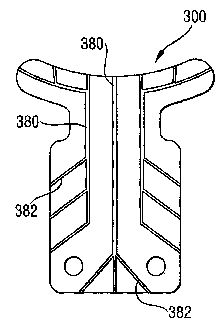
FIG. 9 is a plan view of an ophthalmic implant according to a third exemplary
embodiment of
the present invention, showing the implant also having lateral drainage
passageways.
FIG. 10 is a side view of an ophthalmic implant according to a fourth
exemplary embodiment
of the present invention, showing the implant having drainage passageways
formed by
channels in both outer surfaces of the implant.
FIG. 11 is a side view of an ophthalmic implant according to a fifth exemplary
embodiment of
the present invention, showing the implant made of two layers and having
drainage
passageways formed by channels in inner-facing surfaces of the implant.
FIG. 12 is a side view of an ophthalmic implant according to a sixth exemplary
embodiment
of the present invention, showing the implant made of three layers and having
drainage
passageways formed by voids in the inner layer of the implant.
DETAILED DESCRIPTION OF THE EXAMPLARY EMBODIMENTS
The present invention may be understood more readily by reference to the
following detailed
description of the invention taken in connection with the accompanying drawing
figures,
WO 2004/056294 CA 02516976 2005-08-22
PCT/EP2003/014532
- 6 -
which form a part of this disclosure. It is to be understood that this
invention is not limited to
the specific devices, methods, conditions or parameters described and/or shown
herein, and
that the terminology used herein is for the purpose of describing particular
embodiments by
way of example only and is not intended to be limiting of the claimed
invention. Also, as
used in the specification including the appended claims, the singular forms
"a," "an," and
"the" include the plural, and reference to a particular numerical value
includes at least that
particular value, unless the context clearly dictates otherwise. Ranges may be
expressed
herein as from "about" or "approximately" one particular value and/or to
"about" or
"approximately" another particular value. When such a range is expressed,
another
embodiment includes from the one particular value and/or to the other
particular value.
Similarly, when values are expressed as approximations, by use of "about,"
"approximately,"
or the like, it will be understood that the particular value forms another
embodiment.
The present invention provides ophthalmic implants and surgical methods for
implanting
them in the eyes of people or animals suffering from glaucoma to reduce
intraocular
pressure (10P). When using these implants and methods, outflow of ocular
fluid, the
aqueous humor, from the anterior chamber of the patient's eyes is increased
while better
stabilizing the implant in the eye. This eliminates or at least significantly
reduces the
likelihood of glaucoma resulting in blindness.
Referring now to the drawing figures, wherein like reference numerals
represent like parts
throughout the several views, FIGS. 1 and 2 show an ophthalmic implant
according to a first
exemplary embodiment of the present invention, generally referred to as the
implant 10. The
implant 10 has a body 12, one or more feet 14, and a neck 16 between the body
and the
feet. The body 12 can be positioned under a flap and in a recess surgically
created in the
sclera, the feet 14 can be positioned in the anterior chamber, and the neck 16
can be
positioned in an opening surgically created in the sclera between the scleral
recess and the
anterior chamber.
Preferably, the implant body 12 is generally rectangular. Alternatively, the
body 12 can be
triangular, polygonal, or it can have another shape. In a typical commercial
embodiment, the
body 12 is about 4.0 mm long and about 3.5 mm wide.
WO 2004/056294 CA 02516976 2005-08-22
PCT/EP2003/014532
- 7 -
The implant body 12 has outer portions 18 that can be tucked into undercuts
created at
bottom corners of and extending outward from the scleral recess. With the
outer portions 18
of the body 12 tucked into the undercuts, the implant 10 is held more securely
in place in the
eye. In a typical commercial embodiment, the body 12 has about 0.1 to .05 mm
outer
portions 18 at three sides. Alternatively, the body can have the outer
portions 18 at only one
or two sides. The outer portions 18 are typically the same thickness as the
rest of the body
12.
In addition, the implant body 12 is manufactured with suture holes 20 for
receiving sutures to
secure the implant to the sclera. Conventional implants typically do not have
suture holes,
so the surgeon has to pierce the implant body during the implantation surgery
to suture the
implant in place in the eye. The suture holes 20 in the body 12 simplify the
operation by
eliminating the need for surgeons to create the suture holes during surgical
implantation.
The implant feet 14 have a curvature 22 that is approximately the same as the
curvature of
the anterior chamber at the sclera of the patient. In a typical commercial
embodiment, the
curvature 22 has a radius of about 5.5 mm. This junction of the sclera and the
anterior
chamber (formed by the space under the cornea) is known as the limbus corneae,
or the
anterior chamber angle, or simply, the angle. Because the feet 22 are so
curved, they seat
with a close fit against the limbus corneae. While the seating edge 24 of the
feet 14 is so
curved, the opposite edge need not be curved.
In addition, the feet 14 have outer portions 26 that extend beyond the width
of the body 12.
In a typical commercial embodiment, the feet 14 extend about 1.5 mm from the
neck and the
outer portions 26 extend about 1.0 mm beyond the body 12. Because of the outer
portions
26, the feet 14 are long enough that they do not work their way out of the
anterior chamber
through the sclera! opening. So if the sutures were to fail, the outer
portions 26 of the feet
14 keep the implant 10 securely in place on the eye.
The implant neck 16 has a reduced width, relative to the body 12 and feet 16.
Additionally,
the implant neck 16 has a greater length than previously cited implants. With
the previously
cited implants having very short necks provided by a slit or notch, the
surgeon must cut the
scleral recess close (within a small fraction of a millimeter) to the limbus
corneae. If the cut
is too deep, it will penetrate into the anterior chamber and the opening may
allow the feet 14
WO 2004/056294 CA 02516976 2005-08-22 PCT/EP2003/014532
- 8 -
to pass through it even when unfolded. Because the feet 14 could then migrate
out of the
anterior chamber and into the sclera!, this site cannot then be used for the
conventional
implant. And because of the position of the rectus muscle, there are only four
scleral sites
where the implant can be readily implanted. But the neck 16 of the present
implant 10 is
sufficiently long that the body 12 is spaced apart from the anterior chamber
so that the
scleral recess does not need to be created immediately adjacent to the
anterior chamber. In
a typical commercial embodiment, the neck 16 is about 0.8 mm long, about one-
fifth of the
length of the body 12.
In addition, the length of the neck 16 is less than the length of the scleral
opening created
between the scleral recess and the anterior chamber. For example, as just
mentioned the
neck 16 can be about 0.8 mm long and the scleral opening can be made about 1.0
mm long,
which is the approximate thickness of the limbus corneae transition between
the sclera and
the cornea. So when the implant 10 is positioned in the eye, the neck 16 is
under tension.
And the part of the sclera between the scleral recess and the anterior chamber
is under
compression. By placing the neck 16 under tension, the implant is less able to
shift and
migrate in the eye.
Preferably, the implant 10 is made of regenerated cellulose, though other
materials with the
desired strength, softness, flaccidness, and ocular biocompatibility may be
selected.
Preferably, the material is flexible for conforming the shape of the eye and
so the feet can
fold in for implanting. The implant 10 can be manufactured by die-cutting or
other fabrication
techniques. In a typical commercial embodiment, the implant 10 has a generally
uniform
thickness of about 80 to 250 microns.
Referring now to FIGS. 3 ¨ 6, the present invention also provides surgical
techniques for
implanting the ophthalmic implant 10 in the eye of a person or animal with
glaucoma to
reduce 10P. It will be understood that these exemplary methods can be used
with other
implants as long as they have the body outer portions 18 and/or the neck 16 of
the implant
as described above.
FIG. 3 shows a portion of an eye 50 that has been surgically prepared for
implanting the
ophthalmic implant 10. The eye 50 has a sclera 52, a cornea 54, an angle 56 at
the junction
of the sclera and the cornea, an iris 58, an anterior chamber 60 between the
cornea and the
WO 2004/056294 CA 02516976 2005-08-22 PCT/EP2003/014532
- 9 -
iris, a posterior chamber 62 behind the iris, and a conjunctiva 64 covering
the sclera. The
surgical preparation includes the steps of creating a flap 66 and thus a
recess 68 under the
flap in the sclera 52, and creating an opening 70 in the sclera between the
sclera! recess 66
and the anterior chamber 60. The sclera! recess 68 is made with a size and
shape for
receiving the implant body 12 (but not the outer portions 18 of the body), and
the scleral
opening 70 is made with a size and shape for passing through it the implant
neck 16 and the
implant feet 14 when folded in.
As shown in FIGS. 4 and 5, the surgical preparation further includes the step
of creating
undercuts 72 in the sclera 52 extending outward from the sclera! recess 68.
The undercuts
72 are made with a size and shape for receiving the outer portions 18 of the
implant body
12. For example, the undercuts 72 can be made about 0.1 to 0.5 mm outward from
the
sclera! recess 68 for receiving outer portions 18 of about the same size. The
undercuts 72
can be made at three sides of the sclera! recess 68, or at fewer or more sides
if so desired.
Preferably, the undercuts 72 are made at bottom corners 74 of the sclera!
recess 68. The
sclera! recess 68, sclera! opening 70, and scleral recess undercuts 72 are
preferably made
by cutting with a scalpel 76, but they could alternatively be made by a laser
or by another
surgical technique.
FIG. 6 shows the implant 10 inserted into the eye 50. The insertion steps
include folding in
the implant feet 14 and inserting the folded feet and the neck 16 through the
scleral opening
70 so that the feet are inserted into the anterior chamber 60 and the neck is
positioned in the
scleral opening 70. The elasticity of the implant material causes the feet 14
to unfold so that
they do not migrate down out of the anterior chamber. The insertion steps
further include
inserting the implant body 12 into the sclera! recess 68 under the sclera!
flap 66 and
inserting the body outer portions 18 into the undercuts 72. With the implant
body 12 nested
within the scleral recess 68 and the body outer portions 18 tucked into the
sclera! undercuts
72, the implant 10 is constrained from shifting around on the eye 50.
After the implant 10 is inserted in the desired position in the eye 50,
sutures can be sewn
into the sclera 52 through the suture holes 20 in the implant 10 to further
stabilize it in place.
And the scleral flap 66 is sutured close to promote proper healing and help
stabilize the
implant 10.
WO 2004/056294 CA 02516976 2005-08-22 PCT/EP2003/014532
- 10 -
In another exemplary implantation method shown in FIG. 7, the sclera! opening
70 is made
with a length that is greater than the length of the implant neck 16. To do
this, the sceral
flap 66 and recess 68 are not made as close to the cornea 54. For example,
when using an
implant 10 with a 0.8 mm long neck 16, the scleral opening 70 may be made
about 1.0 mm
long, which is about the thickness of the limbus corneae, so the sceral flap
66 and recess 68
are made up to about 1.0 mm from the cornea 54. This puts the neck 16 under
tension
when it is implanted into the eye 50, and compresses the portion of the sclera
52 between
the sclera! recess 68 and the anterior chamber 60. With the implant neck 16
under tension,
it is better held in place and stabilized in the eye. It will be understood
that this method can
be performed in conjunction with the undercutting method described above or
separately.
Turning now to FIGS. 8-12, in other exemplary embodiments of the present
invention the
implant has one or more drainage passageways for the ocular fluid to flow
through out of the
anterior chamber 60 and into the sclera 52. After implanting the implant, the
sclera! flap 66
tends to heal back into its original position. After healing, ocular fluid
need not flow out of
the scleral through the scleral flap incisions because the lymphatic vessels
in the sclera 52
absorb and disperse the ocular fluid. In order to increase the amount of
ocular fluid drained
out of the anterior chamber 60, the implant is provided with the drainage
passageways to
facilitate ocular fluid drainage to the sclera 52 for dispersing by the
lymphatic vessels. The
drainage passageways are formed in the outer surfaces of the implant, in the
interior of the
implant, or in both. The drainage passageways may be formed by die-stamping,
laser
ablation or by other fabrication techniques.
FIG. 8 shows a second exemplary embodiment of the invention with the implant
200 having
drainage -passageways provided by longitudinal drainage passageways 280. The
longitudinal drainage passageways 280 provide a route for the ocular fluid to
flow through,
instead of just migrating through the cellulose material of the implant 200.
Any number of
the longitudinal drainage passageways 280 can be provided, depending on the
amount of
drainage desired and limitations imposed by the size of the implant 200.
FIG. 9 shows a third exemplary embodiment of the invention with the implant
300 having
drainage passageways provided by longitudinal drainage passageways 380 as well
as lateral
drainage passageways 382. The lateral drainage passageways 382 deliver the
ocular fluid
across the implant 300 to its sides for dispersing the fluid over a larger
area into the sclera
WO 2004/056294 CA 02516976 2005-08-22PCT/EP2003/014532
-11 -
52 for better absorption by the lymphatic vessels. If desired, main portions
of the
longitudinal drainage passageways 380 and/or the lateral drainage passageways
382 may
be thicker or deeper than branch portions, so as not to create a bottleneck in
the fluid flow
delivery system. Any number of the lateral drainage passageways 382 may be
provided,
depending on the amount of drainage desired and limitations imposed by the
size of the
implant 200. Of course, the implant 300 may be provided with the lateral
drainage
passageways 382 but without the longitudinal drainage passageways 380, if so
desired.
FIG. 10 shows a fourth exemplary embodiment of the invention with the implant
400 having
the drainage passageways formed by channels 484 in both outer surfaces 486 of
the
implant. The channels 484 may be configured in alignment with each other (one
over
another), or they may be staggered in a regular or irregular pattern.
FIG. 11 shows a fifth exemplary embodiment of the invention with the implant
500 made of
two layers 588 and 589, and having the drainage passageways formed by channels
590 and
591 in inner-facing surfaces 592 and 593 of the implant layers. The layers 588
and 589 may
be laminated together or folded over each other, and more than two layers may
be provided,
if desired. The channels 590 and 591 may be configured to align with each
other (one over
another), or they may be staggered in a regular or irregular pattern.
FIG. 12 shows a sixth exemplary embodiment of the invention with the implant
600 made of
three layers, with two outer layers 694 and one inner layer 695. The drainage
passageways
are formed by one or more voids 696 in the inner layer 695 of the implant 600.
The layers
694 and 695 may be laminated together or folded over each other, and more than
one inner
layer may be provided, if desired. And the layers 694 and 695 may have also
surface
channels in alignment with the voids 696 or staggered in a regular or
irregular pattern.
In another embodiment contemplated by the present invention, the implant feet
extend
directly from the body instead of indirectly from the body with the neck in
between. The body
does not migrate through the sclerat opening into the anterior chamber,
however, because
the sutures hold it in place. In still another embodiment, the implant has
dimensions that are
larger than or smaller than those of the typical commercial embodiments for an
average-
sized adult as described above. For example, smaller implants could be used
for children
and/or pets, and larger ones for large adults and/or animals.
WO 2004/056294 CA 02516976 2005-08-22
PCT/EP2003/014532
- 12 -
Accordingly, the present invention provides an improved ophthalmic implant for
lowering the
10P in the eyes of glaucoma patients. In some embodiments, the ophthalmic
implant has
one or more drainage passageways formed in the outer surfaces and/or the
interior of the
implants to drain more ocular fluid out of the anterior chamber of the eye. In
further
embodiments, the implant has a uniquely configured body, feet, and/or neck to
increase the
stability of the implant in the eye. Furthermore, the present invention
provides surgical
implantation methods including providing undercuts and scleral openings sized
to better
stabilize the implants securely in place. And the implants are preferably of a
simple
construction using known materials such that they are time and cost-effective
to
manufacture and implant.
While the invention has been disclosed in exemplary forms, those skilled in
the art will
recognize that many modifications, additions, and deletions can be made
therein without
departing from the spirit and scope of the invention as set forth in the
following claims.