Note: Descriptions are shown in the official language in which they were submitted.
CA 02518587 2005-09-08
WO 2004/086042 PCT/FI2004/000179
Multiple-channel test device, method for producing the same and use thereof
Technical Field of the Invention
The invention relates to an immunodiffusion-based multiple-channel test device
that enables the
simultaneous or parallel performing of several different analyses. The
production and use of the
device are also disclosed in the invention.
Background of the Invention
Methods and test devices based on immunodiffusion are known for example from
the following
patents and patent applications: US 4,757,002, US 3,990,852, US 4,562,147, and
EP 0 250 137.
Immunochromatographic methods based on immunodiffusion have been disclosed for
example
in the following patents and patent applications: EP 0 291 194, EP 0 284 232
(FI 93150), and
WO 86/03839.
The above-mentioned test devices based on immunodiffusion and
immunochromatography are
most often only used for performing one analysis per test device. In many
cases, making a
definite diagnosis and selecting the suitable method of treatment for a
patient suffering from a
specific illness or syndrome might require performing several different
analyses. If the
recognition of the cause of a disease causing the symptoms and the illness or
the exclusion of
specific illnesses requires performing several different tests, making a
diagnosis would become
so expensive that usually only one or a very restricted number of analyses are
performed, these
analyses being randomly chosen based on the judgment of the treating physician
or among
commonly used tests, in which case other alternatives might be left undefined.
The use of more than one test channel is known from US B1 6,171,870 and US Al
2003/0040021. US B1 6,171,870 relates to a method for producing such channels
by applying a
water-repellant substance, such as wax on a porous Garner. The treated areas
form blocking
segments between the channels. In the method described in US Al 2003/0040021
samples
moves along treated channels, while untreated areas are left between the
channels.
Advantages of the present invention over prior art methods and test devices
include a decrease
in reagent and material consumption, sample volume and freight, as well as
improvements in
CA 02518587 2005-09-08
WO 2004/086042 PCT/FI2004/000179
2
environmental friendliness, shelf life and user-friendliness. An increment in
environmental
friendliness is achieved on acco~~r!~ of a diminished environmental load
caused by used test
devices. Due to the savirbs in reagent consumption and labour costs, the
method according to
the invention enables the production of the test devices with extremely low
costs.
The problems connected with conventional test devices based on immunodiffusion
and
immunochromatography are overcome with the present invention, the
characteristics of which
are disclosed in the following claims.
Summary of the Invention
The immunodiffusion-based test device of the present invention comprisies a
porous Garner
material and in it, a specific binding reagent (immunoreagent) in the form of
a zone or a blot. A
detectable label to which a second specific binding reagent (immunoreagent) is
coupled, is
supplied separately or pre-applied to the porous Garner, and is mobilizable by
the sample. A
sample application site, optionally provided with a filter, is also situated
on the test device. The
porous carrier material of the test device is preferably made of
nitrocellulose and comprises a
network of channels, which is etched into the porous carrier material with
laser treatment. The
network of channels comprises two or more channels (1), separated by a treated
area (2), one or
more specific binding reagents (3) immobilized in them, an optional label site
(4) placed in the
channel near the sample application site (5), or in the sample application
site (5) itself, which is
placed in a way that enables an even distribution of the sample into each
channel.
The test device of the present invention enables diagnosis of different
diseases or syndromes
based ona simultaneously or parallelly performed recognition of several
syndrome-producing
agents or analytes. The test device may be used to simultaneously recognize
several allergy-
producing agents, myocardial infarction markers, venereal disease-producing
agents, blood
screening analytes, respiratory infection-producing agents, infectious disease-
producing agents
and/or cancer markers.
Example of useful specific binding reagents are antibodies, antibody
fragments, recombinant
antibodies, recombinant antibody fragments, antigens, lectins, receptors
and/or ligands. Useful
label reagents are for example latex, gold, metal or colouring agent
particles, or fluorescent
substances.
CA 02518587 2005-09-08
WO 2004/086042 PCT/FI2004/000179
3
The invention dislcoses a method for producing the test device, wherein the
specific
recognizing immunoreagent is immobilized in a porous material, which has been
rendered inert
with a substancet, and wherein the sample application site is optionally
provided with a filter
and a detectable label to which a specific binding reagent is bound. A channel
network of a
desired shape and size is formed on the porous material by etching the porous
material with
laser. The network comprises two or more channels (1) and a treated area (2);
and one or more
recognizing specific binding reagents are immobilized in each channel (1) as a
zone or blot (3).
Useful substance by which the porous material is rendered inert is a mixture
comprising natural
or synthetic polymers, such as albumin and casein or PEG (polyethylene glycol)
and PVA
(polyvinyl alcohol), nonionic detergents such as hexane sulphonic acid and
TRITON-X-100,
BRIJ, and preservatives such as sugar, for example sucrose and trehalose, or
their derivatives.
The storability of the test device is highly improved by drying and keeping
the the test device in
a relative humidity of not over ~ % and hermetically packed after application
of the reagents.
The multichanneled test device is useful for performing simultaneously or
parallelly several
assays, which enable the diagnoses of a target disease and/or syndrome, which
may be diffiult
to diagnose using only one marker. The sample is applied to the application
site (5) of the test
device containing a label mobilizable by the analyzable sample and a second
specific binding
reagent coupled to it, from which site the sample and the reagents migrate
evenly into the
channels of the channel network formed by the laser treatment, where they
either react or do not
react with the specific binding reagent in its immobilization site (3), from
which site the
positive or negative results are directly readable.
The present invention is also related to a test device for simultaneous or
parallel performing of
several assays for recognition of the target disease and/or syndrome. The the
sample is applied
to an application site (5) of the test device, from which it migrates into a
channel where it is
mixed to and reacts with the label mobilizable by the sample and the second
specific binding
reagent bound to it, after which the sample and reagents migrate evenly into
the channels of the
channel network formed by a laser treatment, where they either react or do not
react with the
specific binding reagent in its immobilization point (3), from where the
positive or negative
results are directly readable.
CA 02518587 2005-09-08
WO 2004/086042 PCT/FI2004/000179
4
The present invention is also related to the use of the test device for
simultaneous or parallel
performing of several assays for recognition of a target disease andlor
syndrome. The sample is
mixed with a separate label having a second specific binding reagent coupled
to it and the
mixture is applied to an application point (5) of the test device, from which
the sample and the
reagents migrate evenly into the channels of the channel network formed by a
laser treatment,
where they either react or do not react with the specific binding reagent in
its immobilization
point (3), from where the positive or negative results are directly readable.
Brief Description of the Drawings
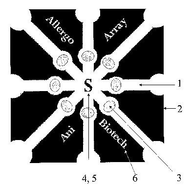
Figure 1 shows a network of eight channels, which is one of the preferred test
devices for
performing up to eight different assays. The label site 4 simultaneously
functions as the sample
application point 5 and the channels 1 separated by treated areas 2 contain
the specific binder as
the zone 3.
Figure 1 a shows an advanced embodiment of the multiple-channel test device
for performing
up to eight different assays according to Figure 1. The label point 4
simultaneously functions as
the sample application point 5 and the channels 1 separated by treated area 2
contain specific
binder as the zone 3. The device is marked with information concerning the
tests and the
manufacturer.
Figure 2 shows a test device that can be used for performing six different
assays and their
control reactions or, optionally, up to twelve different assays by six
different labels. One of the
channels 1 branches into two channels la, lb that are separated by treated
area 2. The sample
application point is located in the middle of the channel network and each
channel 1 comprises
a label point 4 and one or more binder zones 3.
Figure 3 shows a test device that can be used for performing four different
assays and their
control reactions, or optionally, up to eight different assays by four
different labels.
Figure 4 shows a test device that can be used for performing four different
assays and their
control reactions, or optionally, up to eight different assays by four
different labels.
CA 02518587 2005-09-08
WO 2004/086042 PCT/FI2004/000179
Figure 5 shows a test device that can be used for performing up to six
different assays, or
optionallythree assays and their control reactions.
Figure 6 shows a test device that can be used for performing twelve different
assays by four
different labels.
Figure 7 shows a network of eight channels, which is one preferred test device
for performing
up to twenty-four different assays. Each channel 1 comprises three binder
zones 3.
Figure 8 shows a test device that can be used for simultaneously performing
sixteen different
assays.
Figure 9 shows a test device that can be used for performing twenty different
assays by five
differentlabels.
Figure 10 shows a test device that can be used for performing two assays and a
control reaction.
The device is marked with information concerning the tests and the
manufacturer.
Figure 11 shows a test device that can be used for performing two assays and a
control reaction.
The device is marked with information concerning the tests and the
manufacturer.
Figure 12 shows a test device produced by laser etching that can be used for
performing three
parallel assays or, optionally, three individual assays and their control
reactions. The device is
marked with information concerning the tests and the manufacturer.
Detailed Description of the Invention
The object of the invention is an immunodiffusion-based multiple-channel test
device
comprising porous Garner material and in this material, one or more specific
binding reagents
(immunoreagents) in the form of zones) or blots) and a sample application
point that is
optionally equipped with a filter for example for the removal of blood cells,
and which
optionally comprises a label mobilizable by the analyzable sample, coated with
a second
specific binding reagent (immunoreagent). The label is on the test device or
it is added to the
sample.
CA 02518587 2005-09-08
WO 2004/086042 PCT/FI2004/000179
6
The test device according to the invention is characterized in that it
comprises a channel
network produced by etching a network on a porous carrier material for example
by laser. The
channels of the test device have been provided with one or more identical or
different specific
binding agents that are selected from different marker groups, whichcan be
used together and
are required for diagnosing a specific syndrome. Thereby, the diagnosis is
enabledby the
simultaneous performing of several assays for the recognition of a specific
target illness and/or
syndrome. The sample application site, which can be in the form of a dot or a
blot or a line or a
zone, is located for example in the middle or the other end of the strip in a
way that enables an
even distribution of the sample into each of the channels. If the same
specific binding reagent is
used in the channels in different concentrations, a semi-quantitative result
can be obtained.
The test device according to the invention comprises several channels that can
be used for the
simultaneous or parallel determination of several analytes. In the test
device, it is possible to
optionally group together reagents that recognize disease-producing agents
associated with
various syndromes, for example allergens, myocardial infarction markers,
suitable reagents for
the recognition of venereal diseases and for blood screening, markers that
recognize respiratory
infection producing agents, IgG, IgA and IgM antibodies, markers that
recognize other
infectious disease producing agents as well as various cancer markers, for
their simultaneous or
parallel determination.
An additional object of the invention is a method of producing said test
device. The production
method is characterized by that a multiple-channel channel network of a
desired shape and size
(see Figs. 1-12) is etchedon a porous material by laser-techniques, which make
a certain shape
on the substrate. Different recognizing specific binding reagents are
permanently attached, i.e.
immobilized, in the porous substance of each of the channels. The analysis
results for the test
reactions are readable from these points.
After this, the porous material is treated with a substance that renders the
free reactive sites
inert, i.e. they do not react in an undesirable way, for example by slowing
down or preventing
the analytes or labels from migrating into the permanently immobilized reagent
zone or blot.
The detectable, optionally visible label, on which a second specific binding
reagent is bound, is
applied to the sample application point or to the test channel on a pre-
determined place. The
CA 02518587 2005-09-08
WO 2004/086042 PCT/FI2004/000179
label to which the binding reagent is bound can also be added to the sample,
in which case the
label is transferred into the device as sample is added to the sample
application point.
In addition, the use of the test device for the simultaneous or parallel
performing of several
assays to recognize a certain illness and/or syndrome is described in the
invention. The
branched channels may also be used for controlling the appropriate functioning
of the device,
using one of the channels as a test channel and the other as a functionality
or a control channel.
A separate control channel indicating the functionality of the device and the
reagents can also
be added to the test device.
The multiple-channel test device according to the invention as well as its use
in diagnostic
methods differ from the prior art test devices disclosed in the above-
mentioned patents and
patent applications in that one or more different or identical parallel tests,
preferably 2-40,
more preferably 4-30, and most preferably 8-24, parallel tests can be
performed by using an
extremely small object produced from a porous material.
The use of laser for producing the channel network is a preferred method for
the manufacture of
the test devices according to the invention. The machine used in this
production method is
inexpensive, and operating costs mainly consist of the amount of electricity
used. As compared
to the use of a water-repellent substance such as hot wax, by laser
manufacturing the use of
chemicals during the manufacturing process can be avoided. By using laser, a
precise etching
result (printing) can be obtained, and because of the preciseness of the
printing (impression),
the testing devices may, if desired, be marked in connection with the
manufacturing process,
thereby diminishing the risk of mixing up the devices.
Large amounts of tests may be manufactured by etching single channels,
parallel channels or
channel networks on a roll and adding the required reagents and treatments on
the same
production line and finally separating the tests from each other by a desired
manner into single
tests for one or several assays or into test combinations of several parallel
tests.
In addition, the test device can be used for analyzing small volumes of
patient samples, such as
urine, blood, plasma, serum, saliva, tissue fluids, faeces, environmental
samples, etc. The small
sample volume, even as small as 1,0-50 ~,1, preferably 2,0-40 ~,1, more
preferably 4,0-20 ~.1, or
most preferably 5,0-10,0 ~,1 per test device being sufficient, advantages are
gained especially in
CA 02518587 2005-09-08
WO 2004/086042 PCT/FI2004/000179
8
regard to performing assays on samples collected from small children or forger-
tip blood
samples.
Producing the test device
The production of the test device comprises six main steps. Forming the
channel network,
applying and immobilizing the specific reagent, rendering the porous channel
inert, applying
the label coated with a second specific reagent, producing the sample
application point and
stabilizing the test device and ensuring its storing properties. Between the
main steps
mentioned, the test strip may be dried. If the label is added to the sample,
the step of applying
the label to the test device can be omitted.
In the test device according to the invention several different or identical
parallel tests (Figs. 1
12) can be performed on a very compact item of a porous material by
channelling the test
device.
Producing the channels
The channelling of the reagents and the samples can be achieved by treating a
porous, water-
transmitting material, for example nitrocellulose, polysulphonate, nylon or
paper, with a water-
repellent or partly water-repellent substance, such as wax, polyolefmes,
polyacrylamide paints
or mixtures thereof. The application of water-repellent materials on the
porous material is
performed at their melting temperature, preferably at a temperature between 50
°C and 80 °C,
advantageously by using a printing, brushing or spraying technique, thus
forming on the porous
material a water-repellent figure that is of a desired shape and that encloses
a channel network
of several channels separated from each other by the treated area, along which
channels the
sample migrates due to the effect of capillary action and diffusion. After the
application, the
substrate is immediately cooled down to room temperature. It is possible to
apply the reagents
into the channel network in a way that maintains their reactivity.
Naturally, the channels can also be produced by stamping out a suitable-size
piece of carrier
material to form a channel network of a desired shape and size.
However, according to the present invention the channelling is preferably
performed by treating
a porous and water-transmitting material, such as nitrocellulose, with laser.
By using suitable
laser power, nitrocellulose can be etched away from such areas of the
nitrocellulose substrate
CA 02518587 2005-09-08
WO 2004/086042 PCT/FI2004/000179
9
into which fluid flow is not wanted, leaving the treated areas covered only by
the plastic film
(Mylar film) underlying the cellulose. Figures of different shapes (Figs. 1-
12) are formed by
the channels containing nitrocellulose or some other porous material and the
channel edges or
treated areas that extend all the way to the substrate edges and have been
laser-etched by using
a suitable pre-programmed computer program, using 10-90 % of the maximum
output power of
the device, preferably 10-80 %, more preferably 12-60 %, most preferably 15-40
%, an
etching speed that preferably ranges from 100 mm/s to 1 500 mm/s, more
preferably from 400
mm/s to 1 000 mm/s, and a resolution that ranges from 50/1 000 to 1 000/1 000,
preferably
from 100/1 000 to 800/1 000, more preferably from 150/1 000 to 500/1 000, most
preferably
from 200/1 000 to 300/1 000.
The treated area separating the channels may extend all the way to the edges
of the test device
or, optionally, untreated area may be left on figure edges. The test device
may be marked with
information concerning the manufacturer or the tests by etching, for example
by laser treatment.
The application of the reagents into the carrier
It is possible to apply and, if needed, to immobilize the various reagents
required for
performing the tests in the channel network. The reagents may be used in very
small volumes.
The required reagents comprise at least two specific binding reagents, of
which one is
immobilized and the other bound to the label mobilizable by the sample
solution, and at least
one detectable label.
Specific binding reagents
Suitable substances include various binding reagents, specific immunochemical
reagents such
as antibodies, antibody fragments, recombinant antibodies, recombinant
antibody fragments,
antigens, but also other ligands such as receptors, lectins, biotin, avidin,
etc. Especially suitable
are monoclonal and polyclonal antibodies, antigens and fragments thereof. As
regards allergy
testing, suitable allergens include extracts prepared from the pollen of
trees, grass or weed,
extracts prepared from spores of moulds, acarids, household dust, animal skin
epithelium,
insects, latex, parasites, drugs or foodstuff, among others.
Binding reagents may be immobilized or chemically or physically attached by a
known method
to the structure of the porous earner on the desired points, and a binding
reagent for a control
CA 02518587 2005-09-08
WO 2004/086042 PCT/FI2004/000179
reaction may also optionally be immobilized or chemically or physically
attached to the same or
a branched channel.
Renderin _~ the porous substance inert
After producing the channel network and applying the specific binding reagent,
the porous
carrier substance is rendered inert by using a so-called blocking agent,
whereby the free
reactive sites of the carrier substance and the non-specific binding sites of
the specific binding
reagents are eliminated by a suitable mixture containing natural and/or
synthetic polymers such
as albumin or casein and/or PEG (polyethylene glycol) and PVA (polyvinyl
alcohol), nonionic
detergents such as hexane sulphonic acid and TRITON-X-100, BRIJ, and
preservatives such as
sugar, for example sucrose and trehalose, or their derivatives. After this,
the test device is dried.
If desired, the blocking agent may be added only after the production of the
test device, in
connection with the application of the sample.
Producing; the label point
Suitable labelling agents include various plastic or metallic microparticles
mobilizable by
sample flow, such as latex, gold, liposomes, colouring agents, fluorophors,
fluorochromes or
other such particles of a metallic substance or a colouring agent that are
able to bind the
analytes and bind to the specific binders such as antibodies, receptors,
lectin or other ligands.
Also fluorescent particles, fluorescent colouring agents or superparamagnetic
particles may
function as labels.
The label may be immobilized in the porous carrier, to the sample application
point of the test
device or to an another point separate from the sample application point. The
labelling agent
may also be added to the analyzable sample before it is applied on the test
strip.
Producing the sample application point
The sample application point may optionally be provided with a filter by
placing on the sample
application point a filter that prevents for example blood cells from being
carried into the
channels. The sample application point may be provided with a label to which a
second specific
binding reagent has been bound.
CA 02518587 2005-09-08
WO 2004/086042 PCT/FI2004/000179
11
Stabilizing the test device and ensuring its storing pro ep rties
The test device can be made storable by treating it for example by drying to
give it a relative
humidity of below 8 %, after which the test device is hermetically packed and
stored dry in
order to maintain the relative humidity of the test device below 8 %. Thus,
the device maintains
its functionality for as long as 24-36 months, without any substantial
decrease in functionality,
including sensitivity or specificity.
When necessary, the test device may be placed inside a casing made of
paperboard or plastic,
which can, where needed, be equipped with instructions describing the
operation and use of the
test device.
The structure of the test device
The structure of the test device and its possible variations are described in
Figures 1-12.
The test device comprises of several, preferably 2-10, more preferably 3-8,
channels 1 which
may on the branch point further divide into several, preferably 2-5 (la,
lb...), more preferably
2-3, branched channels. A treated area 2 separates the channels 1 from each
other and directs
the migration of the sample in the test device.
The channels 1 or branched channels (1a, lb...) contain one or more binding
reagents as a
binder zone or blot 3. In addition, the test device comprises one or more
label points 4, which
may optionally be combined with the sample application point 5. The sample
application point
may optionally be provided with a filter.
Figure 1 shows a test device wherein a nitrocellulose film of a desired size,
e.g. 25 x 25 mm,
has been divided into eight identical channels 1. The channels have been
prepared by printing a
figure on a porous material, for example on nitrocellulose filin, and
transfernng hot, melted,
about 60 °C polyolefin on the treated water-repellent zones 2 of the
nitrocellulose film with a
stamping device. Each channel contains a specific binder 3. The label point 4
in the middle of
the test device contains a second label reagent directed against the analyte
to be determined.
The sample application point 5 simultaneously functions as the label point 4
of the test device.
The test device according to Figure 1 is characterized in that each of the
channels may contain a
different specific binder (e.g. an allergen) and that a particle label (e.g.
anti-human IgE)
common to each of the binders is located in the middle of the channels.
CA 02518587 2005-09-08
WO 2004/086042 PCT/FI2004/000179
12
Figure la shows a test device wherein the nitrocellulose film has been divided
into eight
identical channels 1. The channels have been produced by laser-etching a
figure on a porous
material such as nitrocellulose film in a way that a treated area 2 is left
between the channels,
on which area markings concerning the tests and the manufacturer have been
added during the
laser treatment. Each of the channels contains a specific binder 3. Located in
the middle of the
test device, the label point 4 contains a second label reagent against the
analyte to be
determined. The sample application point 5 simultaneously functions as the
label point 4 of the
test device. The test device according to Figure 1 is characterized in that
each of the channels
may contain a different specific binder (e.g. an allergen), and that a
particle label (e.g. anti-
human IgE) common to each of the binders is located in the middle of the
channels.
Figure 2 shows a test device wherein one of the channels 1 comprises branched
channels 1 a and
lb containing the specific binder in the binder zone 3. Other channels 1
comprise two binder
zones 3, of which the other may be a control zone. All channels 1 contain a
separate label point
4. At its centre, the device comprises a separate sample application point 5.
Figure 3 shows a test device wherein four identical or non-identical channels
1 comprise the
branched channels l a, lb that contain specific binders in the binder zone 3
and downstream
from the branch points of the channels four separate particle label points 4
that have been
coated with specific binders. The device comprises a separate sample
application point 5.
Figure 4 shows a test device with four identical or non-identical channels 1
that branch into
channels la, lb, which in turn contain specific binders in the binder zone 3
and downstream
from the branch points of the channels four separate particle label points 4
coated with specific
binders. The device comprises a separate sample application point 5.
Figure 5 shows a test device wherein the sample application point is located
at one end of the
test device or strip. The device comprises three channels 1 with two binder
zones 3 located into
each of them. The label point 4 is the same as the sample application point 5.
Figure 6 shows a test device that comprises up to twelve different binders in
twelve separate
binder zones 3 in four different channels 1 that further branch into three
channels 1 a, lb and 1 c.
Furthermore, the device comprises four separate label points 4. The test
device comprises a
separate sample application point 5.
CA 02518587 2005-09-08
WO 2004/086042 PCT/FI2004/000179
13
Figure 7 shows a test device comprising eight identical channels 1 and between
them treated
areas 2. Each chasmel 1 contains three specific binders, optionally against
different analytes, in
three separate binder zones 3. The label point 4 in the middle of the test
device contains another
label reagent against the analyte to be determined.
Figure 8 shows a test device comprising eight channels 1, each branching into
two channels la
and lb and containing a binder zone 3. The device comprises a combined label
and sample
application point 4, 5.
Figure 9 shows a test device comprising five channels 1, each channel
comprising four binder
zones 3. The device comprises four separate label points 4 and a sample
application point 5.
Figure 14 shows a test device for detecting virus antigens, in which the
sample application point
is placed at one of the ends of the test device or strip. The device comprises
three channels 1,
each of them containing a binder zone and a separated label point 4. The
device is marked with
information concerning the test and the manufacturer.
Figure 11 shows a test device for myocardial infarct detection, in which the
sample application
point 5 is placed at one of the ends of the test device or strip. The device
comprises three
channels 1, each of them containing a binder zone 3 and a separated label
point 4. The device is
marked with information concerning the test and the manufacturer.
Figure 12 shows three test devices, in which the sample application point 5 is
placed at one of
the ends of the test device. The devices comprise a label point 4 and binder
zones 3, of which
the other is a control point.
The form and size of the above described test devices are only to be seen as
examples of those
that the process according to the present invention enables to produce.
Use of the test device to perform a simultaneous or parallel assav
The present invention is based on fluid flow in a porous material such as
nitrocellulose taking
place practically in every direction at the same speed due to diffusion and
capillary action as is
well known. The said radial or lateral flow enables the movement of the
sample, that contains
CA 02518587 2005-09-08
WO 2004/086042 PCT/FI2004/000179
14
the analyzable analytes and test reagents, into several channels that
simultaneously serve as
different tests, as far as the channelization of the porous material has been
taken care of in a
secure maxu~.er. The sample applied to the test device reacts with the label
added to the sample
or with one or more labels applied to the device and this complex in turn
reacts with the
immobilized binders placed further in different channels of the test device.
Also immunological
reagents can be immobilized without interfering with their functionality. The
application of the
sample to the test device is performed by applying a small amount of the
sample or its dilution
to the sample application point 5, from where it moves to the label points) 4
and further along
the channels into the test zones 3, where the reaction between the specific
binder of the label,
the analyte in the sample and the other specific binder bound to the solid
carrier takes place.
This reaction is rendered visible to the naked eye if the label has been a
coloured particle or it
can be read under UV-light by naked eye as a light emitting dot, blot, line or
other figure or it
can be read and interpreted with a suitable device, photometer, fluorometer or
a device that
measures the changes of the magnetic field.
In another embodiment the label point 4 is placed in the sample application
point 5, in which
case the analyte in the sample and the specific binder of the label form a
complex before the
sample flows along the channels.
In a further embodiment the application of the label to the sample is
performed before adding
the sample to the test device.
The test device is especially useful in allergy testing, where a relatively
large amount of serum
is now required to define the allergy specific antibodies of class IgE. When
applying the present
invention, the sensibilization of a patient to up to 24 or more different
allergens can be detected
from a very small amount of sample, even only 10-4.0 ~1 of serum, plasma or
whole blood,
simultaneously with the same test device by dosing several different allergens
to each channel
of the test device.
In one preferred embodiment of the present invention several different labels
are attached to the
test device. Thus, one small sample mobilizes the labels that are optionally
different from each
other and placed in different channels. A test device like this is well suited
for immunoglobulin
specific antibody assays against different disease producing agents.
CA 02518587 2005-09-08
WO 2004/086042 PCT/FI2004/000179
Examples of applying the test
The following embodiments are to be seen as examples and it should be
understood that other
possible applications of the present invention are obvious to a person skilled
in the art.
Example 1. Forming a channel network by laser
The figure shown in Figure la is prepared on a porous nitrocellulose carrier
by etching
nitrocellulose from the areas to which fluid flow is not wanted by using a
Domino DGM-1
"High Resolution Laser Marker" device. A network that comprises eight channels
1 separated
by treated areas 2 is formed on the test device. To avoid the risk of the
devices getting mixed
up, markings concerning the product and the manufacturer are formed by leaving
nitrocellulose
unetched .
The desired figure pre-programmed into the computer programme is etched by
using a marking
speed of 700 mm/sec of 10-2500 mm/sec and 20 % of the maximum output power of
20W
Laser (~5-132 V/170-260 V, I Phase input and 20W output) and a resolution of
250/1000.
The thus produced test device matrix with a channel network is used according
to the examples
below for performing several assays simultaneously.
Example 2. A test device for allergy testing
Specific analyzable allergens are dosed as solutions (in a concentration of 1-
5 mg/ml) to the
channels 1 of the network of the test device produced according to Example 1,
so that they form
insoluble zones 3 as they attach to the nitrocellulose of the sample
application point. Different
allergens are applied to each channel (extracts prepared from the pollen of
trees, grass or weeds,
extracts prepared from mould spores, acarids, household dust, animal skin
epithelium, insects,
latex, parasites, drugs or foodstuff). After this, the reactive nitrocellulose
of channels 1 is
blocked, i.e. rendered inert, by using a solution containing bovine serum
albumin (BSA) (0,1-
5,0 %), hexane sulphonic acid and trehalose (1,0-3,0 %). The solution (10-100
~,1) is applied
into the middle of the test device, after which it migrates into each channel
due to diffusion and
capillary action and binds, i.e. blocks, the free reactive points of the
nitrocellulose.
CA 02518587 2005-09-08
WO 2004/086042 PCT/FI2004/000179
16
After this, the test device is dried at room temperature until its relative
humidity is below ~ %.
After drying, 1,0-10,0 ~.1 of an aqueous solution containing coloured latex
particles coated with
anti-IgE (0,2-1,0 %), the mixture further containing 0,1-1,0 % of BSA, 0,01-
0,05 % of Tween
20 and 0,5-1,5 % of trehalose is manually applied into the middle of the test
device. The
particle sol is dried into the middle of the test device.
When using the test, 10-50 ~l of a senun or its dilution from a person
suffering from an allergy
is applied into the middle of the test device to the sample application point
5. The sample
dissolves the label in the middle. The anti-IgE antibodies of the label react
with the allergy
specific IgE molecules and diffuse into the test channels due to capillary
action.
If the analyzable sample contains specific IgE antibodies against one or more
allergens in the
zones 3 of the test device, a coloured zone is formed in the reaction channel
that contains the
antibody of the allergen in question.
Example 3. A test device for allergy testing
Using a suitable stamp, a figure formed as shown in Figure 1 is printed on the
porous
nitrocellulose carrier, wherein a channel network is formed, by transferring
coloured about 60
°C polyolefin on the porous carrier so that it blocks the pores of the
Garner in the desired areas
and a network that contains eight channels 1 separated by treatedareas 2 is
formed on the test
device.
The specific allergens to be tested (in a concentration of 1-5 mg/ml) are
applied to the channels
of this network so that they form insoluble zones 3 attaching to the
nitrocellulose of the sample
application point. Different allergens are applied to each channel (extracts
prepared from the
pollen of trees, grass or weeds, extracts prepared from mould spores, acarids,
household dust,
animal skin epithelium, insects, latex, parasites, drugs or foodstuff). After
this, the reactive
nitrocellulose of the channels 1 is blocked, i.e. rendered inert, by a
solution containing bovine
serum albumin (BSA) (0,1-5,0 %), hexane sulphonic acid and trehalose (1,0 -3,0
%). The
solution (10-100 ~,1) is applied into the middle of the test device, after
which it migrates into
CA 02518587 2005-09-08
WO 2004/086042 PCT/FI2004/000179
17
each channel due to diffusion and capillary action and binds, i.e. blocks, the
free reactive points
of the nitrocellulose.
After this, the test device is dried at room temperature until its relative
humidity is below ~ %.
After drying, 1,0-10,0 ~,1 of an aqueous solution containing coloured latex
particles coated with
anti-IgE (0,2-1,0 %), where the mixture further contains BSA 0,1-1,0 %, Tween
20 0,01-0,05
and trehalose 0,5-1,5 %, is applied in the middle of the test device. The
particle sol is dried
into the middle of the test device.
When using the test, 10-50 ~,1 of a serum or its dilution from a person
suffering from an allergy
is applied into the middle of the test device to the sample application point.
The sample
dissolves the label placed in the middle. The anti-IgE antibodies of the
sample react with the
allergy specific IgE molecules and diffuse into the test channels due to
capillary action.
If the analyzable sample includes specific IgE antibodies against one or more
allergens placed
in the zones 3 of the test device, a coloured zone is formed in the reaction
channel that contains
the antibody of the allergen in question.
Example 4. A test device for venereal disease testing and blood screening
Example 4 describes the application of the present invention to venereal
disease testing where
both antibodies and viral- and bacterial antigens, are determined
simultaneously from patient
samples.
The test device shown in Figure 2, in which a multiple-channel network is
produced as
described in Example 1, is further prepared so that the first channel 1 that
branches into two
parts forms test channels for HIV 1 and HIV2 antibody tests. The polypeptide
or recombinant
antigens typical of the virus in question, are placed in these channels as
lines (0,1-0,5 p,l), in a
concentration of 0,5-5,0 mg/ml. A coloured label point 4, produced by coating
for example
gold particles with a third polypeptide or recombinant antigen that recognizes
HIV1 and HIV2
viruses, is placed in the channel 1.
The second channel 1 of the same test device is used to detect a HIV virus
antigen by placing
for example a line or blot of a monoclonal antibody (0,5-5,0 mg/ml) produced
against the p24-
CA 02518587 2005-09-08
WO 2004/086042 PCT/FI2004/000179
1~
antigen of the HIV virus into that channel and a so-called control reagent
zone comprising a
monoclonal antibody against the same label antibody in question in the same
unbranched
channel 1.
The third channel 1 is used to detect antibodies of the leukaemia virus (HTLV-
1/2) by
producing a test line 3 and a control line 3 in the way described above of a
recombinant antigen
typical of this virus and placing the required label, which is produced by
coating gold particles
with another recombinant antigen typical of the HTLV 1/2 virus in the label
point 4 of the same
channel.
In a similar manner, the reagent zones 3 prepared from specific recombinant
antigens suitable
for detecting the Ti~epouema pallidum-bacterium antibodies are placed in the
fourth channel 1.
In the fifth channel 1, a test system detecting the surface antigen of
Hepatitis B virus is placed
using two specific antibodies produced against the surface antigen, one in the
test zone 3 and
the other in the label point 4.
The required reagents to detect Hepatitis C virus are placed in the sixth
channel 1. A
recombinant antigen typical of HCV is placed in the test line 3 and the label
point 4 is prepared
by coating gold particles with anti-human IgG.
All channels 1 of the test device are treated with a so-called blocking
solution containing
albumin (BSA) (0,1-5,0 %), TRITON-X-100, BRI and sucrose by dosing a
sufficient amount
of this to the sample application point 5 of the test device, from where it
diffuses into each
channel and fills the reactive points of the nitrocellulose. The drying of the
test device is
performed in a vacuum cabinet to accelerate drying.
After this, the required above-mentioned labels that are characteristic to
each test are dosed to
the predetermined label points 4 of the test device by using a suitable
automatic dispenser
device. The drying is performed as described in the above-mentioned examples.
When analyzing patient samples, 10-50 ~,l of serum, plasma or whole blood is
applied into the
middle of the test device and the results can be read after a reaction time of
1-10 minutes. In
CA 02518587 2005-09-08
WO 2004/086042 PCT/FI2004/000179
19
case of a positive result, a coloured line or blot appears in the test zone
and a second coloured
zone in the control zones of those channels or branched channels that have
one.
Example 5. A test device for venereal disease testing and blood screening
Example 5 describes the application of the present invention to venereal
disease testing, where
both antibodies and virus- and bacterium antigens are simultaneously
determined from patient
samples.
To detect a HIV virus antigen, the test device shown in Figure 3, in which the
channelization is
made as described in Example l, is produced by placing a blot prepared from a
polyclonal
antibody (0,5-5,0 mg/ml) produced against p24 antigen of the HIV virus into
the first channel
1, and a so-called control reagent zone that is a polyclonal antibody against
the label antibody
in question into the same unbranched channel.
In a similar manner, the reagent zones 3 prepared from the specific
recombinant antigens
suitable for detecting the antibodies of Ti°eponen2a pallidum-bacterium
are placed in the second
channel 1.
In the third channel, a test system that detects the surface antigen of
Hepatitis B virus is placed
using two specific antibodies produced against the surface antigen, one in the
test zone 3 and
the other in the label point 4.
The required reagents for the detection of the Hepatitis C virus are placed in
the fourth channel
1. A recombinant antigen typical of HCV is placed in the test line 3 and the
label point 4 is
prepared by coating gold particles with anti-human IgG. All channels 1 of the
test device are
treated with a so-called blocking solution containing casein (0,1-5,0 %),
hexane sulfonic acid
and trehalose (1,0-3,0 %) by applying a sufficient amount of this to the
sample application
point 5, from where it diffuses into each channel and fills the reactive zones
of the
nitrocellulose. The drying of the test device is performed in a vacuum cabinet
to accelerate
drying.
CA 02518587 2005-09-08
WO 2004/086042 PCT/FI2004/000179
After this, the above-described required labels that are characteristic to
each test are dosed to
the predetermined label points 4. The drying is performed as described in the
above-mentioned
example.
Example 6. A test device for myocardial infarct testing
An example that well describes the applications of the present invention is a
test device for
detecting myocardial infarct from a whole blood, plasma or serum sample of a
patient.
The test device according to Figure 4 is produced for four different analytes.
The test device
produced as described in Example 1 comprises four identical channels that each
further branch
into two separate channels. The test device can thus be used to simultaneously
determine from a
single sample the presence of Troponin I, Myoglobin, Creatine kinase MB
isoenzyme (CKMB)
and C-reactive protein (CRP) in a patient sample.
As regards to the channels la and lb of the test device, 0,1-0,5 ~.1 of an
antibody of Troponin I
in a concentration of 1,0-5,0 mg/ml is applied into the channel la and 0,1-0,5
~.l of an anti-
mouse antibody in a concentration of 1,0-5,0 mglml into the channel lb. The
channel la forms
a so-called test channel and the channel lb forms a so-called internal
functionality control
channel.
The other reaction pair is formed of coloured particles coated with an
antibody produced
against Troponin I and applied as described in Example 1 to the predetermined
label point 4 in
the channel 1.
In addition to Troponin I specific tests for Myoglobulin, CIM and C-reactive
protein (CRP)
are produced in the test device in the above-described manner.
All channels 1 in the test device are treated with a so-called blocking
solution containing
polyethylene glycol (PEG), TRITON-X-100 and trehalose (1,0-3,0 %) by dosing a
sufficient
amount of this to the sample application point 5 of the test device, from
where it diffuses into
each channel and fills the reactive zones of the nitrocellulose. The drying of
the test device is
performed in a vacuum cabinet to accelerate the drying.
CA 02518587 2005-09-08
WO 2004/086042 PCT/FI2004/000179
21
After this, the labels characteristic to each test are applied to the
predetermined label points 4 of
the test device. The drying is performed as described in the above-mentioned
examples.
The sample is applied in the middle of the test device, from where it diffuses
radially to each
identical channel initiating the test and control reactions if the patient
sample contains an
analyte corresponding to myocordial infarct markers. The analysis is performed
either from a
serum, plasma or whole blood sample, in which case a suitable filtering system
removes the
erythrocytes and leucocytes from the whole blood sample.
Example 7. A test device for myocordial infarct testing
A test device according to Figure 11 is produced as described in Example 1
containing
markings with information concerning specific tests for Troponin I and
Myoglobulin.
The specific tests for Troponin I and Myoglobulin are produced as described in
Example 6.
Also the blocking of the channels and the dosing of the labels are performed
according to
Example 6. One of the channels acts as a control channel ensuring that the
test has been
correctly stored and that the reagents function.
Example 8. A test device for respiratory infection testing
The following example describes the application of the present invention in
respiratory
infection cases, in which it is desired to measure class specific antibodies
against a sought
bacterium or virus antigen from serum, plasma or whole blood samples of a
patient.
A test device according to Figure 6 is produced as described in Example 1. The
antigen
prepared from each analyzable bacterium or virus is placed in points 3 in test
channels 1 in a
concentration of 0,1-0,5 mg/ml, the total volume of each reagent being 0,1-0,5
~,1. The non-
reactive zones of the channels 1 are blocked and the test devices are dried as
described in
Example 1. Conjugates made of anti-IgG, anti-IgM or anti-IgA antibodies are
placed in the
label point 4 of the branch point of each test channel, respectively. They are
dried as described
above.
CA 02518587 2005-09-08
WO 2004/086042 PCT/FI2004/000179
22
A patient sample (serum, plasma or whole blood) is applied to the sample
application point 5 in
the middle of the test device, from where it diffuses and transfers first to
the conjugate points
due to capillary action and reacts in the label points 4 in question with
particle labels and
subsequently migrates further towards test zones 3, where a reaction takes
place if the sample
contains the subclass specific antibody in question against the analyzable
bacterium or virus.
Positive results are detected in a similar manner as disclosed in the above
examples.
Example 9. A test device for cancer diagnostics
Example 9 describes the application of the present invention to a cancer
diagnostical test
device. The test device according to Figure 9 is produced as described in
Example 1. The test
device comprises five channels that are practically identical. 0,1-0,5 ~.1 of
monoclonal
antibodies CA 125 (Cancer Antigen 125), PSA (prostate-specific antigen), pKAc
(protein
kinase A catalytic subunit), CEA (carcinoembryonic antigen), AFP
(alphafetoprotein) produced
against cancer markers are dosed in a concentration of 0,1-5,0 mg/ml for each
test point in each
channel (from left to right). After applying the antibodies, sufficient drying
is performed, after
which blocking is performed using polyvinyl alcohol (PVA), hexane sulphonic
acid and
sucrose. The test device is subsequently dried, after which it is ready for
the dosing of the
labels.
Respectively, a specific label made for the corresponding cancer marker, in
which label gold
particles are coated with a second antibody produced against the marker in
question as
described in earlier examples, is dosed in the label zone 4 in the beginning
of each test channel.
After the dosage, the test device is dried and packed in a protecting plastic
casing.
pl of a patient sample (serum, plasma, whole blood, etc.) is applied to the
sample application
point 5, from where it migrates into channels 1 corresponding to each cancer
marker, dissolving
the label from the label point 4 and further to the test zones 3. Provided
that the analyzable
cancer marker is present in the sample, a visible test result emerges in the
channel in question.
Example 10. A test device for virus antigen detection
Example 10 describes the application of the present invention in a test device
for detection of
virus antigens.
CA 02518587 2005-09-08
WO 2004/086042 PCT/FI2004/000179
23
To detect the Rota virus, the test device shown in Figure 10, in which the
channelization is
made as described in Example 1, is produced by placing in the first channel 1
a blot made of a
specific antibody produced against the virus antigen into the test zone 3
which is an antibody
against the label antibody used, of the label point 4.
The third channel 1 of the same test device is used to detect an Adeno virus
antigen by placing
in that channel a line or blot to the test zone 3 comprising a specific
antibody against the label
antibody in question placed in the label zone 4.
The second channel of the test device is used as a control channel to test the
functionality of the
test device. A non-specific conjugate is added into the label point and the
anti-mouse antibody
is added to the test zone.
The reactive points of channels 1 are blocked and the test devices are dried
as described in
Example 1. Labels characteristic to each assay are dosed to the label point 4
of the branch point
of each test channel. They are dried as described above.
A patient sample (diluted faecal sample) is applied to the sample application
point 5 of the test
device, from where it diffuses and migrates first into the conjugate points
due to capillary action
and reacts with the particle labels in the label points 4 in question and
subsequently continues to
the test zones 3, where a reaction takes place if the sample contains the
analyzable virus
antigen. The positive results are detected in a similar manner as disclosed in
the above
examples.