Note: Descriptions are shown in the official language in which they were submitted.
CA 025190e5 2011-09-30
54346-3
-1-
LARGE VOLUME EX VIVO ELECTROPORATION METHOD
BACKGROUND OF THE INVENTION
Technical Field
The present invention relates generally to ex vivo
electroporation methods, and, more particularly, to
15 electroporation methods especially adapted for clinical
and industrial applications.
Background Art
Delivering large molecules into living cells for
20 therapeutic purposes, using ex vivo or in vitro
electroporation, has been described in the literature for
many years. The purpose of electroporation is to enhance
the movement of molecules into and out of living cells or
non-living vesicles. The practical uses are many and
25 vary according to the complexity of material delivered,
the site of delivery and the purpose for delivery.
Complexity ranges from small drug molecules that are
otherwise difficult to get into cells to complex mixtures
of polynucleotides.
30 The site of delivery is broadly divided into in vivo
and ex-vivo delivery. The choice of an in-vivo site is .
based upon the location of the tissue to be treated and
whether or not local or systemic treatment is desired.
Clinical and industrial applications of this process
35 are possible. Often, in clinical and industrial
applications, it is desirable to insert large molecules
into large numbers of cells and to insure that all cells
CA 02519065 2005-09-13
WO 2004/083379
PCT/US2004/005237
- 2 -
-
have been processed equally. To do that, it is desirable
to process all cells simultaneously to guarantee that all
cells are subjected to the same process conditions.
Therapeutic purposes for delivery are many. Some
examples are gene replacement therapy, therapeutic
genetic medicine for acquired diseases, polynucleotide
vaccines, immunotherapy, enhanced chemotherapy and many
others. Industrial and agricultural applications are
equally varied. Some examples of industrial uses are
extraction of material from cells produced in a
fermenter, large scale transfection for production of
recombinant protein, modification of cells for industrial
use, sterilization of liquids or vaccine production.
Some examples of agricultural uses are vaccines for
livestock (to include ungulates, avian species and
aquatic animals) and modification of genes for
improvement of selected traits.
For standard in vitro electroporation, cuvettes are
usually used. These are chambers that consist of
parallel plate electrodes encased in plastic and have
limited capacity. Volumes used in these cuvettes are
under one milliliter. The limited volume limits the
total capacity for treating cells.
Typical cell densities used are in the range of 1
million to 10 million cells per milliliter. The cells
are typically placed in a physiological medium with high
ionic content such as phosphate buffered saline, which
has a conductivity of 0.017 Siemens/cm (17,000 mS/cm) per
centimeter.
In electroporation, cell density is an important
parameter. If the cells are not dense enough,
therapeutic or other material is wasted. If the cells
are too dense the electric field in the proximity of each
cell is not uniform in direction or in intensity. To
produce consistent results that are required for clinical
applications the electric fields close to the cells must
be both uniform in direction and intensity. According to
CA 02519065 2011-09-30
54346-3
- 3 -
=
Fomekong et al in "Passive electrical properties of RBC
suspensions: changes due to distribution of relaxation
times in dependence on the cell volume fraction and
medium conductivity", in Bioelectrochemistry and
Bioenergenetics, 1998, Vol 47: 81-88), the effect of
cells on the electrical properties of cell suspensions is
dependent upon the packed cell volume of the cells. For
packed cell volumes less than 10% the distance between
cells increases rapidly and therefore the interfering
effect of one cell to another in the electric field
decreases rapidly below a packed cell volume of 10%. A
typical cell of 15 microns in diameter would be at 10%
packed cell volume at approximately 60 million cells/ml
(calculated using a mean cell volume of 0.000001767
mm3/cell). Thus cell densities under 60 million cells/ml
should be used and normally cell densities under 30
million cells/ ml are used.
TABLE 1
Electrode Chamber Volume milliliters
Number Number Cell Density
Out In
Million/m Million/ 20 40
1 ml million/ million/m
ml 1
10 20 1 0.5
100 200 20 5
1000 2000 100 50
Clinical application generally requires 10 million
to 500 million cells in which the large molecules have
been properly inserted. If a treatment requires 10
million cells per dose (treatment) and 5 doses are
required, at least 50 million therapeutic cells must be
prepared. If the efficiency of the electroporation
process is assumed to be 50% And cells are treated at a
concentration of 20 million cells/ml then a 5 ml capacity
electrode would be required (50 million X 2 / 20
CA 02519065 2005-09-13
WO 2004/083379
PCT/US2004/005237
- 4
million). If 100 million therapeutic cells are required,
a 10 ml capacity electrode would be needed.
Simply increasing the size of the electrode to
achieve the desired capacity is not practical because
this causes a proportionate increase in amperage due to a
decrease in resistance in the electrode. As the size of
the electrode increases, the resistance of the electrode
decreases as long as the conductivity of the medium used
remains constant.
If a 100 million therapeutic cells are required and
the input cell density is 20 million cells per milliliter
then a 20 ml electrode is required.
In this case just scaling the size of the electrode
up to 20 milliliters does not work. As the volume of the
electrode increases the resistance of the electrode due
to the conductivity of the media decreases. The
resistance of the media in the electrode is calculated as
follows:
Formula 1:
1 gap
R= ______________________________________ ohms
area
where a = conductivity in Siemens/cm, gap is in cm and
plate area is in cm2. In addition:
Formula 2:
volume = gap* area cm'
and
Formula 3:
1 gqp2
R= ohm
crvohtme
FORMULAS 1, 2, and 3 are taken from Electroporation
and Electrofusion in Cell Biology, edited by Eberhard
Neumann, Arthur Sowers, and Carol Jordan, Plenum Press,
CA 02519065 2005-09-13
WO 2004/083379
PCT/US2004/005237
-5-
1989, mentioned hereinabove.
The TABLE 2 below shows the electrode chamber
resistance as a function of volume for a 4-millimeter gap
and media conductivity of 0.017 Siemens/cm.
TABLE 2
Electrod Media
Resistan
Volume ce
ml ohms
0.5 19.2
1 9.6
5 1.92
0.96
50 0.19
When the electrode chamber volume is above 1 ml the
resistance of the ionic solution becomes impractically
small; significant solution heating will occur due to the
high pulse current destroying the cells.
To address this problem a flow though technique was
developed. In this process the large volume of media
flows through a small treatment chamber, and the voltage
pulse waveform is applied to the parallel plates in the
chamber. The problems with this process are:
1. Not all the cells are exposed to the same
electric field intensity and direction.
2. There is no guarantee that the density of the
material to be inserted and the cell density are
constant.
3. Only uniform pulse voltages may be applied.
Variable rectangular pulse waveforms such as disclosed in
U. S. patent 6,010, 613 cannot be used.
In a flow through process there is no guarantee that
all cells will be subjected to the same electric field
CA 02519065 2005-09-13
WO 2004/083379 PCT/US2004/005237
- 6 -
-
intensity and
direction. In this respect, because of the properties of
laminar and turbulent flow, not all of the cells will be
treated for the same period of time in a flow through
process. Lamina proximal to walls of flow through
conduits travel slower than lamina distal to the walls.
Flow through processes are used in both food processing
where the electric field intensity is over 20,000
volts/cm and in inserting molecules into cells for
therapeutic purposes.
A large body of prior art exists in the field of
electroporation, and a number of aspects of this body of
art are of particular interest herein. For example, of
particular interest herein are disclosures of the
electroporation medium, with special attention directed
to medium parameters. In this respect, TABLE 3 herein
sets forth a number of references relating to
electroporation medium parameters such as cations,
anions, osmolarity, and buffering.
TABLE 3
The following table summarizes the current state of the
art:
Publicati Conductivi Cations Anions Osmolarit Buffer
on ty
(11s/cm) High Low
Conc. Conc.
Invention Low (50- None Ca, Mg Organic L-N Histidin
150)
5,124,259 High K Ca, Mg Organic N
6,040,184 Very low None None None L-N None
6,338,965 Very low None None None L-N None
6,368,784 High K Ca, Mg Cl N Phos,
HEPES
Djuzenova Moderate Na, K Ca Cl, N Phos.
1996 to high Sulfate
(800-
14000)
Kinosita High Na Cl Phos.
1977
Dimitrov Low to Na Phos., Phos.
1990 Moderate Cl
Rols 1989 Low and Na Cl Phos.
high
Pucilar Low and Na, K Mg Cl, N Phos
2001 high (if Sulfate
CA 02519065 2005-09-13
WO 2004/083379
PCT/US2004/005237
- 7 -
_
used)
More particularly with respect to TABLE 3, United
States Patent: 5,124,259 describes an electroporation
medium that provides high transfection efficiency. The
medium has potassium ions (35-105 milligram
equivalents/Liter) and organic anions and is essentially
devoid of chloride ions. The medium is highly conductive
as a result of the potassium ions. The use of low
conductive medium to allow the use of large
electroporation electrodes is not discussed.
United States Patents 6,040,184 and 6,338,965
describe an electroporation medium with essentially no
ions. The medium is made non-ionic through the use of
sugars and no inorganic ions. The patent describes
increased transfection efficiency in bacteria with the
non-ionic medium. The patent does not mention the
addition of a small amount of organic ions to provide
some conductivity and therefore some current to maintain
an electric field during electroporation.
United States Patent: 6,368,784 describes an
electroporation buffer that is also a cryoprotectant. It
also describes the use of this material for freezing
cells prior to transfection. The medium used has a high
concentration of potassium ions similar to that in
intracellular cytoplasm and similar to that described in
patent 5,124,259. The patent does not describe the use
of electroporation medium with lower conductivity to
allow the use of larger capacity electrodes.
Conductivity of the medium affects the movement of
material into cells. Djuezenova (Djuzenova et al,
Biochemica et Biophysica Acta V 1284, 1996, p 143-152)
showed that the uptake of small molecules is increased in
lower conductivity medium down to 1 mS/cm, the lowest
conductivity used in the study. Others have concurred
CA 02519065 2005-09-13
WO 2004/083379
PCT/US2004/005237
- 8
that lower conductivity increases the permeability of
cells to small molecules. during electroporation.
(Kinosita, K, Tsong, TY, Proc. Natl. Acad Sci, uah, 1977
V74:1923-1927) (Kinosita, K, Tsong, TY Nature, 1977
V268:438-440) (Dimitrov, DS, Sowers, AE, Biochem.
Biophys. Acta. 1990, V 1022:381-392).
Kinosita found that with a given electric field,
media of high conductivity allowed leakage of small ions
(sodium and potassium) and medium of lower conductivity
allowed passage of larger molecules (sucrose but not
proteins) through red blood cell membranes. More
specifically, Kinosita et al disclose hemolysis of human
erythrocytes employing an electroporation step. With
respect to the cell used for electroporation, there is no
disclosure of electrode surface area. Therefore, and of
key importance, cell chamber volume is indeterminable. A
broad range of medium conductivities is stated. A broad
range of electrode gaps is stated. Yet, there is no
teaching provided for choosing any particular set of
medium conductivity and electrode gap.
Dimitrov showed that leakage of a fluorescent dye
from electroporated red blood cells was less in medium
with a moderate conductivity compared to medium with a
low conductivity. Using a sensitive assay for
permeability of small molecules one group (Pucihar, G et
al, Bioelectrochemistry 2001, V 54: 107-115) showed that
lowering the conductivity of an electroporation buffer
resulted in no change of permeability at given electric
fields but an increase in viable cells. The assay used,
electroporation using bleomycin, detects small amounts of
uptake of small molecules and would not be sensitive to
differences in amount of electroporation in a given cell.
Others have found just the opposite effect, such as
disclosed in "Better permeability of cells to small
molecules was seen during electroporation using media of
higher conductivity" (Rols, MP, Tiessie, Eur. J Biochem
1989 V 179:109-115). Rols and Tiessie showed that
CA 02519065 2005-09-13
WO 2004/083379
PCT/US2004/005237
- 9 -
permeability to a small molecule, Trypan Blue, was
greater in high sodium medium at equal field strength and
equal number of pulses. Others (vnd den Hoff, MJ,
Christoffels, VM, Labruyere, WT, Moorman, AF, Lamers, WH,
Electrotransfection with "intracellular" buffer, 1995,
Methods Mol. Biol. V48:185-197) used high levels of
potassium to mimic intracellular ionic content in an
effort to preserve cell viability. A more recent
study(Baron, S et al, J. Immunol. Meth., 2000 V 242:
115-126) used commercially available medium with a high
potassium content ( VisSpan, Belzer UW cold-storage
solution, DuPont Pharmaceuticals) to increase
electroporation efficacy. The material delivered during
this study was macromolecules such as proteins and DNA.
None of the above references discussed the use of
medium with lower conductivity to achieve the movement of
macromolecules into mammalian cells. None of the
references discussed the use of medium with lower
conductivity to allow the use of larger capacity
electrodes.
Other components of the medium contribute both to
transfection and to cell viability. One component that
has been used is potassium. Potassium in physiological
levels equal to intracellular amounts tends to increase
viability in electroporated cells. This was shown by van
den Hoff (van den Hoff et al., Nucleic Acids Res., vol.
20, No. 11, 1992, p. 2902) and others. The addition of
potassium to electroporation medium increases the
conductivity of the medium and makes the medium less
desirable for use in larger electrodes.
Calcium ions also are reported to increase viability
of cells following electroporation. The reason for the
increase in viability is reported to be a contribution by
calcium in the resealing process after electroporation.
The increase in viability due to calcium is slightly
offset by decreased uptake of small molecules, presumably
by the same mechanism of increased pore closure due to
CA 02519065 2005-09-13
WO 2004/083379
PCT/US2004/005237
- 10 -
-
calcium. The increase in viability due to small amounts
of calcium (0.1 mM), is obtained at a low cost in terms
of increased conductivity because of the small amount
used. Therefore, the addition of calcium to
electroporation medium is desirable.
Osmolarity of the medium affects cell viability and
the efficiency of movement of large molecules through
cell membranes. Most electroporation is done using media
with normal osmolarity. However, the use of hypoosmolar
media can increase the efficiency of DNA transfection.
(van den Hoff et al, Nucleic Acids Res., vol. 18, No. 21,
1990, P. 6464) (Golzio et al., Biophys. J., vol. 74,
1998, pp. 3015-3022). Osmolarity can be adjusted in
electroporation media using non-ionic compounds such as
sugars, sugar alcohols, aminosugars of other non-toxic
organic compounds. These materials do not add to the
conductivity. Conductivity can be precisely controlled
using inorganic anions with inorganic or organic cations.
The use of non-ionic organic material to adjust
osmolarity without affecting conductivity is desirable.
Other references include:
Melkonyan et al., "Electroporation efficiency in
mammalian cells is increased by dimethyl sulfoxide
(DMSO)", Nucleic Acids Res., vol. 24, No. 21, 1996, pp.
4356-4357 and Rols et al., "Control by ATP and ADP of
voltage-induced mammalian-cell-membrane permeabilization,
gene transfer and resulting expression", Eur. J.
Biochem., vol. 254, 1998, pp. 382-388.
Other parameters are of interest herein with respect
to electroporation methods and apparatus disclosed in the
prior art. Of particular interest are the parameters of
capacity, environment for cell treatment (static or
flow), treated material, whether clinical use is provided
for, and media or buffer used. TABLE 4 sets forth a
number of U. S. patents with respect to these parameters.
CA 02519065 2005-09-13
WO 2004/083379 PCT/US2004/005237
- 11 -
TABLE 4
,
Patent Capacity Static or Treated Clinical Media or
flow material use buffer
used
4,695,472 Large Flow Food N Food
4,695,547 Small Static Cells N Any
4,838,154 Large Flow Food -N Food
4,849,089 Small Static Cells N Any
4,882,281 Small Static Cells N Any
5,048,404 Large Flow Food IN Food
5,098,843 Large Flow* Cells Possibly Non-Ionic
5,128,257 Small Static Adherent N Ionic
cells
5,134,070 Small Static Adherent N Ionic
cells
5,137,817 Small ** Static Cells Y Any
5,173,158 Small Static (on Cells Possibly Any
filter)
5,186,800 Small Static Bacteria N Low ionic
5,232,856 Small Static Adherent N Any
cells
5,235,905 Large Flow Food N Food
5,283,194 Small Static Cells Possibly Any
5,545,130 Large Flow Blood Y Ionic
5,676,646 Large Flow Blood Y Ionic
5,720,921 Large Flow Blood Y Ionic
5,776,529 Large Flow Food N Ionic
5,874,268 Small Static Adherent N Any
cells
6,001,617 Small Static Adherent N Any
cells
6,074,605 Large Flow Blood Y Ionic
Notes for TABLE 4:
*Electric field is always on, no pulses, effective pulse
width determined by flow rate
** Electrodes plated onto surface
More specifically with respect to the patents set
forth in TABLE 2, United States Patent 4,695,472
describes the treatment of food by electroporation using
a large volume flow-through chamber. Cannot reduce
conductivity of food, has large effective capacity, no
clinical use.
United States Patent 4,695,547 describes round
electrodes for electroporation within round tissue
CA 02519065 2005-09-13
WO 2004/083379
PCT/US2004/005237
- 12 -
-
culture plates. No low conductive medium, no large size,
no clinical use
United States Patent 4,838,154 describes the
treatment of food by electroporation using a large volume
flow-through chamber. Cannot reduce conductivity of
food, has large effective capacity, no clinical use.
United States Patent 4,849,089 describes round
electrodes for electroporation using fully enclosed
chambers. No low conductive medium, no large size, no
clinical use
United States Patent 4,882,281 describes round
electrodes for electroporation within round tissue
culture plates. No low conductive medium, no large size,
no clinical use.
United States Patent 5,048,404 describes the
treatment of food by electroporation using a large volume
flow-through chamber. Cannot reduce conductivity of
food, has large effective capacity, no clinical use.
United States Patent 5,098,843 describes a flow
through electroporation chamber for transfection of
cells. The pulse is always on and the effective pulse
width is determined by the time in the chamber (flow
rate). Non-ionic medium is described, large volume
capacity, possible clinical use but not described.
United States Patent 5,128,257 describes an
apparatus for transfecting cells grown as adherent cells.
Apparatus consists of multiple parallel plates placed on
a monolayer of cells. Only buffer described is PBS
(highly ionic), large capacity difficult due to monolayer
of cells. Clinical use not described.
United States Patent 5,134,070 describes a chamber
for culturing cells on an optically transparent surface
that is conductive. The chamber is for electroporation
of the adherent cells. Low-ionic medium is mentioned in
the claims but no specific formula is discussed. Large
capacity difficult because of adherent cells, no clinical
use mentioned.
CA 02519065 2005-09-13
WO 2004/083379
PCT/US2004/005237
- 13
United States Patent 5,137,817 describes a variety
of electrodes. The exami4e used non-ionic medium,
however it mentions that a variety of different ionic
strength media can be used. In vivo and in vitro
electrodes are described. The in vitro electrodes are
small capacity because they have electrodes plated onto
surfaces (not easily scalable). Low ionic medium used,
small capacity, clinical uses mentioned for in vivo
electrodes.
United States Patent 5,173,158 mentions the
electroporation of cells that are trapped in pores of a
non-conducting membrane. Low voltages are possible
because all current flows through the membrane pores.
Electroporation medium conductivity or ionic content is
not mentioned. No clinical use is mentioned. Small
capacity due to the need to trap cells in a pore.
United States Patent 5,186,800 describes the
transfection of prokaryotes (bactreria). Low ionic
medium is used. Does not describe the use of low ionic
medium with mammalian cells. States mall capacity is
desired. No clinical use described.
United States Patent 5,232,856 describes
electroporation where one electrode is partially
conductive. A tilted electrode may be used on one of the
electrodes to compensate for the uneven electric fields
generated using one partially conductive electrode.
Although not clear in the claims, the partially
conductive electrode is for adherence of cells to its
surface. Ionic content of medium not mentioned.
Adherence would limit size. Clinical use is not
mentioned.
United States Patent 5,235,905 describes the use of
electroporation to process liquid food. Large capacity
flow through electrode is described. Ionic content of
food is not adjustable. Large static capacity is not
described. Clinical use is not described.
United States Patent 5,283,194 mentions the
CA 02519065 2005-09-13
WO 2004/083379
PCT/US2004/005237
- 14 -
-
electroporation of cells that are trapped in pores of a
non-conducting membrane. Low voltages are possible
because all current flows through the membrane pores.
Electroporation medium conductivity or ionic content is
not mentioned. No clinical use is mentioned. Small
capacity due to the need to trap cells in a pore.
United States Patent 5,514,391 describes the use of
electroporation to process liquid food. Large capacity
flow through electrode is described. Ionic content of
food is not adjustable. Large static capacity is not
described. Clinical use is not described.
United States Patent 5,545,130 and United States
Patent 5,676,646 describe a flow through electroporation
device. It is designed to treat whole blood. Material
can be added to the blood that is not ionic but blood is
highly ionic. Large capacity is due to flow through.
Low conductivity is not mentioned for increasing
capacity. Large static capacity is not described.
Clinical use is described.
United States Patent 5,720,921 describes a flow
through electroporation chamber. A modification is made
to add flexible walls to buffer pressure changes. The
main example given is to treat red blood cells by
introducing material in them that increases the release
of oxygen from the cells. An electroporation medium is
used that is conductive. Large capacity is due to flow
through. Low conductivity is not mentioned for
increasing capacity. Large static capacity is not
described. Clinical use is described.
United States Patent 5,776,529 describes the use of
electroporation to process liquid food. Large capacity
flow through electrode is described. Ionic content of
food is not adjustable. Large static capacity is not
described. Clinical use is not described.
United States Patent 5,874,268 describes an
electroporation chamber designed to electroporated
adherent cells. The intent of the invention is to reduce
CA 02519065 2005-09-13
WO 2004/083379
PCT/US2004/005237
- 15 -
the number of cells needed. Large capacity is not
mentioned. Specific electroporation buffers are not
mentioned (just a statement about using any
electroporation buffer). Clinical use is not described.
United States Patent 6,001,617 describes an
optically transparent electroporation chamber fro
treatment of adherent cells. Size is limited by adherent
cells. No low ionic medium is discussed. No clinical
use is discussed.
United States Patent 6,074,605 describes a flow
through electroporation chamber. The main example given
is to treat red blood cells by introducing material in
them that increases the release of oxygen from the cells.
An electroporation medium is used that is conductive.
Large capacity is due to flow through. Low conductivity
is not mentioned for increasing capacity. Large static
capacity is not described. Clinical use is described.
Another aspect of the prior art relates to the
parameters of conductivity in conjunction with electrode
dimensions (height, width, and gap), presence or absence
of a cuvette, volume, and dimension, such as shown in
TABLE 5.
TABLE 5
1.TABLE 5
2.Electrode Dimensions
Static, no adherent cells
Publicati Conductivi Electrode Cuvett Volume Dimension
on ty Dimensions
(Rs/cm) Heigh Width Gap
mm mm mm ml
5,124,259 High 2 87.5 4 N 0.7 0.23
(-10K)
6,040,184 Very low Y 0.1-0.4
6,338,965 Very low Y 0.1-0.4
6,368,784 High 4 0.4
(-17K)
Djuzenova Moderate 6 N 1.2 0.3
1996 to high
(800-
14000)
*Kinosita Saline and 5-100 2- N, Not
1977 sucrose 10 cross determinab
sectio le from
CA 02519065 2005-09-13
WO 2004/083379 PCT/US2004/005237
- 16 -
n= 50-
publicatio
200
mm^2
Reimann PBS 30 30 10 0.11
1975
Dimitrov Low to 2 N .003 66
1990 Moderate
(-100-10K)
Pucilar 0.0011 - 2 .05 0.8
2001 1.61 S/m
Baron High 4 .4 0.4
2000 (-17K)
Schwister PBS 30 30 10 N 10 0.11
1985
Mussauer 1.5-3.5 2 .4 0.1
2001 mS/cm
Mussauer 1-8 mS/cm 6 1.1 0.33
1999
Fomekong 0.064- 5 .884 0.28
1998 1.447 S/cm
5,128,257 Saline 10-20 50-80 0.5-
1.5
5,186,800 Water 0.5 - 0.001- 0.5-
2.5 1 hundreds
Having discussed prior art above, it is clear that
the foregoing body of prior art does not teach or suggest
electroporation methods and apparatus which have the
following combination of desirable features: (1) can be
used for clinical and therapeutic purposes wherein all
cells, ex vivo or in vitro, are subject to substantially
the same process conditions; (2) is scalable so that
substantially large volumes of ex vivo or in vitro cells
can be processed in a relatively short period of time;
(3) achieves increased cell capacity without increasing
the size of electrodes resulting in excessively large
amperage requirements; (4) limits heating within the
treatment cell to low levels; (5) exposes substantially
all ex vivo or in vitro cells to the same electric field
intensity and direction; (6) provides that the density of
the material to be inserted into the treatment chamber
can be held constant; (7) permits variable rectangular
pulse waveforms such as disclosed in U. S. Patent No.
6,010,613 can be employed; (8) avoids problems in flow
CA 02519065 2011-09-30
=
54346-3
- 17 -
through treatment cells that are due to laminar and turbulent flow conditions;
(9)
permits the use of medium with lower conductivity to achieve the movement of
macromolecules into mammalian cells and to allow the use of larger capacity
electrodes; and (10) is easily scalable to large capacity without using a flow
through
treatment chamber for cells to be treated.
The foregoing desired characteristics are provided by the unique large
volume ex vivo electroporation method of the present invention as will be made
apparent from the following description thereof. Other advantages of the
present
invention over the prior art also will be rendered evident.
DISCLOSURE OF INVENTION
Some embodiments disclosed herein may provide a large volume
ex vivo electroporation method which can be used for clinical and therapeutic
purposes wherein all cells, ex vivo or in vitro, are subject to substantially
the same
process conditions.
Some embodiments disclosed herein may provide a large volume
ex vivo electroporation method that is scalable so that substantially large
volumes of
ex vivo or in vitro cells can be processed in a relatively short period of
time.
Some embodiments disclosed herein may provide a large volume
ex vivo electroporation method which achieves increased cell capacity without
increasing the size of electrodes resulting in excessively large amperage
requirements.
Some embodiments disclosed herein may provide a large volume
ex vivo electroporation method that limits heating within the treatment cell
to low
levels.
CA 02519065 2011-09-30
54346-3
- 18 -
Some embodiments disclosed herein may provide a large volume
ex vivo electroporation method which exposes substantially all ex vivo or in
vitro cells
to the same electric field intensity and direction.
Some embodiments disclosed herein may provide a large volume of
ex vivo electroporation method that provides that the density of the material
to be
inserted into the treatment chamber can be held constant.
Some embodiments disclosed herein may provide a large volume
ex vivo electroporation method which permits variable rectangular pulse
waveforms
such as disclosed in U.S. Patent No. 6,010,613 can be employed.
Some embodiments disclosed herein may provide a large volume
ex vivo electroporation method that avoids problems in flow through treatment
cells
that are due to laminar and turbulent flow conditions.
Some embodiments disclosed herein may provide a large volume
ex vivo electroporation method that permits the use of medium with lower
conductivity
to achieve the movement of macromolecules into mammalian cells and to allow
the
use of larger capacity electrodes.
Some embodiments disclosed herein may provide a large volume
ex vivo electroporation method which is easily scalable to large capacity
without
using a flow through treatment chamber for cells to be treated.
For a better understanding of some embodiments of the invention, their
operating advantages and the specific objects attained by their uses,
reference
should be had to the accompanying drawings and descriptive matter in which
there
are illustrated preferred embodiments of the invention.
Some embodiments may provide a static chamber with large volume to
insure all cell are subject to the same electric field intensity and direction
and the
density of the cells and material are uniform. With this invention any
waveform may
CA 02519065 2013-02-07
54346-3
- 19 -
be used. Some embodiments include a voltage waveform generator connected to an
electrode with parallel plates, a low conductivity media, and a cell density
of
20 million cells or less. Some embodiments use media with conductivity between
50 pS/cm and 500 pS/cm. Some embodiments may be used in clinical applications
and have a closed sterile chamber into which the cells and large molecules are
inserted and removed.
In accordance with one aspect of the invention, a method is provided of
treating vesicles with exogenous material for insertion of the exogenous
material into
the vesicles includes the steps of:
a. retaining a suspension of the vesicles and the exogenous material in
a treatment volume in a chamber which includes electrodes, wherein the chamber
has a geometric factor (cm-1) defined by the quotient of the electrode gap
squared
(cm2) divided by the chamber volume (cm3), wherein the geometric factor is
less than
or equal to 0.1(cm-1), wherein the suspension of the vesicles and the
exogenous
material is in a medium which is adjusted such that the medium has
conductivity in a
range spanning 0.01 to 1.0 milliSiemens/cm, wherein the suspension is enclosed
in
the chamber during treatment, wherein the resistance of the suspension in the
chamber is greater than 1 ohm, and
b. treating the suspension enclosed in the chamber with one or more
pulsed electric fields,
wherein in accordance with a. and b. above, the treatment volume of
the suspension is scalable, and wherein the time of treatment of the vesicles
in the
chamber is substantially uniform.
Preferably, the chamber is a closed chamber. Preferably, the chamber
has at least a 2 milliliter capacity. The chamber and the contents thereof can
be
sterile. Preferably, the chamber includes entry and exit ports for entry and
removal of
the suspension. Preferably, the electrodes are parallel plate electrodes.
CA 02519065 2011-09-30
54346-3
- 20 -
In some embodiments, the electric fields are substantially uniform
throughout the treatment volume. The electric fields can include a rectangular
voltage pulse waveform to produce a uniform pulse electric field between
parallel
plate electrodes greater than 100 volts/cm and less than 5,000 volts/cm,
substantially
uniform throughout the treatment volume.
The vesicles can be living cells, and the medium can be a physiological
medium and has a conductivity between 50 and 500 pS/cm. The number of living
cells that are treated in the chamber at one time can be more than 10 million
in
number. Furthermore, the number of living cells that are treated in the
chamber at
one time can be more than 20 million in number.
The vesicles can be autologous cells that are to be returned to a donor
after treatment with the exogenous material. The vesicles can be syngeneic
cells
that are to be given to a recipient other than the donor. The vesicles can be
xenogeneic cells. The vesicles can be artificial liposomes.
The pulsed electric fields can be from electrical pulses which are in a
sequence of at least three non-sinusoidal electrical pulses, having field
strengths
equal to or greater than 100 V/cm, to the material. The sequence of at least
three
non-sinusoidal electrical pulses has one, two, or three of the following
characteristics
(1) at least two of the at least three pulses differ from each other in pulse
amplitude,
(2) at least two of the at least three pulses differ from each other in pulse
width, and
(3) a first pulse interval for a first set of two of the at least three pulses
is different
from a second pulse interval for a second set of two of the at least three
pulses.
With the method of some embodiments of the invention, the
temperature rise during vesicle treatment is miniscule.
The method of some embodiments of the invention is scalable in a
range spanning 2 to 10 milliliters. The method of some embodiments of the
invention
can be carried out in sequential batches.
CA 02519065 2013-02-07
54346-3
- 21 -
The exogenous material can be a therapeutic material. The exogenous
material can be a therapeutic product formed from the treatment of the
vesicles with
exogenous material. The exogenous material can be selected from the following
group: a polynucleotide; DNA; RNA; a polypeptide; a protein; and an organic
compound.
The exogenous material can include numerous base pairs, for example,
at least eight base pairs.
In some embodiments, the chamber has a chamber volume, the
suspension has a suspension volume, and the suspension volume is greater than
the
chamber volume. In this respect, an initial portion of the suspension volume
is
moved into the chamber, retained and treated in the chamber, and moved out
from
the chamber. Then, an additional portion of the suspension volume is moved
into the
chamber, retained and treated in the chamber, and moved out from the chamber.
Still further portions of the suspension volume are sequentially moved
into the chamber, retained and treated in the chamber, and moved out from the
chamber. These steps can be repeated until the suspension volume is depleted.
In accordance with another aspect of the invention, an electroporation
apparatus is provided which includes a chamber which has a chamber volume of
at
least 2 milliliters. A pair of electroporation electrodes are contained within
the
chamber. An electroporation medium, carrying vesicles in suspension, is
contained
in the chamber between the electroporation electrodes. The medium has a
conductivity between 50 and 500 mS/cm, and a resistance of greater than 1 ohm.
A
source of pulsed voltages is electrically connected to the electroporation
electrodes,
and means for adding material to the chamber for electroporation treatment
therein.
Also, means are provided for removing treated material from the chamber.
Preferably, sealing means are connected to the chamber for providing a
sealed chamber. The sealing means can include a quantity of elastomer
material.
CA 02519065 2011-09-30
54346-3
-22 -
Preferably, the sealed chamber is sterile inside the chamber.
Preferably, the chamber includes vent means for venting air when fluid is
moved into
the chamber. The vent means can include a filter member in a wall of the
chamber.
Alternatively, the vent means can include a vent cell in fluid communication
with the
chamber.
In some embodiments, the chamber includes a chamber inlet and a
chamber outlet.
In some embodiments, a first reservoir can be provided in fluid
communication with the chamber inlet, for containing the vesicle-bearing
electroporation medium prior to introduction into the chamber. A second
reservoir
can be provided in fluid communication with the chamber inlet, for containing
a
chamber flushing material for flushing treated vesicle-bearing medium out from
the
chamber. A third reservoir can be provided in fluid communication with the
chamber
outlet, for receiving treated, vesicle-bearing medium that is flushed out from
the
chamber.
In some embodiments, the first reservoir, the second reservoir, and the
third reservoir can be comprised of flexible bags. An inlet valve can be
connected
between the chamber inlet and the first reservoir and the second reservoir,
and an
outlet valve can be connected between the chamber outlet and the third
reservoir.
According to another aspect of the present invention, there is provided
a method of treating vesicles with exogenous material for insertion of the
exogenous
material into the vesicles, comprising the steps of: a. retaining the vesicles
and the
exogenous material in a medium in a suspension in a treatment volume in a
chamber
which includes electrodes, wherein the chamber has a geometric factor (cm-1)
defined by the quotient of the electrode gap squared (cm2) divided by the
chamber
volume (cm3), wherein said geometric factor is less than or equal to 0.1 (cm-
1),
wherein the suspension of the vesicles, the exogenous material, and the medium
is
adjusted, such that the suspension has conductivity in a range spanning 0.001
to 100
CA 02519065 2011-09-30
=
= = 54346-3
- 23 -
milliSiemens/cm, wherein the resistance of the suspension in the chamber is
greater
than one ohm, wherein the suspension is enclosed in the chamber during
treatment,
and b. treating the suspension enclosed in the chamber with one or more pulsed
electric fields, wherein in accordance with a. and b. above, the treatment
volume of
the suspension is scalable while maintaining a suspension resistance of more
than
said one ohm.
Non-limiting examples of embodiments of the present invention will now
be described with reference to the drawings, in which:
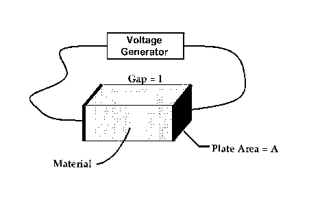
FIG. 1 is a schematic illustration of apparatus employed with carrying
out the method of an embodiment of the invention.
FIG. 2 is a graph illustrating the operating range of the method of an
embodiment of the invention, inside the triangle, and how the operating range
of the
invention is outside operating ranges of prior art electroporation methods,
indicated
by small blocks outside the triangle.
FIG. 3 is a graph illustrating the relationship between charging time (in
microseconds) of biological cells and media conductivity (in microSiemens/cm)
for
cells having three different diameters, namely 1 micrometer, 10 micrometers,
and
100 micrometers.
FIG. 4 is a graph showing Time Constant versus Conductivity as it
relates to the method of an embodiment of the invention.
CA 02519065 2011-09-30
= 54346-3
- 23a -
DESCRIPTION OF EMBODIMENTS
As previously described a significant problem is the conductivity of the
media use in electroporation. In this process a low conductivity media is
employed to
keep the total resistance of the media small and virtually eliminates heating.
Not just
any media conductivity can be used. As the ionic content of the media is
reduced the
number of free ions that are available to build charge (voltage) across the
cell
member is decreased. The effect is to increase the amount of time it takes to
charge
the membrane. This process is described by the equation in Electroporation and
Electrofusion in Cell
CA 02519065 2005-09-13
WO 2004/083379
PCT/US2004/005237
- 24 -
-
Biology, edited by Eberhard Neumann, Arthur Sowers, and
Carol Jordan, Plenum Press, 1989, on page 71. Assuming a
typical cell diameter of 10 microns, the charging time is
20 microseconds at 80 11S/cm. Below 80 11S/cm the charging
time become too long and the pathways in cell membrane
stop forming. The TABLE 6 below illustrates the
resistance of the media as a function of electrode
chamber volume and conductivity.
TABLE 6
Electrod Media Resistance - ohms
Volume
ml
17,000 200 80
11S/cmS/cm
11S/cm
0.5 19.2 1600 4000
1 9.6 800 2000
5 1.92 160 400
10 0.96 80 200
50 0.19 16 40
Ex vivo electroporation has been demonstrated in
numerous published research projects. At this point
commercial applications, such as clinical transfection to
produce a vaccine for the patient, requires large
electrodes or chambers to process millions of cells at
one time. The static parallel plate chamber provides the
most uniform amplitude and most uniform electric field
direction of any configuration available. This
uniformity is required to insure uniform treatment of the
target cells. It is also important not to use very
high-density cell concentration such as 30 million
cells/ml to insure local uniform electric fields about
the cells. This invention applies to chambers larger
than 1 milliliter.
Using larger chambers results in high current flow
CA 02519065 2005-09-13
WO 2004/083379
PCT/US2004/005237
- 25 -
-
when voltage is applied. The equations for chamber
resistance vs. conductivity of the cell and media mixture
and the chamber dimensions are as follows:
Volume of material =lx A
1 1 1 112 GF
Resistance of Material = p ¨ = ¨ ¨ = ohms
AuAuuu
p = resistivity in
ohm-cm
a = 1/p in Siemens/cm
u = volume of material
being treated
There is a Geometric Factor (GF), which is a
constant for any chamber dimension. As the volume of the
chamber gets larger the resistance of the material in the
chamber gets smaller thus increasing current flow.
The present invention uses an electrode with large
capacity in combination with an electroporation buffer of
defined low conductivity. This process exposes all cells
to the same treatment conditions, provides control over
the amperage required and can process large numbers of
cells. Since the cell suspension statically remains in
the chamber during application of pulsed electric fields,
complex waveforms can be used.
Another aspect of the invention further increases
capacity by alternately filling and emptying the gap
between the electrodes. In this manner, all desired
properties are met during a specific treatment and the
electrodes can be re-used for subsequent treatments in an
intermittent batch process.
This present invention specifies a range of material
conductivities, which can be used versus the chamber
dimensions, the larger the volume the smaller the
CA 02519065 2005-09-13
WO 2004/083379
PCT/US2004/005237
- 26 -
-
conductivity. This invention specifies an operating area
for use with the larger volume electrodes. This is
illustrated in FIG. 2. Operating points of prior art
published results are also presented in FIG. 2 as
squares. For chambers with a Geometric Factor less than
0.1 there are two limiting factors, which are related.
The first is the absolute value of the chamber
resistance. In this invention the chamber resistance is
one ohm or greater. Operating below one ohm is view as
impractical. The other constraint is the conductivity of
the medium in the chamber. AS the conductivity decreases
the charging time of the cell membrane increases because
there are fewer ions external to the cell membrane.
The relationship between the Transmembrane Voltage
(TMV) and conductivity and cell diameter is as follows,
taken from Neumann et al stated below:
Transmembrane Voltage = TMV
/711V=-1.5Ericos8lf(2)
where: E = electric filed in volts/cm
r = cell radius in cm
8 = angle from electric field line in
degrees
f(X) = composite conductivity
2,0
f(2)= ___________________________________________________
(2/10 ) + (2¨) (20 ¨
where: ko = conductivity of media external to
cell milliSiemens/cm
Xi = conductivity of cytoplasm
X, = conductivity of cell membrane
d = thickness of cell membrane
CA 02519065 2005-09-13
WO 2004/083379
PCT/US2004/005237
- 27 -
-
Reference:
Electroporation and Electrofusion in Cell
Biology
Edited by Eberhard Neumann, Arthur Sowers, and
Carol Jordon
Plenum Press, 1989
Below 1 microSiemens/cm there are so few ions that
the time to change the cell membrane is unrealistically
large.
The preferred operating region of the present
invention is then:
Cell diameter > 1 micrometer
Chamber volume > 1 milliliter
Conductivity of Material to be treated > 1
microSiemens/cm
Total resistance of material to be
treated in chamber > 1 ohm
Geometric Factor of Chamber < 0.1 cm-1
The invention uses a static chamber with large
volume to insure that all cells are subject to the same
electric field intensity and direction and the density of
the cells and treating material are uniform. With this
invention any waveform may be used. This invention is a
voltage waveform generator connected to an electrode with
parallel plates with has low conductivity medium, a cell
density of 20 million cells or less.
A component of the invention is the use of low
conductivity medium within a defined range to limit
amperage and heat while simultaneously providing enough
ions to effectively electroporate cells. Typically the
medium used will have a conductivity between 50 mS/cm and
500 mS/cm.
CA 02519065 2005-09-13
WO 2004/083379
PCT/US2004/005237
=
- 28 -
-
The invention may be used in clinical applications
and with a closed sterile chamber into which the cells
and large molecules are inserted and removed.
One aspect of the invention further increases
capacity by alternately filling and emptying the
electrode. In this manner, all desired properties are
met during a specific treatment and the electrode can be
re-used for subsequent treatments in an intermittent
batch process.
The conductivity of the medium used in
electroporation is an important aspect of this invention.
In this process, a low conductivity medium is employed
to keep the total resistance of the medium small and
virtually eliminate heating. There is a limit to the
lower conductivity medium that can be used. As the ionic
content of the medium is reduced the number of free ions
that are available to build charge (voltage) across the
cell membrane is decreased. The effect is to increase
the amount of time it takes to charge the membrane. This
process is described by the equation in Neumann, p71.
Assuming a typical cell diameter of 10 microns, the
charging time is 20 microseconds at 80 mS/cm. Below 80
mS/cm the charging time becomes too long and the pathways
in cell membranes stop forming.
Using an electrode with a 4 mm gap, TABLE 6
illustrates the resistance of the medium as a function of
electrode chamber volume and conductivity.
In one aspect of the invention, a chamber with two
electrodes is used as shown in FIG 1. An example of
electrode dimensions that can be used is a gap of 0.4 cm,
electrode height of 2 cm and electrode length of 10 cm.
The chamber can be used with a commercial electroporator
such as the Cyto Pulse Sciences, Inc. PA-4000
electroporator.
An example of a medium that can be used with the
chamber is one with the following formula:
Sorbitol 280 millimoles
CA 02519065 2005-09-13
WO 2004/083379
PCT/US2004/005237
- 29 -
Calcium Acetate, 0.1 millimoles
Magnesium Acetate, 0.5 millimoles
FIG. 3 is a graph illustrating the relationship
between charging time (in microseconds) of biological
cells and media conductivity (in microSiemens/cm) for
cells having three different diameters, namely 1
micrometer, 10 micrometers, and 100 micrometers. From
FIG. 3 it is clear that for media conductivity below 1
microSiemen/cm, the charging time would be so large that
electroporation would not work.
As to the manner of usage and operation of the
instant invention, the same is apparent from the above
disclosure, and accordingly, no further discussion
relative to the manner of usage and operation need be
provided.
It is apparent from the above that the present
invention accomplishes all of the objects set forth by
providing a large volume ex vivo electroporation method
which may advantageously be used for clinical and
therapeutic purposes wherein all cells, ex vivo or in
vitro, are subject to substantially the same process
conditions. With the invention, a large volume ex vivo
electroporation method is provided which is scalable so
that substantially large volumes of ex vivo or in vitro
cells can be processed in a relatively short period of
time. With the invention, a large volume ex vivo
electroporation method is provided which achieves
increased cell capacity without increasing the size of
electrodes resulting in excessively large amperage
requirements. With the invention, a large volume ex vivo
electroporation method is provided which limits heating
within the treatment cell to low levels. With the
invention, a large volume ex vivo electroporation method
is provided which exposes substantially all ex vivo or in
vitro cells to the same electric field intensity and
CA 02519065 2005-09-13
WO 2004/083379
PCT/US2004/005237
- 30 -
-
direction. With the invention, a large volume ex vivo
electroporation method provides that the density of the
material to be inserted into the treatment chamber can be
held constant. With the invention, a large volume ex
vivo electroporation method is provided which permits
variable rectangular pulse waveforms such as disclosed in
U. S. Patent No. 6,010,613 can be employed. With the
invention, a large volume ex vivo electroporation method
is provided which avoids problems in flow through
treatment cells that are due to laminar and turbulent
flow conditions. With the invention, a large volume ex
vivo electroporation method is provided which permits the
use of medium with lower conductivity to achieve the
movement of macromolecules into mammalian cells and to
allow the use of larger capacity electrodes. With the
invention, a large volume ex vivo electroporation method
is provided which is easily scalable to large capacity
without using a flow through treatment chamber for cells
to be treated.
With respect to the above description, it should be
realized that the optimum dimensional relationships for
the parts of the invention, to include variations in
size, form function and manner of operation, assembly and
use, are deemed readily apparent and obvious to those
skilled in the art, and therefore, all relationships
equivalent to those illustrated in the drawings and
described in the specification are intended to be
encompassed only by the scope of appended claims.
While the present invention has been shown in the
drawings and fully described above with particularity and
detail in connection with what is presently deemed to be
the most practical and preferred embodiments of the
invention, it will be apparent to those of ordinary skill
in the art that many modifications thereof may be made
,without departing from the principles and concepts set
forth herein. Hence, the proper scope of the present
invention should be determined only by the broadest
CA 02519065 2005-09-13
WO 2004/083379
PCT/US2004/005237
- 31 -
-
interpretation of the appended claims so as to encompass
all such modifications and equivalents.