Note: Descriptions are shown in the official language in which they were submitted.
CA 02519177 2005-09-14
WO 2004/084988 PCT/EP2004/003270
1
Electrostimulating system
The invention refers to an electrostimulating system comprising
means for producing an electric stimulation that consists of
bioactive neuromodulation of the neurovegetative system, of the
striated-muscle system, of the smooth muscle and of the mixed
nervous structure, particularly suitable for producing inter
alia phenomena of muscular contraction and relaxation by means
of emulation of the action of the nerve fibre that innerves a
skeletal muscle or of the neuroceptors of the sympathetic
system that interact with the smooth muscle of the vessel8.
Equally, depending on the type of electric stimulation and on
the configuration parameters, a consequent induced bioactive
neuromodulation can be generated that is suitable for producing
vasoactive phenomena in the microcirculaton and in the
macrocirculation, which are in turn mediated by phenomena
connected with the direct stimulation of the smooth muscle and
by essentially catecholamine energy phenomena by means of
stimulation of the postsynaptic receptors.
The system thus produces stimulation sequences that induce
reproducible and constant neurophysiological responses; in
particular, but not restricted thereto, the sequences of
activation of the microcirculation (ATMC) and relaxation of the
muscle fibre (DCTR) are able to stimulate different functional
contingents, including but not limited to the striated muscle,
the smooth muscle and the peripheral mixed nerve.
The stimulation sequences are assembled on three fundamental
parameters: the width of the stimulus, the frequency of the
stimulus and the time wherein different combinations of
width/frequency follow each other. The general operating model
reflects the digital-analogue transduction that occurs in
nervous transmission.
WO 02/09809 discloses an apparatus for the treatment of
muscular, tendinous and vascular pathologies by means of which
a series of electric pulses lasting from 10 to 40 microsecs are
CONFIRMATION COPY
CA 02519177 2005-09-14
WO 2004/084988 PCT/EP2004/003270
2
applied to a patient and at variable intensity, depending on
the impedance and conductance of the tissue subjected to
stimulation, typically from 100 to 170 microampere.
These electric pulses are able to produce a relaxing, anti-
inflammatory and vasoactive effect. Such levels of current and
the connected level of energy transferred, below 5 microjoules,
cannot create polarisation or ionisation of metallic structures
and are therefore absolutely compatible with the presence in
the stimulated organism of, for example, metal prostheses, or
of intrauterine-coil contraceptive devices and
cardiostimulators or implanted defibrillators (pacemakers).
US 5,725,563 discloses a method and a system of adrenergic
stimulation of the sympathetic nervous system relative to the
circulation of the patient wherein electric pulses are
generated and simultaneously impedance of the cytoplasm
contained in the space between the stimulation electrodes is
measured. In this case, the specific effects of the disclosed
system are cited, namely the vasoconstriction that is a
consequence of activation of the alphaadrenergic postsynaptic
receptors that modify the venous tone, thereby producing
vasoconstriction and consequent vascular and lymphatic
drainage. In this case, to obtain this specific effect,
stimulations are proposed in a range of frequencies absolutely
below 2 Hz and preferably of 1.75 Hz with currents below 350
microAmperes and preferably below 250 microAmperes with energy
transfer around 10 microJoule. In particular, the pulses
generated by the above-mentioned stimulator are subordinated to
the measurement of impedance so as to vary the width of the
pulse in function thereof.
However, this system produces only the effect of a
"peristaltic pump" due to the periodical "vasoconstriction" and
subsequent "long" period of "relaxation" and is obtained by
means of the delivery of very low frequency pulses (< 2 Hz) to
the smooth muscles of the vessel. However, in addition to being
CA 02519177 2008-08-06
3
limiting and requiring careful measuring of impedance, it
produces limited effects and requires stimulations that are
extremely prolonged over time to obtain visible and effective
effects.
On the contrary, this invention also solves all the problems
that beset the prior art and significantly increases the
disclosed positive effects, having a direct action on
postsynaptic activity, it produces direct effects on synapses
or the motor plate of the skeletal muscle involved.
The invention provides a combination of: an electrostimulating
apparatus for applying electrical stimuli to biological
tissues; heat exchanging means, arranged to exchange heat with
said tissues.
The invention also provides for an electrostimulating
apparatus that generates a relaxing sequence suitable for
stimulating striated muscle fibre, based on three fundamental
parameters: the width of the electric stimulation, the
frequency of the stimulation and the intervals of time wherein
a plurality of width/frequency combinations follows.
Moreover, the invention provides for an electrostimulating
apparatus that generates a vasoactive sequence of activation
of the microcirculation suitable for stimulating the smooth
muscle fibre and the postsynaptic neuroceptors, based on three
fundamental parameters: the width of the electric stimulation,
the frequency of the stimulation and the time wherein a
plurality of combinations of width/frequency follow.
Advantageously, the apparatus and the method provided by the
invention exploit the principle of achieving significant
bioreaction variations.
The invention may be better understood with reference to the
attached drawings that illustrate certain embodiments by way
of non-limiting example, wherein:
CA 02519177 2008-08-06
3a
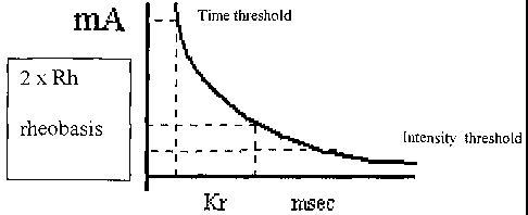
Figure 1 shows a Cartesian graph of time/intensity of current,
disclosing the intensity and time thresholds;
Figure 2 shows a graph illustrating a relaxing sequence, or
DCTR sequence, according to the invention;
Figure 3 shows a DCTR sequence plot, carried out on a healthy
subject;
Figure 4 shows a plot like the one in Figure 3, but carried
out on a further healthy subject;
Figure 5 shows three surface electromyograms, with stimulation
frequencies of 1, 15 and 30 Hertz;
Figure 6 shows a graph illustrating a reactivation sequence of
the microcirculation, or ATMC sequence, according to the
invention;
CA 02519177 2005-09-14
WO 2004/084988 PCT/EP2004/003270
4
Figure 7 shows a polygraph recorded during administration of
an ATMC sequence to a healthy subject, in the presence of
electric stimulation;
Figure 8 shows a polygraph like the one in Figure 7, but
conducted in the absence of electric stimulation;
Figure 9 shows a graph highlighting the discontinuous
variation of the bioreaction obtained during administration of
an ATMC sequence;
Figure 10 shows graphic histograms of flow plots recorded in
the presence and/or absence of ATMC sequences;
Figure 11 shows flow variations recorded at the same time as
the administration of an ATMC sequence like the one
illustrated in Figure 7;
Figure 12 shows flow variations similar to those in Figure 11,
but recorded during the administration of an ATMC sequence
like the one illustrated in Figure 8;
Figure 13 shows further flow variations like those in Figure
12;
Figure 14 illustrates a combination of an ATMC sequence with a
thermal heating stimulus.
The nervous cell is responsible for the formation and
transmission of the nervous pulses, which regulate the
operation of the entire organism. This nervous cell is formed
by a cell body or "soma" wherefrom branches lead: the
"dendrites" along which the pulse has a centripetal direction
(i.e. towards the cell body) and the "axon", along which the
pulses are mediated by the soma to the periphery, i.e. in a
centrifugal direction. The pulses that do not arise from the
soma of the cell are transmitted to the latter by other nervous
cells or by specialised structures (receptors) or originate
directly with the fibres, as in the case of free nerve ends
responsible for collecting painful stimuli.
The pulse can travel towards the centre or vice versa. In the
first case it is defined as being afferent and the result,
CA 02519177 2005-09-14
WO 2004/084988 PCT/EP2004/003270
analysed at the level of the Central Nervous System, is the
acquisition of conscious information (sensitive stimulation) or
unconscious information (e.g. automatic regulation of balance).
The pulse that travels from the centre to the periphery is
5 therefore defined as efferent and is able to cause the
stimulation of the innerved organ or tissue.
The result of this may be muscular contraction, a glandular
secretion, variations in cell metabolism, vasodilatation,
vasoconstriction, and so on. Transmission of the pulse between
the nerve fibres and the cells of a tissue occurs with the help
of synapsis. The latter is terminal dilation (terminal button)
of the axon that is in contact with the membrane of the cell to
which the pulse is transmitted. A diminution of membrane
potential in turn causes depolarisation that subsequently
extends to the entire cell. The pulse that runs along the nerve
fibre is merely the propagation of a depolarisation wave called
action potential.
The nervous pulse may arise directly from the cell, but more
often it originates from the stimulation of one of its parts,
stimulated for example by pressure or a painful sensation.
The striated muscle fibre consists of thousands of myofibrils,
consisting of two types of filamentous protein, that are
arrayed in an alternating manner: the bigger the myosin the
thinner the actin. The actin has light streaks defined as I
bands, whereas with actin and myosin dark streaks known as A
bands are created. The complex formed by an A band and by two
adjacent semibands I is given the name "sarcomere". Between two
adjacent sarcomeres there exists a contact zone and a
sarcoplasmic reticulum for the control of the contraction
consisting of two different types of tubules: T tubules and
longitudinal tubules.
Each muscle fibre receives pulses from the motor nerve fibre
via the neuromuscular junction, which takes the name motor
plate.
CA 02519177 2005-09-14
WO 2004/084988 PCT/EP2004/003270
6
When the pulse arrives this causes depolarisation known as
"plate potential" which generates action potential along the
entire length of the muscle fibre, which causes it to contract.
It is at this point opportune to recall the definition of the
"chronaxy" and "rheobasis" parameters regarding the
excitability characteristics of the nerve and muscle fibres.
Chronaxy (Kr) is defined as the time (expressed in msec)
required by a current intensity to reach a value that is twice
the rheobasis (muscle sensitivity) . Rheobasis (Rh) is in turn
defined as the minimum (liminal) measurable current intensity
required to excite a cell.
If the stimulating current is limited to a short time of the
order of msec it will be observed that the shorter the width of
the current is, the greater its intensity will have to be to
reach the threshold. As shown in Figure 1, by plotting the
intensity-time curve two intensity and time thresholds are
defined. The theoretical construction of the curve is achieved
on the basis of the capacitive features of the axon membranes.
The higher excitability is, the more concave the curve will be
in relation to the axes because smaller products (i t), i.e.
smaller quantities of electricity will correspond to its
points. When one wishes to determine the excitability of a
nerve or muscle in vivo chronaxy is used. Chronaxy and
rheobasis are in fact interconnected as characteristics of the
nerve fibre. By means of "Lorenz stimulation with modulated
frequency and amplitude" the excitation of the nerve fibres can
be obtained by means of the summation effect of several
subthreshold signals that are not able to excite the fibre,
which however, by combining their effects together, are able at
a certain point to excite the fibre. The summation effect, with
the same produced pulse amplitude, will depend on the amplitude
of the signal and on the bioreaction that is therefore
connected to frequency, which in turn interact with the
rheobasis-chronaxy ratio.
CA 02519177 2005-09-14
WO 2004/084988 PCT/EP2004/003270
7
To demonstrate this behaviour, an analytical study of the
physiological responses was conducted in combination with
"Lorenz stimulation" by applying two different experimental
procedures.
A first procedure is based on the use of a relaxing action
sequence or DCTR, whose frequency and width characteristics are
set out in Figure 2.
The aim of the reported experiment is to prove the validity of
the hypothesis that such a sequence, disclosed in WO 02/09809
and appropriately designed to have a relaxing effect on the
muscle fibres, has a prevailing action on the activity of the
skeletal muscle. Stimulation was achieved by measuring with
sophisticated digital polygraph laboratory instruments with the
possibility of sampling high-speed and high-frequency signals.
The latter were recorded at the level of the short adductor
muscle of the thumb and palm of the hand. For the short
adductor muscle of the thumb a pair of plate electrodes (Ag +
Cl -) was used through preamplification of the analogue signal
at 5000 gains, passband 5 Hz-3 KHz. To the palm of the hand an
electro-resistant transducer was applied comprising two surface
electrodes, with 1:10 pohm preamplification.
The DCTR stimulation sequence was administered to two different
healthy subjects. For each of them four polygraphs were
recorded (as described previously), for three identical DCTR
sequence cycles run consecutively. Two of the above polygraphs,
obtained from different subjects, were illustrated in figures 3
and 4. The stimulator electrodes were placed near the recording
seats, along the route of the median nerve on the palmar
surface of the wrist.
In both plots, carried out on healthy subjects, the median
nerve was stimulated at the wrist with the DCTR sequence
repeated three times, measuring on the short adductor muscle of
the thumb of the thenar eminence with a transducer of skin
impedance.
CA 02519177 2005-09-14
WO 2004/084988 PCT/EP2004/003270
8
Each polygraph contains three plots separated into: top, middle
and bottom.
The top plot shows the muscle responses obviously after
discounting the stimulation artefacts, which responses are
expressed in frequency histograms, whilst in the intermediate
plot the skin conductance variations appear. In the bottom plot
the stimulation sequence is shown, wherein the graphically
"densest" part represents the rapid increase phases of the
frequency.
As can be seen from the analysis of the DCTR sequence, the
basic variation is the variation in the frequency of stimuli
whereas widths remain constant at 40 microseconds.
In both polygraphs one notes the reproducible skin conductivity
response (intermediate plot) in close temporal relationship, at
about 500 msec latency, with the frequency increase phase of
the stimulation. In both cases, the average conductance trend
tends to fall. However, the absolutely original element and
result of the disclosed invention consists of the close
reproducibility of the responses regardless of the manner that
they assume compared with the three phases of stimulation
frequency.
This indicates that there is a direct dose-response
relationship between the variability of the frequency of the
electric stimuli which have a constant amplitude and are below
the pain threshold and catecholaminergic vegetative efferents,
inasmuch as skin conductance is directly influenced by local
sweating, which is in turn carried, in the palm of the hand, by
sympathetic innervation.
With regard to variation in skin conductance, some
characteristics have emerged that are practically constant and
independent of the subject subjected to stimulation and are
disclosed below.
Above all, during the phase of rapid increase in stimulation
frequency, a complex twin, triple or quadruple negative
CA 02519177 2005-09-14
WO 2004/084988 PCT/EP2004/003270
9
deflection phase occurs that is constant in each test during
the three increase phases in both subjects and is therefore
independent of the subjects themselves.
Again, the average trend of conductance under stimulation
appeared to be indifferently ascending or descending in the
different polygraphs. Characteristic trends and morphologies of
the polyphase response belong to each subject.
Lastly, the overall duration of the polyphase response during
the increase phase varies from 14 to 19 seconds; the greatest
negative deflection is always the last of the complex and
always occurs following the cessation of the incremental phase
of the stimulus, with latency of approximately 1.5 sec. The
negative components of the complex, which are variable between
subjects and over the course of different measurements, always
appear in relation to the first seconds of increase of the
stimulation frequency.
In terms of the surface electromyogram, in both subjects and in
all the measurements made, the same phenomena were ascertained,
as described below.
During the preparatory stimulation, at a frequency of 1 Hz,
there was no muscle response; during the increase phase
composite motor unit potential was formed with increasingly
shorter latency and increasingly higher amplitude until the
formation of composite muscle action potential (cMAPSs) with
minimum latency and maximum amplitude at the peak of the
stimulation frequency.
The minimum appearance latencies of the cMAPSs correspond to
the latencies that are detectable by means of
electroneurography using standard methods. On the other hand,
compared with the above-mentioned method of detection of the
cMAPSs, the amplitudes are reduced by about 30%.
Each cMAP follows on from each stimulus and the isoelectric
line of the plot returns after the cMAP to the value 0.
The top plot simply describes the production of composite motor
CA 02519177 2005-09-14
WO 2004/084988 PCT/EP2004/003270
potentials (cMAPs) in close temporal relation with the stimuli
of the sequence. The inventive and original element consists of
the fact that the first cMAPs appear only in the phase of
increase of the frequency of the stimulation, according to a
5 model that is absolutely analogous to the temporal recruitment
of stimuli of the same amplitude, but placed in an increasing
sequence over time (in a completely analogous manner to what
occurs in the classical nerve-muscle physiological model).
The second phenomenon should also be pointed out, i.e. the one
10 according to which, in addition to recruiting in frequency the
number of cMAPs, the increase in stimulation determines the
total amplitude of the cMAPS. This means that DCRT-type
stimulation can perfectly emulate the action of a nerve fibre
that innerves a skeletal muscle.
A second experimental procedure is based on the use of a
reactivation sequence of the microcirculation, or ATMC, whose
frequency and width characteristics are disclosed in the graph
in Figure 6.
This second procedure had the object of showing the validity of
the hypothesis that an ATMC sequence, suitably designed to
obtain the desired effect, has a prevalent action on the
motility of the microcirculation, i.e. of the smooth sphincters
of the arterioles and venules of the subcutaneous layer.
In this case, and for this object, stimulation was carried out
by recording with a doppler flow laser-apparatus that is able
to measure the degree of perfusion of the microcirculation,
i.e. of the subcutaneous haematic flow, in addition to other
correlated and synergic parameters, i.e.: 02 saturation, CO2
saturation and skin temperature.
To view the significant components of this sequence, with
reference to figures 5, 7, 8 and 9 the constitution of the ATMC
sequence in three subsequences known as Sl, S2, S3 is discussed
below.
S1 and S3 are both characterized by a frequency increase phase,
CA 02519177 2005-09-14
WO 2004/084988 PCT/EP2004/003270
11
with distinct time modes, whilst S2 is mainly constituted for
producing variability in the width of the different stimuli, in
a gradually increasing range of frequencies but in such a way
as to reduce the bioreaction until it is stabilised.
More in detail, during the S1 subsequence, a sequence that
typically has a relaxing effect and which is very similar to
the DCTR sequence disclosed above, different subphases are
carried out wherein, after a first subphase with a 1-Hz
frequency of mere adaptation, the frequency with a constant
amplitude is gradually increased, thereby also gradually
decreasing the bioreaction. Subsequently, the frequency is
increased much more rapidly up to the target of 19 Hz.
Subsequently, the subsequence S2 is carried out, which in turn
is subdivided into four parts, S2-A, S2-B, S2-C and S2-D. In
this subsequence, after a phase wherein the amplitude is
rapidly increased up to the instant 1 (S2-A), the frequency is
made to gradually increase, and as a result the bioreaction
rapidly falls to the instant 2 (S2-B). At this point the
amplitude is reset, which will again increase at a constant
frequency up to instant 3 (S2-C); the frequency will thereafter
once again gradually increase at constant amplitude, as a
result the bioreaction will also gradually fall to the instant
3 (S2-D).
In this way, the bioreaction is made to vary in a discontinuous
manner, producing points of variation of the jump gradient,
i.e. the points 1, 2 and 3.
To conduct the experiments, the sensor of the laser apparatus
was placed on the extensor surface of the wrist (non-smooth
skin). The stimulation electrodes were placed with the anode
(stimulator) on the route of the radial nerve on the extensor
surface of the third distal of the forearm and with the cathode
placed near the proximal capitulum of the second phalanx.
Furthermore, measuring electrodes of skin conductivity were
positioned, in the same way as the first experimental procedure
CA 02519177 2005-09-14
WO 2004/084988 PCT/EP2004/003270
12
described above used to vary the effects of the DCTR sequence.
The ATMC sequence was administered also in this case to two
healthy subjects.
On the first a polygraph was first recorded during electric
stimulation with an ATMC sequence and subsequently another
polygraph of similar width was recorded but in absence of
electric stimulation.
On the second subject two polygraphs were recorded, one of
which compares responses during and after raising local skin
temperature to 44 C. This thermal shock was induced by the
instrument itself, whose laser probe in contact with the skin
is provided with a thermistor able to heat the face of the
probe in contact with the skin until a desired temperature is
reached.
In this context it is important to stress that that was done
because skin thermal stimulation is reported in the literature
to be the maximum stimulation to obtain vasodilatation.
Therefore in this case the intention is also to carry out a
comparison.
Any stimulation carried out is made up of three basic identical
sequences of the ATMC type.
The parameters that are most subject to variation are local
flow, temperature and skin conductance, whereas oxygen and
carbon-dioxide saturation do not show suggestive variations in
relation to the sequence of the different stimulation phases.
The analysis that is suggested by the detailed evaluation of
the recorded plots enables the apparent synchronisation and
desynchronisation of flow variation to be checked in relation
to the incremental phases of the stimulation sequences. In
fact, during the first subphase consisting of 30 seconds of
constant stimulation at 1 Hz and at 40 microseconds of pure
preparation (considerable ineffective stimulation), there is an
increase in the average oscillation frequency of the flow
signal by means of doppler laser, which instead enters at lower
CA 02519177 2005-09-14
WO 2004/084988 PCT/EP2004/003270
13
frequencies in a temporal relationship with the increase and
decrease phases of the stimulation sequence.
In Figure 10, the frequency spectra of the flow plot for each
stimulation subsequence have been analysed by a Fourier
transform in the field of frequencies, and compared with the
spectrum over a period of recording without ATMC stimulation
(base datum) and having a similar width (about 50 sec).
It can be noted that during the period without stimulation the
oscillation frequencies are rather dispersed and prevalent on
the 1-2 Hz band, i.e. the typical frequency of the heartbeat,
whilst during the three stimulation subsequences frequencies
are drastically synchronised on the 0-1 Hz range.
In detail, the response mode of the flow in relation to
specific moments of the stimulation sequence is displayed. In
the two subjects subjected to polygraph, the most constant flow
variations could be observed during the subsequence S2.
In the plot recorded for subject 1 during the subsequence S2
and illustrated in figure 11, the bottom line indicated the
frequency trend of stimulation, the top line indicated the
virtually constant polyphase trend of the local subcutaneous
flow variation.
In the plot recorded for subject 2 during the subsequence S2
and illustrated in Figure 12, the flow line has a`peaks'
pattern whereas the line of the stimulation frequencies has a
`steps' pattern.
Although apparently random, the flow oscillation phases
coincide perfectly with the different frequency variation
phases of the stimulus.
The close correlation between the trend of the subsequence S2
and the flow response can be displayed through individuation of
flow peaks that coincide with the instants 1, 2, 3 disclosed
previously.
With reference to Figure 13, at the points of flow peak a
reversal occurs of the second derivative of the bioreaction and
CA 02519177 2005-09-14
WO 2004/084988 PCT/EP2004/003270
14
of the energy transferred to the tissue, and therefore of the
determining chronaxy/rheobasis correlated therewith, in view of
the characteristic of the phenomenon of temporal summation that
occurs, i.e. a drastic jump variation of the first derivative
thereof.
In practice, the system produces a sequence of vasodilatations
and vasoconstrictions with sequential increases and decreases
of the haematic flow of the microcirculation that produce a
"pump" effect that is evidently produced by neuromodulation of
the neurovegetative and of the sympathetic system, which
influences vasoactivity through the smooth muscle of the
smaller blood vessels (arterioles, capillary blood vessels).
During the subsequence S2 of the ATMC sequence, characterized
by alternating variations of the rheobasis, a vasoactive effect
occurs comprising a succession of alternating phases of
vasodilatation and vasoconstriction. This without doubt also
produces a draining effect and above all elasticisation of the
microcirculation and its modulation around a main carrying
event that causes its average variation.
In a series of experiments conducted after those described
above, this type of vasoactive ATMC stimulation was associated
with a vasodilative or vasoconstrictive stimulus. If the ATMC
stimulus is accompanied by a vasodilative carrying stimulus,
for example thermal heating stimulation, as in the case
illustrated in Figure 14, this association substantially
enhances vasodilatation and the dose/response ratio.
On the other hand, if the ATMC stimulus is accompanied by a
vasoconstrictive carrying stimulus, such as for example
thermal cooling stimulation, this association substantially
enhances vasoconstriction.
In this case LorenzTM stimulation by means of the ATMC sequence
creates effective neuromodulation that is able to amplify the
excitation phenomena of the primary and secondary neuroceptors.
Consequently, it is possible to use the ATMC vasoactive
CA 02519177 2005-09-14
WO 2004/084988 PCT/EP2004/003270
sequence also in combination with hyperthermia and cryotherapy
treatments to enhance the effects of the latter.
In this way localised neoplasms and solid tumours can be
treated by the combination of temperature effects with
5 vasoactive effects.
If cryotherapy is combined with the vasoactive ATMC sequence
the vasoconstrictive effects are increased, thereby producing
localised hypoxia in a tumour mass, with consequent necrosis of
the latter.
10 Similarly, by combining the vasoactive ATMC sequence with a
hyperthermic therapy important vasodilatation is obtained that
amplifies the necrotizing effect of the hyperthermia on a
tumour mass.
In conclusion, it can certainly be stated that the Lorenz
15 TherapyTM stimulation sequences induce reproducible and constant
neurophysiological responses; the ATMC and DCTR sequences are
able to stimulate different functional contingents, including
the striated muscle, the smooth muscle and the mixed peripheral
nerve.
The stimulation sequences are assembled on three fundamental
parameters: the width of the stimulus, the frequency of the
stimulus and the time wherein different combinations of
width/frequency follow. The general operating model reflects
the digital-analogue transmission that occurs in nervous
transmission.