Note: Descriptions are shown in the official language in which they were submitted.
CA 02520028 2005-09-22
WO 2004/090145 PCT/US2004/010045
COMPOUNDS AND METHODS TO ENHANCE
rAAV TRANSDUCTION
Cross-Reference to Related Aunlications
The present application claims the benefit of the filing date of U.S.
application Serial No. 60/459,323, filed March 31, 2003, and U.S. application
Serial No. 60/512,347, filed October 16, 2003, the disclosures of which are
incorporated by reference herein.
Statement of Government Rights
This invention was made, at least in part, with a grant from the
Government of the United States of America (grant HL58340 from the National
Institutes of Health). The Government may have certain rights in the
invention.
Background of the Invention
Recombinant adeno-associated virus (rAAV) has several characteristics
that underscore its potential as a gene therapy vector for numerous target
organs
and inherited or acquired diseases, a vaccine vector or for diagnostics.
Moreover, rAAV vector systems potentially offer major advantages over other
gene delivery vehicles, including adenoviruses and retroviruses. These include
the ability of rAAV to readily transduce non dividing or slowly dividing cells
and persist essentially for the lifetime of the cell, the lack of undesirable
cellular
immune responses since all viral genes can be deleted from the vector, and the
fact that AAV has never been associated with human disease.
A serotype 2 rAAV (rAAV-2) vector expressing the CFTR gene was the
first AAV vector to be utilized in clinical trials. This vector has
demonstrated
promise in patients with cystic fibrosis and has advanced to phase II trials
(Flotte
et al., 1996; Wagner et a1.,1999; Wagner et al., 2002; Aitken et al., 2001).
In
recent years, additional rAAV 2 vectors have been or are currently being
advanced to clinical trials to treat a number of disease states including a
rAAV2
factor IX vector in phase I clinical trials in patients with hemophilia B (Kay
et
al., 2000), and a rAAV2 sarcoglycan vector in phase I clinical trials of
patients
with CNS disease. Additionally, clinical trials for rAAV expressing proteins
to
treat Parkinsons disease (RDAC, 2001), and Canavan's disease (Janson et al.,
2001), have been proposed.
CA 02520028 2005-09-22
WO 2004/090145 PCT/US2004/010045
Other serotypes of AAV are known to exist, although they are all closely
related at the functional, structural, and genetic level (see, e.g., Blacklow,
1988;
and Rose, 1974). All AAV serotypes exhibit very similar replication properties
mediated by homologous rep genes; all bear three related capsid proteins such
as
those expressed in AAV-2, and all contain 5'-3' ITR sequences. Currently, 8
serotypes of AAV have been described with the complete genome sequence
information available for AAV-1-AAV-6 (Srivastava et al., 1983; Miramatsu et
al., 1996; Chiorini et al., 1967; Xiao et al., 1999; Chiorini et al., 1999;
Bantel-
Schaal et al., 1999; Rutledge et al., 1998) and capsid gene sequence for AAV-7
and AAV-8 (Gao et al., 2002). AAV-6 has been shown to be a recombinant
between AAV-1 and AAV-2. In addition, there are two isolates and sequences
of AAV-3 that differ from each other in a number of amino acids in both rep
and
cap (Rutledge et al., 1998). AAV-5 is the most distantly related of the
serotypes,
and displays a serotype-specific terminal resolution site (trs) in its ITR
(Chiorini
et al., 1999). Even though rep proteins from other serotypes bind the AAV-5
ITR, they do not efficiently cleave at the trs. In addition to recent
developments
in AAV and rAAV serotypes, numerous groups are experimenting with rAAV
pseudotypes.
Variations in cell surface receptor usage for binding of rAAV to cell
membranes exists among various serotypes and may be in part responsible for
the differences .in transduction efficiencies in various tissue and cell
types.
Although conflicting data exists, it has become apparent that there are
differences among the serotypes in the efficiency of transgene expression in
various tissues and cell types. For example, rAAV-l and rAAV-7 overall appear
several orders of magnitude superior for transduction of marine muscle tissue
although rAAV-5 also demonstrates enhancement compared to rAAV-2 (Gao et
al., 2002; Chao et al., 2000; Rabinowitz et al., 2002; Hildinger et al.,
2001).
rAAV-8 transduces marine liver up to 100-fold higher (Gao et al., 2002) than
rAAV-2, and rAAV-5 appears superior in transduction of cells of the marine
respiratory tract (Zabner et al., 2000; Aurichio et al., 2002). rAAV-5
generally
appears to be superior to rAAV-2-based vectors in all tissue types tested so
far
including CNS, muscle, liver and retina (Chao et al., 2000; Rabinowitz et al.,
2002; Hildinger et al., 2001; Aurichio et al., 2002; Davidson et al., 2000;
Mingozzi et al., 2002). Similarly, rAAV-6 is more efficient than rAAV-2 in
2
CA 02520028 2005-09-22
WO 2004/090145 PCT/US2004/010045
transducing marine airway epithelia and alveoli, while rAAV-3 is superior in
transducing smooth muscle cells (Halbert et al., 2001; Halbert et al., 2000).
rAAV-4 transducer ependymal cells in the marine CNS almost exclusively,
while rAAV-5 transducer both neurons and astrocytes (Davidson et al., 2000).
S In retina, a number of studies have demonstrated large differences among
serotypes in the ability to transduce photoreceptor cells and the retinal
pigmented
epithelium (Walters et al., 2001; Aurricchio et al., 2001; Yang et al., 2002).
Despite the fact that rAAV has a very broad host tropism in a variety of
human, simian, and rodent cell lines (Lebkowski et al., 1988; Muzyczka, 1992),
the overall transduction efficiency in human airway epithelia and other
tissues
seems to be quite low. Previous studies have suggested that single to double
strand conversion of the rAAV genome may be the rate-limiting step for AAV-
mediated gene transfer (Ferrari et al., 1996; Fisher et al., 1996). These
studies
demonstrated that adenovirus E4orf6 enhances the conversion of single-stranded
DNA genomes to linear, double-stranded replication form dimers (Rfd) and
monomers (Rfin), through a pathway characteristic of the lytic phase of rAAV
replication. The structure of these replication forms consists of head-to-head
and
tail-to-tail orientated linear concatamers with one covalently linked end
(Ferrari
et al., 1996; Fisher et al., 1996). In contrast, recent studies have
elucidated an
alternative pathway for the conversion of rAAV genomes to double-stranded
circular intermediates with head-to-tail monomer and concatamer structures
(Duan et al., 1999; Duan et al., 1998; Sanlioglu.et al., 1999). The distinct
pathways leading to the formation of either circular AAV genomes or Rf
intermediates appear to be regulated by different cellular factors. For
example,
adenoviral E4orf6 expression decreases circular genome formation while
adenovirus E2a enhances its formation (Duan et al., 1999). Similarly, UV
irradiation also enhances AAV circular intermediate formation but not Rf
intermediates (Sanlioglu et al., 1999).
More recently, when cellular binding protein FK506- (FKBP-52) was
phosphorylated at tyrosine residues (by the epidermal growth factor receptor
protein tyrosine kinase), FKBP-52 was demonstrated to be bound to the single-
stranded D-sequence of the AAV ITR causing an impairment in second strand
synthesis (Qing et al., 2001; Qing et al., 2003. The efficiency of rAAV
transduction in a number of cell types in vit~~o and in vivo correlates with
the
CA 02520028 2005-09-22
WO 2004/090145 PCT/US2004/010045
phosphorylation state of FKBP-52. For example, in HeLa cells, overexpression
of a cellular phosphatase (TC-PTP), led to dephosphorylation of the FKBP-52,
an increase in AAV second-strand DNA synthesis, and an increase in transgene
expression. Transgenic mice expressing either the wild type (wt) or a
catalytically mutant form of TC-PTP, were created. Hematopoietic stem cells
from transgenic mice expressing the wt TC-PTP phosphatase were transduced by
a rAAV2, but those from the phosphatase-negative mutant were not. These
results suggest that the block to second-strand DNA synthesis is due to
binding
of FKBP-52 to the D-sequence of infecting vector genomic DNA and that this
binding is regulated by phosphorylation. Thus, numerous strategies aimed at
increasing the transduction frequency for AAV have focused on enhancing the
molecular conversion of nonfunctional viral genomes to expressible forms
(Fisher et al., 1996; Sanlioglu et al., 1999) or by increasing transcription
and
translation efficiencies by altering the transgene expression cassettes
(Zabner et
al., 1996; Xiao et al., 1998).
A second approach aimed at improving transduction efficiencies of
rAAV has focused on the binding of rAAV to cell surface receptors. Many
primary and secondary cell surface receptor molecules have been identified for
the various AAV serotypes. The primary receptors identified (heparin sulfate
and sialic acid) are found on many cell types and are also utilized by a large
number of viruses besides AAV. This suggests that additional receptors that
lend more specificity to attachment and penetration of cells might exist and
several such co-receptors have been identified. Thus, additional strategies to
improve rAAV transduction efficiency have focused on manipulation of cell
surface receptors (Qing et al., 1997) and/or receptor ligands in the virus
coat
proteins (Wickham et al., 1996a; Wickham et al., 1996b; Bartlett et al.,
1999).
While binding to the cell surface membrane and successful conversion to
a double stranded DNA genome are important, the efficiency of these events
does not necessarily correlate with the overall ability or efficiency of rAAV
to
transduce a given cell type. This has been increasingly apparent in recent
years
as a more detailed understanding of the trafficking and uncoating of rAAV has
been accumulated (Duan et al., 1998; Seisenberger et al., 2001; Hanson et al.,
2001; Bantel-Schaal et al., 2002; Yan et al., 2002). For example, polarized
human airway epithelial cells are transduced with varying efficiencies by rAAV-
4
CA 02520028 2005-09-22
WO 2004/090145 PCT/US2004/010045
2 depending on the route of delivery; entry from the basolateral surface
results in
about a 200-fold increase in.gene expression in the cells compared to vector
administered from the apical surface (Duan et al., 1998; Duan et al., 2000).
Surprisingly, the difference in rAAV cell surface binding between two cell
surfaces is only about 5-fold. This finding led to the discovery that the
vectors
traffic differently in these cells depending on the route of delivery (Duan et
al.,
1998; Duan et al., 2000).
Previous reports have clearly demonstrated that intracellular trafficking
to the nucleus for rAAV-2 and canine parvovirus is a slow, rate-limiting
process
for certain cell types (Parker et al., 2000; Hanson et al., 2001; Hanson et
al.,
2000; Duan et al., 2000). Canine parvovirus and rAAV-2 have also been
demonstrated to be endocytosed through clathrin-dependent receptor endocytosis
and processed through endosomal compartments in a similar fashion to
transferrin, but not a fluid phase marker such as dextran (Parker et al.,
2000;
Bartlett et al., 2000; Benson et al., 2000; Duan et al., 1999). Transferrin
trafficking has been extensively studied and shown to move through the early
endosome to perinuclear recycling endosome (PNRE) (Sonnichsen et al., 2000;
Ren et al., 1998). The recycling of transfernn through the PNRE requires the
coordinated interactions of several small GTPases (RabS, Rab4, and Rabl l)
which direct the movement and fusion of early endosomes to the PNRE
compartment (Sonnichsen et al., 2000).
Studies designed to develop agents to improve the efficiency of rAAV
transduction have demonstrated that proteosome inhibitors such as the
tripeptides LLnL and Z-LLL can enhance transduction of rAAV. Agents of this
class affect ubiquitination of rAAV by inhibiting calpains, cathepsins,
cysteine
proteases as well as the chymotrypsin-like protease activity of proteasomes in
polarized cell types (Duan et al., 2000; Yan et al., 2002). Additionally,
agents
affecting DNA metabolism including hydroxyurea, novobiocin, amsacrine, and
etopside were tested for the ability to enhance rAAV transduction based on the
hypothesis that these drugs would increase the rate of conversion of the
single
stranded rAAV genome to a double stranded form. Results demonstrated that
etoposide, hydroxyurea, and campothecin were effective at enhancing rAAV
transduction when utilized singularly but when used in combination produced no
additive or synergistic effects (Russet et al., 1995). Furthermore, these
agents
5
CA 02520028 2005-09-22
WO 2004/090145 PCT/US2004/010045
were only stated as effective in enhancing rAAV transduction in cell types for
which gene conversion is rate limiting. Steps which proceed gene conversion
(i.e., intracellular trafficking and processing of virions) appear to be
critical rate-
limiting steps in primary cells and differentiated tissues such as the airway
(Hansen et al., 2000; Duan et al., 2000).
There exists a need for improved transduction efficiencies for rA.AV
vectors.
Thus, what is needed is the identification of agents that can alter, e.g.,
increase or
enhance, rAAV transduction or rA.AV transduction frequencies in vivo. What is
also needed is the identification of agents that increase or enhance the
expression
of a rAAV heterologous transgene in non-dividing or slowly dividing cells or
tissues, such as those in the liver and the airway.
Summary of the Invention
The invention provides a method to identify an agent, or a combination
of agents, that alters adeno-associated virus (AAV) transduction of a
eukaryotic
cell, e.g., a mammalian cell such as a mammalian lung or liver cell, or a
population of eukaryotic cells, e.g., in tissues or organs. For example, the
invention provides a method to identify agents that enhance rAAV transduction,
e.g., enhance rAAV endocytosis, enhance trafficking and processing of the
rAAV through the intracellular compartments, including without limitation
proteosomes, endosomes, and trans-golgi, decrease viral nucleic acid or
protein
degradation, increase viral uncoating and/or increase nuclear transport of
virus or
the viral genome, e.g., via cytoskeletal components such as microtubules or
microfilaments. The method comprises contacting the cell or population of
cells
with one or more agents and virus. Then it is determined whether virus
transduction is altered. Preferred cells include those of mammals, birds,
fish,
and reptiles, especially domesticated mammals and birds such as humans, non-
human primates, cattle, sheep, pigs, horses, dogs, cats, mice, rats, rabbits,
chickens, and turkeys. For example, polarized human airway epithelial cells
grown at an air-liquid interface or human bronchial xenografts are useful to
identify agents which alter viral transduction.
In one embodiment, agents to be tested are selected from agents
including those having desirable properties, e.g., therapeutic properties or
6
CA 02520028 2005-09-22
WO 2004/090145 PCT/US2004/010045
functional andJor structural properties of other agents identified as altering
rAAV transduction. An agent or library of agents may be randomly screened in
the methods of the invention. Alternatively, agents to be tested may be
selected
from agents having desirable properties for a particular cell type, tissue
type or
disease type to be treated with rAAV. Moreover, agents may be selected from
agents that modulate the proteosome, e.g., agents that bind to a proteosome,
alter
the interaction of virus and the proteosome, alter a function of the
proteosome,
stabilize the proteosome, or alter the trafficking of the proteosome, but do
not
inhibit the proteolytic activity of the proteosome. As used herein, agents
that are
"proteosome modulating agents" do not include agents that inhibit the
proteolytic activity of the proteosome. For example, to identify an agent
useful
to enhance transduction of a CFTR rAAV vector for delivery to the lungs of
patients with cystic fibrosis, agents may be selected from agents used or
approved for use in cystic fibrosis patient populations, agents in clinical
trials or
having FDA approval, and/or agents associated with viral transduction, e.g.,
rAAV endocytosis, trafficking and processing of the rAAV through intracellular
compartrnent(s), e.g., endosomal compartments, decreased viral nucleic acid or
protein degradation, increased viral uncoating, or increased nuclear transport
of
virus or the viral genome, agents that interact with cytoskeletal elements,
e.g.,
microtubules or microfilarnents. In one embodiment, the agent is not an agent
inhibits proteolytic activity of the proteosome. In one embodiment, the agent
alters, e.g., enhances, transduction of a mammalian cell by rAAV after viral
binding to the cell membrane and before second strand synthesis which yields
an
expressible form of the viral genome. Randomized screening may be performed
using a transgene expressing rAAV, e.g., a reporter transgene encoding GFP, or
high throughput screening of chemical libraries on indicator cell lines.
Transduction may be assessed using expression of the rAAV encoded reporter
transgene. In one embodiment, chemical libraries are selected based on
chemical structures known to interact with the proteasome, virus, or other
intracellular processing pathways, e.g., endosomal compartments, through which
virus is processed. Alternatively, peptide libraries may be screened to
identify
agents that enhance rAAV transduction, for instance, via an interaction with a
proteosome that affects rAAV transduction.
7
CA 02520028 2005-09-22
WO 2004/090145 PCT/US2004/010045
The agents of the inventions may be tested and/or used with any serotype
or pseudotype of rAAV vectors. It is envisioned that agents identified as
enhancing rAAV transduction may function with all AAV serotypes and
pseudotypes although there may be variations in the degree of enhancement,
cell
or tissue type specificities or concentrations employed for enhancement.
Agents of the invention may be used alone or in combination to produce
additive or synergistic transduction effects, to increase the efficiency of
transduction for multiple cell or tissue types, to increase the time period of
rAAV heterologous transgene expression, to shorten the time period to
expression of the transgene, or to reduce the amount of virus needed to
achieve a
therapeutic or prophylactic effect compared to transduction by or expression
of
the same vector in the absence of the agent or agents, or when an agent is
used
singularly. It is also contemplated that one or more agents of invention may
be
used in combination with agents that increase binding to cellular receptors,
promote conversion of the single stranded rAAV vector to the double stranded
form, or inhibit proteosome proteolytic functions, to produce additive or
synergistic transduction effects, to increase the efficiency of transduction
for
multiple cell or tissue types, to increase the time period of rAAV
heterologous
transgene expression, to decrease lag time between contact of the host cell
with
rAAV and expression of the transgene, or to reduce the amount of virus needed
to achieve a desired effect, compared to transduction by or expression of the
same vector in the absence of the agent, a single agent or less than all of
the
agents. Agents of the invention used in combination may be synergistic or
additive in enhancing rAAV transduction. Examples of additive effects of
rAAV transduction include, e.g., a shortened lag time between infection and
expression of the transgene and an overall longer time period of expression.
Accordingly the invention provides a method to enhance rAAV
transduction of a mammalian cell. The method includes contacting the
mammalian cell with at least one rAAV and at least two agents, e.g., in an
amout
effective to additively or synergistically enhance rAAV transduction.
Agents of the invention may be employed with a rAAV vector that
contains only a single heterologous transgene, i.e., one not derived from AAV
sequences, e.g., not encoding a AAV protein, or with dual vector strategies
wherein the rAAV vectors contain more than one heterologous transgene and/or
8
CA 02520028 2005-09-22
WO 2004/090145 PCT/US2004/010045
transcriptional regulatory elements as described in Duan et al. (2000); Yan et
al.
(2000); and Duan et al. (2001). The gene being expressed can be either a DNA
segment encoding a polypeptide, catalytic RNA, or antisense RNA, with
whatever control elements (e.g., promoters, operators) are desired.
The invention includes agents that modulate proteosomes including
agents that bind to, andlor alter one or more activities of a proteosome, the
association of virus with the proteasome, and/or subcellular positioning of
the
proteasome. Proteosomes are the main proteolytic complex in the cytosol and
nucleus, and can be transported between the cytoplasm and nucleus. For
instance, the 26S proteosome complex comprises a 19S regulatory unit and a
20S catalytic core which has chymotrypsin-like activity, i.e., cleavage after
large
hydrophobic residues, trypsin-like activity, i.e., cleavage after basic
residues,
post-glutamyl hydrolase activity, i.e., cleavage after acidic residues,
branched
amino acid cleavage activity and small neutral amino acid cleavage activity.
As
described herein, doxorubicin, an approved antibiotic, also enhances rAAV
transduction. Doxorubin may facilitate viral binding to the proteasome and/or
subsequent transportation into the nucleus. In contrast, proteasome
inhibitorsv
such as LLnL and Z-LLL more significantly inhibit core proteolytic activity of
the proteasome.
Hence, the combined use of agents that individually have different or
overlapping properties that alter rAAV transduction, as well as agents with
similar or identical properties, can result in an additive and/or synergistic
effect
and so enhance rAAV transduction. Thus, agents that enhance virus
transduction are particularly useful in gene therapy that employs rAAV to
introduce and/or express a therapeutic peptide or polypeptide, or in vaccines
that
employ rAAV to introduce and/or express an immunogenic prophylactic
polypeptide or peptide, such as one from a virus, fungus, bacterium, yeast or
cancer cell, so as to induce an immune response to that polypeptide or
peptide.
The agents of the invention are also useful for the development of diagnostic
markers to aide in in vivo cellular marking of cells or tissue, or to track
and/or
target chemotherapeutic strategies. Further, agents of the invention may
enhance
rAAV transduction of cells, tissues, or animals employed for the production of
therapeutic proteins, e.g., growth hormone, cytokines or other recombinant
proteins.
9
CA 02520028 2005-09-22
WO 2004/090145 PCT/US2004/010045
Further, the cells to be transduced may be contacted with one or more
agents prior to viral infection, concurrently with viral infection, subsequent
to
viral infection, or any combination thereof. Cells to be transduced may be
contacted with one or more agents at a single time point, e.g., a single dose
of
one agent or a single dose of two or more agents, or at multiple time points,
as
described above, e.g., multiple doses of one agent, multiple doses of a
combination of two or more agents, sequential or alternating doses of two or
more agents.
As described hereinbelow, virus binding, e.g., the restricted distribution
of viral receptors, and endocytosis of AAV-2 at the apical membrane of airway
epithelia is not the major rate limiting step in transduction of this tissue
type. In
fact, differentiated human airway epithelia internalize rAAV-2 quite
efficiently
from the apical surface. Rather, endosomal processing and trafficking of
internalized virus to the nucleus is the major obstacle encountered by AAV-2
following infection from the apical membrane of the airway. In contrast to
basolateral infection that led to the efficient conversion of single stranded
AAV
DNA to circular form genomes, apical infection gave rise to persistent
intracellular single stranded viral DNA in a transcriptionally inactive state
for up
to 50 days. Using proteasome inhibitors which increase the efficiency of
endosomal processing of AAV-2 and intracellular routing to the nucleus, a
significantly enhanced transduction from the apical surface of more than 200-
fold was observed, to nearly that of transduction from the basolateral
surface. It
was also found that AAV capsid proteins are ubiquitinated following
endocytosis, and that ubiquitin-mediated proteasome degradation of incoming ,
virus can be blocked by treatment with either proteasome or ubiquitin ligase
inhibitors.
Moreover, importantly, in vivo application of proteasome inhibitor in
mouse lung augmented rAAV gene transfer from undetectable levels to a mean
of 10.4 +/- 1.6% of the epithelial cells in large bronchioles. Thus, the use
of one
or more agents that alter rAAV endocytosis, trafficking and processing of the
rAAV through the intracellular compartments, including without limitation
proteosomes, endosomes, and trans-golgi, viral nucleic acid or protein
degradation, viral uncoating and/or nuclear transport of virus or viral
genome,
e.g., via cytoskeletal components such as microtubules or microfilaments, to
CA 02520028 2005-09-22
WO 2004/090145 PCT/US2004/010045
circumvent the major endosomal processing barriers to transduction in the
airway may provide clinically useful strategies for in vivo AAV-mediated gene
therapy of respiratory disorders such as cystic fibrosis, as well as for other
tissues in which viral processing appears to be a rate limiting event, or
strategies
for in vivo rAAV-mediated vaccines.
As also described hereinbelow, the transduction efficiency of a
recombinant AAV-2 construct with an RSV LTR promoter driving a luciferase
reporter that was packaged into both AAV-2 and AAV-5 capsid particles was
compared in a number of cell lines and in lung ih vivo. Co-administration of
the
viruses with agents of the invention including a proteosome modulator and/or a
proteosome inhibitor in vitro not only increased the transduction efficiency
of
AAV-2, it also augmented AAV-5 mediated gene transfer although often to a
slightly lower extent. Increased transgene expression in the presence of
proteasome inhibitor was independent of viral genome degradation since no
significant difference of the amount of internalized viral DNA was detected 24
hours after infection. Western blot assays of immunoprecipitated viral
proteins
from infected HeLa cell lysates and in vitro reconstitution experiments
revealed
evidence for ubiquitin conjugation of both AAV-2 and AAV-5 capsids. These
studies suggest that the previously reported barrier involving the
ubiquitin/proteasome pathway for rAAV-2 is also active for rAAV-5 capsid
entry pathways. In vivo co-administration of a pseudotyped rAAV and the
proteosome inhibitor Z-LLL induced whole lung luciferase expression 17.2- and
2.1-fold at 14 and 42 days post-infection, respectively, while co-
administration
of rAAV and a different rAAV transduction enhancing agent, doxorubicin,
induced tracheal and bronchial luciferase expression at higher levels at 14,
42
and 90 days post-infection relative to expression levels induced by Z-LLL.
Surprisingly, at 42 days, luciferase expression in trachea and bronchi in mice
co-
administered virus and Z-LLL and doxorubicin was more than additive.when
administered by endotracheal instillation. In human polarized airway
epithelia,
combined doxorubicin and LLnL enhanced luciferase expression from rAAV
synergistically more that 1000-fold while individually doxorubicin and LLnL
enhanced transduction by 100- and 10-fold, respectively. As also described
herein, other agents that bind to proteosomes and/or modulate proteosome
activity, can alter rAAV transduction efficiency.
11
CA 02520028 2005-09-22
WO 2004/090145 PCT/US2004/010045
-112/27
Further, the activity of agents that alter virus transduction, e.g., a
modulator of rAAV endocytosis, trafficking and processing of rAAV through
the intracellular compartment, viral nucleic acid or protein degradation,
viral
uncoating and nuclear transport, may be enhanced by the addition of agents,
such as EDTA or EGTA, which may alter molecules in pathways associated with
virus transduction, e.g., agents such as calcium chelators or modulators of
intracellular calcium levels. Thus, one or more agents that alter virus
transduction may be employed with an agent that enhances the activity of, or
acts synergistically with, the one or more agents that alter virus
transduction.
Thus, the invention also provides for compositions or kits comprising two or
more agents, e.g., a first agent that alters virus transduction and a second
agent
which enhances the activity of the first agent or acts synergistically with
the first
agent.
Therefore, the invention also provides a method to alter adeno-associated
virus transduction of a eukaryotic cell or a population of cells. The method
comprises contacting the cell or a population of cells with an amount of virus
and an amount of at least one agent of the invention effective to alter virus
transduction. The agent may be contacted with the cell concurrently with the
virus, prior to contacting the cell with virus or after contacting the cell
with
virus. In one embodiment, the invention provides a method in which a
mammalian cell is contacted with at least one rAAV and at least.one agent that
alters rAAV endocytosis, rAAV trafficking or processing in intracellular
compartments, viral nucleic acid or protein degradation, and/or nuclear
transport
of virus or viral genomes. In one embodiment, the agent is not an inhibitor of
proteosome proteolytic activity. The agents) andlor virus may each be
administered once, or in repeated dosing, so as to achieve the desired effect,
i.e.,
to enhance rAAV transduction. Since AAV has been shown to have a broad host
range (e.g., for pulmonary expression) and persists in muscle, rAAV may be
employed to express a gene in any animal, and particularly in mammals, birds,
fish, and reptiles, especially domesticated mammals and birds such as cattle,
sheep, pigs, horses, dogs, cats, chickens, and turkeys. Both human and
veterinary uses are particularly preferred.
In one embodiment, at least one rAAV and/or pseudotyped rAAV and
one or more agents of the invention may be employed in methods to alter, e.g.,
12
CA 02520028 2005-09-22
WO 2004/090145 PCT/US2004/010045
-111/27
increase, transduction efficiency and transgene expression, methods to detect
or
determine transgene expression efficiency, methods to screen for promoter
strength and/or RNA stability, as well as in therapeutic or prophylactic
therapies
in which the vectors axe useful include blood disorders (e.g., sickle cell
anemia,
thalassemias, hemophilias, and Fanconi anemias), neurological disorders, such
as Alzheimer's disease and Parkinson's disease, and muscle disorders involving
skeletal, cardiac or smooth muscle, as well as diseases ofthe lung, e.g.,
cystic
fibrosis and asthma. In particular, therapeutic genes useful in the vectors of
the
invention include a a globin gene, ~3-globin gene, 'y globin gene, Factor VIII
gene, Factor IX gene, cystic fibrosis transmembrane conductance regulator gene
(CFTR), Fanconi anemia complementation group, dystrophin gene, an antisense
gene, low density lipoprotein (LDL) gene, tyrosine hydroxylase gene
(Parkinson's disease), glucocerebrosidase gene (Gaucher's disease),
arylsulfatase A gene (metachromatic leukodystrophies), erythropoietin gene, as
well as genes encoding immunogenic polypeptides or peptide, such as those
useful for vaccines, or genes encoding other gene products such as other
peptides, polypeptides or proteins. In one embodiment, the rAAV encodes a
catalytic RNA, e.g., a ribozyme or siRNA, e.g., one useful to decrease
expression of a particular RNA expressed in a cell.
~ Also within the scope of the invention is the inclusion of more than one
open reading frame in a rAAV vector, i.e., a plurality of genes may be present
in
an individual vector. Further, as rAAV may form concatamers after infection,
each monomer of that concatamer may comprise a different gene, or a portion
thereof.
Circularized intermediates of recombinant adeno-associated virus may impart
episomal persistence to linked sequences.
Further, co-infection with two or more different rAAV may, through
intermolecular recombination, yield a concatamer having one or more copies of
any particular rAAV. The implications of intermolecular recombination of
rAAV genomes to form a single molecule, e.g., a nuclear episome, which may
be a~concatamer comprising at least two different rAAV genomes, is
particularly
relevant for gene therapy with rAAV, as large regulatory elements and genes
beyond the packaging capacity of rAAV can be brought together by co-infecting
cells or tissue of an organism with two independent rAAV vectors. For example,
13
CA 02520028 2005-09-22
WO 2004/090145 PCT/US2004/010045
-110/27
enhancers and/or promoters may be introduced into one vector while DNA
comprising an open reading frame, e.g., a gene of interest, with or without a
minimal promoter, is introduced into a second vector. Thus, after co-infection
with the two vectors, the transgene cassette size is increased beyond that for
a
single AAV vector alone and the DNA comprising the opening reading frame is
linked to the enhancer and/or promoter. In another embodiment of the
invention,
vectors encoding two independent regions of a gene are brought together to
form
an intact splicing unit. Without being bound by theory, agents of the
invention
may increase concatamerization and/or intermolecular recombination of rAAV
by increasing the steady state abundance of viral genomes resulting in
enhanced
transduction frequencies of rAAV compared to cells not treated with agents of
the invention. Agents of the invention that alter processing of rAAV virions
in
the cytoplasm and/or nucleus may also influence the presentation of viral DNA
in the nucleus and hence alter gene conversion products. Such altered
presentation may affect concatamerization by allowing for more localized
accumulation of virions at specific sites within the nucleus. Alternatively,
ubiquitination of associated factors with viral DNA (i.e., Rep or host cell
proteins) may affect the biologic properties of these associated factors and
influence linear, circular, and/or concatamerization processes. Thus, agents
of
this invention may influence intramolecular concatamerization and the
efficiency
of multiple vector technologies by the mechanisms discussed above.
Accordingly, the use of multiple rAAV vectors is useful to overcome the
current size limitation for transgenes within rAAV vectors, and allows for the
incorporation of a larger transcriptional regulatory region, e.g., a stronger
heterologous promoter or an endogenous promoter, e.g., the CFTR endogenous
promoter, or one or more enhancer sequences.
Therefore, two or more, e.g., a plurality, of DNA segments, each in an
individual rAAV vector, may be delivered to a cell, so as to result in a
single
DNA molecule having DNA segments from more than one rAAV. In one
embodiment of the invention, one rAAV may comprise a first recombinant DNA
molecule comprising linked: a first DNA segment comprising a 5' ITR of AAV;
a second DNA segment which does not comprise AAV sequences (nonAAV
sequences), i.e., heterologous sequences; and a third DNA segment comprising a
3' ITR of AAV. A second recombinant AAV comprises a second recombinant
14
CA 02520028 2005-09-22
WO 2004/090145 PCT/US2004/010045
-109/27
DNA molecule comprising linked: a first DNA segment comprising a 5' ITR of
AAV; a second DNA segment which does not comprise AAV sequences and
which second DNA segment has sequences which are different than the
sequences in the second DNA segment of the first recombinant DNA molecule;
and a third DNA segment comprising a 3' ITR of AAV. One of the rAAV may
be a pseudotype.
In one embodiment of the invention, one rAAV vector comprises a first
DNA segment comprising a 5' ITR linked to a second DNA segment comprising
the 5' end of an open reading frame (but optionally not an entire open reading
frame), optionally operably linked to a promoter, e.g., a heterologous
promoter,
and a 5' splice site linked to a third DNA segment comprising a 3' ITR. The
second rAAV vector comprises a first DNA segment comprising a 5' ITR linked
to a second DNA segment comprising a 3' splice site and the 3' end (the
remainder) of an open reading frame, i.e., the second DNA segment of the
second vector together with the second DNA segment of the first vector encodes
a functional gene product linked to a third DNA segment comprising a 3' ITR. A
"functional" gene product is one which has a detectable activity or is capable
of
having a detectable activity when present in an appropriate cell, tissue or
organism, e.g., has at least one activity, and preferably substantially the
same
activity, as a reference, e.g., corresponding, gene product, for example, a
wild-
type (full-length) polypeptide or ribozyme. Preferably, the second DNA
segments together comprise DNA encoding, for example, CFTR, factor VIII,
dystrophin, or erythropoietin. The second DNA segments may be obtained or
derived from cDNA, genomic DNA or a combination thereof. For example, the
second DNA segment of the first vector may comprise one or more, but not all
of the exons of a gene comprising more than one exon and the second DNA
segment of the second vector may comprise at least one exon of the gene that
is
not present in the first vector, or one or more exons from a different gene
(thereby coding for a chimeric polypeptide). The second DNA segment of the
first vector may comprise the endogenous promoter of the respective gene,
e.g.,
the epo promoter, or a heterologous promoter.
In another embodiment, one rAAV vector comprises a first DNA
segment comprising a 5' ITR linked to a second DNA segment comprising a
promoter andlor enhancer linked to a third DNA segment comprising a 3' ITR.
CA 02520028 2005-09-22
WO 2004/090145 PCT/US2004/010045
-108/27
Optionally, the first rAAV vector does not include a splice donor and/or a
splice
acceptor. A second rAAV vector comprises a first DNA segment comprising a
5' ITR linked to a second DNA segment comprising at least a portion of an open
reading frame optionally linked to a promoter (a different promoter than in
the
S first vector or a second copy of the promoter in the first vector) linked to
a third
DNA segment comprising a 3' ITR. For example, the second DNA segment of
the first recombinant DNA molecule comprises at least one heterologous
enhancer and/or at least one heterologous promoter, i.e., the enhancer andlor
promoter sequences are not derived from AAV sequences. Preferably, the
second DNA segment of the second recombinant DNA molecule comprises a
portion of an open reading frame which encodes a functional gene product.
In one embodiment, co-infection of a cell with at least one pseudotyped
rAAV, e.g., a transgene containing vector, and a second vector comprising at
least one, preferably at least two or more, enhancer sequences, may result in
an
enhancement of transgene expression from a minimal promoter. Furthermore,
an enhancement can also be achieved by cis-activation of ITRs in transgene-
containing vectors without a promoter. Thus, large regulatory elements
including tissue-specific enhancers can be introduced into cells by a separate
rAAV vector to regulate the expression of a second transgene-containing AAV
vector in cis following intracellular concatamerization.
In yet another embodiment of the invention, the second DNA segment of
the first recombinant DNA molecule comprises a cis-acting integration
sequences) for a recombinase and also encodes a recombinase or integrase that
is specific for the integration sequence(s), e.g., Cre/lox system of
bacteriophage
P1 (U.S. Patent No. 5,65,772), the FLP/FRT system of yeast, the Gin
recombinase of phage Mu, the Pin recombinase of E. coli, the R/RS system of
the pSRl plasmid, a retrotransposase or the integrase from a lentivirus or
retrovirus. The second DNA segment in the second recombinant DNA molecule
comprises at least a portion of an open reading frame, and preferably a
promoter
operably linked to the open reading frame. The formation of a concatamer
comprising the first and the second recombinant DNA molecules, and the
expression of the recombinase or integrase, will enhance the integration of
the
concatariler, or a portion thereof, into the host genome. Also, rAAV vectors
comprising cis-acting integration sequences and the corresponding recombinase
16
CA 02520028 2005-09-22
WO 2004/090145 PCT/US2004/010045
-107/27
or integrase are useful to drive directional recombination, which, as
discussed
above, may be particularly useful when employing two or more rAAV vectors.
Accordingly, the vectors of the invention are useful in a method of
delivering andlor expressing one or more genes in a host cell, to prepare host
cells having the vector(s), and in the preparation of a composition comprising
rAAV(s). A host cell may be contacted with each rAAV individually, e.g.,
sequentially, with or without an agent of the invention. To deliver the genes)
to
the host cell, a recombinant adenovirus helper virus may be employed.
Thus, the invention also provides a method to express a gene product in a
host cell. The host cell is preferably a mammalian host cell, e.g., a marine,
canine, feral or human cell, and may be a lung, neuron or muscle cell. The
method comprises contacting the host cell with at least one rAAV vector and at
least one agent of the invention. In one embodiment, the host cell is
contacted
with at least two different rAAV vectors. In one embodiment, one of the rAAV
vectors is a pseudotyped rAAV. The host cell is preferably contacted with the
vectors concurrently, although it is envisioned that the host cell may be
contacted with each vector at a different time relative to the contact with
the
other vector(s), as discussed herein. Two or more agents of the invention may
also be employed in the method, and may be contacted with the cell prior to,
concurrent with, or subsequent to contact of the cell with the vector(s). In
one
embodiment, the agent modulates microfilament or microtubule synthesis,
formation or degradation, modulates rAAV endocytosis, modulates rAAV
trafficking in a cell, modulates rAAV processing in a cell, modulates rAAV
nucleic acid degradation in a cell, modulates rAAV protein degradation in a
cell,
modulates rAAV transport to the nucleus and/or modulates viral genome
transport to the nucleus. In one embodiment, two agents that modulate
microfilament or microtubule synthesis, formation or degradation, rAAV
endocytosis, rAAV.trafficking in a cell, rAAV processing in a cell, rAAV
nucleic acid degradation in a cell, rAAV protein degradation in a cell, rAAV
transport to the nucleus and/or viral genome transport to the nucleus, are
employed.
Also provided is a method to detect expression of a transgene in a cell.
The method comprises contacting a host cell with a rAAV of the invention
which comprises a transgene comprising a non-AAV promoter linked to an open
17
CA 02520028 2005-09-22
WO 2004/090145 PCT/US2004/010045
-106/27
reading frame, e.g., a marker gene or an open reading frame having one or more
genetic modifications relative to a corresponding wild-type open reading
frame.
One or more agents of the invention may also be employed in the method, and
may be contacted with the cell prior to, concurrent with, or subsequent to
contact
of the cell with the vector(s). The expression of the transgene is then
detected or
determined, e.g., relative to a host cell contacted with a rAAV comprising a
transgene linked to a different promoter or a transgene with the same promoter
but linked to a wild-type open reading frame or one not contacted with an
agent
of the invention.
Thus, one embodiment, the invention provides a method to prevent,
inhibit or treat a condition associated with aberrant expression of an
endogenous
gene product. The method includes contacting a mammal at risk of or having
the condition, with an effective amount of at least one agent that enhances
AAV
transduction and an effective amount at least one rAAV comprising a transgene
encoding at least a portion of a functional gene product, the expression of
which
in the mammal inhibits the aberrant expression of the corresponding endogenous
gene product, e.g., via a dominant negative, antisense or catalytic RNA, or
encodes a functional gene product, thereby preventing or inhibiting one or
more
symptoms of the condition. In one embodiment, the agent is a
chemotherapeutic, a lipid lowering agent, an antibiotic or a food additive. In
one
embodiment, the agent is not campthothecin. In another embodiment, the agent
is not cisplatin.
The invention provides use of two or more agents that enhance rAAV
transduction of a mammalian cell for the preparation of a medicament to
additively or synergistically enhance rAAV transduction in a mammal in need of
rAAV mediated gene therapy. Further provided is use of a chemotherapeutic, a
lipid lowering agent, an antibiotic or a food additive, or use of at least one
agent
that alters rAAV endocytosis, rAAV trafficking or processing in intracellular
compartments, viral nucleic acid or protein degradation, nuclear transport of
virus or viral genome, for the preparation of a medicament to enhance
transduction of at least one rAAV comprising a transgene encoding at least a
portion of a functional gene product in a mammal having a condition associated
with aberrant expression of an endogenous gene product that is capable of
being
inhibited or treated with the transgene.
1~
CA 02520028 2005-09-22
WO 2004/090145 PCT/US2004/010045
-105/27
Brief Description of the Figures
Figures lA-E. Luciferase activity in HeLa cells transfected with rAAV
FLAG-Luc in the presence or absence of various agents. HeLa cells were
contacted with 100 ppc AAV FLAG-Luc for 2 hours, and cells were harvested
48 hours later. N=3, average + standard deviation.
Figure 2. In vivo enhancement of rAAV transduction with Doxil. Male
Balblc mice intravenously administered Doxil were endotracheally instilled
with
1 x 10¹¹ DRP AAV2FLAG-Luc (01:004)
Figure 3. In vivo enhancement of rAAV transduction of Factor VIII in
Rag-1 mice treated with Doxil. Rag-1 mice intravenously administered Doxil
(20 mg/kg) were infected with 1 R 1 O12 DRP AAVS-FVIII, and Factor VIII
levels at sacrifice determined. Data are the average + standard deviation.
Figure 4. Luciferase activity in HeLa cells after infection with A)
AV2.RSVLuc or AV2.RSVIucCapS (100 or 1000 ppc) or B) AV2CMVluc or
AV2CMVluc Caps (500 ppc), and co-administration of LLnL (40, 200 or 400
~, Z-LLL (4 pM), or doxorubicin (0.5, 1.0 or 5.0 ~ or a combination of
LLnL (4, 10, 20 or 200 p,M) and doxorubicin (0.5, 1.0 or 5.0 ~. C)
Comparison of CMV and RSV promoters in AAV-2 vectors in HeLa cells.
Figure 5. Luciferase activity in A549 cells after infection with
AV2CMVluc or AV2CMVluc Caps (500 ppc), and co-administration of LLnL
(40, 200 or 400 ~, Z-LLL (4 pM), or doxorubicin (5 ~M). A) Comparison of
AV2CMVluc and AV2CMVIucCapS. B) Dose response for varying amounts of
LLnL in A549 cells infected with AV2CMVluc.
Figure 6. Luciferase activity in ferret fibroblasts after infection with
AV2CMVluc or AV2CMVluc Caps (500 ppc), and co-administration of LLnL
(40, 200 or 400 pM), Z-LLL (4 pM), or doxorubicin (1 ~.IVn. A) Comparison of
AV2CMVluc and AV2CMVIucCapS. B) RLU at 1 and 5 days for AV2CMVluc
and AV2CMVlucCapS in ferret fibroblasts.
Figure 7. Comparison of luciferase activity in HeLa (A), ferret ftbroblast
(B) and A549 (C) cells with one or two proteosome modulators.
Figure 8. Luciferase activity in polarized airway epithelial cells at 3 days
and 15 days after apical infection with 5 x 10⁹ AV2RSVLuc or
AV2RSVlucCapS and co-administration of LLnL (40 ~ or doxorubicin (1.0
or 5.0 ~M) or a combination of LLnL (40 p.M) and doxorubicin (1.0 or 5.0 ~.
19
CA 02520028 2005-09-22
WO 2004/090145 PCT/US2004/010045
-104/27
Figure 9. Luciferase activity in C57B16 mouse lung or trachea and
bronchi at 2 weeks (A) or 6 weeks (B) after infection (via nasal aspiration)
with
AV2RSVIucCapS (3 times with 10 ,u1 of 2 x 10⁹ particles/~,l in 40 ~.1, for a
total
of 6 x 101° particles) and co-administration of Z-LLL (200 p,M),
doxorubicin
(200 ~,M), or a combination of Z-LLL (200 ~,M) and doxorubicin (200 ~.M). For
each group, n =12. Lung and trachea with some bronchial tissue was isolated
and, after extraction, luciferase activity/total protein in the tissue
extraction
determined.
Figure 10. Luciferase activity in mouse lung or trachea and bronchi at 2
weeks, 6 weeks or 3 months after infection with AV2RSVIucCapS and co-
administration of Z-LLL (200 p,M), doxorubicin (200 p,M) or a combination of
Z-LLL (200 p.M) and doxorubicin (200 ~,M). The luciferase assay was
performed at ~0% sensitivity. Lung and trachea with some bronchial tissue was
isolated and, after extraction, luciferase activity/total protein in the
tissue
extraction determined.
Figure 11. The effects of proteasome inhibitors LLnL and Doxorubicin
(Dox) on AV2Luc and AV2/SLuc transduction of immortalized human airway
cell lines IB3 (panel A) and A549 (panel B) were evaluated. Proteasome-
modulating agents were co-administered with each rAAV vector (MOI of 500
particles per cell) at the time of infection and transduction was evaluated 24
hours later. Various concentrations of each chemical were evaluated as
indicated in each graph. Data represent the mean (+/-SEM) relative luciferase
activity experiment (N=4).
Figure 12. LLnL and Dox both facilitate translocation of rAAV to the
nucleus. IB3 cells were infected with AV2eGFP (MOI =1000 particles/cell) in
the presence or absence 40 ~.M LLnL or 1 p.M Dox. At 24 hours post-infection,
cytoplasmic (Cyt) and nuclear (Nuc) fractions were isolated. (A) Viral DNA in
each fraction was detected by slot-blot hybridization against a P32 labeled
eGFP
probe and visualized using a BioRad phosphoimager (N = 3 isolations are shown
for each condition). P32 signal was quantified using BioRad software. (B) The
percentage distribution of the signals in nuclear and cytoplasmic fractions
was
calculated based on the mean signal for the three experimental points. (C)
Results of S35-capsid labeled rAAV2 localization in polarized human airway
epithelia by ira situ autoradiography. Infections were performed in the
presence
CA 02520028 2005-09-22
WO 2004/090145 PCT/US2004/010045
-103/27
and absence of LLnL treatment.
Figure 13. Dox and LLnL provide additive induction of rAV2
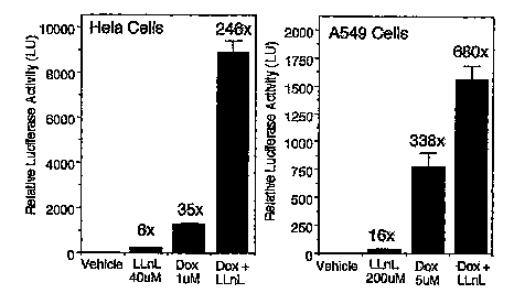
transduction. Hela cells (left panel) and A549 cells (right panel) were
infected
with rAAV (MOI 500 particles/cell) in the presence of the indicated drug
combinations and the expressed transgene was assessed at 24 hours post-
infection (Mean +/-SEM, N = 4). Fold induction relative to vehicle-treated
rAAV-infected cells is indicated above each bar.
Figure 14. The effect of proteasome inhibitor LLnL on (A) AV2Luc and
(B) AV2/SLuc transduction was evaluated following apical and basolateral
infection of human polarized airway epithelia at an MOI of 10,000
particles/cell
in the presence and absence of LLnL (40 ~.M). Luciferase activity was measured
at 5 and 14 days post-infection. Values represent the mean (+/-SEM) relative
luciferase activity for three independent tissue samples (N = 6-9 total
transwells).
. Figure 15. Analysis of full-length and selfcomplementary eGFP
expressing AAV vectors. HeLa cells were infected at an MOI =1000
particles/cell. (A) Quantification of relative eGFP-expressing area for
AV2eGFP and scAV2eGFP vectors. The values represent the mean (+/-SEM)
for three independent infections and quantification of 10 random fields for
each
experimental point. (B) Response of AV2eGFP and scAV2eGFP vectors to
treatment with hydroxyurea (5 mM) with gene expression analyzed at 24 hours
post-infection. (C) Southern blot analysis of Hirt DNA harvested from
AV2eGFP-infected (lanes 1 and 2) and scAV2eGFP-infected (lanes 3 and 4)
Hela cells at 24 hours post-infection. A 32P-labeled eGFP DNA probe was used
for Southern blots.
Figure 16. Quantification of eGFP expression following apical infection
of polarized human airway epithelia with self complementary and full-length
eGFP vectors. The relative mean area of fluorescence was evaluated following
transduction with AV2eGFP and scAV2eGFP vectors in the presence or absence
of LLnL (40 ,uM) at an MOI of 10,000 particles/cell on l, 3, 7, 15 and 30 days
post-infection. The values represent the mean (+/-SEM) for three independent
tissue samples. For each tissue sample, 3 transwells were evaluated by imaging
10 random fields in each sample at the various time points (N = 9 total
transwells for each experimental point).
21
CA 02520028 2005-09-22
WO 2004/090145 PCT/US2004/010045
-102/27
Figure 17. Combined administration of proteasome-modulating agents
can synergistically induce rAAV transduction from the apical surface of
polarized human airway epithelia. (A) 1 x 10⁹ particles of AV2Luc were applied
to the apical surface of polarized human airway epithelia cultures in the
absence
and presence of various combinations of LLnL (40 ,uM) and/or Dox (5 ~.M).
Luciferase expression was assayed at 3 and 17 days post-infection (B-E).
Similar results were observed following apical infection with a self
complementary (2.3 kb) scAV2eGFP vector at 15 days post-infection. (F)
Combined administration of LLnL and Dox augments dual vector heterodimer-
mediated delivery of a traps-spliced LacZ gene product. 101° particles
of
AV2LacZdonor (indicated by D) and/or AV2LacZacceptor (indicated by A)
were used to infect each transwell of the polarized airway epithelia in'~the
presence or absence of co-administered LLnL (40 ,uM) and Dox (5 ,uM). (3-
galactosidase activity was evaluated at 15 days post-infection. Data
represents
the mean (+/-SEM) relative luciferase or (3-galactosidase activity (per 1/10
sample) for 3 independent experiments.
Figure 18. IfZ vivo gene transfer to the mouse lung. AV2 and AV2/5
luciferase vectors were used to evaluate the ability of proteasome-modulating
agents to induce transduction. Results depict the mean (+/-SEM) luciferase
expression from (N = 5) mouse lungs at 14 days post-infection for each
condition.
Figure 19. Complementation of CFTR chloride transport abnormalities
in CF airway epithelia using combined CFTR rAAV and proteasome inhibitor
treatment. Results depict the mean +/-SEM (N=9) delta Isc response to
IBMX/forskolin in CF airway epithelia treated under the indicated conditions.
Assays were performed at 15 days post-infection and a non-CF untreated control
is given as a reference for fully functional CFTR.
Figure 20. Screening for anthracycline proteosome modulators. A)
Graph of luciferase activity versus concentration of tested agent. B) Fold
change
in luciferase activity for various treatments.
Figure 21. In vivo results for anthracycline proteosome modulators.
Detailed Description of the Invention
Definitions
22
CA 02520028 2005-09-22
WO 2004/090145 PCT/US2004/010045
-101/27
A "vector" as used herein refers to a macromolecule or association of
macromolecules that comprises or associates with a polynucleotide and which
can be used to mediate delivery of the polynucleotide to a cell, either in
vitro or
ira vivo. Illustrative vectors include, for example, plasmids, viral vectors,
liposomes and other gene delivery vehicles. The polynucleotide to be
delivered,
sometimes referred to as a "target polynucleotide" or "transgene," may
comprise
a coding sequence of interest in gene therapy (such as a gene encoding a
protein
of therapeutic or interest), a coding sequence of interest in vaccine
development
(such as a polynucleotide expressing a protein, polypeptide or peptide
suitable
for eliciting an immune response in a mammal), and/or a selectable or
detectable
marker.
"AAV" is adeno-associated virus, and may be used to refer to the
naturally occurring wild-type virus itself or derivatives thereof. The term
covers
all subtypes, serotypes and pseudotypes, and both naturally occurring and
recombinant forms, except where required otherwise. As used herein, the term
"serotype" refers to an AAV which is identified by and distinguished from
other
AAVs based on capsid protein reactivity with defined antisera, e.g., there are
eight serotypes of primate AAVs, AAV-1 to AAV-8. For example, serotype
AAV2 is used to refer to an AAV which contains capsid proteins encoded from
the cap gene of AAV 2 and a genome containing 5' and 3' ITR sequences from
the same AAV2 serotype. Pseudotyped AAV as refers to an AAV that contains
capsid proteins from one serotype and a viral genome including 5'-3' ITRs of a
second serotype. Pseudotyped rAAV would be expected to have cell surface
binding properties of the capsid serotype and genetic properties consistent
with
the ITR serotype. Pseudotyped rAAV axe produced using standard techniques
described in the art. As used herein, for example, rAAVS may be used to refer
an AAV having both capsid proteins and 5'-3' ITRs from the same serotype or it
may refer to an AAV having capsid proteins from serotype 5 and S'-3' ITRs from
a different AAV serotype, e.g., AAV serotype 2. For each example illustrated
herein the description of the vector design and production describes the
serotype
of the capsid and 5'-3' ITR sequences. The abbreviation "rAAV" refers to
recombinant adeno-associated virus, also referred to as a recombinant AAV
vector (or "rAAV vector").
23
CA 02520028 2005-09-22
WO 2004/090145 PCT/US2004/010045
-100/27
"Transduction" or "transducing" as used herein, are terms refernng to a
process for the introduction of an exogenous polynucleotide, e.g., a transgene
in
rAAV vector, into a host cell leading to expression of the polynucleotide,
e.g.,
the transgene in the cell. The process includes 1) endocytosis of the AAV
after
it has bound to a cell surface receptor, 2) escape from endosornes or other
intracellular compartments in the cytosol of a cell, 3) trafficking of the
viral
particle or viral genome to the nucleus, 4) uncoating of the virus particles,
and
generation of expressible double stranded AAV genome forms, including
circular intermediates. The rAAV expressible double stranded form may persist
as a nuclear episome or optionally may integrate into the host genome. The
alteration of any or a combination of endocytosis of the AAV after it has
bound
to a cell surface receptor, escape from endosomes or other intracellular
compartments to the cytosol of a cell, trafficking of the viral particle or
viral
genome to the nucleus, or uncoating of the virus particles, and generation of
expressive double stranded AAV genorne forms, including circular
intermediates, by an agent of the invention, may result in altered expression
levels or persistence of expression, or altered trafficking to the nucleus, or
altered types or relative numbers of host cells or a population of cells
expressing
the introduced polynucleotide. Altered expression or persistence of a
polynucleotide introduced via rAAV can be determined by methods well known
to the art including, but not limited to, protein expression, e.g., by ELISA,
flow
cytometry and Western blot, measurement of and DNA and RNA production by
hybridization assays, e.g., Northern blots, Southern blots and gel shift
mobility
assays. The agents of the invention preferably alter, enhance or increase
viral
endocytosis, escape from endosomes or other intracellular cytosolic
compartments, and trafftcking into or to the nucleus, uncoating of the viral
particles in the nucleus, and/or increasing concatamerization or generation of
double stranded expressible forms of the rAAV genome in the nucleus, so as to
alter expression of the introduced polynucleotide, e.g., a transgene in a rAAV
vector, ira vitro or ira vivo. Methods used for the introduction of the
exogenous
polynucleotide include well-known techniques such as transfection,
lipofection,
viral infection, transformation, and electroporation, as well as non-viral
gene
delivery techniques. The introduced polynucleotide may be stably or
transiently
maintained in the host cell.
24
CA 02520028 2005-09-22
WO 2004/090145 PCT/US2004/010045
-99/27
"Increased transduction or transduction frequency", "altered transduction
or transduction frequency", or "enhanced transduction or transduction
frequency" refers to an increase in one or more of the activities described
above
in a treated cell relative to an untreated cell. Agents of the invention which
increase transduction efficiency may be determined by measuring the effect on
one or more transduction activities, which may include measuring the
expression
of the transgene, measuring the function of the transgene, or determining the
number of rAAV vector particles necessary to yield the same transgene effect
compared to host cells not treated with the agents.
"Proteosome modulator" refers to an agent or class of agents which alter
or enhance rAAV transduction or rAAV transduction frequencies by interacting
with, binding to, or altering the function of, and/or trafficking or location
of the
proteosome. Proteosome modulators may have other cellular functions as
described in the art, e.g., such as doxyrubicin, an antibiotic. Proteosome
modulators of the current invention do not include proteosome inhibitors,
e.g.,
such as tripeptidyl aldehydes (Z-LLL or LLnL), agents that inhibit calpains,
cathepsins, cysteine proteases, andlor chymotrypsin-like protease activity of
proteasomes (Wagner et al., 2002; Young et al., 2000; Seisenberger et al.,
2001).
"Generation of double stranded expressible forms" or "conversion of
single to double strand rAAV genomes" refers to the process of replicating in
the
nucleus of an rAAV infected host cell a complimentary strand of the rAAV
single stranded vector DNA genome and annealing of the complimentary strand
to the vector genome to produce a double stranded DNA rAAV genome. Agents
of the invention described herein to increase, alter, or enhance rAAV
transduction include agents which increase the rate of nuclear transport or
the
steady state of single stranded viral DNA genomes in the nucleus which can
drive gene conversion events via steady state mechanisms. For the purposes of
the invention described herein, agents which enhance conversion of single to
double strands do not include agents which increase the concentration of DNA
repair enzymes or activate alternate DNA repair mechanism described by Russel
et al. (1995).
"Gene delivery" refers to the introduction of an exogenous
polynucleotide into a cell for gene transfer, and may encompass targeting,
binding, uptake, transport, localization, replicon integration and expression.
CA 02520028 2005-09-22
WO 2004/090145 PCT/US2004/010045
-98/27
"Gene transfer" refers to the introduction of an exogenous polynucleotide
into a cell which may encompass targeting, binding, uptake, transport,
localization and replicon integration, but is distinct from and does not imply
subsequent expression of the gene.
"Gene expression" or "expression" refers to the process of gene
transcription, translation, and post-translational modification.
A "detectable marker gene" is a gene that allows cells caxrying the gene
to be specifically detected (e.g., distinguished from cells which do not carry
the
marker gene). A large variety of such marker genes are known in the art.
A "selectable marker gene" is a gene that allows cells carrying the gene
to be specifically selected for or against, in the presence of a corresponding
selective agent. By way of illustration, an antibiotic resistance gene can be
used
as a positive selectable marker gene that allows a host cell to be positively
selected for in the presence of the corresponding antibiotic. A variety of
positive
and negative selectable markers are known in the art, some of which are
described below.
An "rAAV vector" as used herein refers to an AAV vector comprising a
polynucleotide sequence not of AAV origin (i.e., a polynucleotide heterologous
to AAV), typically a sequence of interest for the genetic transformation of a
cell.
In preferred vector constructs of this invention, the heterologous
polynucleotide
is flanked by at least one, preferably two AAV inverted terminal repeat
sequences (ITRs). The term rAAV vector encompasses both rAAV vector
particles and rAAV vector plasmids.
An "AAV virus" or "AAV viral particle" refers to a viral particle
composed of at least one AAV capsid protein (preferably by all of the capsid
proteins of a wild-type AAV) and an encapsidated polynucleotide. If the
particle
comprises a heterologous polynucleotide (i.e., a polynucleotide other than a
wild-type AAV genome such as a transgene to be delivered to a mammalian
cell), it is typically referred to as "rAAV".
A "rAAV vaccine " as used herein refers to an AAV vector comprising a
polynucleotide sequence not of AAV origin (i.e., a polynucleotide heterologous
to AAV), that encodes a peptide, polypeptide, or protein capable of eliciting
an
immune response in a host contacted with the vector. Expression of the
polynucleotide may result in generation of a neutralizing antibody response
26
CA 02520028 2005-09-22
WO 2004/090145 PCT/US2004/010045
-97/27
and/or a cell mediated response, e.g., a cytotoxic T cell response. In
preferred
vector constructs of this invention, the heterologous polynucleotide is
flanked by
at least one, preferably two AAV inverted terminal repeat sequences (ITRs).
A "helper virus" for AAV refers to a virus that allows AAV (e.g., wild-
s type AAV) to be replicated and packaged by a mammalian cell. A variety of
such helper viruses for AAV are known in the art, including aderioviruses,
herpes viruses and poxviruses such as vaccinia. The adenoviruses encompass a
number of different subgroups, although Adenovirus type 5 of subgroup C is
most commonly used. Numerous adenoviruses of human, non-human
mammalian and avian origin are known and available from depositories such as
the ATCC. Viruses of the herpes family include, for example, herpes simplex
viruses (HSV) and Epstein-Barr viruses (EBV), as well as cytomegaloviruses
(CMV) and pseudorabies viruses (PRV); which are also available from
depositories such as ATCC.
An "infectious" virus or viral particle is one that comprises a
polynucleotide component which it is capable of delivering into a cell for
which
the viral species is trophic. The term does not necessarily imply any
replication
capacity of the virus.
A "replication-competent" virus (e.g., a replication-competent AAV, sometimes
abbreviated as "RCA") refers to a phenotypically wild-type virus that is
infectious, and is also capable of being replicated in an infected cell (i.e.,
in the
presence of a helper virus or helper virus functions). In the case of AAV,
replication competence generally requires the presence of functional AAV
packaging genes. Preferred rAAV vectors as described herein are replication-
incompetent in mammalian cells (especially in human cells) by virtue of the
lack
of one or more AAV packaging genes. Preferably, such rAAV vectors lack any
AAV packaging gene sequences in order to minimize the possibility that RCA
are generated by recombination between AAV packaging genes and an incoming
rAAV vector. Preferred rAAV vector preparations as described herein are those
which contain few if any RCA (preferably less than about 1 RCA per 102 rAAV
particles, more preferably less than about 1 RCA per 104 rAAV particles, still
more preferably less than about 1 RCA per 108 rAAV particles, even more
preferably less than about 1 RCA per 1012 rAAV particles, most preferably no
RCA).
27
CA 02520028 2005-09-22
WO 2004/090145 PCT/US2004/010045
-96/27
The term "polynucleotide" refers to a polymeric form of nucleotides of
any length, including deoxyribonucleotides or ribonucleotides, or analogs
thereof. A polynucleotide may comprise modified nucleotides, such as
methylated or capped nucleotides and nucleotide analogs, and may be
interrupted by non-nucleotide components. If present, modifications to the
nucleotide structure may be imparted before or after assembly of the polymer.
The term polynucleotide, as used herein, refers interchangeably to double- and
single-stranded molecules. Unless otherwise specified or required, any
embodiment of the invention described herein that is a polynucleotide
encompasses both the double-stranded form and each of two complementary
single-stranded forms known or predicted to make up the double-stranded form.
A "transcriptional regulatory sequence" or "TRS," as used herein, refers
to a genomic region that controls the transcription of a gene or coding
sequence
to which it is operably linked. Transcriptional regulatory sequences of use in
the
present invention generally include at least one transcriptional promoter and
may
also include one or more enhancers and/or terminators of transcription.
"Operably linked" refers to an arrangement of two or more components,
wherein the components so described are in a relationship permitting them to
function in a coordinated manner. By way of illustration, a transcriptional
regulatory sequence or a promoter is operably linked to a coding sequence if
the
TRS or promoter promotes transcription of the coding sequence. An operably
linked TRS is generally j oined in cis with the coding sequence, but it is not
necessarily directly adjacent to it.
"Heterologous" means derived from a genotypically distinct entity from
that of the rest of the entity to which it is compared. For example, a
polynucleotide introduced by genetic engineering techniques into a different
cell
type is a heterologous polynucleotide (and, when expressed, can encode a
heterologous polypeptide). Similarly, a TRS or promoter that is removed from
its native coding sequence and operably linked to a different coding sequence
is
a heterologous TRS or promoter.
"Packaging" as used herein refers to a series of subcellular events that
results in the assembly and encapsidation of a viral vector, particularly an
AAV
vector. Thus, when a suitable vector is introduced into a packaging cell line
under appropriate conditions, it can be assembled into a viral particle.
Functions
28
CA 02520028 2005-09-22
WO 2004/090145 PCT/US2004/010045
-95/27
associated with packaging of viral vectors, particularly AAV vectors, are
described herein and in the art.
A "terminator" refers to a polynucleotide sequence that tends to diminish
or prevent read-through transcription (i.e., it diminishes or prevent
transcription
originating on one side of the terminator from continuing through to the other
side of the terminator). The degree to which transcription is disrupted is
typically a function of the base sequence and/or the length of the terminator
sequence. In particular, as is well known in numerous molecular biological
systems, particular DNA sequences, generally referred to as "transcriptional
termination sequences" are specific sequences that tend to disrupt read-
through
transcription by RNA polymerase, presumably by causing the RNA polymerase
molecule to stop and/or disengage from the DNA being transcribed. Typical
example of such sequence-specific terminators include polyadenylation
("polyA") sequences, e.g., SV40 polyA. In addition to or in place of such
sequence-specific terminators, insertions of relatively long DNA sequences
between a promoter and a coding region also tend to disrupt transcription of
the
coding region, generally in proportion to the length of the intervening
sequence.
This effect presumably arises because there is always some tendency for an
RNA polymerise molecule to become disengaged from the DNA being
transcribed, and increasing the length of the sequence to be traversed before
reaching the coding region would generally increase the likelihood that
disengagement would occur before transcription of the coding region was
completed or possibly even initiated. Terminators may thus prevent
transcription from only one direction ("uni-directional" terminators) or from
both directions ("bi-directional" terminators), and may be comprised of
sequence-specific termination sequences or sequence-non-specific terminators
or
both. A variety of such terminator sequences are known in the art; and
illustrative uses of such sequences within the context of the present
invention are
provided below.
"Host cells," "cell lines," "cell cultures," "packaging cell line" and other
such terms denote higher eukaryotic cells, preferably mammalian cells, most
preferably human cells, useful in the present invention. These cells can be
used
as recipients for recombinant vectors, viruses or other transfer
polynucleotides,
and include the progeny of the original cell that was transduced. It is
understood
29
CA 02520028 2005-09-22
WO 2004/090145 PCT/US2004/010045
-94/27
that the progeny of a single cell may not necessarily be completely identical
(in
morphology or in genomic complement) to the original parent cell.
A "therapeutic gene," "prophylactic gene," "target polynucleotide,"
"transgene," "gene of interest" and the like generally refer to a gene or
genes to
be transferred using a vector. Typically, in the context of the present
invention,
such genes are located within the rAAV vector (which vector is flanked by
inverted terminal repeat (ITR) regions and thus can be replicated and
encapsidated into rAAV particles). Target polynucleotides can be used in this
invention to generate rAAV vectors for a number of different applications.
Such
polynucleotides include, but are not limited to: (i) polynucleotides encoding
proteins useful in other forms of gene therapy to relieve deficiencies caused
by
missing, defective or sub-optimal levels of a structural protein or enzyme;
(ii)
polynucleotides that are transcribed into anti-sense molecules; (iii)
polynucleotides that are transcribed into decoys that bind transcription or
translation factors; (iv) polynucleotides that encode cellular modulators such
as
cytokines; (v) polynucleotides that can make recipient cells susceptible to
specific drugs, such as the herpes virus thymidine kinase gene; and
(vi) polynucleotides for cancer therapy, such as ElA tumor suppressor genes or
p53 tumor suppressor genes for the treatment of various cancers. To effect
expression of the transgene in a recipient host cell, it is preferably
operably
linked to a promoter, either its own or a heterologous promoter. A large
number
of suitable promoters are known in the art, the choice of which depends on the
desired level of expression of the target polynucleotide; whether one wants
constitutive expression, inducible expression, cell-specific or tissue-
specific
expression, etc. The rAAV vector may also contain a selectable marker.
A "gene" refers to a polynucleotide containing at least one open reading
frame that is capable of encoding a particular protein after being transcribed
and
translated.
"Recombinant," as applied to a polynucleotide means that the
polynucleotide is the product of various combinations of cloning, restriction
and/or ligation steps, and other procedures that result in a construct that is
distinct from a polynucleotide found in nature. A recombinant virus is a viral
particle comprising a recombinant polynucleotide. The terms respectively
CA 02520028 2005-09-22
WO 2004/090145 PCT/US2004/010045
-93/27
include replicates of the original polynucleotide construct and progeny of the
original virus construct.
A "control element" or "control sequence" is a nucleotide sequence
involved in an interaction of molecules that contributes to the functional
regulation of a polynucleotide, including replication, duplication,
transcription,
splicing, translation, or degradation of the polynucleotide. The regulation
may
affect the frequency, speed, or specificity of the process, and may be
enhancing
or inhibitory in nature. Control elements known in the art include, for
example,
transcriptional regulatory sequences such as promoters and enhancers. A
promoter is a DNA region capable under certain conditions of binding RNA
polymerase and initiating transcription of a coding region usually located
downstream (in the 3' direction) from the promoter. Promoters include AAV
promoters, e.g., P5, P19, P40 and AAV ITR promoters, as well as heterologous
promoters.
An "expression vector" is a vector comprising a region which encodes a
polypeptide of interest, and is used for effecting the expression of the
protein in
an intended target cell. An expression vector also comprises control elements
operatively linked to the encoding region to facilitate expression of the
protein in
the target. The combination of control elements and a gene or genes to which
they are operably linked for expression is sometimes referred to as an
"expression cassette," a large number of which are known and available in the
art or can be readily constructed from components that are available in the
art.
"Genetic alteration" refers to a process wherein a genetic element is
introduced into a cell other than by mitosis or meiosis. The element may be
heterologous to the cell, or it may be an additional copy or improved version
of
an element already present in the cell. Genetic alteration may be effected,
for
example, by transfecting a cell with a recombinant plasmid or other
polynucleotide through any process known in the art, such as electroporation,
calcium phosphate precipitation, or contacting with a polynucleotide-liposome
complex. Genetic alteration may also be effected, for example, by transduction
or infection with a DNA or RNA virus or viral vector. Preferably, the genetic
element is introduced into a chromosome or mini-chromosome in the cell; but
any alteration that changes the phenotype and/or genotype of the cell and its
progeny is included in this term.
31
CA 02520028 2005-09-22
WO 2004/090145 PCT/US2004/010045
-92/27
A cell is said to be "stably" altered, transduced or transformed with a
genetic sequence if the sequence is available to perform its function during
extended culture of the cell ~ih vitro. In preferred examples, such a cell is
"inheritably" altered in that a genetic alteration is introduced which is also
inheritable by progeny of the altered cell.
The terms "polypeptide" and "protein" are used interchangeably herein to
refer to polymers of amino acids of any length. The terms also encompass an
amino acid polymer that has been modified; for example, disulfide bond
formation, glycosylation, acetylation, phosphonylation, lipidation, or
conjugation with a labeling component. Polypeptides such as "CFTR" and the
like, when discussed in the context of gene therapy and compositions therefor,
refer to the respective intact polypeptide, or any fragment or genetically
engineered derivative thereof, that retains the desired biochemical function
of the
intact protein. Similarly, references to CFTR, and other such genes for use in
gene therapy (typically referred to as "transgenes" to be delivered to a
recipient
cell), include polynucleotides encoding the intact polypeptide or any fragment
or
genetically engineered derivative possessing the desired biochemical function.
An "isolated" plasmid, virus, or other substance refers to a preparation of
the substance devoid of at least some of the other components that may also be
present where the substance or a similar substance naturally occurs or is
initially
prepaxed from. Thus, for example, an isolated substance may be prepared by
using a purification technique to enrich it from a source mixture. Enrichment
can be measured on an absolute basis, such as weight per volume of solution,
or
it can be measured in relation to a second, potentially interfering substance
present in the source mixture. Increasing enrichments of the embodiments of
this invention are increasingly more preferred. Thus, for example, a 2-fold
enrichment is preferred, 10-fold enrichment is more preferred, 100-fold
enrichment is more preferred, 1000-fold enrichment is even more preferred.
A preparation of AAV is said to be "substantially free" of helper virus if
the ratio of infectious AAV particles to infectious helper virus particles is
at least
about 102:1; preferably at least about 104:1, more preferably at least about
106:1;
still more preferably at least about 108:1. Preparations are also preferably
free of
equivalent amounts of helper virus proteins (i.e., proteins as would be
present as
a result of such a level of helper virus if the helper virus particle
impurities noted
32
CA 02520028 2005-09-22
WO 2004/090145 PCT/US2004/010045
-91/27
above were present in disrupted form). Viral and/or cellular protein
contamination can generally be observed as the presence of Coomassie staining
bands on SDS gels (e.g., the appearance of bands other than those
corresponding
to the AAV capsid proteins VP1, VP2 and VP3).
"Efficiency" when used in describing viral production, replication or
packaging refers to useful properties of the method: in particular, the growth
rate and the number of virus particles produced per cell. "High efficiency"
production indicates production of at least 100 viral particles per cell;
preferably
at least about 10,000 and more preferably at least about 100,000 particles per
cell, over the course of the culture period specified.
An "individual" or "subj ect" treated in accordance with this invention
refers to vertebrates, particularly members of a mammalian species, and
includes
but is not limited to domestic animals, sports animals, and primates,
including
humans.
"Treatment" of an individual or a cell is any type of intervention in an
attempt to alter the natural course of the individual or cell at the time the
treatment is initiated, e.g., eliciting a prophylactic, curative or other
beneficial
effect in the individual. For example, treatment of an individual may be
undertaken to decrease or limit the pathology caused by any pathological
condition, including (but not limited to) an inherited or induced genetic
deficiency, infection by a viral, bacterial, or parasitic organism, a
neoplastic or
aplastic condition, or an immune system dysfunction such as autoimmunity or
immunosuppression. Treatment includes (but is not limited to) administration
of
a composition, such as a pharmaceutical composition, and administration of
compatible cells that have been treated with a composition. Treatment may be
performed either prophylactically or therapeutically; that is, either prior or
subsequent to the initiation of a pathologic event or contact with an
etiologic
agent.
The practice of the present invention will employ, unless otherwise
indicated, conventional techniques of molecular biology, virology,
microbiology,
recombinant DNA, and immunology, which are within the skill of the art. Such
techniques are explained fully in the literature. See, e.g., Sambrook et al.,
1989;
Gait, 1984; Freshney, 1987; the series Methods in Enz olo~y (Academic
33
CA 02520028 2005-09-22
WO 2004/090145 PCT/US2004/010045
-90/27
Press, Inc.); Miller et al., 1987; Weir et al., 1996; Ausubel et al., 1998;
Coligan
et al., 1991; Coligan et al., 1995; and Scopes 1994.
I. rAAV vectors
Recombinant AAV vectors are potentially powerful tools for human gene
therapy, particularly for diseases such as cystic fibrosis and sickle cell
anemia.
A,major advantage of rAAV vectors over other approaches to gene therapy is
that they generally do not require ongoing replication of the target cell in
order
to become stably integrated into the host cell.
rAAV vectors and/or viruses are also potentially powerful for the
development of therapeutic or prophylactic vaccines to prevent infection,
progression, and/or severity of disease. A major advantage of rAAV vectors for
vaccine development is that they are capable of persisting for essentially the
lifetime of the cell as a nuclear episome and therefore provide long term
expression of the peptide, polypeptide, or protein of immunologic interest.
Transgenes of interest include viral gene e.g. the envelope (env) or gag genes
of
HIV; bacterial genes e.g., streptococcal cell wall proteins; fungi, e.g.,
cocidomycosis; parasites, e.g., Leischmaniosis, or cancer genes, e.g. p53.
rAAV vectors and/or viruses may also contain one or more detectable
markers. A variety of such markers are known, including, by way of
illustration,
the bacterial beta-galactosidase (lacZ) gene; the human placental alkaline
phosphatase (AP) gene and genes encoding various cellular surface markers
which have been used as reporter molecules both ih vitro and in vivo. The rAAV
vectors and/or viruses may also contain one or more selectable markers.
Recombinant AAV vectors and/or viruses can also comprise
polynucleotides that do not encode proteins, including, e.g., polynucleotides
encoding for antisense mRNA (the complement of mRNA) which can be used to
block the translation of normal mRNA by forming a duplex with it, and
polynucleotides that encode ribozymes (RNA catalysts).
II. Selection and Preparation of AAV Vector
Adeno-associated viruses of any serotype are suitable to prepare rAAV,
since the various serotypes are functionally and structurally related, even at
the
genetic level (see, e.g., Blacklow, 1988; and Rose, 1974). All AAV serotypes
apparently exhibit similar replication properties mediated by homologous rep
genes; and all generally bear three related capsid proteins such as those
34
CA 02520028 2005-09-22
WO 2004/090145 PCT/US2004/010045
-89/27
expressed in AAV2. The degree of relatedness is further suggested by
heteroduplex analysis which reveals extensive cross-hybridization between
serotypes along the length of the genome; and the presence of analogous self
annealing segments at the termini that correspond to ITRs. The similar
infectivity patterns also suggest that the replication functions in each
serotype
are under similar regulatory control. Among the various AAV serotypes, AAV2
is most commonly employed.
An AAV vector of the invention typically comprises a polynucleotide
that is heterologous to AAV. The polynucleotide is typically of interest
because
of a capacity to provide a function to a target cell in the context of gene
therapy,
such as up- or down-regulation of the expression of a certain phenotype. Such
a
heterologous polynucleotide or "transgene," generally is of sufficient length
to
provide the desired function or encoding sequence.
Where transcription of the heterologous polynucleotide is desired in the
intended target cell, it can be operably linked to its own or to a
heterologous
promoter, depending for example on the desired level and/or specificity of
transcription within the target cell, as is known in the art. Various types of
promoters and enhancers are suitable for use in this context. Constitutive
promoters provide an ongoing level of gene transcription, and are preferred
when it is desired that the therapeutic or prophylactic polynucleotide be
expressed on an ongoing basis. Inducible promoters generally exhibit low
activity in the absence of the inducer, and are up-regulated in the presence
of the
inducer. They may be preferred when expression is desired only at certain
times
or at certain locations, or when it is desirable to titrate the level of
expression
using an inducing agent. Promoters and enhancers may also be tissue-specific:
that is, they exhibit their activity only in certain cell types, presumably
due to
gene regulatory elements found uniquely in those cells.
Illustrative examples of promoters are the SV40 late promoter from
simian virus 40, the Baculovirus polyhedron enhancer/promoter element, Herpes
Simplex Virus thymidine kinase (HSV tk), the immediate early promoter from
cytomegalovirus (CMV) and various retroviral promoters including LTR
elements. Inducible promoters include heavy metal ion inducible promoters
(such as the mouse mammary tumor virus (mMTV) promoter or various growth
hormone promoters), and the promoters from T7 phage which are active in the
CA 02520028 2005-09-22
WO 2004/090145 PCT/US2004/010045
-88/27
presence of T7 RNA polymerase. By way of illustration, examples of tissue-
specific promoters include various surfactin promoters (for expression in the
lung), myosin promoters (for expression in muscle), and albumin promoters (for
expression in the liver). A large variety of other promoters are known and
generally available in the art, and the sequences of many such promoters are
available in sequence databases such as the GenBank database.
Where translation is also desired in the intended target cell, the
heterologous polynucleotide will preferably also comprise control elements
that
facilitate translation (such as a ribosome binding site or "RBS" and a
polyadenylation signal). Accordingly, the heterologous polynucleotide
generally
comprises at least one coding region operatively linked to a suitable
promoter,
and may also comprise, for example, an operatively linked enhancer, ribosome
binding site and poly-A signal. The heterologous polynucleotide may comprise
one encoding region, or more than one,encoding regions under the control of
the
same or different promoters. The entire unit, containing a combination of
control elements and encoding region, is often referred to as an expression
cassette.
The heterologous polynucleotide is integrated by recombinant techniques
into or preferably in place of the AAV genomic coding region (i.e., in place
of
the AAV rep and cap genes), but is generally flanked on either side by AAV
inverted terminal repeat (ITR) regions. This means that an ITR appears both
upstream and downstream from the coding sequence, either in direct
juxtaposition, preferably (although riot necessarily) without any intervening
sequence of AAV origin in order to reduce the likelihood of recombination that
might regenerate a replication-competent AAV genome. However, a single ITR
may be sufficient to carry out the functions normally associated with
configurations comprising two ITRs (see, for example, WO 94/13788), and
vector constructs with only one ITR can thus be employed in conjunction with
the packaging and production methods of the present invention.
The native promoters for rep are self regulating, and can limit the amount
of AAV particles produced. The rep gene can also be operably linked to a
heterologous promoter, whether rep is provided as part of the vector
construct, or
separately. Any heterologous promoter that is not strongly down-regulated by
rep gene expression is suitable; but inducible promoters are preferred because
36
CA 02520028 2005-09-22
WO 2004/090145 PCT/US2004/010045
-87/27
constitutive expression of the rep gene can have a negative impact on the host
cell. A large variety of inducible promoters are known in the art; including,
by
way of illustration, heavy metal ion inducible promoters (such as
metallothionein promoters); steroid hormone inducible promoters (such as the
MMTV promoter or growth hormone promoters); and promoters such as those
from T7 phage which are active in the presence of T7 RNA polymerase. An
especially preferred sub-class of inducible promoters are those that axe
induced
by the helper virus that is used to complement the replication and packaging
of
the rAAV vector. A number of helper-virus-inducible promoters have also been
described, including the adenovirus early gene promoter which is inducible by
adenovirus ElA protein; the adenovirus major late promoter; the herpesvirus
promoter which is inducible by herpesvirus proteins such as VP 16 or 1 CP4; as
well as vaccinia or poxvirus inducible promoters.
Methods for identifying and testing helper-virus-inducible promoters
have been described (see, e.g., WO 96/17947). Thus, methods are known in the
art to determine whether or not candidate promoters are helper-virus-
inducible,
and whether or not they will be useful in the generation of high efficiency
packaging cells. Briefly, one such method involves replacing the p5 promoter
of
the AAV rep gene with the putative helper-virus-inducible promoter (either
known in the art or identified using well-known techniques such as linkage to
promoter-less "reporter" genes). The AAV rep-cap genes (with p5 replaced),
preferably linked to a positive selectable maxker such as an antibiotic
resistance
gene, are then stably integrated into a suitable host cell (such as the HeLa
or
A549 cells exemplified below). Cells that are able to grow relatively well
under
selection conditions (e.g., in the presence of the antibiotic) are then tested
for .
their ability to express the rep and cap genes upon addition of a helper
virus. As
an initial test for rep and/or cap expression, cells can be readily screened
using
immunofluorescence to detect Rep and/or Cap proteins. Confirmation of
packaging capabilities and efficiencies can then be determined by functional
tests for replication and packaging of incoming rAAV vectors. Using this
methodology, a helper-virus-inducible promoter derived from the mouse
metallothionein gene has been identified as a suitable replacement for the p5
promoter, and used for producing high titers of rAAV particles (as described
in
WO 96/17947).
37
CA 02520028 2005-09-22
WO 2004/090145 PCT/US2004/010045
-86/27
Given the relative encapsidation size limits of various AAV genomes,
insertion of a large heterologous polynucleotide into the genome necessitates
removal of a portion of the AAV sequence. Removal of one or more AAV
genes is in any case desirable, to reduce the likelihood of generating
replication-
s competent AAV ("RCA"). Accordingly, encoding or promoter sequences for
rep, cap, or both, are preferably removed, since the functions provided by
these
genes can be provided in trans.
The resultant vector is referred to as being "defective" in these functions.
In order to replicate and package the vector, the missing functions are
complemented with a packaging gene, or a plurality thereof, which together
encode the necessary functions for the various missing rep andlor cap gene
products. The packaging genes or gene cassettes are preferably not flanked by
AAV ITRs and preferably do not share any substantial homology with the rAAV
genome. Thus, in order to minimize homologous recombination during
replication between the vector sequence and separately provided packaging
genes, it is desirable to avoid overlap of the two polynucleotide sequences.
The
level of homology and corresponding frequency of recombination increase with
increasing length of homologous sequences and with their level of shared
identity. The level of homology that will pose a concern in a given system can
be determined theoretically and confirmed experimentally, as is known in the
art. Typically, however, recombination can be substantially reduced or
eliminated if the overlapping sequence is less than about a 25 nucleotide
sequence if it is at least 80% identical over its entire length, or less than
about a
50 nucleotide sequence if it is at least 70% identical over its entire length.
Of
course, even lower levels of homology are preferable since they will further
reduce the likelihood of recombination. It appears that, even without any
overlapping homology, there is some residual frequency 'of generating RCA.
Even further reductions in the frequency of generating RCA (e.g., by
nonhomologous recombination) can be obtained by "splitting" the replication
and encapsidation functions of AAV, as described by Allen et al., WO
98/27204).
The rAAV vector construct, and the complementary packaging gene
constructs can be implemented in this invention in a number of different
forms.
38
CA 02520028 2005-09-22
WO 2004/090145 PCT/US2004/010045
-85/27
Viral particles, plasmids, and stably transformed host cells can all be used
to
introduce such constructs into the packaging cell, either transiently or
stably.
In certain embodiments of this invention, the AAV vector and
complementary packaging gene(s), if any, are provided in the form of bacterial
plasmids, AAV particles, or any combination thereof. In other embodiments,
either the AAV vector sequence, the packaging gene(s), or both, are provided
in
the form of genetically altered (preferably inheritably altered) eukaryotic
cells.
The development of host cells inheritably altered to express the AAV vector
sequence, AAV packaging genes, or both, provides an established source of the
material that is expressed at a reliable level.
A variety of different genetically altered cells can thus be used in the
context of this invention. By way of illustration, a mammalian host cell may
be
used with at least one intact copy of a stably integrated rAAV vector. An AAV
packaging plasmid comprising at least an AAV rep gene operably linked to a
promoter can be used to supply replication functions (as described in U.S.
Patent
5,658,776). Alternatively, a stable mammalian cell line with an AAV rep gene
operably linked to a promoter can be used to supply replication functions
(see,
e.g., Trempe et al., WO 95/13392); Burstein et al. (WO 98/23018); and Johnson
et al. (U.S. No. 5,656,785). The AAV cap gene, providing the encapsidation
proteins as described above, can be provided together with an AAV rep gene or
separately (see, e.g., the above-referenced applications and patents as well
as
Allen et al. (WO 98/27204). Other combinations are possible and included
within the scope of this invention.
III. Generating rAAV
To generate recombinant AAV particles useful for such purposes as gene
therapy, the packaging cell line is preferably supplied with a recombinant AAV
vector comprising AAV inverted terminal repeat (ITR) regions surrounding one
or more polynucleotides of interest (or "target" polynucleotides).
The target polynucleotide is generally operably linked to a promoter,
either its own or a heterologous promoter. A large number of suitable
promoters
are known in the art, the choice of which depends on the desired level of
expression of the target polynucleotide (i.e., whether one wants constitutive
expression, inducible expression, cell-specific or tissue-specific expression,
etc.).
39
CA 02520028 2005-09-22
WO 2004/090145 PCT/US2004/010045
-84/27
Preferably, the rAAV vector also contains a positive selectable marker in
order to allow for selection of cells that have been infected by the rAAV
vector.
Negative selectable markers can also be included; as a means of selecting
against
those same cells should that become necessary or desirable. In a preferred
embodiment, one can make use of the "bifunctional selectable fusion genes"
described by S. D. Lupton; see, e.g., PCT/LTS91/08442 and PCT/LTS94/05601.
Briefly, those constructs involve direct translational fusions between a
dominant
positive selectable marker and a negative selectable marker. Preferred
positive
selectable markers are derived from genes selected from the group consisting
of
hph, neo, and gpt, and preferred negative selectable markers are derived from
genes selected from the group consisting of cytosine deaminase, HSV-I TK,
VZV TK, HPRT, APRT and gpt. Especially preferred markers are bifunctional
selectable fusion genes wherein the positive selectable marker is derived from
hph or neo, and the negative selectable marker is derived from cytosine
deaminase or a TK gene.
Useful target polynucleotides can be employed in rAAV vectors for a
number of different applications. Such polynucleotides include, but are not
limited to: (i) polynucleotides encoding proteins useful in other forms of
gene
therapy to relieve deficiencies caused by missing, defective or sub-optimal
levels
of a structural protein or enzyme; (ii) polynucleotides that are transcribed
into
anti-sense molecules; (iii) polynucleotides that are transcribed into decoys
that
bind transcription or translation factors; (iv) polynucleotides that encode
cellular
modulators such as cytokines; (v) polynucleotides that can make recipient
cells
susceptible to specific drugs, such as the herpes virus thymidine kinase gene;
and (vi) polynucleotides for cancer therapy, such as the wild-type p53 tumor
suppressor cDNA for replacement of the missing or damaged p53 gene
associated with some lung and breast cancers, or the ElA tumor suppressor gene
.
which is capable of inhibiting tumorigenesis and/or metastasis of a variety of
different cancers including breast and ovarian cancers.
Since the therapeutic or prophylactic specificity of the resulting
recombinant AAV particle is determined by the particular vector or pro-vector
introduced, the same basic packaging cell line can be modified for any of
these
applications. For example, a vector comprising a specific target
polynucleotide
can be introduced into the packaging cell for production of the AAV vector by
CA 02520028 2005-09-22
WO 2004/090145 PCT/US2004/010045
-83/27
any of several possible methods; including, for example, electroporation or
transfection of a plasmid comprising an rAAV pro-vector, or infection with an
rAAV or helper virus comprising an rAAV vector or pro-vector.
Helper virus can be introduced before, during or after introduction of the
rAAV vector. For example, the plasmid can be co-infected into the culture
along
with the helper virus; and the cells can then be cultured for a sufficient
period,
typically 2-5 days, in conditions suitable for replication and packaging as
known
in the art (see references above and examples below). Lysates are prepared,
and
the recombinant AAV vector particles are purified by techniques known in the
art.
In a preferred embodiment, also illustrated in the Examples below, a
recombinant AAV vector is itself stably integrated into a mammalian cell to be
used for packaging. Such rAAV "producer cells" can then be grown and stored
until ready for use. To induce production of rAAV particles from such producer
1 S cells, the user need only infect the cells with helper virus and culture
the cells
under conditions suitable for replication and packaging of AAV (as described
below).
Alternatively, one or more of the AA~V split-packaging genes or the
rAAV vector can be introduced as part of a recombinant helper virus. For
example, the El, E3 and/or the E4 genes of adenovirus can be replaced with one
or more split-packaging genes or an rAAV vector. Techniques for facilitating
cloning into adenovirus vectors, e.g., into the E1 andlor E3 regions, are
known in
the art (see, e.g., Bett, A. J. et al., Proc. Natl. Acad. Sci. USA, 91, 8802-
8806
(1994)). Thus, a helper virus such as a recombinant adenovirus, can be used to
provide helper virus functions as well as AAV packaging genes andlor an rAAV
pro-vector, since (as is known in the art) a number of genes in such a helper
virus (e.g., the E3 gene of adenovirus) can be replaced without eliminating
helper virus activity. Additional genes can be inserted into such a helper
virus
by providing any necessary helper virus functions in trans. For example, human
293 cells contain adenoviral genes that can complement adenoviral E1 mutants.
Thus, heterologous genes can also be cloned into an adenovirus in which the E1
genes have been deleted, for use in cells that can effectively provide such
adenoviral functions in trans. Alternatively, the use of a helper virus can be
41
CA 02520028 2005-09-22
WO 2004/090145 PCT/US2004/010045
-82/27
eliminated by providing all necessary helper virus functions in the packaging
cell.
IV. Introduction of Genetic Material Into Cells
As is described in the art, and illustrated both herein and in the references
cited above, genetic material can be introduced into cells (such as mammalian
"producer'.' cells for the production of AAV) using any of a variety of means
to
transform or transduce such cells. By way of illustration, such techniques
include, for example, transfection with bacterial plasmids, infection with
viral
vectors, electroporation, calcium phosphate precipitation, and introduction
using
any of a variety of lipid-based compositions (a process often referred to as
"lipofection"). Methods and compositions for performing these techniques have
been described in the art and are widely available.
Selection of suitably altered cells may be conducted by any technique in
the art. For example, the polynucleotide sequences used to alter the cell may
be
introduced simultaneously with or operably linked to one or more detectable or
selectable markers as is known in the art. By way of illustration, one can
employ a drug-resistance gene as a selectable marker. Drug-resistant cells can
then be picked and grown, and then tested for expression of the desired
sequence, i.e., a packaging gene product, or a product of the heterologous
polynucleotide, as appropriate. Testing for acquisition, localization and/or
maintenance of an introduced polynucleotide can be performed using DNA
hybridization-based techniques (such as Southern blotting and other procedures
as is known in the art). Testing for expression can be readily performed by
Northern analysis of RNA extracted from the genetically altered cells, or by
indirect immunofluorescence for the corresponding gene product. Testing and
confirmation of packaging capabilities and efficiencies can be obtained by
introducing to the cell the remaining functional components of AAV and a
helper virus, to test for production of AAV particles. Where a cell is
inheritably
altered with a plurality of polynucleotide constructs, it is generally more
convenient (though not essential) to introduce them to the cell separately,
and
validate each step seriatim. References describing such techniques include
those
cited herein.
V. Selection and Preparation of Helper Virus
42
CA 02520028 2005-09-22
WO 2004/090145 PCT/US2004/010045
-81/27
As discussed above, AAV is a parvovirus that is defective for self
replication, and must generally rely on a helper virus to supply certain
replicative functions. A number of such helper viruses have been identified,
including adenoviruses, herpes viruses (including but not limited to HSV1,
cytomegalovirus and HHV-6), and pox viruses (particularly vaccinia). Any such
virus may be used with this invention.
Frequently, the helper virus is an adenovirus of a type and subgroup that
can infect the intended host cell. Human adenovirus of subgroup C,
particularly
serotypes 1, 2, 4, 6, and 7, are commonly used. Serotype 5 is generally
preferred.
The features and growth patterns of adenovirus are known in the art. The
reader may refer, for example, to Horowitz (195). The packaged adenovirus
genome is a linear DNA molecule, linked through adenovirus ITRs at the left-
and right-hand termini. through a terminal protein complex to form a circle.
Control and encoding regions for early, intermediate, and late components
overlap within the genome. Early region genes are implicated in replication of
the adenovirus genome, and are grouped depending on their location into the
E1,
E2, E3, and E4 regions.
Although not essential, in principle it is desirable that the helper virus
strain be defective for replication in the subject ultimately to receive the
genetic
therapy. Thus, any residual helper virus present in an rAAV preparation will
be
replication-incompetent. Adenoviruses from which the ElA or both the ElA
and the E3 region have been removed are not infectious for most human cells.
They can be replicated in a permissive cell line (e.g., the human 293 cell
line)
which is capable of complementing the missing activity. Regions of adenovirus
that appear to be associated with helper function, as well as regions that do
not,
have been identified and described in the art (see, e.g., P. Colosi et al.,
W097/17455, and references cited therein).
VI. Uses of rAAV for Gene Therapy
AAV vectors can be used for administration to an individual for purposes
of gene therapy or vaccination. Suitable diseases for rAAV therapy include but
are not limited to those induced by viral, bacterial, or parasitic infections,
various malignancies and hyperproliferative conditions, autoimmune conditions,
and congenital deficiencies.
43
CA 02520028 2005-09-22
WO 2004/090145 PCT/US2004/010045
-80/27
Gene therapy can be conducted to enhance the level of expression of a
particular protein either within or secreted by the cell. Vectors of this
invention
may be used to genetically alter cells either for gene marking, replacement of
a
missing or defective gene, or insertion of a therapeutic gene. Alternatively,
a
polynucleotide may be provided to the cell that decreases the level of
expression.
This may be used for the suppression of an undesirable phenotype, such as the
product of a gene amplified or overexpressed during the course of a
malignancy,
or a gene introduced or overexpressed during the course of a microbial
infection.
Expression levels may be decreased by supplying a therapeutic or prophylactic
polynucleotide comprising a sequence capable, for example, of forming a stable
hybrid with either the target gene or RNA transcript (antisense therapy),
capable
of acting as a ribozyme to cleave the relevant mRNA or capable of acting as a
decoy for a product of the target gene.
The introduction of rAAV vectors by the methods of the present
invention may involve use of any number of delivery techniques (both surgical
and non-surgical) which are available and well known in the art. Such delivery
techniques, for example, include vascular catheterization, cannulization,
inj ection, inhalation, endotracheal, subcutaneous, inunction, topical, oral,
percutaneous, infra-arterial, intravenous, and/or intraperitoneal
administrations.
Vectors can also be introduced by way of bioprostheses, including, by way of
illustration, vascular grafts (PTFE and dacron), heart valves, intravascular
stems,
intravascular paving as well as other non-vascular prostheses. General
techniques regarding delivery, frequency, composition and dosage ranges of
vector solutions are within the skill of the art.
In particular, for delivery of a vector of the invention to a tissue, any
physical or biological method that will introduce the vector to a host animal
can
be employed. Vector means both a bare recombinant vector and vector DNA
packaged into viral coat proteins, as is well known for AAV administration.
Simply dissolving an AAV vector in phosphate buffered saline has been
demonstrated to be sufficient to provide a vehicle useful for muscle tissue
expression, and there are no known restrictions on the Garners or other
components that can be coadministered with the vector (although compositions
that degrade DNA should be avoided in the normal manner with vectors).
Pharmaceutical compositions can be prepared as injectable formulations or as
44
CA 02520028 2005-09-22
WO 2004/090145 PCT/US2004/010045
-79/27
topical formulations to be delivered to the muscles by transdermal transport.
Numerous formulations for both intramuscular inj ection and transdermal
transport have been previously developed and can be used in the practice of
the
invention. The vectors can be used with any pharmaceutically acceptable
carrier
for ease of administration and handling.
For purposes of intramuscular injection, solutions in an adjuvant such as
sesame or peanut oil or in aqueous propylene glycol can be employed, as well
as
sterile aqueous solutions. Such aqueous solutions can be buffered, if desired,
and the liquid diluent first rendered isotonic with saline or glucose.
Solutions of
the AAV vector as a free acid (DNA contains acidic phosphate groups) or a
pharmacologically acceptable salt can Ibe prepared in water suitably mixed
with a
surfactant such as hydroxypropylcellulose. A dispersion of AAV viral particles
can also be prepared in glycerol, liquid polyethylene glycols and mixtures
thereof and in oils. Under ordinary conditions of storage and use, these
preparations contain a preservative to prevent the growth of microorganisms.
In
this connection, the sterile aqueous media employed are all readily obtainable
by
standard techniques well-known to those skilled in the art.
The pharmaceutical forms suitable for injectable use include sterile
aqueous solutions or dispersions and sterile powders for the extemporaneous
preparation of sterile injectable solutions or dispersions. In all cases the
form
must be sterile and must be fluid to the extent that easy syringability
exists. It
must be stable under the conditions of manufacture and storage and must be
preserved against the contaminating action of microorganisms such as bacteria
and fungi. The earner can be a solvent or dispersion medium containing, for
example, water, ethanol, polyol (for example, glycerol, propylene glycol,
liquid
polyethylene glycol and the like), suitable mixtures thereof, and vegetable
oils.
The proper fluidity can be maintained, for example, by the use of a coating
such
as lecithin, by the maintenance of the required particle size in the case of a
dispersion and by the use of surfactants. The prevention of the action of
microorganisms can be brought about by various antibacterial and antifungal
agents, for example, parabens, chlorobutanol, phenol, sorbic acid, thimerosal
and
the like. In many cases it will be preferable to include isotonic agents, for
example, sugars or sodium chloride. Prolonged absorption of the injectable
CA 02520028 2005-09-22
WO 2004/090145 PCT/US2004/010045
-78/27
compositions can be brought about by use of agents delaying absorption, for
example, aluminum monostearate and gelatin.
Sterile injectable solutions are prepared by incorporating the AAV vector
in the required amount in the appropriate solvent with various of the other
ingredients enumerated above, as required, followed by filtered sterilization.
Generally, dispersions are prepared by incorporating the sterilized active
ingredient into a sterile vehicle which contains the basic dispersion medium
and
the required other ingredients from those enumerated above. In the case of
sterile powders for the preparation of sterile injectable solutions, the
preferred
methods of preparation are vacuum drying and the freeze drying technique
which yield a powder of the active ingredient plus any additional desired
ingredient from the previously sterile-filtered solution thereof.
For purposes of topical administration, dilute sterile, aqueous solutions
(usually in about 0.1 % to 5% concentration), otherwise similar to the above
parenteral solutions, are prepared in containers suitable for incorporation
into a
transdermal patch, and can include known carriers, such as pharmaceutical
grade
dimethylsulfoxide (DMSO).
Of particular interest is the correction of the genetic defect of cystic
fibrosis, by supplying a properly functioning cystic fibrosis transmembrane
conductance regulator (CFTR) to the airway epithelium. Thus, rA.AV vectors
encoding native CFTR protein, and mutants and fragments thereof, are all
preferred embodiments of this invention.
Compositions of this invention may be used in vivo as well as ex vivo. Ira
vivo gene therapy comprises administering the vectors of this invention
directly
to a subject. Pharmaceutical compositions can be supplied as liquid solutions
or
suspensions, as emulsions, or as solid forms suitable for dissolution or
suspension in liquid prior to use. For administration into the respiratory
tract, a
preferred mode of administration is by aerosol, using a composition that
provides either a solid or liquid aerosol when used with an appropriate
aerosolubilizer device. Another preferred mode of administration into the
respiratory tract is using a flexible fiberoptic bronchoscope to instill the
vectors.
Typically, the viral vectors are in a pharmaceutically suitable pyrogen-free
buffer such as Ringer's balanced salt solution (pH 7.4). Although not
required,
46
CA 02520028 2005-09-22
WO 2004/090145 PCT/US2004/010045
-77/27
pharmaceutical compositions may optionally be supplied in unit dosage form
suitable for administration of a precise amount.
An effective amount of virus is administered, depending on the
objectives of treatment. An effective amount may be given in single or divided
doses. Where a low percentage of transduction can cure a genetic deficiency,
then the objective of treatment is generally to meet or exceed this level of
transduction. In some instances, this level of transduction can be achieved by
transduction of only about 1 to 5% of the target cells, but is more typically
20%
of the cells of the desired tissue type, usually at least about 50%,
preferably at
least about 80%, more preferably at least about 95%, and even more preferably
at least about 99% of the cells of the desired tissue type. As a guide, the
number
of vector particles present in a single dose given by bronchoscopy will
generally
be at least about 1 ~ 10g, and is more typically 5 X 10⁸, 1 ~ 101°, and
on some
occasions 1 x 10¹¹ particles, including both DNAse-resistant and DNAse-
susceptible particles. In terms of DNAse-resistant particles, the dose will
generally be between 1 X 10⁶ and 1 ~ 1014 particles, more generally between
about 1 ~ 10$ and 1 ~ 1012 particles. The treatment can be repeated as often
as
every two or three weeks, as required, although treatment once in 180 days may
be sufficient.
To confirm the presence of the desired DNA sequence in the host cell, a
variety of assays may be performed. Such assays include, for example,
"molecular biological" assays well known to those of skill in the art, such as
Southern and Northern blotting, RT-PCR and PCR; "biochemical" assays, such
as detecting the presence of a polypeptide expressed from a gene present in
the
vector, e.g., by immunological means (immunoprecipitations, immunoaffinity
columns, ELISAs and Western blots) or by any other assay useful to identify
the
presence and/or expression of a particular nucleic acid molecule falling
within
the scope of the invention.
To detect and quantitate RNA produced from introduced DNA segments,
RT-PCR may be employed. In this application of PCR, it is first necessary to
reverse transcribe RNA into DNA, using enzymes such as reverse transcriptase,
and then through the use of conventional PCR techniques amplify the DNA. In
most instances PCR techniques, while useful, will not demonstrate integrity of
the RNA product. Further information about the nature of the RNA product may
47
CA 02520028 2005-09-22
WO 2004/090145 PCT/US2004/010045
-76/27
be obtained by Northern blotting. This technique demonstrates the presence of
an RNA species and gives information about the integrity of that RNA. The
presence or absence of an RNA species can also be determined using dot or slot
blot Northern hybridizations. These techniques are modifications of Northern
blotting and only demonstrate the presence or absence of an RNA species.
While Southern blotting and PCR may be used to detect the DNA
segment in question, they do not provide information as to whether the DNA
segment is being expressed. Expression may be evaluated by specifically
identifying the polypeptide products of the introduced DNA sequences or
evaluating the phenotypic changes brought about by the expression of the
introduced DNA segment in the host cell.
Thus, the effectiveness of the genetic alteration can be monitored by
several criteria, including analysis of physiological fluid samples, e.g.,
urine,
plasma, serum, blood, cerebrospinal fluid or nasal or lung washes. Samples
1 S removed by biopsy or surgical excision may be analyzed by in situ
hybridization, PCR amplification using vector-specific probes, RNAse
protection, immunohistology, or immunofluorescent cell counting. When the
vector is administered by bronchoscopy, lung function tests may be performed,
and bronchial lavage may be assessed for the presence of inflammatory
cytokines. The treated subject may also be monitored for clinical features,
and
to determine whether the cells express the function intended to be conveyed by
the therapeutic or prophylactic polynucleotide.
The decision of whether to use in vivo or ex vivo therapy, and the
selection of a particular composition, dose, and route of administration will
depend on a number of different factors, including but not limited to features
of
the condition and the subject being treated. The assessment of such features
and
the design of an appropriate therapeutic or prophylactic regimen is ultimately
the
responsibility of the prescribing physician.
The foregoing description provides, inter alia, methods for generating
high titer preparations of recombinant AAV vectors that are substantially free
of
helper virus (e.g., adenovirus) and cellular proteins. It is understood that
variations may be applied to these methods by those of skill in this art
without
departing from the spirit of this invention.
VII. Exemplary Methods to Identiy Useful Agents
48
CA 02520028 2005-09-22
WO 2004/090145 PCT/US2004/010045
-75/27
The utility of rAAV as a gene therapy vector is based on its transduction
properties. Methods to detect AAV transduction are known in the art, including
those described herein, and are useful to screen libraries, types and classes
of
agents for the ability to improve AAV transduction by means other than by
affecting binding to cell surface receptors, or the rate of infra-nuclear
genome
conversion of the single stranded rAAV vector to a double stranded genome.
Transduction may be defined by protein expression of a heterologous
transgene contained in the vector or steady state levels thereof. Hence,
transduction is a measurable functional endpoint of successful gene delivery
with a viral vector, e.g., rAAV. A variety of transgenes have been expressed
from cells infected with AAV vectors, and include intracellularly expressed
proteins such as the green fluorescent protein (GFP), cell membrane associated
proteins such as the cystic fibrosis transmembrane protein (CFTR), and
secretory
proteins such as Epo, FVIZI, and FIX. However, not all transgenes are capable
of fully assessing the extent of transduction with a given vector and tissue
target.
For example, secreted proteins do not give indication of the number of cell
types
expressing a given transgene. Furthermore, functional markers of gene
expression are dependent on the ability of a given transgene protein product
to
function properly within a given target cell type.
VIII. Agents Useful in the Practice of the Invention
rAAV must undergo a number of complex intracellular events between
binding and conversion to dsDNA that may be rate limiting for transduction
efficiency including but not limited to rAAV endocytosis, trafficking and
processing of the rAAV through the appropriate intracellular compartments
(including without limitation proteosomes, endosomes, and trans-golgi),
transport into the nucleus, and viral uncoating. Furthermore, agents that
alter the
efficiency of these intracellular processing events can also have an end
result of
increasing the amount of viral DNA in the nucleus and hence, through steady
state, the abundance of gene conversion products. Thus, an increase in genome
conversion products following enhancement of rAAV intracellular processing
does not necessarily indicate an increased rate of gene conversion. Methods of
enhancing transduction with rAAV are expected to increase the extent of double
stranded genome conversion products in the nucleus. This is an important
distinction with previous methods aimed at directly enhancing genome
49
CA 02520028 2005-09-22
WO 2004/090145 PCT/US2004/010045
-74/27
conversion using DNA damaging agents, topoisomerase inhibitors, or adenoviral
early gene products (Alexander et al., 1997; Alexander et al., 1996; Ferrari
et al.,
1996; Fisher et al., 1996; Halbert et al., 1997; Russel et al., 1995) which
essentially change the level of gene conversion enzymes in the nucleus.
However, in cases where gene conversion in the nucleus is not rate limiting,
intracellular viral processing events that limit transduction may predominate.
Thus, agents useful in the practice of the invention include agents which
alter rAAV transduction efficiency, e.g., rAAV endocytosis, trafficking and
processing of rAAV through the intracellular compartment, viral nucleic acid
or
protein degradation, viral uncoating and nuclear transport of virus or viral
genomes or otherwise modulate proteosomes. Preferred agents are those which
enhance or increase rAAV transduction. Classes of agents useful in the
invention include but are not limited to antibiotics, chemotherapeutics, e.g.,
anthracyclines, proteosome modulators, lipid lowering agents, and food
additives. Exemplary agents include proteasomes (Wagner et al., 2002; Young
et al., 2000; Seisenberger et al., 2001), as well as agents that modulate the
proteosome and ubiquitin pathways, e.g., bind to proteosomes and/or modulate
the activity of proteosomes, ubiquitin, ubiquitin carrier protein, or
ubiquitin
ligase, but do not substantially alter the activity of the proteosome, e.g.,
the
proteolytic activity of the proteasome or of ubiquitin, ubiquitin carrier
protein, or
ubiquitin ligase. Examples of these agents include without limitation
antibiotics,
e.g., epoxomicin, lipid lowering drugs, e.g., simvastatin, food additives,
e.g.,
tannic acid, and chemotherapeutics, e.g., cisplatin, anthracyclines such as
doxorubicin; and camptothecin.
IX. Dosages, Formulations and Routes of Administration of the A~Lents of
the
Invention
Administration of the agents identified in accordance with the present
invention may be continuous or intermittent, depending, for example, upon the
recipient's physiological condition, whether the purpose of the administration
is
therapeutic or prophylactic, and other factors known to skilled practitioners.
The
administration of the agents of the invention may be essentially continuous
over
a preselected period of time or may be in a series of spaced doses. Both local
and systemic administration is contemplated. When the agents of the invention
CA 02520028 2005-09-22
WO 2004/090145 PCT/US2004/010045
-73/27
are employed for prophylactic purposes, agents of the invention are amenable
to
chronic use, preferably by systemic administration.
One or more suitable unit dosage forms comprising the agents of the
invention, which, as discussed below, may optionally be formulated for
sustained release, can be administered by a variety of routes including oral,
or
parenteral, including by rectal, transdermal, subcutaneous, intravenous,
intramuscular, intraperitoneal, intrathoracic, intrapulmonary and intranasal
routes. For example, for administration to the liver, intravenous
administration
is preferred. For administration to the lung, airway administration is
preferred.
The formulations may, where appropriate, be conveniently presented in discrete
unit dosage forms and may be prepared by any of the methods well known to
pharmacy. Such methods may include the step of bringing into association the
agent with liquid carriers, solid matrices, semi-solid carriers, finely
divided solid
carriers or combinations thereof, and then, if necessary, introducing or
shaping
the product into the desired delivery system.
When the agents of the invention are prepared for oral administration,
they are preferably combined with a pharmaceutically acceptable carrier,
diluent
or excipient to form a pharmaceutical formulation, or unit dosage form. The
total active ingredients in such formulations comprise from 0.1 to 99:9% by
weight of the formulation. By "pharmaceutically acceptable" it is meant the
carrier, diluent, excipient, andlor salt must be compatible with the other
ingredients of the formulation, and not deleterious to the recipient thereof.
The
active ingredient for oral administration may be present as a powder or as
granules; as a solution, a suspension or an emulsion; or in achievable base
such
as a synthetic resin for ingestion of the active ingredients from a chewing
gum.
The active ingredient may also be presented as a bolus, electuary or paste.
Pharmaceutical formulations containing the agents of the invention can
be prepared by procedures known in the art using well known and readily
available ingredients. For example, the agent can be formulated with common
excipients, diluents, or carriers, and formed into tablets, capsules,
suspensions,
powders, and the like. Examples of excipients, diluents, and carriers that are
suitable for such formulations include the following fillers and extenders
such as
starch, sugars, mannitol, and silicic derivatives; binding agents such as
carboxymethyl cellulose, HPMC and other cellulose derivatives, alginates,
51
CA 02520028 2005-09-22
WO 2004/090145 PCT/US2004/010045
-72/27
gelatin, and polyvinyl-pyrrolidone; moisturizing agents such as glycerol;
disintegrating agents such as calcium carbonate and sodium bicarbonate; agents
for retarding dissolution such as paraffin; resorption accelerators such as
quaternary ammonium compounds; surface active agents such as cetyl alcohol,
glycerol monostearate; adsorptive carriers such as kaolin and bentonite; and
lubricants such as talc, calcium and magnesium stearate, and solid polyethyl
glycols.
For example, tablets or caplets containing the agents of the invention can
include buffering agents such as calcium carbonate, magnesium oxide and
magnesium carbonate. Caplets and tablets can also include inactive ingredients
such as cellulose, pregelatinized starch, silicon dioxide, hydroxy propyl
methyl
cellulose, magnesium stearate, microcrystalline cellulose, starch, talc,
titanium
dioxide, benzoic acid, citric acid, corn starch, mineral oil, polypropylene
glycol,
sodium phosphate, and zinc stearate, and the like. Hard or soft gelatin
capsules
containing an agent of the invention can contain inactive ingredients such as
gelatin, microcrystalline cellulose, sodium lauryl sulfate, starch, talc, and
titanium dioxide, and the like, as well as liquid vehicles such as
polyethylene
glycols (PEGS) and vegetable oil. Moreover, enteric coated caplets or tablets
of
an agent of the invention are designed to resist disintegration in the stomach
and
dissolve in the more neutral to alkaline environment of the duodenum.
The agents of the invention can also be formulated as elixirs or solutions
for convenient oral administration or as solutions appropriate for parenteral
administration, for instance by intramuscular, subcutaneous or intravenous
routes.
The pharmaceutical formulations of the agents of the invention can also
take the form of an aqueous or anhydrous solution or dispersion, or
alternatively
the form of an emulsion or suspension.
Thus, the therapeutic agent may be formulated for parenteral
administration (e.g., by injection, for example, bolus injection or continuous
infusion) and may be presented in unit dose form in ampules, pre-filled
syringes,
small volume infusion containers or in multi-dose containers with an added
preservative. The active ingredients may take such forms as suspensions,
solutions, or emulsions in oily or aqueous vehicles, and may contain
formulatory
agents such as suspending, stabilizing and/or dispersing agents.
Alternatively,
52
CA 02520028 2005-09-22
WO 2004/090145 PCT/US2004/010045
-71/27
the active ingredients may be in powder form, obtained by aseptic isolation of
sterile solid or by lyophilization from solution, for constitution with a
suitable
vehicle, e.g., sterile, pyrogen-free water, before use.
These formulations can contain pharmaceutically acceptable vehicles and
adjuvants which are well known in the prior art. It is possible, for example,
to
prepare solutions using one or more organic solvents) that islare acceptable
from the physiological standpoint, chosen, in addition to water, from solvents
such as acetone, ethanol, isopropyl alcohol, glycol ethers such as the
products
sold under the name "Dowanol", polyglycols and polyethylene glycols, Cl-C4
alkyl esters of short-chain acids, preferably ethyl or isopropyl lactate,
fatty acid
triglycerides such as the products marketed under the name "Miglyol",
isopropyl
myristate, animal, mineral and vegetable oils and polysiloxanes.
The compositions according to the invention can also contain thickening
agents such as cellulose and/or cellulose derivatives. They can also contain
gums such as xanthan, guar or carbo gum or gum arabic, or alternatively
polyethylene glycols, bentones and montmorillonites, and the like.
It is possible to add, if necessary, an adjuvant chosen from antioxidants,
surfactants, other preservatives, film-forming, keratolytic or comedolytic
agents,
perfumes and colorings. Also, other active ingredients may be added, whether
for the conditions described or some other condition.
For example, among antioxidants, t-butylhydroquinone, butylated
hydroxyanisole, butylated hydroxytoluene and a tocopherol and its derivatives
may be mentioned. The galenical forms chiefly conditioned for topical
application take the form of creams, milks, gels, dispersion or
microemulsions,
lotions thickened to a greater or lesser extent, impregnated pads, ointments
or
sticks, or alternatively the form of aerosol formulations in spray or foam
form or
alternatively in the form of a cake of soap.
Additionally, the agents are well suited to formulation as sustained
release dosage forms and the like. The formulations can be so constituted that
they release the active ingredient only or preferably in a particular part of
the
intestinal or respiratory tract, possibly over a period of time. The coatings,
envelopes, and protective matrices may be made, for example, from polymeric
. substances, such as polylactide-glycolates, liposomes, microemulsions,
microparticles, nanoparticles, or waxes. These coatings, envelopes, and
53
CA 02520028 2005-09-22
WO 2004/090145 PCT/US2004/010045
-70/27
protective matrices are useful to coat indwelling devices, e.g., stems,
catheters,
peritoneal dialysis tubing, and the like.
The agents of the invention can be delivered via patches for transdermal
administration. See U.S. Patent No. 5,560,922 for examples of patches suitable
for transdermal delivery of an agent. Patches for transdermal delivery can
comprise a backing layer and a polymer matrix which has dispersed or dissolved
therein an agent, along with one or more skin permeation enhancers. The
backing layer can be made of any suitable material which is impermeable to the
agent. The backing layer serves as a protective cover for the matrix layer and
provides also a support function. The backing can be formed so that it is
essentially the same size layer as. the polymer matrix or it can be of larger
dimension so that it can extend beyond the side of the polymer matrix or
overlay
the side or sides of the polymer matrix and then can extend outwardly in a
manner that the surface of the extension of the backing layer can be the base
for
an adhesive means. Alternatively, the polymer matrix can contain, or be
formulated of, an adhesive polymer, such as polyacrylate or acrylate/vinyl
acetate copolymer. For long-term applications it might be desirable to use
microporous and/or breathable backing laminates, so hydration or maceration of
the skin can be minimized.
Examples of materials suitable for making the backing layer are films of
high and low density polyethylene, polypropylene, polyurethane,
polyvinylchloride, polyesters such as polyethylene phthalate), metal foils,
metal
foil laminates of such suitable polymer films, and the like. Preferably, the
materials used for the backing layer are laminates of such polymer films with
a
metal foil such as aluminum foil. In such laminates, a polymer film of the
laminate will usually be in contact with the adhesive polymer matrix.
The backing layer can be any appropriate thickness which will provide
the desired protective and support functions. A suitable thickness will be
from
about 10 to about 200 microns.
Generally, those polymers used to form the biologically acceptable
adhesive polymer layer are those capable of forming shaped bodies, thin walls
or
coatings through which agents can pass at a controlled rate. Suitable polymers
are biologically and pharmaceutically compatible, nonallergenic and insoluble
in
and compatible with body fluids or tissues with which the device is contacted.
54
CA 02520028 2005-09-22
WO 2004/090145 PCT/US2004/010045
-69/27
The use of soluble polymers is to be avoided since dissolution or erosion of
the
matrix by skin moisture would affect the release rate of the agents as well as
the
capability of the dosage unit to remain in place for convenience of removal.
Exemplary materials for fabricating the adhesive polymer layer include
polyethylene, polypropylene, polyurethane, ethylene/propylene copolymers,
ethylene/ethylacrylate copolymers, ethylene/vinyl acetate copolymers, silicone
elastomers, especially the medical-grade polydimethylsiloxanes, neoprene
rubber, polyisobutylene, polyacrylates, chlorinated polyethylene, polyvinyl
chloride, vinyl chloride-vinyl acetate copolymer, crosslinked polymethacrylate
polymers (hydrogel), polyvinylidene chloride, polyethylene terephthalate),
butyl rubber, epichlorohydrin rubbers, ethylene vinyl alcohol copolymers,
ethylene-vinyloxyethanol copolymers; silicone copolymers, for example,
polysiloxane-polycarbonate copolymers, polysiloxane-polyethylene oxide
copolymers, polysiloxane-polymethacrylate copolymers, polysiloxane-alkylene
copolymers (e.g., polysiloxane-ethylene copolymers), polysiloxane-
alkylenesilane copolymers (e.g., polysiloxane-ethylenesilane copolymers), and
the like; cellulose polymers, for example methyl or ethyl cellulose, hydroxy
propyl methyl cellulose, and cellulose esters; polycarbonates;
polytetrafluoroethylene; and the like.
Preferably, a biologically acceptable adhesive polymer matrix should be
selected from polymers with glass transition temperatures below room
temperature. The polymer may, but need not necessarily, have a degree of
crystallinity at room temperature. Cross-linking monomeric units or sites can
be
incorporated into such polymers. For example, cross-linking monomers can be
incorporated into polyacrylate polymers, which provide sites for cross-linking
the matrix after dispersing the agent into the polymer. Known cross-linking
monomers for polyacrylate polymers include polymethacrylic esters of polyols
such as butylene diacrylate and dimethacrylate, trimethylol propane
trimethacrylate and the like. Other monomers which provide such sites include
allyl acrylate, allyl methacrylate, diallyl maleate and the like. ,
Preferably, a plasticizer and/or humectant is dispersed within the
adhesive polymer matrix. Water-soluble polyols are generally suitable for this
purpose. Incorporation of a humectant in the formulation allows the dosage
unit
to absorb moisture on the surface of skin which in turn helps to reduce skin
CA 02520028 2005-09-22
WO 2004/090145 PCT/US2004/010045
-68/27
irritation and to prevent the adhesive polymer layer of the delivery system
from
failing.
Agents released from a transdermal delivery system must be capable of
penetrating each layer of skin. In order to increase the rate of permeation of
an
agent, a transdermal drug delivery system must be able in particular to
increase
the permeability of the outermost layer of skin, the stratum corneum, which
provides the most resistance to the penetration of molecules. The fabrication
of
patches for transdermal delivery of agents is well known to the art.
For administration to the upper (nasal) or lower respiratory tract by
inhalation, the agents of the invention are conveniently delivered from an
insufflator, nebulizer or a pressurized pack or other convenient means of
delivering an aerosol spray. Pressurized packs may comprise a suitable
propellant such as dichlorodifluoromethane, trichlorofluoromethane,
dichlorotetrafluoroethane, carbon dioxide or other suitable gas. In the case
of a
pressurized aerosol, the dosage unit may be determined by providing a valve to
deliver a metered amount.
Alternatively, for administration by inhalation or insufflation, the
composition may take the form of a dry powder, for example, a powder mix of
the agent and a suitable powder base such as lactose or starch. The powder
composition may be presented in unit dosage form in, for example, capsules or
cartridges, or, e.g., gelatine or blister packs from which the powder may be
administered with the aid of an inhalator, insufflator or a metered-dose
inhaler.
For infra-nasal administration, the agent may be administered via nose
drops, a liquid spray, such as via a plastic bottle atomizer or metered-dose
inhaler. Typical of atomizers are the Mistometer (Wintrop) and the Medihaler
(Biker).
The local delivery of the agents of the invention can also be by a variety
of techniques which administer the agent at or near the site of disease.
Examples
of site-specific or targeted local delivery techniques are not intended to be
limiting but to be illustrative of the techniques available. Examples include
local
delivery catheters, such as an infusion or indwelling catheter, e.g., a needle
infusion catheter, shunts and stems or other implantable devices, site
specific
carriers, direct injection, or direct applications.
56
CA 02520028 2005-09-22
WO 2004/090145 PCT/US2004/010045
-67/27
For topical administration, the agents may be formulated as is known in
the art for direct application to a target area. Conventional forms for this
purpose include wound dressings, coated bandages or other polymer coverings,
ointments, creams, lotions, pastes, jellies, sprays, and aerosols. Ointments
and
creams may, for example, be formulated with an aqueous or oily base with the
addition of suitable thickening andlor gelling agents. Lotions may be
formulated
with an aqueous or oily base and will in general also contain one or more
emulsifying agents, stabilizing agents, dispersing agents, suspending agents,
thickening agents, or coloring agents. The active ingredients can also be
delivered via iontophoresis, e.g., as disclosed in U.S. Patent Nos. 4,140,122;
4,383,529; or 4,051,842. The percent by weight of an agent of the invention
present in a topical formulation will depend on various factors, but generally
will
be from 0.01% to 95% of the total weight of the formulation, and typically 0.1-
25% by weight.
Drops, such as eye drops or nose drops, may be formulated with an
aqueous or non-aqueous base.also comprising one or more dispersing agents,
solubilizing agents or suspending agents. Liquid sprays are conveniently
delivered from pressurized packs. Drops can be delivered via a simple eye
dropper-capped bottle, or via a plastic bottle adapted to deliver liquid
contents
dropwise, via a specially shaped closure.
The agent may further be formulated for topical administration in the
mouth or throat. For example, the active ingredients may be formulated as a
lozenge further comprising a flavored base, usually sucrose and acacia or
tragacanth; pastilles comprising the composition in an inert base such as
gelatin
and glycerin or sucrose and acacia; and mouthwashes comprising the
composition of the present invention in a suitable liquid carrier.
The formulations and compositions described herein may also contain
other ingredients such as antimicrobial agents, or preservatives. Furthermore,
the active ingredients may also be used in combination with other agents, for
example, bronchodilators.
The agents of this invention may be administered to a mammal alone or
in combination with pharmaceutically acceptable carriers. As noted above, the
relative proportions of active ingredient and Garner are determined by the
57
CA 02520028 2005-09-22
WO 2004/090145 PCT/US2004/010045
-66/27
solubility and chemical nature of the compound, chosen route of administration
and standard pharmaceutical practice.
The dosage of the present agents will vary with the form of
administration, the particular compound chosen and the physiological
characteristics of the particular patient under treatment. Generally, small
dosages will be used initially and, if necessary, will be increased by small
increments until the optimum effect under the circumstances is reached.
The invention will be further described by, but is not limited to, the
following examples. In particular, the following Examples are provided to
exemplify various methods to detect rAAV transduction, which methods are
described in WO 00/75365.
5~
CA 02520028 2005-09-22
WO 2004/090145 PCT/US2004/010045
-65/27
Examule 1
Endosomal Processing Inhibitors May Increase rAAV
Transduction in Polarized Airwa. Cells
Materials and Methods
Primary culture of human bronchial epithelia and reagents utilized.
Primary human airway epithelial cells were collected by enzymatic digestion of
bronchial samples from lung transplants, as previously described (Kondo et
al.,
1991; Zabner et al., 1996). Isolated primary airway cells were seeded at a
density of 5 X 10⁵ cells/cm2 onto collagen-coated Millicell-HA culture inserts
(Millipore Corp., Bedford, MA). Primary cultures were grown at the air-liquid
interface for more than 2 weeks, by which time differentiation into a
mucociliary
epithelium occurs. The culture medium, used to feed only the basolateral side
of
the cells, contained 49% DMEM, 49% Ham's F12 and 2% Ultraser G (BioSepra,
Cedex, France). Dimethyl Sulphoxide (DMSO), camptothecin (Camp),
etoposide (Etop), aphidicolin (Aphi), hydroxyurea (HL)] and genistein (Geni)
were purchased from Sigma (St. Louis, MO). Tripeptidyl aldehyde proteasome
inhibitors N-Acetyl-L-Leucyl-L-Leucyl-Norleucine (LLnL) and
benzyloxycarbonyl-L-leucyl-L-leucyl-L-leucinal (Z-LLL) were purchased from
Calbiochem-Novabiochem Corporation (La Jolla, CA). Ubiquitin ligase (E3)
inhibitors were obtained from Bachem Bioscience Inc. (King of Prussia, PA).
Anti-AAV capsid monoclonal antibody (Anti-VP1,2 and 3) was purchased from
American Rese~.rch Products (Belmont, MA) and anti-ubiquitin antibody was
purchased from Santa Cruz Biotechnology Inc. (Santa Cruz, CA).
Production of recombinant AAV viral stocks. Recombinant AAV was
produced by a CaP04 co-transfection protocol and purified through three rounds
of isopycnic cesium chloride ultracentrifugation. The proviral plasmid
pCisAV.GFP3ori is described in Duan et al. (1998). The proviral plasmid
pCisRSV.Alkphos, which encodes the alkaline phosphatase reporter gene under
the transcriptional control of the RSV promoter and SV40 poly-adenylation
signal, was used to generate AV.Alkphos (Yang et al., 1999). The proviral
plasmid pCisRSV.LacZ used for AV.LacZ production was generated by first
inserting 3474 by Not I digested,-galactosidase gene (from pCMV~3, Clontech)
into the Not I site of the pRep4 (Invitrogene). The entire ~i-galactosidase
expression cassette, including the RSV promoter, ~3-galactosidase reporter
gene
59
CA 02520028 2005-09-22
WO 2004/090145 PCT/US2004/010045
-64/27
and SV40 polyA signal, was excised by Sal I and subsequently cloned into the
pSub201 backbone by blunt end ligation (Samulski et al., 1987). Recombinant
viral stocks were heated at 58°C for 60 minutes to inactivate
contaminating
helper adenovirus. Typical yields were
5 X 10⁵ to 5 X 10⁹ particlesl~.l based on DNA slot blot hybridization assays
against plasmid standards. The level of adenoviral contamination, as based on
a
second reporter assay (Duan et al., 1997) for the recombinant adenovirus used
for propagation (Ad.CMVAlkphos for AV.~GFP3ori, and Ad.CMVLacZ for
AV.Alkphos, Ad.CMVGFP for AV.LacZ), was less than one functional particle
per 1 x 101° rAAV particles used for infection of 293 cells in the
presence of
adenovirus. Transfection with Rep/Cap encoding plasmids served as controls for
antibody staining of Rep protein. Virus was dialyzed in PBS prior to in vitro
or
ih vivo infections.
Transduction of polarized airway epithelial cells and primary human
fibroblasts. rAAV infection of fully differentiated bronchial cells was
performed
as described in Duan et al. (1998). For infections from the apical surface of
the
airway cells, 5 ~1 rAAV was mixed with 50 ,u1 of culture media and applied
directly onto the apical compartment of Millicell inserts (MOI=10,000
particles/cell). During apical infection, the basolateral side of the
Millicell was
continuously bathed in culture media. Gene transfer to the basal side was
performed by inverting Millicell inserts and applying viral vector to the
bottom
of the supporting filter membrane in a 50 ~,1 volume for 2 hours.
Subsequently,
Millicell inserts were returned to the upright position, in the continued
presence
of the original viral inoculum plus an additional 450 ~.1 of media. For both
apical and basolateral infections, rAAV containing media was removed after 24
hours and replaced with either fresh culture media (for the basal side) or
exposed
to air (for the apical side). To test the effect of different agents on the
efficiency
of AAV transduction in polarized airway cells, 1 ,u1 of each solution was
mixed
with AAV prior to infection of airway epithelia. Agents were usually presented
during the 24 hours AAV infection period unless indicated otherwise. Most of
the agents were dissolved in DMSO except for hydroxyurea (dissolved in
phosphate buffered saline), H-Leu-Ala-OH (dissolved in 0.9% glacial acetic
acid) and H-His-Ala-OH (dissolved in 50% methanol). The working
concentrations of the agents were as follows: 0.1 p,M camptothecin, 10 ~,M
CA 02520028 2005-09-22
WO 2004/090145 PCT/US2004/010045
-63/27
etoposide, 5 pg/ml aphidicolin, 40 mM hydroxyurea, 50 ~,M ge~stein, 40 p.M
LLnL and 4 p,M Z-LLL. When the ubiquitin ligase (E3) inhibitors (H-Leu-Ala-
OH and H-His-Ala-OH) were used, airway cells were pretreated with a
combination of both inhibitors at a final concentration of 2 mM for 60 minutes
prior to infection, followed by the continued presence of inhibitor (0.2 mM)
during the entire 24 hours infection period from the basolateral surface.
Studies
involving EGTA treatment were performed by transiently treating the apical
membrane of polarized airway epithelia with 3 mM EGTA in water for 10
minutes (Duan et al., 1998). Following hypotonic EGTA treatment, cultures
were washed twice with culture medium and infected with rAAV in the presence
or absence of 40 ,uM LLnL. Human primary fibroblast cells (P4) were
maintained in 10% fetal bovine serum (FBS), 1% penicillin and streptomycin,
and 89% DMEM. Infection with AV.GFP3ori was performed with 80%
confluent fibroblasts at an MOI of 1000 DNA particles/cell in 2% FBS DMEM
for 24 hours.
S35 labeling of rAAV. The methionine residue in the capsid protein of
rAV.GFP3ori was labeled during the generation of radioactive viral stocks
according to a previously published protocol with modifications (Mizukami et
al., 1996). Briefly, twenty 150 mm plates of subconfluent 293 cells were
infected with Ad.LacZ (5 pfulcell) for 1 hour followed by calcium phosphate
transfection of pCisAV.GFP3ori (250 ,ug) and pRepCap (750 ~.g). Cells were
incubated for an additional 10 hours, at which time the medium was changed to
2% FBS Methionine-free DMEM for 45 to 60 minutes. The medium was
changed once again to labeling medium containing 15 mCi of S35-methionine per
400 ml of 2% FBS Methionine-free DMEM (final=1.49MBq/ml), and cells were
pulsed for 1.5 hours at 37°C. Following labeling, L-methionine was
added back
to a final concentration of 30 mg/L, and cells were incubated for an
additional 30
hours at 37°C. Cell lysates were prepared and virus was purified by
isopycnic
cesium chloride ultracentrifugation as described above. Typical specific
activities of labeled virus preparations were 5 x 10'6 cpm/particle, which is
slightly higher than the 5.5 x 10-~ cpmlparticle specific activity reported by
other
investigators (Bartlet et al., 1999).
Viral bindin~/entry assays and in situ localization of viral particles. To
assess the binding of rAAV to polarized bronchial epithelia cells, S35-labeled
61
CA 02520028 2005-09-22
WO 2004/090145 PCT/US2004/010045
-62/27
AV.GFP3ori was applied to either the apical or basal surface (MOI=50,000
particles/cell), followed by incubation at 4°C for 60 minutes. Combined
binding/entry of rAAV into differentiated airway epithelia was measured under
the same conditions, except that the cultures were incubated at 37°C
for an
S additional 2-24 hours before they were harvested. These combined viral
binding/entry assays were performed under identical infection conditions to
those used for functional studies of rAAV transduction with transgene
expression as an endpoint. After washing three times in PBS, cells were lysed
in
situ by the addition of S ml of liquid scintillation cocktail at room
temperature
for S minutes, and the radioactivity was quantitated in a scintillation
counter.
To.analyze the subcellular localization of the rAAV particles within
polarized human bronchial epithelial cells, infection was performed by
applying
S35 labeled virus (MOI=50,000 particles/cell) to either the mucosal or serosal
surface. At 2 hours post-infection, transwells were washed with medium three
1 S times and fixed in 4% paraformaldehyde overnight prior to cryoprotection
and
embedding for frozen sectioning. 10 ,um frozen sections were overlaid with
photoemulsion and developed for S weeks according to a previously published
protocol (Duan et al., 1990.
Molecular analysis of rAAV viral ~enomes following infection of
polarized airwa~epithelial cultures. The molecular state of bound and
endocytosed virus was assayed at different times following rAAV infection. To
examine the amount of virus attached to the cell surface, rAAV infection was
performed at 4°C for 1 hour. Following binding, the extent of viral
internalization was assessed by continuing incubations in the presence of
virus at
2S 37°C for 4-24 hours. Viral DNA was extracted according to a modified
Hirt
protocol and Southern blots performed with Hybond N+ nylon membrane
(Amersham) (Duan et al., 1997). The 1.6 kb single stranded viral DNA, the 2.7
kb double stranded circular intermediate, and the 4.7 kb double stranded
replication from viral genome were detected with a transgene EGFP specific
probe at S x 106 cpm/ml. Blots were washed at a stringency of
0.2 X SSC/0.1%SDS at SS°C for 20 minutes twice. In studies aimed at
evaluating viral internalization, virus attached to the cell surface was
removed by
trypsinization with 1 ml of buffer containing O.S% trypsin, and S.3 mM EDTA at
37°C for 10 minutes (S00 ,u1 buffer was added to the apical and
basolateral
62
CA 02520028 2005-09-22
WO 2004/090145 PCT/US2004/010045
-61/27
compartment of the Millicell inserts), followed by washing with ice-cold PBS
twice. Externally bound AAV virus was determined by the intensity of the 1.6
kb viral genome band in Hirt DNA extracted from cells infected at 4°C
for 60
minutes. The internalized virus was determined by the intensity of the 1.6 kb
S viral genome band in Hirt DNA extracted from trypsinized cells after
infection at
37°C for 4 and 24 hours. The dynamic changes in the molecular structure
of the
internalized virus were assayed at 2, 10, 30 and 50 days after virus was
removed
from culture medium.
Detection of ubiquitinated AAV capsid proteins by immunoprecipitation.
To analyze the effect of the proteasome inhibitor on AAV ubiquitination, human
primary fibroblasts were lysed at 6 hours post-viral infection in 1X RIPA
buffer.
Cell lysates were then cleared with 30 ~.1 Protein A-Agarose. The supernatant
was incubated with 10 p1 ofmonoclonal anti-VP1, 2, and 3 antibody (Clone Bl,
ARP) followed by the addition of 30 ,u1 Protein A-Agarose. The pellets were
washed 4 times with IX RIPA buffer and resolved on a 10% SDS-PAGE. After
transfer to a nitrocellulose filter, blots were probed with a 1:1000 dilution
of
anti-ubiquitin monoclonal antibody (clone P4D1, Santa Cruz, catalogue #sc-
8017), followed by 1:500 HRP-conjugated secondary antibody (BMB). After
the final washings, immunoreactivity was visualized using the ECL system
(Amersham).
Ih. vivo studies in mice. Animal studies were performed in accordance
with the institutional guidelines of the University of Iowa. To determine the
effect of the proteasome inhibitor on AAV mediated gene transfer in mouse
lung, 6 week-old BALB/c mice were lightly anesthetized using a methoxyflurane
chamber. A,V.LacZ (5 x 101° particles) was administered alone or with
400 ,uM
Z-LLL in a 10 p1 instillation by nasal aspiration as described by Walters et
al.
(2000). To prevent unforeseen toxicity of DMSO solvent, the proteasome
inhibitor Z-LLL was dissolved in ethanol as a 40 mM stock solution and was
included in the viral inoculum at 1% final concentration. Viral infection
controls
in the absence of Z-LLL also contained a 1 % final concentration of ethanol.
Since studies in both primary cultured human airway cells and fibroblasts have
demonstrated similar enhancement efficiency between 40 p,M LLnL and 4 ~,M
Z-LLL, and also due to the poor solubility of LLnL in ethanol (a low dose in
DMSO had previously been administered to the trachea), only Z-LLL was tested
63
CA 02520028 2005-09-22
WO 2004/090145 PCT/US2004/010045
-60/27
in this particular mouse lung study. The animals were euthanized at 2, 10 and
150 days post infection and PBS (10 ml) was instilled into the right
ventricle,
followed by removal of the lungs and heart as an intact cassette. The trachea
was intubated and instilled at 10 cm of water pressure with the following
solutions in order: PBS, 0.5% glutaraldehyde, 1 mM MgCl2/PBS, and finally X-
gal staining reagent for an overnight incubation at room temperature. The X-
gal
stained mouse lungs were then post fixed in 10% neutral buffered formalin for
48 hours at room temperature and cryopreserved in serial 10°l°,
20% and 30%
sucrose/PBS solutions. Lungs (N=3 for each condition) were embedded in OCT
(optimal cutting temperature; Baxter, Warrendale, PA) and 15 ,um serially
sections were analyzed for gene transfer by calculating the percentage of
positive
cells in the airway epithelium. The diameter of the airway was recorded for
classification (> 360 ,um, 260-350 ,um, 160-250 ~.m, < 150 gum) of results
following morphometric analysis. Greater than 150 airway cross-sections were
quantified for each experimental condition.
Results
Molecular analysis of rAAV ~enomes in polarized airw~ epithelia.
Recent studies revealed a lack of AAV-2 receptor, heparin sulfate
proteoglycan,
and co-receptors, FGFR-1 and aV~iS integrin, at the apical surface of
differentiated airway epithelia (Duan et al., 1998; Duan et al., 1999; Hughes
et
al., 1993; Goldman et al., 1999). However, differences in the binding of
radioactive virus at the apical and basolateral membranes were only 4-7 fold
(basolateral > apical) (Duan et al., 1998). These differences in binding are
insufficient to explain the 200-fold variance observed in the polarity of
infection
(basolateral » apical) with rAAV-2 (Duan et al., 1998). These findings
suggested that viral binding and/or uptake were not the sole limiting factors
contributing to inefficient mucosal transduction in airway epithelia. To this
end,
the molecular state of rAAV DNA at 50 days following apical and basolateral
infection of air-liquid interface cultured human bronchial epithelia was
evaluated. At this time point, gene expression measured from an EGFP reporter
was > 200-fold higher in basolaterally infected cultures (data not shown)
(Duan
et al., 1998). Hirt DNA from the cultures was evaluated by Southern blot
hybridization with 32P-labeled EGFP probes. A significant amount of apically
applied rAAV was able to infect airway cells. However, only single stranded
64
CA 02520028 2005-09-22
WO 2004/090145 PCT/US2004/010045
-59/27
viral genomes (ssDNA) were detected at this time point (50 days). This result
clearly suggests that rAAV can be endocytosed from the mucosal surface and
that the endocytosed viral ssDNA was stably sequestered in some unknown
subcellular compartment. In contrast, the majority of basolaterally applied
S rAAV was converted into double stranded forms that migrated at 2.8 kb and >
12 kb in 1 % non-denaturing agarose gels. Consistent with previous reports
(Sanlioglu et al., 1999; Duan et al., 1999), subsequent restriction enzyme
mapping of Hirt DNA and Southern blots confirmed this 2.8 kb band to be a
supercoiled, circular episomal molecule (data not shown). The identity of the
>
12 kb band, which is significantly more intense following basolateral
infection,
is currently unknown but may represent episomal circular concatamers of the
AAV genome. Taken together, these results suggest that inefficient molecular
conversion of AAV viral DNA to circular genomes represents a significant
obstacle for rAAV mediated gene transfer from the apical surface of the
airway.
Furthermore, circularization, not linear replication though self priming, is
the
predominant pathway for rAAV gene conversion in polarized airway epithelia.
Proteasome inhibitors dramatically enhance rAAV infection inpolarized
airway epithelia. Given the fact that rAAV appears to remain latent within
some
cellular compartments) following apical infection in the airway, and that
agents
that alter the molecular conversion of the viral genome might enhance rAAV
transduction in airway epithelia, several agents were tested in this regard,
including DNA damaging agents (Alexander et al., 1994), DNA synthesis and
topoisomerase inhibitors (Russell et al., 1995), and cellular tyrosine kinases
inhibitors (Qing et al., 1997; Man et al., 1998). Application of camptothecin,
etoposide, hydroxyurea, and genistein resulted a 10 to 60 fold enhancement in
rAAV transduction from the basolateral surface. Interestingly, however, none
of
these agents facilitated rAAV transduction from the apical surface (data not
shown). Since chemicals known to affect infra-nuclear events involved in rAAV
transduction in other cell types (Sanlioglu et al., 1999) did not enhance rAAV
apical infection in the airway, other agents affecting endocytic processing,
such
as the ubiquitin-proteosome pathway, were tested. Proteasome systems are
known to modulate the intracellular processing of many foreign and endogenous
molecules, including viruses such as HIV (Schwartz et al., 1998). Several
specific, cell permeable, peptide aldehyde inhibitors of proteasome pathways
CA 02520028 2005-09-22
WO 2004/090145 PCT/US2004/010045
-58/27
have recently been discovered (Rock et al., 1994; Fenteany et al., 1995).
These
inhibitors bind to the active sites of proteolytic enzymes within the
proteasome
core and reversibly block their function (Rubin et al., 1995). To test whether
proteasomes represent an intracellular compartment that sequesters rAAV
following infection, the tripeptidyl aldehyde proteasome inhibitor (a cysteine
protease inhibitor) N-acetyl-L-leucinyl-L-leucinal-L-norleucinal (LLnL, also
called Calpain inhibitor I) was applied to polarized cultures of human
bronchial
epithelial cells at the time of rAAV infection. Surprisingly, a greater than
200
fold augmentation in transgene expression was obtained at 2 days post
infection
when 40 ~,M LLnL was applied to the serosal surface along with rAAV. A
similar result was achieved when another ubiquitin-proteasome pathway
inhibitor, N-carbobenzoxyl-L-leucinyl-L-leucinyl-L-leucinal (Z-LLL, also
called
MG132) (Jensen et al., 1995), was tested (data not shown). However, the most
important finding was that these proteasome inhibitors also substantially
increased rAAV transduction from the mucosal surface (see below). When
compared with other agents, proteasome inhibitors were found to ~be the most
potent enhancers of rAAV transduction in airway epithelium.
Proteasome inhibitors augment rAAV transduction in airway epithelia in
a polarized fashion. Although proteosome inhibitors appear to significantly
increase the efficacy of rAAV transduction from the serosal surface, the route
most germane to clinical application of gene delivery in the airway is the
mucosal surface. To test the effect of proteasome inhibitors on rAAV
transduction from apical membrane, a side-by-side kinetic comparison of rAAV
transduction from both mucosal and serosal surfaces of airway epithelia
following treatment with LLnL was performed. Co-administration of LLnL and
rAAV to the mucosal surface resulted a sustained augmentation in AAV
transduction, which peaked at 22 days post-infection. In contrast to mucosal
infection, rAAV infection from the serosal surface in the presence of LLnL
resulted only in a transient peak in gene expression at 72 hours post-
infection,
which returned to the levels equivalent to that of the untreated samples by 22
days. These results suggested that the proteasome inhibitor LLnL produces
different augmentation profiles when AAV virus is applied to either the apical
or
the basolateral membranes. To exclude potential effects caused by polarized
uptake of LLnL by airway epithelia, different combinations of rAAV and LLnL
66
CA 02520028 2005-09-22
WO 2004/090145 PCT/US2004/010045
-57/27
administration from both apical and basolateral surfaces were tested. Similar
augmentation patterns for AAV transduction were achieved, regardless of
whether LLnL was applied to the same or opposite surface as rAAV during
infections (data not shown).
S To determine whether LLnL administration augmented rAAV
transduction of particular airway cell types, a rAAV vector encoding the
alkaline
phosphatase gene (Alkphos) was utilized. Transduced cell types were evaluated
by standard histochemical staining for Alkphos to address this question. In
the
absence of LLnL, rAAV preferentially transduced basal cells at 3 days
following
serosal application of virus. Consistent with previous findings utilizing
AV.GFP3ori virus, co-administration of LLnL resulted in a dramatic increase in
AV.Alkphos transduction. Interestingly, ciliated cell transduction was most
significantly increased by treatment with LLnL at the time of rAAV infection.
In contrast, basal cells were the least responsive to LLnL treatment. These
findings indicated that the mechanisms of LLnL action may have some cell
specific components, which differs in polarized (i.e., ciliated) and non-
polarized
(i.e., basal) cell types.
Optimization of LLnL enhanced rAAV transduction. With the aim of
further improving the enhancement in rAAV transduction achieved in the
presence of LLnL, several detailed kinetic studies were performed which
altered
the timing and number of LLnL administrations following rAAV infection.
Several important conclusions arose from these studies. First, following
basolateral infection, administration of LLnL once every three days increased
length of peak transgene expression, despite the fact that by the end of 30
days
levels were similar to that of cultures treated once at the time of infection.
Second, continual administration of LLnL was toxic to cells and ablated
transgene expression by 10 days. Third, re-infection of cultures with rAAV in
the presence of LLnL at 7, 10 and 15 days resulted in a similar pattern of
augmentation and, as expected, elevated the final level of transgene
expression
observed at 30 days (only data from the second infection at 15 days are
shown).
Most notably however, all the cultures infected from the basolateral side
produced similar long-term transgene expression levels within 2 to 3 fold of
each
other, regardless of whether LLnL was administered.
67
CA 02520028 2005-09-22
WO 2004/090145 PCT/US2004/010045
-56/27
Despite the fact that LLnL administration at the time of the viral
infection augmented rAAV transduction from both the apical and basolateral
surfaces, the kinetics of this induction were significantly different.
Enhancement
following basolateral infection was transient, while enhancement following
apical infection was long-term. Furthermore, although induction with LLnL
from the apical membrane was long-lasting, by 30 days the maximal level of
transgene expression was only one eighth of that resulting from basal
infection.
The application of hypotonic EGTA solution has been shown to increase AAV
transduction from the apical surface by 7 to 10 fold (Duan et al., 1998;
Walters
et al., 2000). Therefore the combined administration of EGTA and LLnL could
provide yet a further increase in rAAV transduction from the apical surface.
Interestingly, treatment of airway cultures with EGTA prior to infection with
rAAV in the presence of LLnL gave a transient peak in transduction within the
first three days, and a significantly increased (200-fold), prolonged level of
transgene expression out to 30 days. This prolonged level of transgene
expression, while comparable to rAAV infection from the basal surface, was
much above the level observed in apically infected epithelia treated with EGTA
alone. In summary, these results demonstrate that EGTA and LLnL have
synergistic effects on rAAV transduction, allowing for transduction from the
apical surface at levels normally only seen following basolateral infection.
Viral binding and internalization are not affected by LLNL treatment.
The action of LLnL has been typically attributed to it selective and
reversible
inhibition of the proteasome system. However, it was important to rule out any
possible effect on viral binding and/or endocytosis. As has been found for
type 1
herpes simplex virus (Everett et al., 1998), LLnL treatment had no significant
effect on 4°C rAAV binding. Similarly, the uptake of S35 labeled rAAV
for a 2
hour infection period at 37°C was not altered by LLnL treatment. Given
these
results, LLnL acts at points distal to virus binding and entry. Interestingly,
at 24
hours post-infection a very significant decrease in the amount of
intracellular
radioactivity was observed in epithelia treated with LLnL, regardless of which
surface was infected. Given the concordant increase in transgene expression at
this time point, LLnL may be accelerating processing and routing of the virus
to
the nucleus, wherein uncoating and clearance of S35 labeled capsid proteins
68
CA 02520028 2005-09-22
WO 2004/090145 PCT/US2004/010045
-55/27
occur. By this mechanism, S35 isotope would be diluted into the culture medium
and could explain the decrease in cell-associated counts.
LLnL enhances endosomal processing and nuclear trafficking of rAAV.
To test the hypothesis that LLnL increases trafficking of rAAV to the nucleus,
in situ localization of the S35-labeled rAAV particles following infection
from
the apical and basolateral surfaces was performed in the presence and absence
of
LLnL. Since loss of intact radiolabeled capsid proteins occurred at 24 hours
post-infection, a 2 hour time point was chosen for this analysis. Using
photoemulsion overlay, the subcellular distribution of S35-labeled rAAV
particles was evaluated by blinded morphometric analysis. The majority of
viral
particles localized to the cytoplasm in the absence of LLnL. This was the case
regardless of whether infection was performed from the apical or basolateral
surface. In contrast, LLnL treatment substantially changed the intracellular
distribution of radiolabeled rAAV particles, resulting in a significant shift
to
nuclear associated grains. These results substantiated the findings from whole
cell counts at 24 hours post-infection, which suggested that LLnL increases
viral
uncoating and the subsequent loss of S35 isotope into the media.
LLnL augment rAAV transduction within a specific time frame after
infection. Evidence thus far has suggested that LLnL may act to increase
intracellular routing of rAAV to the nucleus. Additionally, LLnL action is
independent of the epithelial surface to which it is administered (i.e.,
serosal
application of LLnL augments mucosal infection and vice versa). This indicates
that LLnL need not be endocytosed with AAV particles to enhance transduction.
Thus, LLnL may act at a specific time following rAAV endocytosis but during
endosomal processing. To provide functional support for this hypothesis, a
kinetic analysis of LLnL action at various times after infection from the
basolateral surface was performed. In these experiments, LLnL was added to the
culture medium either at the time of AAV infection or at various time points
after infection. Viral-mediated transgene expression was quantified at 24 hour
intervals following infection. Augmentation was achieved regardless of whether
LLnL was administrated at 0, 24, 4~, and 72 hours after viral infection.
However, addition of LLnL at 24 or 4~ hours gave the strongest level of
augmentation. The ability of LLnL to reduce AAV expression appeared to
decline by 72 hour post-infection and was completely lost by 15 days after the
69
CA 02520028 2005-09-22
WO 2004/090145 PCT/US2004/010045
-54/27
initial AAV infection (data not shown). Taken together, it appears that after
rAAV enters the cell, it may be targeted to an intracellular compartment that
is
sensitive to proteasome inhibitor-facilitated liberation. In addition, the
loss of an
LLnL augmentation effect at 15 days post-infection suggests that enhanced
transcription, translation, andlor stability of the transgene products do not
likely
contribute to the mechanism responsible for this observation.
Combined treatment of LLnL and EGTA prevents de~Tadation of
internalized rAAV. To further clarify the molecular mechanisms) responsible
for augmentation of rAAV transduction by LLnL, rAAV genomes in infected
cells were analyzed by Southern blotting Hirt DNA. Consistent with studies
using S35 labeled virus, rAAV binding to either surface of polarized airway
epithelia was not affected by LLnL treatment. Southern blotting also
demonstrated 2 to 7 fold higher viral binding from the basal surface, which
varied among different tissue samples (data not shown). The extent of virus
internalization was compared after stripping surface bound virus with trypsin.
Confirming previous results, a significant amount of rAAV was endocytosed
from the apical surface during the infection period, although viral uptake was
more active from basolateral surface. LLnL alone also did not substantially
prevent enzymatic degradation of the internalized AAV viral DNA, indicating
that enhanced viral trafficking into the nucleus might be more important in
the
observed augmentation by LLnL. However, treatment with both hypotonic
EGTA and LLnL substantially increased the amount of virus internalized from
apical surface. Since hypotonic EGTA treatment alone only slightly increased
persistence of AAV DNA or AAV-mediated gene expression (Duan et al., 1998;
Waiters et al., 2000) following apical infection, the predominant mechanism
responsible for the combined effects of EGTA and LLnL might be due to
reduced degradation of the internalized virus and an increased rate of
endocytosis. These synergistic effects of EGTA and LLnL augment rAAV
transduction from the apical membrane more than 200-fold. Additionally, the
conversion of single stranded viral genomes to linear replication or circular
forms has been associated with enhanced AAV transduction by adenoviral early
gene products or UV irradiation, respectively (Fisher et al., 1996; Sanlioglu
et
al., 1999; Duan et al., 1999). Southern blots of Hirt DNA from cultures co-
infected with Ad.d1802 and rAAV showed LLnL enhanced AAV transduction
CA 02520028 2005-09-22
WO 2004/090145 PCT/US2004/010045
-53/27
was clearly not mediated through the formation of linear replication
intermediates (4.7 kb band) as seen in the presence of adenoviral E4orf6
protein
produced by Ad.d1802 co-infection.
Ubiguitination of viral capsid proteins following rAAV infection in the
airway alters the efficiency of transduction. Proteasome-dependent degradation
of ubiquitinated molecules represents a major pathway for disposal of both
endogenous and foreign proteins (Schwartz et al., 1999). Several distinct
steps
in the regulation of this pathway have been identified, including: activation
of
ubiquitin by its activating enzyme (E1), transfer of the activated ubiquitin
to the
~ ubiquitin carrier protein (E2), and subsequent delivery of the activated
ubiquitin
to the protein substance by ubiquitin ligase (E3). Ultimately, ubiquitinated
proteins are degraded by the 26S proteasome through an ATP-dependent
process. To test whether enhancement of rAAV transduction by proteasome
inhibitors involves liberation of ubiquitinated virus from an endosomal
compartment, the extent of ubiquitin side chains on AAV capsid proteins
following infection was examined as well as whether treatment with proteasome
inhibitors altered the extent of ubiquitination. AAV capsid proteins were
immunoprecipitated using anti-VP 1,2, 3 antibody from rAAV infected human
polarized airway cells and confluent human fibroblasts at 6 hours post-viral .
infection. Subsequent Western analysis with anti-ubiquitin specific antibodies
demonstrated a significant increase in the cellular level of ubiquitinated AAV
capsid in fibroblasts following proteasome treatment. Ubiquitination
significantly increased the molecular weight of capsid proteins (63 kd, 73 kd,
and 87 kd) to 220-250 kd and is consistent with the size change following
ubiquitination for other molecules (Bregman et al., 1996). Unfortunately, the
limited amount of virus retrievable from air-liquid interface cultured human
airway cells precluded the ability to detect ubiquitinated capsid in this
system
(data not shown). Nonetheless, confluent primary fibroblasts also demonstrated
augmentation (10-fold) of transgene expression following treatment with
proteasome inhibitors. Thus, proteosome inhibitors increase rAAV transduction
by decreasing the targeting and/or degradation of ubiquitinated AAV in the
proteosome. The net result of such proteasome inhibition would be expected to
increase the abundance of ubiquitinated viral capsid.
71
CA 02520028 2005-09-22
WO 2004/090145 PCT/US2004/010045
-52/27
Because a technical limitation in polarized airway model prevented direct
detection of ubiquitinated viral capsid, it was determined whether modulation
of
other steps in the ubiquitin proteasome pathway could also increase rAAV
transduction similarly to that seen with proteasome inhibitors LLnL and Z-LLL.
S Several dipeptides, such as H-Leu-Ala-OH and H-His-Ala-OH, are known to
inhibit ubiquitin ligase E3 (Obin et al., 1999). Application of these
ubiquitin
ligase inhibitors indeed enhanced rAAV transduction from the basolateral
surface of human airway cells. Taken together, data in both fibroblasts and
polarized airway epithelia suggest that AAV capsid is ubiquitinated following
endocytosis, and that this process is a barrier to rAAV transduction. The most
plausible mechanism responsible for the augmentation of rAAV transduction by
tripeptide proteasome inhibitors involves the prevention of ubiquitinated
virus
degradation and/or targeting to the proteasome.
Long-term enhancement of rAAV transduction by proteasome inhibitor
in vivo. To evaluate the potential utility of proteasome inhibitors for in
vivo gene
therapy, both the toxicity and efficacy of these agents for in vivo rAAV
mediated
gene transfer in the mouse lung was tested. To assess the toxicity of these
proteasome inhibitors in mice, 10, 100, and 1000 fold higher effective doses
of
LLnL or Z-LLL were administered than used to induce gene transfer in polarized
airway cells, using both infra-tracheal and systemic (IV) delivery. No
toxicity
was indicated by histologic evaluation of the lung and liver or was evidenced
by
the death of animals. To investigate whether these proteasome inhibitors could
improve rAAV transduction in vivo, AV.LacZ (S ~ 101° particles) was
delivered
either alone or in the presence of 400 ,uM Z-LLL by intranasal administration.
Mouse lungs were harvested at 3, 10 and 150 days post-infection to evaluate
short and long term effects. Proteasome inhibitor treatment from basal
surface,
or in conjunction with EGTA from apical surface, resulted in pronounced,
immediate enhancement on rAAV transduction, however, X-gal staining of the
lung tissues at 3 and 10 days post infection demonstrated no detectable
transgene
expression in either proteasome inhibitor treated or untreated groups. In
contrast, significant transduction was achieved at 150 days in Z-LLL treated
samples. Targeted transgene expression was predominantly confined to the
conducting airways, rather than in the parenchyma. Alveolar cells were rarely
transduced. Although on average only about 5.88% of airway cells were.
72
CA 02520028 2005-09-22
WO 2004/090145 PCT/US2004/010045
-51/27
transduced by AV.LacZ, and LacZ positive cells were observed throughout the
entire conducting airway, a characteristic transduction profile was evident.
The
transduction efficiency in larger bronchioles (> 350 mm) reached a mean of
10.36 ~ 1.63% of the airway epithelium, while 1.37 ~ 0.41 % of airways cells
in
the smaller bronchioles (< 150 mm) expressed the ~3-galactosidase transgene.
The range of transgene expression in distal and proximal airways was 0 to 4%
and 5 to 18%, respectively. This transduction profile demonstrating a higher
and
more consistent transduction in larger airways likely reflects a more uneven
delivery of virus to regions of the lung encompassing the smaller airways.
Examination of cryo-sections from lungs infected by AV.LacZ alone revealed
only 2 lacZ positive cells in a total of 315 airway sections (n=3 animals).
Discussion
Inefficient gene transfer from the apical surface of the airway has been a
major obstacle in numerous gene therapy approaches for cystic fibrosis
utilizing
recombinant adenovirus (Welters et al., 1999; Pickles et al., 1998), adeno-
associated virus (Duan et al., 1998), retrovirus (Wang et al., 1998), and non-
viral
liposome vectors (Chu et al., 199). It has been generally thought that
inefficient viral mediated gene delivery through the apical membrane of airway
epithelia is predominantly due to the lack of receptors or co-receptors on
this
surface.
Molecular analysis of rAAV infection in polarized airway epithelia has
revealed several unique findings. First, there is conclusive evidence that the
previously reported lack of known AAV-2 receptor and co-receptors (Duan et
al., 1999) at the apical membrane of airway epithelia does not abrogate AAV
infection. Although transduction (as determined by transgene expression) from
the basolateral surface is 200-fold more efficient than from the apical
membrane,
quantitative and semi-quantitative analyses of viral endocytosis with either
S3s-
labeled virus or Southern blotting have demonstrated that viral uptake from
the
apical surface is only 2-7 fold less efficient than from the basolateral
membrane.
Therefore, it is reasonable to assume that previously unidentified alternative
receptor/co-receptors and/or receptor-independent mechanisms) might be
involved in AAV uptake from the mucosal surface of the airway.
Polarity is widely recognized to significantly influence endosomal
processing of many proteins, and distinct sorting mechanisms have been
73
CA 02520028 2005-09-22
WO 2004/090145 PCT/US2004/010045
-50/27
described for the apical and basolateral compartments (Odorizzi et al., 1996;
Rodriguez-Boulan et al., 1993). The lack of a direct correlation between the
efficiency of viral uptake and transgene expression following basolateral and
apical infection suggest that additional post-endocytic barriers exist for
rAAV
mediated gene transfer. Differences in the extent of AAV nuclear trafficking
following basolateral versus apical routes of infection suggest that basal and
apical cellular compartments possess distinct biologic properties that may
influence the polarity of AAV transduction. Endosomal processing barriers to
rAAV transduction may not be limited to polarized epithelial cells. In support
of
this notion, impaired intracellular trafficking of viral particles to the
nucleus has
been observed in NIhI 3T3 cells. In addition, rAAV can remain in an inactive
state for as long as 7 days in confluent primary fibroblast cells until
rescued by
UV irradiation to a functionally active state. Thus, post-endocytic barriers
to
infection exist in multiple cell types.
In the airway, the major rate-limiting steps in rAAV transduction from
the mucosal surface appear to involve inefficient endosomal processing of the
internalized virus. Regulated intracellular proteolysis through proteasomes
plays
a critical role in many physiological and pathological conditions (Schwartz et
al.,
1999; Kato, 1999). Recent identifications of many specific proteasome
inhibitors has set the foundation for pharmacologic intervention in this
cellular
enzymatic system as a novel therapeutic approach. For example, several cell
permeable synthetic tripeptide aldehydes (such as LLnL and Z-LLL used in this
study) have been demonstrated to be promising cancer therapy agents or anti-
inflammatory drugs (Goldberg et al., 1995; Kloetzel, 1998; Wojcik, 1999).
Additionally, the proteasome has been suggested to have antiviral functions in
HIV infection (Schwartz et al., 1998), implying that the inhibition of
proteosome
function could be beneficial in promoting transduction with recombinant
viruses.
Based on the molecular evidence that apical infection of rAAV in the airway is
significantly limited by post-entry events, ubiquitin/proteasome pathways
appear
to be instrumental in this blockage. The proteasome is commonly know as a
compartment for clearance of endogenous and foreign proteins. However, recent
studies also suggested that the proteasome system is involved in regulating
endocytosis (Bonifacino et al., 1998; Strous et al., 1999). From the
standpoint of
gene delivery, proteasome inhibitors have been shown to protect transfected
74
CA 02520028 2005-09-22
WO 2004/090145 PCT/US2004/010045
-49/27
plasmid DNA from degradation (Coonrod et al., 1997). The results described
herein clearly demonstrate that rAAV mediated gene transfer to the airway
epithelia is also significantly enhanced by proteasome inhibitors.
Furthermore,
this enhancement is correlated with proteasome inhibitor stimulated viral
trafficking to the nucleus. Although proteasome inhibitors increased long-term
levels of AAV transduction form the apical surface, their effect on
basolateral
infection appeared predominantly to alter the rate, rather than the long-term
levels, of transduction. These differences highlight fundamentally distinct
pathways involved in rAAV transduction from apical and basolateral surfaces.
Several findings also support the notion that ubiquitination of virus
following endocytosis may be a critical mechanism for sorting incoming AAV.
First, treatment of airway epithelia with proteasome inhibitors know to block
ubiquitin-dependent degradation of proteins enhances rAAV gene transfer.
Second, inhibition of ubiquitin E3 ligase activity in airway epithelia also
enhances transduction. Lastly, rAAV capsid proteins are ubiquitinated
following
infection in confluent human fibroblasts, and that the extent of this
ubiquitination is increased by inhibition of ubiquitin-proteasome degradative
pathways.
From an applied standpoint, one of the most important findings in this
study is the persistent high level of rAAV transduction induced by proteasome
inhibitor in mouse lung. Co-administration of Z-LLL with rAAV increased
transgene expression from undetectable levels to 10.36+/-1.63% of proximal
bronchial epithelial cells at 150 days post-infection. This level of gene
expression is thought to be sufficient for therapeutic correction of CFTR
deficiency (Crystal, 1999). The feasibility of this strategy for clinical
application
is further supported by the lack of a detectable local or systemic toxicity
following proteasome inhibitor administration to mice. Furthermore,
preliminary studies in several other organs, e.g., heart skeletal muscle and
liver,
have suggested that ubiquitination of rAAV2 may occur in an organ-specific
fashion. The application of proteasome inhibitors in skeletal and cardiac
muscle
had no effect on either short-term or long-term rAAV mediated gene transfer.
However, application of Z-LLL in the liver led to a 7-fold increase in rAAV
transduction at 1 month post-infection. These findings suggest that tripeptide
CA 02520028 2005-09-22
WO 2004/090145 PCT/US2004/010045
-48/27
proteasorne inhibitors could be used to increase gene transfer in organs other
than the lung, depending on the cell biology of virus processing.
In conclusion, a significant barner to apical infection in the airway with
rAAV-2 lies at the level of endosomal processing and ubiquitination.
Modulation of the ubiquitin-proteasome system has revealed innovative
strategies to enhance rAAV transduction from the mucosal surface of the airway
for gene therapy of cystic fibrosis.
Examule 2
Expression of the LacZ Gene in Lung
Airway Epithelium and Liver In vivo
The ih vivo activity of rAAV in the presence or absence of an agent of
the invention in the lung or liver may be tested using the LacZ gene. A rAAV
vector containing the LacZ gene, recombinant AV.LacZ (about 5 x l Olo
panicles), was administered to mouse lung either as virus alone in PBS or
virus
in combination with 40 ~.cM LLnL in PBS. Virus was directly instilled into
C57Balb/c mice trachea with a 30 G needle in a total volume of 30 ~.1. To
insure
the spread of the virus in mouse lung, 50 ~,l air was pumped into lung through
the same syringe immediately after virus was administrated. Ninety days after
infection, lungs were harvested intact and fixed in 4% paraformaldehyde
followed by cryosection. AAV-mediated transgene expression was evaluated by
10 ~Cm tissue sections staining for LacZ.
Recombinant AV.LacZ (about 5 ~ 101° particles) was also
administered
to mouse liver either as virus alone in PBS, virus in combination with 40 ~.M
Z-
LLL in PBS, or virus in combination with 20 ~.M LLnL in PBS. Virus was
directly instilled into portal vein of the C57B6 mice. AAV-mediated LacZ
transgene expression was evaluated by histology staining at 2 and 4 weeks post
infection in frozen tissue sections.
Example 3
Methods to Determine Additional Agents Useful to Enhance rAAV Transduction
A. To screen for agents that enhance rAAV transduction, any number of
cells can be used. A range of concentrations of the agent to be tested can be
determined based on, for instance desirable profiles of the agent, desirable
76
CA 02520028 2005-09-22
WO 2004/090145 PCT/US2004/010045
-47/27
toxicity profiles of the agent and/or concentration of the agents employed ih
vivo. The usefulness of the cell type chosen for the screen can be confirmed
by
testing compounds, e.g., proteosome inhibitors described in Example 1 such as
LLnL and ZLL which are known to increase rAAV transduction. For example, a
AAV2 FLAG-Luc vector was employed to transduce HeLa, ferret fibroblasts,
IB3 and Huh (liver) cells in the presence or absence of the proteosome
inhibitor
MG132. MG132 was confirmed to enhance AAV transduction in all cell types
tested: HeLa cell transduction was enhanced about 500-fold at 80 p.M, and 200-
fold at 40 p,M, MG132; ferret fibroblast cell transduction was enhanced about
200-fold at 20 ~.M, and 17-fold at 4 pM, MG132; IB3-1 cell transduction was
enhanced about 30 to 70-fold at 20 to 80 p,M MG132; and Huh-7 cell
transduction was enhanced about 15- fold at 20 to 80 p.M MG132. There was no
difference in rAAV transduction efficiency in HeLa cells when either DMSO or
ETOH was used as a vehicle for MG132.
B. HeLa cells were selected to screen for additional agents that enhance
rAAV transduction, although any cell strain or line; or primary cells, may be
employed. Agents were selected from various classes, such as anti-
inflammatories (e.g., dexamethasone and cyclosporin A), NSAIDs (e.g.,
ibuprofen), (3-adrenergics (e.g., albuterol), antibiotics (e.g.,
ciprofloxacin,
colison, gentamycin, tobramycin, and epoxomycin), lipid lowering agents (e.g.,
lovastatin, simvastatin and eicosapentaenoic acid), food additives (e.g.,
tannic
acid), viral protease inhibitors (e.g., Norvir, Kaletra, and Viracept),
chemotherapeutics (e.g., aclacinomycin A, doxorubicin, doxil, camptothecin,
taxol and cisplatin) and protease inhibitors (e.g., chymostatin, bestatin and
chloroquine). The range of concentrations of the agents to be tested were
selected based on solubility profiles, toxicity profiles and/or concentrations
previously employed in vivo.
HeLa cells were infected for 2 hours with an MOI of 100 rAAV in the
presence of agents, e.g., ritonavir (Norvir) (1, 10 and 100 p,M), cyclosporin
A
(2.5, 25 and 250 ~.g/ml), epoxomicin (1, 10 and 50 ~.M), alcacinomycin A (5,
50
or 500 pM), chymostatin (1, 10 and 100 p,M), bestatin (1, 10 and 100 pM),
doxorubicin (adriamycin) (0.1, 1 and 10 p,M), camptothecin (camptosar) (1, 10
and 100 p.M), eicosapentanoic acid (1, 10 and 100 ~.M), tannic acid (2, 20,
200
77
CA 02520028 2005-09-22
WO 2004/090145 PCT/US2004/010045
-46/27
and 2000 ~.M), simvastatin, prodrug (2, 20 and 200 ~.M), cisplatin (0.2, 2 and
20
p.g/mL), and chloroquine (4, 40 and 400 ~,M). Forty-eight hours after
infection,
cells were harvested for analysis. rAAV transduction was measured by
removing the media from the cell cultures, adding 100 p,L reporter lysis
buffer
(RLB) and freezing. The supernatant was thawed and transferred to microfuge
tubes, freeze thawed an additional 2 times, clarified by centrifugation for 10
minutes and then analyzed for reporter gene expression on the luminometer.
Protein was determined by Bradford analysis and results were expressed as
relative light units per mg protein (RLU/mg). Data is presented in Figures lA-
E.
Doxorubicin, epoxomicin, and camptothecin all showed a dose-
dependent increase in transduction at the dose ranges tested. At the doses
tested
doxorubicin and epoxomicin increased transduction efficiency up to 169-fold
and 120-fold, respectively, camptothecin increased transduction efficiency by
15-fold, tannic acid increased transduction efficiency by 17-fold, cisplatin
increased transduction efficiency by 16-fold, and simvastatin increased
transduction efficiency by 4-fold.
It should be noted with respect to simvstatin and the lovastatin, that these
drugs are formulated as prodrugs and conversion to the activated open ring
forms was not confirmed which may have contribute to the negative results.
Similarly, the liposomal formulation of doxorubicin, doxil could not be
confirmed to be bioavailable to cell culture cells. Thus, agents which
initially
screened as statistically negative may be reflective of formulations that are
not
readily bioavailable to cell culture cells or may be reflective of the limited
dose
range or exposure time.
Epoxomicin, a naturally occurring antibiotic isolated from
Actinomycetes known to inhibit NF-KB-mediated signaling in vivo and in vitro,
inhibits proteosomes by inhibiting a proteosome-specific chyrnotrypsin-like
protease. Doxorubicin, an anti-tumor antibiotic which inhibits topoisomerase
II
and inhibits nucleic acid synthesis, is translocated by a 20S proteosome from
the
cytoplasm to the nucleus. Camptothecin, a reversible DNA topoisomerase
inhibitor, down regulates topoisomerase via an ubiquitin/26S proteosome
pathway. Simvastatin is an agent that modulates proteosome activity, taxmic
acid
inhibits chymotrypsin-like activity and is a cancer chemopreventative, and
cisplatin is a chemotherapeutic which crosslinks DNA.
7~
CA 02520028 2005-09-22
WO 2004/090145 PCT/US2004/010045
-45/27
C. To determine whether combinations of agents that enhance rAAV
transduction efficiency have synergistic or additive effects when used in
combination, cells were contacted with the proteosome modulator, doxorubicin,
and the proteosome inhibitor Z-LLL or LLnL. Different AAV vectors were
tested, including splicing vectors and pseudotyped rAAV. Viral stocks utilized
were as follows: Av2RSVluc, 5 x 10⁸ particle/p,l; Av2RSVlucCapS (also
referred to as Av2/5 CMVLuc), 2 x 10⁹ particle/p,l; Av2CMVluc, 1.3 x 10⁹
particle/~,1; and Av2CMVlucCapS, 1.1 x 10⁹ particle/~.1. Combinations of
agents
were compared to the agents used alone to determine the efficiency of
transduction. LLnL was used at 40, 200 or 400 ~M, Z-LLL at 4 ~.M and
doxorubicin at 0.5 or 1 p.M when employed alone. When a combination of LLnL
and doxorubicin was used, LLnL was used at 4, 10, 20, 40, 200 or 400 p,M and
doxorubicin at 1 or 5 ~,M. The apical surface of polarized airway epithelia,
HeLa cells or ferret fibroblast was contacted with the agents and rAAV (5 x
109
1 S particles per well).
The results showed that LLnL enhances transduction in HeLa, ferret
fibroblast and polarized epithelial cells at 40 ~.M and A549 cells at 200 to
400
~,M. Doxorubicin enhanced transduction in HeLa and ferret fibroblast cells at
1
~,M and A549 or polarized airway cells at 5 p.M, and enhanced transduction
about 100 fold when ferret fibroblasts were infected with lacZ splicing
vectors.
Doxorubicin also enhanced AAV2 and AAVS transduction to a greater extent
than LLnL. Synergistic effects were noted when doxorubicin and LLnL were
co-administered.
. In the absence of agent administration, transduction from the apical
surface of polarized epithelial cells was greater with AAV vectors with AAVS
capsid than AAV vectors with AAV2 capsid. In the presence of doxorubicin, a
200 to 600-fold induction was observed for. AAV2 and. AAVS apical infection of
polarized cells. Thus, agents of the invention can enhance rAAV transduction,
including in serotype, pseudotype and multiple vector strategies.
D. Endotracheal administration of 101 ~ AV2FLAG-luc rAAV particles to
male Balb/c mice in conjunction with intravenous administration of Doxil
(dosed
in a range of 2, 10, or 20 mg/kg), a liposomal preparation of doxorubicin, to
mice enhanced AV2FLAG-luc transduction by 2 logs by day 7 at the 20 mk/kg
79
CA 02520028 2005-09-22
WO 2004/090145 PCT/US2004/010045
-44/27
dose of doxil. Specifically, at 20 mg/kg doxil, transduction was enhanced on
the
average of 67-fold by day 7 and 4-fold by day 30 (Figure 2). It is worth
noting
that doxil previously tested negative in cell line screening while the free
compound doxorubicin tested positive in cell line screening (Figures lA-E ).
Liposomal formulations have desirable properties for ih vivo use including
their
increased stability or circulation half life making them more bioavailable ijz
vivo.
Those same characteristics make liposomal formulations less desirable for i~
vitro screening as described above. Thus, one skilled in the art can design
formulation strategies for agents of the invention to tailor them to the
desired
application. In addition to formulation design, one skilled in the art can
tailor
routes of delivery in order to maximize rAAV transduction efficiencies.
In additional experiments, a pseudotyped rAAV vector encoding FVIII
was tested in male Rag-1 mice. Rag-1 mice were used because as described in
the art, normal mice produce inhibitors of human FVIII that can obscure
protein
detection in the serum. Rag-1 mice are known to be deficient in the pathways
necessary to produce these inhibitors and thus will either produce no
inhibitors,
lower levels of inhibitors or have extended time periods for development of
inhibitors. The rAAV vector was constructed containing serotype 5 capsid
proteins and 5'-3' ITRs of AAV-2 flanking a heterologous transgene comprised
of the minimal liver specific element HNF3/EBP and a human B-domain deleted
FVIII gene (a second construct was identical except it contained a B-domain
deleted canine FVIII gene). Animals were administered lOla rAAV vector
particles intravenously via the lateral teil vein concurrently with 20 mg/kg
of
doxil at day 0. Circulating, bioavailable FVIII activity was measured from the
serum at days 31, 53 and 90 by techniques known in the art including ELISA
and Coatest. Data presented in Figure 3 demonstrate that animals not treated
with doxil had barely detectable levels of FVIII in the range of 0.99 ng/ml
for
days 31 and 53 which decreased to 0.13 ng/ml by day 90. In contrast, animals
dosed with 20 mg/kg of doxil had over 40 times the levels of FVIII protein.
Interestingly, the decline in FVlII protein seen in animals not treated with
doxil
at day 90 (0.13 ng/ml) was not evident in animals treated with doxil (40.16
ng/ml) indicating that doxil not only enhanced rAAV transduction as evident at
the shorter time period, but the agent of the invention also prolonged
expression.
In order to demonstrate that doxil was affecting rAAV transduction and not
~0
CA 02520028 2005-09-22
WO 2004/090145 PCT/US2004/010045
-43/27
merely affecting the FVIII protein translation or stability, RS-PCR was
performed on liver tissue at the day 53 time point. The data presented for
individual animals in Table 1 demonstrates that the increase in FVIII protein
noted in animals treated with doxil correlates with the levels of mRNA
detected.
The increase ira vivo rAAV transduction produced by doxil was further
confirmed utilizing the same vectors and protocol described above in male
FVIII knockout mice tolerized to the human FVIII protein utilizing a cytoxan
mediated tolerization strategy as described in the art. Animals were treated
with
weekly injection of 50 mg/kg cytoxan beginning at the time of rAAV vector
delivery. Data presented in Table 2 confirmed the previously described results
when tested by ELISA or Coatest at days 14 and 25, namely animals dosed with
doxil demonstrated at least a ten-fold enhancement of rAAV transduction.
~l
CA 02520028 2005-09-22
WO 2004/090145 PCT/US2004/010045
-42/27
Table 1
Animal Treatment - Molecules ~',~T1T
;' :: ~ ; ,, :x VIII protein
Number ~ r ~/celX ,. F (n mL
,a
#26 2.1 0.68
5
#27 ~V2/5 HNF3/EBP_ <0
_ 63
0.91
#28 1.98 .
97 -
0
#29 2.06 .
1.45
#30 2.45 0
77
#31 2.29 ,
<0.63
#59 AAV2/5 HNF3/EBP65.47 31
85
#60 FVIII __ .
41.4 37.75
#61 + 99.43 51
9
#62 Doxil 49.44 .
38.65
#63 43.9 40.55
#64 57.54 31.55
Animal Treatment Molecules FVIII
Number mRNA/cell FVIII Protein
(ng/mL
#26 2.15 0
68
#27 AAV2/5 HNF3/EBP0.91 .
<0
63
#28 1.98 .
0
97
#29 2.06 .
' 1
45
#30 2.45 .
0, 77
#31 2.29 <0.63
#59 AAV2/5 HNF3/EBP65.47 31.85
#60 FV~ 41.4 37
75
#61 + 99.43 .
51
9
#62 Doxil 49.44 .
3 8
65
#63 43.9 .
40
55
#64 57.54 .
31.55
Table 2. In Vivo Enhancement of FVIII rAAV Transduction
Day 14 Results
Sample Animal # and Final Result (DF*nglmL~ Coatest (mU/mLl
Group 1 Vehicle
801 < 0.63 0
804 < 0.63 0
805 < 0.63 0
847 < 0.63 0
Group 2 AAV2/5-HFN3/EBP-FVIII
816 < 0.63 0
817 < 0.63 0
818 0.92 0
819 < 0.63 0
820 < .63 0
82
CA 02520028 2005-09-22
WO 2004/090145 PCT/US2004/010045
-41/27
834 0.9 0
Group 2 AAV2l5-HFN3BBP-FVIII + Doxil
870 60.45 171
871 26.29 p
872 12.395 14
873 44.3 30
874 12.135 122
875 31.04 94
2.X.10, Day 25 FVIII ELISA
Sample ~ Animal # and Final Result
Group 1 Vehicle
806 < 0.63 0
807 < 0.63 0
808 < 0.63 0
849 < 0.63 p
Group 2 AAV2/5-HFN3lEBP-FVIII
821 < 0.63 0
822 < 0.63 0
823 < 0.63 0
824 1.27 p
825 0.72 0
833 0.74 0
Group 3 AAV2/5-HFN3/EBP-FVIII + Doxil (no spikes)
841 16.785 49.833
842 12.425 37.282
843 13.685 41.466
844 35.225 91.842
845 7.815 12.974
846 24.02 54.853
Thus, agents that interact with molecules in intracellular AAV trafficking
pathways, such as proteosomes or molecules in the ubiquitin pathway, by
binding to those molecules and/or inhibiting their activity, are useful to
enhance
rAAV transduction.
Example 4
Proteasome Involvement in rAAV-2 and rAAV-5 Transduction
of Polarized Airwa~pithelia In vitro and In vivo
Inhibition of the proteasome with small tripeptide inhibitors such as
LLnL can significantly augment rAAV-2 transduction from the apical membrane
R3
CA 02520028 2005-09-22
WO 2004/090145 PCT/US2004/010045
-40/27
of both polarized human airway epithelia in vitro and mouse lung in vivo (Duan
et al., 2000). As AAV-5 has been reported to have higher tropism for, and
alternate receptors on, the apical membrane of airway epithelia, increased
transduction of airway epithelia from the apical membrane with rAAV-5 might
be due to altered proteasome involvement. Co-administration of a proteosome
modulator and a proteosome inhibitor was found to augment transduction of
both serotypes in a cell type dependent manner (Figures 4-6).
To better understand serotype-specific differences in airway transduction,
the effect of proteasome inhibitors on rAAV-2 and rAAV-5 transduction in
polarized human airway epithelial cultures and mouse lung was examined
(Figure i~). A proviral construct containing S' and 3' ITRs from AAV-2
flanking
a transgene was packaged into both AAV-2 and AAV-5 capsid to generate
AV2.RSVluc and AV2.RSVlucCapS viruses which express the luciferase
transgene. rAAV-2, but not rAVV-5, demonstrated a significant difference in
transduction from the apical versus basolateral surface. Transduction with
AV2.RSVluc was 36- and 103-fold greater from the basolateral membrane at 5
and 14 days post-infection, respectively. In contrast, AV2.RSVlucCapS
transduced epithelia from the apical and basolateral membranes with similar
efficiencies at both time points.
LLnL augments AV2.RSVluc transduction from the apical and
basolateral surfaces. However, application of LLnL selectively increased
AV2.RSVlucCapS transduction 12-fold only when virus was applied to the
apical surface. These results suggest an interesting difference in the
involvement
of the proteasome for various AAV capsid entry pathways that are effected by
cell polarity.
The proteasome inhibitor Z-LLL was found to induce long-term (5
month) transduction with rAAV-2 in mouse lung. To determine in vivo
transduction efficiency of AV2.RSVIucCapS, mice were infected with 6 x lOlo
particles of AV2.RSVIucCapS by nasal aspiration alone (control) or in
combination with 200 p,M Z-LLL, 200 ~.M doxorubicin or 200 ~,M Z-LLL and
200 ~,M doxorubicin (12 mice per group). Co-administration of Z-LLL induced
whole lung luciferase expression 17.2- and 2.1-fold at 14 (2 weeks) and 42 (6
weeks) days post-infection, respectively (Figure 9). Interestingly, luciferase
expression was further reduced at 3 months post-infection (Figure 10).
~4
CA 02520028 2005-09-22
WO 2004/090145 PCT/US2004/010045
-39/27
Co-administration of doxorubicin induced whole lung luciferase
expression at levels almost ten times higher than those for Z-LLL at 2 weeks.
Doxorubicin also induced tracheal and bronchi luciferase expression at higher
levels than Z-LLL at 2 weeks. At six weeks, a similar pattern was observed for
Z-LLL and doxorubicin alone, however, luciferase levels were more than
additive in trachea and bronchi in mice co-administered virus, Z-LLL and
doxorubicin. By three months post-infection, the synergism was no longer
observed. These observations suggest a striking difference in the kinetics and
longevity of induction by Z-LLL between in vivo studies with rAAV-2 and
rAAV-5. Since in vivo transduction is significantly more efficient with rAAV-5
compared to rAAV-2, altering proteasome activity may simply enhance the rate
of transduction with rAAV-5. In the case of rAAV-2, this basal rate may be
significantly reduced from the apical membrane in vivo rendering more
sustained
augmentation of transduction by proteasome inhibitors.
These results also highlight the use of different agents and vectors to
achieve different results. For example, agents and vectors that result in a
steady
increase in transgene expression in particular cells over time may be useful
for
certain disorders or conditions while agents and vectors that result in a high
burst
of transgene expression may be useful for metabolic disorders such as
hemophilia.
Ubiquitination and proteasome activity can influence a myriad of
intracellular processes that control protein degradation and intracellular
trafficking. The following examples are designed to identify the molecular
mechanisms of rAAV transduction that are controlled by the
ubiquitin/proteasorne system. These studies may lead to a clearer
understanding
of pathways and/or molecules that influence rate-limiting steps in rAAV
transduction and can also be used to identify further useful agents to enhance
processing of rAAV (i.e., endosomal escape, trafficking to the nucleus, and
uncoating) and hence transduction.
Example 5
Endosomal Pathways for Serotypes of rAAV
To delineate the intracellular pathways) of rAAV trafficking in airway
epithelial cell lines, the pathway of intracellular trafficking for type 2 and
5
CA 02520028 2005-09-22
WO 2004/090145 PCT/US2004/010045
-38/27
rAAV is determined using co-localization techniques with fluorescently-labeled
rAAV and intracellular endosomal markers, biochemical purification of various
endosomal compartments, and inhibition of endosomal movement using
exogenously-expressed specific dominant negative Rab proteins. Rabs are small
GTPases that provide for programmed delivery of endosomal compartments to
various subcellular domains, and facilitate membrane fusion through GTP-
dependent mechanism. Single cell microinjection of fluorochrome-quenching
antibodies was used to determine the endosomal compartment from which rAAV
escapes based on a color changes of dually-labeled rAAV. rAAV2 and rAAVS
may traffic through multiple endocytic compartments (Late endosome [LE),
golgi, perinuclear recycling endosorne [PNRE]), but only one of these
compartments is the point of exit into the cytoplasm.
A. Intracellular Accumulation of rAAV2
Previous studies have demonstrated that Cy3-labeled rAAV2 co-localizes
with FITC-labeled transferrin but not FITC-labeled Dextran when visualized in
Hela cells (Baxtlett et al., 2000; Duan et al., 1999; Sanlioglu et al., 2000).
Although virus begins to accumulate in the nucleus by 1 hour post-infection, a
significant amount accumulates in a perinuclear organization. Transferrin is
known to traffic through the PNRE (also called pericentriolar recycling
endosome), which is an intracellular warehouse for intracellular sorting of
receptors. Thus, rAAV2 may also traffic through this compartment.
Rab proteins encompass a group of small GTPases that are well known
for their importance in vesicular sorting and membrane fusion. Many of these
Rab proteins have been extensively characterized as markers for various
intracellular sorting pathways. GFP-Rab fusion proteins as intracellular
markers
by which to compare rAAV2 trafficking to the transfernn sorting pathway
(RabS-~Rab4~Rab11) (Ren et al., 1998; Trischler et al., 1996).
Methods
Labeling. rAAV was labeled with a monovalent Cy3 fluorochrome as
previously described in Duan et al. (1999) and Sanlioglu et al. (2000).
Typically
about 2 fluorochromes label the rAAV capsid with greater than 85% retention of
functional activity. To facilitate quality control analysis of labeled. rAAV,
rAAV
that expresses the luciferase genes was used. For all labeling procedures,
rAAV
is generated by triple transfection as previously described in Duan et al.
(2001).
86
CA 02520028 2005-09-22
WO 2004/090145 PCT/US2004/010045
-37/27
The labeling procedure was modified to include a G50 Sephadex gel-filtration
step to isolate virus from free fluorochrome. Fractions were then assessed by
slot blot and functional activity, and peak fractions were then analyzed by
EM.
Intracellular GFP-ta~~ed endosomal markers. cDNAs that express N-
terminal GFP-tagged proteins that mark various intracellular compartments
including Rab4, 5, 7, 9, and 11 endosomes, golgi, and the proteasome were
obtained, as well as dominant negative constructs for each of these Rabs that
prevents GTP hydrolysis (a function required for endosomal fusion mediated by
each Rab) (Table 3).
Table 3
ConstructProtein Endosomal cDNA Source
Expressed Compartment and (Ref)
Use
RabS-GFP GFP-tagged Marks Early EndosomeSonnichsen et
al.
RabSa (2000)
dnRabS RabSa(S34N)Block Movement Li et al. (1993)
out of
Early Endosome
RabS-HA HA-tagged Immunoaffinity
RabSa Isolation of RabS
Endosomes
Rab4-GFP GFP-tagged Block Movement Sonnichsen et
into al.
Rab4 the Rab4 compartment(2000)
dnRab4 Rab4(S22N) Immunoaffinity Sonnichsen et
al.
Isolation of Rab4 (2000)
Endosomes
Rab4-HA HA-tagged Immunoaffinity
Rab4 Isolation of Rab4
Endosomes
Rabl 1- GFP-tagged Marks the PNRE Sonnichsen et
al.
GFP Rab 11 a (2000)
dnRab Rab 11 a(S25Block Movement
11
through the PNRE
Rab 11-HAHA-tagged Immunoaffinity
Rab 11 a Isolation of Rab
11
Endosomes
Rab7-GFP GFP-tagged Marks the Late Bucci et al.
(2000)
Rab7 Endosome to
Lysosome athway
dnRab7 Rab7(T22N) Block Movement Bucci et al.
(2000)
through the late
endosome
Rab7-HA HA-tagged Immunoaffinity
Rab7 Isolation of Rab7
Endosomes
S7
CA 02520028 2005-09-22
WO 2004/090145 PCT/US2004/010045
-36/27
ConstructProtein Endosomal cDNA Source
Expressed Compartment and (Ref)
Use
Rab9-GFP GFP-tagged Marks Rab7 Late Barbero et al.
Rab9 Endosome to Golgi (2002)
Movement
dnRab9 Rab(S21N) Blocks endosomal Iversen et al.
(2001)
movement to the
golgi
Rab9-HA HA-tagged Immunoaffinity
Rab9 Isolation of Rab9
Endosornes
TGN38- GFP-tagged Marks the traps-golgiGirotti et al.
(1996)
GFP TGN38 network
TGN38- HA-tagged Immunoaffinity
HA TGN38 Isolation traps-golgi
network
LMP2- GFP-tagged Marks the proteasomeReits et al.
(1997)
GFP LMP2 subunit LMP2
Co-localization of Cy3-rAAV2 with GFP-labeled Rab compartments.
HeLa cells were transfected with GFP-tagged Rab4, RabS, and Rab 11
expression constructs using standard protocols and lipofectamine/DNA
complexes. At 48 hours following transfection, HeLa cells were infected with
Cy3-rAAV2 at an MOI of 50,000 particles/cell on glass coverslips at
4°C for 1
hour. Cells were then washed extensively and either fixed for analysis or
shifted
to 37°C for 1 hour. Samples were then evaluated by confocal microscopy
for the
co-localization of Cy3 and GFP signal.
Results and Conclusions
GFP-tagged RabS and Rab4 show similar patterns of distribution in HeLa
cells at 48 hours post-transfection consistent with their overlapping
distribution
within the early endosomal recycling compartment (Trischler et al., 1999). In
contrast, Rabl 1-GFP, which marks the PNRE, demonstrated a very unique
distribution within the cell. Co-localization experiments with Cy3-rAAV2 and
Rabl 1-GFP demonstrate a large percentage of overlap at 1 hour following
infection. However, as expected, no overlap in signal was detected with bound
Cy3-rAAV prior to initiated endocytosis at 37°C. These findings
suggest that
rAAV2 traffics through the Rabl l compartment. In HeLa cells, this
compartment predominantly demarcates the PNRE. However, some overlap
exists with traps-golgi as Rabl l has also been shown to control movement from
the PNRE to the golgi. The Golgi-specific marker TGN-38 is employed to
88
CA 02520028 2005-09-22
WO 2004/090145 PCT/US2004/010045
-35/27
evaluate this possibility. Although a large extent of overlap between Cy3 and
Rabl l signals was observed, specific intracellular and intranuclear domains
contained regions of no overlap in signal. The intracellular domains are other
potential endosomal compartments through which rAAV2 may migrate (i.e., late
endosome, golgi, lysosome).
B. Localization of rAAV with RabS and Rabl1
Endosomal purification techniques were employed to evaluate the
trafficking patterns of rAAV and the effect to which proteasome inhibitors
alter
the manner in which virus moves through the cell.
Methods
Densit~gradients. Density gradient centrifugation was used to isolate
mixed populations of endosomes according to their size and buoyant density.
Iodixanol is used as the medium for fractionation since it can provide an iso-
osmotic condition with low viscosity over a wide range of density.
Vesicular isolation. Confluent monolayers of IB2, Hela, or A549 cells
grown on one 150 mm dish were incubated with 0.~ mg/ml biotin-transfernn
(Sigma Co., St. Louis, MO) or AV2Luc (MOI = 10,000) in prewarmed MEM
supplemented with 10 mM Hepes for 30 minutes at 37°C. The cells were
harvested by trypsinization (which removes external, membrane-bound rAAV2
(Duan et al., 2001; Duan et al., 2000), washed in ice-cold PBS three times,
and
harvested into ice-cold homogenization buffer (0.25 M sucrose, 10 mM
triethanolamine, 1 mM EDTA, 1 mM PMSF, 100 ,ug aprotinin). Cells were then
homogenized in a Duall tissue grinder and centrifuged at 1000 x g at
4°C for 10
minutes. The supernatant, which contains intracellular vesicular compartments
and membranes, but not the nuclei, was designated the post-nuclear supernatant
(PNS). The PNS was subsequently combined with 60% iodixanol solution to
obtain a final concentration of 32% and then loaded into an SWSSTi centrifuge
tube and overlaid with two-step gradients of 24% and 20% iodixanol. All
iodixanol solutions were prepared in homogenization buffer. Samples were
centrifuged at 30,500 rpm for 1 hour at 4°C. Fractions were collected
from the
top to bottom of the centrifuge tube at 4°C (320-500 ul/fraction) since
vesicular
fractions migrate at the interphase between 24%/20% iodixanol.
Western blot analysis. 50 ,u1 of each fraction was loaded on SDS-PAGE
gel and Western blotted for RabS (Santa Cruz Biotechnology), Rabl 1
~e
CA 02520028 2005-09-22
WO 2004/090145 PCT/US2004/010045
-34/27
(Transduction Laboratories), and/or biotin-transfernn (Zymed Laboratories).
Western blots were developed using ECL chemiluminescence using HRP
conjugated streptavidin or secondary antibodies.
TaaMan PCR quantification of viral DNA. PCR primers and the
Taqman probe for AV2Luc DNA quantification were selected using the Primer
Express software program. The forward primer, P1 (5'-
TTTTTGAAGCGAAGGTTGTGG-3 ; SEQ ID NO:l), and the reverse primer,
P2~(5'-CACACACAGTTCGCCTCTTTG -3 ; SEQ ID N0:2), were chosen to
amplify a 32-by fragment in the promoter region of AV2Luc DNA. The
Taqman probe (5'-ATCTGGATACCGGGAA.AACGCTGGGCGTTAAT-3 ;
SEQ ID NO:3) was designed following the general rules outlined by the
manufacturer. The Taqman probe carried a 5' reporter dye, 6-carboxy
fluorescein (FAM), and a 3' quencher dye, 6-carboxy tetramethyl rhodamine,
and was synthesized by Genosys. The 25 p1 PCR mixture consisted of 10 p1
AV2Luc gradient sample, primers P1 and P2 (final concentration 500 nM),
Taqman probe (final concentration 100 riM), and 12.5 p,1 Taqman Universal
Master Mix (PE Applied Biosystems). For AV2Luc DNA amplification, l cycle
at 50°C for 2 minutes and 1 cycle at 95°C for 10 minutes were
followed by a
two-step PCR procedure consisting of 15 seconds at 95°C and 1 minutes
at 60°C
for 40 cycles. Amplification, data acquisition, and analysis were performed
using the ABI Prism 7700 Sequence Detector System (PE applied Biosystems).
All standard dilutions of purified AV2Luc, controls, and samples from the
subcellular fractionation were run in duplicate, and the average value of the
copy
number was used to quantify AV2Luc. The standard curve for AV2Luc was
accepted when the slope was between -3.74 and -3.32 and the coefficient of
correlation was > 0.990.
Results and Conclusions
Subcellular fractionation of purified endosomes was shown by isolating
intact endosomes containing preloaded biotin-transferrin. As expected,
transferrin immunoreactivity co-fractionates with the RabS and Rabl 1
compartments as detected by Western blotting (fraction 3 and 4). The remaining
immunoreactivity in the bottom of the tube represents lysed endosomes and free
Rabs or transferrin in the PNS. The addition of free biotin-transferrin to the
PNS
of unloaded cells does not lead to detectable immunoreactivity in peak
vesicular
CA 02520028 2005-09-22
WO 2004/090145 PCT/US2004/010045
-33/27
fractions (data not shown).
To investigate whether this method could be used to isolate viral-
containing endosomes, similar evaluations were performed on AV2Luc-infected
Hela cells (MOI=10,000). Peak RabS/Rabl1 positive vesicular fractions (#2-4)
co-isolate with internalized rAAV genomes, following a 30 minute infection at
37°C. Approximately 50% of rAAV DNA was contained within the endosomal
fraction. There was also a significant portion of rAAV DNA in the PNS at the
bottom of the tube. This likely represents either free rAAV that has exited
the
endosome or endosomal lysis during the processing. However, without the use
of more refined methods proposed in the experimental plan, the interpretation
that vector remaining in the PNS had exited the endosome should be interpreted
cautiously.
C. Use of HA-tag;~ed Rab Proteins for Purification
Based on a previous report describing the immuno-affinity isolation of
RabS and Rabl l endosomal compartments to study transferrin movement
through cells (Trischler et al., 1999), a novel approach was developed to
immuno-isolate numerous endosomal compartments using HA-tagged Rab
marker proteins. These HA-tagged constructs as described below, partition to _
the endogenous sites of their Rab counterparts as well as our ability to
immuno-
isolate the RabS compartment.
Methods
N-terminal HA-tagged Rabs were generated by PCR for RabS, Rab7, and
Rab 11 using a forward primer containing the HA epitope. A CMV-driven
plasmid expression construct was employed to express HA-RabS and HA-Rabl 1
in Hela cells following lipofectamine transfection. At 72 hours post-
transfection, endosomal fractions were purified and various fractions from the
Iodixanol gradient were evaluated by Western blotting for HA, RabS, and
Rabll. Mixed populations of endosomes were then used for the immuno-
affinity isolation strategy described below.
Imrnuno-affinity isolation of HA-tagged RabS endosomal compartments.
RabS endosomes were isolated based on a previous method (Trischler et al.,
1999) with modifications. Hela cells were transfected with HA-RabS expression
plasmid and peak vesicular fractions (#3, 4, and 5) were combined from the
Iodixanol gradient and immuno-affinity-purified using Dynabeads M-500
91
CA 02520028 2005-09-22
WO 2004/090145 PCT/US2004/010045
-32/27
(Dynal Inc) bound to anti-HA antibodies. Secondary antibody (40 ug anti-
rabbit) was conjugated to Dynabeads (200 ,u1 containing 4 x 10⁸ beads/ml) in
0.1
M borate buffer (pH 9.5) for 24 hours at 25°C with slow rocking.
The beads
were then placed into the magnet for 3 minutes to remove the supernatant and
washed three times in 0.1 %' (w/v) BSA/PBS for S minutes at 4°C. A
final wash
in 0.2 M Tris (pH ~.5)/BSA was performed for 24 hours. Finally, the beads
were resuspended in BSA/PBS and conjugated to 4 ,ug primary anti-HA antibody
per 10' beads O/N at 4°C and washed in BSA/PBS. Vesicular fractions
(300 ~.1)
from 2 x 10' cells expressing the various HA-tagged Rabs were mixed with 700
p1 coated beads in PBS containing 2 mM EDTA, 5%BSA, and protease
inhibitors. The mixture was then incubated for 6 hours at 4°C with slow
rocking,
followed by magnetic capture and washing in the same tube three times (15
minutes each). Beads and enriched endosomes were then resuspended in PBS
for Western blotting to assess enrichment of the RabS compartment.
Results and Conclusions
Exogenously-expressed HA-RabS and HA-Rabl l partition in an
Iodixanol gradient to fractions typically containing the endosome. To assess
the
co-localization of endogenous Rab counterparts with the exogenously-expressed
HA-tagged fusion, Western blots of peak vesicular fractions using anti-HA,
anti-
RabS, and anti-Rab 11 were performed. fiA immunoreactivity was only seen in
endosomes from cells transfected with the HA-tagged Rabs. This
immunoreactivity coincided with the peak immunoreactivity for each of the Rab
proteins. These results demonstrated that the tagged Rabs properly incorporate
into endosomes and partition with the endogenous membrane-bound Rab
counterparts.
Using an immuno-affinity isolation strategy with anti-HA bound
Dynabeads beads, the peak endosomal fraction from HA-RabS-transfected Hela
cells (#4) was used for RabS endosomal isolation. Immuno-isolation was
performed in the presence of 1° anti-HA and 2° anti-rabbit
antibodies or with 2°
anti-rabbit antibody alone as a control for specificity. The results
demonstrate
approximately 30% immuno-isolation of RabS-containing vesicles using the
HA-RabS marker and undetectable contamination when 2° anti-rabbit
antibody
was used alone.
D. Dual Fluorochrome Labeling of rAAV to Follow Endosomal Escape
92
CA 02520028 2005-09-22
WO 2004/090145 PCT/US2004/010045
-31/27
One of the most challenging but important aspects of intracellular
trafficking of rAAV is determining the exact endosomal compartment from
which virions exit into the cytoplasm. Proteasome inhibitors may modulate this
aspect of the rAAV life cycle by either changing the rate of endosomal escape
and/or the comparhnent from which rAAV enters into the cytoplasm.
Methods
To study endosomal escape, single-cell imaging and microinjection of
quenching antibodies against one of two fluorochromes on a dual-labeled rAAV
capsid were performed. The Alexa Fluor system from Molecular Probes was
chosen as a system for which multiple fluorochromes could be linked to the
rAAV capsid at similar efficiencies. Three dyes (Alexa Fluor~ 488 [green],
Alexa Fluor~ 568 [Red] and Alexa Fluor~ 647 [blue]) were selected as useful
in this regard. Preferably, dual labeling of rAAV does not change the
infection
pattern. Also preferably, microinjection of quenching antibodies against Alexa-
488 (Molecular Probes) can shift fluorescence of dual-labeled rAAV. The
general approach to assess endosomal escape is to inject the cytoplasm of
living
cells with anti-Alexa-488 following infection with rAAV that is dual labeled
with Alexa-488 and one of the other dyes. Alexa-488/568 dual-labeled rAAV, a
shift in fluorescence of virus from yellow to red (i.e., quenching of the
green
fluorochrome) indicates movement of virus into the cytoplasm. This approach is
used in combination with GFP-tagged endosomal compartments and/or
dominant negative Rabs to evaluate the compartment from which rAAV moves
into the cytoplasm.
Alexa labeling of rAAV. The monovalent Alexa succinimidyl ester
reactive dye (Alexa-488 and/or Alexa-568) was dissolved in 50 ~,1 of 1 M
bicarbonate. 0.5 x10~~ particles (determined by slot blot) of purified AV2Luc
in
0.5 ml Hepes buffer was added to the reaction mixture and incubated for 2
hours.
When dual labeling was performed, equal molar amounts of the two
fluorochromes was used and the reaction time was extended to 3 hours. The
labeled rAAV2 was separated from the free dye by exclusion chromatography.
The fractions were tested for infectious titers on HeLa cells using luciferase
assays. The 5 peak fractions were then combined and used for fluorescent
imaging studies. Imaging studies were performed.
Results and Conclusions
93
CA 02520028 2005-09-22
WO 2004/090145 PCT/US2004/010045
-30/27
Assessment of functional particles demonstrated that greater than 85%
activity was retained following label with Alexa dyes (data not shown). This
was similar to results observed with Cy3 labeling. Results from Hela cells
infected with Alex-568-labeled rAAV2 demonstrated a significant overlap in
signal with the GFP-tagged Rabl l compartment. The distribution observed was
very similar to that seen with Cy3-labeled rAAV2. From these studies, it was
concluded that Alexa-labeling of rAAV can be performed, and it was slightly
more sensitive than Cy3-labeling. In these studies, approximately 3-4
fluorochromes were labeled on each rAAV capsid. To investigate whether dual
labeling procedures could also be adapted to efficiently label rAAV, studies
were conducted that compared dual Alexa-488/568 and Alexa-568-labeled
rAAV2 following a 1 hour infection of Hela cells. These studies, which
demonstrate overlap in the Alexa-488/568 signal, as compared to Alexa-568
alone, confirm that the predominance of rAAV virions are dual-labeled when
both dyes are added to the conjugation reaction.
To begin to develop assays for visualizing endosomal release of rAAV
into the cytoplasm, it was determined single cell injection ofAnti-Alexa-488
could quench green fluorescence from dual-labeled Alexa-488/568 once rAAV
entered into the cytoplasm. Results from these experiments are show in Figure
17C and depict the fluorescence of Alexa-4881568 dual-labeled AV2Luc in Hela
cells at 2 hour post-infection following injection with Anti-Alexa-488. Three
cells are shown in the field, of which two were microinjected with antibody
(closed arrowheads). From this study, it is obvious that the level of Alexa-
488
fluorescence is significantly quenched by injection of anti-Alexa-488 while
leaving red channel fluorescence of Alexa-568 intact. In contrast,
fluorescence
of both fluorochromes remains quite high in uninfected cells (open arrow). The
remaining Alexa-488 fluorescence in injected cells is interpreted as virus
still
remaining in the endosomal compartment protected from antibody binding.
These findings suggest that a significant portion of rAAV may be free in the
cytoplasm by two hrs post-infection.
E. Intracellular Trafficking Patterns of rAAV-2 Demonstrate Significant Cell-
Type S~ecificitx
To further investigate the intracellular mechanisms of rAAV-2
transduction that might vary between cell types, immunofluorescent
localization
94
CA 02520028 2005-09-22
WO 2004/090145 PCT/US2004/010045
-29/27
of Cy3-rAAV-2 was performed following transduction of HeLa and IB3 cells.
Despite the fact that rAAV-2 enters these two cell types with similar
efficiency,
HeLa cells are much more transducible with rAAV-2 than IB3 cells. However,
IB3 cells demonstrate a much higher responsiveness to tripeptidyl proteosome
inhibitor induction of transduction than HeLa cells. These differences in
transduction may be reflected by variations in the intracellular trafficking
patterns of rAAV-2 between HeLa and IB3 cells.
Methods
Luciferase-expressing rAAV2 was labeled with Cy3 and purified by
column chromatography. Rabl l, Rab7, and Rab9 were cloned into a pEGFP-C3
vector such that N-terminal EGFP-Rab fusions were generated. IB3 and HeLa
cells were transfected with various EGFP-tagged Rabs using lipofectamine and
infected with Cy3-labeled rAAV-2 at 4°C for 30 minutes with an MOI of
10,000
particles/cell. Cells were then washed and shifted to 37°C for 30
minutes to 2
hours. Cells were fixed and evaluated by fluorescent microscopy.
Results
Fluorescent microscopy was used to evaluate the primary vesicular
compartments in which Cy3-labeled rAAV-2 accumulated following infection of
HeLa and IB3 cells. A substantial degree of co-localization of Cy3-AAV-2 and
EGFP-Rabl 1 was observed in HeLa cells from 30 minutes to 2 hours post-
infection. This pattern, however, was not observed in IB3 cells. In contrast,
the
Cy3-labeled rAAV-2 was primarily co-localized with EGFP-Rab9 in IB3 cells.
In HeLa cells, the degree of co-localization of Cy3-AAV and EGFP-Rab9 was
not predominant. These findings suggest that rAAV-2 traffics through a
diversity of intracellular compartments in a cell type specific manner.
CA 02520028 2005-09-22
WO 2004/090145 PCT/US2004/010045
-28/27
EXamnle 66
Altered Trafficking of rAAV
Proteasome-modulating agents act to increase rAAV transduction
through one or more of the following mechanisms: 1) increasing the rate at
which rAAV accumulates in the primary compartment through which it emerges
to the cytoplasm without changing the pathway of intracellular trafficking; 2)
altering the pathway of rAAV intracellular trafficking in a manner that leads
to
more efficient accumulation in a compartment through which it emerges to the
cytoplasm; 3) increasing the efficiency at which rAAV breaks out of the
endosomal compartment; and/or 4) enhancing the rate of nuclear trafficking of
free rAAV in the cytoplasm.
Several lines of evidence suggest that proteasome inhibitors may act to
enhance rAAV transduction by increasing the rate of viral transport to the
nucleus (Duan et al., 2000) and/or enhancing viral processing of the capsid
(Yan
1 S et al., 2002). First, proteasome inhibitors such as the tripeptides LLnL
and Z-
LLL enhance transduction of both rAAV2 or rAAVS, viruses without enhancing
1) endocytosis of virus, 2) stability of viral DNA within the cell, or 3)
promoter
activity which drives transgene expression (Duan et al., 2000; Yan et al.,
2002).
Second, proteasome inhibitors can be added up to a week following infection of
polarized human airway epithelia and still enhance transduction (i.e., gene
expression). Third, viral capsids for type 2 and type 5 show enhanced
ubiquitination in vivo in the presence of proteasome inhibitors, and purified
virus
can also be ubiquitinated ih vitro (Yan et al., 2002). Together, these
findings
strongly suggest that modulating proteasome activity enhances rAAV
transduction for at least two serotypes and that the mechanism of enhancement
involves some aspect of intracellular viral processing.
A. Proteasome Inhibitors Increase Transport of rAAV2 and rAAV2/5 cell to the
Nucleus
A large number of various classes of proteasome inhibitors were
screened to identify those that had the largest effect. Two classes of
compounds
(the tripeptidyl aldehyde LLnL and an anthracycline derivative doxorubicin),
and their ability to induce rAAV2 and rAAV2/5 transduction in two airway cell
lines (IB3 and A549) are described below.
96
CA 02520028 2005-09-22
WO 2004/090145 PCT/US2004/010045
-27/27
Methods
LLnL and Z-LLL are two tripeptidyl aldehydes shown to inhibit calpains,
cathepsins, cysteine proteases as well as the chymotrypsin-like protease
activity
of proteasomes (Wagner et al., 2002; Donkor, 2000; Sasaki et al., 1990).
Doxorubicin has also been shown to inhibit chymotrypsin-like protease activity
of proteasomes (Kiyomiya et al., 2002). Both classes of proteasome inhibitors
bind tightly to the proteasome complex. Dose response curves for these two
proteasome-modulating agents were evaluated on IB3, A549, Hela, and primary
fibroblasts. The responses were consistent for a number of cell lines and for
three different promoters driving luciferase expression. For one set, CMV-
driven luciferase constructs with an AAV2-based genome were employed that
were packaged into AV2 or AVS capsids. Cells were infected at various doses
of AV2Luc and AV2/SLuc (MOIs 100 to 1000 particles/cell). At the time of
infection, cells were treated with various concentrations of LLnL or
Doxorubicin
and gene expression was assayed at 24 hours post-infection. The effect of
proteasome inhibitors on nuclear uptake of virus was evaluated using a
previously-described protocol for fractionating viral DNA in the cytoplasm and
nucleus (Xiao et al., 2002). Viral DNA content in the cytoplasmic and nuclear
fractions was then evaluated by slot blot hybridization against a Luciferase
DNA
probe.
Results and Conclusions
Results from this analysis demonstrated that both LLnL and Dox can
significantly augment rAAV2 and rAAV2/5 transduction in two independent
airway cell lines (Figure 11). Although the trends were similar between these
two cell lines and the two serotypes of rAAV, several features of the
induction
are worth noting. First, transduction in IB3 cells was most significantly
inducible (> 200-fold) by LLnL, while A549 cells required much higher
concentration of LLnL to achieve 10-fold lower levels of induction. Hence, IB3
cells appear to be particularly sensitive to LLnL induction of rAAV. Second,
rAAV transduction in both cell lines was highly inducible (200-fold) by Dox.
Given previous findings in polarized human airway epithelial cells that
treatment with LLnL increased movement of rAAV to the nucleus (Duan et al.,
2000), it was determined whether LLnL and Dox treatment at the time of
infection also enhanced rAAV movement to the nucleus. Subcellular
97
CA 02520028 2005-09-22
WO 2004/090145 PCT/US2004/010045
-26/27
fractionation of nuclei and cytoplasmic extracts from rAAV2-infected IB3
cells,
demonstrated that both Dox and LLnL significantly increased the fraction of
viral DNA in the nuclear compartment (Figure 12A and B). These findings
suggest that these two proteasome-modulating agents act to increase rAAV
transduction by mobilizing virus to the nucleus. In summary, these findings
support a growing body of work that the ubiquitin/proteasome system acts in
some manner to control intracellular processing of rAAV and its movement to
the nucleus.
B. LLnL and Dox Act through Distinct Mechanisms to Modulate the
Proteasome and Enhance rAAV Transduction.
To test the hypothesis that LLnL and Dox might augment rAAV
transduction through distinct mechanistic interactions with the proteasome,
their
effects on rAAV transduction were assessed when added in combination. If each
of these drugs acted to augment transduction by distinct mechanistic
interactions
with the proteasome, then their cumulative effect would be greater than either
individually.
Methods
Hela, A549, IB3, and primary fetal fibroblasts were evaluated for
AV2Luc and AV2/SLuc transduction in the presence of LLnL, Dox, or LLnL +
Dox at various concentrations. The data shown is from Hela and A549 cells at
the most optimal dose combination that induces rAV2Luc transduction to a
greater extent than each compound alone.
Results and Conclusions
Cooperative inhibition of the proteasome by multiple proteasome
inhibitors can provide increased augmentation in rAAV transduction (Figure
13).
The observation that combined Dox and LLnL treatment enhances rAAV
transduction greater than either compound alone does not, in and of itself,
prove
that the mechanisms of induction are independent of one another. There are
several potential reasons why such drugs might cooperatively enhance rAAV
transduction. First LLnL and/or Dox might alter endosomal routing of rAAV,
enhance endosomal escape, and/or mobilize rAAV in the cytoplasm to the
nuclear pore. Each of these compounds might affect any one or more of these
processes to differing extents and allow for additive or synergistic affects
on
rAAV transduction. Hela cells appear to provide a greater additive effects of
9~
CA 02520028 2005-09-22
WO 2004/090145 PCT/US2004/010045
-25/27
Dox and LLnL on rAAV transduction than A549 cells. Furthermore, it should
be noted that in primary fetal fibroblasts, no additive effect on transduction
is
seen (data not shown). In this cell line, Dox most significantly enhances
transduction of rAAV2 and rAAVS, and LLnL provides no additional induction
despite the fact it induced transduction 10-fold by itself. These interesting
cell-
specific differences also imply that certain cellular processes that alter
rAAV
transduction may be uniquely controlled by LLnL and Dox interactions with the
proteasome.
Example 7
The mechanisms) by which proteasome inhibitors augment rAAV
transduction from the apical membrane of airway epithelia may be reflected in
the biologic differences in intracellular trafficking in apical and
basolateral
compartments. Co-infection studies from the apical and basolateral membranes
of epithelia with two different fluorochrome-labeled rAAV viruses were used to
directly visualize how polarity alters intracellular trafficking, as endocytic
pathways from the apical and basolateral membranes of polarized airway
epithelia may differentially utilize the ubiquitin/proteasome system to
modulate
vesicular trafficking and processing of rAAV. Endosomal trafficking pathways
for rAAV2 and rAAVS identified using cell lines and polarized airway models
may be in vivo using human and mouse bronchial xenograft models with
recombinant adenoviruses expressing either GFP-tagged intracellular markers or
dominant negative Rab proteins.
A. Epithelial Polarity and the Ubiquitin/Proteasome System Uniquely Affect
rAAV transduction from Apical and Basolateral Membranes
Of particular importance to understanding the intracellular barriers to
rAAV transduction from the apical membrane is an appreciation of how
epithelial polarity alters intracellular trafficking of virus from the apical
and
basolateral membranes. It was previously reported that rAAV2 transduces from
the basolateral membrane of polarized human airway epithelia 200-fold more
effectively then from the apical membrane (Duan et al., 1998). Interestingly,
this reduced transduction from the apical membrane correlated with the
partitioning of high-affinity heparan sulfate proteoglycan (HSPG) AAV2
receptor to the basolateral membrane, but did not correlate with a substantial
99
CA 02520028 2005-09-22
WO 2004/090145 PCT/US2004/010045
-24/27
difference in viral endocytosis from the apical vs basolateral membranes (Duan
et al. 1999, Duan et al., 2000). These findings suggest that an unidentified
alternative apical receptor for AAV2 may be present on the apical surface of
human airway epithelia which leads to endocytosis but also to poor
intracellular
processing of rAAV2 in the absence of applied proteasome inhibitors (Duan et
al., 2000). In contrast, rAAVS has been suggested to infect the apical surface
of
airway epithelia more effectively than rAAV2 due to its use of an alternative
receptor that resides on both the apical and basolateral surfaces (Waiters et
al.,
2001; Zabner et al., 2000). This finding raises the possibility that the
different
receptors for rAAV2 and rAAVS may also utilize different endosomal
processing pathways.
Methods
Polarized human airway epithelia were generated as previously described
in Duan et al. (1998), Duan et al. (2000), and Duan et al. (1998). Both AV2Luc
and pseudotyped AV2/SLuc viruses were utilized in these studies. Infections
were performed by applying equal amounts of virus, in 500 ~Cl of cell culture
media, to the apical or basolateral membrane in the presence of LLnL (40 ,uM)
for 16 hours as previously described in Duan et al. (1998). After infection,
the
epithelia were washed and re-fed with media lacking LLnL or virus and
harvested for luciferase assays at 5 and 14 days post-infection.
Results and Conclusions
Comparison of AV2Luc and AV2/SLuc transduction from the apical and
basolateral membrane of airway epithelia yielded several interesting findings
(Figure 14). First, these studies confirmed previous findings demonstrating a
>
200 fold higher efficiency of rAAV2 transducing from the basolateral as
compared to the apical membrane. Second, they also demonstrated a slightly
higher level of transduction from the apical membrane with AV2/SLuc as
compared with AV2Luc, although the difference was not as great as previously
reported by Zabner et al. (2000). Third, only rAAV2 demonstrated a polarity of
infection from the apical and basolateral membranes. Lastly, only apical
transduction for both AAV serotypes was enhanced by the addition of the
proteasome inhibitor LLnL. Thus, the data suggest a common link between
rAAV transduction and the proteasome independent of the receptor entry
pathway.
100
CA 02520028 2005-09-22
WO 2004/090145 PCT/US2004/010045
-23/27
B. rAAV Gene Conversion is not a Rate-Limitin Ste in Transduction of
Polarized Airway Epithelium from the Apical Membrane
Thus far the data suggests that inhibition of the proteasome increases the
ability of rAAV to migrate to the nucleus and express its encoded gene. Since
rAAV is a single-stranded DNA virus which packages + or - stands, it must
convert its genome to duplex double-stranded form in order to express encoded
transgenes. If proteasome inhibition also affects this process, the mechanism
of
action could be more complicated than proposed. Since increased nuclear
uptake of virus in the presence of proteasome inhibitors will undoubtedly also
increase genome conversion of rAAV, differentiating between a direct
proteasome inhibitor effect on the level of conversion enzymes and increased
nuclear transport of virus or viral genome, e.g., via cytoskeletal components
such as microtubules or microfilaments.
To address whether second strand synthesis might also be rate-limiting in
the airway epithelia and enhanced by the proteasome inhibition, self
complementary AAV vectors (scAAV, also known as double-stranded AAV or
dsAAV) that do not require second strand synthesis were used. These viruses
which contain half length genomes (< 2.Skb) have been shown to package either
two annealed single-strand genomes ( i.e., dsAAV) or replication form (Rf)
monomer genomes composed of a covalently joined end ( ~ i.e., scAAV)
(McCarty et al., 2001). Since scAAV vectors have been shown to not require
second strand synthesis to express an encoded transgene, their onset of gene
expression is much more rapid. scAAV vectors and full-length AAV vectors
were employed to demonstrate that intracellular processing, and not second
strand synthesis, is the primary rate-limiting step in apical transduction of
human
airway epithelia.
Methods
A set of viral vectors was prepared of half genome length was prepared.
Four GFP-based viruses were generated for this analysis (AV2eGFP,
scAV2eGFP, AV2/SeGFP, and scAV2/SeGFP that had either 4.7 kb or 2.4 kb
length genomes packaged into AAV2 or AAVS capsids. The data AAV2 capsid
viruses were identical to data for rAAVS viruses. Functional confirmation of
scAAV/dsAAV structure was performed by analysis of gene conversion and
gene expression rates in Hela cells, sensitivity of transduction in Hela cells
to the
101
CA 02520028 2005-09-22
WO 2004/090145 PCT/US2004/010045
-22/27
DNA synthesis inhibitor hydroxyurea (HCT), and by denaturing NaHO gel
electrophoresis. Polarized airway epithelia were infected with the various
vector
constructs from the apical membrane in the presence and absence of applied
LLnL at the time of infection. Gene expression was monitored by quantitative
morphometry of GFP fluorescence at various post-infection time points.
Results and Conclusions
Evaluation of full-length rAAV and scAAV vectors on Hela cells
demonstrated the previously reported faster rate of onset and higher levels of
gene expression for scAV2eGFP as compared to AV2eGFP (Figure 15A)
(McCarty et al., 2001 ). Furthermore, pretreatment of Hela cells with SmM HU
significantly decreased gene expression from AV2eGFP but not scAV2eGFP
virus (Figure -1 SB). These findings support the notion that scAAV vectors do
not require DNA synthesis to express encoded transgenes (McCarty et al.,
2001).
Furthermore, molecular characterization of Hirt DNA from infected Hela cells
demonstrated a much higher percentage of full-length scAV2eGFP genomes (2.4
kb) at 24 hours post-infection as compared to AV2eGFP, which was
predominantly single-stranded migrating at 1.6 kb in a native gel (Figure
15C).
Additionally, denaturing NaHO gel analysis of viral DNA demonstrated that .
approximately 75% was Rf (data not shown). Given the Hirt DNA analysis, we
assume the remainder is likely dsAAV. In contrast to the clear enhancement of
gene expression seen with scAV2eGFP vector on Hela cells, results from
analysis of scAV2eGFP and full-length AV2eGFP vector on airway epithelia
demonstrated no discernable difference in apical transduction in the presence
or
absence of proteasome inhibitor (Figures 16-17). Although data is only shown
for rAAV2 serotypes, the results were identical for rAAV2/5. These findings
strongly suggest that second strand synthesis is not the major rate-limiting
step
in rAAV transduction of human airway epithelia. Additionally, the finding that
.
LLnL did not alter the profile of expression between scAV2eGFP and full-length
AV2eGFP viruses also suggests that this proteasome inhibitor does not alter
the
rate of second strand synthesis in airway epithelia.
C. Proteasome Inhibitors Enhance the Efficacy ofrAAV-Mediated Functional
Correction of CFTR in Polarized Human Airway Epithelia
As described below, combined administration of LLnL and Dox
synergistically act to augment rAAV transduction from the apical membrane of
102
CA 02520028 2005-09-22
WO 2004/090145 PCT/US2004/010045
-21/27
human polarized airway epithelia to a level which can restore near normal
levels
of CFTR-mediated chloride current. Furthermore, analysis of type 5 and type 2
rAAV vectors in mouse lung using. these approaches suggest species-specific
differences in both the synergistic response to proteasome inhibitors and the
optimal AAV serotype when compared to human airway epithelia (Figure 18).
Methods
Several vectors were used for this analysis including, AV2.Luc,
AV2/SLuc, scAV2eGFP, AV2LacZdonor, AV2LacZacceptor, AV2tgCF,
AV2/StgCF, AV2CF83, and AV2/SCF83. AV2LacZdonor and
AV2LacZacceptor virus are two traps-splicing vectors that reconstitute LacZ
expression following intermolecular recombination a,nd have been previously
described in Duan et al. (2001). These vectors were used to establish the
utility
of combined proteasome inhibitor treatment to augment delivery using this
approach. AV2tgCF is the current clinically-used AAV2-based full-length CFTR
vector in which expression of CFTR is driven off the ITR (Aitken et al., 2001;
Wagner et al., 2002). AV2/StgCF virus has the identical proviral structure to
AV2tgCF but is packaged into AAVS capsid. AV2CF83 and AV2/SCF83
viruses have an additional 83 by minimal promoter inserted into the AV2tgCF
proviral genome to increase gene expression and are packaged into AAV2 and
AAVS capsids, respectively. All the CFTR vectors used in the current study
were provided by Target Genetics Incorporated. Infections of polarized human
CF and non-CF airway epithelia were all performed from the apical membrane at
a dose of 10,000 particles/cell for 24 hours in the presence of Dox and LLnL.
In vivo Assessment of Gene Transfer in Mouse Lun . Ih vivo gene
delivery to the lung of BL6 mice was performed by nasal inhalation of 1 x 10l
particles of AV2Luc or AV2/SLuc in the presence of proteasome inhibitor (200
,uM Z-LLL and/or 200 ~.M Dox) as previously described in Duan et al. (2000).
For i~ vivo studies, it is necessary to use the tripeptide Z-LLL was employed
in
place of LLnL because solubility in ethanol is much higher for Z-LLL. LLnL
and Z-LLL perform similarly to augment rAAV in human polarized airway
epithelia (Duan et al., 2000). For in vivo use, the concentration of
tripeptide
proteasome inhibitor is 5 to 10-fold higher to augment rAAV transduction and
LLnL is insoluble in ethanol at 200 ,uM. Mouse lung and tracheas were
harvested separately for analysis at various post-infection time points and
103
CA 02520028 2005-09-22
WO 2004/090145 PCT/US2004/010045
-20/27
assayed for luciferase activity as previously described in Duan et al. (2000).
Data presented here shows only 14-day time points for comparison to studies
with human polarized airway epithelia (Figure 19).
Functional assays for CFTR complementation. Complementation of
CFTR chloride transport abnormalities in polarized CF airway epithelia was
performed as previously described in Liu et al. (2002). Short circuit currents
of
epithelia were measured in Ussing chambers at 15 days following a 24 hour
apical infection in the presence of Dox and LLnL. CFTR-mediated transport of
chloride was interpreted as the increase in current generated following
addition
of 0.1 mM IBMX/10 ,uM Forskolin to the luminal bath of epithelia equilibrated
with low luminal chloride and 100 ~.M amiloride. All CFTR-mediated current
was reversibly blocked by the addition of 100 ,uM bumetanide to the
basolateral
bath.
Results and Conclusion
Experiments evaluating the affect of LLnL and/or Dox treatment of
polarized airway epithelia demonstrated a dramatic synergistic affect on
transduction efficiency with rAAV transduction. As seen in Figure 15,
enhancement of transduction from the apical surface increased 10 and 100-fold
in the presence of LLnL and Dox, respectively. Remarkably, the combined
addition of Dox and LLnL at the time of infection enhanced transduction 1000-
fold with both full-length AV2Luc (Figure 17A) or self complementary
scAV2eGFP (Figure 17B-E). This high level of augmentation was also capable
of facilitating high level dual vector traps-splicing reconstitution of LacZ
(Figure 17F). Expression of the reconstituted LacZ gene product was only seen
in epithelia co-infected with both AV2LacZdonor and AV2LacZacceptor
viruses.
CFTR complementation studies in CF polarized airway epithelia using
CFTR rAAV vectors, which compared both AAV2 and AAVS capsid-mediated
transduction in the presence and absence of optimal proteasome inhibitor
combinations (Dox and LLnL), demonstrated several interesting findings (Figure
19). First, it was evident that rAAV2 capsid vectors performed as well or
slightly better than rAAVS in the absence of proteasome inhibitor. These
findings are similar to those previously discussed using luciferase vectors
(Figure 19) but differ from one previous report (Zabner et al., 2000). Second,
as
104
CA 02520028 2005-09-22
WO 2004/090145 PCT/US2004/010045
-19/27
seen with luciferase-based vectors, rAAV2-mediated CFTR delivery performed
dramatically better in the presence of proteasome inhibitors than that seen
with
rAAVS. These findings suggest that in the presence of proteasome inhibitors,
rAAV2 capsid-based vectors are perhaps the better vector for gene therapy of
CF. Third, there was a tangible increase in correction seen with the AV2CF~3
minimal promoter as compared to the current clinical vector AV2tgCF that
utilizes the ITR as a promoter. Cumulatively, these findings demonstrate the
need to circumvent intracellular barriers by modulating the proteasome to
achieve functional expression of CFTR and support the current clinical
observations with AV2tgCF (also called tgAAVCF) that substantial vector DNA
can be found in airway epithelia without RT PCR detectable mRNA (Aitken et
al., 2001). Thus fax, results have demonstrated a direct correlation of CFTR
functional correction with mRNA expression from the vectors.
Studies comparing transduction of rAAV2 and rAAV2/5 vectors and the
1 S effect of Dox and Z-LLL on transduction in mouse lung have demonstrated
several notable differences to those seen in human polarized airway model.
First, AV2/5 vectors perform substantially better (100-fold) in mouse lung and
trachea as compared to AV2 vectors. This finding in mice supports several
other
reports in the field comparing AV2 to AV2/S (Aurrichio et al., 2002; Zabner et
al., 2000), but is notably different than observations in polarized human
airway
epithelia that demonstrate near equivalent transduction with these two
serotypes.
Second, Z-LLL and Dox both substantially increased transgene expression from
AV2/5 vectors to a level of 10 and 100-fold induction, respectively. Third,
the
lack of synergism in the induction of AV2/5 vectors when both Dox and Z-LLL
were given at the time of infection. In fact, the combination of the two drugs
appeared to inhibit overall transduction (Figure 19).
These differences between rAV2 and rAV2/5 transduction in mouse and
human airways are relevant to evaluating mechanisms of proteasome
involvement in rAAV transduction in the airway for several reasons. First, the
mouse is extensively used as a preclinical model and knowledge about
differences in transduction biology between humans is made. Second,
tripeptidyl aldehyde (i.e., LLnL and Z-LLL) and anthracycline derivatives
(i.e.,
Dox) may enhance intracellular processing of rAAV through overlapping yet
distinct mechanisms. Hence, the lack of synergism in the induction of rAAV
105
CA 02520028 2005-09-22
WO 2004/090145 PCT/US2004/010045
-18/27
transduction in the mouse airway may provide clues as to the mechanism of
action of these compounds.
Example 8
In vitro and Ih vivo Activities of Additional Proteasome Modulators
Based on results with doxorubicin, a small number of FDA approved
anthracyclines were tested for their relative in vitro and ih vivo activities
on
AAV transduction. HeLa cells were infected with 100 ppc AAV2FLAG-Luc for
2 hours in the presence of different anthracyclines, e.g., doxorubicin,
, daunarubicin (Cerubidine), epirubicin (EllenceTM), and idarubicin
(Idamycin~),
and cells harvested 48 hours later. The anthracyclines were pharmaceutical
grade, and prepared according to the manufacturer's instructions. Prior to
use,
the agents were diluted in sterile water to an equal mass, e.g., 0.6 ~,g/mL, 3
~Cg/mL and 6 ~.g/mL. The results are shown in Figure 20. For example, 3 ,ug/mL
idamycin increased luciferase expression by over 5000-fold while doxorubin
increased luciferase expression by 58-fold. Generally, the potency was as
follows: idarubicin > daunarubicin > epirubicin > doxorubicin.
Six groups of ten, five-to-seven week-old, Balb/c mice (5 male and 5
female per group) were employed in a comparison of the relative in vivo
potency
and safety of different anthracycline derivatives at a single dose after
intranasal
delivery. Treatment was administered as shown in Table 4. Animals were
followed for seven days post dose.
Table 4
rAAV ProteasomeRoute of
Modulator
Grou R TreatmentProteasome AdrmmstrationDay
of
" (Dose ModulatorDose (rAAV/InhibitSacrifice
in
DRP) (% of or)
HDE)
1 ontrol Vehicle Vehicle 0 Intranasall7
Intranasal
Vector 1 x 10" Intranasal/7
controlAAV2- Intranasal
GFP + Vehicle 0
1x10"
AAV2-Luc
Test 1 x 10" Intranasal/7
1
AAV2- Intranasal
GFP + Doxorubicin10
1 x 101'
AAV2-Luc
106
CA 02520028 2005-09-22
WO 2004/090145 PCT/US2004/010045
-17/27
rAAV ProteasomeRoute of
Modulator
TreatmentProteasome 'castrationDay
of
GroupR (Dose ModulatorDose (r~''~V/InhibitSacrifice
" in
DRP) (oho or).
of
~E)
Test 1 x 10" Intranasal/7
2
AAV2- Intranasal
4 GFP + Idamycin 10
1x10~~
AAV2-Luc
Test 1 x 10" Intranasal/7
3
AAV2- Intranasal
GFP + Doxil 10
1x10"
AAV2-Luc
Positive1 x 10"
controlAAV2-
Intranasall
6 GFP + Doxil 75
1 x 10 ' Intravenous
~
AAV2-Luc
The dose of modulator was based on the Human Dose Equivalent (HDE)
and is summarized below in Table 5. For intranasal dose administration, the
dose was held constant at 10% of the HDE. For the intravenous positive control
5 (Doxil), a dose of 10 mg/kg (75%) of the HDE was used. This represented the
lowest dose that gave a 10% increase in mean and median luciferase expression
in earlier studies.
Table 5. Human dose eauivalent calculations
k Dru;3:;~ '.., 1 4 ' ~' 7 =
.~ g Dru Human . 0 . 10/ p- :~~olunie
$ .. . aofHuman 7
g ~10/oHuman lQ~Oof
, , ,
a, F ,
1 ' ' ' . '
': '
Concen dose Dose- c ~ ,'=of
,~ ' human stock
, :dace y .-
rng/kg
~ ..z " . ,
., ,
tratioz~~ ;,s. m ~~ far ~ .,.~...
,-r ''l~r my aE dose -. ,.
l~g ) ";'o s a ::t
) ~ ~ in 20 -.1;.~,
a d
: :
b f _. , p 5 ,
. ~_ ~,gr r rug
~ ~ ~ 4, ~
n
'
4 a t , i m~.
,~ > (?n~~)'~ -' ~a(dose i~,tam,rGr
mg/m213~ f ~'
~ . ( )
P .
~' ' ~ ' # '~ c ~ ' , :
~ ~ mquse moy~se
~ '
: . a i . ; . ~ ~ '
~ , x y ., ,a ~
7 s..r _~' '1' ~
t s : '
l '
....m , , :
w ~ I ) . ~
~ e. , J
J .i!
'
Adriam 2 40-75 7.5 2.5 m 0.05 0.025
cin m mL
Idam 1 10-12 1.2 0.4 m 0.008 0.008
cin m mL
Doxil 2 10-40 4.0 1.3 m /k 0.026 0.013
m mL
Dose
calculation:
Animal
(mouse)
dose
in
mg/kg
x
3
(mouse
kln)
=
dose
in
mg/m2.
mg
per
mouse
=
Dose
in
mg/kg
x
0.02
kg
mouse
Safety endpoints included morbidity and mortality, clinical observations,
body weights, gross necropsy observations and histopathology. Transduction
endpoints included luciferase and GFP analysis.
On the day of sacrifice, the left lung was clamped off at the level of the
extrapulmonary bronchi, removed and frozen on dry ice. The left lung was
homogenized and processed for luciferase expression using Promega's luciferase
107
CA 02520028 2005-09-22
WO 2004/090145 PCT/US2004/010045
-16/27
assay system (Madison, WI). Luminescence was measured using the Berthold
AutoLumat LB953 instrument. Samples were normalized for total protein using
Pierces Coomassie Plus Protein Assay Reagent (Rockford, IL).
Intranasal administration of doxorubicin and idamycin at 10% HDE were
both associated with early mortality of some animals, ruffled hair coats and
sick
mice. In addition, those animals that survived also lost considerable body
weight over the week. The intranasally doxil treated mice did better than the
doxorubicin- or idamycin-treated animals in that there was no early mortality
and they appeared clinically normal. However, they also lost weight. The
intravenously doxil treated mice fared the best.
Intranasal treatment of doxorubicin and idamycin resulted in increased
luciferase expression (Figure 21 and Table 6). Treatment with doxil at a 10%
HDE (both intravenously and intranasally) resulted in an average increase in
luciferase expression by 49- and 74-fold, respectively, 7 days post-dose.
Table 6. Fold increase in luciferase expression
~, ' ' ' ~ -' ~ t ~' Standard. Fo~'d
# : ' , 2~
.,. ; =vera ~ E ,
~e; eviatron ncrease
, ' i
n
Vehicle - M 1.28E+03 4.05E+02
Vehicle - F 1.32E+03 6.64E+02
Vehicle 1.30E+03 5.19E+02
No Rx - M 7.28E+03 S.OlE+03 1
No Rx - F 3.56E+03 1.27E+03 1
No Rx 5.63E+03 4.12E+03 1
Doxorubicin (10% *4.21E+06*2.06E+06 578
HDE) -M
Doxorubicin (10% 5.44E+05 4.OOE+OS 153
HDE) - F
Doxorubicin (10% 1.77E+06 2.13E+06 314
HDE)
Idamycin (10% HDE) 8.1 lE+OS2.81E+05 111
- M
Idamycin (10% HDE) 2.02E+05 LOSE+OS 57
- F
Idamycin (10% HDE) 5.06E+05 3.80E+05 90
Doxil (10% HDE) - 6.68E+05 2.57E+05 92
M
Doxil (10% HDE) - 1.65E+05 7.15E+04 46
F
Doxil (10% HDE) 4.16E+05 3.19E+05 74
Doxil (75% HDE iv) 3.16E+05 2.69E+05 43
- M
Doxil (75% HDE iv) 2.31E+05 1.21E+05 65
- F
Doxil (75% HDE iv) 2.73E+05 2.02E+05 49
*Average and standard deviation were calculated from two numbers
108
CA 02520028 2005-09-22
WO 2004/090145 PCT/US2004/010045
-15/27
References
Afione et al., J. Virol., 70:3235 (1996).
Aitken et al., Hum. Gene Ther., 12:1907 (2001).
Alexander et al., Hum. Gene Ther., 7:841 (1996).
Alexander et al., J. Virol., 68:8282 (1994).
Animal Cell Culture (R. I. Freshney, Ed., 1987).
Aurichio et al., J. Clin. Invest., 110:499 (2002).
Aurrichio et al., Hum. Mol. Genetics, 10:3075 (2001).
Bantel-Schaal et al., J. Virol., 73:939 (1999).
Bantel-Schaal et al., J. Virol., 76:2340 (2002).
Barbero et al., J. Cell Biol., 156:511 (2002).
Bartlett et al., Hum. Gene Ther., 9:1181 (1998).
Bartlett et al., J. Virol., 74:2777 (2000).
Bartlett et al., Nat. Biotechnol., 17:181 (1999).
Basak et al., J. Virol., 63:3164 (1989).
Basak et al., Virolo~y, 16:368 (1992).
Benson et al., J. Virol., 74:9184 (2000):
Berg et al., J. Biol. Chem.. 273:21883 (1998).
Bertran et al., Ann. N.Y. Acad. Sci., 850:163 (1998).
Blacklow, Parvoviruses and Human Disease, J. R. Pattison, ed., pp. 165-
174 (1988).
Blau et al., Virolo~y, 210:91 (1995).
Blommaart et al., Eur. J. Biochem., 243:240 (1997).
Bonifacino et al., Ann. Rev. Cell Dev. Biol., 14:19 (1998).
Bregman et al., Proc. Natl. Acad. Sci. USA, 93:11586 (1996).
Britten et al., Radiat. Res., 148:308 (1997).
Bucci et al., Mol. Biol. Cell, 11, 467 (2000).
Ceresa et al., Mol. Cell Biol., 18:3862 (1998).
Chao et al., Mol. Ther., 2:619 (2000).
Chen et al., J. Cell Biol., 101:85 (1985).
Chiorini et al., J. Virol., 68:797 (1994).
Chiorini et al., J. Virol., 71:6823 (1967).
Chiorini et al., J. Virol., 73:1309 (1999).
109
CA 02520028 2005-09-22
WO 2004/090145 PCT/US2004/010045
-14/27
Chorini et al., J. Virol., 73:4293 (1999).
Chu et al., Hum. Gene Ther., 10:25 (1999).
Clayson et al., Mol. Cell Biol., $:3391 (1988).
Conforti et al., Cell Adhes. Commun., 1:279 (1994).
Conrad et al., Gene Ther., 3:658 (1996).
Coonrod et al., Gene Ther.. 4:1313 (1997).
Crystal et al., J. Clin. Invest., 104:1491 (1999).
Current Protocols in Immunolo~y (J. E. Coligan, A. M. Kruisbeek, D. H.
Margulies, E. M. Shevach and W. Strober, eds., 1991).
Current Protocols in Molecular Biolo~y (F. M. Ausubel, R. Brent, R. E.
Kingston, D. D. Moore, J. G. Siedman, J. A. Smith, and K. Struhl,
eds., 1987).
Current Protocols in Protein Science (John E. Coligan et al., eds., Wiley
and Sons, 1995).
Davidson et al., Proc. Natl. Acad. Sci. USA, 97:3428 (2000).
Duan et al., Am. J. Respir. Cell Mol. Biol., 18:750 (1998).
Duan et al., Hum. Gene Ther., 10:1553 (1998).
Duan et al., Hum. Gene Ther., 10:1553 (1999).
Duan et al., Hum. Gene Ther., 9:2761 (1998).
Duan et al., J. Clin. Invest., 105:1573 (2000).
Duan et al., J. Virol., 72:8568 (1998).
Duan et al., J. Virol., 73:10371 (1999).
Duan et al., J. Virol., 73:161 (1999).
Duan et al., Virus Res., 48:41 (1997).
Engelhard et al., J. Clin. Invest., 90:2598 (1992).
Engelhardt et al., Development, 121:2031 (1995).
Engelhardt et al., Nat. Genet., 2:240 (1992).
Engelhardt et al., Nat. Genet., 4:27 (1993).
Everett et al., EMBO J., 17:161 (1998).
Fenteany et al., Science, 268:726 (1995).
Ferrari et al., J. Virol., 70:3227 (1996).
Fisher et al., J. Virol., 70:520 (1996).
Flotte et al., Gene Ther., 2:357 (1995).
Flotte et al., Hum. Gene Ther., 7:1145 (1996).
110
CA 02520028 2005-09-22
WO 2004/090145 PCT/US2004/010045
-13/27
Flotte et al., Proc. Natl. Acad. Sci. USA, 90:10613 (1993).
Flotte et al., Proc. Natl. Acad. Sci. USA, 93:10163 (1993).
Folli et al., Gastroenterolo~y, 113:954 (1997).
Fuller et al., Cell, 38:65 (1984).
Gao et al., Proc. Natl. Acad. Sci. USA, 99:11854 (2002).
Gene Transfer Vectors for Mammalian Cells (J. M. Miller and M. P.
Calos eds. 1987).
Gibson et al., J. Cell Biol., 143:81 (1998).
Girotti et al., J. Cell Sci., 109:2915 (1996).
Goldberg et al., Chem. Biol., 2:503 (1995).
Goldenthal et al., J. Histochem. Cytochem., 36:391 (1988).
Goldman et al., Gene Ther., 3:811 (1996).
Goldman et al., J. Virol., 69:5951 (1995).
Gommerman et al., J. Biol. Chem., 272:30519 (1997).
Gottlieb et al., J. Cell Biol., 120:695 (1993).
Griffiths et al., Cell, 52:329 (1988).
Halbert et al., J. Virol., 71:5932 (1997).
Halbert et al., J. Virol., 74:1524 (2000).
Halbert et al., J. Virol., 75:6615 (2001).
Handbook of Experimental hnmunology, (D. M. Weir and C. C.
Blackwell, Eds.).
Hansen et al., J. Virol., 74:992 (2000).
Hansen et al., J. Virol., 75:4080 (2001).
Hansen et al., Mol. Ther., 4:289 (2001).
Hildinger et al., J. Virol., 75:6199 (2001).
. ~Hirt, J. Mol. Biol., 26:365 (1967).
Hoggan et al., Proceedings of the Fourth Lepetit Colloqium, pp. 41-47,
. North Holland, Amsterdam.
Horowitz, Fundamental Virolo~y, Fields et al., eds., pp. 771-816 (1985).
Hughes et al., Lab Invest., 69:173 (1993).
Iqbal et al., J. Med. Chem., 38, 2276 (1995).
Iversen et al., Mol. Biol. Cell, 12:2099 (2001).
Janson et al., Hum. Gene Ther., 13:1391 (2002).
Jeggo et al., Int. J. Radiat. Biol., 66:573 (1994).
111
CA 02520028 2005-09-22
WO 2004/090145 PCT/US2004/010045
-12/27
Jensen et al., Cell, 83:129 (1995).
Jiang et al., J. Cell Biol., 143:645 (1998).
Kao et al., J. Biol. Chem., 273:25450 (1998).
Kaplitt et al., Nat. Genet., 8:148 (1994).
Kato, Hum. Mutat., 13:87 (1999).
Kay et al., Nat. Genetics, 24:257 (2000).
Kloetzel, Gene Ther., 5:1297 (1998).
Kondo et al., Am. J. Physiol., 261:L106 (1991).
Kotin et al., Proc. Natl. Acad. Sci. USA, 87:2211 (1990).
Lebkowski et al., Mol. Cell Biol., x:3988 (1988).
Lee et al., Ch. 10 in Proteosomes: TheWorld of Regulatory Proteolysis,
Hut et al., eds (2000).
Li et al., J. Biol. Chem., 268:24475 (1993).
Li et al., J. Virol., 72:2055 (1998).
Li et al., J. Virol., 72:8806 (1998).
Lim et al., Proc. Natl. Acad. Sci. USA, 95:10146 (1998).
Linden et al., Proc. Natl. Sci. USA, 93:11288 (1996).
Mah et al., J. Virol., 72:9835 (1998).
Martys et al., J. Biol. Chem., 271:10953 (1996).
Memmo et al., J. Cell Sci., 111:425 (1998).
Meresse et al., J. Cell Sci., 108:3349 (1995).
Mingozzi et al., J. Virol., 76:10497 (2002).
Minutes of Recombinant DNA Advisory Committee Meeting of June
14015, 2002. Hum. Gene Ther., 12:2129 (2001).
Miramatsu et al., Virolo~y, 221:208 (1996).
Mizukami et al., Virolo~y, 217:124 (1996).
Mu et al., J. Biol. Chem., 270:13503 (1995).
Muzyczka et al., In: BN Fields, DM Knipe, PM Howley, eds., Virology,
New York,: Raven Press, 2327-2360 (2001).
Muzyczka, Curr. Top. Microbiol. Immunol., 158, 97 (1992).
Naim et al., J. Cell Biol., 129:1241 (1995).
Obin et al., J. Biol. Chem., 274:11789 (1999).
Odorizzi et al., J. Cell Biol., 135:139 (1996).
Odorizzi et al., J. Cell Biol., 137:1255 (1997).
112
CA 02520028 2005-09-22
WO 2004/090145 PCT/US2004/010045
-11/27
Oligonucleotide Synthesis (M. J. Gait Ed., 1984).
Paillard, Hum. Gene Ther., 10:337 (1999).
Park et al., Proc. Natl. Acad. Sci. USA, 89:11416 (1992).
Parker et al., J. Virol., 74:1919 (2000).
Peters et al., Embo J., 13:3296 (1994).
Pickles et al., J. Virol., 72:6014 (1998).
Ponnazhagan et al., Hum. Gene Ther., 8:275 (1997).
Powell et al., Am. J. Respir. Cell Mol. Biol., 19:563 (1998).
Prasad et al., Virolo~y, 214:360 (1995).
Protein Purification: Principles and Practice (Robert K. Scopes, Springer-
Verlag, 1994).
Prydz et al., J. Cell Biol., 119:259 (1992).
Qing et al., J. Virol., 71:5663 (1997).
Qing et al.,.J. Virol., 75:8968 (2001).
Qing et al., J. Virol., 77:2741 (2003).
Qing et al., Nat. Med., 5:71 (1999).
Qing et al., Proc. Natl. Acad. Sci. USA, 94:10879 (1997).
Rabinowitz et al., J. Virol., 76:791 (2002).
Rabinowitz et al., Virolo~y, 278:301 (2000).
Recchia et al., Proc. Natl. Acad. Sci. USA, 96:2615 (1999).
Reits et al., EMBO J., 16:6087 (1997).
Ren et al., PNAS USA, 95:6187 (1998).
Rock et al., Cell, 78:761 (1994).
Rodriguez et al., J. Virol., 65:494 (1991).
Rodriguez-Boulan et al., J. Cell Sci. Suppl., 17:9 (1993).
Rose, Comprehensive Virology, 3: 1 (1974).
Rose, Comprehensive Virology, 3:1 (1974).
Rubin et al., Curr. Biol., 5:854 (1995).
Russell et al., Proc. Natl. Acad. Sci. USA, 91:8915 (1994).
Russell et al., Proc. Natl. Acad. Sci. USA, 92:5719 (1995).
Rutledge et la., J. Virol., 72:309 (1998).
Sambrook, Fritsch, and Maniatis, Molecular Clonine~: A Laboratory
Manual, Second Edition (1989).
Samulski et al., EMBO J., 10:3941 (1991).
113
CA 02520028 2005-09-22
WO 2004/090145 PCT/US2004/010045
-10/27
Samulski et al., EMBO J., 11:1228 (1992).
Samulski et al., J. Virol., 61:3096 (1987).
Samulski, In Press, 2003.
Sanlioglu et al., Gene Ther., 6:1427 (1999).
Sanlioglu et al., Hurnan Gene Therapy, 10:591 (1999).
Sanlioglu et al., Virolo~y, 74:9184 (2000).
Sato et al., J. Biochem. (Tokyo), 119:887 (1996).
Schwartz et al., Annu. Rev. Med., 50:57 (1999).
Schwartz et al., J. Virol., 72:3845 (1998).
Seglen, Methods Enzymol., 96:737 (1983).
Seisenberger et al., Science, 294:1029 (2001).
Sharma et al., Am. J. Respir. Cell Mol. Biol., 19:30 (1998).
Shiomi et al., Mutat. Res., 314:167 (1994).
Snyder et al., Nat. Genet., 16:270 (1997).
Sonnichsen et al., J. Cell Biol., 149:901 (2000).
Srivastava et al., J. Virol., 45:555 (1983).
Strouss et al., J. Cell Sci., 112:1417 (1999).
Sumrnerford et al., J. Virol., 72:1438 (1998).
Summerford et al., Nat. Med., 5:78 (1999).
Teramoto et al., J. Virol., 72:8904 (1998).
Thompson et al., Mol. Cell Biol., 10:6160 (1990).
Thompson et al., Proc. Natl. Acad. Sci. USA, 91:6855 (1994).
Tomkinson et al., Mutat. Res., 407:1 (1998).
Tugizov et al., J. Gen. Virol., 77:61 (1996).
Vihinen-Ranta et al., J. Virol., 72:802 (1998).
Wacher et al., J. Pharma. Sci., 87, 1322 (1998).
Wagner et al., Hum. Gene Ther., 13:1349 (2002).
Wagner et al., Lar~n oscope, 109:266 (1999).
Waite et al., Clin. Immunol. Immunopathol., 86:81 (1998).
Waiters et al., J. Biol. Chem., 274:10219 (1999).
Waiters et al., J. Biol. Chem., 276:20610 (2001).
Waiters et al., J. Virol., 74:535 (2000).
Wang et al., J. Virol., 72:3455 (1998).
Wang et al., J. Virol., 72:9818 (1998).
114
CA 02520028 2005-09-22
WO 2004/090145 PCT/US2004/010045
-9/27
Weber et al., Mol. Cell Biol., x:1137 (1988).
Westerveld et al., Nature, 310:425 (1984).
Wickham et al., J. Virol., 70:6831 (1996).
Wickham et al., Nat. Biotechnol., 14:1570 (1996).
Wills et al., Epithelial transport: a wide to methods and experimental
analysis, 1St ed., Chapman & Hall, London; New York (1996).
Wojcik, Drug Discovery Today, 4:188 (1999).
Xiao et al., J. Virol., 70:8098 (1996).
Xiao et al., J. Virol., 71:941 (1997).
Xiao et al., J. Virol., 72:10222 (1998).
Xiao et al., J. Virol., 73:3994 (1999).
Xiao et al., J. Virol., 76:11505 (2002).
Yan et al., J. Virol., 76:2043 (2002).
Yang et al., J. Virol., 73:9468 (1999).
Yang et al., J. Virol., 76:7651 (2002).
Young et al., J. Virol., 74:3953 (2000).
Yukawa et al., Atherosclerosis, 141:125 (1998).
Zabner et al., Gene Ther., 3:458 (1996).
Zabner et al., J. Clin. Invest., 100:1144 (1997).
Zabner et al., J. Virol., 70:6994 (1996).
Zabner et al., J. Virol., 74:3652 (2000).
Zhang et al., Am. J. P~siol., 270:C1326 (1996).
Zhang et al., Hum. Gene Ther., 9:635 (1998).
Zhang et al., J. Clin. Invest., 96:2997 (1995).
Zwacka et al., Nat. Med., 4:698 (1998).
All publications, patents and patent applications are incorporated herein
by reference. While in the foregoing specification, this invention has been
described in relation to certain preferred embodiments thereof, and many
details
have been set forth for purposes of illustration, it will be apparent to those
skilled
in the art that the invention is susceptible to additional embodiments and
that
certain of the details herein may be varied considerably without departing
from
the basic principles of the invention.
115