Note: Descriptions are shown in the official language in which they were submitted.
CA 02522787 2005-10-18
WO 2004/095187 PCT/US2004/012025
SYSTEMS AND METHODS FOR CORRECTING HIGH ORDER
ABERRATIONS IN LASER REFRACTIVE SURGERY
BACKGROUND OF THE INVENTION
[0001] This invention generally relates to optical correction, and in
particular provides
methods, devices, and systems that reduce optical aberrations or inhibit
refractive surgery
induced aberrations.
[0002] To some degree, all eyes deviate from a perfect optical system. These
deviations, or
aberrations, include imperfections, irregularities, or distortions of the
optical quality of the
eye, and can lead to refractive or visual errors. Aberrations can be
classified into low order
and high order aberrations, and can be described mathematically, for example,
by Zemike
polynomials.
[0003] Low order aberrations include prismatic, spherical, and cylindrical
errors. First
order, or prismatic, errors include vertical prism and horizontal prism
errors. Second order,
or defocus and astigmatism, errors include myopia, hyperopia, 45/135
astigmatism, and
90/180 astigmatism, for example. Traditional forms of optical correction
involve measuring
low order aberrations and prescribing sphero-cylindrical lenses in the form of
glasses, contact
lenses, and refractive surgery.
[0004] High order aberrations, on the other hand, are aberrations of the
optical system that
go beyond nearsightedness, farsightedness, and astigmatism. For example, third
order
aberrations include trefoil and coma. Fourth order aberrations include Z(4,-
4), Z(4,-2),
spherical aberration, Z(4,2), and Z(4,4) errors. Generally, high order
aberrations include third
order errors and above. Such aberrations are typically not corrected with
glasses or contact
lenses. High order optical errors. of the human eye can be responsible for
reduced visual
acuity in spite of an optimal spherical or cylindrical refraction.
[0005] Wavefront-guided refractive surgery provides one method for measuring
and
treating low order and high order optical distortions in the eye. Wavefront
systems measure
how light is distorted as it passes into the eye and then is reflected back.
An optical map of
the eye can be created, detailing specific imperfections. There are several
ways of analyzing
the optical system of the eye using wavefront technology. One of the more
common
approaches involves the Hartmann-Shack wavefront sensing method.
CA 02522787 2011-09-13
[0006] Refractive surgery, including wavefront-guided custom ablation
treatment, is effective
in laser vision correction. However, current systems and methods may be less
than ideal, and
may even introduce or amplify high order aberrations. In light of the above,
it would be
desirable to have improved methods, devices, and systems that reduce optical
aberrations or
inhibit refractive surgery induced aberrations. Relatedly, it would be
desirable to have
improved methods, devices, and systems that determine, predict, or otherwise
characterize
optical aberrations or refractive surgery induced aberrations.
BRIEF SUMMARY OF THE INVENTION
[0007] The present invention provides a novel approach to evaluating and
improving
refractive surgery systems. Further, the present invention provides novel
approaches to error
source control and adjustment or optimization of ablation profiles or other
optical data, for
addressing high order aberrations. Relatedly, the present invention provides a
novel
simulation approach to identifying and characterizing system factors that can
contribute to, or
that can be adjusted to inhibit, optical aberrations. What is more, the
present invention
provides an approach to modeling limitations on system components so that
adjustment of the
system parameters such as, for example, the accuracy of registration, the
accuracy of fitting in
the ablation algorithm, the tracker speed, the accuracy and system latency
time of tracking,
and/or the laser beam uniformity and variability can be obtained for a certain
level of high
order aberration correction.
[0008] In a first aspect, the invention provides a method of inhibiting an
induced aberration
resulting from refractive surgery, the method comprising: (a) inputting a
target optical surface
shape to an input device of a computer system; (b) determining a model optical
surface shape
based on the target optical surface shape and a set of refractive surgery
system parameters
with a determination module of the computer system, wherein the set of
refractive surgery
system parameters is embodied within a data file of a refractive surgery
system; (c)
comparing the target optical surface shape and the model optical surface shape
to determine
an aberration induced by the set of refractive surgery system parameters
embodied within the
data file of the refractive surgery system with a comparison module of the
computer system;
and (d) adjusting the set of refractive surgery system parameters embodied
within the data file
2
CA 02522787 2011-09-13
of the refractive surgery system so as to inhibit the induced aberration with
an adjustment
module of the computer system.
[0008a] The set of refractive surgery system parameters can include at least
one member
selected from the group consisting of a wavefront device variable, a laser
ablation profile
variable, a laser registration and tracking system variable, a microkeratome
variable, and a
healing effect variable. The adjustment of the set of refractive surgery
system parameters can
be based on a metric selected from the group consisting of an accuracy
variable, a heating
variable, and a treatment time variable. The accuracy variable can be based on
a root mean
squares error factor, the heating variable can be based on a temperature
factor, and the
treatment time variable can be based on an ablation time factor.
[0009] In some aspects, the aberration may include a high order aberration. In
other aspects,
the target optical surface shape can be configured to address a low order
aberration. The
wavefront device variable can include a member selected from the group
consisting of a spot
identification factor, an accommodation factor, and a reconstruction factor.
The reconstruction
factor can include a member selected from a group consisting of uncompensated
residual error
portion, a measurement error portion, and a remaining error portion. The laser
ablation profile
variable can include a member selected from the group consisting of a pulse
size factor, a spot
size variability factor, a beam uniformity factor, and a laser pulse
repetition rate factor. The
microkeratome variable can include a member selected from the group consisting
of a central
flattening and peripheral thickening effect factor and a hinge effect factor.
The laser
registration and tracking system variable can include a member selected from
the group
consisting of a registration factor, a linear tracking factor, and a torsional
tracking factor. In
some aspects, the wavefront device variable can be configured to address a
high order
aberration. The wavefront device variable can include a gridsize factor
adjusted to about 100
m, and the laser ablation profile variable can include a flying spot scanning
factor adjusted
to range from about 1 mm to about 1.6 mm. The flying spot scanning factor can
be adjusted to
about 1.5 mm. The wavefront device variable can include a spot identification
error adjusted
to about 0.05 microns. The wavefront device variable can include a wavefront
reconstruction
error adjusted to about 0.05 microns. Similarly, the wavefront device variable
can include an
3
CA 02522787 2011-09-13
accommodation error adjusted to about 0.25D, equivalent to about 0.325 microns
RMS error
for an approximately 6mm pupil.
100101 In some related aspects, the microkeratome variable can include an
induced positive
spherical aberration adjusted to between about 0.1 microns and about 0.3
microns. The
microkeratome variable can include a coma in the direction of the
microkeratome hinge
adjusted to between 0.1 microns and 0.3 microns. The healing effect variable
can include a
Gaussian kernel adjusted to about 2 micron in height and about 0. 5mm in full
width at half
maximum (FWHM).
[00111 In other related aspects, the set of refractive surgery system
parameters can be
adjusted such that a post-operative total high order RMS of about 0.3 m is
achieved. In some
aspects, the pre-operative total high order RMS may be about 0.3 m. In some
aspects, each
component of the total high order RMS does not exceed about 0.113 m. The set
of refractive
surgery system parameters can be adjusted such that a post-operative total
high order RMS of
about 0.1 m is achieved. In some aspects, each component of the total high
order RMS does
not exceed about 0.038 m.
[00121 In still other aspects, the laser ablation profile variable can include
a variable spot
scanning factor, and the laser registration and tracking system variable can
include a
registration accuracy adjusted to less than about 10 m in both the vertical
and horizontal
directions and a rotational error adjusted to less than about 0.5 . The laser
ablation profile
variable can include a flying spot scanning factor, and the laser registration
and tracking
system variable can include a registration accuracy adjusted to less than
about 10 m in both
the vertical and horizontal directions and a rotational error adjusted to less
than about 0.5 .
The laser ablation profile variable can include a variable spot scanning
factor, and the laser
registration and tracking system variable can include a tracking accuracy
adjusted to less than
about 20 m in both the vertical and horizontal directions, a latency time
adjusted to less than
about 10 ms, and a tracking speed adjusted to about 60 Hz or greater. The
laser ablation
profile variable can include a flying spot scanning factor, and the laser
registration and
tracking system variable can include a tracking accuracy adjusted to less than
about 5 m in
both the vertical and horizontal directions, a latency time adjusted to less
than 5 ms, and a
tracking speed adjusted to about 200 Hz or greater. The laser ablation profile
variable can
4
CA 02522787 2011-09-13
include a variable spot scanning factor, and the laser registration and
tracking system variable
can include a cyclo-torsional tracking angular accuracy adjusted to 0.5 or
better. The laser
ablation profile variable can include a flying spot scanning factor, and the
laser registration
and tracking system variable can include a cyclo-torsional tracking angular
accuracy adjusted
to 0.25 or better. The laser ablation profile variable can include a variable
spot scanning
factor, and the laser registration and tracking system variable can include a
laser energy
fluctuation adjusted to less than 4%. The laser ablation profile variable can
include a flying
spot scanning factor, and the laser registration and tracking system variable
can include a laser
energy fluctuation adjusted to less than 2%.
[0013] In some embodiments, the target optical surface shape can include a set
of 6-order
Zernike polynomials, and the set of refractive surgery system parameters is
adjusted such that
each component of a post-operative total high order RMS does not exceed about
0.022 m.
The target optical surface shape can include a set of 6-order Zernike
polynomials, and the set
of refractive surgery system parameters is adjusted such that each component
of a post-
operative total high order RMS does not exceed about 0.0073 m.
[0014] In some embodiments, the set of refractive surgery system parameters
can be adjusted
such that a post-operative total high order RMS is substantially equivalent to
a pre-operative
total high order RMS. The set of refractive surgery system parameters can be
adjusted such
that a post-operative total high order RMS is less than a pre operative total
high order RMS.
The set of refractive surgery system parameters can be adjusted such that a
post-operative
total high order RMS is about one third the amount of a pre operative total
high order RMS.
[0015] In a second aspect, the present invention provides a method of altering
aberration
distribution resulting from optical surface refractive surgery, the method
comprising: (a)
inputting a target optical surface shape to an input device of a computer
system; (b)
determining a model optical surface shape based on the target optical surface
shape and a set
of refractive surgery system parameters with a determination module of the
computer system,
wherein the set of refractive surgery system parameters is embodied within
machine readable
data of a tangible storage media of a refractive surgery system; (c) comparing
the target
optical surface shape and the model optical surface shape to determine an
aberration
distribution with a comparison module of the computer system; and (d)
adjusting the set of
5
CA 02522787 2011-09-13
refractive surgery system parameters embodied within machine readable data of
the tangible
storage media of the refractive surgery system so as to alter the aberration
distribution with an
adjustment module of the computer system.
[0016] In a third aspect, the present invention provides a method of
inhibiting a refractive
surgery induced aberration, the method comprising: (a) inputting a target
optical surface shape
to an input device of a computer system; (b) determining a model optical
surface shape based
on the target optical surface shape and a set of refractive surgery system
parameters, the
model optical surface shape having an aberration with a determination module
of the
computer system, wherein the set of refractive surgery system parameters is
embodied within
a storage module of a refractive surgery system; and (c) adjusting the set of
refractive surgery
system parameters embodied within the storage module of the refractive surgery
system so as
to inhibit the aberration with an adjustment module of the computer system.
[0017] There is also disclosed a system for inhibiting an induced aberration
resulting from
refractive surgery. The system can include an input that accepts a target
optical surface shape;
a module that determines a model optical surface shape based on the target
optical surface
shape and a set of refractive surgery system parameters; and a module that
adjusts the set of
refractive surgery system parameters so as to inhibit an aberration in the
model optical surface
shape. The set of refractive surgery system parameters can include at least
one member
selected from the group consisting of a wavefront device variable, a laser
ablation profile
variable, a laser registration and tracking system variable, a microkeratome
variable, and a
healing effect variable. The module that adjusts the refractive surgery system
parameters can
include a metric selected from the group consisting of an accuracy variable, a
heating
variable, and a treatment time variable. The accuracy variable can be based on
a root mean
squares error factor, the heating variable can be based on a temperature
factor, and the
treatment time variable can be based on an ablation time factor.
[0018] In some aspects, the aberration may include a high order aberration. In
other aspects,
the target optical surface shape can be configured to address a low order
aberration. The
wavefront device variable can include a member selected from the group
consisting of a spot
identification factor, an accommodation factor, and a reconstruction factor.
The reconstruction
factor can include a member selected from a group consisting of uncompensated
residual error
6
CA 02522787 2011-09-13
portion, a measurement error portion, and a remaining error portion. The laser
ablation profile
variable can include a member selected from the group consisting of a pulse
size factor, a spot
size variability factor, a beam uniformity factor, and a laser pulse
repetition rate factor. The
microkeratome variable can include a member selected from the group consisting
of a central
flattening and peripheral thickening effect factor and a hinge effect factor.
The laser
registration and tracking system variable can include a member selected from
the group
consisting of a registration factor, a linear tracking factor, and a torsional
tracking factor. In
some aspects, the wavefront device variable can be configured to address a
high order
aberration. The wavefront device variable can include a gridsize factor
adjusted to about 100
m, and the laser ablation profile variable can include a flying spot scanning
factor adjusted
to range from about 1 mm to about 1.6 mm. The flying spot scanning factor can
be adjusted to
about 1.5 mm. The wavefront device variable can include a spot identification
error adjusted
to about 0.05 microns. The wavefront device variable can include a wavefront
reconstruction
error adjusted to about 0.05 microns. Similarly, the wavefront device variable
can include an
accommodation error adjusted to about 0.25D, equivalent to about 0.325 microns
RMS error
for an approximately 6mm pupil.
[0019] In some related aspects, the microkeratome variable can include an
induced positive
spherical aberration adjusted to between about 0.1 microns and about 0.3
microns. The
microkeratome variable can include a coma in the direction of the
microkeratome hinge
adjusted to between 0.1 microns and 0.3 microns. The healing effect variable
can include a
Gaussian kernel adjusted to about 2 micron in height and about 0.5mm in full
width at half
maximum (FWHM).
[0020] In other related aspects, the set of refractive surgery system
parameters can be
adjusted such that a post-operative total high order RMS of about 0.3 m is
achieved. In some
aspects, the pre-operative total high order RMS may be about 0.3 m. In some
aspects, each
component of the total high order RMS does not exceed about 0. 113 m. The set
of refractive
surgery system parameters can be adjusted such that a post-operative total
high order RMS of
about 0.1 pm is achieved. In some aspects, each component of the total high
order RMS does
not exceed about 0.038 m.
7
CA 02522787 2011-09-13
[0021] In still other aspects, the laser ablation profile variable can include
a variable spot
scanning factor, and the laser registration and tracking system variable can
include a
registration accuracy adjusted to less than about 10 gm in both the vertical
and horizontal
directions and a rotational error adjusted to less than about 0.5 . The laser
ablation profile
variable can include a flying spot scanning factor, and the laser registration
and tracking
system variable can include a registration accuracy adjusted to less than
about 10 gm in both
the vertical and horizontal directions and a rotational error adjusted to less
than about 0.5 .
The laser ablation profile variable can include a variable spot scanning
factor, and the laser
registration and tracking system variable can include a tracking accuracy
adjusted to less than
about 20 gm in both the vertical and horizontal directions, a latency time
adjusted to less than
about 10 ms, and a tracking speed adjusted to about 60 Hz or greater. The
laser ablation
profile variable can include a flying spot scanning factor, and the laser
registration and
tracking system variable can include a tracking accuracy adjusted to less than
about 5 gm in
both the vertical and horizontal directions, a latency time adjusted to less
than 5 ms, and a
tracking speed adjusted to about 200 Hz or greater. The laser ablation profile
variable can
include a variable spot scanning factor, and the laser registration and
tracking system variable
can include a cyclo-torsional tracking angular accuracy adjusted to 0.5 or
better. The laser
ablation profile variable can include a flying spot scanning factor, and the
laser registration
and tracking system variable can include a cyclo-torsional tracking angular
accuracy adjusted
to 0.25 or better. The laser ablation profile variable can include a variable
spot scanning
factor, and the laser registration and tracking system variable can include a
laser energy
fluctuation adjusted to less than 4%. The laser ablation profile variable can
include a flying
spot scanning factor, and the laser registration and tracking system variable
can include a laser
energy fluctuation adjusted to less than 2%.
[0022] In some embodiments, the target optical surface shape can include a set
of 6-order
Zernike polynomials, and the set of refractive surgery system parameters is
adjusted such that
each component of a post-operative total high order RMS does not exceed about
0.022 gm.
The target optical surface shape can include a set of 6-order Zernike
polynomials, and the set
of refractive surgery system parameters is adjusted such that each component
of a post-
operative total high order RMS does not exceed about 0.0073 gm.
8
CA 02522787 2011-09-13
[0023] In some embodiments, the set of refractive surgery system parameters
can be adjusted
such that a post-operative total high order RMS is substantially equivalent to
a pre-operative
total high order RMS. The set of refractive surgery system parameters can be
adjusted such
that a post-operative total high order RMS is less than a pre operative total
high order RMS.
The set of refractive surgery system parameters can be adjusted such that a
post- operative
total high order RMS is about one third the amount of a pre operative total
high order RMS.
[0024] These and other aspects will be apparent in the remainder of the
figures, description,
and claims.
BRIEF DESCRIPTION OF THE DRAWINGS
[0025] Figure 1 illustrates a laser ablation system according to an embodiment
of the present
invention.
[0026] Figure 2 illustrates a simplified computer system according to an
embodiment of the
present invention.
[0027] Figure 3 illustrates a wavefront measurement system according to an
embodiment of
the present invention.
[0028] Figure 3A illustrates another wavefront measurement system according to
an
embodiment of the present invention.
[0029] Figure 4 illustrates a system flow diagram according to an embodiment
of the present
invention.
[0030] Figure 5 illustrates a system flow diagram according to an embodiment
of the present
invention.
[0031] Figure 6 illustrates a simulator flow diagram according to an
embodiment of the
present invention.
8a
CA 02522787 2005-10-18
WO 2004/095187 PCT/US2004/012025
[0032] Figure 7 illustrates RMS error due to accommodation according to an
embodiment
of the present invention.
[0033] Figure 8 illustrates RMS error due to reconstruction according to an
embodiment of
the present invention.
[0034] Figure 8A illustrates high order aberrations of a therapeutic eye
according to an
embodiment of the present invention.
[0035] Figure 9A illustrates a flying spot scanning profile according to an
embodiment of
the present invention.
[0036] Figure 9B illustrates a variable ',spot scanning profile according to
an embodiment of
the present invention.
[0037] Figure 9C illustrates a fitting error comparison according to an
embodiment of the
present invention.
[0038] Figures 10A illustrates fitting performance according to an embodiment
of the
present invention.
[0039] Figures 11A illustrates fitting performance according to an embodiment
of the
present invention.
[0040] Figure 12A illustrates an RMS distribution graph for FSS according to
an
embodiment of the present invention.
[0041] Figure 12B illustrates a PV distribution graph for FSS according to an
embodiment
of the present invention.
[0042] Figures 13A-E illustrate registration error analysis according to an
embodiment of
the present invention.
[0043] Figure 14A illustrates real X motion according to an embodiment of the
present
invention.
[0044] Figures 14B-D illustrate additional real and simulated eye motions
according to an
embodiment of the present invention.
[0045] Figures 15A-F illustrate a comparison of VSS and FSS observed tracking
according
to an embodiment of the present invention.
9
CA 02522787 2005-10-18
WO 2004/095187 PCT/US2004/012025
[0046] Figure 16A illustrates VSS torsional tracking efficiency according to
an
embodiment of the present invention.
[0047] Figure 16B illustrates torsional tracking efficiency with RMS error for
VSS, with
respect to tracking error, according to an embodiment of the present
invention.
[0048] Figure 16C illustrates torsional tracking efficiency with RMS error for
VSS, with
respect to system latency, according to an embodiment of the present
invention.
[0049] Figure 16D illustrates. torsional tracking efficiency for FSS, with
respect to tracking
speed, according to an embodiment of the present invention.
[0050] Figure 16E illustrates-torsional tracking efficiency for FSS, with
respect to tracking
error, according to an embodiment of the present invention.
[0051] Figure 16F illustrates torsional tracking efficiency for FSS, with
respect to system
latency, according to an embodiment of the present invention.
[0052] Figure 17A illustrates a tracking error comparison between VSS and FSS
according
to an embodiment of the present invention.
[0053] Figure 17B illustrates a torsional error comparison between VSS and FSS
according
to an embodiment of the present invention.
[0054] Figure 17C illustrates a torsional error (no tracking) comparison
between VSS and
FSS according to an embodiment of the present invention.
[0055] Figure 18A illustrates a beam uniformity induced RMS error in VSS
according to an
embodiment of the present invention.
[0056] Figure 18B illustrates a beam variability induced RMS error in VSS,
with respect to
laser energy decay, according to an embodiment of the present invention.
[0057] Figure 18C illustrates a beam variability induced RMS error in VSS,
with respect to
laser pulse repetition rate, according to an embodiment of the present
invention.
[0058] Figure 18D illustrates a beam uniformity induced RMS error in FSS
according to an
embodiment of the present invention.
[0059] Figure 18E illustrates a beam variability induced RMS error in FSS,
with respect to
laser energy decay, according to an embodiment of the present invention.
CA 02522787 2005-10-18
WO 2004/095187 PCT/US2004/012025
[0060] Figure 18F illustrates a beam variability induced RMS error in FSS,
with respect to
laser pulse repetition rate, according to an embodiment of the present
invention.
[0061] Figure 19A illustrates a beam variability error comparison between VSS
and FSS
according to an embodiment of the present invention.
[0062] Figure 19B illustrates a beam uniformity error comparison between VSS
and FSS
according to an embodiment of the present invention.
[0063] Figure 20 illustrates an input myopic ablation profile pre- and post-
healing
according to an embodiment of the present invention.
[0064] Figure 21 illustrates an input hyperopic profile pre- and post-healing
according to an
embodiment of the present invention.
[0065] Figure 22 illustrates an error comparison between VSS and FSS, without
a healing
effect.
[0066] Figures 23A and 23B illustrate comparisons of VSS and FSS for various
error
sources according to an embodiment of the present invention.
DETAILED DESCRIPTION OF THE INVENTION
[0067] The present invention can be readily adapted for use with existing
laser systems,
wavefront measurement systems, and other optical measurement devices. While
the systems,
software, and methods of the present invention are described primarily in the
context of a
laser eye surgery system, it should be understood the present invention may be
adapted for
use in alternative eye treatment procedures and systems such as spectacle
lenses, intraocular
lenses, contact lenses, corneal ring implants, collagenous corneal tissue
thermal remodeling,
and the like.
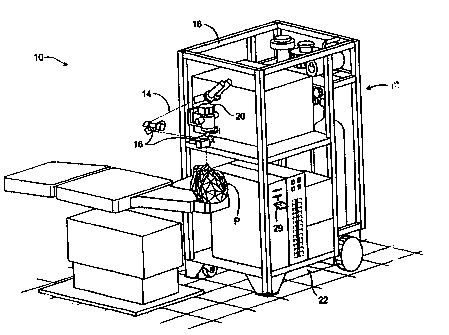
[0068] Turning now to the drawings, Fig. 1 illustrates a laser eye surgery
system 10 of the
present invention, including a laser 12 that produces a laser beam 14. Laser
12 is optically
coupled to laser delivery optics 16, which directs laser beam 14 to an eye E
of patient P. A
delivery optics support structure (not shown here for clarity) extends from a
frame 18
supporting laser 12. A microscope 20 is mounted on the delivery optics support
structure, the
microscope often being used to image a cornea of eye E.
11
CA 02522787 2005-10-18
WO 2004/095187 PCT/US2004/012025
[0069] Laser 12 generally comprises an excimer laser, ideally comprising an
argon-fluorine
laser producing pulses of laser light having a wavelength of approximately 193
mn. Laser 12
will preferably be designed to provide a feedback stabilized fluence at the
patient's eye,
delivered via delivery optics 16. The present invention may also be useful
with alternative
sources of ultraviolet or infrared radiation, particularly those adapted to
controllably ablate
the corneal tissue without causing significant damage to adjacent and/or
underlying tissues of
the eye. Such sources include, but are not limited to, solid state lasers and
other devices
which can generate energy in the ultraviolet wavelength between about 185 and
205 nm
and/or those which utilize frequency-multiplying techniques. Hence, although
an excimer
laser is the illustrative source of an ablating beam, other lasers may be used
in the present
invention.
[0070] Laser system 10 will generally include a computer or programmable
processor 22.
Processor 22 may comprise (or interface with) a conventional PC system
including the
standard user interface devices such as a keyboard, a display monitor, and the
like. Processor
22 will typically include an input device such as a magnetic or optical disk
drive, an internet
connection, or the like. Such input devices will often be used to download a
computer
executable code from a tangible storage media 29 embodying any of the methods
of the
present invention. Tangible storage media 29 may take the form of a floppy
disk, an optical
disk, a data tape, a volatile or non-volatile memory, RAM, or the like, and
the processor 22
will include the memory boards and other standard components of modern
computer systems
for storing and executing this code. Tangible storage media 29 may optionally
embody
wavefront sensor data, wavefront gradients, a wavefront elevation map, a
treatment map, a
corneal elevation map, and/or an ablation table. While tangible storage media
29 will often
be used directly in cooperation with a input device of processor 22, the
storage media may
also be remotely operatively coupled with processor by means of network
connections such
as the internet, and by wireless methods such as infrared, Bluetooth, or the
like.
[0071] Laser 12 and delivery optics 16 will generally direct laser beam 14 to
the eye of
patient P under the direction of a computer 22. Computer 22 will often
selectively adjust
laser beam 14 to expose portions of the cornea to the pulses of laser energy
so as to effect a
predetermined sculpting of the cornea and alter the refractive characteristics
of the eye. In
many embodiments, both laser beam 14 and the laser delivery optical system 16
will be under
computer control of processor 22 to effect the desired laser sculpting
process, with the
processor effecting (and optionally modifying) the pattern of laser pulses.
The pattern of
12
CA 02522787 2012-04-25
pulses may by summarized in machine readable data of tangible storage media 29
in the form
of a treatment table, and the treatment table may be adjusted according to
feedback input into
processor 22 from an automated image analysis system in response to feedback
data provided
from an ablation monitoring system feedback system. Optionally, the feedback
may be
manually entered into the processor by a system operator. Such feedback might
be provided
by integrating the wavefront measurement system described below with the laser
treatment
system 10, and processor 22 may continue and/or terminate a sculpting
treatment in response
to the feedback, and may optionally also modify the planned sculpting based at
least in part on
the feedback. Measurement systems are further described in U.S. Patent No.
6,315,413.
[00721 Laser beam 14 may be adjusted to produce the desired sculpting using a
variety of
alternative mechanisms. The laser beam 14 may be selectively limited using one
or more
variable apertures. An exemplary variable aperture system having a variable
iris and a
variable width slit is described in U.S. Patent No. 5,713,892. The laser beam
may also be
tailored by varying the size and offset of the laser spot from an axis of the
eye, as described in
U.S. Patent Nos. 5,683,379, 6,203,539, and 6,331,177.
[00731 Still further alternatives are possible, including scanning of the
laser beam over the
surface of the eye and controlling the number of pulses and/or dwell time at
each location, as
described, for example, by U.S. Patent No. 4,665,913, using masks in the
optical path of laser
beam 14 which ablate to vary the profile of the beam incident on the cornea,
as described in
U.S. Patent No. 5,807,379, hybrid profile-scanning systems in which a variable
size beam
(typically controlled by a variable width slit and/or variable diameter iris
diaphragm) is
scanned across the cornea; or the like. The computer programs and control
methodology for
these laser pattern tailoring techniques are well described in the patent
literature.
[00741 Additional components and subsystems may be included with laser system
10, as
should be understood by those of skill in the art. For example, spatial and/or
temporal
integrators may be included to control the distribution of energy within the
laser beam, as
described in U.S. Patent No. 5,646,791. Ablation effluent evacuators/filters,
aspirators, and
other ancillary components of the laser surgery system are known in the art.
Further details of
suitable systems for performing a laser ablation procedure can be found in
commonly
assigned U.S. Pat. Nos. 4,665,913, 4,669,466, 4,732,148, 4,770,172, 4,773,414,
5,207,668,
13
CA 02522787 2012-04-25
5,108,388, 5,219,343, 5,646,791 and 5,163,934. Suitable systems also include
commercially
available refractive laser systems such as those manufactured and/or sold by
Alcon, Bausch &
Lomb, Nidek, WaveLight, LaserSight, Schwind, Zeiss-Meditec, and the like.
Basis data can
be further characterized for particular lasers or operating conditions, by
taking into account
localized environmental variables such as temperature, humidity, airflow, and
aspiration.
[00751 Fig. 2 is a simplified block diagram of an exemplary computer system 22
that may be
used by the laser surgical system 10 of the present invention. Computer system
22 typically
includes at least one processor 52 which may communicate with a number of
peripheral
devices via a bus subsystem 54. These peripheral devices may include a storage
subsystem
56, comprising a memory subsystem 58 and a file storage subsystem 60, user
interface input
devices 62, user interface output devices 64, and a network interface
subsystem 66. Network
interface subsystem 66 provides an interface to outside networks 68 and/or
other devices,
such as the wavefront measurement system 30.
[00761 User interface input devices 62 may include a keyboard, pointing
devices such as a
mouse, trackball, touch pad, or graphics tablet, a scanner, foot pedals, a
joystick, a
touchscreen incorporated into the display, audio input devices such as voice
recognition
systems, microphones, and other types of input devices. User input devices 62
will often be
used to download a computer executable code from a tangible storage media 29
embodying
any of the methods of the present invention. In general, use of the term
"input device" is
intended to include a variety of conventional and proprietary devices and ways
to input
information into computer system 22.
[00771 User interface output devices 64 may include a display subsystem, a
printer, a fax
machine, or non-visual displays such as audio output devices. The display
subsystem may be
a cathode ray tube (CRT), a flat-panel device such as a liquid crystal display
(LCD), a
projection device, or the like. The display subsystem may also provide a non-
visual display
such as via audio output devices. In general, use of the term "output device"
is intended to
14
CA 02522787 2005-10-18
WO 2004/095187 PCT/US2004/012025
include a variety of conventional and proprietary devices and ways to output
information
from computer system 22 to a user.
[0078] Storage subsystem 56 can store the basic programming and data
constructs that
provide the functionality of the various embodiments of the present invention.
For example,
a database and modules implementing the functionality of the methods of the
present
invention, as described herein, may be stored in storage subsystem 56. These
software
modules are generally executed by processor 52. In a distributed environment,
the software
modules may be stored on a plurality of computer systems and executed by
processors of the
plurality of computer systems. Storage subsystem 56 typically comprises memory
subsystem
58 and file storage subsystem 60.
[0079] Memory subsystem 58 typically includes a number of memories including a
main
random access memory (RAM) 70 for storage of instructions and data during
program
execution and a read only memory (ROM) 72 in which fixed instructions are
stored. File
storage subsystem 60 provides persistent (non-volatile) storage for program
and data files,
and may include tangible storage media 29 (Fig. 1) which may optionally embody
wavefront
sensor data, wavefront gradients, a wavefront elevation map, a treatment map,
and/or an
ablation table. File storage subsystem 60 may include a hard disk drive, a
floppy disk drive
along with associated removable media, a Compact Digital Read Only Memory (CD-
ROM)
drive, an optical drive, DVD, CD-R, CD-RW, solid-state removable memory,
and/or other
removable media cartridges or disks. One or more of the drives may be located
at remote
locations on other connected computers at other sites coupled to computer
system 22. The
modules implementing the functionality of the present invention may be stored
by file storage
subsystem 60.
[0080] Bus subsystem 54 provides a mechanism for letting the various
components and
subsystems of computer system 22 communicate with each other as intended. The
various
subsystems and components of computer system 22 need not be at the same
physical location
but may be distributed at various locations within a distributed network.
Although bus
subsystem 54 is shown schematically as a single bus, alternate embodiments of
the bus
subsystem may utilize multiple busses.
[0081] Computer system 22 itself can be of varying types including a personal
computer, a
portable computer, a workstation, a computer terminal, a network computer, a
control system
in a wavefront measurement system or laser surgical system, a mainframe, or
any other data
CA 02522787 2005-10-18
WO 2004/095187 PCT/US2004/012025
processing system. Due to the ever-changing nature of computers and networks,
the
description of computer system 22 depicted in Fig. 2 is intended only as a
specific example
for purposes of illustrating one embodiment of the present invention. Many
other
configurations of computer system 22 are possible having more or less
components than the
computer system depicted in Fig. 2.
[0032] Referring now to Fig. 3, one embodiment of a wavefront measurement
system 30 is
schematically illustrated in simplified form. In very general terms, wavefront
measurement
system 30 is configured to sense local slopes of a gradient map exiting the
patient's eye.
Devices based on the Hartmann-Shack principle generally include a lenslet
array to sample
the gradient map uniformly, over an aperture, which is typically the exit
pupil of the eye.
Thereafter, the local slopes.-of the gradient map are analyzed so as to
reconstruct the
wavefront surface or map.
[0083] More specifically, one wavefront measurement system 30 includes an
image source
32, such as a laser, which projects a source image through optical tissues 34
of eye E so as to
form an image 44 upon a surface of retina R. The image from retina R is
transmitted by the
optical system of the eye (e.g., optical tissues 34) and imaged onto a
wavefront sensor 36 by
system optics 37. The wavefront sensor 36 communicates signals to a computer
system 22'
for measurement of the optical errors in the optical tissues 34 and/or
determination of an
optical tissue ablation treatment program. Computer 22' may include the same
or similar
hardware as the computer system 22 illustrated in Figs. 1 and 2. Computer
system 22' may
be in communication with computer system 22 that directs the laser surgery
system 10, or
some or all of the components of computer system 22, 22' of the wavefront
measurement
system 30 and laser surgery system 10 may be combined or separate. If desired,
data from
wavefront sensor 36 may be transmitted to a laser computer system 22 via
tangible media 29,
via an I/O port, via an networking connection 66 such as an intranet or the
Internet, or the
like.
[0084] Wavefront sensor 36 generally comprises a lenslet array 38 and an image
sensor 40.
As the image from retina R is transmitted through optical tissues 34 and
imaged onto a
surface of image sensor 40 and an image of the eye pupil P is similarly imaged
onto a surface
of lenslet array 38, the lenslet array separates the transmitted image into an
array of beamlets
42, and (in combination with other optical components of the system) images
the separated
beamlets on the surface of sensor 40. Sensor 40 typically comprises a charged
couple device
16
CA 02522787 2005-10-18
WO 2004/095187 PCT/US2004/012025
or "CCD," and senses the characteristics of these individual beamlets, which
can be used to
determine the characteristics of an associated region of optical tissues 34.
In particular,
where image 44 comprises a point or small spot of light, a location of the
transmitted spot as
imaged by a beamlet can directly indicate a local gradient of the associated
region of optical
tissue.
[0085] Eye E generally defines an anterior orientation ANT and a posterior
orientation
POS. Image source 32 generally projects an image in a posterior orientation
through optical
tissues 34 onto retina R as indicated in Fig. 3. Optical tissues 34 again
transmit image 44
from the retina anteriorly toward wavefront sensor 36. Image 44 actually
formed on retina R
may be distorted by any imperfections in the eye's optical system when the
image source is
originally;.transmitted by optical tissues 34. Optionally, image source
projection optics 46
may be configured or adapted to decrease any distortion of image 44.
[0086] In some embodiments, image source optics 46 may decrease lower order
optical
errors by compensating for spherical and/or cylindrical errors of optical
tissues 34. Higher
order optical errors of the optical tissues may also be compensated through
the use of an
adaptive optic element, such as a deformable mirror (described below). Use of
an image
source 32 selected to define a point or small spot at image 44 upon retina R
may facilitate the
analysis of the data provided by wavefront sensor 36. Distortion of image 44
may be limited
by transmitting a source image through a central region 48 of optical tissues
34 which is
smaller than a pupil 50, as the central portion of the pupil may be less prone
to optical errors
than the peripheral portion. Regardless of the particular image source
structure, it will be
generally be beneficial to have a well-defined and accurately formed image 44
on retina R.
[0087] In one embodiment, the wavefront data may be stored in a computer
readable
medium 29 or a memory of the wavefront sensor system 30 in two separate arrays
containing
the x and y wavefront gradient values obtained from image spot analysis of the
Hartmann-
Shack sensor images, plus the x and y pupil center offsets from the nominal
center of the
Hartmann-Shack lenslet array, as measured by the pupil camera 51 (Fig. 3)
image. Such
information contains all the available information on the wavefront error of
the eye and is
sufficient to reconstruct the wavefront or any portion of it. In such
embodiments, there is no
need to reprocess the Hartmann-Shack image more than once, and the data space
required to
store the gradient array is not large. For example, to accommodate an image of
a pupil with
an 8 mm diameter, an array of a 20 x 20 size (i.e., 400 elements) is often
sufficient. As can
17
CA 02522787 2012-04-25
be appreciated, in other embodiments, the wavefront data may be stored in a
memory of the
wavefront sensor system in a single array or multiple arrays.
[00881 While the methods of the present invention will generally be described
with reference
to sensing of an image 44, it should be understood that a series of wavefront
sensor data
readings may be taken. For example, a time series of wavefront data readings
may help to
provide a more accurate overall determination of the ocular tissue
aberrations. As the ocular
tissues can vary in shape over a brief period of time, a plurality of
temporally separated
wavefront sensor measurements can avoid relying on a single snapshot of the
optical
characteristics as the basis for a refractive correcting procedure. Still
further alternatives are
also available, including taking wavefront sensor data of the eye with the eye
in differing
configurations, positions, and/or orientations. For example, a patient will
often help maintain
alignment of the eye with wavefront measurement system 30 by focusing on a
fixation target,
as described in U.S. Patent No. 6,004,313. By varying a position of the
fixation target as
described in that reference, optical characteristics of the eye may be
determined while the eye
accommodates or adapts to image a field of view at a varying distance and/or
angles.
[00891 The location of the optical axis of the eye may be verified by
reference to the data
provided from a pupil camera 52. In the exemplary embodiment, a pupil camera
52 images
pupil 50 so as to determine a position of the pupil for registration of the
wavefront sensor data
relative to the optical tissues.
[00901 An alternative embodiment of a wavefront measurement system is
illustrated in Fig.
3A. The major components of the system of Fig. 3A are similar to those of Fig.
3.
Additionally, Fig. 3A includes an adaptive optical element 53 in the form of a
deformable
mirror. The source image is reflected from deformable mirror 98 during
transmission to retina
R, and the deformable mirror is also along the optical path used to form the
transmitted image
between retina R and imaging sensor 40. Deformable mirror 98 can be
controllably deformed
by computer system 22 to limit distortion of the image formed on the retina or
of subsequent
images formed of the images formed on the retina, and may enhance the accuracy
of the
resultant wavefront data. The structure and use of the system of Fig. 3A are
more fully
described in U. S. Patent No. 6,095, 651.
[00911 The components of an embodiment of a wavefront measurement system for
measuring
the eye and ablations may comprise elements of a VISX WaveScan , available
from VISX,
18
CA 02522787 2012-04-25
INCORPORATED of Santa Clara, California. One embodiment includes a WaveScan
with a
deformable mirror as described above. An alternate embodiment of a wavefront
measuring
system is described in U.S. Patent No. 6,271,915. It is appreciated that any
wavefront
aberrometer could be employed for use with the present invention.
1. TARGET OPTICAL SURFACE SHAPE
[00921 Refractive surgery is typically based on a target optical surface shape
that is selected
or determined to treat a vision condition in a patient. A target optical
surface shape can be
based on or represented by any of a variety of target optical surface shape
data or data
formats. In this context, a vision condition can be analogous to a refractive
case. Examples of
refractive cases include the following.
Refractive Case Optical Zone x Ablation Zone
1. Myopic (-4D) 6mm X 8mm
2. Hyperopic (+2D) 5mm X 9mm
3. Myopic Astigmatism (-2DS/-1DC X 34 ) 6mm X 8mm
4. Hyperopic Astigmatism (+2DS/-1DC X 65 ) 5mm X 9mm
5. Mixed Astigmatism (+2DS/-3DC X 45 ) 5mm X 9mm
6. Therapeutic (+2.35DS/-3.51DC X 17 ) 6mm X 8mm
100931 Refractive cases 1 through 5 represent hypothetical refractive cases,
and the
therapeutic eye of case 6 represents a real eye case having more than a 1 urn
high order
aberration RMS with large coma and spherical components (for example a single
high order
Zernike mode Z88, or Z45, with 1 gm RMS error). The optical zone can be based
on a
hypothetical pupil diameter. In the real eye case, the optical zone can
correspond to a pupil
diameter under standard lighting conditions used during wavefront evaluation.
The high order
part of the exemplary therapeutic eye refractive case is shown in Fig. 8A. In
one embodiment
of the present invention, the target optical surface shape includes a set of 6-
order Zernike
polynomials.
100941 Refractive cases such as these can be determined with a wavefront
sensing device,
which can determine both low and high order aberrations. In some cases, the
target optical
surface shape can be configured to address a low order aberration. Some
refractive cases may
present both low and high order aberrations, and may benefit from a combined
target optical
surface shape treatment. As shown in Fig. 4, given a particular vision
condition or
19
CA 02522787 2005-10-18
WO 2004/095187 PCT/US2004/012025
refractive case, it is possible to generate a corresponding high resolution
target optical surface
shape, or input profile, for treating the condition.
II. REFRACTIVE SURGERY SYSTEM PARAMETERS
[0095] Given a target optical surface shape, it is possible to determine a
model optical
surface shape based on the target shape and a set of refractive surgery system
parameters.
The refractive surgery system parameters correspond to the individual system
components of
the system. For example, as shown in Fig. 4, one embodiment of the refractive
surgery
system can include components such as a wavefront device, a laser ablation
profile, and a
la per-servo system such as a laser registration and tracking system. These
components can
introduce errors into the model optical surface shape. The surgery system can
have error
soi rces including, for example, a wavefront device measurement error, a
wavefront surface
fitting error or algorithm imperfection, a laser beam uniformity and
variability error, a
registration error, and a tracking error. Thus, the model surface shape can
include aberrations
that are introduced or amplified by the surgery system parameters, and these
aberrations can
be described and evaluated by certain mathematical equations.
[0096] Fig. 5 illustrates another embodiment of an overall refractive system
according to
the present invention, which can include components such as a wavefront
device, a laser
ablation profile, a laser registration and tracking system, and a laser
delivery system. Such a
refractive surgery system can have error sources including, for example, a
wavefront device
measurement error, a laser beam profile error, a laser registration and
tracking system error,
and a laser delivery system error. Accordingly, a set of refractive surgery
system parameters
can be selected from the group consisting of a wavefront device variable, a
laser ablation
profile variable, a laser registration and tracking system variable, a
biomechanical variable,
and a healing effect variable.
[0097] As noted above, different components of the refractive surgery system,
as
represented by the surgery system parameters, can by their own accord
introduce different
errors or aberrations into the model optical surface shape, and they can
exacerbate different
errors or aberrations present in the target optical surface shape.
Consequently, there may be
different RMS values or other error values associated with the different
system components.
The present invention provides a numerical approach to characterizing or
identifying error
sources in such a system.
CA 02522787 2005-10-18
WO 2004/095187 PCT/US2004/012025
[0098] To evaluate the error sources, it is helpful to consider the overall
system. Assuming
that all of the error sources are statistically independent, the overall error
associated with the
system embodiment shown in Fig. 4 can be represented as
2 2 2
A= ~WF + (7 r1B+(7 RT (1)
where Ãb'WF2 represents a WF (wavefront) measurement induced error or
variance, aAB 2
represents an ablation profile related variance or fitting error, and aRT2
represents a laser
system registration and tracking error or variance. This total error is a
representation of the
system source errors that can contribute aberrations to a model optical
surface shape.
[0699] In another example, the total error associated with the surgical system
parameters
can be written as
2 2 2 2 2 2
6total -6w +6 f +6r +Cyt +6b (2)
where o 2 represents a measurement error in the wavefront device, of
represents an error
induced in surface fitting with respect to a certain algorithm such as a
simulated annealing
algorithm, ur2 represents an error induced by registration, at2 represents a
tracking error, and
ab2 represents an error due to laser beam uniformity and variability.
[0100] As shown in Fig. 5, another exemplary surgical system can include a
wavefront
device, a laser ablation profile, a laser registration and tracking component,
and a laser
delivery system. As indicated in the figure, errors introduced by a
microkeratome can also be
factored into the total system error analysis. When assuming that all the
error sources are
statistically independent, the overall error can be represented as
61orat = h1 7 -I- U f + 6r + 6, -I- 66 -F' 6 2 (3)
where HQ represents a non-linear healing operator, a represents a total error
in the
wavefront device, a f represents an error induced in surface fitting with
respect to a certain
algorithm such as a simulated annealing algorithm, a r represents an error
induced by the
registration, a; represents a tracking error, U2 represents an error due to
laser beam
uniformity and variability, and a õ2 represents an error induced from the LARK
flap, or
biomechanical effect. The individual error sources are discussed in further
detail below.
[0101] In some embodiments, the set of refractive surgery system parameters
can be
adjusted such that a post-operative total high order RMS is substantially
equivalent to a pre-
21
CA 02522787 2005-10-18
WO 2004/095187 PCT/US2004/012025
operative total high order RMS. In other embodiments, the set of refractive
surgery system
parameters can be adjusted such that a post-operative total high order RMS is
less than a pre
operative total high order RMS. In still other embodiments, the set of
refractive surgery
system parameters can be adjusted such that a post-operative total high order
RMS is about
one third the amount of a pre operative total high order RMS.
[0102] The set of refractive surgery system parameters can be adjusted such
that a
post-operative total high order RMS of about 0.1 gm to about 0.3 gm is
achieved. In related
embodiments, the set of refractive surgery system parameters can be adjusted
such that each
system component of the total high order RMS does not exceed from about 0.038
gm to
about 0.113 gm. In other embodiments, where the total RMS error is about 0.1
gm to about
0.3 gm and the system includes 3 components, the set of refractive surgery
system parameters
can be adjusted such that each system component of the total high order RMS
does not
exceed from about 0.0577 gm to about 0.173 gm. In yet other embodiments, where
the total
RMS error is about 0.1 gm to about 0.3 gm and the system includes 10
components, the set of
refractive surgery system parameters can be adjusted such that each system
component of the
total high order RMS does not exceed from about 0.0316 gm to about 0.0949 gm.
[0103] In one embodiment of the present invention, the target optical surface
shape
includes a set of 6-order Zernike polynomials, and the set of refractive
surgery system
parameters is adjusted such that each component of a post-operative total high
order RMS
does not exceed about 0.022 gm. In another embodiment, the target optical
surface shape
includes a set of 6-order Zernike polynomials, and the set of refractive
surgery system
parameters is adjusted such that each component of a post-operative total high
order RMS
does not exceed about 0.0073 gm. In other embodiments, where the total RMS
error is about
0.1 gm to about 0.3 gm and the system includes 3 components, the set of
refractive surgery
system parameters can be adjusted such that each system component of the total
high order
RMS does not exceed from about 0.0111 gm to about 0.0333 gm. In yet other
embodiments,
where the total RMS error is about 0.1 gm to about 0.3 gm and the system
includes 10
components, the set of refractive surgery system parameters can be adjusted
such that each
system component of the total high order RMS does not exceed from about 0.0061
gm to
about 0.0111 gm.
A. Wavefront Device Parameters
22
CA 02522787 2005-10-18
WO 2004/095187 PCT/US2004/012025
[0104] A wavefront device measurement error, which can be represented as 6 t,
, can
originate from errors associated with any of a variety of parameters. For
example, as shown
in Figs. 4 and 5, a wavefront device can include parameters such as spot
identification (e.g.
Hartmann-Shack spot pattern identification), accommodation, and
reconstruction.
Accordingly, a wavefront device variable can be selected from the group
consisting of a spot
identification factor, an accommodation factor, and a reconstruction factor.
In some cases,
the wavefront device variable is configured to address a high order
aberration.
1. Accommodation Error
[0105] Accommodation error can be due to partial accommodation or micro-
accommodation of the patient, which can be translated to a root mean squares
(RMS)- error.
Micro-fluctuation, or accommodation drift, can be present in a patient as they
gaze at, but are
unable to fixate upon, a distant target. Most patients will accommodate at
least slightly, and
micro-accommodation corresponds to slight changes in the relaxation of
accommodation. To
the extent a patient cannot fully relax and therefore accommodates during this
procedure, this
accommodation can become part of the error. Assuming the random error of
accommodation
is a for an eye with pupil radius of R, an RMS accommodation error can be
expressed as
=a 2 R 4
aY ac 48 (4)
where a is given in diopters and R is given in mm. a can represent a variable
averaged
microaccommodation for several patients being measured.
[0106] In a practical clinical setting, it may be difficult to keep the
accommodation drift, or
accommodation error, under 0.1D. In some cases, accommodation error of more
than one
diopter has been observed. Taking 0.1 D as a limit, the minimum RMS
accommodation error
for a 6mm pupil is then 0.15 microns. In some embodiments, the wavefront
device variable
includes an accommodation error of 0.25D, equivalent to 0.325 microns RMS
error for a
6mm pupil.
[01071 For the accommodation error, Fig. 7 shows the contribution of the
accommodation
error to the total RMS accommodation error for different pupil sizes. It is
clear to see that
larger pupil sizes correspond with larger total RMS errors when the amount of
accommodation remains constant.
2. Reconstruction Error
23
CA 02522787 2005-10-18
WO 2004/095187 PCT/US2004/012025
[0108] For the wavefront device error, it is also possible to consider error
induced from a
wavefront reconstruction error. The sources of a reconstruction error can
include an
uncompensated error due to truncation of the number of basis functions, such
as Zernike
polynomials; a measurement error; and a remaining error due to aliasing of the
derivatives of
basis functions. A complete theoretical analysis is given by Dai, in "Modal
wave-front
reconstruction with Zernike polynomials and Karhunen-Lowe functions," J Opt.
Soc. Am. A
13, 1218-1225 (1996), which is incorporated herein by reference in its
entirety for all
purposes. In one embodiment, the reconstruction error can be written as
2 _ 2 2 2
arc -Uuc+~sp+~rs. (5)
where auC2 is this the uncompensated error, 6p2 is this the spot
identification error, and crrs2 is
this the remaining error.
[0109] The uncompensated error may be difficult to estimate due to the lack of
statistics of
the Zernike expansion for human eye's aberrations. However, a treatment with
consideration
of the Hartmann-Shack sensor configuration is possible. The measurement error,
which is
often directly related to spot identification, can be treated as spot
identification error. Finally,
the remaining error can be small, especially when the number of sub-apertures
is relatively
small. For example, in one embodiment the VISX WaveScan device uses 37 sub-
apertures.
[0110] For a wavefront reconstruction error, it is possible to assume that
different error
sources result in the final uncertainty in a slope estimate. These error
sources include CCD
detector noise, noise in pixel round-off position error, as well as error
contained in the
reconstruction algorithm, and these could affect spot identification error and
reconstruction
error. Fig. 8 shows the contribution of slope estimation error to the total
RMS error for four
example cases, illustrating that an error in slope estimation can affect the
total RMS error. In
some embodiments, the wavefront reconstruction error can be about 0.05 m.
[0111] A spot identification error can be an error due to round off of pixel
position (integer
pixel position), low contrast spots due to corneal reflection, or low signal
to noise (SIN) ratio.
A complete theoretical derivation of the total spot identification error is
not given here. It is
possible to use a simple Gaussian random noise model for the simulation of
spot
identification error. However, a general formulation can be given as
24
CA 02522787 2005-10-18
WO 2004/095187 PCT/US2004/012025
2 _ 2 2
6'sp 6ro + 6 sn = (6)
where ar 2 is this the round off error and G,n2 is this the signal to noise
ratio error. In some
embodiments, the spot identification error can be about 0.05 tm.
3. Total Wavefront Device Error
[0112] The total wavefront device error can be written as
2 2 2
~w = ~"rc -I - ac (7)
1`0 which in some embodiments is the final formula for RMS calculation for a
wavefront device,
and reflects the sum of the accommodation error and the reconstruction error.
[0113] , In one embodiment, a reconstruction error can reflect a typical slope
estimation
error of 0.001 corresponding to an RMS reconstruction error of about 0.2
microns. In this
embodiment, the total RMS error from a wavefront device can be at the order of
0.25
microns, assuming an RMS accommodation error of 0.1 microns.
[0114] A large.portion of the wavefront device error may often be manifested
as errors in
low orders, or mostly in the sphere error. Therefore, the end result, or true
ablation (e.g. the
model optical surface shape) may be a random over correction or under
correction. If the
total RMS wavefront device error is entirely a low order aberration, it may
correspond to a
0.2D refractive error, which can be considered as small. The statistical
trend, though, would'
result in relatively small total RMS wavefront device error. The truly induced
high order
total RMS error, which typically originates from the system parameters, will
often be below
0.1 microns.
[0115] One approach to correcting or inhibiting high order aberrations
involves controlling
the overall wavefront error to a certain limit. For instance, using a 100 m
scanning
resolution of a Gaussian spot (FWHM = 0.75 mm) to correct a -4DS eye without
high order
aberrations can induce 0.21 m high order aberration (HOA). In some
embodiments, the
wavefront device variable includes a 100 m gridsize factor.
E. Laser Ablation Profile Parameters
[0116] Laser ablation profile errors are sometimes referred to as wavefront
surface fitting
errors, or algorithm errors. Wavefront surface fitting errors can be the
result of a numerical
CA 02522787 2005-10-18
WO 2004/095187 PCT/US2004/012025
solution of a multi-dimensional problem in fitting individual laser pulses to
the expected
wavefront surface, or model optical surface shape. Laser ablation profile
errors, which can
be represented as a f , can originate from errors associated with any of a
variety of
parameters. Accordingly, a laser ablation profile variable can include a pulse
size factor, a
spot size variability factor, a beam uniformity factor, and a laser pulse
repetition rate factor.
[0117] In one embodiment of the present invention, as shown in Fig. 4, a laser
ablation
profile can include parameters such as pulse profile, spot size variability,
beam uniformity,
and laser pulse repetition rate. In another embodiment of the present
invention, as shown in
Fig. 5, a laser ablation profile can include parameters such as laser beam
profile, grid
geometry, and ablation algorithm. In Fig. 5, the beam uniformity and laser
pulse repetition
rate are characterized as laser delivery system'parameters, and are further
discussed under
that section heading below.
1. Laser Pulse Profile Fitting Errors
[0118] Laser ablation pulse profiles can be generated in a variety of ways. In
the following
examples, the Y(r) function describes how to generate the ablation pulse
profile, where a
represents the standard deviation of a Gaussian profile, and FWHM represents
the full width
at half maximum of the Gaussian profile. Different types of pulse profiles can
contribute
different amounts of error to the laser ablation profile error. For example, a
laser ablation
profile variable can include a variable spot scanning factor or a flying spot
scanning error.
a. Flying Spot Scanning (FSS) Pulse Profile
[0119] A Flying Spot Scanning (FSS) pulse profile can be represented by the
following
formula:
Y(r) = -0.4 exp [(-81n2/a2)(4-r)2] (8)
where a = D/2 and D is the spot size, and where FWHM is D/48 = 0.3536D. Y(r)
represents
the ablation depth, and r represents the distance from the pupil center, in
mm. Thus, for a
0.75 mm FWHM spot, D = 2 mm. This profile is depicted in Fig. 9A.
[0120] In one embodiment, a -4 Diopter input is used to generate the basis
function, or
basis data, for a Flying Spot Scanning profile. The following results were
obtained: 5481
pulses, PV = 1.03 gm, RMS = 0.14 pm (OPD), which is the profile fitting error.
All
measurements are in optical path difference (OPD). PV is a peak to valley
measurement, and
represents the difference between maximum and minimum in sequence of values.
It reflects
26
CA 02522787 2005-10-18
WO 2004/095187 PCT/US2004/012025
the magnitude of fluctuation. The RMS is similar to standard deviation. In
some
embodiments, the flying spot scanning factor can be about 1.5 mm.
b. Variable Spot Scanning (VSS) Pulse Profile
[0121] In a Variable Spot Scanning (VSS) profile laser, a top hat shape can be
used. This
profile is depicted in Fig. 9B, at 15 different diameters. In the -4 Diopter
input embodiment
describe above, for the VISX Variable Spot Scanning profile, the following
results were
obtained: 339 pulses, PV = 0.78 m, RMS = 0.11 pan, which is the profile
fitting error. All
measurements are in optical path difference (OPD).
c. Comparison Between Various Pulse Profiles
[0122] The fitting errors, or f , for the following laser pulse profiles were
evaluated with
respect to a variety of refractive cases, and the results are shown in Fig.
10A. This assumes
that all other laser ablation profile parameters, such as spot size
variability and grid geometry,
were equal, and there were no other error sources.
2. Spot Size Variability Errors
[0123] A spot size variability error can also contribute to a laser ablation
profile error. Fig.
12A illustrates an RMS Distribution Graph, showing the fitting RMS error for
several
different refractive cases (refractive powers) at various spot sizes. Fig. 12B
illustrates a PV
Distribution Graph. The units on the x-axis represent the spot size diameter
in millimeters.
Based on the example shown in these figures, an optimal spot size (i.e. lowest
error) for
Flying Spot Scanning (FSS) can range from about 1.0 mm to about 1.6mm, and
more
specifically can be about 1.5 mm or about 0.5 mm FWHM. The FWHM is often about
one
third of the spot size. In this way, an optimal spot size can be determined
for each refractive
case, which can confer the maximum inhibition of aberration in model optical
surface shape.
In this way, it is possible to control the amount of error by controlling the
spot size.
[0124] A simple spherical refraction is shown in Figs. 12A and 12B. The RMS
and PV
change may not be very significant over a big range (e.g. +3D to -6D), except
possibly for the
low refraction cases as shown in these examples. The optimal spot size may not
appear to
change with refractions, again except for very low refraction cases such as,
for example,
0.5D or cases close to emmetropia.
[0125] The VSS spot can range between 0.65 mm and 6.5 mm. Though only discrete
number of spots are shown, it should be recognized that the spot sizes can be
continuous.
27
CA 02522787 2012-04-25
VSS can have an ablation depth of about 0. 25 gm (tissue) while the FSS can
have an optimal
spot size of 1.5 mm with 0.5 mm FWHM and 0.4 gm depth Gaussian profile. Often,
there is
no variability of the FSS spot size, meaning that the spot size can be fixed.
In some
embodiments, the laser ablation profile variable includes a flying spot
scanning factor ranging
from about 1 mm to about 1.6 mm.
3. Grid Geometry
[0126] See Fig. 6, reference number 240. Grid geometry decides the solution
space for the
simulated annealing algorithm. In some embodiments, the wavefront device
variable may
include a 100 gm gridsize factor.
4. Ablation Algorithm
[0127] It is also possible to use fitting performance, which is related to the
fitting error, to
determine the number of ablation pulses employed to perform the ablation, as
shown in Fig.
I IA. In this example, the Flying Spot Scanning profile can have about 10,000
pulses, for
example, for each of the refractive cases, whereas the Variable Spot Scanning
profile can
have less than about 1,000. Consequently, in this example if VSS operates at a
laser pulse
repetition rate of 20 Hz it takes about 50 seconds for the ablation. To
perform an FSS ablation
in the same amount of time, the system should operate at a laser pulse
repetition rate of 200
Hz.
[0128] An analysis of fitting error comparison of all 6 exemplary profile
cases in shown in
Fig. 9C, including fitting (algorithm), registration, tracking and beam
uniformity. These 6
cases can be evaluated for RMS error with VSS and FSS being the two sets of
beams. Laser
pulse repetition rate for VSS is 10 Hz and FSS is 100 Hz. Based on Fig. 9C, it
is clear that
VSS is superior to FSS in all of the six refractive cases, based on the RMS
error, with
therapeutic and myopic astigmatism having the biggest gain, or difference
between VSS and
FSS.
[0129] Suitable ablation algorithm approaches are also discussed in US patent
number
6,673,062, issued January 6, 2004.
C. Laser Registration and Tracking System Parameters
28
CA 02522787 2005-10-18
WO 2004/095187 PCT/US2004/012025
[0130] As shown in Figs. 4 and 5, a laser registration and tracking system can
include
parameters corresponding to registration source errors (c r ), and linear and
torsional tracking
source errors (o , ). Accordingly, a laser registration and tracking system
variable may be
selected from the group consisting of a registration factor, a linear tracking
factor, and a
torsional tracking factor. In one embodiment, the laser registration and
tracking system
variable can include a registration accuracy less than about 10 gm in both the
vertical and
horizontal directions and a rotational error less than about 0.5 .
1. Registration Error
[0131] Registration error, position error, and rotational alignment error can
be modeled or
determined. The registration error is typically the sum of the position error
and the rotational
alignment error. In general, the pupil center at the time the wavefront is
taken with an
aberrometer or phoropter can differ from the pupil center at the time the
treatment procedure
is performed. This shift in pupil center position error can induce a high
order aberration.
Similarly, rotational alignment error can also contribute to an increase in
high order
aberrations. Further, pupil size changes and rotational or alignment errors
can also contribute
to aberrations. These aberrations can be modeled as randomly distributed
errors.
[0132] Figs. 13A-E illustrate registration error analysis. For linear
registration induced
RMS error, VSS and FSS appear to perform similarly, as shown in Figs. 13A and
13C, which
depict linear, or X/Y, registration. VSS can be volunmable (larger) on mixed
astigmatism.
For the comparison, values of 20 gm accuracy in position and 5 in the
rotational alignment
can be used. In the case of a cylinder with a large angle, VSS registration
can be larger than
FSS registration, but theoretically, if there is no fitting error, they may be
similar. Figs. 13B
and 13D depict VSS and FSS torsional registration induced RMS error, and based
on these
figures, torsional registration appears to be correlated to the refractive
case. VSS and FSS
torsional registration induced RMS error appear to behave similarly, and for
mixed
astigmatism, VSS appears to exhibit slightly more error which may be due to
mixed
astigmatism having the least symmetric profile. Fig. 13E shows a comparison
between VSS
and FSS for the combined linear and torsional registration error. Again, VSS
and FSS
combined linear and torsional registration induced RMS error appear to behave
similarly, and
for mixed astigmatism, VSS appears to exhibit slightly more error which may be
due to
mixed astigmatism having the least symmetric profile.
29
CA 02522787 2005-10-18
WO 2004/095187 PCT/US2004/012025
2. Linear and Torsional Tracking Errors
[0133] It may be desirable to keep the center of the pupil tracked during the
whole course
of ablation as the eye moves vertically, horizontally, and cyclo-rotationally.
Thus, the
tracking component can account for vertical, horizontal, and cyclo-torsional
tracking.
Parameters such as eye motion speed, duration of each motion, tracking speed,
tracking
accuracy, and the system latency time can be considered.
[0134] It can be helpful to evaluate eye motion during visual fixation.
Without fixation,
eye movement can be large. During fixation, the eye can undergo micro-motion,
which can
be modeled as a set of random walks with fixed speed and duration. This is
often true for
linear motion, although for torsional movement a similar approach can also be
applied in
terms of an error source parameter. In some cases, the linear motion can have
a speed of
about 5 -10 /second, or about 20 m with duration of about 22 ms. Hence, a
grid size of
1001 X 1001 can be used because the grid spacing will be 10 m, which
represents the
distance between each location. The amount of torsional movement can be, for
example,
between 0.4 :L0.3 .
[0135] 45 real eye motions were captured with a 200 Hz eye motion camera and a
60 Hz
VISX Eye Tracker. Figs. 14A and 14B show the X and 'Y real motion of the 19th
OS. For
eye Y motion, the tracker can have tracking error ranging from about 0.5 mm to
about 1.0
mm. Spectral analysis can provide another approach to eye motion analysis. In
some
embodiments, tracking can compensate for a certain amount of eye motion, and
the
uncompensated motion can contribute to a high order aberration.
[0136] Types of eye movement include (a) saccadic motion, (b) smooth pursuit,
(c) tremor,
and (d) nystagmus. From the example shown in Figs. 14A and 14B mostly smooth
pursuit
and saccadic motion is observed. The standard deviation can be about 0.1 mm.
There are
zones of deviation that are present toward the left and right side of the
graphs, where the error
is more easily observed. Figs. 14C and 14D show simulated eye movement
examples that
are comparable to real eye movement shown in Figs. 14A and 14B.
[0137] Figs. 15A - 15F illustrate a comparison of VSS and FSS observed
tracking
efficiency. Tracking efficiency can be based on tracking speed, tracking
accuracy, and
system latency time. A model for eye tracking in the X-Y (or
vertical/horizontal) direction
can be constructed, based on the following input parameters:
CA 02522787 2005-10-18
WO 2004/095187 PCT/US2004/012025
parameter description
num number of treatment
gridsize grid size for wavefront
speed eye movement (mm/s)
duration eye movement time (s)
laser pulse repetition rate 10 Hz VSS
100 Hz FSS
tracking rate tracking speed (Hz)
tracking error accuracy (mm)
system latency time how fast system responds (s)
[0138] In one example, a 2 mm/s eye speed and 0.022s duration were used.
According to
the results, the tracking speed can be exponential (Figs.15A and 15D), the
tracking error can
be linear (Figs. 15B and 15E), and the latency time can be quadratic (Figs.
15C and 15F).
In Figs. 15A and 15D, no tracking accuracy and system latency errors were
assumed. In
Figs. 15B and 15E, no tracking speed and system latency errors were assumed.
In Figs. 15C
and 15F, no tracking speed and tracking accuracy errors were assumed.
[0139] Figs. 16A - 16F show a comparison of torsional tracking efficiency
between VSS
and FSS. Based on this data, torsional tracking error values appear to be
lower than
horizontal and vertical tracking error values. Overall, the linear tracking
appears to be one
magnitude larger than torsional tracking error. However, when the torsional
tracker is not on,
the error is larger, especially for FSS. In general, the tracker error is
about twice as large for
FSS, as compared to VSS.
[0140] Figs. 17A-C show linear and torsional tracking error comparisons
between VSS and
FSS. For linear tracking, 60 Hz tracking for FSS1, VSS as well as for
torsional was used, and
120 Hz was used for FSS2. This example also used 2 mm/s with 0.022 ms duration
for linear
eye movement, 0.1 /s with 5 s duration for torsional movement. Linear 0.05 mm
accuracy
and 0.1 s latency time, torsion 0.5 accuracy and 0.1 Is latency time values
were observed.
Based on these figures, it appears that VSS confers lower errors than FSS.
[0141] In one embodiment, the laser ablation profile variable includes a
variable spot
scanning factor, and the laser registration and tracking system variable
includes a tracking
accuracy less than about 20 m in both the vertical and horizontal directions,
a latency time
less than about 10 ms, and a tracking speed of about 60 Hz or greater. In
another
31
CA 02522787 2005-10-18
WO 2004/095187 PCT/US2004/012025
embodiment, the laser ablation profile variable includes a flying spot
scanning factor, and the
laser registration and tracking system variable includes a tracking accuracy
less than about 5
nn in both the vertical and horizontal directions, a latency time less than 5
ms, and a tracking
speed of about 200 Hz or greater. Relatedly, the laser ablation profile
variable can include a
variable spot scanning factor, and the laser registration and tracking system
variable can
include a cyclo-torsional tracking angular accuracy of about 0.5 or better.
Likewise, the
laser ablation profile variable can include a flying spot scanning factor, and
the laser
registration and tracking system variable can include a cyclo-torsional
tracking angular
accuracy of about 0.25 or better.
[0142] In other embodiments, the laser ablation profile variable includes a
variable spot
scanning factor, and the laser registration and tracking system variable
includes a laser energy
fluctuation less than about 4%. Similarly, the laser ablation profile variable
can include a
flying spot scanning factor, and the laser registration and tracking system
variable can include
a laser energy fluctuation less than about 2%.
D. Laser Delivery System Parameters
[0143] As noted above, a laser ablation profile variable can be selected from
the group
consisting of a pulse size factor, a spot size variability factor, a beam
uniformity factor, and a
laser pulse repetition rate factor.
1. Beam Uniformity and Variability
[0144] Laser beam uniformity source errors can be represented as c b , and can
result from
the laser beam profile deviating from the theoretically claimed shapes due to
micro-
fluctuations in the energy profile. It is also possible that the laser energy
fluctuates over time
due to physical or mechanical reasons producing laser beam variability.
[0145] For laser beam uniformity, the laser energy can be fluctuating during
ablation. This
energy fluctuation can cause deviation of ablation depth in each laser pulse.
This deviation
can eventually translate to high order RMS errors. Typically, laser beam
uniformity is not
dependent on the laser pulse repetition rate.
[0146] Figs. 18A and 18D show laser beam uniformity analysis, and Figs.
18B,18C,18E,
and 18F show laser beam variability analysis. Micro-fluctuations and
variability may be due
to laser energy decay because of ozone formation. Fig. 18A considers
uniformity only,
without decay. Often, the first 20 seconds the laser is pulsed are considered
when
32
CA 02522787 2012-04-25
determining whether high order aberrations are induced. Exponential time decay
(half time)
can be about 7 seconds. Two factors to consider are decay half time and laser
pulse repetition
rate. Laser beam variability may be dependent on laser pulse repetition rate.
[01471 Figs. 19A and 19B are derived from known uniformity and variability
calculations.
Such analysis can include factors such as ozone buildup, laser decay, and
uniformity change,
which can cause or amplify high order aberrations and/or under-correction. For
the VSS case,
a 7 second decay half time is assumed, as is the case for FSS1. For FSS2 the
decay half time
is 2 seconds. For uniformity, 1% error in laser energy is assumed for VSS.
Apparently, the
error induced by laser energy micro-fluctuation is much smaller. For
uniformity for FSS,
FSSI used 1% error in laser energy while FSS 2 used 0.5% error. Laser beam
uniformity
and variability are further discussed in US No. 7,836,893, filed March 15,
2004.
2. Laser Pulse Repetition Rate
[01481 Another laser ablation profile variable is the laser pulse repetition
rate factor. In some
VSS embodiments, the laser pulse repetition rate can range from 10 Hz to about
20Hz. In
some FSS embodiments, the laser pulse repetition rate can range from about 100
Hz to about
200 Hz.
E. Microkeratome Parameters
[01491 Microkeratome source errors can be represented as a 2m, and can result
from
aberrations associated with, for example, a LASIK flap. The LASIK flap is
typically
generated from a LASIK procedures, but not PRK or LASEK. The LASIK flap may
tend to
induce spherical aberrations and coma, and the orientation of the coma may be
consistent with
the orientation of the LASIK flap hinge. Microkeratome-induced errors,
represented by
Zernike polynomials, may spread to all modes. The general consideration lies
on the
biomechanical changes of the stroma both long term and short term. The water
content
redistribution and stress changes in different layers of lamella cause
deformation of the
cornea.
[01501 Biomechanical effects of microkeratome incision were described in more
detail by
Cynthia Roberts ("The cornea is not a piece of plastic", J Refract. Surg.
16(4):407-413
(2000), while aberration effects were studied by Jason Porter et al.
("Separate effects of the
microkeratome incision and laser ablation on the eye's wave aberration", Am. J
Ophthalmol.
33
CA 02522787 2012-04-25
136 (2):327-337 (2003).
[0151] It is possible to take an approach that only considers a population
average effect on the
induced spherical aberrations as well as coma. It is possible to claim, on
average, 0.1 micron
in spherical aberration and a 0.1 microns in coma (at the same orientation as
flap hinge).
Therefore, the combined effect, represented as a lasik flap error, can often
be at the order of
0.15 microns. Microkeratome parameters may include spherical aberrations (e.
g. central
flattening and peripheral thickening effects), hinge effects, and orientation
effects. A LASIK
flap box may also reflect a biomechanical effect that it based on a
laser/tissue interaction.
1. Spherical Aberration
[0152] After a flap cut, there may be a central flattening and a peripheral
thickening of the
cornea, hence inducing positive spherical aberrations. In some embodiments,
the
microkeratome variable can include an induced positive spherical aberration
between about
0.1 microns and about 0.3 microns. In some embodiments, the microkeratome
variable can
include a coma in the direction of the microkeratome hinge in an amount
between 0.1 microns
and 0.3 microns.
2. Hinge Effect
[0153] However, due to the hinge of the flap, the spherical aberration induced
might not be
circularly symmetric. Therefore, a small amount of coma can also be induced.
3. Orientation Effect
[0154] It is possible to model the flap effect as a random process to induce
positive spherical
aberration as well as direction oriented (toward hinge direction) coma as
a2m = (72sph + 62coma, (10)
where a2m represents the total error of the flap effect, 02 ph represents the
error induced by the
positive spherical aberration, and a2coma represents error induced by the
coma.
F. Healing Effect
[0155] Finally, the healing effect is a smoothing process, which can be
modeled as a Gaussian
kernel applied to the final wavefront. In general, this is an error reduction
process
for random noise but an error generation process for uniform error-free
shapes. It is possible
that the smoothing effect of healing can reduce a local RMS error while not
reducing the
overall RMS error. The healing effect can be expressed as H (.) The healing
effect can be
34
CA 02522787 2012-04-25
considered as a low pass filter. The healing effect can be thought of as a
first order
Butterworth low-pass filter. It may be desirable to simplify the model to be a
standard
Gaussian filter. There are two reasons for doing that. First, the first order
Butterworth filter
can be approximated with a standard Gaussian filter. Second, the Gaussian
filter is often more
commonly used, and may be easier to implement than Butterworth. See David
Huang et al.
("Mathematical model of corneal surface smoothing after laser refractive
surgery", Am. J.
Ophthalmol. 135 (3): 267-278 (2003).
[01561 In some embodiments, the healing effect variable includes a Gaussian
kernel with 2
micron in height and 0.5mm in full width at half maximum (FWHM).
[01571 Fig. 20 shows the healing effect with a Gaussian filter for a-1D myopic
treatment
profile (Munnerlyn shape) with a 6mm pupil. Fig. 21 shows the healing effect
with a Gaussian
filter for a +1D hyperopic treatment profile (Munnerlyn shape) with a 6mm
pupil. In both
cases the resulting healing effect compared very well with those shown in
Huang et al. (see
above).
[01581 With the application of a Gaussian filter, the end effect is that it
may alter the general,
or ideal, shape to induce some low order aberrations as well as some lower
high order
aberrations such as coma and spherical aberrations. However, at the same time,
it can also
smooth out some higher order aberrations due to its nature of smoothing out
rapid
fluctuations.
[01591 Fig. 22 illustrates the error from different error sources without
consideration of
healing effect. Apparently, registration, tracking, wavefront device error and
flap effect may
be somewhat more important than the laser beam variability as well as fitting
error. The error
due to healing may reduce the high order RMS errors because it has a
"smoothing" effect. It
may also induce additional somewhat lower order and some lower high order
aberrations such
as sphere, cylinder, comas and spherical aberrations. It is possible to
consider healing as one
of the factors that affects the overall refractive surgery outcome.
III. DETERMINING A MODEL OPTICAL SURFACE SHAPE BASED ON THE
TARGET OPTICAL SURFACE SHAPE AND A SET OF REFRACTIVE SURGERY
SYSTEM PARAMETERS
CA 02522787 2005-10-18
WO 2004/095187 PCT/US2004/012025
[0160] Given a target optical surface shape (e.g. refractive case) and a set
of refractive
surgery system parameters, it is possible to determine or predict a model
optical surface
shape. Essentially, this is the target optical surface shape "as applied' by
the surgery system,
and can also represent an optical surface shape after healing. In one
embodiment, the present
invention provides a system for inhibiting an induced aberration resulting
from refractive
surgery, where the system includes an input that accepts a target optical
surface shape; a
module that determines a model optical surface shape based on the target
optical surface
shape and a set of refractive surgery system parameters; and a module that
adjusts the set of
refractive surgery system parameters so as to inhibit an aberration in the
model optical
surface shape.
IV. COMPARING THE TARGET OPTICAL SURFACE SHAPE AND THE MODEL OPTICAL
SURFACE SHAPE To DETERMINE AN ABERRATION DISTRIBUTION
[0161] The comparison of the target optical surface shape and the model
optical surface
shape can be based on a metric selected from the group consisting of an
accuracy variable, a
heating variable, and a treatment time variable. The accuracy variable can be
based on a root
mean squares error factor. The heating variable can be based on a temperature
factor. The
treatment time variable can be based on an ablation time factor.
V. ADJUSTING THE SET OF REFRACTIVE SURGERY SYSTEM PARAMETERS So As To
INHIBIT THE ABERRATION
[0162] The adjustment of the set of refractive surgery system parameters can
be based on a
metric selected from the group consisting of an accuracy variable, a heating
variable, and a
treatment time variable. The accuracy variable can be based on a root mean
squares error
factor. The heating variable can be based on a temperature factor. The
treatment time
variable can be based on an ablation time factor.
VI. SIMULATION
[0163] The present invention provides an approach to modeling components of a
refractive
correction system. Any of the system parameters discussed above that can
introduce errors or
contribute to or exacerbate aberrations can also be simulated. Because system
parameter
error sources can contribute to or amplify aberrations in a model optical
surface shape,
adjustment of system parameters such as the accuracy of registration, the
accuracy of fitting
in the ablation algorithm, the tracker speed, the accuracy and system latency
time of tracking,
36
CA 02522787 2005-10-18
WO 2004/095187 PCT/US2004/012025
the laser beam uniformity and variability, or any other system component, can
have effects on
those aberrations.
[0164] Simulated laser systems can be useful in modeling the effects of
various system
components, and the present invention provides laser simulators for simulating
laser ablation.
An exemplary flow diagram of a laser refractive surgery system simulator is
shown in Fig. 6.
The simulator includes an input refraction module 210, a laser beam profile
module 230, a
grid geometry module 240, a simulated annealing algorithm module 220, a
treatment table
module 250, an ideal ablation module 260, a real ablation module 270, a
comparison module,
280, and a random sample module 290.
A. Input Refractions
[0165] A simulator may include an input refraction module 210 that can present
various
target optical surface shapes, or refractive cases, to the simulator. Such
target optical surface
shapes may include low order refractive cases such as myopic, hyperopic,
myopic
astigmatism, hyperopic astigmatism, and mixed astigmatism, and may also
include high order
refractive cases such as a therapeutic case from a real eye that has more than
1.0 microns high
order total RMS error. Often refractive cases are determined from a wavefront
measurement
device.
B. Laser Beam Profiles
[0166] A simulator may include a laser beam profile module 230. In simulating
fitting
errors for, example, a laser beam profile 230, a 100 micron grid size can be
used. A
validator, which can simulate the laser ablation, and a pulse instruction,
which can simulate
the characteristic of each laser pulse, can be used to formulate the basis
data for different
cases, such as VSS and FSS.
[0167] For a 100 [im sampling resolution in the algorithm fitting of the
surface to be
solved, flying Gaussian small spot scanning with spot size of or around 1.5mm
(FWHM
0.5mm (FSS)) was observed to give a smaller amount of RMS errors compared to
other spot
sizes.
[0168] A laser simulator can be constructed such that given a set of commands
(e.g. beam
size and location) the accumulated tissue surface during the ablation can be
modeled. The
basis laser beam shapes, or the craters created by each individual pulse can
be, for example,
the flat top shapes for the variable spot scanning (VSS) or a Gaussian shape
with 0.5mm
37
CA 02522787 2005-10-18
WO 2004/095187 PCT/US2004/012025
FWHM for the flying spot scanning (FSS). A laser registration and tracking
component, as
well as a laser delivery component, can be incorporated into the real ablation
module 270.
[0169] For the fitting error, the input shape can be based on a wavefront
surface. This
shape is given to the fitting algorithm for the ideal ablation solution. A
calculated list of
commands (e.g. spot size and position), when passed through the laser
simulator, can form
another surface, the real ablation solution. The difference in the two
surfaces can be
represented as an RMS error. In general, it is possible to obtain a more
accurate result if the
spot size is smaller. However, due to the limitation in grid size, smaller
spot size may not
result in smaller fitting error. In addition, if the spot size can be
variable, the fitting error can
also be smaller.
C. Grid Geometry
[0170] A simulator may include a grid geometry module 240. As discussed above,
it is
helpful to use at least 1001 X 1001 grid size, that is, 10 m resolution for
the simulation. But
the program can be designed with any configuration for grid size or grid
geometry.
D. Simulated Annealing Algorithm
[0171] A simulator may include a simulated annealing algorithm module 220. The
example wavefronts are then used for the algorithm module 220 to determine an
ablation
solution. Algorithm module 220 here is analogous to the algorithm box of Fig.
4. Laser
beam profiles 230 can provide basis data for constructing laser delivery beam
profiles to
drive a simulated annealing algorithm, and grid geometry 240 can determine the
solution
space for the simulated annealing algorithm.
E. Treatment Tables
[0172] A simulator may include a treatment table module 250. It is possible to
calculate
the treatment table 250 with a simulated annealing algorithm for both VSS and
FSS. The
same treatment table 250 can be used to adjust variation on the specific basis
data for any
pulse profile (e.g. VSS or FSS) to determine both the ideal ablation 260 and
the real ablation
270. Thus, VSS can be associated with a real ablation and an ideal ablation,
and similarly
FSS can be associated with a real ablation and an ideal ablation. For each
case (e.g. VSS,
ideal ablation), there can be 6 input shapes (e.g. myopia, hyperopia, myopic
astigmatism,
hyperopic astigmatism, mixed astigmatism, and a high-order based therapeutic
case). A
fitting error can be included prior to table creation. Typically, the
treatment table is not
38
CA 02522787 2005-10-18
WO 2004/095187 PCT/US2004/012025
considered to contain significant errors. However, a simulated annealing
algorithm may
introduce certain errors.
[0173] In the instance where the treatment table 250 is used to determine the
ideal ablation
260, no device errors are assumed, and essentially the target optical surface
shape becomes
the ideal ablation. In the instance where the treatment table 250 is used to
determine the real
ablation 270, certain device errors are assumed, and essentially the target
optical surface
shape is used to determine the model optical surface shape.
[0174] For tracking in vertical and horizontal directions, the limitation can
be set to 20gm;
tracking accuracy and system -can respond quicker than 1 Oms, and tracking
speed of 60 Hz
for VSS. In the case of FSS, the vertical and horizontal tracking accuracy
should be less than
5,m and the system latency time shorter than 5ms with a 200 Hz tracking speed.
[0175] For cyclo-torsional tracking, the angular accuracy can be within half a
degree for
VSS and within a quarter degree for FSS. As for laser beam uniformity and
variability, VSS
benefits from laser energy fluctuation less than 4% and FSS benefits from
fluctuation less
than 2%.
[0176] Of the various error sources, tracking error and registration error
appear to be
relatively large, as can be seen from Figs. 23A and 23B, which show a
comparison of VSS
and FSS for various error sources for a myopia case (Fig. 23A) and for
tracking error only for
all refractive cases (Fig. 23B). In general, VSS performs better than FSS in
terms of fitting,
tracking, and beam variability. In order to correct high order aberrations,
limitations on each
component in a refractive surgery system can be considered in the system
design phase. To
achieve same-level error reduction, FSS systems may benefit from tighter
restrictions.
[0177] For tracking errors, 60Hz tracking speed, 0.05 mm tracking accuracy,
and 0.1s
system latency time can be assumed. As for registration, 0.025 nun and 2
alignment error
can be used. Finally, in beam variability simulation, a 1 % fluctuation in
delivered laser
energy can be assumed. Different parameters with all six refractive cases can
be applied to
all simulations.
[01781 Cyclo-torsional registration was observed to induce only about one
tenth RMS error
as compared to registration in vertical and horizontal position errors. Cyclo-
torsional
tracking was observed to induce only about one tenth RMS error as compared to
tracking in
vertical and horizontal eye movements.
39
CA 02522787 2005-10-18
WO 2004/095187 PCT/US2004/012025
[0179] Simulations of tracking can be based on studies of eye motion.
Parameters such as
eye motion speed, duration of each motion, tracking speed, tracking accuracy,
and the system
latency time can be used in the simulation.
F. Ideal Ablation
[0180] A simulator may include an ideal ablation module 260. Typically, the
ideal ablation
will contain no errors. When applied with no device errors, the overall
induced root-mean-
squares (RMS) error should be zero. Ideal shapes can be useful, for example,
in evaluating or
calculating the healing effect, in which a shape-based propagation may be
used. For
refractive cases, a Munnerlyn shape or equation can be used to construct an
ideal shape... For
10, therapeutic applications, a wavefront shape or Zernike equation can be
used.
G. Real Ablation
[0181] A simulator may include a real ablation module 270. Due to the
imperfection of the
components in the refractive surgery system, error can be introduced in the
real ablation.
Most, if not all, of the components can induce high order aberrations, and
some errors appear
to be random. As data from the treatment table is processed by the simulator,
the source
errors are compiled in the real ablation module 270. This may reflect laser
tissue interaction,
which deals with biomechanics on the cornea. It may also include the healing
effect, which
can be modeled as a low-pass filter. Relatedly, as a LASIK flap is cut, a
microkeratome
effect can be included. Registration error as well as tracking error can be
considered. The
laser delivery system itself can induce further errors. Healing effects can
also be considered.
H. Comparison
[0182] A simulator may include a comparison module 280. Two surfaces can be
compared, on a point by point basis. The results on error can be the overall
RMS error. This
process can be repeated with random samples 290 to simulate the randomness of
different
errors.
[0183] After healing, it may be desirable to compare the final shape to the
original, or ideal,
shape, repeatedly several times against each modified real ablation to
calculate the statistics.
Simulator errors, except for fitting error and wavefront device error, can be
represented in
real ablation 270.
[0184] About 100 to about 1000 cases can be generated, each of which would
have random
walk based on time, so that the pulses in the treatment table may not exactly
land on the
expected location. The simulator can add up the pulses and can calculate the
difference
CA 02522787 2005-10-18
WO 2004/095187 PCT/US2004/012025
between the target shape and the model shape. Finally averages for those about
100 to about
1000 wavefronts can be calculated to obtain the RMS error.
1. Random Samples
[0185] A simulator may include a random sample module 290. Each of the random
samples generated by module 290 can contain different errors. In some
embodiments,
module 290 can generate 100 random samples, and each is iteratively cycled
through the real
ablation module 270 for comparison with the ideal ablation module 260.
[0186] Modeled ablations of multiple (e.g. 100) surfaces can be used to
simulate the
random errors introduced by system parameters such as registration, tracking,
and laser beam
variability. Root-mean-squares (RMS) errors can be used as a performance
metric.,, Multiple
simulations can be run to eliminate bias.
[0187] To simulate the eye motion, a model of combined smooth pursuit and
saccadic
motion can be used. The model is a random walk of position (constructed with
known
distance and random angle) in a certain speed with certain duration. These are
the parameters
considered for smooth pursuit. The saccadic motion part can be constrained
such that when
the accumulated eye motion deviates a certain amount from a fixed target, then
it quickly
drifts back to origin. The model speed can be 2mm/s and the duration can be
22ms. The
saccadic limit can be 0.25mm with a weight of (0.5 + rand()).
[0188] The rand() represents a random number generator, and generates a number
having a
value ranging from 0 to 1. If the eye motion goes beyond the weighted limit,
one saccadic
motion would move it back to zero.
[0189] Fig. 15A depicts the simulated X motion, and Fig. 15B depicts the
simulated Y
motion. When comparing Figs. 14A and 14B to Figs. 15A and 15B, the simulated
motion
appears similar to the real eye motion.
Simulation
StDev X = 0.087 mm Y = 0.099 mm
Mean X = -0.027 mm Y = -0.029 mm
Real eye
St Dev X = 0.093 mm Y = 0.134 mm
41
CA 02522787 2005-10-18
WO 2004/095187 PCT/US2004/012025
J. Adjustments
[0190] The present invention also provides for simulation of system parameter
adjustments.
It may be desirable to adjust a set of refractive surgery system parameters
based on a metric
such as an accuracy variable, a heating variable, or a treatment time
variable. In some
embodiments, the model optical surface shape can correspond to a post-
operative optical
surface shape such as a healed cornea. By adjusting the set of parameters, it
is possible to
inhibit aberrations in the post operative optical surface shape. Simulators
can assist in
evaluating parameter adjustments to inhibit aberrations.
[0191] For example, to inhibit an increase in high order aberrations post-
operatively, the
wavefront device can have a per-term accuracy of about 0.01 83 to about 0.0333
gin when a
:set of 6-order Zernike polynomials are used. Relatedly, the target optical
surface shape can
include a set of 6-order Zernike polynomials, and the set of refractive
surgery system
parameters can be adjusted such that each component of a post-operative total
high order
RMS does not exceed about 0.0061 to about 0.0111 gm. Further, the set of
refractive surgery
system parameters can be adjusted such that a post-operative total high order
RMS is
substantially equivalent to a pre-operative total high order RMS. What is
more, the set of
refractive surgery system parameters can be adjusted such that a post-
operative total high
order RMS is less than a pre operative total high order RMS. The set of
refractive surgery
system parameters can also be adjusted such that a post-operative total high
order RMS is
about one third the amount of a pre operative total high order RMS. The
procedure includes
error evaluation of wavefront device, registration, fitting, tracking, and
laser beam uniformity
and variability.
[0192] Relatedly, to correct or inhibit high order aberrations, the total RMS
error can be
limited to less than the pre-operative high order RMS. In general, the pre-
operative eyes
have an average high order RMS on the order of 0.3 gm. Assuming a 7 component
system,
each component having an equal limitation, this leads to a maximum limit of
0.113 gm for
each component. This can keep a high order RMS from increasing post-
operatively. In other
embodiments, where the total RMS error is about 0.1 gm to about 0.3 gm and the
system
includes 3 components, the set of refractive surgery system parameters can be
adjusted such
that each system component of the total high order RMS does not exceed from
about 0.0111
gm to about 0.0333 gm. In yet other embodiments, where the total RMS error is
about 0.1
gm to about 0.3 gm and the system includes 10 components, the set of
refractive surgery
42
CA 02522787 2005-10-18
WO 2004/095187 PCT/US2004/012025
system parameters can be adjusted such that each system component of the total
high order
RMS does not exceed from about 0.0061 m to about 0.0111 m.
[0193] To correct high order aberrations, however, it is possible the
limitation can be a few
times lower as evaluated on terms of total RMS error. For example, assuming a
three-fold
increase, that is, 0.1 m total high order RMS, 0.03 8 m is the limitation for
each component.
This can result in a reduced post-treatment error as compared to the pre-
treatment error.
[0194] Similarly, to correct high order aberrations, the registration accuracy
can be within
m in both vertical and horizontal direction and rotational error within half a
degree for
V'S S. For FSS, the registration accuracy should be less than 10 m and the
rotation accuracy
10 should be less than half a degree. The healing effect can be modeled after
each source of
error is evaluated. This healing effect, which can be modeled as low pass
filter, can
effectively decrease the total RMS errors.
[0195] The principles of the present invention can be used as guidelines for
developing
next generation refractive systems, and can also be used as guidelines for
individual eye
surgeries with existing systems. These principles can also be used to develop
strategies for
improving parameters of existing systems. For example, by idealizing certain
components,
i.e. attributing no error to all components but one, it is possible to
determine how much error
of the total error is associated with the specific component.
[0196] In some embodiments, several components of the wavefront/laser system
can be
controlled to correct high order aberrations in, for example, VISX variable
spot scanning
(VSS) or flying spot scanning systems (FSS). In a refractive surgery system,
discrete pulses
can be applied to fit the surface or model optical surface shape. The total
error can be
associated with model surface shape, or total RMS after surgery.
[0197] Once a laser simulator is constructed, and simulation procedures are
followed,
design guidance can be obtained by plugging in the maximum allowable high
order RMS
errors for each component. Essentially, design guidance is the design of a
laser ablation
system involving different components. Relatedly, a simulator can be used to
evaluate error
source effects.
[0198] System performance evaluation for laser vision correction can include
all possible
error sources, such as errors from wavefront device, fitting, registration,
tracking, laser
delivery system, LASIK flap effect as well as healing effect. Each component
can be
43
CA 02522787 2012-04-25
considered separately. To evaluate the overall effect, it may be desirable to
consider all
components at the same time. In some embodiments, tracking, registration,
wavefront device
as well as LASIK flap effect are the major error sources; whereas fitting and
laser beam
variability are the minor sources.
[01991 It can be appreciated by one of skill in the art that all parameters,
variables, factors,
and the like can be incorporated into method steps or system modules. While
the specific
embodiments have been described in some detail, by way of example and for
clarity of
understanding, a variety of adaptations, changes, and modifications will be
obvious to those
of skill in the art. Although the invention has been described with specific
reference to a
wavefront system using lenslets, other suitable wavefront systems that measure
angles of light
passing through the eye may be employed. For example, systems using the
principles of ray
tracing aberrometry, scheming aberrometry, and dynamic skiascopy may be used
with the
current invention. The above systems are available from TRACEY Technologies of
Bellaire,
Texas, Wavelight of Erlangen, Germany, and Nidek, Inc. of Fremont, California,
respectively.
The invention may also be practiced with a spatially resolved refractometer as
described in
U.S. Patent Nos. 6,099,125; 6,000,800; and 5,258,791. Treatments that may
benefit from the
invention include intraocular lenses, contact lenses, spectacles and other
surgical methods in
addition to lasers.
44