Note: Descriptions are shown in the official language in which they were submitted.
CA 02523803 2009-12-03
1
SCREW-DEVICE FOR ANASTOMOSIS
DESCRIPTION:
Technical Field
The present invention relates to an anastomosis device, more particularly, the
screw-
device 10 is capable of anastomosing the end of a blood vessel 12 to the side
of
another blood vessel 14 (end to side - see figure 1) or the side of a blood
vessel 12 to
the side of another blood vessel 14 (side to side - see figure 2).
BACKGROUND ART
Vital Cells (in brain, heart, muscles, organs) demand nutrition, oxygen,
energy
(glucose) at a constant supply. These components are found in the blood which
runs
in a healthy vascular system. As a pump system, the heart ensures the
circulation of
the blood (nutrients) through the body. When there is a discrepancy between
demand
of oxygen or nutrients to the cell and the delivery capacity of the
bloodstream to the
cell, there is cell damage or even cell death. In some vital organs this warm
ischemia
before cell death is very short. For brain cells, death occurs after 3
minutes. The
reason for this insufficiency is mostly a vascular disease (arteriosclerosis),
stenosis,
occlusion of small or large vessels, or a heart problem (coronary disease).
Most
important risk factors of arteriosclerosis are: hypertension, elevated serum
lipids,
cigarette smoking, diabetes mellitus, decreased physical activity and obesitas
(i.e.,
consuming more calories than those expended as energy).
TREATMENT OPTIONS
To treat a clogged or occluded blood vessel, pharmacotherapy and surgery have
been
practised. Pharmacotherapy is useful for treatment at the initial stage but
not when
there is further progression of the disease, leading to occlusion of the
vessel and
eventually to an infarct (of the heart, brain, kidney, etc).
The medical diagnostic tools and the technical developments available to the
doctor
have increased enormously, MRI-, MRA-, CT- and CTA- scan. This has enabled an
early diagnosis and a more successful treatment of patients with vascular
diseases.
In vascular surgery we have, firstly, the endovascular procedures, for
example: stents,
which can expand the occluded vessel in heart surgery (coronary artery) and in
vascular surgery (stenosis of the carotic artery); stents remodelling
aneurysma of the
aorta abdominalis; and recently the brain stents remodelling fusiform
aneurysma of
the basilar artery.
In vascular surgery we have, secondly, the revascularisation operations, viz.
anastomosis and bypass operations. These revascularisation operations are
carried out
when there is (risk of) ischaemia, (risk of) infarct (of the heart, brain,
limb, etc).
Bypass procedures in general vascular surgery, in heart surgery (coronary
bypass),
and in neurosurgery want to bypass huge vascular malformations (giant
aneurysmas).
In tumor surgery, bypass operations want to avoid the risk of brain-infarct
after and/or
before removing the tumour.
CA 02523803 2009-12-03
2
The graft can be an arterial graft (in the case of heart surgery it would be
the lima, or
rima; in the case of neurosurgery, it would be the temporal artery or the
occipital
artery), or a venous graft (v.s.m), or even prosthetic material. These highly
complex
operations require a competent surgical team, delicate instruments, advanced
microsurgical equipment, magnifying loops, or - for neurosurgeons - an
operating
microscope.
In microvascular surgery, ultra fine suture material is used to suture the
blood vessels
onto each other (anastomosis). This technique of suturing is time consuming
and it
demands extremely advanced microsurgical skills. Moreover, it never results in
a
completely smooth joint, the stitches producing microscopic creases in the
vessel
wall. Various methods have been developed to perform anastomosis with
mechanical
devices in a short time without suturing. Most of these devices are complex
and time-
consuming to apply (for example, in brain-surgery, a microvascular anastomosis
takes
on average, twenty to thirty minutes). During this time, there is a high risk
of bleeding
and infection in all forms of microvascular surgery mentioned above. Reducing
this
time is of the utmost importance for the well-being of the patient.
SUMMARY OF INVENTION
The object was to invent a really easy and extremely quick way to perform
vascular
anastomosis.
The present invention, the screw-device 10, provides a device capable of
anastomosing the side of a vessel to another vessel (side to side) or the end
of a vessel
to the side of another vessel (end to side) without use of a suture. As a
result there is a
perfectly smooth joint without any creases in the vessel wall.
The screw-device 10 is very easy to apply on the vessel wall, it takes only a
few
seconds to screw the screw-device 10 into the vessel wall of the receptor
vessel and to
screw it into the donor vessel. This procedure can also be reversed, screwing
the
device into the donor vessel and screwing this onto the receptor vessel.
It can be used in every surgical operation dealing with vascular problems,
like
anastomosis or bypass operations done in vascular surgery, heart surgery and
neurosurgery.
The opening of the receptor vessel wall can be done without occlusion of the
receptor
vessel or with a temporary occlusion of the receptor vessel. It can easily be
combined
with existing laser technologies for opening the receptor vessel in a non-
occlusive
manner.
BRIEF DESCRIPTION OF THE DRAWINGS
Further aspects and advantages of the present invention will become better
understood
with reference to the description in association with the following Figures,
in which
similar references used in different Figures denote similar components,
wherein:
Figure 1 is a lateral view of an anastomosis with the screw-device of the
present
invention screwed to the end of a donor vessel to the side of a receptor
vessel;
CA 02523803 2009-12-03
3
Figure 2 is a lateral view of an anastomosis with the screw-device of the
present
invention screwed to the side of a donor vessel to the side of a receptor
vessel;
Figure 3a is a lateral view of the single-ended screw-device;
Figure 3b is a top view of the single-ended screw-device;
Figure 3c is an inside view of the single-ended screw-device screwed inside
the
receptor vessel;
Figure 3d is a lateral view of the single-ended screw-device screwed into the
donor
vessel;
Figure 4a is a lateral view of the double-ended screw-device / ring-form;
Figure 4b is a top view of the double-ended screw-device / ring-form;
Figure 4c is an inside view of the double-ended screw-device / ring-form
screwed
inside the receptor vessel;
Figure 5a is a lateral view of the double-ended screw-device / spiral form;
Figure 5b is a top view of the double-ended screw-device / spiral form;
Figure 6a is a lateral view of the key-ring screw-device;
Figure 6b is a top view of the key-ring screw-device;
Figure 7a is a lateral view of the screw-device with removable head;
Figure 7b is a top view of the screw-device with removable head;
Figure 7c is an in situ view of the screw-device with removable head in the
vessel
wall;
Figure 7d is a lateral view of the way in which the removable head is attached
to the
remainder of the screw-device with removable head;
Figure 8a is a lateral view of the screw-cutter;
Figure 8b is a top view of the screw-cutter;
Figure 8c is a lateral view of the sharp end of the screw-cutter, consisting
of two
regular windings and one sharp winding.
DETAILED DESCRIPTION OF THE SCREW-DEVICE
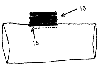
Form: There are five main forms.
1. SINGLE-ENDED SCREW-DEVICE 16. This device 16 is a spring with four to
six windings guaranteeing elasticity. The first three windings are closely
adjacent,
i.e. there is just the smallest space between them (the space enabling the
screw-
device 16 to dig itself into the vessel wall). On the one end of the single-
ended
CA 02523803 2009-12-03
4
screw-device 16 there is an extremely sharp end 18, meant to perforate the
vessel
wall. The other end 20 is blunt.
The sharp end 18 is round, i.e. non-cutting but capable of perforating the
vessel
wall. The sharp, round point is bent inwardly and downwardly in an angle of 10
to
20 degrees (a) (see figure 3a, 3b, 3c, 3d). Alternatively, this sharp, round,
non-
cutting point 18 may bend downwardly in an angle of 90 degrees (a). In this
case,
the end 18 resembles a cork-screw, but the end 18 is not situated in the
middle of
the final winding but rather on the periphery.
2. DOUBLE-ENDED SCREW-DEVICE / RING-FORM 22. This device 22 is a
spring with four to six windings guaranteeing elasticity. The first three
windings
are closely adjacent, i.e. there is just the smallest space between them (the
space
enabling the screw-device 22 to dig itself into the vessel wall). On the one
end the
double-ended screw-device / ring-form 22 takes the form of a ring with two
sharp,
round, non-cutting points 24, 26, pointing in the same direction but 180
degrees
apart from each other (see figure 4a, 4b, 4c). These two points 24, 26 are
bent
inwardly and downwardly in an angle of 10 to 20 degrees. Alternatively, these
sharp, round, non-cutting points 24, 26 may bend downwardly in an angle of 90
degrees. In this case, they resemble a cork-screw, but the ends are not
situated in
the middle of the final winding but rather on the periphery. The other end 23
or 25
of the double-ended screw-device 22 is blunt.
3. DOUBLE-ENDED SCREW-DEVICE / SPIRAL FORM 28. This device 28 is a
spring with four to six windings guaranteeing elasticity. The first three
windings
are closely adjacent, i.e. there is just the smallest space between them (the
space
enabling the screw-device 28 to dig itself into the vessel wall). The double-
ended
screw-device / spiral-form 28 consists of two sharp, round, non-cutting points
30,
32, the first coming from the end 30, the second 32 coming from the beginning
but bent in such a way as to align itself with the other sharp point (see
figure 5a,
5b). Again, these two points 30, 32 point in the same direction but stand 180
degrees apart from each other. They bend inwardly and downwardly in an angle
of
to 20 degrees. Alternatively, these sharp, round, non-cutting points 30, 32
may
bend downwardly in an angle of 90 degrees. In this case, the end resembles a
cork
-screw, but the ends are not situated in the middle of the final winding but
rather
on the periphery.
4. KEY-RING SCREW-DEVICE 34. This device 34 consists of two to three
windings, resembling a key-ring. On the one end 36, there is a sharp, round,
non-
cutting point, bending inwardly and downwardly in an angle of 10 to 20
degrees.
Alternatively, this point 36 may bend downwardly in an angle of 90 degrees. In
this case, the end 36 resembles a cork-screw, but the end is not situated in
the
middle of the final winding but rather on the periphery. The other end 38 is
blunt.
Where the two ends meet, there is a twist in the ring (see figure 6a, 6b).
5. SCREW-DEVICE WITH REMOVABLE HEAD 40. This device 40 consists of
two basic parts, the removable head 42 (with applicator 44) and a hollow screw
46
of three windings, which remains in place (i.e. in the blood-vessel).
5.1 The head 42 consists of two windings, and ends in the form of a cork-screw
(see figure 7a, 7b). This is, again, a round, sharp, non-cutting point. The
CA 02523803 2009-12-03
head 42 forms one whole with the applicator 44, i.e. a long, thin shaft with a
handle 48 used to drill the head 42 into the vessel wall. Once the head 42 is
in place (i.e. in the middle of the vessel 14 (see figure 7c), it is removed -
together with the applicator 44 - from the rest of the screw-device 40 that
stays within the vessel wall 14.
5.2. The other part of the screw-device 40 consists of three hollow windings
attached to the head 42 by means of internal, anti-clockwise windings (see
figure 7d). Every winding is wider than the previous one, thus expanding the
vessel wall and the opening in it made by the head 42. This opening is made
in a non-occlusive way, i.e. the receptor vessel 14 need not be temporarily
occluded.
Additional tool. In the fifth form, i.e. the screw-device with removable head
40, no
additional tools are needed to open the vessel wall.
In the other forms, the hole in the vessel wall can be made by traditional
means -
basically: the occlusive manner using a surgical knife, or the non-occlusive
manner
using a laser - or by means of a screw-cutter 50. This specially designed
device 50
operates in a non-occlusive manner. It takes the form of a hollow cylinder 52
in which
a long shaft 54 with a handle 56 on top moves up and down (see figure 8a, 8b).
This
shaft 54 ends in a screw consisting of three windings. The first two 54 of
these take
the form of a cork-screw, so that the sharp point 56 is in the middle. They
keep the
vessel wall in its place, whereas the third winding - forming a full circle of
360
degrees - actually cuts and removes the part of the vessel wall where the hole
is to be
made. The third winding has its sharp edges pointing downward, whereas the
first two
windings are horizontal, like in an ordinary screw (see figure 8c).
Diameter: Depending on the sort of blood-vessel, the diameter of the five
screw-
devices 10 may vary from 1 millimetre to plus 2 centimetres.
Substance: The screw-device 10 is made of inox material, or titanium, or super-
elastic
materials such as nitinol, or synthetic materials, or even resorbable
materials.
Thickness of material: Depending on the diameter of the blood-vessel, the
material
may vary from 0,1 mm to any desirable thickness.
Elasticity: Depending on the material.
DESCRIPTION OF OPERATION TECHNIQUE WITH THE SCREW-
DEVICE
A. For the first four forms of the screw-device 10 - that is: single-ended
screw-device
16, double-ended screw-device / ring-form 22, double-ended screw-device /
spiral-
form 28, key-ring screw-device 34 - the technique is as follows:
1. End-to-side
In the first step, the receptor vessel 14 is exposed by means of the
techniques current
in vascular surgery. When a venous graft is used end-to-side, the screw-device
10 is
screwed into the graft (donor vessel 12) or sutured to the donor vessel 12.
CA 02523803 2009-12-03
6
In the second step, the donor vessel 12 containing the screw-device 10 is
screwed into
the receptor vessel 14.
Alternatively, the screw-device 10 can first be screwed into the receptor
vessel 14 and
then the donor vessel 12 can be attached to it.
The screw-device 10 is turned into the vessel clock-wise and completes only
one turn,
that is: it is in its proper place after 360 degrees.
In the third step, the wall of the receptor vessel 14 is opened by means of
existing
techniques, such as laser or the surgical knife.
2. Side-to-side
First, the donor vessel 12 is clamped and opened. The screw-device 10 is
screwed into
and through the vessel wall, thus perforating the donor vessel 12 with two
windings.
These windings are then screwed into the receptor vessel 14 (clockwise and 360
degrees). A hole is then made into the receptor vessel 14 wall by means of
existing
techniques, such as laser or the surgical knife.
B. For the fifth form, that is the screw-device with removable head 40, the
techniques
mentioned sub A are applied in the same way, but they are followed by the
removal of
the head 42.
In all these forms, the screw-device 10 can be used in an occlusive or non-
occlusive
manner, depending on the preferences of the surgeon.
MANUFACTURING AND INDUSTRIAL APPLICABILITY
The screw-device 10 can be manufactured commercially and be employed to
anastomose two vessels of different or identical sizes. It can be used in all
domains of
vascular surgery, heart surgery, and neurosurgery.