Note: Descriptions are shown in the official language in which they were submitted.
CA 02524272 2005-10-31
WO 2004/088322 PCT/AU2004/000403
I
MULTIPLEX SCREENING FOR LYSOSOMAL
STORAGE DISORDERS (LSDs)
RELATED APPLICATIONS:
[0001] This application claims priority to the following applications: (1)
Australian
Provisional Patent Application, Serial Number 20031901451, entitled "AN
IMPROVED
METHOD OF SCREENING FOR LYSOSOMAL STORAGE DISORDERS," filed on March
31, 2003, having Hopwood et al., listed as inventors; (2) Australian
Provisional Patent
Application, Serial Number 2003!904174, entitled "MULTIPLEX SCREENING FOR
LSD's," filed on August 8, 2003, having Hopwood et al., listed. as inventors;
(3) Australian
Provisional Patent Application, Serial Number 2003/904720, entitled "MULTIPLEX
SCREENING FOR LSD'S," filed on September 2, 2003, having Hopwood et al.,
listed as
inventors. The entire content of each of the above identified applications is
hereby
incorporated by reference.
BACKGROUND:
[0002] The present invention is generally related to diagnostics that
determine
Lysosomal Storage Disorders ("LSDs") and related diseases in a subject. More
particularly,
this invention pertains to compounds, reagents, and methods for identifying
and quantifying
the levels and ratios of multiple target antigens that are used to accurately
diagnose LSD.
The target antigens are naturally present in biological fluids or tissues of
either LSD or non-
LSD patients.
[0003] LSDs represent a group of over 40 distinct genetic diseases that
generally
affect young children. Individuals that are affected with a LSD present a wide
range of
clinical symptoms that depend upon the specific disorder or a particular
genotype involved.
The clinical symptoms associated with LSD's can have a devastating impact on
both the child
and the family of affected individuals. For example, central nervous system
dysfunction,
behavioral problems, and severe mental retardation are characteristic of many
LSDs. Other
clinical symptoms may include skeletal abnormalities, organomegaly, corneal
clouding and
dysmorphic features (Neufeld and Muenzer, 1995). Patients are usually born
without the
visible features of a LSD, but early stage symptoms can quickly develop into a
progressive
CA 02524272 2005-10-31
WO 2004/088322 PCT/AU2004/000403
2
clinical concern. In severe cases, the affected children require constant
medical management
but still often die before adolescence.
[0004] The significance of LSDs to health care becomes obvious when comparing
the
group incidence rate for a LSD (1:5,000 births) to the group incidence rate of
other with well-
s known and intensively studied genetic disorders, such as phenylketonuria
(1:14,000) and
cystic fibrosis (1:2,500), wherein these figures reflect incidence rates for
Caucasian
populations.
[0005] Once an individual begins to present the symptoms of a LSD, the actual
clinical diagnosis of the disease is still a complex process. A clinical
diagnosis of a LSD
often requires multiple visits to a range of specialists, which can take
months or even years.
This long process is extremely stressful on the patient and family.
Fortunately, there has been
considerable progress in the diagnosis of LSDs over the past 20 years. For
example, the
development and introduction of chromatographic-based urine screens for a
specific group of
LSDs called mucopolysaccharidoses ("MPS") and oligosaccharidoses has
facilitated
screening of clinically selected patients for these disorders. Following a
clinical index of
suspicion for the disorders, the next stage of diagnosis involves a urine
screen, wherein a
"positive" urine screen is then followed by specific enzymatic analysis.
Although the
chromatographic-based screening methods are simple to perform, they are
relatively labor-
intensive and often require experience to accurately interpret results. One
example includes a
method of identifying and quantitating biochemical markers ("biomarkers") that
are present in
biological fluids or tissues of a patient having a MPS or related disorders
comprises
determining a target quantity of a target MPS biomarker oligosaccharide from a
target
biological sample taken from the target animal, and then comparing the target
quantity to a
reference quantity of a reference MPS biomarker oligosaccharide for the
diagnosis,
characterization, monitoring, and clinical management of MPS and related
disease, as
described in PCT Application AU03/00731 entitled "identification of
Oligosaccharides and
their Use in the Diagnosis and Evaluation of Mucopolysaccharidoses and Other
Related
Disorders," filed on June 13, 2003 with Hopwood et al., listed as inventors
(the entire content
of PCT Application AU03/00731 is hereby incorporated by reference).
Consequently,
chromatographic-based screening tests for LSDs are not used in some centers.
Furthermore,
CA 02524272 2005-10-31
WO 2004/088322 PCT/AU2004/000403
3
these chromatographic-based screens are not readily amenable to automation,
which has
further limited their utilization in screening strategies for newborns.
[0006] The production of specific substrates and antibody capture assays has
made the
enzymatic analyses for LSDs more accurate. Although not wanting to be bound by
theory,
the majority of LSDs result from a reduction in levels of a particular
enzymes) involved in a
specific LSD, and the identification of the specific enzymes) steady state in
normal
individuals will help identify the particular form of LSD in the affected
individual. The
ability to quickly and accurately determine the levels of the more than 40
enzymes known to
be involved with LSDs will assist in the development of better and more
economical
screening assays. Unfortunately, many of the chromatographic-based screens and
enzyme
assays mentioned above are time-consuming, invasive, complex, and require
cultured cells, or
tissue biopsies, which tends to make such assays inconvenient and expensive.
As a result,
testing for a LSD is often not a first line strategy for an affected child
with early stage
symptoms. Newborn screening for LSDs promises to provide early detection of
the LSD, but
all newborns must be screened in order to detect the disease early. Patients
having a family
history of LSDs may have a justifiable reason to perform an early screen for a
LSD.
However, the cost of an early screen of the LSD in individuals not having a
family history
may not be justified economically. Therefore, it would be beneficial that any
LSD screening
process be capable of economically screening large numbers of newborns.
[0007] One common feature of LSDs is the accumulation and storage of materials
within lysosornes. It is generally recognized that the accumulation and
storage of material in
LSD affected individuals results in an increase in the number and the size of
lysosomes within
a cell from approximately 1 % to as much as 50% of total cellular volume. In
non-affected
individuals, such materials are typically degraded into degradation products
within the
lysosome and then transported across the lysosomal membrane. Certain lysosomal
proteins
are present at elevated levels in the lysosomes of affected individuals
(Meikle et al., 1997;
Hua et al., 1998). These identified proteins are useful biomarkers for an
early diagnosis of all
LSDs. For example, sensitive immunoquantification assays have been developed
to monitor
the level of useful biomarkers such as the lysosome-associated membrane
proteins
("LAMPs"), saposins, and oc-glucosidase. Although the determination of either
LAMP-1 or
LAMP-2 levels alone in an 'at-increased-risk' group will identify up to 65% of
LSD affected
CA 02524272 2005-10-31
WO 2004/088322 PCT/AU2004/000403
4
individuals, the combination of a LAMP with one of the saposins increase
identification of
LSD affected individuals to approximately 85%. Therefore, a method to identify
two or more
biomarkers simultaneously would increase the accuracy of diagnosing a specific
LSD as
compared to any single assay. An automated multiplex assay that could perform
a
simultaneous screen on each of the known LSD deficient enzymes would reduce
time and
cost for accurate LSD diagnosis.
[0008] Multiplexing Bead Technology is built around 3 core technologies. The
first is
the family of fluorescently dyed microspheres having specific biomolecules
bound to the
surface of the microsphere. The second is a flow cytometer with 2 lasers and
associated optics
to measure biochemical reactions that occur on the surface of the
microspheres, and the third
is a high-speed digital signal processor to efficiently manage the fluorescent
output. This type
of system has been described in, for example: United States Patents 6,449;562;
6,524,793 and
United States Patent Application SN 09/956,857. United States Patent 6,449,562
("the '562
Patent") entitled "Multiplexed Analysis of Clinical Specimens Apparatus and
Method,"
having Chandler et al. listed as inventors was issued on September 10, 2002.
The '562 Patent
discloses a method for the multiplexed diagnostic and genetic analysis of
enzymes, DNA
fragments, antibodies, and other biomolecules comprising the steps of
constructing an
appropriately labeled headset, exposing the headset to a clinical sample, and
analyzing the
combined sample/beadset by flow cytometry. Flow cytometric measurements are
used to
classify, in real-time, beads within an exposed headset and textual
explanations, based on the
accumulated data obtained during real-time analysis, are generated for the
user. The inventive
technology of the '562 Patent enables the simultaneous, and automated,
detection and
interpretation of multiple biomolecules or DNA sequences in real-time while
also reducing
the cost of performing diagnostic and genetic assays. However, the '562 Patent
does not
describe how to utilize the technology for diagnosing LSD's.
[0009] United States Patent 6,524,793 ("the '793 Patent") entitled
"Multiplexed
Analysis of Clinical Specimens Apparatus and Method," having Chandler et al.
listed as
inventors, was issued on February 25, 2003. The '793 Patent discloses a method
for the
multiplexed diagnostic and genetic analysis of enzymes, DNA fragments,
antibodies, and
other biomolecules comprising the steps of constructing an appropriately
labeled headset,
exposing the headset to a clinical sample, and analyzing the combined
sample/beadset by flow
CA 02524272 2005-10-31
WO 2004/088322 PCT/AU2004/000403
cytometry. Flow cytometric measurements are used to classify, in real-time,
beads within an
exposed beadset and textual explanations, based on the accumulated data
obtained during
real-time analysis, are generated for the user. The '793 Patent enables the
simultaneous, and
automated, detection and interpretation of multiple biomoIecules or DNA
sequences in real-
5 time while also reducing the cost of performing diagnostic and genetic
assays. However, the
'793 Patent does not describe how to utilize the technology for diagnosing
LSD's.
[0010] United States Patent Application Serial No. 09/956,857 ("the '857
Application") entitled "Multiple Reporter Read-out for Bioassays" was
published on March
20, 2003. The '857 Application describes a method for. detecting a plurality
of reactive sites
on an analyte, comprising allowing reactants on an addressable microsphere and
the reactive
sites to react, forming reactant-reactive site pairs distinguishable by
fluorescence intensity.
The '857 Application also provides a method for detecting a plurality of
analytes in a sample
using addressable microspheres in combination with one or more reporter
reagents. Also
provided are a method for determining allele zygosity of a genetic locus
having two alleles or
more alleles using microparticles, and a method for detecting a plurality of
SNPs in nucleic
acid molecules. The '857 Application also provides a composition comprising an
addressable
microsphere carrying at least two fluorescent reactants capable of forming
reactant-analyte
pairs distinguishable by their fluorescence intensity, and kits comprising the
inventive
composition and a plurality of reporter reagents. However, the '857
Application does not
describe how to utilize the technology for diagnosing LSD's. The entirety of
each of the
applications or patents listed above is hereby specifically incorporated by
reference.
[0011] Accordingly, there is a need for the development of a fast, accurate
and
economical screen for early diagnosis of LSDs, which is amenable to
automation. The ability
to identify specific LSD enzymes in an automated multiplex assay will have a
significant
impact on the development of a newborn screening programs, as well as the
ability to address
a number of other issues associated with the early diagnosis and treatment of
LSDs. The
present invention provides compounds, reagents, and methods for a LSD
diagnostic multiplex
assay.
CA 02524272 2005-10-31
WO 2004/088322 PCT/AU2004/000403
6
FIGURES
[0012] Figure 1 shows LAMP-1 levels in plasma from LSD individuals wherein the
box length is the interquartile range that covers 25th to 75th percentile, the
outliers are
represented by (circles) each of these cases represent values between 1.5 and
3 box lengths
from the upper or lower edge of the box, and the extreme outlier (stars) are
cases with values
more than 3 box lengths from the upper or lower edge of the box;
[0013] Figure 2 shows saposin C levels in plasma from LSD individuals wherein
the
box length is the interquartile range that covers 25th to 75th percentile, the
outliers are
represented by (circles) each of these cases represent values between 1.5' and
3 box lengths
from the upper or lower edge of the box, and the extreme outlier (stars) are
eases with values
more than 3 box lengths from the upper or lower edge of the box;
[0014] Figure 3 shows a-Glucosidase in plasma from LSD affected individuals,
wherein the box length is the interquartile range that covers 25th to 75th
percentile, the
outliers are represented by (circles) each of these cases represent values
between 1.5 and 3
box lengths from the upper or lower edge of the box, and the extreme outlier
(stars) are cases
with values more than 3 box lengths from the upper or lower edge of the box;
[0015] Figure 4 shows analysis of patient blood spots for LAMP-1 wherein
the box length is the interquartile range that, covers 25th to 75th
percentile, the outliers are
represented by (circles) each of these cases represent values between 1.5 and
3 box Lengths
from the upper or lower edge of the box, and the extreme outlier (stars) are
cases with values
more than 3 box lengths from the upper or lower edge of the box;
[0016] Figure 5 shows Analysis of patient blood spots for saposin C wherein
the box
length is the interquartile range that covers 25th to 75th percentile, the
outliers are represented
by (circles) each of these cases represent values between 1.5 and 3 box
lengths from the upper
or lower edge of the box, and the extreme outlier (stars) are cases with
values more than 3 box
lengths from the upper or lower edge of the box;
[0017] Figure 6 shows a-Glucosidase protein/activity determination
in dried blood spots, wherein the box length is the interquartile range that
covers 25th to 75th
percentile, the outliers are represented by (circles) each of these cases
represent values
CA 02524272 2005-10-31
WO 2004/088322 PCT/AU2004/000403
7
between 1.5 and 3 box lengths from the upper or lower edge of the box, and the
extreme
outlier (stars) are cases with values more than 3 box lengths from the upper
or lower edge of
the box;
[0018] Figure 7 shows a-Glucosidase protein distribution in neonates;
[0019] Figure 8 shows the newborn population distribution of LAMP-1 and
saposin C
[0020] Figure 9 shows target populations representing each LSD of interest
analyzed;
[0021] Figure 10 shows a microsphere capture sandwich immunoassay having a
microsphere with two spectrally distinct fluorophores, the target LSD capture
antibody and
the unique LSD target protein or target antigen bound to the target LSD
capture antibody and
a reporter molecule;
[0022] Figure 11 shows a list of antibody reagents available for lysosomal
proteins for
utilization of LSD's screened by multiplex technology;
[0023] Figure 12 shows a calibration curve for a-glucosidase in a rnicrosphere
based
assay;
[0024] Figure 13 shows multiplexed calibration curves in a microsphere based
assay;
[0025] Figure 14A and Figure 14B show calibration curves of a-glucosidase
using
bead technology and measured using Bio-Plex~ Protein Array system (Bio-Rad);
[0026] Figure 15 shows the multiplex technology having at least a 4-plex for
LSD's;
[0027] Figure 16 shows calibration curves for a 4-plex immune quantification
of
lysosomal proteins;
[0028] Figure 17 shows multiplex analysis of control and MPS I plasma, wherein
the
box length is the interquartile range that covers 25th to 75th percentile, the
outliers are
represented by (circles) each of these cases represent values between 1.5 and
3 box lengths
from the upper or lower edge of the box, and the extreme outlier (stars) are
cases with values
more than 3 box lengths from the upper or lower edge of the box;
CA 02524272 2005-10-31
WO 2004/088322 PCT/AU2004/000403
8
[0029] Figure 18 shows box plots of plasma concentrations of Lamp-1 (A),
saposin C
(B), a-glucosidase (C) and a-iduronidase (D) from a control group and 6
different LSD
wherein, the center line within the box represents the median, the top of the
box is the 75~
and the bottom of the box is the 25~ percentile, error bars represent the
largest and smallest
values that are not outliers, outliers represented by open circles, are values
more than 1.5 box
lengths from the 75~ and 25~ percentile and extremes represented by stars are
values more
than 3 box-lengths from the 75~ and 25~ percentile.
[0030] Figure 19 shows box plots of concentrations of Lamp-1 (A), saposin C
(B), oc-
glucosidase (C) and a-iduronidase (D) from dried blood spots, the samples were
measured in
a control group, a newborn group, and a group of 3 LSD patients.
[0031] Figure 20 shows target protein markers for LSD screening;
[0032] Figure 21 shows the antibodies and bead regions used for the 7-plea
assay;
[0033] Figure 22 shows the calibration curves for each of the protein assays;
[0034] Figure 23 shows the individual and average adult control protein values
in the
7 plex assay obtained for each sample with the standard deviation, minimum and
maximum of
each group;
[0035] Figure 24 shows the individual and average newborn protein values in
the 7
Alex assay for each sample with the standard deviation, minimum and maximum of
each
group;
[0036] Figure 25 shows the Pearson correlation coefficient between each pair
of
protein analytes;
[0037] Figure 26 shows the protein concentrations of the LSD individuals
compared
to adult control group;
[0038] Figure 27 shows the protein concentrations of the LSD individuals
compared
to the newborn control group;
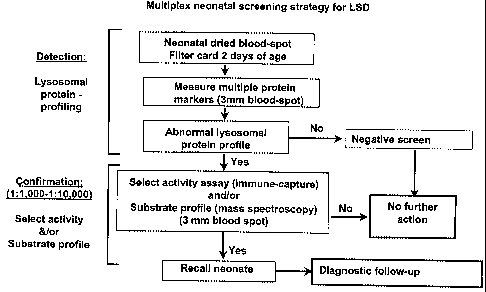
[0039] Figure 28 shows the multiplex neonatal screening strategy for LSD;
CA 02524272 2005-10-31
WO 2004/088322 PCT/AU2004/000403
9
[0040] Figure 29 shows the derivatization of oligosaccharides for MS/MS
analysis;
[0041] Figure 30 shows MS/MS analysis of a-mannosidosis urine
(Precursor ion scan of m/z 175);
[0042] Figure 31 shows retrospective analysis of HNAcS in newborn blood spots
vs
blood spot age;
[0043] Figure 32 shows retrospective analysis of HNAc-UA-HNAc-UA in newborn
blood spots;
[0044] Figure 33 shows a summary of retrospective analysis of newborn blood
spots;
[0045] Figure 34 shows protein markers for LSD screening using multiplex
assays for
LSD.
CA 02524272 2005-10-31
WO 2004/088322 PCT/AU2004/000403
SIJl~IARY
[0046] Lysosomal Storage Disorders ("LSDs") represent a group of over 40
distinct
genetic diseases that generally affect young children. Individuals that are
affected with a LSD
present a wide range of clinical symptoms that depend upon the specific
disorder or a
5 particular genotype involved. The present invention is generally related to
a multiple
screening diagnostic for LSD and related diseases. More particularly, this
invention pertains
to compounds, reagents, and methods for identifying and quantifying multiple
target enzymes
and proteins that are used to accurately diagnose a LSD. These target enzymes
and proteins
are naturally present in biological fluids or tissues of patients. The
invention also pertains to a
10 Multiplexing Bead Technology for simultaneous screening of specific LSD
enzymes.
[0047] A first aspect of the current invention is a composition used for
diagnosing a
LSD. The composition comprises a capture antibody capable of binding a target
antigen, and
a microsphere having the capture antibody conjugated to the microsphere. The
target antigen
is a LSD associated biomolecule that comprises a-iduronidase, a-glucosidase,
saposin C,
LAMP-1, LAMP-2, ~i-glucosidase, a-galactosidase A, iduronate-2-sulphatase, N-
acetylgalactosamine 4-sulphatase, galactose 6-sulphatase, acid
sphingomyelinase,
galactocerebrosidase, arylsulphatase A, saposin B, heparan-N-sulphatase, a-N-
acetylglucosaminidase, acetylCoA: glucosamine N-acetyltransferase, N-
acetylglucosamine 6-
sulphatase, [3-galactosidase, (3-glucuronidase, aspartylglucosaminidase, acid
lipase, (3-
hexosamindase A, (3-hexosamindase B, GM2-acitvator, acid ceramidase, a-L-
fucosidase, a-D-
mannosidase, [3-D-mannosidase, neuraminidase, phosphotransferase,
phosphotransferase g-
subunit, palmitoyl protein thioesterase, tripeptidyl peptidase I, cathespsin
K, a-galactosidase
B, or sialic acid transporter. The microsphere having the conjugated capture
antibody has a
diameter of about 5 pm and at least a first fluorophore and a second
fluorophore. The first
fluorophore being spectrally distinct from the second fluorophore. The
composition may
further comprise a detection antibody, wherein the detection antibody is
capable of binding
the target antigen, but is different from the capture antibody, and the
detection antibody is
conjugated to any detectable label known in the art (e.g. a fluorescent
label).
[0048] A second aspect of the current invention comprises a protein profiling
method
for diagnosing a pre-clinical status, or a clinical status of a LSD. The
method determines at
least a first- and second- target antigen quantity from a target biological
sample having an
CA 02524272 2005-10-31
WO 2004/088322 PCT/AU2004/000403
1I
unknown clinical status of LSD. At least a first- and a second- reference
antigen quantity are
also determined from a reference biological sample having a known clinical
status of LSD.
The target antigens are LSD associated biomolecules that comprise a-
iduronidase, a-
glucosidase, saposin C, LAMP-1, LAMP-2, or other biomarkers associated with
LSD. By
calculating a target proportion between the first- and second- target antigen
quantities, an
adjusted target quantity can be assigned. Similarly, an adjusted reference
quantity can be
assigned by calculating a reference proportion between the first- and second-
reference
antigen quantities. The pre-clinical status or the clinical status of an LSD
can then be
determined by comparing a deviation of the adjusted target quantity to the
adjusted reference
quantity. In one specific embodiment, the target biological sample and the
reference
biological sample of this method are selected from a cellular extract, blood,
plasma, or urine.
Alternatively, the second target antigen and the second reference antigen
comprise a
biomarker indicator of cell number, organelle number, cell size, organelle
size, cell volume,
or organelle volume.
[0049] A third aspect of the current invention comprises a method for
determining an
amount of at least a first target antigen and at least a second target antigen
indicative of a LSD
in a target biological sample using a composition of capture antibody
microspheres. The
method comprises incubating at least a first capture antibody microsphere and
at least a
second capture antibody microsphere with the target biological sample forming
a capture
suspension. The first capture antibody microsphere and the second capture
antibody
microsphere are then recovered from the capture suspension. These first- and
second-
recovered microspheres are then hybridized with a first- and a second-
detection antibody,
respectively. The first recovered antibody microsphere and the second
recovered antibody
microsphere having a bound detection antibody can be detected when they are
passed through
an examination zone. Data is then collected that relates to one or more
microsphere
classification parameters, the presence or absence of the first- or second-
detection antibody;
and the amount of first- or second- detection antibody is quant~ed. In a
specific
embodiment, the target biological sample is selected from a cellular extract,
blood, plasma, or
urine. In another specific embodiment, the first target antigen and second
target antigens are
each a-iduronidase, a-glucosidase, saposin C or other biornarkers associated
with a LSD. The
second target antigen may also comprise an indicator of cell number, organelle
number, cell
size, organelle size, cell volume, or organelle volume.
CA 02524272 2005-10-31
WO 2004/088322 PCT/AU2004/000403
12
[0050] A fourth aspect of the current invention comprises a method of
detecting
multiple LSD target antigens in a sample. The specif c subset of LSD antigens
comprises a-
iduronidase, a-glucosidase, saposin C or other biomarkers associated with LSD.
The method
comprises exposing a pooled population of target capture microspheres to the
sample. Each
of the target capture microspheres have distinct subsets, and each distinct
subset has: (i) one
or more characteristic classification parameters that distinguishes one target
capture
microsphere of one subset from those of another target capture microsphere
subset according
to a predetermined discriminate microsphere function table, which includes
fluorescence
emission intensities; and (ii) a distinct capture antibody that can bind a
specific subset of LSD
antigens. After the pooled population of target capture microspheres has been
exposed to the
sample, the exposed pooled population of target capture microspheres is passed
through an
examination zone. The identity and quantity of each specific subset of LSD
target antigen of
interest is determined, if present, in the sample by (i) collecting data
relating to one or more
subsets of target capture microsphere classification parameters that
distinguishes one target
capture antibody microsphere of one subset from those of another target
capture antibody
microsphere subset according to a predetermined discriminate function table,
including the
fluorescence emission intensities, (ii) collecting data relating to the
presence or absence of a
corresponding subset of specific LSD antigen, (iii) quantifying each
corresponding subset of
specific LSD antigen on ,each subset of capture antibody microsphere. In a
specific
embodiment, the method further comprises adding a pooled population of
detection antibodies
to the exposed pooled population of the target capture microspheres prior to
passing the target
capture microspheres through the examination zone.
CA 02524272 2005-10-31
WO 2004/088322 PCT/AU2004/000403
13
DETAILED DESCRIPTION
Terms:
[0051] The term "a" or "an" as used herein in the specification may mean one
or more.
As used herein in the claim(s), when used in conjunction with the word
"comprising", the
words "a" or "an" may mean one or more than one. As used herein "another" may
mean at
least a second or more.
[0052] The term "animal," "subject," or "patient" as used herein may be used
interchangeably and refers to any species of the animal kingdom. In preferred
embodiments it
refers more specifically to humans.
[0053] The term "biomolecule" as used herein is understood to represent the
target
molecule, such as a protein, an antibody, a metabolite, a DNA sequence, an RNA
sequence; a
biologic with activities used or measured for the purposes multiplexing and
profiling of target
biomolecules, or a combination thereof, for the composition and method of
determining LSD,
used in administering, monitoring, or modifying an LSD therapy.
[0054] The term "clinical status" as used herein refers to patients that are
being
studied or treated by physicians for a LSD.
[0055] The term "comprise," or variations such as "comprises" or "comprising,"
as
used herein may be used to imply the inclusion of a stated element or integer
or group of
elements or integers, but not the exclusion of any other element or integer or
group of
elements or integers.
[0056] The term "fluorophore" as used herein refers to any fluorescent
compound or
protein that can be used to quantify the LSD antigens.
[0057] The term "normalize" as used herein refers to bringing a target,
reference, or
other samples into conformity with a standard, pattern, model, etc. For
example, in one
embodiment, urine samples from LSD patients and non-LSD patients were
normalized by
using a 1 ~.mol equivalent of creatinine from each sample.
CA 02524272 2005-10-31
WO 2004/088322 PCT/AU2004/000403
14
[0058] The term "phenotype" as used herein refers to the manifest
characteristics of
an organism collectively, including anatomical and psychological traits, that
result from both
its heredity and its environment:
[0059] The term "preclinical status" as used herein refers to the period of a
disease
, before any of the clinical symptoms appear.
[0060] The term "lysosornal storage disorder ("LSD") associated biomolecule"
as
used herein refers to any biomolecule that has been linked to any LSD. In
preferred
embodiments, a LSD associated biomolecule includes, but is not limited to: a-
iduronidase, a-
glucosidase, saposin C, LAMP-l, LAMP-2, (3-glucosidase, a-galactosidase A,
iduronate-2-
sulphatase, a-iduronidase, N-acetylgalactosamine 4-sulphatase, galactose 6-
sulphatase, acid
sphingomyelinase, galactocerebrosidase, arylsulphatase A, saposin B, heparan-N-
sulphatase,
a-N-acetylglucosaminidase, acetylCoA: glucosamine N-acetyltransferase, N-
acetylglucosamine 6-sulphatase, (3-galactosidase, (3-glucuronidase,
aspartylglucosaminidase,
acid lipase, (3-hexosamindase A, (1-hexosamindase B, GMZ-acitvator, acid
ceramidase, a-L-
fucosidase, a-D-mannosidase, (3-D-rnannosidase, neuraminidase,
phosphotransferase,
phosphotransferase g-subunit, palmitoyl protein thioesterase, tripeptidyl
peptidase I,
cathespsin K, a-galactosidase B, or sialic acid transporter. As shown below,
Table 1 indicates
some enzyme deficiencies for LSDs.
CA 02524272 2005-10-31
WO 2004/088322 PCT/AU2004/000403
Table 1 Enzymes deficient in some common lvsosomal storage disorders
Disease Clinical PhenotypeEnzyme Deficiency Australian
Prevalence
Gaucher disease types Glucocerebrosidase 1 in 57,000
I / II / III Gaucher
disease
((3-gIucosidase)
Cystinosis Cystine transporter1 in 192,000
Fabry disease Fabry disease a-Galactosidase 1 in 117,000
A
Glycogen storage Pompe disease a-Glucosidase 1 in 146,000
disease II
MucopolysaccharidosisHurler/Scheie a-L-Idurorudase 1 in 88,000
type I
syndrome
MucopolysaccharidosisHunter syndromeIduronate-2-sulphatase1 in 136,000
type II ~
MucopolysaccharidosisMaroteaux-Lamy N-acetylgalactosamine1 in 235,000
type VI 4-
syndrome , sulphatase
MucopolysaccharidosisMorquio syndromeGalactose 6-sulphatase1 in 169,000
type
IVA
Niemann-Pick disease Acid sphingomyelinase1 in 248,000
types A / Niemann-Pick
disease
B
Globoid cell leucodystrophyKrabbe disease Galactocerebrosidase1 in 201,000
Metachromatic leucodystrophy Arylsulphatase A 1 in 92,000
Metachromatic leucodystrophy Saposin B
MucopolysaccharidosisSanfilippo syndromeHeparan-N sulphatase1 in 114,000
type
MucopolysaccharidosisSanfilippo syndromeoc N Acetylglucosaminidase1 in 211,000
type
MucopolysaccharidosisSanfilippo syndromeAcefylCoA:N- 1 in 1,407,000
type
TIIC acetyltransferase
MucopolysaccharidosisSanfilippo syndromeN-Acetylglucosamine1 in 1,056,000
type 6-
lIID sulphatase
MucopolysaccharidosisMorquio syndrome(3-Galactosidase
type
MucopolysaccharidosisSly (3-Glucurorudase 1 in 2,111,000
type VII
Niemann-Pick diseaseNiemann-Pick , 1 in 211,000
type C1 disease Cholesterol trafficking
.
Niemann-Pick diseaseNiemann-Pick Cholesterol trafficking
type C2 disease
Aspartylglucosaminuria Aspartylglucosaminidase1 in 2,111,000
Cholesterol ester Wolman disease Acid lipase 1 in 528,000
storage disease
GM1-Gangliosidosis (3-Galactosidase 1 in 384,000
types
I/II/III
GM2-Gangliosidosis Tay Sachs diseasea-Hexosaminidase 1 in 201,000
type I A
GM2-Gangliosidosis Sandhoff disease(3-Hexosaminidase 1 in 384,000
type II A & B
GM2-Gangliosidosis GM2-activator deficiency
Farber LipogranulomatosisFarber disease Acid ceramidase
Fucosidosis ~ of L-Fucosidase > 1 in
2,000,000
Galactosialidosis Protective protein
types I / II
a-Mannosidosis types a-D-Mannosidase 1 in 1,056,000
I / II
(3-Mannosidosis (3-D-Mannosidase
Mucolipidosis type Sialidosis typesNeuraminidase
I I / II
Mucolipidosis types I-cell disease;Phosphotransferase 1 in 325,000
II / III
Mucolipidosis type pseudo-Hurler Phosphotransferase
IIIC g-subunit
polydystrophy
Mucolipidosis type Unknown
IV
Multiple sulphatase Multiple sulphatases1 in 1,407,000
deficiency
Neuronal Ceroid Batten disease Palmitoyl protein
thioesterase
CA 02524272 2005-10-31
WO 2004/088322 PCT/AU2004/000403
16
Lipofuscinosis, CLN1
Neuronal Ceroid Batten disease Tripepiidyl peptidase I
Lipofuscinosis, CLN2
Neuronal Ceroid Vogt Spielmeyer Protein function not known
Lipofuscinosis, CLN3 disease
Neuronal Ceroid Batten disease Protein function not known
Lipofuscinosis, CLN5
Neuronal Ceroid Northern fipilepsy Protein function not known
Lipofuscinosis, CLN8
Pycnodysostosis Cathepsin K
Sialic acid storage disease Schindler disease a Galactosidase B
Sialic acid storage disease Sialuria; salla disease Sialic acid transporter 1
in 528,000
Prevalence figures quoted from Miekle et al., JAMA 281:249-254 (1999).
Prevalence and ratio of
lysosomal storage disorders may vary from country to country
CA 02524272 2005-10-31
WO 2004/088322 PCT/AU2004/000403
17
[0061] The term "reference quantity" as used herein refers to a known,
normalized
amount of a LSD biomarker in a biological fluid. The reference quantity is
determined from
an animal, or group of animals having a defined clinical status, preclinical
status, or
phenotype of a LSD disease. The reference quantity may refer to a table
compiled from
various animals or groups of animals having correlations between relative
amounts of LSD
biomarkers in a biological fluid, and a known clinical status, preclinical
status, or phenotype.
LYSOSOMAL STORAGE DISORDERS
[0062] The LSD's represent a group of over 40 distinct genetic diseases that
generally
affect young children. Patients are usually born without the visible features
of a LSD, but
early stage symptoms can quickly develop into a progressive clinical concern.
Although
some effective LSD therapies have been developed it is paramount that therapy
be started as
soon as the LSD has been diagnosed. Unfortunately, a clinical diagnosis of a
LSD often
requires multiple visits to a range of specialists requiring time-consuming,
invasive, complex,
inconvenient; and expensive assays. The current process for an accurate
diagnosis of LSD for
a patient not having a family history of LSD can take months to years, which
is unacceptable
when effective LSD therapies are needed earlier.
[0063] It is generally recognized that the accumulation of storage materials
in the
lysosomes of LSD affected individuals will increase from approximately 1% to
as much as
50% of the total cellular volume. Certain lysosomal proteins are present at
altered levels in
the LSD affected individuals (Meikle et al., 1997; Hua et al., 1998), as
indicated in Figures 1-
6. The values for the individual immunoassays in plasma samples were
determined as follows
arid shown in Figures 1-6. Unless stated otherwise all regents were of
analytical grade and
were obtained from Sigma Chemical Company, MO USA. Preparation of recombinant
proteins, antibodies and calibration standards for Lamp-1 and saposin C.
Recombinant Lamp-
1 (minus tail) was isolated from CHO-Kl cells as detailed in Isaac et al
[Isaac EL,
Karageorgous LE, Brooks DA, Hopwood JJ and Meikle PJ. Experimental Cell
Research
2000, 254: 204-209]. Recombinant Saposin C was a gift from Dr GA Grabowski and
was
prepared by the method of Qi and Grabowski [Qi TL and Grabowski GA J Biol Chem
1994,
269:16746-16753].
CA 02524272 2005-10-31
WO 2004/088322 PCT/AU2004/000403
18
' [0064] The anti Lamp-1 monoclonal antibody (BB6) was generated using intact
Lamp-1 protein by the method of Carlsson and Fukada [Carlsson SR and Fukada M
JBC
(1989) 264(34): 20526-20531] and 7B2 (anti Saposin C) monoclonal antibody was
produced
using the recombinant protein by the method described in [Zola H and Brooks D.
Techniques
for the production and characterization of monoclonal hybridoma antibodies.
In: Hurrell
JGR, ed. Monoclonal hybridoma antibodies: techniques and applications. Boca
Raton, FL:
CRC Press, 1982:1-57]. Polyclonal antibodies were generated for both Lamp-1
and Saposin
C by immunizing separate rabbits with 200~.g of each recombinant protein per
inoculation
(four inoculations in total) based upon the method of Leonova et al, 1996,
[JBC 271:17312-
20]. All antibodies were purified using 5ml HitrapTM Protein G afFmity column
(Pharmacia,
Uppsala, Sweden). The polyclonal antibodies were aff'mity purified further by
column
chromatography using their respective recombinant proteins coupled to Affi-
Gel~ 10 Gel
(Bio-Rad #153-6046, CA, USA) according to manufacturers instructions.
[0065] Blood spot calibrators containing final concentrations of 2000, 1000,
500, 250,
62.5 and 0 p,g/L for Lamp-1 and saposin C were prepared as detailed in
Umapathysivam et al
[Umapathysivam K, Whittle AM, Ranieri E, Bindloss C, Ravenscroft EM, van
Diggelen OP,
Hopwood JJ and Meikle PJ Clin Chem 46(9): 1318-1325 2000]. Two blood spot
controls
containing low (Lamp-1 400~.g/L; saposin 200~.g/L) and high (Lamp-1 800~.g/L;
saposin C
500~.g/L) protein concentrations were similarly prepared.
[0066] Quantification of Lamp-1 and Saposin C in dried blood spots containing
EDTA. Lamp-1 and Saposin C were measured in dried blood spots using one step
three tier,
time-delayed fluorescence immunoassays. Microtiter plates (Labsytems,
Helsinki, Finland
#95029180) were coated with either BB6 or 7B2 at a concentration of 5 ~,g/L in
O.lmol/1
NaHC03, pH 8.3 and incubated covered for approximately l6hrs at 4°C.
Plates were washed
twice with wash buffer (0.25mo1/1 NaCl, 0.02mo1/1 Tris containing 0.005% Tween
20 (BDH,
Poole, England) and 0.002°lo Thiomerosal, pH7.8) Non-specific binding
sites on the plates
were blocked by the addition of 100.1 of 0.25M NaCI, 0.02M Tris containing
0.5% skim milk
powder (Diploma, Bonlac Foods, Victoria, Australia), pH 7.8, per well. After a
two hour
incubation at room temperature, the microtiter plates were washed twice with
0.25M NaCI,
0.02M Tris pH 7.8 and tapped dry before being lyophilized and stored
desiccated at 4°C prior
to use.
CA 02524272 2005-10-31
WO 2004/088322 PCT/AU2004/000403
19
[0067] Standard calibrators, controls and patient dried blood spots were
placed in
duplicate into the coated microtiter wells with 2001.1,1 of either polyclonal
antibody diluted in
assay buffer (0.15mo1/1 NaCI, 0.05mo1/L Tris, 20N.moI/L Diethylene triamine-
penta-acetic
acid, containing 0.01% Tween 40, 0.5% bovine serum albumin (A-9647), 0.05%
bovine 'y
globulin (G-7516), and 0.05% sodium azide, pH 7.8). The antibodies were used
at a final
concentration of 200~,g/L and 400~.g/L for the anti-Lamp-I and anti saposin C
polyclonal
respectively. The plates were covered and incubated at room temperature for
one hour with
shaking, then placed overnight at 4°C, followed by an hour incubation
with shaking at room
temperature. The blood spots were removed by suction and the plates washed six
times with
wash buffer. After dilution in assay buffer to final concentration of
O.l~.g/ml, 100.1 of anti
rabbit europium labeled antibody (Wallac, Finland #AD0105), was added to every
well and
incubated for one hour at room temperature with shaking. After washing the
plates a final six
times with wash buffer, 200p.1 of DELFIA° Enhancement solution (Wallac,
Finland) was
added per well and the plates incubated at room temperature for ten minutes
with shaking.
Fluorescence was measured on a DELFIA° 1234 Research Fluorometer,
(Wallac, Finland).
The concentrations of Lamp-1 and Saposin C in the blood spots were calculated
using spline
fit curves generated by Multicalc Data Analysis software (version 2.4 Wallac,
Finland).
[0068] Figure 1 shows the LAMP-1 levels in plasma from LSD individuals that
are
indicated by the box length being the interquartile range that covers .25th to
75th percentile.
Figure 2 shows saposin C levels in plasma from LSD individuals wherein the box
length is
the inter-quartile range that covers 25th to 75th percentile. Figure 3 shows
oc-Glucosidase in
plasma from LSD affected individuals, wherein the box length is the inter-
quartile range that
covers 25th to 75th percentile.
[0069] Target enzymes can also be detected by individual immunoassays in dried
blood spots, as indicated in Figure 4, Figure 5, and Figure 6. For example,
Figure 4 shows
analysis of patient blood spots for LAMP-1, wherein the box length is the
inter-quartile range
that covers 25th to 75th percentile. Figure 5 shows Analysis of patient blood
spots for saposin
C wherein the box length is the inter-quartile range that covers 25th to 75th
percentile. Figure
6 shows a-Glucosidase protein/activity determination in dried blood spots,
wherein the box
length is the inter-quartile range that covers 25th to 75th percentile. Figure
7 shows a-
Glucosidase protein distribution in neonates. Figure 8 shows the newborn
population
CA 02524272 2005-10-31
WO 2004/088322 PCT/AU2004/000403
distribution of LAMP-I and saposin C, and Figure 9 shows target populations
representing
each LSD of interest analyzed.
[0070] Although certain lysosomal target proteins are present at altered
levels in the
affected individuals, the current individual screening assays may be
inaccurate due to
5 variations among individual samples. For example, a given sample is assumed
to contain an
average number of lysosomes or white blood cells ("WBC"), however variations
in these
values between individual samples are not typically considered. Thus,
variations in an
individual having a deficiency in a particular LSD biomolecule (e.g. lysosomal
target protein),
but also having an unusually high WBC count or high numbers of lysosomes in
the test
10 sample may return an assay result that is consistent for individuals that
do not have a LSD.
Consequently, if WBC or high numbers of lysosomes .were controlled in the
sample
preparation a large inaccuracy could be avoided, and a proper diagnosis could
be made during
the first round of LSD screening.
[0071] Determining the quantities of multiple target enzymes increases the
accuracy
15 of diagnosing a specific LSD as compared to any single assay. For example,
using
immunoquantification assays directed toward identifying the levels of the
lysosome-
associated membrane proteins ("LAMPS"), such as LAMP-I or LAMP-2, in an "at-
increased-
risk" group will identify up to 65% of LSD affected individuals. However, the
combination
of LAMP'S with one of the saposins increases identification of LSD affected
individuals to
20 approximately 85%. Therefore, a method to identify two or more biomarkers
simultaneously
would increase the accuracy of LSD diagnosis and reduce the time and cost for
each assay. A
Multiplexing Bead Technology is used to simultaneously detect specific at
least 2 LSD target
antigens is described below or in Table 1.
EXAMPLE 1
[0072] Multiplexing Bead Technology and Target LSD Proteins. The
Multiplexing Bead Technology is built around 3 core technologies. The first is
the family of
fluorescently dyed microspheres having bound biomolecules. The second is a
flow cytometer
with 2 lasers and associated optics to measure biochemical reactions that
occur on the surface
of the microspheres, and the third is a high-speed digital signal processor to
efficiently
manage the fluorescent output. Bio-Rad (Hercules, CA), provides a commercially
available
CA 02524272 2005-10-31
WO 2004/088322 PCT/AU2004/000403
21
protein array system called the "Bio-Plex~". The Bio-Plex~ protein array
system includes
fluorescently dyed microspheres, a flow cytometer with 2 lasers and associated
optics, and a
high-speed digital signal processor. However, neither the Bio-Plex~ protein
array system
nor any other commercially available systems include any specific
biomolecules, methods,
compounds, or reagents needed for the simultaneous screening of specific LSD
enzymes.
[0073] The Bio-Plex~ protein array system uses multiplexing technology to
enable
the simultaneous quantitation of up to 100 different analytes. This technology
uses
polystyrene microspheres internally dyed with differing ratios of 2 spectrally
distinct
~fluorophores. Each fluorophore can have any of 10 possible levels of
fluorescent intensity,
thereby creating a family of 100 spectrally distinct bead sets. In a preferred
embodiment, the
dyed microspheres are conjugated with monoclonal antibodies specific for a
target LSD
protein or peptide thereof. Although not wanting to be bound by theory, each
of the 100
spectrally distinct bead sets can contain a capture antibody specific for a
unique LSD target
protein. In a multiplexed Bio-Plex~ assay, LSD antibody-conjugated beads are
allowed to
react with the sample and a secondary LSD . antibody, or a detection LSD
antibody in a
microtiter plate well to form a capture sandwich immunoassay. Figure 10 shows
a drawing of
a complete microsphere capture sandwich immunoassay having a polystyrene
microsphere
(110) with 2 spectrally distinct fluorophores; the target LSD capture antibody
(120) bound to
the microsphere; a unique LSD target protein or target antigen (130) bound to
the target LSD
capture antibody; a detection LSD antibody (140); and a detection molecule
(150). Once the
complete microsphere capture sandwich immunoassay has formed in solution, the
immunoassay solution is then drawn into the Bio-Plex~ array reader, which
illuminates and
reads the sample. Although not wanting to be bound by theory, there are many
enzyme
deficiencies specific for a particular LSD, and some of these enzymes are
shown in Table 1.
Specific capture antibodies, and detection antibodies for the target compounds
are available
for specific LSD's, as shown in Figure 11. Additional capture antibodies and
detections
antibodies include: j3-glucosidase; a-galactosidase A; iduronate-2-sulphatase;
a-iduronidase;
N-acetylgalactosamine 4-sulphatase; galactose 6-sulphatase; acid
sphingomyelinase;
galactocerebrosidase; arylsulphatase A; saposin B; heparan-N-sulphatase; a-N-
acetylglucosaminidase; acetylCoA: glucosamine N-acetyltransferase; N-
acetylglucosamine 6-
sulphatase; (3-galactosidase; (3-glucuronidase; aspartylglucosaminidase; acid
lipase; (3-
hexosamindase A; ~3-hexosamindase B; GM2-acitvator; acid ceramidase; a-L-
fucosidase; a-
CA 02524272 2005-10-31
WO 2004/088322 PCT/AU2004/000403
22
D-mannosidase; (3-D-mannosidase; neuraminidase; phosphotransferase;
phosphotransferase g-
subunit; palmitoyl protein thioesterase; tripeptidyI peptidase I; cathespsin
K; a-galactosidase
B; sialic acid transporter.
[0074] When a red diode "classification" Iaser (635 nm) in the Bio-Plex~ array
reader illuminates a dyed bead, the bead's fluorescent signature identifies it
as a member of
one of the 100 possible bead sets. Bio-Plex~ Manager software correlates each
bead set to
the assay reagent that has been coupled to it (for example, a first LSD
capture antibody
coupled to bead set #22, and a second LSD capture antibody coupled to bead set
#42). In this
way the Bio-Plex~ protein array system can distinguish between the different
assays
combined within a single microtiter well. A green "reporter" laser (532 nm) in
the array
reader simultaneously excites a third fluorescent dye (phycoerythrin, "PE")
bound to the
detection LSD antibody in the assay. Although not wanting to be bound by
theory, the
amount of green fluorescence is proportional to the amount of target analyte
captured in the
immunoassay. Extrapolating the captured amount of target analyte to a standard
curve allows
quantitation of each LSD analyte in the sample. The digital signal processing
algorithms
provide simultaneous real-time data acquisition of classification and reporter
signal output
from thousands of beads per second, supporting up to 100 x 96 = 9,600 analyte
measurements
from each 96-well plate.
EXAMPLE 2
[0075] Designing and Producing LSD Target Microspheres. The BioPlex Protein
Array System was used as one embodiment to demonstrate the type and nature of
the reagents
necessary for a LSD multiplex diagnostic assay. Four target proteins (e.g.
LAMP-1, a-
iduronidase, a-glucosidase, and saposin C ) were used to design target capture
microspheres
and target reporter antibodies.
[0076] The monoclonal capture antibody for LAMP-1 was BB6 developed and
provided by Sven Carlsson (Carlsson et al., 1989). The monoclonal reporter
antibody for a-
glucosidase (43D1) was obtained from Pharming, Inc. and has been described
(Fransen et al.,
1988). The polyclonal reporter antibody for LAMP-1, the rabbit polyclonal
reporter antibody
for saposin C, the sheep polyclonal capture antibody for a-glucosidase, and
the monoclonal
capture antibody ("7B2") for saposin C were prepared within the Lysosomal
Diseases
CA 02524272 2005-10-31
WO 2004/088322 PCT/AU2004/000403
23
Research Unit at the WCH in Adelaide, Australia using standard techniques,
known in the art,
and briefly described below. The availability and production of specific
monoclonal and
polyclonal antibodies are know to one of ordinary skill in the art. Production
of the specific
antibodies uses in the current examples are given below:
[0077] Polyclonal Antibodies. Sheep polyclonal antibody was produced against
recombinant proteins. A sheep was injected sub-cutaneously with 2mg of protein
in 1 mL of
an emulsion of phosphate buffered saline (pH 7.4) and complete Freunds
adjuvant, followed
by four booster injections (2mg each) with incomplete Freunds adjuvant, each
three weeks
apart. One week after the last injection the sheep was bled out and serum
collected. Rabbit
polyclonal antibody was produced in the same manner, except 0.2-1.0 mg of
protein was used
per immunisation. Sheep polyclonal antibody was purified on a 5 mL Hitrap ~
Protein G
affinity column (Pharmacia Biotech, Uppsala, Sweden) followed by an affinity
column
prepared from the recombinant protein used for the immunisation. Recombinant
protein
affinity columns were prepared by coupling 5 mg of the recombinant protein to
2.5 mL of
Affi-gel 10 (Bio-Rad, Hercules, CA, USA) as per manufacturer's instructions.
[0078] Briefly, 5 mL of sheep serum was diluted with 5 mL of phosphate
buffered
saline (pH 7.4) and centrifuged (2200g, 10 min, 4°C). The centrifuged
serum was passed
through a 0.2 ~.tsn filter, and then loaded on to the Protein G column at a
flow rate of 0.5
mL/min. The column was washed with phosphate buffered saline, pH 7.4 and the
antibody
eluted with 0.1 mol/L H3P04/NaH2P04, pH 2.5 and immediately neutralised by
adding 1.0
mol/L Na2HP04 (1/10' vol). The protein content was estimated by absorbance at
280nrn
(absorbance =1.4 for 1.0 g/L of protein). The eluate was diluted four fold and
then loaded on
to the appropriate recombinant protein affinity column at the same flow rate.
The column was
washed and eluted as described for. the Protein G column.
[0079] Monoclonal Antibodies. Monoclonal antibodies were produced in Balb/C
mice using standard immunisation protocols (Harlow et al., 1988). Mice were
immunised
with recombinant enzyme using established protocols. Plasma cells from these
immunised
mice were fused with P3.653 myeloma cells (Zola et al., 1982) and the
resulting hybridoma
cell lines screened for antibodies against the recombinant protein by direct
ELISA (Harlow et
' al., 1988). Monoclonal antibodies were purified from cell culture
supernatants by
CA 02524272 2005-10-31
WO 2004/088322 PCT/AU2004/000403
24
ammonium sulfate precipitation followed by affinity purification on Hitrap~
Protein G
affinity column (Pharmacia Biotech, Uppsala, Sweden).
[0080] Coupling Antibodies to Microspheres. The target capture antibodies were
coupled to Bio-Rad carboxylated ("COOH") beads as follows: anti-LAMP-1 to bead
#(17),
anti-saposin C to bead #(19), and anti-a-glucosidase to bead #(21). The
coupling of the target
capture antibodies to the polystyrene microspheres was performed using the
BioRad bead
coupling kit (Catalog number 171-406001, BioRad, Hercules, CA). The Bio-PIexTM
amine
coupling kit includes 4 ml bead wash buffer, 85 ml bead activation buffer, 135
ml PBS, pH
7.4, 10 ml blocking buffer, 25 m1 storage buffer, 105 ml staining buffer, 40
coupling reaction
tubes. The Bio-PIexTM amine coupling kit provides the buffers necessary to
covalently couple
6-150 kD proteins to 5.5 ~Cm dyed carboxylated polystyrene beads in under 5
hr. The
covalent couple of the target capture antibody to the carboxylated polystyrene
bead is
achieved via carbodiimide reactions involving the protein primary amino groups
and the
carboxyl functional groups bound on the surface of polystyrene beads. The
covalent
attachment is permanent, leaving no unbound protein after cleanup, even after
months of
storage. The protein-coupled beads can then be used in multiplex protein-
protein binding
studies or in the development of multiplex assays that can be analyzed with
the Bio-Plex~
protein array system. The bead yield per coupling reaction is approximately
80%, or enough
protein-coupled beads for two 96-well rnicrotiter plates using 5,000 beads per
well.
[0081] Once the coupling reaction was completed, the target capture antibody-
coupled
beads. weie enumerated and the efficiency of the protein coupling reaction was
validated,
according to the manufacturer's protocol with modifications. In this
procedure, the protein-
coupled beads were reacted with a phycoerythrin ("PE")-labeled antibody that
binds to the
coupled protein, which was then analyzed using the Bio-PIexTM protein array
system. This
procedure was performed by reacting the beads with a PE-labeled antibody.
Alternatively, a
reaction using a biotinylated antibody followed by streptavidin-PE may be
used. Although
not wanting to be bound by theory, the intensity of the fluorescent signal of
this reaction is
directly proportional to the amount of protein on the surface of the beads. A
successful
coupling typically yields a mean fluorescent intensity ("MFI") signal that is
greater than
2,000. The protein coupling validation procedure provided a rapid relative
assessment of the
amount of protein coupled to the beads, but could not verify the functionality
of the protein.
CA 02524272 2005-10-31
WO 2004/088322 PCT/AU2004/000403
[0082] Coupling of the phycoerythrin reporter molecule to the detection
antibodies in
the LAMP-1, saposin C and a-glucosidase assays was achieved using the
Molecular Probes
(Eugene, Oregon, USA) Protein-Protein Coupling Kit, as per manufacturer's
instructions with
modifications. There are several published methods known in the art for
preparation of
5 phycobiliprotein conjugates with antibodies and other proteins. Generally,
the coupling
chemistry used to crosslink a phycobiliprotein to another protein includes:
(a) treating the
antibody or other protein with a succinimidyl ester maleimide derivative at pH
7.5, which
converts some lysine residues of the antibody to thiol-reactive maleimides;
(b) preparing a
thiolated phycobiliprotein by reducing the appropriate SPDP-modified
phycobiliprotein with
10 dithiothreitol ("DTT") or with tris-(2-carboxyethyl)phosphine ("TCEP"); (c)
mixing the
above two dialyzed protein conjugates to yield a stable thioether crosslink;
and (d)
chromatographically separating the phycobiliprotein conjugates from the
unreacted proteins.
[0083] A calibration curve was generated using liquid calibrator proteins in a
microsphere based assay using calibrator protein capture antibodies and bead
sets #17, #19
15 and #21 respectively (BioRad, Hercules, CA, USA). Figure 12 shows a
calibration curve for
a single assay for oc-glucosidase. The detection capability for the amount of
calibrator protein
present in each well reaction was linear in the range of 0 to 4 ng/well of the
assay. The MFI.
was the average of the total fluorescence detected for the beads in the
defined bead region.
Calibration curves were also established, using liquid calibrators, for LAMP-1
(open square),
20 saposin C (open circle), and oc-glucosidase (open triangle), as shown in
Figure 13. Increased
MFI for the oc-glucosidase protein, when compared to Figure 12, is the result
of improvements
in the capture antibody labeling of the microspheres and the phycoerythrin
reporter labeled
antibodies.
[0084] Figure 13 also indicates that the detection capability for a multiplex
assay of
25 three calibrators was linear from 0 to 2 ng/well of the assay. The
sensitivity of the
microsphere assay system was also demonstrated with the target capture sheep
polyclonal
antibody for a-glucosidase and bead set (#19) using a biotinylated reporter
antibody with
streptavidin-phycoerythrin conjugate (Molecular Probes #S-866). As shown in
Figure 14, oc-
glucosidase was detectable down to a level of 10 pg lwell using this assay.
Figure 14A shows
the calibration curve in the range 0-2.5 ng/well, and Figure 14B shows the
same calibration
curve expanded in the range 0-0.156 ng/well.
CA 02524272 2005-10-31
WO 2004/088322 PCT/AU2004/000403
26
EXAMPLE 3
[0085] Four-plex Assay for the Determination of LAMP-1, a-Iduronidase, a-
Glucosidase and Saposin C. A high sensitivity, four-plex assay for target
antigens LAMP-1,
a-iduronidase, a-glucosidase, and saposin C was developed using the
microsphere
technology based upon Luminex LABMAPTM technology. As a general illustration,
Figure
shows a drawing of a microsphere collection of capture sandwich immunoassays
for the 4-
plex having: 4 spectrally distinct polystyrene microsphere (510-513); 4 target
LSD capture
antibody (520-523) bound to the microsphere; 4 unique LSD target proteins or
target antigens
and representing saposin, LAMP-l; a-iduronidase and a-gulucosidase (530-533)
bound to the
10 corresponding target LSD capture antibody; 4 unique detection LSD antibody
(540-543); and
a detection molecule (550).
[0086] Specific Target Capture Microspheres and Target Reporter Antibodies.
Specific target capture microspheres and target reporter antibodies were
produced using
antibodies directed against four specific target proteins (e.g. LAMP-1, a-
iduronidase, a-
15 glucosidase, and saposin C), as described above. The sheep anti-a-
iduronidase~ and anti-a-
glucosidase polyclonal antibodies were initially purified by ammonium sulphate
precipitation.
The ammonium sulphate precipitation purified antibodies were further purified
using a protein
G affinity purification (Amersham Pharmacia 5m1 #17-0404-01). The protein G
aff'mity
purified antibodies were finally purified using an Hi trap NHS-activated HP
column
(Amersham Pharmacia 5m1 #17-0717-O1) coupled with either a a-iduronidase or a-
glucosidase protein. The antibodies for anti-LAMP-1, anti-oe iduronidase, anti-
a-glucosidase,
and anti-saposin C were purified from hybridoma supernatant using protein G
affinity
purification according to manufacturer's specifications (Amersham Pharmacia
5ml #17-0404-
Ol).
[0087] Specific target capture microspheres and target reporter antibodies
were
produced using antibodies directed against four specific target proteins (e.g.
LAMP-I, a-
iduronidase, a-glucosidase, and saposin C). Specific target capture
microspheres and target
reporter antibodies were produced using antibodies directed against four
specific target
proteins (e.g. LAMP-1, a-iduronidase, a-glucosidase, and saposin C). The
capture
antibodies were coupled to microsphere beads by a 2-step carbodiimide reaction
according to
CA 02524272 2005-10-31
WO 2004/088322 PCT/AU2004/000403
27
manufacturers instructions (Bio-Rad, Amine coupling kit 171-406001). For
example, sheep
anti-a-iduronidase QandO, anti-a-glucosidase polyclonal antibodies and anti-
saposin C
monoclonal antibody (7B2) were coupled to dyed polystyrene beads using the
antibody
protein amino group via carbodiirnide chemistry according to manufacturer's
instructions at a
concentration of 9~,g of IgG to 1.4x 10⁶ beads.
[0088] One with ordinary skill in the art is aware of the several published
methods
known for efficiently biotinylating antibodies and other proteins. For
example, the purified
anti-LAMP-l, anti-a-iduronidase (IdlA), anti-a-glucosidase (43D1), and anti-
saposin C
(S13C1) monoclonal antibodies were biotinylated using manufacturer's
instructions for a
FluoReporter~ Biotin-XX Protein labeling kit F-2610 purchased from Molecular
Probes
(Eugene, OR). Generally, the FluoReporter~ Biotin-XX Protein Labeling Kit
contains a
biotin-XX succirlimidyl ester, which reacts with primary amines of proteins or
other
biomolecules to form stable biotin conjugates. The long spacer between the
biotin and the re-
active group in biotin-XX succinimidyl ester enhances the ability of the
conjugated biotin to
interact with the relatively deep biotin-binding sites of avidin and
streptavidin. The
biotinylated protein was purified from the excess biotin using a gel
filtration column. The
degree of biotinylation was determined using an avidin-HABA complex and a
control
biotinylated goat IgG.
[0089] Development of Four-plex Assays. LSD target antigen capture
microspheres
were diluted in PBS containing 1% BSA (assay buffer). The diluted LSD target
antigen
capture microspheres were then added to stock beads in a 96 well filtration
plate (Millipore
#MABVS1210), wherein the diluted LSD target antigen capture microspheres and
stock
beads had a total volume of l~tl per well. Each microwell containing the beads
was then
washed 3 times with PBS containing 0.05% Tween 20 (wash buffer) under vacuum
using a
manifold (Millipore #MAVM0960R). Standard solutions containing LAMP-1, oc-
iduronidase, a-glucosidase, and saposin C protein (50.1) were added in serial
2-fold dilutions
in assay buffer, as indicated. Standards were generated by using the
recombinant form of
each specific target protein. Biotinylated antibodies (50.1) were added to
each well, wherein
the final concentration of each antibody was l6ng/well in assay buffer. The
plate was
covered and incubated for 2 hours at room temperature with shaking. The wells
were washed,
incubated with Streptavidin R-phycoerythrin conjugate (Molecular _ Probes # S-
866)
CA 02524272 2005-10-31
WO 2004/088322 PCT/AU2004/000403
28
(50ng/well) in assay buffer for 10 minutes at room temperature with shaking.
After a final
wash, 1251.11 of assay buffer was added per well and the plate shaken for 5
minutes at room
temperature. Fluorescence was measured using the Bio-Plex~ Protein Array
system in
combination with the Bio-Plex~ software version 2.0 (Bio-Rad, Hercules, CA).
Figure I6
shows the resulting calibration curves for LAMP-1 (solid square), oc-
iduronidase (open
circle), oc-glucosidase (open square), and saposin C (open triangle) of the
four-plex assay.
[0090] Samples. Plasma and blood samples were collected from infants, children
and
adults. Although plasma samples and dried blood spots were used as example
samples, other
suitable sample types are also embodied for this invention (e.g. amniotic
fluid, cellular
extract, urine, etc.) The plasma and blood spot samples used to demonstrate
the four-plex
were obtained from the National Referral Laboratory and Neonatal Screening
Laboratory
Women's and Children's Hospital (Adelaide, Australia) and research
laboratories at the
Lysosomal Disease Research Unit (Adelaide, Australia). Blood collection and
blood spotting
techniques are well established, and known by one with ordinary skill in the
art.
[0091] The bead assays were performed in 96 well filtration plates (Millipore
MAV
B VS 12) and protected from light. Although 96 well filtration plates were
utilized, one with
ordinary skill in the art understand that other types of sample holders can be
used without
diverting the scope and spirit of the invention. Plasma samples were diluted
in PBS
containing 1% BSA (Sigma A-9647) pH 7.2 (assay buffer) at a final
concentration of
3~.1/well. Samples derived from 3mm dried blood spots were pre-eluted
overnight at 4°C in
100~t.1 of assay buffer in 96 well low protein binding plates (Greiner
655101), wherein 50E,~1 of
each eluted sample was then transferred to a filtration plate. Sample assays
and standard
assays were performed in duplicate with the exception of the newborn sample
blood spots,
wherein only a single sample for each newborn was measured.
[0092] Following sample preparation, the capture antibody beads were prepared
for
the multiplex assay. Each individual multiplex assay contained a mixture of
capture antibody
beads for each of the LAMP-1, a-iduronidase, a-glucosidase, and saposin C
capture antibody
beads describe above. About 5,000 capture antibodies beads were placed in each
sample well
of a pre-wetted filtration plate. The mixtures of capture antibody beads were
washed 3 times
under vacuum in the filtration plate using a wash buffer (PBS, 0.05% Tween 20,
pH 7.2),
CA 02524272 2005-10-31
WO 2004/088322 PCT/AU2004/000403
29
forming a washed/capture bead mixture. Diluted mixed standards or samples
prepared as
described above were added to the microtiter wells containing the
washed/capture bead
mixture forming an antigen/bead-set mixture. A mixture of the four
biotinylated reporter
antibodies (i.e. LAMP-1, a-iduronidase, a-glucosidase, and saposin C) was
added to the
antigen/bead-set mixture completing assay components.
[0093] The plates were sealed and incubated for about 1 hour at room
temperature
with shaking, then placed at 4°C overnight under static conditions. The
plates were then
incubated at room temperature with shaking for about 1 hour. It will be
apparent to one
skilled in the art of antibody hybridization that incubation conditions can be
modified without
altering the scope and spirit of the invention. Following incubation, the
plates were washed 3
times with wash buffer (PBS, 0.05% Tween 20, pH 7.2) under vacuum.
Streptavidin
conjugated to phycoerythrin (Molecular Probes S-866) was added to the wells
and the plates
were incubated at room temperature for 10 minutes. The plates were placed in a
Bio-Plex
suspension array system (Bio-Rad) and data was collected using Bio-Plex~
Manager
software version 3.0 software and counting 100 beadslregion. Analysis of the
data was
determined using a Mann-Whitney U tests (MWU) and box plots using the SPSS
statistical
package Version 10.0 (SPSS Inc. Chicago, IL, USA). Percentile cut offs were
generated
using a standard computer spreadsheet.
[0094] Plasma Samples. The concentrations of LAMP-1, a-iduronidase, a-
glucosidase, and saposin C in plasma samples, as determined by the four-plex
assay are
shown in Figure 17 and Figure 18. Briefly, Figure 17 shows multiplex analysis
of control
and MPS I plasma, wherein the box length is the interquartile range that
covers 25th to 75th
percentile, the outliers are represented by (circles) each of these cases
represent values
between 1.5 and 3 box lengths from the upper or lower edge of the box, and the
extreme
outlier (stars) are cases with values more than 3 box lengths from the upper
or lower edge of
the box. Figure 18 shows box plots of plasma concentrations of LAMP-1 (A),
saposin C (B),
a-glucosidase (C) and a-iduronidase (D) from a control group and 6 different
LSD. The
center line within the box represents the median. The top of the box is the
75~ and the
bottom of the box is the 25~ percentile. Error bars represent the largest and
smallest values
that are not outliers. Outliers represented by open circles and are considered
values that are
CA 02524272 2005-10-31
WO 2004/088322 PCT/AU2004/000403
more than 1.5 box lengths from the 75~ and 25~ percentile. The extremes are
represented by
stars having values more than 3 box-lengths from the 75~ and 25~ percentile.
[0095] Plasma LAMP-1 concentrations (Figure 18 A) were significantly elevated
above controls for the LSD samples measured (MWU Test p<0.05). However saposin
C
5 (Figure 18 B) was only elevated in the Gaucher plasma (MWU Test p<0.05).
Plasma a-
iduronidase levels (Figure 18 D) were significantly decreased in lysosomal
diseases tested
with respect to controls (MWU Test p<0.05), except for MPS IIIA. MPS I plasma
is
normally expected to have negligible if not zero a-iduronidase levels,
however, one of the
MPS I plasmas has an exceptionally high level of a-iduronidase, which,
although not wanting
10 to be bound by theory, probably result from mistargeting of the protein
into circulation. From
a screening point of view this patients plasma would be flagged for further
investigation.
Pompe plasma was the only disease group with significantly lower (MWLT Test
p<0.05) oc-
glucosidase levels (Figure 18 C) when compared to control samples.
[0096] Although not wanting to be bound by theory, the pattern of LAMP-1
elevation
15 in the 3 disorders as compared to controls observed in the plasma samples
was not as apparent
in a direct comparison of the target proteins in the sample blood spots
(Figure 19 A) (e.g.
none of the disorders had elevated LAMP-1). Similarly no disorder was elevated
for saposin
C (Figure 19 B). Although not wanting to be bound by theory, there is an
extremely broad
range of these four markers in newborns when compared to a tighter range of
the same 4
20 markers in blood spots from older control individuals (i.e. age range 6
months to 47 years).
The broad range of absolute levels of the marker protein in infants hinders a
defined standard
of "elevated" levels of Lamp-l and saposin C in newborns. Additionally, there
was no
detectable oc-iduronidase and negligible oc-glucosidase protein for MPS I and
Pompe disease
respectively (Figure 19 C and Figure 19 D) as compared to the control group in
blood spots
25 (MWU Test p<0.001). Some of the newborn control a-iduronidase levels appear
to overlap
with the zero levels found in MPS I samples but the lowest level for a-
iduronidase in
newborns was 0.243ug/L. In contrast to absolute marker measurements, the
multiplex allows
each protein to be compared using ratios. For example, there was one four
month old Pompe
patient who had a-glucosidase blood spots levels in the lower range of the
control group
30 (Figure 19 C), this patient would have been missed in a typical screening
program if the
CA 02524272 2005-10-31
WO 2004/088322 PCT/AU2004/000403
31
determined cut-offs used only absolute protein levels. However, using the
ratios with the
multiplex data, a-glucosidase can be compared against either saposin C (e.g.
ratio of 0.271) or
LAMP-1 (e.g. ratio of 0.019), whereby flagging this patient as an affected in
the 2nd
percentile. Similar ratio values for the older control range were about 0.339
and about 0.021
for a-glucosidase/ saposin C and a-glucosidase/ LAMP-1 respectively.
[0097] The multiplex data generated for Pompe patients was used to produce a
ratio of
a-glucosidase to Lamp-l, and this ratio could distinguish 3 / 3 Pompe plasma
samples and 9 /
9 Pompe blood spot samples from the corresponding plasma and blood spot
samples from
non-LSD patients. Similarly, the MPS I multiplex ratio data for a-iduronidase
to LAMP-l
nd
was below the 2 percentile cut-off for 16/17 plasmas and 4/4 blood spots. As
mentioned
previously the one rogue MPS I plasma that does not fit the pattern, but still
have been
flagged as suspicious due to the very high a-iduronidase levels.
[0098] Although not wanting to be bound by theory, the specific example of a 4-
plea
assay supports the invention that a mufti-plex assay combined with protein
profiling of two or
more lysosomal proteins improves the detection of MPS I and Pompe affected
individuals in
both plasma and blood spots. Determination of protein profiles that look at
two, three, four or
more than four-protein concentrations or corresponding ratios give even more.
discriminating
power to the LSD multiplex assay. ~ne aspect of this invention allows the
ratios of LAMP-1
and saposin C to be used as markers to normalize the population for the
lysosomal content of
the patient sample. For such disorders, these proteins profiles provide
additional
discriminatory power by showing an increase in concentration relative to the
non-disease
state. Multiplex technology improves the detection rate for most LSD and has
an application
in newborn screening programs for these diseases.
[0099] As shown in the above examples of the multiplex concept combined with
the
protein profile/fingerprint concept, there are many ways the profile can be
analyzed. Levels
of proteins, ratios of proteins and discriminate analysis have been described,
but other
examples could include the use of neural networks. Therefore, it will be
readily apparent to
one skilled in the art that various substitutions and modifications may be
made in the
invention disclosed herein without departing from the scope and spirit of the
invention.
CA 02524272 2005-10-31
WO 2004/088322 PCT/AU2004/000403
32
EXAMPLE 4
[0100] 7-plex Lysosomal Protein Profile Assay. Protein markers for several LSD
disorders are shown in Figure 20. A 7-plex assay for target antigens LAMP-1,
saposin C, a-
iduronidase, a-glucosidase, a-galactosidase, (3-glucosidase and N-
acetylgalactosamine-4-
sulphatase was developed using the microsphere technology based upon Luminex
LABMAPTM technology.
[0101 Specific Target Capture Microspheres and Target Reporter Antibodies.
Specific target capture microspheres and target reporter antibodies were
produced using
antibodies directed against the seven specific target proteins (e.g. LAMP-1,
saposin C, a-
iduronidase, a=glucosidase, a-galaetosidase~ ~3-glucosidase and N-
acetylgalactosamine-4-
sulphatase), and the coupling method as outlined above in Example 3. Briefly,
the capture
antibodies for LAMP-1, saposin C, a-iduronidase, a-glucosidase, a-
galactosidase, (3-
glucosidase and N-acetylgalactosamine-4-sulphatase were coupled to microsphere
beads by
a 2-step carbodiimide reaction according to manufacturers instructions (Bio-
Rad, Amine
coupling kit 171-406001). Reporter antibodies were biotinylated according to
manufacturers
instructions (Molecular Probes, FluroReporter Biotin-XX protein labelling kit
F-2610). The
recombinant form of each protein were generated and used as standards. The
dried blood
spots that were collected from newborns, children and adults and used..in this
study were
samples submitted to the National Referral Laboratory and Neonatal Screening
Laboratory
Women's and Children's Hospital. Additional samples were collected from within
the
Lysosomal Disease Research Unit. Figure 21 shows the antibodies and bead
regions used for
the 7-plea assay.
[0102] Sample Preparation and Method for Multiplexed Assays (7-plex). A 3mm
dried blood spots were pre-eluted in 130g,~1 of filtered (0.2~.m) PBS
containing 0.5% BSA
(Sigma A-9647), 0.05% y globulin (Sigma G-7516) and 0.05% Tween 20, pH7.2,
(assay
buffer) for 1 hour at room temperature with shaking, followed by 16 h at
4°C in 96 well, low
protein binding plates (Greiner 655101). The blood spots were incubated a
further 1 hour at
room temperature with shaking and 100,1 of each eluted sample was used for the
multiplex
assay. Bead assays were performed in 96 well filtration plates (Millipore MAB
VNS1250)
CA 02524272 2005-10-31
WO 2004/088322 PCT/AU2004/000403
33
sealed and protected from light. Samples and standards were performed in
duplicate except
for the newborn blood spots where only single samples were used.
[0103] Antibody coated beads (5,000/well) for each individual assay were mixed
and
placed into pre-wetted filtration plates, and the supernatant removed by
vacuum. Diluted pre-
y mixed standards or samples were added to the beads followed by the 7 pre-
mixed biotinylated
reporter antibodies. The plates were incubated for 1 hour at room temperature
with shaking,
then placed at 4°C overnight. After a further 1 hour incubation at room
temperature with
shaking, the plates were washed 3 times with filtered (0.2~.m) PBS containing
0.05% Tween
20, pH 7.2 (wash buffer) under vacuum. Streptavidin conjugated ~o
phycoerythrin (Molecular
Probes S-866) was diluted in assay buffer (1.5 ug/mL) and added to the wells
(100 ~.1/well),
then the plates were incubated at room temperature with shaking for 10
minutes. The plates
were then read on the Bio-Plex suspension array system (Bio-Rad) using version
3.0 software
and counting 100 beads/region.
[0104] Results for Multiplexed Assays (7-plex). Control blood spots from 12
adult
and 28 newborn control individuals were assayed for the 7 lysosomal proteins;
LAMP-1,
saposin C, a-iduronidase, ~ o~-glucosidase, ' oc-galactosidase, (3-glucosidase
and N-
acetylgalactosamine-4-sulphatase. Figure 22 shows the calibration curves for
each of the
protein assays. Figure 23 shows the individual and average adult control
protein values in the
7 plex assay obtained for each sample with the standard deviation, minimum and
maximum of
each group. Figure 24 shows the individual and average newborn protein values
in the 7 plex
assay for each sample with the standard deviation, minimum and maximum of each
group.
Standard deviation, minimum and maximum are also presented as multiples of the
mean
(MOM). Comparison of the standard deviation, minimum and maximum MOM values
for the
adult and newborn groups show that the newborn group has a wider range than
the adult
group.
[0105] Figure 25 shows the Pearson correlation coefficient between each pair
of target
protein analytes. With the exception of a-iduronidase, the target antigens
showed a
significant correlation to the other target antigens.
CA 02524272 2005-10-31
WO 2004/088322 PCT/AU2004/000403
34
[0106] Dried blood spot samples from 16 LSD affected individuals representing
5
different disorders were also analysed with the 7-plex protein profile. The
results of these
analyses are shown in Figure 26 (compared to the adult control group) and
Figure 27
(compared to the newborn control group). The LSD patients were clearly
distinguished from
the control groups.
EXAMPLE 5
[0107] Multiplex Method to Screen the Newborn Population for Major LSD's. A
general multiplex neonatal screening strategy for LSD is illustrated in Figure
28. A neonatal
screening strategy for LSD's can be customized depending upon the geographic
region and
LSD prevalence. For example, the following 14-Plex is an example of an assay
suitable for
use in North America and Europe. Twelve specific LSDs were chosen because of
their
relatively high prevalence in North America and Europe, together with the
availability of
effective therapies that would benefit from early diagnosis. A multiplex assay
for the
following 14 target proteins can test for the associated LSD that is shown in
parentheses:
LAMP-1 (generic LSD), saposin C (generic LSD), a-glucosidase (Pompe), a-
galactosidase A
(Fabry), glucocerebrosidase or ~3-glucosidase (Gaucher), oc-iduronidase (MPS
I), iduronate-2-
sulphatase (MPS II), heparan-N-sulphatase (MPS IIIA), oc-N-
acetylglucosaminidase (MPS
IIIB), galactose-6-sulphatase (MPS lVA), (3-galactosidase or
galactocerebrosidase (Krabbe),
galactose-3-sulphatase (MLD), sphingomyelinase (Niemann-Pick A/B) and N-
acetylgalactosamine-4-sulphatase (MPS VI).
[0108] The protein profiling multiplex technology enables combinations of LSD
target
antigens to be modified as LSD treatment methods improve, as new LSD are
identified, or
screening needs change in different geographic areas. Antibodies to each of
the 14 LSD
target antigens are needed for this 14-plex assay.
[0109] The present invention improves the accuracy and detection of each of
the
LSD's in a single multiplex assay. The target antigens LAMP-1 and saposin C
are used as
markers to normalize the population for the lysosomal content of the patient
sample. For
some disorders, these proteins may provide additional discriminatory power by
showing an
increase in concentration relative to the non-disease state. By calculating
the ratio of these
CA 02524272 2005-10-31
WO 2004/088322 PCT/AU2004/000403
proteins to the individual proteins deficient in each LSD, greater
discriminatory power can be
attained. This concept can be extended beyond the calculation of ratios of
individual proteins
to the determination of protein profiles that encompasses many different
target antigen protein
concentrations for a given sample. The use of discriminate analysis or other
statistical
5 methods can provide improved discrimination between control and affected
populations.
[0110] Protein profiling will improve the sensitivity and specificity of
determining an
LSD, wherein false negatives can be optimized to a sensitivity of 0-20% for
most LSD's and
false positives can be predicted between 0.2% and 0.01%. Additionally,
confirmation assays
can be performed on all positive assays prior to recalling the patient.
Confirmation testing
10 for LSD type following Multiplex protein profiling can be completed by
methods such as
specific enzyme analysis, substrate storage analysis, or genotyping. Enzyme
analysis
comprises immune capture assay for specific lysosomal enzymes that are
performed on a
second blood spot. The substrate storage analysis comprises oligosaccharide
and glycolipid
analysis performed on second and third blood spots. Genotyping from dried
blood spots
15 comprises screening for common mutations where appropriate on a further
blood spots.
[0111] It is understood that proteins characteristic of other LSD types can be
replaceed, or added to the 14 target antigen lysosomal proteins listed above
and that such
modifications may depend on the frequency of individual LSD's for particular
geographic
regions. For example, the relative prevalence of individual LSD's is different
in North
20 America, Japan and China. It is also understood that other biomolecules can
represent the
specific LSD target antigens or target molecules, such as antibodies, DNA
sequences or RNA
sequences or protein activities may be used or measured for the purposes
multiplexing and
profiling of target biomolecules this invention.
EXAMPLE 6
25 [0112] Developing a Multiplex Profiles for a LSD. In one embodiment of the
invention a series of at least two lysosomal proteins (e.g. a-glucosidase, (3-
glucosidase, a-
galactosidase, oc-iduronidase, iduronate-2-sulphatase and N-
acetylgalactosamine-4-sulphatase,
etc.) are multiplexed. Samples from a control population (n >_ 100) are
analyzed with the
multiplexed assay to determine the normal range for each of the analytes. Each
analyte is
CA 02524272 2005-10-31
WO 2004/088322 PCT/AU2004/000403
36
normalized to general lysosomal markers (e.g. LAMP-1 and saposin C) in
addition to the
other specific markers to produce a series of ratios, or a fingerprint table.
These ratios are
then used to provide a profile of the control population. Samples from. a
target population
(Pompe, Gaucher, and other LSD affected individuals) (n >_ 20) axe analyzed
and the results
normalized as described in previous examples. The specific ratios that best
differentiate the
control and target populations are then utilized develop a specific
profile/fingerprint of the
LSD disease state.
EXAMPLE 7
[0113] Multiplex Profiles for Specific a LSD. In one embodiment of the
invention a
series of at least two lysosomal proteins (e.g. oc-glue~sidase, (3-
glucosidase, a-galactosidase,
a-iduronidase, iduronate-2-sulphatase and N-acetylgalactosamine=4-sulphatase,
etc.) are
multiplexed and utilized as a specific disease diagnostic (e.g. Pompe,
Gaucher, Fabry, MPS,
Niemann-Pick, Krabbe, etc.). Samples from a control population of patients are
analyzed
with the specific LSD multiplexed assay to determine the normal range for each
of the
analytes in the control population. Each analyte is normalized to the general
lysosomal
markers (e.g. LAMP-1 and saposin C) in addition to the other specific markers
to produce a
series of ratios. These ratios are then used to provide a profile of the
control population.
Samples from a target population of patients (e.g. Pompe, Gaucher, Fabry, MPS,
Niemann-
Pick, Krabbe, etc.) are also analyzed to determine the diseased state
reference range of each
analyte in a target disease population. The level of each analyte in the
target population is
identified as being elevated, decreased or unchanged, relative to the control
population. This
provides a protein profile or fingerprint for the target disease state. Target
populations
representing each LSD of interest can be analyzed by this method and specific
profiles/fmgerprints can be obtained. Samples from patients with an unknown
LSD clinical
status are then analyzed and the resulting patterns compared with the
available target protein
profiles to identify the specific LSD disease.
EXAMPLE 8
[0114] Multiplex Profiles for LSD Disease Progression and Therapy Monitoring.
At least two lysosomal proteins (e.g. LAMP-1, saposin C, a-glucosidase, (3-
glucosidase, a-
CA 02524272 2005-10-31
WO 2004/088322 PCT/AU2004/000403
37
galactosidase, a-iduronidase, iduronate-2-sulphatase and N-acetylgalactosamine-
4-sulphatase,
etc) are multiplexed. Samples from a control population are analyzed with the
multiplexed
assay to determine the normal range for each of the analytes. Samples fram a
population of
individuals affected with a specific LSD (e.g. Pompe, Gaucher, Fabry, MPS,
Niemann-Pick,
Krabbe, etc.) in axe also analyzed to determine the reference range of each
analyte in the LSD
affected population. The two sets of data are used as a training data set to
perform
discriminate analysis. This discriminate analysis will allow the
identification of the LSD
disease affected individuals from the control population and classification
for each LSD
patient that is correlated to the disease severity (phenotype), or provide a
prediction of
phenotype (disease progression) in asymptomatic patients. Samples taken from a
LSD
affected individual at different times during the course of therapy are
analysed. The
discriminate function is used to determine the degree of normalisation of the
protein profile
for that individual (i.e how close does it approach the control profile) and
thereby monitor the
efficacy of therapy.
EXAMPLE 9
[0115] Multiplex Profiles for Pompe. In one embodiment of the invention a
series
of at least two lysosomal proteins (e.g. a-glucosidase, (3-glucosidase, a-
galactosidase, a-
iduronidase, iduronate-2-sulphatase and N-acetylgalactosamine-4-sulphatase,
etc.) are
multiplexed and utilized as a specific disease diagnostic for Pompe disease.
Samples from a
control population (n >_ 100) of patients are analyzed with the specific LSD
multiplexed assay
to determine the normal range for each of the analytes in the control
population. Each analyte
is normalized to the general lysosomal markers (e.g. LAMP-1 and saposin C) in
addition to
the other specific markers to produce a series of ratios. These ratios are
then used to provide a
profile of the control population. Samples from a target population of
patients Pompe (n >
20) are also analyzed to determine the LSD state reference range of each
analyte in Pompe
disease population. The level of each analyte in the Pompe population is
identified as being
elevated, decreased or unchanged, relative to the control population. This
provides a protein
profile or fingerprint for the Pompe disease state. Target populations
representing each level
of Pompe severity of interest can be analyzed by this method and specific
profiles/fingerprints
can be obtained. Samples from patients with an unknown LSD clinical status are
then
analyzed and the resulting patterns compared with the available target protein
profiles to
CA 02524272 2005-10-31
WO 2004/088322 PCT/AU2004/000403
38
identify the specific Pompe LSD disease. Additionally, the discriminate
function can be
used determine the degree of normalization of the protein profile for that
individual (i.e. how
close do the values approach the control profile) and thereby monitor the
efficacy of a
therapy.
EXAMPLE 10
[0116] Multiplex Newborn Screening. In one embodiment of the invention a
series
of lysosomal proteins (LAMP-1, saposin C, oc-glucosidase, (3-glucosidase, oc-
galactosidase, oc-
iduronidase, iduronate-2-sulphatase and N-acetylgalactosamine-4-sulphatase)
are multiplexed.
Samples (e.g., dried blood spots) from newborns in a given population are
analyzed for a
specific LSD (e.g. none, Pompe, Gaucher, Fabry, MPS, Niemann-Pick, Krabbe,
etc.) based on
protein profiles/fingerprints of discriminate functions as described in
Examples 5 - 9 above.
The newborns are then assigned a probability of being affected by a LSD,
wherein further
testing may be required for newborns verification.
EXAMPLE 11
[0117] Multiplex and Cancer. At least two lysosomal proteins (e.g. LAMP-1,
saposin C, o~-glucosidase, ~3-glucosidase, oc-galactosidase, oc-iduronidase,
iduronate-2-
sulphatase and N-acetylgalactosamine-4-sulphatase, etc) or cancer antigens are
multiplexed.
Samples from a control population are analyzed with the multiplexed assay to
determine the
normal range for each of the analytes. Samples from a target population (e.g.
cancer affected
individuals) are also analyzed to determine the reference range of each
analyte in this
population. The two sets of data are used as a training set to perform
discriminate analysis
and identify a discriminate function that will enable the separation of the
cancer affected
individuals from the control population. The discriminate function is then
used to identify
patients having an unknown protein profile consistent with the particular
cancer under
investigation. This embodiment thereby provides early identification of the
cancer.
[0118] Multiplex LSD protein profiling provides solutions to many issues
relating to
newborn screening assays. For example, multiplex LSD protein profiling
provides sensitivity
and specificity required to diagnose a specific selection of LSD disorders to
be screened,
CA 02524272 2005-10-31
WO 2004/088322 PCT/AU2004/000403
39
wherein additional lysosomal proteins can be added if needed. Multiplex LSD
protein
profiling also provides a platform technology to undertake screening ~ for
other LSD
populations (e.g. Renal/cardiac clinics for Fabry disease; accociation of
Fabry disease w/ end-
stage renal failure, and association of Fabry disease w/idiopathic
cardiomyopathy, muscle
fatigue/soreness for adult Pompe disease, and altered lysosomal function and
protein levels in
some types of cancers). Multiplex LSD protein profiling is also flexable to
incorporate non-
lysosomal protein markers (e.g. thyroid stimulating hormone, immunoreactive
trypsin and
others.)
[0119] This invention comprises lysosornal protein profiling for LSD, which
encompasses the use of protein marker ratios using existing LSD target markers
that are
increased with LSD, and the use additional LSD target markers that are
decreased with LSD.
Protein profiling also utilizes a ratio of the LSD target markers to improve
discrimination.
Some LSD markers can be used to correct for lysosome/leukocyte levels.
Additionally ratio
specific markers (e.g. LAMP-1) can be utilized to correct for different
lysosomal content and
other ratio markers can be utilized to correct for white cell content (e.g.
CD45). Protein
profiles incorporate different proteins markers that are measured to improve
discrimination.
Although Multiplex bead technology has been used as a specific example, other
methods of
multiple LSD target protein measurements can be utilized to perform protein
profiling. Such
methods do not deviate from the spirit and scope of the claimed invention.
CA 02524272 2005-10-31
WO 2004/088322 PCT/AU2004/000403
REFERENCES CITED
The following references, to the extent that they provide exemplary procedural
or other details
supplementary to those set forth herein, are spec~cally incorporated herein by
reference.
5 U.S. PATENT DOCITMENTS
United' States Patent 6,449,562 ("the '562 Patent") entitled "Multiplexed
Analysis of
Clinical Specimens Apparatus and Method," having Chandler et al. listed as
inventors was issued on September 10, 2002.
United States Patent 6,524,793 ("the '793 Patent") entitled "Multiplexed
Analysis of
10 Clinical Specimens Apparatus and Method," having Chandler et al. listed as
inventors, was issued on February 25, 2003.
United States Patent Application Serial No. 09/956,857 ("the '857
Application")
entitled "Multiple Reporter Read-out for Bioassays" was published on March
20, 2003.
15 PCT Application AU03/00731 entitled "identification of Oligosaccharides and
their
Use in the Diagnosis and Evaluation of Mucopolysaccharidoses and Other
Related Disorders,' having Hopwood et al., listed as inventors; filed on June
13, 2003
Other Publications
20 Carlsson, S.R., M. Fukuda, Structure of human lysosomal membrane
glycoprotein 1,
Assignment of disulfide bonds and visualization of its domain arrangement., J.
Biol.
Chem. 264:20526-20531 (1989).
Fransen, J.A., L.A. Ginsel, P.H. Cambier, J. HIumperman, R.P. Oude Elferink,
J.M. Tager,
hnmunocytochemical demonstration of the lysosomal enzyme alpha-glucosidase in
25 the brush border of human intestinal epithelial cells, Eur J Cell Biol
47:72-80 (1988).
Harlow, E., D. Lane, Antibodies, A laboratory manual, Cold Spring Harbor
Laboratory
(1988).
Hua, C.T. et al., Evaluation of the lysosome-associated membrane protein LAMP-
2 as a
marker for lysosomal storage disorders, Clin. Chem. 44(10): 2094-2102 (1988).
30 Isbrandt, D., G. Arlt, D.A. Brooks, J.J. Hopwood, K. von Figura, and C.
Peters,
Mucopolysaccharidosis VI (Maroteaux-Lamy syndrome): Six unique arylsulfatase B
gene alleles causing variable disease phenotypes, Am J Hum Genet 54(3): 454-63
( 1994).
Meikle et al., Prevalence of lysosomal storage disorders, JAMA 281: 249-254
(1999).
CA 02524272 2005-10-31
WO 2004/088322 PCT/AU2004/000403
41
Neufeld, E.F. and J. Muenzer, The mucopolysaccharidoses, The Metabolic &
Molecular:
Basis of Inherited Disease, 7~ Edition., pp. 2465-2494 (1995).
Umapathysivam, K., J.J. Hopwood, P.J. Meikle, Determination of acid alpha-
glucosidase
activity in blood spots as a diagnosis for Pompe Disease, Clin. Cherrc. 47(8):
1378
~ I383 (2001).
Zola, H., D. Brooks, Techniques for the Production and Characterization of
Monoclonal
Hybridorna Antibodies, Monoclonal Hybridoma Antibodies: Techniques and
Applications (1982).