Note: Descriptions are shown in the official language in which they were submitted.
CA 02532754 2006-O1-16
WO 2005/010162 PCT/US2004/023222
AUTOMATED CELL CULTURE SYSTEM AND PROCESS
CROSS-REFERENCE TO RELATED PATENT APPLICATIONS
This application is a US Application 60/488,068, filed 07/17/2003,
incorporated herein by reference in its entirety (1).
BACKGROUND OF THE INVENTION
The present invention relates generally to the field of cell culture, which is
a
laboratory process used primarily for the growth, propagation, and production
of cells
for analysis and the production and harvesting of cell products. Living cells
are
~o usually seeded onto a plastic surface in a growth media containing many of
the
nutrients and growth factors present in their natural environment. The cells,
sitting on
the bottom of a plastic vessel, such as a Petri dish or a flask, are then
placed into an
incubator which provides a warm, moist, and appropriately gassed environment
to
grow. There is virtually no limit to the number and variety of cells that can
be
~ s cultured, and valuable products and data that can be obtained from cells
in culture.
Cultured cells can be used to screen large medicinal compound libraries for
potential
pharmaceutical activity, and secreted proteins and nucleic acids from cultured
cells
may have significant value as pharmaceutical products. In addition, cell
culture has a
wide range of laboratory research applications, such as drug discovery
programs in
2o pharmaceutical laboratories, and human, animal and plant cells for cell
based
therapeutics.
The bulk of traditional cell culture depends on the use of flat bottom dishes
on
which cells of interest are grown. Petri dishes, and other cell culture ware,
provide a
surface on which anchorage dependent cells can attach and grow. A traditional
Petri
2s dish has a surface area of 78.52 cm and can support the growth of over
1x10⁶ cells
when fully confluent. Improvements on the Petri dish have included the use of
cell
flasks, roller bottles, and growing cells on fibers in culture vessels.
Microcarriers have been developed as an alternative to growing cells on the
surface of the growth media container or culture vessel. Microcarners have
been
-1-
CA 02532754 2006-O1-16
WO 2005/010162 PCT/US2004/023222
created out of a variety of materials such as plastic, glass, gelatin and
calcium-alginate
(2, 3, 4, 5), in order to increase the surface area available on which cells
can grow.
However, microcarriers must be stirred in order to grow cells on their
surface. Prior
art describes a spinner flask requiring a suspended impeller driven by an
external
rotating magnet under the base of the spinner flask to maintain the
microcarriers in
suspension. However, impellers impart hydrodynamic stress on growing cells (6)
that
can damage cells or alter their morphology. Impellers are usually suspended in
the
cell culture media and are stirred via a direct coupling to an overhead motor,
or
through magnetic induction from a rotating magnet in the base of the support
for the
~ o culture flask. Impellers can be expensive since they have to be made out
of material
that can be cleaned and sterilized and do not impart any contaminating
substances in
the cell culture media.
Additionally, the majority of laboratories perform conventional cell culture
manually that includes thawing cells from the freezer, seeding them in a
culture vessel
~ s or flask, growing, feeding and splitting them to eventually scrape or
detach them with
enzymes for assay and freezing away if necessary.
Thus, there is a need to improve conventional cell culture regarding the
handling of the cells during the culturing, maintenance and analysis of the
cells and to
improve the status or health of the cells in culture and the conditions in
which the
2o cells are grown so in some cases the cells are grown in an environment more
like the
environment in which the cells are grown in nature. This improvement in growth
conditions will provide more accurate analyses and observation because the
cell
culture conditions will mimic or be a more accurate representation of the
physiological conditions of cell in the organism from which it originally was
2s obtained, such as humans, non-human mammals, animals, plants, and others.
In terms
of reduction in manipulative steps, in some embodiments, the present invention
can
reduce the labor required to handle the cells by approximately 75% to
eliminate the
traditional manipulative steps of seeding, growing, feeding, splitting and
assaying the
cells or cell products.
so SUMMARY OF THE INVENTION
The present invention is directed to an engineered microcarner suitable for
growing cells comprising a hydrogel composition capable of providing a
substrate
-2-
CA 02532754 2006-O1-16
WO 2005/010162 PCT/US2004/023222
that will support the growth of cells in culture, wherein said gel composition
further
comprises at least one material which renders the microcarrier responsive to
at least
one physical force. The cells may grow inside of and outside on the surface of
the
engineered microcarriers,which have been produced to respond to, to be
manipulated
s by or to be controlled by at least one physical force when used in a cell
culture
system. The present invention also is directed to methods of making these
engineered
microcarriers and methods of use to grow cells for analysis and production of
cell
products..
In another embodiment, the present invention further is directed to a
bioreactor
~o comprising the engineered microcarriers as described herein contained in a
culture
vessel or bioreactor and a source for emitting a force into, around and/or
outside of
the culture vessel that will control the movement of the engineered
microcarriers
within the culture vessel, wherein the source is controlled by a
In a further embodiment, the present invention additionally is directed to an
~ s automated cell culture system comprising the engineered microcarriers, a
culture
vessel or bioreactor and a source for emitting a force into, around and/or
outside of
the culture vessel that will control the movement of the engineered
microcarriers
within the culture vessel , wherein the source is controlled by a control
system. and
bioreactors to achieve the goals of culturing cells.
zo One embodiment of the invention relates to an automated cell culture system
and monitoring system comprising the engineered microcarriers, a culture
vessel or
bioreactor and a source for emitting a force into, around and/or outside of
the culture
vessel that will control the movement of the engineered microcarriers within
the
culture vessel , wherein the source is controlled by an integrated control
system and
25 further comprising a monitoring system that view, measures, records, and
transmits
data to an integrated computer processor or biochip processor which controls
the
process.
BRIEF DESCRIPTION OF THE DRAWINGS
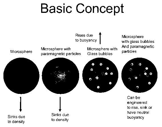
FIG. 1 is a representation of a conventional microsphere, a microsphere with
so paramagnetic particles, a microsphere with buoyant elements and
paramagnetic
particles.
-3-
CA 02532754 2006-O1-16
WO 2005/010162 PCT/US2004/023222
FIG. 2 is a diagram of a representative bioreactor of the present invention
containing
engineered microcarriers of the present invention showing the relationship of
the
source of applied physical force to the culture vessel and an opening for the
addition
and removal of media and/or microcarriers.
s FIG. 3 is a diagram of a representative automated bioreactor of the present
invention
containing engineered microcarriers of the present invention which is
controlled by a
control system that also controls the source of the physical force, the
addition or
removal of the media and microcarriers from the culture vessel.through an
opening
and the monitoring system.
~o FIG. 4 is a diagram of a representative automated bioreactor of the present
invention
containing engineered microcarriers and further showing the relationship to a
microcarrier manufacturing method from which microcarriers are provided
directly
into the culture vessel and its relationship to an assay method which receives
microcarriers from the culture vessel for analysis. The control system
controls the
~s automated culture vessel system in the boxed area as well as the
microcarrier
manufacturing method and the assay method.
FIG. 5 is a representation of one embodiment of the present invention that
utilizes a
single magnet. The figure shows how this magnet is used to move the
microcarriers
within the bioreactors. Two bioreactors comprising a culture vessel and a
source of a
2o physical force, a single electro- or permanent magnet for each culture
vessel are
shown. In the left figure, the magnets represented by the dark disc are moved
down to
the bottom of the culture vessel to pull the microcarriers represented by
small circles
to pull off waste media through the opening on the right side of the culture
vessel. In
the right, the magnets are moved down to the top of the culture vessel to pull
the
is microcarriers representated by small circles to pull off microcarners from
the culture
vessel.
FIG. 6 is a representation of an embodiment of the present invention when a
series of
electromagnetic coils or magnets are used to encircle a culture vessel. This
representation shows that microcarriers can be moved according to their cell
growth
so needs and to facilitate media changing and microcarrier aspiration. The top
coil is
energized to move microcarriers up for aspiration manually or by a robot arm.
All
coils can be energized to keep the microcarners in suspension. The bottom coil
is
-4-
CA 02532754 2006-O1-16
WO 2005/010162 PCT/US2004/023222
energized to move microcarriers to the bottom for removal of waste media and
addition of fresh media.
FIG. 7 is a representation of media change in the left figure and microcarrier
aspiration in the right figure. This figure shows a similar use of the magnets
as FIG. 5
s but with a plurality of the magnets as in FIG. 6.
FIG. 8 is a representation of an alternative magnet arrangement that will
allow
microcarriers to be manipulated according to specific needs. As in FIG. 6, the
top
magnet coil moves the microcarriers up for manual or robotic aspiration of
microcarriers, the bottom magnet coil moves the microcarriers down for manual
or
~o robotic aspiration of used or waste media and all of the coils are
oscillated to keep the
microcarriers in suspension.
FIG. 9 is a further representation of magnetic fields to provide a variety of
microcarrier movements. This figure demonstrates the use of two circular
electromagnets with two poles or multiple poles to effect diagonal movement
through
15 the culture vessel.
FIG. 10 is a representation of a further alternative arrangement of magnets
allowing
more circular and top to bottom mixing of the microcarriers.
FIG. 11 shows a representation of an engineered microcarrier that is
manipulated by
external magnetic fields to induce kinetic energy. The microcarrier is rotated
on its
2o axis to induce shear stress on cells growing on the exterior of the sphere
and cells
around the perimeter are expected to be exposed to greater shear stresses as
compared
to those near the axis as approximated in the Shear Force Profile to the right
of the
sphere.
zs DETAILED DESCRIPTION OF THE PREFERRED EMBODIMENTS
The present invention discloses microcarriers that have been modified from
the conventional microcarners to also contain additives that provide specific
properties that result in the manipulation and physical movement of the
microcarriers
in relation to other microcarriers or simply movement within the culture
vessel. The
so present invention further discloses microcarriers in which the additives
are ligands,
reporters or response elements that report a stimulus or respond to a
stimulus.
The present invention further discloses engineered microcarriers that are made
with a wide variety of substances with virtually unlimited properties. For
example,
-5-
CA 02532754 2006-O1-16
WO 2005/010162 PCT/US2004/023222
such engineered microcarners include but are not limited to gelatin,
polyacrylamide-
copolymerized with collagen or gelatin, polyacrylamide with modified charge,
alginate, and alginate copolymerized with gelatin. A preferred microcarrier is
one
made with a chemical format, such as calcium-alginate and gelatin, as
disclosed in
s Kwon et al. (7). But these conventional microcarriers are then modified to
produce
functionalized microcarriers that act as reporters. We also describe
improvements
over chemical microcarriers that are engineered microcarriers possessing
specific
properties, such as specific buoyant and magnetic and/or paramagnetic
properties, as
descried herein. Thus, the functionalized and/or engineered microcarriers of
the
~o present invention comprise properties of known microcarriers in that they
are
produced from chemical compounds and compositions using known methods and
materials but these conventional microcarriers are further engineered or
modified to
contain or comprise additives that provide these advantageous properties, such
as
particles, molecules and/or gases, introduced into the microcarrier (See FIG.
1) or
~ s alternatively attached to the outside of the microcarrier that impart
changes in density
and/or allow the engineered microcarrier to be moved, steered, agitated or
otherwise
manipulated around the inside of a culture vessel or bioreactor by at least
one applied
physical force that imparts kinetic energy to the engineered microcarriers
inside the
culture vessel.
2o The calcium alginate and gelatin microcarriers are particularly useful for
monitoring cell function since the resulting engineered microcarriers made
from these
compositions have minimal endogenous fluorescence allowing the cells to be
observed using microscopic techniques, such as fluorescence confocal
microscopy. A
preferred embodiment is the creation of microcarriers that are optically clear
when
is compared to other microcarriers that are available. Preferred microcarriers
disclosed
in the present invention retain a large proportion of their optical clarity,
and
functionally do not interfere with observations or quantitative measurements
that are
carried out on the cells inside or outside of the engineered microcarriers,
even when
engineered with additives as described in more detail herein.
so The microcarriers of the present invention provide for an increase in
cellular
density, for example, cancer cells grow to a density of up to 7x10⁵ cells for
every
5x10³ microcarriers. However, when cells have grown to a sufficient density,
as
determined by reporter molecules integral to the microcarriers indicate the
degree of
-6-
CA 02532754 2006-O1-16
WO 2005/010162 PCT/US2004/023222
confluence, or the use of an external monitoring system, they are used
directly for
studies since they contain reporter molecules without the need for the
relatively
disruptive process of releasing them from attachment using enzymes such as
trypsin
(as is necessary in flat bottom vessels).
The present microcarriers convey the benefit of flexibility in that they can
be
created with unlimited geometric, chemical, and functional properties (8, 9).
In some embodiments of the present invention, microcarriers may be made as
spheres
of any diameter, but more reasonably in a range from 1 mm down to less than
the
diameter of the cell of interest. Microcarriers of the present invention
preferably
~ o range in size from 1 pm up to 1 mm in diameter. However, smaller sizes
down to less
than one nanometer and larger sizes up to 2 centimeters or more are
microcarriers that
are useful in the disclosed automated cell culture system and are produced by
the
disclosed techniques. Microcarriers useful in the present invention may also
be
chemically modified to allow non-adherent cells to attach to their surface.
The
15 present invention in some embodiments employs a technology that allows non-
adherent cells to attach (10), but allows such attachment to the further
engineered
microcarriers possessing specific reporting, buoyant and magnetic and/or
paramagnetic properties as descried herein.
The engineered microcarriers of the present invention are useful generally to
2o facilitate harvesting operations; however, the tendency for microcarriers
to settle out
of suspension does not allow them to be easily harvested by automation.
Microcarrier
products have been on the market for several decades, but interest in their
use to
support the high throughput screening process in the pharmaceutical industry
has been
stymied by their difficulty to manipulate and the expensive and complicated
impeller
2s systems or growth vessel rotation systems needed to use them. The use of
engineered
microcarriers of the present invention in an automated cell culture system and
monitoring system as disclosed herein for high throughput screening provides
advantages over previously used high throughput screening systems.
In another embodiment, the present invention discloses manufacturing or
so producing microcarners using conventional techniques, including spraying
into a
liquid containing a polymerizing chemical mixture, or by adding the
microcarrier
matrix to a rapidly stirring oil bath in order to create an emulsion. In
another
embodiment of the present invention, the manufacturing of the engineered
CA 02532754 2006-O1-16
WO 2005/010162 PCT/US2004/023222
microcarriers may optionally be an integrated process within the cell culture
automation platform (see Fig.4). To date, a cell culture system does not exist
that
manufactures the needed engineered microcarriers as they are needed for use in
the
automated cell culture system as disclosed herein. This approach provides a
"just in
time" cell culture production process.
Engineered Microcarriers
Magnetism
The novel engineered microcarriers of the present invention comprise
~o incorporated buoyant elements that effect the density of the engineered
microcarriers
and/or particles that impart magnetic and/or paramagnetic properties to the
engineered
microcarriers. The use and incorporation of miniature magnetic or paramagnetic
particles allows the control of the particles by external magnetic fields. The
choice of
magnetic and/or paramagnetic particles allows one to reduce the size or
orientation of
~s the external magnetic field necessary to impart selected movement or
kinetic energy
on the engineered microcarriers. The benefit of incorporating paramagnetic
particles
in the engineered microcarrier is their lack of inherent magnetism when they
are not
being exposed to an external magnetic field, which would then prevent their
attraction
to each other. In some embodiments, microcarrier aggregation is desirable when
Zo creating useful aggregates of cells, such as building tissues or organs.
Thus,
microcarriers may be induced to aggregate in specific orientations and numbers
by
any combination of internal magnetic or ferromagnetic properties coupled with
any
arrangement of external magnets (either permanent or electromagnet). In other
embodiments aggregation is undesirable, such as in high throughput screening
for
25 novel pharmaceuticals where discrete microcarriers may yield higher
screening
signals. In further embodiments, a combination of paramagnetic and magnetic
material is desirable to impart properties that allow variable response to an
external
magnetic field.
Magnetic properties of the engineered microcarriers may be controlled during
so the manufacturing of these microcarriers, or imparted after the
manufacturing process.
If microcarriers are polymerized or gelled in the absence of a magnetic field,
then the
magnetic or paramagnetic particles will have a random orientation on or within
the
engineered microcarrier. On the other hand, if a magnetic field is applied in
a static or
_g_
CA 02532754 2006-O1-16
WO 2005/010162 PCT/US2004/023222
varying way during the manufacturing process, then one can impart a specific
orientation and magnetic strength (if the particles can be magnetized) to the
particles
on or within the microcarrier. For example, but not meant to limit the present
invention, one might wish to impart a magnetic dipole to each microcarrier so
that
they may be rotated on their axis as the result of exposing the microcarriers
in a liquid
to an external magnetic field. If the user does not desire microcarner
aggregation as a
result of cells growing on their surface sticking to each other, then
imparting an axial
rotation would tend to prevent inter-microcarrier aggregation.
~o Buoyancy
The buoyancy of the microcarriers is controlled by either manufacturing them
out of materials with buoyant properties, or by adding a substance or
substances
which can control buoyancy. Buoyancy is defined herein as the property that
will
make the microcarriers spontaneously move in a direction opposite to gravity
in the
~ s liquid in which they are suspended. In one of many possible embodiments,
the
manufactured microcarriers are doped with both paramagnetic particles and
glass
bubbles exhibiting net positive buoyancy. These substances impart physical
properties to the microcarriers previously unknown in cell culture.
Furthermore, the
density of the microcarriers may be controlled by using various combinations
of
zo ingredients, some with buoyant properties, such that that the density of
the carrier for
cell culture is within the range of 0.8 to 1.4 g/cm, which allows suspension
of the
microcarriers in a culture medium.
Manufacturing
z5 Microcarriers can be manufactured using a plurality of methods, including
but
not limited to spraying, sonicating, suspending, vibrating, or emulsifying the
liquid
containing the raw materials from which the microcarriers are polymerized, in
suspension, and in oil water emulsions. Imparting the engineered properties
described
in this patent, such as the ability to control buoyancy, is accomplished by
adding
so selected material to the microcarrier raw materials, such as glass bubbles,
so that they
distribute themselves in the microcarrier according to the needs of the user.
Alternatively, the material that imparts selected properties to the
microcarrier may be
added after the microcarrier has been manufactured.
-9-
CA 02532754 2006-O1-16
WO 2005/010162 PCT/US2004/023222
Special proteins can be incorporated into the matrix of the microcarrier or in
the surface coating of the microcarner that will promote or enhance cell
adhesion,
growth, differentiation, or promote expression of a selected phenotype
including
morphological changes as well as the expression of biochemicals. For example,
(but
s not limited to) microcarriers into which extracellular matrix proteins have
been
incorporated, such as collagens, fibronectins, peptides, and other proteins
and
biochemicals that may have been used to induce a variety of cellular behaviors
(including those mentioned above). Alternatively, non-specific adhesion and
cellular
behaviors have been inhibited through the use of polymers, biochemicals, and
other
~o substances (10-14). Gelatin has been used to promote cell adhesion to
planar glass
slides (15). Prior art teaches a low density collagen coated microcarrier
method for
culturing, harvesting, and using anchorage dependent cells (16). However, the
present invention discloses the creation of microcarriers that can be
automatically
manipulated by non-impeller based methods with engineered buoyancy to match
the
~ s needs of the cells to be cultured. Furthermore, the engineered
microcarriers of the
present invention can be used directly in applications that call for living
cells, which
is different from what is taught by Hillegas (16), who describes insoluble
microcarriers that are not optically clear. The Hillegas proposal does not
teach the
use of any specific cell or cells and the inherent advantages of their
invention for
zo supporting growth of particular cells. The present invention improves upon
the
teaching of Koichi (10) that allows non-adherent cells to attach to glass
slides for
microarrays. The engineered microcarriers of the present invention improves
upon
the method of Koichi in that they are suspendable, engineered with additives,
and
participate in a cell culture process which takes advantage of their ability
to be
is manipulated in suspension. The advantage of these anchors is that they
allow a
plurality of non-adherent cells, such as blood cells, immunocytes (cells of
the
lymphoid series which can react with antigen to produce antibody or to become
active
in cell-mediated immunity or delayed hypersensitivity reactions; also referred
to a
immunologically competent cells), some cancer cells, stem cells, single cell
so organisms, and other cells to anchor to a variety of substrates (10). In
the case of the
present engineered microcarners, in some embodiments, the biocompatible anchor
material is incorporated in the matrix of the microcarrier, or on a surface
layer coated
onto the microcarrier according to procedures described herein. In one
embodiment,
-10-
CA 02532754 2006-O1-16
WO 2005/010162 PCT/US2004/023222
an anchor promoting material is oleyl polyethylene glycol) ether (10).
Including this
anchor promoting material with the engineered microcarrier that can be
manipulated
in suspension, one has a powerful cell culture technique that will work with
virtually
all anchorage dependent and independent cells.
Alternatively, cells can be held inside an engineered microcarrier in a
microenvironment that allows for cell differentiation or growth, or maintains
the cell
in a steady state non-growth phase. The engineered microcarriers could then be
used
to deliver the cell to a selected location, such as transplantation in a
living being,
where the cells would then be allowed to attach, differentiate into other
clonal cell
lines, or expand to fill a space or need. A breakable or enzymatically
digestible
biocompatible microcarrier may be used allowing the cells to be delivered to a
site of
interest and then the bubble would be digested, broken, collapsed or dissolved
by a
variety of means. An example of this latter method, one could break the
bubbles by
delivering ultrasound energy to the same location as the bubbles either in-
vitro or in-
VlVO.
More specifically, microcarriers may be manufactured from a number of
substances including two major classes of material, namely thermoplastic
polymers,
hydrogel polymers. Thermoplastics are any water soluble substances including
but
not limited to polyacrylates or polyethylene glycol. It is advantageous to use
more
2o gentle, and thus less harmful, manufacturing conditions for cells that are
encapsulated
in hydrogel substances, such as, but not limited to agarose and/or alginate.
As a
specific but not limiting example, alginate is an intracellular matrix
polysaccharide
extracted from brown algae and some bacteria. In order to improve the
viability of
cells on our unique engineered microcarriers we selected alginate from sources
that do
z5 not contain endotoxins. Even those skilled in the art of cell culture on
alginate
microcarriers would benefit from our teaching the use of low or no endotoxin
containing alginate in order to improve cell attachment, health, growth, and
viability
on the microcarriers. Alginate is obtained from either Sigma (St. Louis, MO)
or
Pronova Biomedical (Oslo, Norway) and mixed in an aqueous solution using
so endotoxin free water in a range from 0.1 % by weight sodium alginate to 10%
sodium
alginate. However, the microcarriers are easier to manufacture due to the
viscosity of
the solution when the alginate concentration was 0.8% to 1.2 %. Endotoxin is
measured using a Limulus-lysate assay kit from Sigma and only alginate
solutions
-11-
CA 02532754 2006-O1-16
WO 2005/010162 PCT/US2004/023222
with values of less than 5000 endotoxin units/mL were used for manufacturing
microcarriers with cells on their exterior. The ideal range for culturing
cells was an
endotoxin level of less than 500 endotoxin units/mL. The alginate solution is
mixed
with a 2-4% by weight propylene glycol alginate (PGA) solution (Kelcoloid TM
D; ISP
s Alginate, San Diego, CA) in order to crosslink the alginate [Kwon et al.
(7)]. This
optional procedure can be performed with concentrations of PGA from 0% to 10%
by
weight. In addition, alginate is created by its living source with regions of
mannuronic acid or guluronic acid, or a mixture of both mannuronic and
guluronic
acids. Sources with ratios of these substances that optimize cell health,
growth, and
~o viability are selected. Ratios of mannuronic acid to guluronic acid are
determined by
emission at 445 nm according to the method of Klock (17). The preferred ratios
for
crosslinking with calcium is 90% or greater mannuronic acid although cell
growth is
observed at any ratio of mannuronic acid to guluronic acid. Additional
additives
include glass bubbles (3M Corporation, Maplewood, MN), protein bubbles, or air
~ s bubbles up to 20% by volume. However, we fmd that glass bubble 1 % - 5%
allows
for microcarriers with ideal densities and that are affordably manufactured.
Air (or
other inert gas such as helium) bubbles can be incorporated into the solution
by
vigorously shaking the container to trap air in the form of small non-uniform
bubbles
or by using bubbler (compressed air or gas is pumped into a scintered metal
device
2o that divides the gas into uniform bubbles). Paramagnetic or ferromagnetic
particles
(Spherotech, Libertyville, IL) can also be included in the solution at this
point in order
to impart paramagnetic or ferromagnetic properties to the resulting
microcarriers. Up
to 75% of the internal volume of the microcarrier can be filled with para or
ferromagnetic particles, however, the idea ratio is from one to 1000 particles
per 250
2s um microcarrier. Indicators, such as fluorescent molecules described
elsewhere in
this application can be added, if desired, at this point.
Once the contents of the microcarrier solution has been determined, based on
the specific properties of the resulting microcarrier desired, the
microcarriers are
formed by a variety of methods including adding the alginate/PGA additive
solution
so drop wise to a gently agitated 1.5% (0.135M) calcium chloride solution.
Commercial
micro droplet generators may also be used. Living cells may be added to the
mixture
before the microcarners are created to allow interior cell encapsulated
culture or the
co-culture of cells both inside and outside of the microcarrier using either
similar or
-12-
CA 02532754 2006-O1-16
WO 2005/010162 PCT/US2004/023222
dissimilar cells. The alginate solution is adjusted with a physiologic buffer
if living
cells are intended to be encapsulated. Microcarriers are then used for cell
culture after
washing. Microcarriers can also be coated with gelatin by adding a 1 % gelatin
solution (for example unflavored Knox gelatin from the local grocery store) to
any
s volume of a microcarrier suspension, gently mixing, and then washing the
beads by
repeated changes of fresh buffer. High concentrations of gelatin add greater
rigidity,
thus we have used up to 10% gelatin, but 0.5% to 3% is ideal for cell
attachment.
Additives are placed in the gelatin solution including molecules that enhance
cell
attachment, molecules that can transform cells (DNA, RNA), and indicators as
~ o described elsewhere in this application. The Gelatin can be crosslinked to
give
microcarriers with greater rigidity by transacylating to the alginate by
adding two
volumes of 0.2M NaOH as described by Kwon et al. (7). Various molecules are
incorporated to increase or decrease the microcarrier charge and/or porosity,
such as
but not limited to poly-L-lysine (a cationic amino acid polymer)( 18). The
present
invention discloses the incorporation of substances that control microcarrier
response
to physical forces, which improves upon the use of substances that control
microcarrier permeability, porosity, and strength.
The alginate guluronic molecules and hence the microcarriers are held
together and develop rigidity and hence strength through the addition of
bivalent
2o cations such as calcium (Caz+). Both the guluronic and mannuronic acids are
bound
together using Barium (Ba2+), so the incorporation of Ba2+ is important to
achieve
stronger microcarner properties according to Strand. (19). As apposed to the
art
taught by Strand to use selected cations to increase microcarner rigidity, we
teach the
novel art of using Ba2+ as a bivalent molecular bond in order to avoid the use
of Caz+
is in biochemical assays examining calcium flux and concentrations because
changing
calcium concentrations would both disturb the measurement and would alter the
integrity of the microcarrier.
Microcarriers are manufactured using a variety of methods including the use
of an electromagnetic or piezoelectric driven nozzle equipped to allow laminar-
jet-
so breakup of the alginate solution and additive suspension. The use of a
commercially
available encapsulation system is desirable to allow control of the physical
parameters
affecting microcarrier size (e.g. flow rate, vibration frequency and
amplitude).
Alternatively, the alginate solution is added to a rapidly stirring emulsion
of oil and
-13-
CA 02532754 2006-O1-16
WO 2005/010162 PCT/US2004/023222
buffer containing bivalent cat-ions or cross linking agents. The microcarners
spontaneously form their micro-spherical shape in the emulsion and then
precipitate
out of the oil when the stirring is slowed or stopped if they have a density
greater than
that of the emulsion buffer. If the microcarriers are made with a buoyant
density, then
they can be removed from the emulsion by centrifugation and aspiration from
the
surface, or by pulling them to the side of the vessel if they contain
paramagnetic
particles.
Using engineered properties to facilitate using microcarriers
~ o Growing cells on standard microcarriers that are suspended in the growth
medium allows greater access to nutrients, air, or oxygen, and carbon dioxide
and
random orientation with respect to gravity, yet increases potential damage due
to
uncontrollable shear stress. An added benefit of the engineered microcarriers
of the
present invention is that they provide gentle growth conditions without the
stresses or
~ 5 inconveniences imparted by stirred cultures. The present engineered
microcarriers
allow specific orientation of each microcarrier to be externally controlled.
Additionally, the engineered microcarrier can be easily and quickly harvested
for
subsequent procedures. The amount of microcarriers that may be used to grow
cells
is limited only by the amount of culture media in the vessel. Thus, the use of
the
zo disclosed engineered microcarrier allows for culture scalability from use
in a
microscale culture in microfabricated technologies (20) to culture systems in
excess
of one liter, for example 500 Liters.
Engineered microcarriers have been successfully created that were
approximately 5 um which became partially engulfed by one Cinese Hamster Ovary
2s cell during its growth. This technique will allow cells to remain on their
anchorage
surface while being translocated in a microchannel fluid stream or immobilized
either
temporarily or permanently on a flat surface, three-dimensional surface, or
array.
Furthermore, the present invention discloses the use of microscale
components, such as micromagnets, micro-pressure systems, and micro-detectors
to
so perform many of the same procedures described in this patent application,
only on the
microscale. An important advantage of the present invention is the ability to
steer
cells within microchannel arrays using pressure or magnetism, to which the
engineered microcarriers will respond. The present invention comprising a
-14-
CA 02532754 2006-O1-16
WO 2005/010162 PCT/US2004/023222
suspension culture of engineered microcarriers increases the productivity one
can
expect, of conservatively 400 fold over flat bottom dishes, and over two fold
compared to spinner flasks.
Once cells have reached a desired level of confluence, for example 80%
s coverage of the microcarrier, it is often necessary to remove the cells from
the
microcarriers in order to use them for analysis. The most popular method of
removing viable cells from their anchor surface is through the use of a
proteolytic
enzyme (trypsin) that digests some of the proteins used by the cell to anchor
them on
the microcarrier. Not only does trypsinization strip the cell of many
important cell
~ o surface proteins and it causes a temporary shock to the cells, often
resulting in a low
yield of cells that are released intact from the microcarrier. Also cells may
need a
mechanical shock (such as the energy imparted by rapid deceleration of
microcarriers
in solution) in order to be released from the microcarner. The more dimpled
the
surface of the microcarrier, the less likely those cells will release from the
surface or
~s be sheared off. The specific art of releasing cells from microcarriers was
addressed
by Mundt (21 ), who taught the use of trypsin to release the cells from the
microcarriers. The present invention does not have these problems as the
present
microcarriers are engineered to dissolve spontaneously, as described by Kwon
(7),
thus obviating the challenges associated with using non-specific enzymes to
release
2o cells from their anchorage surface. Thus, the present invention is intended
to
encompass the use of spontaneously dissolving engineered microcarriers that
work in
concert with automation to obviate the need to perform these tasks manually.
Furthermore, the ability to dissolve the microcarriers within a specific time
point and
location within an automated process has not previously been described. For
25 example, engineered microcarriers can be dissociated either partially or
fully during
their transition in a fluid stream prior to analysis in a cell sorter or
fluorescence
activated cell scanner. Our engineered microcarriers may be more quickly
dissociated
by making use of the external control of the properties of the microcarrier.
For
example (but not limited to), increased internal kinetic energy imparted by
rapidly
so moving magnetic or paramagnetic particles as the result of an externally
applied
oscillating magnetic field can quickly dissociate the polymerized alginate
when
calcium is reduced below the polymerization threshold in solution. By
dissolving
microcarners containing paramagnetic particles and/or glass bubbles, one can
retrieve
-15-
CA 02532754 2006-O1-16
WO 2005/010162 PCT/US2004/023222
these substances for either reuse, or to prevent them from contaminating or
adversely
affecting downstream processes.
Another benefit of the engineered microcarriers is that they may be used in
conventional cell culture facilities employing conventional disposable dishes,
pipettes,
culture media, incubators, cell dispensing equipment, rockers, agitators,
plate sealers,
and analytical instruments.
Imparting movement in the growth media
Agitation of microcarriers has been traditionally performed through the use of
~o impellers (see above). But there are many benefits of stirring growth media
without
the use of impellers. The present invention provides alternative approaches to
imparting kinetic energy to growth medium containing living cells grown
through the
use of the engineered microcarriers. Imparting kinetic energy to the growth
medium
assures even distribution of nutrients, assures good gas exchange to all
cells, and
~s prevents clumping of engineered microcarriers. The present invention
provides a
number of methods to impart kinetic energy to the growth media that may be
used
individually or in any combination. For example, the kinetic energy may be in
the
form of using a heat source that induces a thermal gradient in the growth
medium.
The thermal gradient imparts motion in the growth medium as less dense heated
2o media rises in the culture vessel, the more dense cooler media tends to
sink in the
culture vessel and hence imparts movement of the engineered microcarriers. The
thermal gradient is sufficient to induce kinetic energy, but not cause harm to
the
growing cells which generally can tolerate 33°F to 105°F unless
they are
cryoprotected or thermally stabilized, respectively. A temperature
differential from
zs ambient of one degree to greater than 40°F above ambient may be used
to induce
convection currents. The thermal gradient can be controlled by a servo
controller so
that it actually serves as the heat source to warm the culture medium to
temperatures
that impart optimal cell growth.
Pressure can also be applied to the microcarriers can achieve two goals. The
first goal
so is to subject the cells growing in or on the microcarrier to a pressure
profile similar to
that felt by cells growing in living beings. Thus pressure pulses, or
differential
pressures over time, can be applied to the microcarriers at rates found in
nature such
as 5 beats per minute up to 500 beats per minute. The second goal of the
applied
-16-
CA 02532754 2006-O1-16
WO 2005/010162 PCT/US2004/023222
pressure force is to compress gas bubbles incorporated into the microcarrier
to
enhance buoyancy. By compressing the entire container in which the gas bubble
containing microcarriers are held, one can increase the density of said
microcarriers
thus causing them to sink under applied pressure, and rise under reduced
pressure.
Pressure from ambient barometric pressure up to many atmospheres may be used.
Mechanical modulating microcarrier buoyancy
Another embodiment of the present invention exploits particle buoyancy to
increase the kinetic energy in the culture vessel or bioreactor. For example,
particles
~o are introduced that are either composed of compressible gas bubbles, or
contain
compressible gas bubbles. A variety of natural or man-made elastic materials
may be
used to trap a gas bubble(s). Since gas is more compressible than liquids,
compression of the gas by the use of an externally generated energy source,
either
thermal or pressure will impart varying buoyancy to the particles. Particles
exhibiting
~ s variable buoyancy may be exploited to either stir the growth medium
containing
microcarriers supporting cell growth or maintenance, or compressible bubbles
may be
introduced into or on the microcarriers containing cells.
Modulating an external magnetic field
2o Magnetic fields may be used to induce kinetic energy into a fluid, such as
cell
culture medium. A large magnetic flux induces movement at the microscopic, and
ultimately, at the macroscopic level in any liquid. Alternatively, in order to
limit the
amount of magnetic field that has to be generated, in one embodiment, the
present
invention discloses the introduction of ferromagnetic or paramagnetic
particles into
25 (or on) the microcarrier whose motion can be induced by an externally
generated
magnetic field. Ferromagnetic particles constitutively exhibit a magnetic
field,
whereas paramagnetic particles only exhibit a magnetic field while being
exposed to a
magnetic field. The motion of the particles induces motion in the liquid, and
hence
maintains the suspension of microcarriers supporting the growth of cells. The
so paramagnetic particles may be attached to the surface or placed inside the
microcarrier that are supporting the growth or maintenance of cells which are
grown
either inside or on the surface of the microcarrier to produce an example of
an
engineered microcarrier within the meaning of the present invention.
Ferromagnetic
-17-
CA 02532754 2006-O1-16
WO 2005/010162 PCT/US2004/023222
material, or any material that responds to a magnetic field, can be placed in
or on the
microcarrier by adding formed particles or precipitating the material from
solution
during the microcarrier manufacturing process, or introducing it as a coating
once the
microcarner is manufactured. Known magnetic materials include, but are not
limited
s to chromium, iron, nickel, and cobalt and their oxides and derivatives.
These
materials can be added from 0-75% by weight in finely divided nanoparticles so
as to
provide less interference with optical properties, or as a large core so that
the optical
properties of the perimeter of the microcarrier is preserved.
The magnetic field may be modulated by either use of permanent magnets, or
~ o electromagnets placed above, below, or on the side of the cell culture
vessel. The
placement of the magnet will depend on the movement desired from the
microcarriers. The magnetic fields may be continuously applied when a specific
microcarrier orientation within the vessel is desired, such as (but not
limited to)
bringing the engineered microcarriers to the bottom of the vessel to allow the
media to
15 be aspirated, or bringing the microcarriers to the surface of the media so
that they may
be harvested. (See FIGS. 5 and 7) Magnetic fields may be applied with
different
temporal or strength profiles. For example (but not limited to), pulsing the
magnetic
field is useful for maintaining the microcarriers in suspension (See FIGS. 5-
10), yet
limit the amount of heat generated by an electromagnet, or the amount of
mechanical
2o movement of a permanent magnet. Hybrid magnetic fields may be applied, such
that
a field with deep penetrating strength may impart selected movement or
orientation of
the microcarrier, while at the same time a stronger field with less
penetrating strength
may be used to hold microcarriers in a selected orientation.
FIG. 12 shows one embodiment in which the verticle bars represent bar
2s magnets arranged in a circular fashion around the perimeter of the vessel.
By
computer-controlled activation of the magnets and alteration of their
polarity, many
different microcarrier paths can be effected for stirring, media changing,
cell adhesion
operations, or cell harvesting.
A combination of the above techniques can be used to optimize the growth or
so maintenance of cells, depending on the cell type and growth conditions
required. For
example, in one embodiment, the present invention utilized both paramagnetic
particles and bubbles introduced into the same microcarrier simultaneously to
obtain
an engineered microcarrier with a blend of properties that both the
paramagnetic
-I g-
CA 02532754 2006-O1-16
WO 2005/010162 PCT/US2004/023222
particles and bubbles impart to the microcarrier. This combination of
paragmagnetic
particles and bubbles imparts the ability to control buoyancy as well as the
ability to
use a magnetic field to stir and direct the movement and/or orientation of the
magnetic
particles in the bioreactor. Thus, engineered microcarriers may be
manufactured to
s match the specific needs of each cell type, depending on the needs to
control kinetic
energy, density, response to the externally applied magnetic field, and
orientation.
For example, an external magnetic field could be applied to the culture media
containing cells and the engineered microcarriers so that an engineered
microcarrier
with buoyant properties would be attracted to the bottom of the culture vessel
to allow
1o initial cell attachment. The magnetic field could be then removed to allow
buoyant
engineered microcarriers with growing cells attached to rise into the growth
media.
Use of populated engineered microcarriers directly in a High Throughput
System (HTS)
1 s Once engineered microcarriers of the present invention have been populated
with cells, they may be used directly in biochemical or physiologic
procedures. The
ability to maneuver the engineered microcarriers to a specific location as a
result of
their inherent properties or through the use of automation allows them to be
exploited
for use in subsequent research or development procedures. For example, the use
of
2o their buoyancy and/or heat convection to cause the engineered microcarriers
to move
to the upper portion of the cell culture vessel or bioreactor will make them
available
to an automated pipette, or other means of harvesting cells. Alternatively,
the
engineered microcarriers can be gathered at the top of the bioreactor by an
externally
applied magnetic field or induction of lower density for aspiration by a
pipette.
25 Another embodiment of the invention is to maneuver the microcarriers to a
liquid port
in the bioreactor so that they are concentrated and pumped out in the fluid
stream to
be used in a subsequent procedure.
Functionalized microcarriers
3o Biochemical procedures or analyses on cultured cells grown in cell culture
laboratories have a variety of uses, including research, product development,
and drug
discovery. Normally, cells must be digested or otherwise dissociated and
fractionated
so that one can study individual organelles of the cells, or biomolecules
produced by
-19-
1233176.1
CA 02532754 2006-O1-16
WO 2005/010162 PCT/US2004/023222
the cells. Recently, "high content" discovery or screening has resulted from
the
ability to study whole cells. Novel cell culture plates have been developed to
allow
cells adherent to a surface to be examined by intracellular fluorescent
reporting
molecules. However, cells grown on flat dishes do not have similar phenotypes
or
s behaviors as compared to cells in situ. In contrast, cells grown and
maintained on
microcarriers have been shown to be polarized, demonstrate phenotypes more
equivalent to their in situ counterparts, and produce larger quantities of
cell products.
Cell sorters or fluorescence activated cell sorting (FACS) instruments have
been
developed to study cells in suspension. Suspended cells are moved in a narrow
~ o stream of fluid in front of an optically based detector in order to
quantitate size,
fluorescence, and/or electrical properties. Unfortunately, when anchorage
dependent
cells are placed in an environment where they are not anchored, they often
exhibit
negative properties. The presently manufactured engineered microcarriers are
small
enough so that individual cells are supported by the microcarrier matrix.
Thus, the
~ s engineered microcarriers of the present invention are useful for cell
counting and
sorting instrumentation. The paramagnetic and buoyant properties of the
engineered
microcarners are also useful as a means to separate cells from their liquid
environment, to sort cells, or to measure responses to stimuli.
An enhancement to conventional non-engineered microcarriers or to the
2o engineered microcarriers described herein is to functionalize the
microcarriers so that
they contain ligands and or binding molecules that report a stimulus and/or
respond to
a stimulus. For example, microcarriers containing a contractile protein may be
induced to contract and change its buoyancy in response to a stimulus from the
cell, or
the cell culture bioreactor controller. Ligands, reporters, or response
elements may be
2s covalently or non-covalently linked to the surface and/or interior of the
microcarrier.
Reporters may consist of micro (or nano) electronic or micro (or nano)
mechanical
elements that are incorporated in or on the microcarrier which report via
electromagnetic methods, for example but not limited to wireless motes.
Ligands may
be used to cause a reaction from the cells grown on any aspect (outside,
inside, or
so both) of the microcarriers. Microcarriers may be functionalized with
reporters so that
they report changes to their environment as a result of changes in the culture
media or
changes resulting from materials exported or secreted from the cells. In one
embodiment, reporters can signal the presence of and progress of a reaction,
or a
-20-
CA 02532754 2006-O1-16
WO 2005/010162 PCT/US2004/023222
response to a stimulus. A large number of light emitting reporters are
available in the
form of fluorescent and bioluminescent molecules. The choice of reporters will
vary
according to what is to be measured. For example, in one embodiment, a sodium
sensitive reporter for sodium can be placed inside the cell to report
intracellular
s sodium. Similarly, a sodium sensitive dye can be incorporated into the
microcarrier
so that sodium pumped by the cell to its anchorage surface on the surface of
or into
the microcarrier would be reported. The reporter may be organic, inorganic,
and
single or multiple molecules, linked directly to/in the microcarrier, or
linked to a
functional group which was first linked to/in the microcarrier. Our process of
~ o functionalizing microcarriers to report or respond differs from the prior
art described
(15) in that our microcarriers are designed to support living cells which
release
molecules of interest. Furthermore, the present invention discloses the use of
molecules that respond and alter the microcarrier environment, such as in one
embodiment, contractile elements.
15 Engineered microcarriers may be used for a plurality of assays that are of
interest to pharmaceutical companies and basic researchers. For example, there
is
great interest in determining the ability of cancer cells to metastasize, and
to
determine the mechanisms cells use to bind, penetrate, and move into foreign
tissue.
Cell migration and/or metastasis assays are useful to find or refine new
anticancer
2o agents, or examine how arteries form in developing tissues. The engineered
microcarriers disclosed herein are designed to measure cell migration or
invasion
based on biochemical assays. In one embodiment, cancer cell division into the
microcarrier may be monitored by measuring cell number or a signal emitted as
a
result of cell division. For example, in this embodiment, reporter molecules
sensitive
25 to cell surface proteins can be polymerized into the core of the
microcarrier. As cells,
growing on the surface of the microcarrier penetrate toward the core an
increase or
decrease in the fluorescence signaling molecule is measured. Thus, signal
magnitude
is correlated with the ability and avidity of cells to migrate or invade. In
another
embodiment, the microcarrier is coated with a substance that resembles
basement
so membrane or other biological barriers that may be invaded by cells growing
on the
microcarrier. Cells are co-cultured on the surface and/or interior of the
microcarrier
so that invasion is measured from an outer layer of cells toward the center of
the
microcarrier, or cells are observed and measured invading outward, away from
the
-21-
CA 02532754 2006-O1-16
WO 2005/010162 PCT/US2004/023222
core of the microcarrier. Alternatively, the microcarrier containing the
potentially
invading or migrating cells are attracted toward other cells growing on
another
microcarrier (using a magnetic field or buoyancy) to observe and measure
invasion or
migration from one microcarrier to another. Microcarriers additionally may be
s attracted toward cells growing on a conventional anchorage dependent
surface, for
example, in a further embodiment, the surface of a conventional culture flask,
using
gravity,~buoyancy, thermal gradients and/or magnetism. Once they have come
within
a specified distance, then cell migration or invasion from the surface to the
microcarrier or from the microcarrier to the surface is measured.
~ o The effects of shear stress on cellular physiology or biochemistry are
measured using the engineered microcarriers of the present invention. A
rotating
microcarrier will impart shear stresses on the cells on its surface (See FIG.
11 ). Thus,
changes in cellular physiology or biochemistry are measurable in response to
an
externally applied magnetic field that allows for changes in microcarrier
internal or
~ s external kinetic energy, for example, in one embodiment, rotation
according to a user
programmable profile of speed, direction, amplitude, and temporal profile
(such as
pulsatile, ramping, square wave, and other user definable profiles).
Engineered microcarriers of the present invention are useful to mimic the
blood brain barrier. The brain is a difficult place to deliver active
pharmacological
zo compounds. The blood-brain barrier has been actively studied to determine
how this
barrier separates the brain from the circulating blood. Thus, the engineered
microcarriers and the culture system of the present invention provides a model
for
pharmaceutical discovery in methods that can mimic the blood brain barrier and
allow
its study, as well as the development of an in-vitro model of the blood brain
barrier.
zs In this embodiment, brain vessel endothelial cells are grown on engineered
microcarriers that are useful to determine how much of a selected compound
within
the culture media gains access to the interior of the cells and/or microcarner
core or
compounds inside the microcarrier gain access to the interior of the cells or
get
exported to the exterior of the microcarrier cell layer to an reporter layer
or the media.
so This model can be easily deployed in any laboratory using engineered
microcarners.
In a further embodiments, engineered microcarriers are used as an attachment
surface for stem cells that are derived from a variety of sources such as (but
not
limited to) cord blood, adipose tissue, embryos, and peripheral circulation.
The
-22-
CA 02532754 2006-O1-16
WO 2005/010162 PCT/US2004/023222
simulated microgravity environment is favorable for promoting the maintenance
or
differentiation of stem cells into progeny cells.
Bioreactor
s More than 100 biopharmaceutical products are currently approved for use in
humans by the FDA, creating a market of over $100 billion, with an annual
growth
rate of over 100%. Bioreactors or culture vessels are used to produce proteins
under
conditions that are optimized for cell growth (22-31 ). Once cells have
reached
maximum density in a bioreactor, competition for nutrients and oxygen causes
cell
~o death, which leads to system inefficiency. Most bioengineers consider the
bioreactor
as having reached maturity, and thus are seeking more efficient and optimal
processes. Hollow fiber bioreactors (or perfusion based systems) have improved
protein production, but only for cells that secrete the protein of interest.
Hollow fiber
systems become clogged with the products of dead cells as the culture matures,
~ s leading to lower yields compared to many batch systems. Thus, until now,
no one
technique has yielded optimal cell viability and protein productivity.
Bioreactors are operated for as long as 120 days in order to produce proteins
of interest. Therefore, there is a significant amount of labor in monitoring
and
maintaining optimal reactor conditions (pH, nutrient level, temperature,
dissolved gas
2o concentrations). Generally, cells are not removed from the bioreactor.
These large
batches are maintained by adding nutrients or adjusting conditions as the
process
continues. There are resulting monitoring gaps as liquid is removed from the
bioreactor and sent to the laboratory for analysis. Ideally, monitoring of
cell growth
and metabolism should occur in real time, at the cellular level.
2s The automated cell culture system of the present invention comprises
engineered microcarners as described herein which have an indicator imparted
into
their structure that would allow each engineered microcarrier to report the
health and
growth conditions for the cells growing on its surface (or interior). Through
the use
of indicators, a closed loop control system would be able to be implemented on
each
so of the bioreactor modules. In our embodiment, the engineered microcarriers
of the
present invention are engineered to report microscopic conditions at the
cellular level
by incorporating indicators into the matrix of the microcarrier itself. For
example,
such indicators my be but not limited to, fluorescent indicators for pH, and
indicators
-23-
CA 02532754 2006-O1-16
WO 2005/010162 PCT/US2004/023222
for oxygen, carbon dioxide, glucose, urea, bicarbonate, lactate, and ammonia
can be
incorporated into each microcarrier and monitored through the bioreactor or
culture
vessel. Alternatively, a conventional flow through analytical system can be
used to
monitor the components of the culture media.
The bioreactor of the present invention capitalizes on the ability of the
engineered microcarriers to be agitated, rotated, heated, cooled, gassed (with
unique
gas mixtures), pressurized, exposed to magnetic fields (either constant or
varying in
any portion of the electromagnetic spectrum including, but not limited to the
near
infrared to far ultraviolet), in order to move and stir microcarriers.
~ o Another embodiment of the present invention is an engineered microcaxrier
based bioreactor that comprises a single or a plurality of orifices or
openings that
maintain disposable cell culture vessels upright (or vertical) or laying on
its side (or
horizontal). The microcarriers may be introduced into the cell culture media
contained in the bioreactor through one of the orifices or openings in order
to affect
~s an increase in kinetic energy within suspension cell cultures of non-
adherent cells,
such as for example, SF9 insect cells, which is derived from Spodoptera
frugiperda.
The bioreactor also comprises at least one source for generating at least one
physical
force to which said microcarrier is responsive
In a further embodiment, the bioreactor described above contains an further
zo element or elements necessary to levitate and manipulate the microcarriers
in the
growth medium, termed a control system. The control system may consist of
hardware that is operated manually. The control system may be enhanced to
include
mechanical systems that operate automatically. The control system may be
further
enhanced to include software, and control electronics to enable a fully
automated
zs system to operate. For example, hardware, and control electronics and
software
would provide the heat elements whereby the microcarriers would be levitated
by the
thermal gradients (a thermal control system). The bioreactor would contain the
hardware, control electronics and software to provide magnetic fields whereby
the
microcarriers would be moved, rotated, and/or held stationary (a magnetic
control
so system). Magnetic fields could be varied by moving permanent magnets using
robotic devices, servos, and other means. Alternatively, fixed or movable
electromagnets under software control could be employed to manipulate the
microcarriers. Hardware, and control electronics and software would provide
the
-24-
CA 02532754 2006-O1-16
WO 2005/010162 PCT/US2004/023222
means by which pressure transducers could alter the pressure on the bioreactor
to
impart changes in microcarrier density through compression of gases contained
in or
on the microcarriers (a pressure control system). Either temporal or special
pressure
gradients or profiles can be imparted on the vessel to mimic biological shear
or
s compressive stresses to study cell responses, or to induce cells to produce
specific
proteins or exhibit selected behaviors. Each of these control systems can
operate on a
single bioreactor, or a single control system could impart its action on a
plurality of
bioreactors. Alternatively, a plurality of control systems can operate on a
plurality of
automated bioreactors. The capacity of the bioreactor can be increased by
simply
~o increasing the size or number of bioreactors.
The use of magnetic fields to manipulate microcarrier orientation and/or
movement is different than the use of electromagnetic fields to stimulate the
attachment of cells to microcarriers as taught by Wolf (32). In the former
case our
magnetic fields impart changes in the kinetic energy of the microcarrier, in
the latter
~s case Wolf is enhancing the attachment of cells to microcarriers. In one
embodiment ,
the present invention uses individual, linearly spaced electromagnetic coils
oriented at
right angles to the bioreactor to generate a linear magnetic field suitable
for
maintaining ferromagnetic microcarriers in suspension (See Figure 6). The
magnetic
flux and shape of the lines of magnetic force can be varied by controlling the
current,
2o radius of the coil, the number of windings in the coils, diameter of the
wire, number
of coils, and the spacing of the coils. In order to use an electromagnetic
approach, the
current can be varied between 0.1 amps and 100 amps. Windings can be varied
from
one to as many windings that will fit in the space surrounding the cell
culture fluid
column. The spacing can vary so that only one coil or hundreds of coils are in
a 15
25 cm length. The diameter of the coils can be as small as the diameter of the
cell
culture tube to as wide as possible so that a magnetic flux can still impart
movement
to the microcarriers. The use of magnetic coils to control paramagnetic
microcarners
has been previously taught (33). However, they teach the use of this technique
to
hold microcarriers containing enzymes stationary in a moving field so that
waste
so water may be purified, not cells cultured.
Automation
-25-
CA 02532754 2006-O1-16
WO 2005/010162 PCT/US2004/023222
The cell culture process is a tedious and labor-intensive undertaking that has
a
high error rate and is prone to contamination by the people managing the
process.
Many cell cultures and cell culture facilities are contaminated with
mycoplasma,
fungus, yeast and other organisms usually derived from the individuals
performing
cell culture. Cell culture involves countless hours spent by researchers and
technicians in a sterile environment feeding and sub-culturing living cells.
In addition
to the labor costs, cell culture is an expensive process consuming large
quantities of
sterile plastic pipettes, culture dishes, media bottles, and other associated
materials.
Robots have been used to automate (34-36) the steps currently performed
~o manually (37). For example, in performing conventional cell culture, cells
are first
thawed from frozen stocks that are maintained from -80C to -150C. The thawed
stocks are placed in cell culture media in a l2mm by 75mm sterile disposable
culture
flask (often called a T75). The flask is placed into an incubator to allow the
cells to
attach to the surface of the flask and to begin to divide and grow. Continuous
feeding
~s (e.g. three times per week) is necessary to maintain growth rates and cell
viability.
Feeding involves careful aspiration of spent media using a disposable sterile
plastic
pipette introduced into the culture flask. Fresh, warmed media is then
carefully
introduced so as to not disturb the growing cells. When the cells have reached
the
proper degree of confluence, then the cells can be removed from the flask for
use.
Zo Removal of cells involves scraping or detaching by mechanical or enzymatic
methods. In either case, cells are either physically damaged or denuded of
cell
surface proteins during these steps. In order to propagate the cells, the
cells are
usually enzymatically detached from the dish and frozen for long term storage.
The present invention discloses the automation of cell culture through the use
is of the combination of a microcarrier or an engineered and/or functionalized
microcarrier for growing cells, a bioreactor that contains and supports the
use of these
microcarriers, and an automation system that provides for manipulation of
microcarriers, fluids, gases, and bioreactor components. The automation system
can
also comprise a computer system equipped with process control software to
manage
so the automated cell culture process and to provide data on the progress of
the system.
Pluralities of sensors are employed to monitor the actions of the automation
system
and the conditions of the environment so that feedback control of each process
is
maintained. The bioreactor requires the unique properties of the microcarrier,
and the
-26-
CA 02532754 2006-O1-16
WO 2005/010162 PCT/US2004/023222
configuration of the automation depends on the properties of the bioreactor
and
microcarrier. Through the use of automation, many of the manual steps involved
with
cell culture of seeding, growing, feeding, splitting and assaying can be
reduced or
eliminated resulting in less contamination. In addition, since cell scraping
or
s enzymatic digestion is not necessary using microcarriers, healthier cells
may be
introduced directly into a downstream process such as drug discovery.
Continuous
culture of cells for protein production is also supported by the automation
system.
The automation system comprises microcarriers or a microcarrier making
device, a bioreactor, an optional monitoring system, an optional control
system, a
~o method to move liquids, culture vessels, and disposable culture ware. The
mechanical
devices provide a means to gain access to permanent or disposable culture ware
that
supports the use of the microcarriers of the present invention. In one
embodiment, at
least one cup shaped plastic culture vessel that holds cell culture media and
allows
access by liquid handling equipment from above. The vessel may be maintained
as an
~ s open container if the automation system is contained in a sterile
environment.
Sterility can be achieved in conventional ways including (but not limited to)
the use of
ultraviolet light to kill living microbes, pollens, and spores, airborne
bacteria, fungus,
and virus, or by using HEPA filters equipped to remove all particles over a
specified
size. Alternatively, the cell culture vessel may be closed, but equipped with
an orifice
Zo or opening that allows entry and exit of a tool while maintaining closure,
as in a
septum. A septum is a device that is integrated into the culture vessel that
acts as a
port for adding or removing material from the culture vessel. The septum may
be
capped with a pierceable rubber cap that can be penetrated by a rigid pipette.
When
the pipette or syringe needle is removed, the rubber cap reseals. Culture
vessels may
25 be rigid allowing only gas exchange from the open end, or may be
constructed out of
a material that is engineered to allow free exchange of gases such as C02 and
02. In
one embodiment, polyfluorinated culture bags (American Fluoroseal Corporation,
Gaithersburg, MD) are utilized that have excellent gas exchange, but do not
allow
exchange or loss of liquid. The culture bags may be supported in any vessel
that is
so designed to support a culture bag including a standard SO mL centrifuge
tube or a ,
larger rigid structured container to hold the culture bag. In one embodiment,
holes are
drilled in a 50 mL centrifuge tube to allow free exchange of gases. The use of
polyfluorinated bags allows the continuous manufacture and feeding of cell
culture
-27-
CA 02532754 2006-O1-16
WO 2005/010162 PCT/US2004/023222
vessels within the automated system through the septum that is sealed into the
polyfluorinated plastic bag and which is inserted through the cap of the tube
or
container in which the bag is held, and sealed to the cap. For example (but
not limited
to), a roll of polyfluorinated sheet goods could be formed into a culture
vessel by laser
s melting (or welding).
The automation system comprises a means to move liquid in and out of the
culture vessel. For example, an overhead Cartesian robot equipped with
pipetting
tools could be used to aspirate or replenish liquid from the culture vessel.
Alternatively, a cylindrical robot, articulating arm, Stuart platform, or
other robotic
~ o system may be equipped with liquid handling hardware. Further, in one
embodiment,
a means may be provided to use the paramagnetic properties of the engineered
microcarriers in order to facilitate removal of the culture media. For
example, the
culture media can be removed after attracting paramagnetic microcarriers to
the
bottom of the culture vessel through the use of a magnetic force as previously
shown
~s in FIGS. 5 and 7. Once the media is removed, then the pipetting robot could
replenish
fresh media in the culture vessel by either using a pipette of sufficient
volume, using
multiple trips from the source of media to the culture vessel, or by using a
pipette
equipped with a pump to continuously dispense culture media,into the culture
vessel.
The magnetic force may be removed from the engagement vicinity of the
2o microcarriers before, during, or after the media replenishment activity.
The
automation system may contain all the necessary hardware to perform the
culture
operations, or may employ the use of a bioreactor (described above) to perform
various steps of the culture process.
The culture vessel may also be equipped with an inlet and outlet port for
zs liquids that are in direct connection with the culture media, either
through tubing
dipped into the opening of the vessel, or through direct connections to the
culture
vessel that are installed during the vessel's manufacturing process.
Microcarriers may
be moved away from the ports when liquids need to be pumped out or into the
vessel,
or they may be moved toward the port when it is necessary to collect the
so microcarriers. Movement of the microcarriers may be through convection,
microcarrier buoyancy, or through the use of their paramagnetic properties.
The entire automation internal environment is maintained at the appropriate
cell growth temperature, humidity, and gas concentrations suitable for each
cell type.
-2~-
CA 02532754 2006-O1-16
WO 2005/010162 PCT/US2004/023222
Alternatively, selected parts of the automation system may be environmentally
controlled. The bioreactor system or subsystems may be used in the automation
system to provide the appropriate conditions to optimize the use of the unique
microcarriers.
The sequence of events that would transpire in an automated system would be
similar to that experienced when performing manual cell culture. Initially,
cell culture
users would deliver a vial of frozen or growing cells to the automation
system.
Preferably, the cell vial would be bar coded so that a bar code reader could
establish
the identity of the vial and then match this information in a pre-established
database
~o regarding the contents such as cells, operator, type of microcarrier, and
growth
conditions. The vial could also be equipped with a radio frequency
identification chip
(RFID) or other means of labeling. The vial would be placed into an input
device in
the form of a window, port, or orifice. A mechanical assembly would acquire
the vial
and transfer the vial to a device that would warm the vial to 37°C. The
warming
~ s device would be configured to perform a controlled reproducible thawing
profile. In
addition, the means to sterilize the outside of the vial would be engineered
into the
system, such as (but not limited to) bathing the vial in ethanol, isopropanol,
bleach,
hydrogen peroxide, or exposing to a gas plasma. Following the controlled thaw
and
vial sterilization, the vial cap would be removed, or the vial would be
pierced with a
2o pipette on the pipetting effector of the robot under sterile conditions.
The contents
would be aspirated by a pipette and then transferred to the sterile culture
vessel.
Microcarriers, media, and growth factors would be introduced into the culture
vessel
while in the controlled growth environment (incubator), or prior to placing
the culture
vessel into the incubator. A mixture of microcarriers and cells are allowed to
rest for
2s at least one hour in media in the culture vessel so that cells may become
attached to
the surface of the microcarrier. The length of time allowed for the cells to
attach to
the microcarriers will depend on the type of cell being cultured. Once the
cells have
attached to the microcarriers, any of the plurality of physical forces
previously
described to stir or move microcarriers within the cell culture media can be
employed.
so While cells are growing, a plurality of methods for monitoring cell growth
may optionally be employed. Various methods have been tested that demonstrate
their use for determining what percentage of the microcarner surface (on
average) is
covered with growing cells. For example, one might employ (but not be limited
to)
-29-
CA 02532754 2006-O1-16
WO 2005/010162 PCT/US2004/023222
any number of conventional analysis, such as spectroscopy in any wavelength of
the
electromagnetic spectrum, right angle light scatter, image analysis, measuring
cell
autofluorescence, Raman spectroscopy, mass spectroscopy, protein expression,
ability
to take up or exclude vital dyes, thymidine uptake, and other means for
measuring cell
s growth. Once cells have reached confluence, or have been arrested in any
state of
growth, one can then monitor cell health and status using techniques such as
(but not
limited to) ion transport, intracellular pH, and calcium uptake.
Once cells have reached their desired state of confluence or growth, they may
be harvested for a variety of uses including drug discovery, research, and
cell product
~o production. Alternatively, the cells may be used as protein or cell product
factories,
and the media may be harvested by the automated pipettes or pumps.
Microcarriers
may be harvested by a variety of means after the stirring means is
discontinued.
Stirring in this context, does not imply a circular motion, but any motion
that
maintains the microcarriers in suspension. Microcarriers may be harvested from
~ s either the bottom of the culture vessel or the top of the culture vessel
depending
whether one uses sinking or buoyant microcarriers, respectively. Conversely,
the
media may be harvested from the top or the bottom of the cell culture system
depending if the microcarriers are at the top or the bottom. One would
normally want
to harvest media in the absence of microcarriers, or harvest microcarriers in
a minimal
zo amount of media.
Cell assays on harvested microcarriers can be used directly in the various
product production processes or bioassays. Alternatively, as explained above,
microcarriers may be dissociated using chemical or enzymatic means. In the
case of
calcium alginate, the rnicrocarriers will spontaneously dissolve in the
presence of a
25 low Ca2+ medium. The automation system is equipped with the mechanical
systems
necessary to harvest cells or cell products and transport them directly into
the next
process. This feature will obviate the need for laboratory technologist labor,
as well
as reduce the potential for contamination of the cells.
Alternatively, cells may be harvested for long term storage by freezing at -
so 1 SO°C, either directly on the microcarriers or after having been
dissociated from the
microcarriers. The automated system may be programmed to perform the
controlled
freezing protocol by using an automated cooling device. Once the cells have
been
frozen, according to standard freezing protocols, then the cells may be stored
for a
-30-
pass»s.9
CA 02532754 2006-O1-16
WO 2005/010162 PCT/US2004/023222
short period of time (one month or less) in a -80°C freezer (TechCell,
Hopkinton,
MA) or in a -150°C freezer, or directly in a liquid nitrogen freezer.
The use of a
freezer may be obviated by dehydrating the microcarriers containing cells to a
state
that supports the suspended animation of the cells.
Uses of cells on microcarriers
Orothobiologics is the field of growing structural tissues for replacement or
repair. Functionalized andlor engineered microcarriers of the present
invention can
be used to support the growth and differentiation of cells intended for
autologous or
~ o heterologous transplantation in plants, animals, or humans. Implant tissue
should
support the growth of cells on a matrix that may ultimately be absorbed and
replaced
by the body's own support matrix. Various cells can be grown for use in living
beings. In humans, commercially viable replacement cells include chondrocytes
(cartilage cells), oesteocytes (bone cells), oesteoblasts, chondrogenic cells,
15 pluripotential cells and mucosal cells for tissue replacement and/or
coverage.
The microcarrier culture technologies of the present invention (engineered
microcarriers, bioreactor, and automation platform) provides a better source
of cells
for tissue replacement in humans and conventionally grown cells. Cells
produced in
the engineered microcarriers of the present invention will enable more rapid
zo production of cells, less damage due to shear stress and impeller
collisions, an ability
to monitor cell growth and optimize growth conditions in real time, and
initiate and
maintain the culture in a fully automated and sterile environment.
Furthermore, when
human, animal, or plant cells are grown on engineered microcarriers, they can
be
injected directly into tissue for repair or replacement of cells. In this
case,
z5 paramagnetic particles that have been approved by the FDA for implantation
in
humans would be used. Alternatively, a strong magnetic field is used to strip
the
paramagnetic particles from the microcarriers prior to injection. The glass
bubbles
are biologically inert, however, the use of gas bubbles would be preferable
for
injectable microcarriers. The ability of engineered microcarriers to be
kinetically
so manipulated allows formation of microcarrier aggregates, which may have
better in-
vivo viability, or to manipulate microcarriers once they have been placed in
the living
being.
-31-
CA 02532754 2006-O1-16
WO 2005/010162 PCT/US2004/023222
The following additional embodiments that have not already been disclosed
above are here below provided to describe the present invention within the
scope of
the disclosure:
Microcarriers with inherent physical properties
1. A method to create microcarriers of various shapes and composition which
have
inherent properties that allow them to respond to external forces [for
example, but
not limited to microcarriers have inherent dipole moments so they will respond
to
an electrical and/or magnetic field, inherent compressibility and buoyancy so
they
will respond to changes in pressure, and inherent autofluorescence that will
yield a
~o useful signal when measured with the right device].
2. The method of 1, wherein at least one subpopulation of microcarrier can be
any or
all of the following: spherical, triangular, trapezoidal, cubic, extended
cylinder,
hollow, hollow with access openings, tubular (sealed or with an opening at
either
end or anywhere along its length), porous, or planar shape. Any position along
~ s the surfaces of the plurality of shapes that come in direct contact with
cell culture
media may be chemically modified to allow or disallow cell attachment.
2b. The method of 1 and 2 above, wherein said microcarriers is characterized
by a
surface that will support the growth of cells.
2c. The method of 1 -3 above wherein said microcarriers are characterized by
an
2o absence of specific sites capable of supporting the growth of cells.
2d. The method of 1 and 2 wherein said micro-spheres have a mean diameter
between 1 nm and 1 mm. 2e. The method of claim 1-2c wherein said microcarriers
have a mean diameter between 100 nm and 500 um.
2f. The method of 1-2 wherein the microcarriers have a density of the carrier
for
Zs cell culture is within the range of 0.8 to 1.4 g/cm., which makes it
possible to
suspend the microcarriers for cell culture in a culture solution.
2g. The method of 1 above wherein the microcarriers are created by spray
coalescence or emulsion polymerization.
so 3. A breakable biocompatible microcarrier as in 1 or 2 directly above,
allowing the
cells to be delivered to a site of interest and then the bubble would be
broken,
collapsed or dissolved by a variety of means. For example, but not a limiting
-32-
CA 02532754 2006-O1-16
WO 2005/010162 PCT/US2004/023222
application, one could break the bubbles by delivering ultrasound energy to
the
same location as the bubbles either in-vitro or in-vivo.
4. A microcarrier with a modified surface to allow attachment of non-anchor
dependent cells.
Engineered microcarriers
5. A method to impart a detector molecule within or on the microcarrier to
measure
cell growth and/or activity in living cells growing on or in the microcarrier.
6. A detector molecule within or on the microcarrier which amplifies the
signal
emitted by another detector molecule in or on the microcarrier as in 4 above.
~ 0 7. A microcarrier designed for the growth and/or maintenance of anchorage
dependent cells incorporating materials which imparts a magnetic dipole or
wherein the microcarrier is magnetic containing iron or oxides of iron, or
paramagnetic, or wherein the microcarrier has a combination of these features.
8. A microcarrier designed for the growth and/or maintenance of anchorage
~ s dependent cells manufactured with materials which impart an ability to
control the
microcarrier density and/or buoyancy, or contains materials that allow the
density
or buoyancy of the microcarrier to be controlled by outside forces.
9. A microcarrier described in any claims designed for the growth and/or
maintenance of anchorage dependent cells incorporating materials which imparts
2o transparency, and a low autofluorescence relative to the autofluorescence
inherent
in the cells of interest.
Applications of engineered microcarriers
10. Microcarriers that have been engineered as analytical tools that mimic
biological
processes.
2s 11. Microcarriers as in 8 above that have been engineered to monitor and
measure cell
migration, invasion, and metastasis.
12. Microcarriers as in any of 7 and 8 above that are engineered to mimic the
biological activities of various organs including but not limited to the blood
brain
barrier, intestinal track, kidney, liver, heart, lungs, bone marrow, skin, and
blood
so vessels.
13. Microcarners are used as an attachment surface for stem cells that are
derived
from a variety of sources such as (but not limited to) adipose tissue,
embryos, and
-33-
CA 02532754 2006-O1-16
WO 2005/010162 PCT/US2004/023222
peripheral circulation. The simulated microgravity environment is favorable
for
promoting the maintenance or differentiation of stem cells into progeny cells.
Combinations of physical properties
14. Microcarriers which have a combination of properties in any or all of
Claims 1-6.
Kinetic energy
15. A method to control the kinetic energy parameters; acceleration, movement,
velocity of movement, absolute position, and rotational speed of a
microcarrier in
~ o a liquid.
16. A method to control the kinetic energy parameters within a microcarrier in
a
liquid.
17. A method to control the kinetic energy parameters as in 1 and 2 above in
clusters
of microcarriers in a liquid.
Controlling each physical force impinging on the microcarriers
18. A method to control the magnetic forces that impinge on microcarriers as
in any
or all of 1-7 above.
19. A method to control buoyancy or kinetic energy of microcarriers as in any
or all
of 1-7 above by controlling the external pressure on the liquid containing the
microcarriers.
20. A method to control the kinetic energy parameters of any microcarner as
well as
microcarriers as in any or all of 1-7 above by inducing a thermal gradient.
z5 Controlling many physical forces
21. Microcarriers according to any of the previous 1-7 above, which contain
substances indicating their orientation and/or direction of travel.
21b. Microcarriers as in any claim 1-7 and/or 21 that participate in a
feedback loop
where their kinetic energy and or direction of travel and or orientation can
be
so controlled external to the culture vessel based on their orientation as
determined
by a method described in 21 above.
-34-
CA 02532754 2006-O1-16
WO 2005/010162 PCT/US2004/023222
Measuring microcarrier orientation and cell biochemistry and physiology
22. A method for examination of microcarriers as in any of 1-7 above to
determine
orientation, cell growth, and cell health.
22b. The method of 1 above wherein at least one sub-population of
microcarriers has
a luminescent, fluorescent, or colorimetric property and wherein signals
emitted
by said microcarriers can be detected by any method that includes: (a) whole
frame imaging; (b) partial frame imaging, and (c) signal capture as a static
recording or signal measurement or time based recording or signal measurement.
23. A method as in 15 above using any device measuring changes in the
~ o electromagnetic spectrum emitted by cells on or in microcarners, including
(but
not limited to) a spectrophotometer, fluorometer, Raman light scattering
instrument, luminometer, fluorescence polarimiter, and/or light scatter
instrument.
24. A method to detect cellular biochemical signals given off by the
microcarrier [for
example; examining microcarriers in a solution to determine drug absorption].
15 Bioreactor
24b. A bioreactor that contains the microcarrier and media in which it grows.
25. A bioreactor for optimizing the growth of cells on microcarriers
consisting of a
unit containing a vessel to hold cell culture media, a device to supply heat
to the
microcarrier culture to maintain optimal growth and maintenance temperature, a
2o device to supply a constant supply of gas (both CO2, air, and/or oxygen),
an
external control device to control kinetic energy; position, orientation, and
movement of the microcarriers as in 8-13 above, and devices to maintain
sterility
within the bioreactor.
26. A bioreactor as in 18 above, but constructed in a modular fashion so that
multiple
is bioreactors can be used simultaneously and share the same sources for
energy,
gases, and/or external control device, and can allow the media and
microcarriers
to remain sterile.
27. A bioreactor as in 18 and 19 above employing a vessel that allows ample
oxygenation of the cell culture media through the walls of the vessel, for
example,
so a polyfluorinated bag, but does not allow for appreciable loss of moisture
or the
transmission of virus or bacteria.
28. A bioreactor as in 18-20 above which employs an external computing device
to
control the flow of gases, temperature, humidity, sterility.
-3 5-
CA 02532754 2006-O1-16
WO 2005/010162 PCT/US2004/023222
Automation
29. An automated cell culture system consisting of a single or plurality of
bioreactors
as in 18-21 above.
30. An automated cell culture system as in 21 above incorporating a device to
add
media to and withdraw media from the bioreactors.
31. An automated cell culture system as in any of the 21-22 above
incorporating a
device to accept the input of a vial of cells for culture.
32. An automated device that sterilizes the vial of cells provided to the
automated cell
culture device prior to thawing the cells, opening the container and
transferring
~ o the thawed cells to a bioreactor as in any 18-21 above containing cell
culture
media.
33. An automated device that maintains, grows, and monitors the progress of
cultured
cells including maintaining sterility, changing media, maintains optimal
kinetic
energy associated with the microcarriers, and harvests cells at an appropriate
time.
~ s 34. An automated device, as in 26 above, containing a computer system that
monitors
and adjusts the performance of the automated system based on any of the above
21-25 based on the cell culture needs.
35. An automated device that prepares and freezes cells for long term storage
that
uses a controlled freeze profile for lowering the temperature of the cells to
be
2o frozen while they are still attached to the microcarrier.
36. An automated device that prepares and freezes cells as in 27 above, but
employs
strong magnetic field to prevent microcrystallization of ice within the cell
or
microcarrier.
37. An automated device that prepares cells for long term storage but employs
2s desiccation of the cell/microcarrier complex.
38. An automated system that contains the hardware and reagents controlled by
a
software algorithm necessary to produce microcarriers on demand.
Although the invention has been described in detail for the purposes of
illustration, it is to be understood that such detail is solely for that
purpose and that
so variations can be made therein by those skilled in the art without
departing from the
spirit and scope of the invention.
All cited references are herein incorporated in their entirety by reference.
-36-
CA 02532754 2006-O1-16
WO 2005/010162 PCT/US2004/023222
References
1. Felder RA, Gildea J. Automated cell culture system and process. US Patent
60/488,068, Provisional 07.17.03.
2. Mizrahi A. Biologicals produced from animal cells in culture--an overview.
s Biotechnol Adv. 1988;6(2):207-20.
3. Talbot P, Keen MJ. Utilization of DEAE-cellulose as a microcarrier
material.
Dev Biol Stand 1980;46:147-149.
4. Johansson A, Nielsen V. Biosilon a new microcarrier. Dev Biol Stand
1980;46:125-129.
~0 5. Wissemann KW, Jacobson BS. Pure gelatin microcarriers: synthesis and use
in
cell attachment and growth of fibroblast and endothelial cells. In-Vitro Cell
Devl
Biol 1985;21:391-401.
6. Huang YM, Rorrer GL. Cultivation of microplantlets derived from the marine
red
alga Agardhiella subulata in a stirred tank photobioreactor. Biotechnol Prog
~s Mar/Apr 2003;19(2):418-27.
7. Kwon JY, Peng C. Calcium-alginate gel bead cross-linked with gelatin as
microcarrier for anchorage-dependent cell culture. Biotechniques 2002;33:212-
218.
8. Thilly WG, Levine DW. Microcarrier culture: a homogeneous environment for
2o studies of cellular biochemistry. Methods Enzymol. 1979;58:184-194.
9. Van Wezel AL. Growth of cell-strains and primary cells on micro-carriers in
homogeneous culture. Nature 1967;216:64-65.
10. Koichi K, Umezawa K, Funeriu DP, Myake M, Miyake J, Nagamune T.
Immobilized culture of non-adherent cells on an oleyl polyethylene glycol)
ether-
25 modified surface. BioTechniques, 2003;35(5):1014-1021.
11. Zhang SL, Zhang SL, Yan L, Altman M, Lassle M, Nugent H, Frankel F,
Lauffenburger DA, Whitesides GM, Rich A. Biological surface engineering; a
simple system for cell pattern formation. Biomaterials 1999;20;1213-1220.
12. Chen CS, Mrksich M, Huang S, Whitesides GM, Ingher DE. Geometric control
so of cell life and death. Science 1997;276:1425-1428.
13. Chen CS, Mrksich M, Huang S, Whitesides GM, Ingher DE. Micropatterned
surfaces for control of cell shape, position, and function. Biotechnol Prog
1998;14:356-363.
-37-
CA 02532754 2006-O1-16
WO 2005/010162 PCT/US2004/023222
14. Ostuni E, Yan L, Whitesides GM. The interaction of proteins and cells with
self
assembled monolayers of alkane-thiolates on gold and silver. Colloids Surf. B
Biointerfaces 1999;15:3-30.
15. Ziauddin J, Sabatini DM. Microarrays of cells expressing defined cDNAs.
s Nature May 2001;411(6833):107-10.
16. Hillegas WJ, Varani J, Helmreich DL. Collagen-coated polystyrene
microcarrier
beads, US Patent #4,994,388, February 19, 1991.
17. Klock G, Frank H, Houben R, Zekorn T, Horcher A, Siebers U, Wohrle M,
Federlin K, Zimmermann U. Production of purified alginate suitable for use in
~o immunoisolated transplantation. Appl Microbiol Biotechnol 1994;40:638-643.
18. King G, Daugulis A, Faulkner P, Goosen M. Alginate-polylysine
microcapsules
of controlled membrane molecular weight cutoff for mammalian cell culture
engineering. Biotechnol Prog 1987;3:231.
19. Strand BL, Morch YA, Skjak-Braek G. Alginate as immobilization matrix for
~s cells. Minerva Biotecnologica 2001;12:223-233.
20. Thomas N, Andersson U. Microfabricated apparatus for cell based assays. US
Patent Publication #2004/0058408 A1, March 25, 2004.
21. Mundt W. Method for releasing cell cultures from microcarriers. US Patent
#5,100,799, March' 31, 1992.
zo 22. Farghali H, Kamenikova L, Hynie S. The concept of application of
immobilized
and perfused mammalian cells (a bioreactor model) in biomedical research.
Physiological Research, 1994;43(2):117.
23. Hu WS, Aunins JG. Large-scale mammalian cell culture. Current Opinion in
Biotechnology, 1997;8:148.
zs 24. Chu L, Robinson DK. Industrial choices for protein production by large-
scale cell
culture. Current Opinion in Biotechnology 2001;12:180.
25. Meisenholder G. Postmodern culture: maximizing cell culture output at
every
level. The Scientist 1999;13(14):21.
26. Grandics P, Szathmary S, Szathmary Z, O'Neill T. Integration of cell
culture with
ao continuous, on-line sterile downstream processing. Annals of the New York
Academy of Sciences 1991;646:322.
27. Davis JM, Hanak JA. Hollow-fiber cell culture. Methods in molecular
biology
1997;75:77-8.
-3 8-
CA 02532754 2006-O1-16
WO 2005/010162 PCT/US2004/023222
28. Kurkela R, Fraune E, Vihko P. Pilot-scale production of marine monoclonal
antibodies in agitated, ceramic-matrix or hollow-fiber cell culture systems.
Biotechniques 1999;15(4):674.
29. Schwarz RP, Wolf DA. Rotating bioreactor cell culture apparatus. US Patent
#4,988,623, January 29, 1991.
30. Schwarz RP, et al. Cell culture for three-dimensional modeling in rotating-
wall
vessels: An application of microgravity. Journal of Tissue Culture Methods
1992;14:51-8.
31. Shuler ML, Babis JG, Sweeney LM, Johnson BE. Automated,
~o multicompartmental cell culture system. US Patent #5,612,188, March 18,
1997.
32. Wolf DA, Goodwin TJ. Growth stimulation of biological cells and tissue by
electromagnetic fields and uses thereof. US Patent #6,673,597, January 6,
2004.
33. Jovanovic GN, Pinto-Espinoza J, Sornchamni T, Reed B, Wheeler RR Jr.,
Atwater
JE, Akse JR. Development of enabling technologies for magnetically assisted
~s gasification of solid wastes. SAE Technical Paper 2003-O1-2374, presented
33rd
International Conference on Environmental Systems, Vancouver, BC, July 7-10,
2003.
34. Kempner ME, Felder RA. A review of cell culture automation. JALA
2002;7(2):56-62.
20 35. Lutkemeyer D, Poggendorf I, Sherer T, Zhang J, Knoll A, Lehmann J.
First steps
in robot automation of sampling and sample management during cultivation of
mammalian cells in pilot scale. Biotechnology Progress. 2000;16:822.
36. Selznick SH, Thatcher ML, Brown KS, Haussler CA. Development and
application of computer software for cell culture laboratory management. In-
vitro
z5 Cellular and Developmental Biology - Animal 2001;37(1): 55.
37. Hu WS, Piret JM. Mammalian cell-culture processes. Current Opinion in
Biotechnology 1992;3(2):110.
-39-