Note: Descriptions are shown in the official language in which they were submitted.
CA 02533422 2006-01-23
WO 2005/012508 PCT/AU2004/001006
1
TITLE
SKIN REGENERATION SYSTEM
FIELD OF THE INVENTION
THIS INVENTION relates to cell culture. More particularly, this invention
relates to
a medium, system and method for propagating keratinocytes for subsequent use
in
skin growth and regeneration. This invention also relates to compositions for
use in
skin growth and regeneration in situ.
BACKGROUND OF THE INVENTION
The insulin-like growth factors (IGFs), IGF-I and IGF-II, are mitogenic
peptide growth factors involved in a broad range of cellular processes
including
hyperplasia, DNA synthesis, differentiation, cell cycle progression and
inhibition of
apoptosis (Keiss et al., 1994, Hormone Research 41 66; Wood & Yee, 2000, J.
Mammary Gland Biology and Neoplasia 5 1; Jones & Clemmons, 1995, Endocrine
Rev. 16 3). These effects are mediated through binding to their tyrosine-
kinase linked
cell surface receptor, the type 1 IGF receptor (IGF-IR). The IGFs are also
tightly
regulated by a family of specific binding proteins, termed IGFBPs, whose
primary
role is to bind free IGFs and thereby moderate their half-life, specificity
and activity
(Clemmons, 1998, Mol. Cell. Endocrinol. 140 19).
Recently, vitronectin (VN) has been shown to bind directly to IGF-II (Upton
et al., 1999. Endocrinology 140 2928-31) while IGF-I can bind to VN in the
presence
of certain IGFBPs (International Publication WO 02/24219; Kricker et al.,
2003,
Endocrinol. 144 2807-15). The finding that VN, an ECM organization and
adhesion
molecule, binds IGF-II with an affinity that is similar to that of IGF-II for
IGF-IR
(Upton et al., 1999, supra), its biologically relevant receptor, reveals a
specific
physical link between IGF action and VN in the ECM. In addition, IGF-II bound
to
VN, and IGF-I bound to VN via IGFBPs, can stimulate synergistic functional
responses in a diverse range of cells including human keratinocytes in vitro
(International Publication WO 02/24219; Noble et al., 2003, supra; Kricker et
al.,
2003, supra).
Wounds, burns and ulcers are debilitating and painful skin conditions that
require intensive and costly treatments which, in many cases, are only partly
successful. For example, more than 520,000 Australians are currently diagnosed
with
CA 02533422 2006-01-23
WO 2005/012508 PCT/AU2004/001006
2
diabetes, and of these, more than 5% will experience foot ulcers. These wounds
significantly compromise the quality of life of the patient, often lead to
prolonged
hospitalisation, and may ultimately result in amputation. In fact, the vast
majority of
lower limb amputations performed are attributed to a non-healing ulcer.
An increasingly preferred approach to healing wounds, burns and ulcers is to
replace dead or damaged skin with autologous or allogeneic keratinocytes grown
in
vitro. Typically, keratinocytes are grown in defined media in the presence of
exogenous factors such as serum or bovine pituitary extracts, usually with
feeder
cells that optimize keratinocyte growth.
SUMMARY OF THE INVENTION
Typical prior art in vitro cell culture systems are relatively expensive by
virtue of the inclusion of the aforementioned exogenous factors. Furthermore,
animal-derived exogenous factors such as serum and bovine pituitary extracts
are
relatively poorly defined and may harbour infectious agents such as those that
cause
CJD, HIV and other diseases.
To this end, the present inventors have discovered that protein complexes
comprising IGF-II and VN or IGF-I and IGFBP and VN stimulate significant
proliferative responses in primary cell cultures ex vivo in the absence of
serum. More
particularly, protein complexes comprising IGF-II and VN or IGF-I and IGFBP
and
VN can be used to enhance keratinocyte growth for the purposes of skin
replacement,
burn and wound healing and other therapeutic treatments that require skin
growth ex
vivo.
Therefore, in a first aspect, the invention provides a cell culture medium
comprising:
(i) at least an IGF selected from IGF-I and IGF-II; and
(ii) an absence of serum or an amount of serum which in the absence of
said at least an IGF would not support cell growth.
In one embodiment, the culture medium comprises IGF-I and an IGFBP.
In a second aspect, the invention provides a cell culture system comprising a
culture vessel and the cell culture medium of the first aspect.
CA 02533422 2011-08-19
3
It will be appreciated that the culture medium and/or culture system of the
invention may further comprise vitronectin (VN) and/or fibronectin (FN) or a
fragment thereof.
In a third aspect, the invention provides a method of cell culture including
the
step of culturing one or more cells in the cell culture medium of the first
aspect
and/or the cell culture system of the second aspect.
In a fourth aspect, the invention provides a pharmaceutical composition
comprising one or more cells produced cultured according to the method of the
third
aspect together with a pharmaceutically acceptable carrier, diluent or
excipient.
In a preferred embodiment, the pharmaceutical composition is suitable for
aerosol delivery of keratinocytes or keratinocyte progenitor cells.
In a fifth aspect, the invention provides a method of delivering keratinocytes
or keratinocyte progenitor cells for skin regeneration in situ including the
step of
delivering the pharmaceutical composition of the fourth aspect to an
individual to
facilitate skin regeneration.
A preferred embodiment of this aspect provides a method of regenerating skin
in situ, including the steps of-
(i) spraying one or more keratinocytes or keratinocyte progenitor cells
onto the skin of an individual;
and
(ii) growing said keratinocytes or keratinocyte progenitor cells to form
regenerated skin in situ.
Throughout this specification, unless otherwise indicated, "comprise",
"comprises" and "comprising" are used inclusively rather than exclusively, so
that a
stated integer or group of integers may include one or more other non-stated
integers
or groups of integers.
BRIEF DESCRIPTION OF THE FIGURES
Figure 1. Keratinocyte growth in the presence of isolated protein complexes
and
feeder cells in the absence of serum. The average growth of freshly isolated
keratinocytes with VitroGroTM (+3t3 cells) relative to the conventional method
where
both foetal bovine serum and 3t3 cells are present. P0, P 1 and P2 relative to
the
number of times that the cells have been harvested and replated (PO =
performance of
CA 02533422 2011-08-19
4
cells immediately following isolation from a skin sample). The data were
obtained
via staining with MTT which is converted to a coloured substrate by growing
cells.
Error bars are not shown owing to the large variation between different donor
tissues.
Figure 2. Keratinocyte morphology after growth in the presence of isolated
protein complexes and feeder cells in the absence of serum. Cells were grown
for 3
weeks in the presence of foetal bovine serum and mouse 3t3 cells (A) or grown
for 3
weeks in the presence of VitroGroTM (B; vitronectin, IGFBP5 and IGF-I) and
mouse
3t3 cells in the absence of serum. The scale bar is approximately 200
micrometres
( m)=
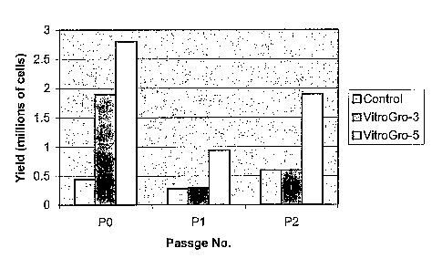
Figure 3. Relative activity of isolated protein complexes containing IGFBP3 or
IGFBP5. Control = standard keratinocyte growth medium supplemented with 10%
foetal bovine serum. VitroGroTM-3 = vitronectin, IGFBP3 and IGF-I (serum-
free).
VitroGroTM-5 = vitronectin, IGFBP5 and IGF-l (serum-free). All cultures were
grown in the presence of gamma irradiated mouse 3t3 cells.
Figure 4. IGF protein complexes support the ex vivo expansion of
keratinocytes.
Keratinocytes derived from adult human skin seeded onto IGF protein complexes
survive and grow at rates comparable to cells seeded onto irradiated mouse 3T3
cells
in the presence of fetal bovine serum. Cell growth was observed by: (a) visual
examination of culture morphology/confluence; and (b) quantified by MTT assay.
(a) from left to right: feeder layer + bovine serum; control without feeder
layer or
serum; IGF-I + IGFBP5 + VN without feeder layer or serum; (b) left to right:
Greens
media + feeder layer + bovine serum; Greens media + feeder layer alone; Greens
media - insulin + IGF-I, + IGFBP-3 + VN; Greens media - insulin + IGF-I, +
IGFBP-
5 + VN. VN is present at 300 ng/well. IGF-I or IGF-II are present at 100
ng/well and
IGFBPs are present at 300 ng/well.
Figure 5. IGF protein complexes supplemented with other growth factors
further enhance growth of cultures of keratinocytes. Keratinocytes derived
from adult
human skin were cultured on IGF protein complexes plus epidermal growth factor
(EGF) and basic fibroblast growth factor (bFGF) and assayed for protein
synthesis by
[3H]-leucine incorporation. Cells seeded on trimeric IGF-I, IGFBP5 and VN or
dimeric IGF-II and VN protein complexes grow at rates equivalent to Defined
Keratinocyte Media (DKM, Invitrogen). IGF protein complexes further
incorporating
EGF (100 ng/well) and bFGF (100 ng/well) significantly enhanced protein
synthesis
CA 02533422 2011-08-19
compared to DKM (p<0.05). IGF-I or IGF-II are present at 100 ng/well, VN at
300
ng/well and IGFBPs are present at 300 ng/well.
Figure 6. Effect of TISSOMATIM on keratinocyte viability. Cell distribution
and growth following spray delivery of keratinocytes into 150 mm diameter
collagen-
5 coated cultures dishes. Cells were sprayed at two different concentrations
to
determine cell numbers required to cover sprayed area. The cultures used for
spraying were originally grown on either control (with serum) or vitronectin
with
IGFBP3 and IGF-I (VitroGroT M). All cultures were prepared in presence of 3t3
cells.
Cultures were then stained with crystal violet after 7 days growth on collagen
coated plates in the presence of serum (designed to mimic conditions following
delivery to wound bed). Spray volume was 0.2 ml, pressure 20 psi, height = 10
cm.
Figure 7. Effect of TISSOMAT'M on keratinocyte viability. Cultures were
established using the conventional culture medium with added serum. (A) The
Trypan Blue exclusion test was performed within minutes following spraying
cells
into a collection tube. Viable cells are not permeable to the dye. (B) The MTT
conversion data is a measure of viability that provides an indication of the
metabolic
activity 24-hours after spraying the cells.
DETAILED DESCRIPTION OF THE INVENTION
The present invention has arisen from the discovery that culture media
comprising IGF-II and VN or IGF-I and IGFBP and VN stimulate significant
proliferative responses in primary cultures of keratinocytes ex vivo in the
absence of
serum, which is typically required for keratinocyte growth ex vivo.
Furthermore, the absolute requirement for feeder cells may be at least partly
eliminated, particularly during later stages of cell culture after cell
cultures have
initially been established.
This invention therefore provides technology that improves current clinical
best practice for ex vivo skin regeneration. In addition, the present
invention also
provides for the derivation and establishment of keratinocytes from tissue
biopsies. In
a preferred form, the invention provides a keratinocyte culture medium and
system
that utilizes autologous vitronectin isolated from a patient's own serum or
produced
recombinantly, thereby further minimizing the use of xenogeneic or allogeneic
CA 02533422 2006-01-23
WO 2005/012508 PCT/AU2004/001006
6
support systems, as well as eliminating use of poorly-defined supplementary
products. This will therefore provide an autologous-cell based tissue
engineering
system that can be translated to approved therapeutic applications.
For the purposes of this invention, by "isolated" is meant material.that has
been removed from its natural state or otherwise been subjected to human
manipulation. Isolated material may be substantially or essentially free from
components that normally accompany it in its natural state, or may be
manipulated so
as to be in an artificial state together with components that normally
accompany it in
its natural state. Isolated material may be in native, chemical synthetic or
recombinant form.
As used herein, by "synthetic" is meant not naturally occurring but made
through human technical intervention. In the context of synthetic proteins and
nucleic
acids, this encompasses molecules produced by recombinant or chemical
synthetic
and combinatorial techniques as are well understood in the art.
By "protein" is meant an amino acid polymer. The amino acids may be
natural or non-natural amino acids, D- or L- amino acids as are well
understood in
the art.
A `peptide" is a protein having less than fifty (50) amino acids.
A `polypeptide" is a protein having fifty (50) or more amino acids.
In particular aspects, the invention provides a cell culture medium and system
comprising at least IGF-I and/or IGF-II, such that exogenous, animal-derived
factors
such as serum are not required or are required at substantially reduced levels
whereby
cell growth and/or viability are maintained.
It will be appreciated that the invention is applicable to any mammalian cell
type that is responsive to IGF-I and/or IGF-II.
Generally, such cells are mesoderm-derived cells such as epithelial cells,
myoblasts and their progenitors, bone marrow and dendritic cells.
In a preferred embodiment, the invention is applicable to epithelial cells
inclusive of skin epithelial cells such as keratinocytes, keratinocyte
progenitors and
corneal epithelial cells. Indeed, both skin and corneal epithelial cells may
be
regarded as "keratinocytes" since they produce keratin proteins.
CA 02533422 2006-01-23
WO 2005/012508 PCT/AU2004/001006
7
Keratinocytes and/or their progenitors may be derived from normal skin, skin
biopsies such as obtained from wounds or ulcers or from outer root sheath
(ORS)
cells of hair follicles, although without limitation thereto.
It will therefore be appreciated that the culture medium, method and system
of the invention may potentially be used to engineer replacement tissues
wherever
epithelial cells are found e.g. the oral and respiratory mucosa (inner lining
of mouth,
nose, trachea and oesophagus) and genito-urinary tissue (e.g vagina, bladder).
These
tissues can also be damaged by bums and other trauma and as such can be
treated
using cultivated grafts grown in a similar way to the skin biopsies.
The invention may also be applicable to human embryonic stem (hES) cells,
which also normally have a requirement for serum during culture.
It will therefore be appreciated that "an absence of serum or an amount of
serum which in the absence of said at least an IGF would not support cell
growth"
means either no serum or a substantially reduced amount or concentration of
serum
than would ordinarily be required for optimal cell growth and/or development
in
vitro.
By "serum" is meant a fraction derived from blood that comprises a broad
spectrum of macromolecules, carrier proteins for lipoid substances and trace
elements, cell attachment and spreading factors, low molecular weight
nutrients, and
hormones and growth factors. Operationally, serum may be defined as the
proteinaceous, acellular fraction of blood remaining after removal of red
blood cells,
platelets and clotted components of blood plasma. The most widely used animal
serum for cell culture is fetal bovine serum, FBS, although adult bovine
serum, horse
serum and protein fractions of same (e.g. Fraction V serum albumin) may also
be
used.
Typically, mammalian cells require between 5-10% serum depending on cell
type, duration of culture, the presence or absence of feeder cells and/or
other cellular
components of a culture system and other factors that are apparent to persons
of skill
in the art.
Thus, in a preferred embodiment, the invention contemplates less than 5%
serum, more preferably less than 2% serum, even more preferably less than 1%
serum or advantageously no more than 0.5%, 0.4%, 0.3% or 0.2 % serum (v/v).
CA 02533422 2006-01-23
WO 2005/012508 PCT/AU2004/001006
8
In particularly advantageous embodiments, the invention contemplates no
serum or no more than 0.1%, or 0.05% serum (v/v).
In embodiments where IGF-I is present, it is preferred that IGF-I is a
component of a protein complex further comprising an IGFBP and vitronectin
(VN).
The IGFBP is selected from IGFBPI, IGFBP2, IGFBP3, IGFBP4, IGFBP5
and IGFBP6.
Preferably, the IGFBP is IGFBP3 or IGFBP5.
More preferably, the IGFBP is IGFBP5.
In embodiments where IGF-II is present, it is preferred that IGF-II is a
component of an isolated protein complex further comprising vitronectin (VN).
It will also be appreciated that vitronectin (VN) may be in monomeric or
multimeric form.
In one particular embodiment, the invention comprises autologous, purified
VN.
Preferably, keratinocytes are cultured in culture vessels as typically used in
the art. It will therefore be appreciated that the respective amounts of IGFs,
VN and
IGFBPs present during culture will depend on factors such as the size of the
culture
vessel, amount of liquid medium present in the vessel, cell density and other
factors
known in the art.
For guidance, in a 1.9 cm2 well, preferred amounts are as follows:
VN: 50-5000 ng, more preferably 100-500 ng or advantageously
250-350 ng;
IGF: 0.1 to 1000 ng, more preferably 10-200 ng or advantageously
50-150 ng; and
IGFBP: 1 to 1000ng, more preferably 30-700 ng or advantageously
300-500 ng.
Suitably, the culture medium of the invention comprises other defined
components. Non-limiting and in some cases optional components include well
known basal media such as DMEM or Hain's media, antibiotics such as
streptomycin
or penicillin, human serum albumin (HSA), phospholipids (eg.
phosphatidylcholine),
amino acid supplements such as L-glutamine, anti-oxidants such as 13-
CA 02533422 2006-01-23
WO 2005/012508 PCT/AU2004/001006
9
mercaptoethanol, transferrin, buffers such as carbonate buffers, HEPES and a
source
of carbon dioxide as typically provided by cell culture incubators.
The invention also contemplates use of additional biologically active proteins
that regulate cell growth, differentiation, survival and/or migration such as
epidermal
growth factor (EGF; Heldin et al., 1981, Science 4 1122-1123), fibroblast
growth
factor (FGF; Nurcombe et al., 2000, J. Biol. Chem. 275 30009-30018), basic
fibroblast growth factor (bFGF; Taraboletti et al., 1997, Cell Growth. Differ.
8 471-
479), osteopontin (Nam et al., 2000, Endocrinol. 141 1100), thrombospondin-1
(Nam
et al., 2000, supra), tenascin-C (Arai et al., 1996, J. Biol. Chem. 271 6099),
PAI-1
(Nam et al., 1997, Endocrinol. 138 2972), plasminogen (Campbell et al., 1998,
Am.
J. Physiol. 275 E321), fibrinogen (Campbell et al., 1999, J. Biol. Chem 274
30215),
fibrin (Campbell et al., 1999, supra) or transferrin (Weinzimer et al., 2001,
J. Clin.
Endocrinol. Metab. 86 1806).
Preferred additional biologically active proteins are EGF and bFGF.
Additional biologically active proteins such as EGF and bFGF may be present
at 0.1 to 1000 ng or advantageously 1-100 ng per 1.9 cm2 culture well.
In a particular embodiment, the invention contemplates use of any growth
factor with a heparin-binding-like domain.
In another particular embodiment, the invention contemplates use of LIF
and/or other agents that inhibit cell differentiation in addition to isolated
protein
complexes.
In yet another particular embodiment, the invention contemplates use of one
or more of poly-L-lysine and poly-L-arginine and secreted cellular material
that
interacts with vitronectin, for example polymers of collagens, fibronectins,
glycosaminoglycans/proteoglycans, laminins, sialoproteins and/or mucins in the
culture medium, system and/or method of the invention.
It is also proposed that the invention may facilitate cell culture in the
absence
of feeder cells, at least after the initial establishment stages of cell
culture.
In the context of keratinocytes and/or keratinocyte progenitors, feeder cells
(such as irradiated 3t3 feeder cells) may be present for the initial 6-7 days
of culture
in the absence of serum, after which time feeder cells may be absent for up to
two
passages.
CA 02533422 2006-01-23
WO 2005/012508 PCT/AU2004/001006
In light of the foregoing and although not wishing to be bound by any
particular theory, it is proposed that IGF-I forms an isolated protein complex
with an
IGFBP and VN while IGF-II forms a complex with VN to exert a biological effect
during cell culture.
5 The term "isolated protein complex" is used herein consistent with that used
in International Publication WO 02/24219 and International. Application
PCT/AU2004/000117.
Isolated protein complexes may be pre-formed and included in the culture
medium of the invention or may form in the culture vessel.
10 Typically, vitronectin and/or fibronectin are bound, immobilized, coated or
otherwise associated with the culture vessel. Addition of an IGF and,
optionally, an
IGFBP, forms a complex with the vitronectin and/or fibronectin bound,
immobilized,
coated or otherwise associated with the culture vessel.
As described in International Application PCT/AU2004/000117, isolated
protein complexes of the invention may comprise a growth factor (e.g. IGF-I
and
IGF-II) , or at least a domain of a growth factor which is capable of binding
a cognate
growth factor receptor (e.g.IGF type 1 receptor).
In this context, by "domain" is meant at least that portion or region of a
growth factor that is capable of binding a cognate growth factor receptor.
Typically,
although not exclusively, the cognate growth factor receptor is expressed by a
cell
and binding or ligation of said cognate growth factor receptor by said at
least a
domain of a growth factor elicits a cellular response such as cell growth,
differentiation, survival and/or migration.
With particular regard to IGF-I, said domain suitably comprises amino acid
residue 24, which is not a leucine residue.
Typically, said residue is tyrosine.
With particular regard to IGF-II, said domain suitably comprises amino acid
residue 27, which is not a leucine residue.
Typically, said residue is tyrosine.
With particular regard to IGF-I, in one embodiment said domain comprises or
consists of residues 1 to 70 of IGF-I.
CA 02533422 2006-01-23
WO 2005/012508 PCT/AU2004/001006
11
In another embodiment, said domain comprises or consists of residues 4 to 70
of IGF-I.
It will also be understood that another component of isolated protein
complexes of the invention is at least an integrin-binding domain of
vitronectin or
fibronectin.
This includes and encompasses any domain of VN or FN which is capable of
binding an a, integrin.
More preferably, the integrin is an av(33 integrin or an a,(35 integrin.
As described in International Application PCT/AU2004/000117, the heparin
binding domain (HBD) of VN (and analagously FN) is not required for the full
biological activity of isolated protein complexes.
With regard to VN, it is most likely the polyanionic region of VN (and
analagously FN) that is required for interaction with IGF-II or IGF-I/IGFBP
complexes.
The polyanionic region is amino acid residues 53-64 of the mature VN
sequence.
In light of the foregoing, the present invention contemplates embodiments of
synthetic chimeric proteins that do not include the HBD and/or the polyanionic
region of VN or FN.
With regard to VN proteins and amino acid sequences thereof that do not
include the HBD and/or the polyanionic region, these may be naturally
occurring
proteins such as the 54kDa chicken yolk VN (lacking a HBD) or may be
engineered
by deletion, mutation or truncation of a VN protein or amino acid sequence so
that
the HBD and/or the polyanionic region are absent or at least substantially non-
functional.
It will be readily appreciated from the foregoing that isolated protein
complexes of the invention may be in the form of non-covalently associated
oligo-
protein complexes, oligo-protein complexes that have been covalently cross-
linked
(reversibly or irreversibly) or in the form of synthetic, chimeric proteins.
Accordingly, in a particular aspect the invention provides an isolated protein
complex in the form of a synthetic chimeric protein.
CA 02533422 2006-01-23
WO 2005/012508 PCT/AU2004/001006
12
As used herein, a "chimeric protein", comprises a contiguous sequence of
amino acids derived from an integrin-receptor binding domain of VN or FN and a
growth factor or at least a receptor-binding domain of a growth factor.
Although not wishing to be bound by any particular theory, it is proposed that
synthetic chimeric proteins may be able to co-ligate and co-activate a cognate
receptor for said growth factor and an integrin receptor for VN or FN to
thereby
stimulate, induce, augment or otherwise promote cell migration.
An advantage of chimeric proteins according to the invention is that they are
readily produced by chemical synthetic or recombinant means and are expected
to be
more stable in vivo, as they do not rely on maintaining the protein-protein
interactions that are required in non-covalent oligo-protein complexes.
In this regard, although isolated protein complexes that comprise receptor
binding domains of IGF-I would also comprise an IGFBP, it is proposed that
according to the aforementioned mode of action, an IGFBP is preferably not
present
in an IGF-I/VN synthetic chimera.
Preferably, chimeric proteins further comprise a "linker sequence" located
between and contiguous with a growth factor sequence and a VN or FN amino acid
sequence.
In one embodiment, said linker sequence comprises one or more glycine
residues and one or more serine residues.
Particular examples of linker sequences may be selected from; Gly4 Ser; Gly4
Sera and (Gly4 Ser)3, although without limitation thereto.
In another embodiment, the linker sequence includes a Plasmin Cleavage
Recognition Site, such as according to the sequence:
Leu Ile Lys Met Lys Pro
In yet another embodiment, the linker sequence includes a Collagenase-3
Cleavage Recognition Site, such as according to the sequence:
Gln Pro Gln Gly Leu Ala Lys
The aforementioned are examples of biologically-active fragments of a
growth factor, growth factor binding protein and/or vitronectin/fibronectin.
In one embodiment, said "biologically-active fragment" has no less than 10%,
preferably no less than 25%, more preferably no less than 50% and even more
CA 02533422 2011-08-19
13
preferably no less than 75%, 80%, 85%, 90% or at least 95% of a biological
activity
of a "full length" protein.
Also contemplated are variant growth factors, growth factor binding proteins
and/or vitronectin/fibronectin. and/or encoding nucleic acids that may be used
according to the invention.
In one embodiment, a "variant" has one or more amino acids that have been
replaced by different amino acids. It is well understood in the art that some
amino
acids may be changed to others with broadly similar properties without
changing the
nature of the activity of the protein (conservative substitutions).
In one embodiment, a variant shares at least 70%, preferably at least 80%,
more preferably at least 90% and advantageously at least 95%, 96%, 97%, 98% or
99% sequence identity with the amino acid sequences described herein.
Preferably, sequence identify is measured over at least 60%, more preferably
at least 75%, even more preferably at least 90% and advantageously over
substantially the full length of the synthetic protein of the invention.
In order to determine percent sequence identity, optimal alignment of amino
acid and/or nucleotide sequences may be conducted by computerised
implementations of algorithms (Geneworks program by Intelligenetics; GAP,
BESTFIT, FASTA, and TFASTA in the Wisconsin Genetics Software Package
Release 7.0, Genetics Computer Group, 575 Science Drive Madison, WI, USA,) or
by inspection and the best alignment (i.e., resulting in the highest
percentage
homology over the comparison window) generated by any of the various methods
selected. Reference also may be made to the BLAST family of programs as for
example disclosed by Altschul et al., 1997, Nucl. Acids Res. 25 3389.
In another example, "sequence identity" may be understood to mean the
"match percentage" calculated by the DNASIS computer program (Version 2.5 for
windows; available from Hitachi Software engineering Co., Ltd., South San
Francisco, California, USA).
A detailed discussion of sequence analysis can be found in Unit 19.3 of
CURRENT PROTOCOLS IN MOLECULAR BIOLOGY Eds. Ausubel et al. (John
Wiley & Sons Inc NY, 1995-1999).
CA 02533422 2006-01-23
WO 2005/012508 PCT/AU2004/001006
14
The invention also contemplates derivatives of a growth factor, growth factor
binding protein and/or vitronectin/fibronectin.
As used herein, "derivative" has been altered, for example by addition,
conjugation or complexing with other chemical moieties or by post-
translational
modification techniques as are well understood in the art
"Additions" of amino acids may include fusion with other peptides or
polypeptides. The other peptide or polypeptide may, by way of example, assist
in the
purification of the protein. For instance, these include a polyhistidine tag,
maltose
binding protein, green fluorescent protein (GFP), Protein A or glutathione S-
transferase (GST).
Other derivatives contemplated by the invention include, but are not limited
to, modification to side chains, incorporation of unnatural amino acids and/or
their
derivatives during protein synthesis and the use of crosslinkers and other
methods
which impose conformational constraints on proteins. Non-limiting examples of
side
chain modifications contemplated by the present invention include
modifications of
amino groups such as by acylation with acetic anhydride; acylation of amino
groups
with succinic anhydride and tetrahydrophthalic anhydride; amidination with
methylacetimidate; carbamoylation of amino groups with cyanate; pyridoxylation
of
lysine with pyridoxal-5-phosphate followed by reduction with NaBH4; reductive
alkylation by reaction with an aldehyde followed by reduction with NaBH4; and
trinitrobenzylation of amino groups with 2, 4, 6-trinitrobenzene sulphonic
acid
(TNBS).
Sulphydryl groups may be modified by methods such as performic acid
oxidation to cysteic acid; formation of mercurial derivatives using 4-
chloromercuriphenylsulphonic acid, 4-chloromercuribenzoate; 2-chloromercuri-4-
nitrophenol, phenylmercury chloride, and other mercurials; formation of a
mixed
disulphides with other thiol compounds; reaction with maleimide, maleic
anhydride
or other substituted maleimide; carboxymethylation with iodoacetic acid or
iodoacetamide; and carbamoylation with cyanate at alkaline pH.
The imidazole ring of a histidine residue may be modified by N-
c arbethoxylation with diethylpyrocarbonate or by alkylation with iodoacetic
acid
derivatives.
CA 02533422 2011-08-19
Examples of incorporating non-natural amino acids and derivatives during
peptide synthesis include but are not limited to, use of 4-amino butyric acid,
6-
aminohexanoic acid, 4-amino-3-hydroxy-5-phenylpentanoic acid, 4-amino-3-
hydroxy-6-methylheptanoic acid, t-butylglycine, norleucine, norvaline,
5 phenylglycine, ornithine, sarcosine, 2-thienyl alanine and/or D-isomers of
amino
acids.
Further examples of chemical derivatization of proteins are provided in
Chapter 15 of CURRENT PROTOCOLS IN PROTEIN SCIENCE Eds. Coligan et.
al., John Wiley & Sons NY (1995-2001).
10 According to the invention, a protein may be prepared by any suitable
procedure known to those of skill in the art.
In one embodiment, proteins may be in substantially pure native form.
One particular example is purified autologous vitronectin.
In another embodiment, a protein may be produced by chemical synthesis.
15 Chemical synthesis techniques are well known in the art, although the
skilled person
may refer to Chapter 18 of CURRENT PROTOCOLS IN PROTEIN SCIENCE Eds.
Coligan et. al., John Wiley & Sons NY (1995-2001) for examples of suitable
methodology.
In yet another embodiment, a protein may be prepared as a recombinant
protein.
Production of recombinant proteins is well known in the art, the skilled
person may refer to standard protocols as for example described in Sambrook et
al.,
MOLECULAR CLONING. A Laboratory Manual (Cold Spring Harbor Press, 1989),
in particular Sections 16 and 17; CURRENT PROTOCOLS IN MOLECULAR
BIOLOGY Eds. Ausubel et al., (John Wiley & Sons, Inc. 1995-1999), in
particular
Chapters 10 and 16; and CURRENT PROTOCOLS IN PROTEIN SCIENCE Eds.
Coligan et al., (John Wiley & Sons, Inc. 1995-1999) in particular Chapters 1,
5 and 6.
Recombinant proteins may further comprise a fusion partner.
Well known examples of fusion partners include, but are not limited to,
glutathione-S-transferase (GST), Fc portion of human IgG, maltose binding
protein
CA 02533422 2006-01-23
WO 2005/012508 PCT/AU2004/001006
16
(MBP) and hexahistidine (HIS6), which are particularly useful for isolation of
the
fusion protein by affinity chromatography. For the purposes of fusion protein
purification by affinity chromatography, relevant matrices for affinity
chromatography are glutathione-, amylose-, and nickel- or cobalt-conjugated
resins
respectively. Many such matrices are available in "kit" form, such as the
QlAexpressTM system (Qiagen) useful with (HIS6) fusion partners and the
Pharmacia
GST purification system.
In some cases, the fusion partners also have protease cleavage sites, such as
for Factor Xa or Thrombin, which allow the relevant protease to partially
digest the
fusion protein of the invention and thereby liberate the recombinant protein
therefrom. The liberated protein can then be isolated from the fusion partner
by
subsequent chromatographic separation.
Fusion partners according to the invention also include within their scope
"epitope tags", which are usually short peptide sequences for which a specific
antibody is available. Well known examples of epitope tags for which specific
monoclonal antibodies are readily available include c-myc, haemagglutinin and
FLAG tags.
Suitable host cells for expression may be prokaryotic or eukaryotic, such as
Escherichia coli (DH5a for example), yeast cells, Sf9 cells utilized with a
baculovirus expression system, CHO cells, COS, CV-1, NIH 3T3 and HEK293 cells,
although without limitation thereto.
The invention further contemplates use of cells, such as keratinocytes or
keratinocyte progenitor cells, capable of expressing at least one recombinant
protein
selected from the group consisting of:
(i) a recombinant IGF;
(ii) a recombinant IGFBP;
(iii) a recombinant vitronectin;
(iv) a recombinant chimeric protein as hereinbefore described; and
(v) an additional biologically active protein such as EGF or bFGF.
According to a particular embodiment, paracrine/autocrine expression of
IGFs, VN and/or IGFBPs may enable keratinocytes or keratinocyte progenitors to
be
CA 02533422 2006-01-23
WO 2005/012508 PCT/AU2004/001006
17
cultured in media without serum and without the need to add one or more of
growth
factors, IGFBPs and/or vitronectin to the culture medium.
Recombinant protein expression may be achieved by introduction of an
expression construct into a keratinocyte or keratinocyte progenitor cell.
Typically, the expression construct comprises a nucleic acid to be expressed
(encoding the recombinant protein) operably linked or operably connected to a
promoter.
The promoter may be constitutive or inducible.
Constitutive or inducible promoters include, for example, tetracycline-
repressible, ecdysone-inducible, alcohol-inducible and metallothionin-
inducible
promoters. Promoters may be either naturally occurring promoters (e.g. alpha
crystallin promoter, ADH promoter, phosphoglycerate kinase (PGK), human
elongation factor (x promoter and viral promoters such as SV40, CMV, HTLV-
derived promoters), or synthetic hybrid promoters that combine elements of
more
than one promoter (e.g. SR alpha promoter).
In a preferred embodiment, the expression vector comprises a selectable
marker gene. Selectable markers are useful whether for the purposes of
selection of
transformed bacteria (such as bla, kanR and tetR) or transformed mammalian
cells
(such as hygromycin, G418 and puromycin).
- Expression constructs may be introduced into mammalian cells such as
keratinocyte or keratinocyte progenitor cells by well known means such as
electroporation, microparticle bombardment, virus-mediated gene transfer,
calcium
phosphate precipitation, DEAE-Dextran, cationic liposoines, lipofectin,
lipofectamine and the like, although without limitation thereto.
For non-limiting particular examples of methodology potentially applicable to
expression of recombinant growth factor proteins in keratinocytes, reference
may be
made to Supp et al., 2000, J. Invest. Dermatol. 114 5 and Supp et al., 2000,
Wound
Repair Regen. 8 26-35.
Pharmaceutical compositions
The invention also provides pharmaceutical compositions that comprise on or
more cells produced using the culture medium and/or system of the invention,
such
CA 02533422 2011-08-19
18
as keratinocytes although not limited thereto, together with a
pharmaceutically
acceptable carrier diluent or excipient.
Pharmaceutical compositions of the invention may be used to promote or
otherwise facilitate cell migration, tissue regeneration and wound healing.
Generally, the compositions of the invention may be used in therapeutic or
prophylactic treatments as required. For example, pharmaceutical compositions
may
be applied in the form of therapeutic or cosmetic preparations for skin
repair, wound
healing, healing of burns and other dermatological treatments.
Preferably, the pharmaceutically-acceptable carrier, diluent or excipient is
suitable for administration to mammals, and preferably, to humans.
In particular embodiments, the pharmaceutical composition comprises
autologous or allogeneic keratinocytes cultured according to the invention.
By "pharmaceutically-acceptable carrier, diluent or excipient" is meant a
solid or liquid filler, diluent or encapsulating substance that may be safely
used in
systemic administration. Depending upon the particular route of
administration, a
variety of carriers, well known in the art may be used. These carriers may be
selected from a group including sugars, starches, cellulose and its
derivatives, malt,
gelatine, talc, calcium sulfate, vegetable oils, synthetic oils, polyols,
alginic acid,
phosphate buffered solutions, emulsifiers, isotonic saline and salts such as
mineral
acid salts including hydrochlorides, bromides and sulfates, organic acids such
as
acetates, propionates and malonates and pyrogen-free water.
A useful reference describing pharmaceutically acceptable carriers, diluents
and excipients is Remington's Pharmaceutical Sciences (Mack Publishing Co.
N.J.
USA, 1991).
Any safe route of administration may be employed for providing a patient
with the composition of the invention. For example, oral, rectal, parenteral,
sublingual, buccal, intravenous, intra-articular, intra-muscular, intra-
dermal,
subcutaneous, inhalational, intraocular, intraperitoneal,
intracerebroventricular,
transdermal and the like may be employed.
Dosage forms include tablets, dispersions, suspensions, injections, solutions,
syrups, troches, capsules, suppositories, aerosols, transdermal patches and
the like.
These dosage forms may also include injecting or implanting controlled
releasing
CA 02533422 2011-08-19
19
devices designed specifically for this purpose or other forms of implants
modified to
act additionally in this fashion.
Controlled release formulations may be effected by coating, for example, with
hydrophobic polymers including acrylic resins, waxes, higher aliphatic
alcohols,
polylactic and polyglycolic acids and certain cellulose derivatives such as
hydroxypropylmethyl cellulose. Controlled release may be effected by using
other
polymer matrices, liposomes and/or microspheres. Non-limiting examples of
controlled release formulations and delivery devices include osmotic pumps,
polylactide-co-glycolide (PLG) polymer-based microspheres, hydrogel-based
polymers, chemically-crosslinked dextran gels such as OctoDEX"M and dex-
lactate-
HEMA, for example.
The above compositions may be administered in a manner compatible with
the dosage formulation, and in such amount as is pharmaceutically-effective.
The
dose administered to a patient, in the context of the present invention,
should be
sufficient to effect a beneficial response in a patient over an appropriate
period of
time. The quantity of agent(s) to be administered may depend on the subject to
be
treated inclusive of the age, sex, weight and general health condition
thereof, factors
that will depend on the judgement of the practitioner.
With regard to pharmaceutical compositions for wound healing, particular
reference is made to U.S. patent 5,936,064 and International Publication
W099/62536.
In one particular embodiment, the composition of the invention is suitable for
spray delivery in situ.
The term "spray" encompasses and includes terms such as "aerosol" or "mist"
or "condensate" that generally describe liquid suspensions in the form of
droplets.
According to the invention, although optional, the spray or aerosol
composition may further comprise at least an IGF selected from IGF-I and IGF-
II, or
in particular embodiments, isolated protein complexes comprising IGFs, VN and
IGFBPs, to promote skin cell growth and migration in situ. Additional
biologically
active proteins such as EGF and/or bFGF may also be included.
Although not wishing to be bound by any particular theory, the invention
contemplates that the inherent "stickiness" of VN in IGF complexes present in
the
CA 02533422 2006-01-23
WO 2005/012508 PCT/AU2004/001006
spray composition will facilitate delivery of IGF-I, IGF-II and other growth
factors
such as EGF and bFGF.
Typically, spray compositions of the invention will be delivered by apparatus
such as a pressurised canister equipped with a delivery outlet.
5 An example of an aerosolised keratinocyte delivery systems such as for
wound healing in a pig model, is provided by Navarro et al., 2000, J Bum Care
Rehabil 21 513. Reference is also made to Grant et al., 2002, Br J Plast Surg
55 219
which describes use of aerosolised keratinocytes in conjunction with fibrin
glue for
wound healing in a pig model.
10 Preferably, spray compositions of the invention are substantially free of
serum.
In one particular embodiment, the skin spray composition of the invention
comprises Tissomat (Baxter Healthcare), which facilitates spray-application
of
fibrin glue and aerosolises liquids via delivery into a stream of compressed
medical
15 grade air controlled by a regulator. Pressures of between 10-30 psi are
suitable, but a
drop in viability is observed within increasing pressure. Cells may be sprayed
at
concentrations of between 0.5 to 1.5 million per millilitre. Application of
0.2
millilitres of cell suspension at 20 psi is sufficient to cover an area of
approximately
square centimetres (based on measurement of surface area covered with cells
after
20 7 days growth in vitro). Cells are preferably delivered in serum free
growth medium,
but may also be suspended in fibrin glue such as the commercially available
Tisseel/Tissucol (Baxter Healthcare).
It is also contemplated that similar efficacy may be achieved using syringe
delivery of composition of the invention (e.g. a syringe fitted with a spray
cap).
25 Therapeutic uses
In particular aspects, the present invention provides methods of treating
burns, wounds and ulcers as well as methods that relate to cosmetic skin
treatments to
improve or enhance skin quality or skin appearance.
These methods are particularly aimed at treatment of mammals, and more
particularly, humans. However, it will also be appreciated that the invention
may
have veterinary applications for treating domestic animals, livestock and
performance
animals as would be well understood by the skilled person.
CA 02533422 2006-01-23
WO 2005/012508 PCT/AU2004/001006
21
In a preferred embodiment, the invention provides a culture medium, system
and method for propagating primary keratinocytes ex vivo, which cells may be
administered to an individual according to the invention.
In particular embodiments, the keratinocytes are autologous or allogeneic
keratinocytes cultured according to the invention.
Such methods include administration of pharmaceutical compositions as
hereinbefore defined, and maybe by way of microneedle injection into specific
tissue
sites, such as described in U.S. patent 6,090,790, topical creams, lotions or
sealant
dressings applied to wounds, burns or ulcers, such as described in U.S. patent
6,054,122 or implants which release the composition such as described in
International Publication W099/47070.
There also exist methods by which skin cells can be genetically modified for
the purpose of creating skin substitutes, such as by genetically engineering
desired
growth factor expression (Supp et al., 2000, J. Invest. Dermatol. 114 5). An
example
of a review of this field is provided in Bevan et al., Biotechnol. Gent. Eng.
Rev. 16
231.
Also contemplated is "seeding" a recipient with transfected or transformed
cells, such as described in International Publication W099/11789.
These methods can be used to stimulate cell migration and thereby facilitate
or progress wound and burn healing, repair of skin lesions such as ulcers,
tissue
replacement and grafting such as by in vitro culturing of autologous skin, re-
epithelialization of internal organs such as kidney and lung and repair of
damaged
nerve tissue.
Skin replacement therapy has become well known in the art, and may employ
use of co-cultured epithelial/keratinocyte cell lines, for example as
described in Kehe
et al., 1999, Arch. Dermatol. Res. 291 600 or in vitro culture of primary
(usually
autologous) epidermal, dermal and/or keratinocyte cells. These techniques may
also
utilize engineered biomaterials and synthetic polymer "scaffolds".
Examples of reviews of the field in general are provided in Terskikh &
Vasiliev, 1999, hit. Rev. Cytol. 188 41 and Eaglestein & Falanga, 1998, Cutis
62 1.
More particularly, the production of replacement oral mucosa useful in
craniofacial surgery is described in Izumi et al., 2000, J. Dent. Res. 79 798.
Fetal
CA 02533422 2006-01-23
WO 2005/012508 PCT/AU2004/001006
22
keratinocytes and dermal fibroblasts can be expanded in vitro to produce skin
for
grafting to treat skin lesions, such as described in Fauza et al., J. Pediatr.
Surg. 33
357, while skin substitutes from dermal and epidermal skin elements cultured
in vitro
on hyaluronic acid-derived biomaterials have been shown to be potentially
useful in
the treatment of burns (Zacchi et al., 1998, J. Biomed. Mater. Res. 40 187).
Polymer scaffolds are also contemplated for the purpose of facilitating
replacement skin engineering, as for example described in Sheridan et al.,
2000, J.
Control Release 14 91 and Fauza et al., 1998, supra, as are microspheres as
agents
for the delivery of skin cells to wounds and burns (LaFrance & Armstrong,
1999,
Tissue Eng. 5 153).
Keratinocyte sheets typically produced for therapeutic use are responsible for
the ultimate closure of burn wounds. This sheet graft technique is applicable
to all
partial thickness burn injuries and is most useful in treating large surface
area wounds
where early permanent closure of both wound and donor sites is nearly
impossible
without external help. This is the type of injury responsible for the death of
patients
burnt in the recent Bali bombing.
Currently, it is possible to grow enough skin from a patient skin biopsy the
size of a fifty-cent piece to cover an entire adult. This culture process
takes 17 days.
However, earlier skin replacement is urgently needed to reduce patient
trauma, risk of infection, scarring and the present requirement for expensive
temporary skin replacements ahead of permanent skin grafting. In addition, a
sheet of
cultured skin comprises many skin cells, some mature and some immature. The
simple act of allowing cultured keratinocytes to reach confluence (necessary
to
produce sheets of skin) causes cells to prematurely loose their primitive
characteristics Le to differentiate. When a sheet of cultured skin is applied,
only the
immature cells are capable of attaching and establishing themselves on the
patient.
Because only small areas adhere, the sheets are very susceptible to damage
arising
from friction or movement of the patient and can sometimes result in the loss
of the
entire graft. Furthermore, in a sheet graft, the more mature skin cells in the
sheet, the
more likely it will be that the graft will not take and the cells themselves
will not
proliferate and migrate on the wound bed itself. Thus it is clear that earlier
application of immature skin cells will result in better graft take and reduce
scarring.
CA 02533422 2006-01-23
WO 2005/012508 PCT/AU2004/001006
23
The present invention therefore provides a spray or aerosol delivery method
to deliver skin cells cultured ex vivo onto a patient's burnt, ulcerated or
wounded skin
to enable a larger surface area of the patient's body to be covered by
immature skin
cells much earlier than existing sheet graft technology. This could be as
early as only
7 days. This would also significantly reduce scar formation, shock and heat
loss and
would enable faster return of skin function in partial thickness and also full
thickness
burns.
According to the invention, although optional, the administered spray or
aerosol may further comprise isolated protein complexes comprising IGFs, VN
and
IGFBPs together EGF and/or bFGF to promote skin cell growth and migration in
situ.
The patients' own skin cells (autologous skin) and donor skin cells
(allogeneic
or heterologous skin) can be grown and used for early burn closure. Donor
cells do
not express transplantation antigens, so they do not cause an immune response
in the
patient. The donor skins cells, however, are eventually replaced by the
patients' own
skin cells.
Although autologous cells are preferred, use of allogeneic or heterologous
cells in a spray-on-skin would allow immediate application to a needy patient.
Alternatively, sufficient autologous skin cells could be cultured in
approximately
seven days for use in a therapeutic spray.
Another treatment contemplated by the present invention is the treatment of
burns patients to achieve early closure of full thickness wounds, because take
of
cultured skin on a wound that has removed both the surface (epidermal) and
deep
layer (dermis) of skin is poor. The invention contemplates use of dermal
substitutes
in conjunction with the spray-on-skin to effect early permanent closure of
these most
horrific injuries. Both biological and synthetic dermal substitutes are
contemplated.
For example, a de-epidermised, de-cellularised cadaveric-derived dermal
scaffold
comprising isolated protein complexes of the invention may be overlayed with a
synthetic epidermis (dressing). After approximately 7 days the dermis the
present
inventors hypothesise that this dermis will be highly infilitrated by
autologous
endothelial cells. At this time, the synthetic dermis will be removed and the
patient's
CA 02533422 2006-01-23
WO 2005/012508 PCT/AU2004/001006
24
own ex-vivo expanded fibroblasts and keratinocytes will be applied to the allo-
dermis.
It is anticipated that the spray-on-skin, rather than epidermal sheets, will
be
successful as the dermal substitute will act as a nutritious stabilising
scaffold
promoting the migration and anchoring of skin cells and other important cells
normally found in the skin. This will result in improved take of cultured skin
cells in
full thickness skin injuries
So that the present invention may be more readily understood and put into
practical effect, the skilled person is referred to the following non-limiting
examples.
EXAMPLES
EXAMPLE 1
PRIMARY HUMAN KERATINOCYTE GROWTH IN AN ABSENCE OF
SERUM
Materials and Methods
Growth factor concentrations/pre-absorption to culture plastic
A standard approach for adding VN, IGF and IGFBP has been used
throughout all studies. Culture plastic is prepared by incubating for 2 hours
at 37
degrees C with vitronectin 150 ng/cm2 in serum-free culture medium. The VN
solution is then removed and replaced with serum-free medium containing IGFBP
(250 ng/cm2), IGF-I (50 ng/cm2) and EGF (50 ng/cm). The growth factors are
left
over night at 4 degrees C (in the fridge) to absorb to the VN treated plastic.
The
following day, the growth factor solution is removed and replaced with growth
medium (defined below) containing 50 ng/ml VN, 50 ng/ml IGFBP, 15 ng/cm IGF-I
and 15 ng/cm EGF. The cells are added at densities given below. The medium is
typically changed once every 3 days. Each culture is grown for approximately 6
days
before passaging: i.e. there are approximately 6 days between each passage.
Growth medium
The base medium is a 3:1 mixture of Dulbecco's Modified Eagle's Medium
(DMEM) with Ham's F12 medium that is routinely supplemented with L-glutamine
(2 mM), cholera toxin (0.1 g/ml), adenine (180 M), hydrocortisone (0.4
g/ml),
and a mixture of non-essential amino acids (1% v/v).
CA 02533422 2006-01-23
WO 2005/012508 PCT/AU2004/001006
Positive control medium contains an additional 10% fetal bovine serum,
insulin (5 g/ml) and epidermal growth factor (EGF, 10 nglml).
Seeding Densities
Cultures were grown in the presence of growth arrested mouse 3t3 cells at a
5 density of 2.5 x 10⁴/cm2. The 3t3 cells are rendered "growth-arrested" by
gamma
irradiation immediately prior to use.
Keratinocytes were seeded at two different densities depending upon the
passage number. Initial cultures (P0) were established by seeding cells at 3.8
x
104/cm2. Subsequent cultures (P1, P2 etc) were established by re-seeding
harvested
10 cells at a density of 6.4 x 10³/cm2. The higher seeding density was used
for the PO
cultures since only a fraction of the freshly harvested cells will display
ongoing
proliferation in culture. Thus, culturing the cells enables expansion of the
proliferating subpopulation.
Results
15 COMPARISON OF CULTURES WITH CONVENTIONAL GROWTH MEDIUM
CONTAINING SERUM.
Referring to FIG. 1, this graph displays the average growth of freshly
isolated keratinocytes with VitroGro (+3t3 cells) relative to the conventional
method
where both foetal bovine serum and 3t3 cells are present. P0, P1 and P2
relative to
20 the number of times that the cells have been harvested and replated (PO =
performance of cells immediately following isolation from a skin sample). The
data
were obtained via staining with MTT. The data show that culture in the
presence of
isolated protein complexes in the absence of serum consistently achieved at
least
90% of the cell growth achieved in the presence of 10% serum.
25 Referring to FIG. 2A and 2B, skin cells grown on VitroGro display a similar
appearance to those grown in the presence of fetal bovine serum (Figure 2A). A
more
detailed comparison based on the presence of molecular markers is currently in
progress to confinn this conclusion. Techniques employed will include
immunocytochemistry, fluorescence activated cell sorting (FACS) analysis,
western
blotting, and polymerase chain reaction (PCR) methods. The use of state-of-the-
art
proteonomic and gene array technologies are also being considered. Major
markers
for investigation will include cytokeratins (CK1 and CK10, CK6, CK14, and
CK19)
CA 02533422 2011-08-19
26
and putative keratinocyte progenitor cell markers (e.g. p63, 0-integrin, a6-
integrinh"/CD71d'"'). Particular attention will be placed on comparing the
expression
of putative progenitor cell markers since these are likely to confer clinical
efficacy in
cultures following grafting. In addition, the responses of cells in routine in
vitro
functional assays may also be performed (attachment, migration,
proliferation).
RELATIVE ACTIVITY OF ISOLATED PROTEIN COMPLEXES CONTAINING
IGFBP3 OR IGFBP5
Referring to FIG. 3, it is apparent that isolated protein complexes comprising
IGFBP5 were more efficient than IGFBP3-containing complexes in terms of
keratinocyte yield.
EXAMPLE 2
PRIMARY HUMAN KERATINOCYTE GROWTH IN THE ABSENCE OF
SERUM WITH AND WITHOUT FEEDER CELLS
Materials and Methods
Primary Keratinocyte Culture
Keratinocytes were isolated from adult human skin using the standard
procedures essentially the same as that originally reported by Rheinwald &
Green,
1977, Nature 265 421. Briefly this involved digestion of the skin sample for
one hour
at 37 C in Dispase fM II solution. The recovered epithelium is subsequently
digested
for a further 10 minutes at 37 C with 0.25% trypsin/0.02% EDTA to dissociate
the
cells. Residual trypsin activity is inactivated and recovered cells are then
washed and
co-seeded into tissue culture dishes in the presence or absence of lethally
irradiated
3T3 mouse fibroblasts. "Control" cells, cultivated using these standard
conditions are
grown in DMEM/F12 medium supplemented with 10% fetal calf serum, 0.1%
penicillin-streptomycin solution, 0.4 pg/ml hydrocortisone, 0.1 .ig/ml cholera
toxin,
10 ng/ml human recombinant epidermal growth factor (EGF), 5 pg/ml insulin, 5
g/ml transferrin and 2 nM tri-iodothyronine, while cells treated with isolated
growth
factor complexes used identical media except that no insulin was present.
Insulin was
not included in media used in conjunction with isolated protein complex
treatments
to minimize competitive binding of insulin to the type-1 IGF receptor. Cells
cultured
on isolated growth factor complex-coated dishes also differed from those
cultured
CA 02533422 2006-01-23
WO 2005/012508 PCT/AU2004/001006
27
following the standard procedure in that the cells will be seeded onto plates
without
irradiated mouse fibroblasts.
Protein Synthesis Assay
Keratinocytes were derived from an adult skin biopsy and expanded until
passage 2 using standard procedures incorporating Greens media, serum and
feeder
cells. These cells were then assessed for the stimulation of protein synthesis
in the
presence and absence of IGF + VN complexes. Here, 24 well plates were coated
for 2
hours with 300 ng of vitronectin and then washed to remove unbound
vitronectin.
Wells were then incubated with the growth factors to be examined, that is;
epidermal
growth factor, basic fibroblast growth factor, insulin-like growth factor-I
and insulin-
like growth factor-II, in combination with insulin-like growth factor binding
protein-
5; were added to the wells and allowed to bind the vitronectin overnight. The
next
day the wells were washed twice to remove any unbound growth factors and the
plates allowed to air dry. Keratinocytes were then harvested and seeded at a
density
of 1 x 10⁵ cells/well in serum-free Dulbecco'5 Modified Eagle Medium (DMEM)
along with 1 uCi/well of [3H]-leucine. In select wells, cells were seeded in
Defined
Keratinocyte Medium (DKM) (Invitrogen), a commercially available product for
the
serum-free culture of keratinocytes. Plates were then incubated for 48 hours
and then
washed to remove any unincorporated [3H]-leucine. Incorporation of [3H]-
leucine
into de novo synthesised protein was determined by sampling solubilised
protein
precipitate for beta-scintillation counting.
MTT-Esta Assay
Human keratinocytes were isolated and the cultures established using
standard culture techniques of fully supplemented Greens Media with a feeder
layer
of lethally irradiated mouse 3T3 cells. Cells were expanded to passage 3 and
seeded
into 24 well plates in Greens media in the presence or absence of Fetal Calf
Serum
(FCS) and 3T3 cells. In select treatments, wells were coated with isolated
protein
complexes. Wells were incubated with 300 ng of vitronectin for 2 hours and
then
aspirated prior to the addition of IGF-I and IGFBP3 or IGFBP5, or IGF-II.
Plates
were incubated overnight and aspirated prior to seeding cells. The cultures
were
assessed for metabolic activity as measured using the MTT-esta assay as
described
previously (Ealey et al. 1988, J Mol Endocrinol 1:R1-R4.).
CA 02533422 2006-01-23
WO 2005/012508 PCT/AU2004/001006
28
Results
In view of the significant enhanced functional responses obtained with
isolated growth factor complexes in cell lines (International Publication WO
02/24219; Noble et al., 2003, supra; Kricker et al., 2003, supra) we recently
extended our studies to cultures of keratinocytes derived from adult skin. In
particular
we examined the potential of isolated growth factor complexes to replace serum
and
feeder cells used in current best clinical practice for ex vivo expansion of
keratinocytes for split thickness autografting. While this procedure has
significantly
advanced therapies available to bums patients, the culture of keratinocytes
derived
from patients is conducted in the presence of fetal bovine serum (FBS), a semi-
defined xenobiotic product that is a potential source of pathogens. In
addition, in the
early stages of keratinocyte derivation and establishment a feeder layer of
cells
derived from a second species, namely murine 3T3 fibroblasts, is used as a
source of
cytokines and matrix elements to encourage cell attachment and growth. FBS-
also
contributes to these effects.
As (i) IGFs account for a large proportion of the cytokines secreted by the
feeder cells; (ii) we have established that VN replaces any requirement for
serum to
facilitate the attachment of primary cultured keratinocytes seeded at low
density to
plasticware; and iii) the effects we have obtained with keratinocyte cell
lines cultured
on isolated growth factor complexes are equivalent to those obtained with
media
containing 10% FBS, we hypothesised that isolated growth factor complex-
supplemented media had the potential to provide a superior product for
autologous
keratinocyte engineering applications. This hypothesis is supported by the
fact that
IGFs are key mitogens that stimulate keratinocyte proliferation, yet
keratinocytes
themselves do not secrete IGF-I. While serum-free media, such as KGMTM
(Clonetics) and EpiLifeTM (Sigma-Aldrich), have been developed commercially
for
keratinocyte expansion, these media require the addition of bovine pituitary
extract,
which is also undefined, a xenobiotic and a potential source of pathogens, or
alternatively, the addition of expensive supplements. Furthermore, most
current
serum-free keratinocyte culture applications demand very high seeding
densities
which defeats the purpose of attempting to culture large quantities of
keratinocytes
CA 02533422 2006-01-23
WO 2005/012508 PCT/AU2004/001006
29
rapidly and accounts for the poor adoption of these practices for routine
clinical
applications.
We have directly tested our hypothesis and the results are illustrated in
Figure
4. In this experiment keratinocytes were derived from adult skin and
established
using usual procedures for 7 days. The cells were then passaged by
trypsinisation and
seeded at low density (8,500 keratinocytes/cm2) on isolated growth factor
complex-
coated tissue culture plastic and grown in the absence of feeder cells, and
minus both
FBS and insulin (FIG. 4) for a further 7 days. Cells grown in these conditions
were
found to expand more rapidly than those grown using only current best clinical
practice (i.e. grown in the presence of FBS and 3T3 mouse feeder fibroblasts;
FIG.
4). The margins of the colony grown in the presence of isolated protein
complexes
demonstrate keratinocytes that are outwardly mobile, healthy and
proliferating. The
innermost cells depicted in FIG. 4 show the typical pavement morphology
observed
in keratinocyte cultures near confluence, with confluence in this case
obtained in just
7 days. Quantification of keratinocyte proliferation in the presence of these
protein
complexes via MTT assay confirms these findings (FIG. 4B).
Subsequent data has tended to suggest that the ability of keratinocytes to
grow
well in the absence of feeder cells (also without serum) is restricted to
later stages of
cell culture as feeder cells appear to be important for the establishment of
the cultures
from the initial biopsies.
The effect of additional growth factors EGF and bFGF is demonstrated in
FIG. 5. We examined passage 3 human skin keratinocytes (derived from an adult
skin
biopsy) and assessed the stimulation of protein synthesis by supplemented IGF
+ VN
complexes over 48 hr. These treatments were tested in parallel with cells
grown in
Defined Keratinoctye-SFM (DKM) (Invitrogen), a commercially available product
for the serum free culture of keratinocytes, containing undefined amounts of
insulin,
EGF and bFGF. DKM was found to stimulate increases in protein synthesis of
148%
above control wells (-VN), which was significantly higher (p<0.05) than the
effect of
VN alone (+VN) or the absence of VN and growth factors (-VN). The dimeric IGF-
II
+ VN and trimeric IGF-II + VN + IGFBP-5 complexes also stimulated significant
increases in protein synthesis of 134% and 161% respectively (p<0.05). Indeed
there
were no significant differences (p>0.05) in the stimulation of protein
synthesis
CA 02533422 2011-08-19
observed for DKM, dimeric and trimeric complexes, indicating that both
complexes
are equally efficient at stimulating keratinocyte protein synthesis as the
commercially
available DKM.
When EGF, bFGF, or both growth factors in combination, were added to the
trimeric
5 complex increases of 216%, 248% and 213% were observed. All of these
responses
were significantly higher than that of DKM (p<0.05). Likewise, when EGF, or
both
EGF and bFGF, were added to dimeric complexes, significant increases in
protein
synthesis of 192% and 198% respectively, were obtained which were also
significantly higher than that of DKM (p<0.05). These results highlight that
10 incorporating EGF and bFGF into isolated protein complexes stimulate
increases in
protein synthesis above that of a commercially available product for the serum-
free
and feeder-free cultivation of keratinocytes.
EXAMPLE 3
SKIN SPRAY TECHNOLOGY
15 Materials and Methods
There are two issues addressed here. First, sufficient numbers of cells are
produced
on VitroGro T M to support application of sprayed cell suspensions within one
week.
This technique is therefore consistent with that already used commercially
(Clinical
Cell Culture Ltd), but has the advantage of being serum-free. Secondly, cells
grown
20 on Vitro GroT M remain viable following spraying. The delivery system that
we have
used is Tissomat (Baxter Healthcare). The Tissomat delivery system is
designed for
the spray-application of fibrin glue and aerosolises liquids via delivery into
a stream of
compressed medical grade air controlled by a regulator. Nevertheless, it is
also our
expectation that similar results can be achieved using alternative spray
methods
25 (syringe fitted with spray cap). Pressures of between 10-30 psi are
suitable, but a drop
in viability is observed within increasing pressure. Cells may be sprayed at
concentrations of between 0.5 to 1.5 million per millilitre. Application of
0.2
millilitres of cell suspension at 20 psi is sufficient to cover an area of
approximately
25 square centimetres (based on measurement of surface area covered with cells
after
30 7 days growth in vitro). Cells can be delivered in serum free growth
medium, but may
also be suspended in fibrin glue such as the commercially available
TisseelTM/TissucolTM (Baxter Healthcare). Our studies indicate that fibrin
glue
CA 02533422 2006-01-23
WO 2005/012508 PCT/AU2004/001006
31
should be adjusted prior to use by diluting to isotonic conditions with
sterile water for
injection and further adjusting the final fibrin glue components with sterile
saline to
between 1-10 mg/ml for fibrinogen and between 10-100 Units/ml for Thrombin.
Results
FIG. 6 demonstrates cell distribution and growth following spray delivery of
keratinocytes into 150 mm diameter collagen-coated cultures dishes.
Importantly,
cells grown on VitroGro display good viability following being sprayed. Cells
were
sprayed at two different concentrations to determine cell numbers required to
cover
sprayed area. The cultures used for spraying were originally grown on either
control
(with serum), vitronectin with IGFBP3 and IGF-I. All cultures were prepared in
the
presence of 3t3 cells. Following spraying, the cells have been grown in the
presence
of serum to mimic conditions that are likely to be experienced on the wound
bed.
The cultures have been stained with crystal violet to demonstrate the cellular
distribution.
As shown in FIG 7, the effects of spraying cultured keratinocytes with the
Tissomat delivery system can be seen. For these preliminary experiments,
cultures
were established using the conventional culture medium with added serum and
feeder
cells. In FIG. 7A, .the Trypan Blue exclusion test was performed within
minutes
following spraying cells into a collection tube and works on the principle
that viable
cells are not permeable to the dye. As can be seen in FIG. 7B, the MTT
conversion
data is a more robust measure of viability as it provides an indication of the
metabolic
activity 24-hours after spraying the cells.
In both FIGS 7A and 7B, it can be seen that an optimal delivery pressure is
10-20 psi, although viability is still acceptable at a delivery pressure of 30
psi.
EXAMPLE 4
SKINSPRAY CLINICAL TRIAL
Harvesting of skin biopsy
A suitable donor site will be selected and prepped by shaving and swabbing
with disinfectant. A split thickness. skin graft of approximately 10 square
centimetres
in area will be removed in theatre under local anesthetic. The biopsy will be
placed in
sterile saline solution with antibiotics and immediately transported to the
skin culture
CA 02533422 2006-01-23
WO 2005/012508 PCT/AU2004/001006
32
laboratory for processing. The donor site will be dressed with Opsite or other
dressing according to the judgement of the attending surgeon.
Isolation and culturing of keratinocytes
Upon arrival at the skin culture facility, each patient biopsy will be washed
in
sterile buffer and incubated for 1 hour at room temperature in antibiotics to
reduce
the likelihood of contamination during subsequent culture. The epidermal and
dermal
layers will be separated by digestion with trypsin. The opposing faces of the
separated tissue will be scraped and the dislodged cells (predominantly basal
keratinocytes) washed and resuspended in serum-free medium containing soybean
trypsin inhibitor. The final cell suspension will be seeded into a 25 cm2
tissue culture
flask containing growth arrested mouse 3t3 fibroblasts (2.5 x 10⁴/cm) and 5 ml
of
DMEM/F12 culture medium supplemented with vitronectin (VN, 50 ng/cm2),
insulin-like growth factor I (IGF-I, 15 ng/cm2), insulin-like growth factor
binding
protein 5 (IGFBP5, 50 ng/cm2), epidermal growth factor (EGF, 15 ng/cm2),
adenine
(180 M, cholera toxin (0.1 g/ml), L-glutamine (2 mM), hydrocortisone (0.4
g/ml)
and non-essential amino acids (1% v/v). Culture flasks will be pre-treated
with VN
(300 ng/cm2), IGF-I (100 ng/cm), IGFBP5 (500 ng/cm2) and EGF (100 ng/cm2) to
promote pre-absorption of protein complexes. Fresh medium will be applied
after 3
days culture. After 6 days culture, the 3t3 cells will be removed by
incubating in
buffered saline containing EDTA. The remaining keratinocytes will be harvested
by
further incubation with Trypsin/EDTA and washed in buffered saline containing
soybean trypsin inhibitor. Recovered cells will be adjusted to a concentration
of 2 x
106/ml and transported to the operating theatre in buffered saline containing
0.2%
human serum albumin.
Preparation and delivery of keratinocyte suspension
The TISSEEL Duo 500 will be thawed according to manufacturers
instructions by placing at 37 C. Once thawed, the fibrinogen and thrombin
syringes
will be removed from their respective holders and dispensed into sterile
plastic tubes.
The fibrinogen component will be diluted 1:1 with sterile water for injection
followed by a further 1:4 dilution with the stock patient cell suspension (2 x
106
cells/ml, as prepared in step 2). The thrombin component will be diluted
1:0.25 (Le
4:1) with sterile water for injection. An equal volume of each modified
component
CA 02533422 2006-01-23
WO 2005/012508 PCT/AU2004/001006
33
(fibrinogen + cells, and thrombin) will be loaded into separate 1 ml syringes
and
attached to TISSOMAT via a Duploject spray nozzle. During application, the two
syringes will be uniformly depressed resulting in a further 1:1 mixture of
each
component. Thus, the final concentrations will be: 0.8 x 10⁶ cells/ml, 170
IU/ml
Thrombin, and 4.7 mg/ml fibrinogen in the sprayed product. Approximately 0.5
ml of
combined solution will be sequentially delivered from a height of 10 cm at 20
psi to
each 20 cm2 of split thickness wound. Thus the average seeding density of
applied
cells will be 0.2 x 10⁵/cm2. The height and interval for each spray will be
approximated using hand width (height) and the combined width of three fingers
(interval). The treated wounds will be created in the course of performing a
routine
split thickness autograft (treatment of burns or contracture releases).
Approximately
half of each wound will be covered with a sterile mask during spraying to
serve as a
non-treated control. Two donor sites may be used: one treated and one left
untreated.
Each wound will be photographed both prior and after applying the cell
suspension.
Treated wounds will be covered with Opsite silicone dressing.
Post-operative clinical care and assessment will be undertaken according to
established protocols.
EXAMPLE 5
GROWTH AND MIGRATION OF ORS-DERIVED CELLS
Primary outer root sheath (ORS) cultures will be derived from anagen-phase
hair follicles harvested from the scalp of consenting diabetic patients and
cultured
using the methods described by Limat & Hunziker, 2002, Cells Tissues Organs
172
79-85 and International Publication WO 01/59442. The cells will be ex vivo
expanded using a pre-formed feeder layer of post-mitotic human dermal
fibroblasts
and fetal calf serum supplemented media as described above for keratinocytes
derived from skin. The cultures will be maintained in a sub-confluent state
for a
maximum of three passages and morphological and functional assessment of the
growth of the cells in the presence of isolated protein complexes examined in
the
absence of serum and feeder cells.
The particular complexes determined to be optimal for skin-derived
keratinocyte growth will be tested.
CA 02533422 2006-01-23
WO 2005/012508 PCT/AU2004/001006
34
Having established that ex vivo expanded ORS-derived keratinocyte
progenitor cells grow and migrate in the presence of isolated protein
complexes, it
will then be determined whether the initial derivation of the cells from the
anagen-
phase ORS and the subsequent primary culture can also be performed in serum-
and
feeder cell-free conditions. Thus the ORS of anagen-phase follicles will be
explanted
onto the microporous membranes of cell culture inserts and rather than coating
the
underside of the membrane inserts with a feeder layer of postmitotic dermal
fibroblasts, the undersurface will instead be coated with isolated protein
complexes.
The cells will be grown in serum-free media alone, or media supplemented with
ai tologous serum obtained from the patient, or media containing isolated
protein
complexes. The growth rate of the ORS-derived cells grown the presence of
isoolated
protein complexes will be compared with cells grown on inserts using standard
procedures.
Epidermal equivalents will also be prepared by exposing the cells to air, as
described by Limat & Hunziker, 2002, supra, and characterized using
histological,
ultrastructural (e.g. basement membrane-like structure, keratohyalin granules,
keratinosomes) and immunohisto-chemical (e.g. keratins, integrins, gp80,
involucrin,
filaggrin) criteria. If successful, use of ORS-derived progenitor cells with
this growth
factor + VN technology will not only significantly reduce manufacturing costs,
but
will also enhance safety, thus expedite regulatory issues associated with the
approval
of a cell-based therapeutic.
EXAMPLE 6
PREPARATION OF PURIFIED VITRONECTIN
Autologous VN purified patient blood (typically present at 0.4 mg/ml) will
be used to support the growth of the patient's own keratinocytes ex vivo. We
will
evaluate monoclonal antibodies produced against vitronectin and that have
successfully been used for purification of vitronectin from human serum
(Underwood
et al., 2001, J Immunol Methods. 247 217-24). The monoclonal antibodies
selected
for evaluation will be coupled to the support purification matrix using
methodologies
similar to those described by to purify VN from serum. At this stage we
estimate we
will need 0.25 mg of VN to culture 1 m2 of patient cells and this should be
readily
obtained from 20 ml of patient blood. The purification procedure by Underwood
et
CA 02533422 2011-08-19
al., 2001, supra will be modified with the emphasis being on minimal
manipulation
and simplicity: the aim being to develop a disposable affinity purification
matrix that
requires ideally, only 2-3 washing steps. As the VN will be from the patients
themselves, the requirement for pure VN is reduced, provided that the VN
obtained is
5 able to still promote cell growth efficiently. Thus VN purified using the
protocols
developed will be evaluated for efficacy in promoting keratinocyte growth as
well as
through standard biochemical analyses such as SDS-PAGE, N-terminal protein
sequencing, electrospray mass analysis, IGF- and IGFBP-binding, and will be
compared with VN purchased from Promega Pty. Ltd.
10 Throughout the specification the aim has been to describe the preferred
embodiments of the invention without limiting the invention to any one
embodiment
or specific collection of features. It will therefore be appreciated by those
of skill in
the art that, in light of the instant disclosure, various modifications and
changes can
be made in the particular embodiments exemplified without departing from the
scope
15 of the present invention.