Note: Descriptions are shown in the official language in which they were submitted.
CA 02534561 2006-02-02
WO 2005/016444 PCT/US2004/020431
METHOD AND APPARATUS FOR ULTRASONIC IMAGING
Background of the Invention
The present invention relates generally to ultrasonic imaging, and
more particularly to a technique for increasing a signal-to-noise ratio and
reducing
speckle in ultrasonic images used for making medical diagnoses.
Ultrasonic imaging, which is used to provide visual representations of
tissues in patients so medical personnel may make appropriate diagnoses, is
performed using apparatus including a transducer having elements that emit
ultrasonic energy into the body of a patient. The energy is reflected by
tissue in
the body and the reflected energy is converted to an electrical signal by
other
elements in the transducer. The intensity of the electrical signal varies with
the
characteristics of the tissue. The elements in the transducer are typically
arranged
in an array and the output from the elements is displayed as an image on a
video
monitor.
The usefulness of ultrasonic imaging is somewhat limited by a low
signal-to-noise ratio in the resulting images. When ultrasonic energy is
reflected
by a specular target such as a tissue interface having relatively large and
generally
planar surfaces, the reflected energy provides a distinct image. However,
energy
reflected from different depths in the body or from curved surfaces may be out
of
phase with other reflected energy. As a result, the energy may either subtract
from
or add to other reflected energy, causing holes and bright spots in the image.
When ultrasonic energy is reflected from small discrete targets such as cell
structures within the tissue having dimensions on the order of the wavelength
of
the ultrasonic energy, the reflected energy scatters in all directions causing
spherical wave fronts. For this reason, these small discrete targets are
referred to
as "scatterers". The spherical wave fronts subtract from and add to each
other,
producing a finely textured salt-and-pepper interference pattern superimposed
on
the image produced by specular targets. This pattern is commonly referred to
as
acoustic speckle and may have an intensity equal to or greater than other
features
of the image. Acoustic speckle blurs the edges of images produced by specular
targets and degrades the resolution of the resulting image. Further, the
speckle
obscures information about the small targets.
1
CA 02534561 2006-02-02
WO 2005/016444 PCT/US2004/020431
Most previous attempts to reduce speckle in ultrasonic images use
averaging techniques that reduce speckle by reducing small scale variations in
the
image. Reducing small scale variations blurs the image. Although blurring the
image can be useful because it reduces pseudo-random variability such as
speckle, it can also significantly reduce image quality by obscuring
boundaries and
small scale features.
Other attempts to reduce speckle in ultrasonic images have used
higher-order statistics. One method discriminates different tissue textures by
assuming a single, well-defined spatial texture scale. Linear and higher order
statistical terms are added, and an estimated noise curve is subtracted from
the
signal to locate features within a feature space. This approach assumes the
return
can be represented as "signal plus noise". Small-scale details are treated as
noise
and subtracted from the ultrasound signal. Thus, this approach is similar to
an
averaging approach. Moreover, subtraction frequently magnifies errors when the
signal includes a large amount of noise, which is not uncommon where the
signal
is highly attenuated.
Another family of approaches for reducing speckle involves
comparing images taken under slightly different conditions and assumes high
speckle regions have a greater relative difference than low speckle regions.
For
example, one method uses a pair of images in which the transducer is moved
slightly between obtaining data for the first image and obtaining data for the
second image. Subtracting the data obtained for the second image from that
obtained from the first image, shows regions of high variability such as
resulting
from speckle. However, since speckle is random, this method does not detect
all
speckle. Further, high variability also results from small features and
boundaries
that may be important in diagnoses, but this method obscures these features.
Moreover, the subtraction technique used in this method sometimes magnifies
errors.
Still other approaches use asymmetric gradient operators. The use
of gradient operators also involves subtraction and has the inherent problems
associated with subtraction such as loss of small scale information and
potential
magnification of errors. Further, since boundaries also produce large
gradients,
important features can be missed because this method regards them as noise.
2
CA 02534561 2006-02-02
WO 2005/016444 PCT/US2004/020431
Summary of the Invention
Briefly, the present invention includes a method for reducing speckle
in an ultrasonic image formed from a digitized scan line including a plurality
of
linearly arranged signal intensity data points obtained from ultrasonic energy
reflected by structures within a body. The method comprises dividing the scan
line
into a plurality of intensity pixels. Each of the intensity pixels includes at
least one
data point of the plurality of signal intensity data points. Further, the
method
comprises determining a raw intensity level for each of the plurality of
intensity
pixels, and determining a feature gain factor for each pixel of the plurality
of
intensity pixels. A corrected intensity level is calculated for each of the
plurality of
intensity pixels by multiplying the raw intensity level for each intensity
pixel by the
feature gain factor for the corresponding intensity pixel. The method also
comprises displaying the corrected intensity level of each of the plurality of
intensity pixels.
In another aspect, a method of the invention comprises dividing the
scan line into a plurality of intensity pixels and determining a raw intensity
level for
each of the plurality of intensity pixels. Further, the method includes
selecting a
feature detection pixel corresponding to each intensity pixel of the plurality
of
intensity pixels. Each of the feature detection pixels includes at least one
data
point of the plurality of signal intensity data points. In addition, the
method
includes developing a normalized power spectrum for each feature detection
pixel
and determining a feature gain factor for each feature detection pixel from
the
normalized power spectrum thereof. A corrected intensity is calculated and
displayed for each of the plurality of intensity pixels.
In still another aspect, the present invention includes apparatus for
reducing speckle in an ultrasonic image formed from a digitized scan line
including
a plurality of linearly arranged signal intensity data points obtained from
ultrasonic
energy reflected by structures within a body. The apparatus comprises a
control
and processor unit having means for dividing the scan line into a plurality of
intensity pixels. Each of the intensity pixels includes at least one data
point of the
plurality of signal intensity data points. The control and processor unit also
includes means for determining a raw intensity level for each of the plurality
of
3
CA 02534561 2006-02-02
WO 2005/016444 PCT/US2004/020431
intensity pixels, means for determining a feature gain factor for each pixel
of the
plurality of intensity pixels, and means for calculating a corrected intensity
level for
each of the plurality of intensity pixels by multiplying the raw intensity
level for each
intensity pixel by the feature gain factor for the corresponding intensity
pixel.
Further, the apparatus comprises a display for displaying the corrected
intensity
level of each of the plurality of intensity pixels.
In a further aspect, the present invention includes apparatus for
producing an ultrasonic image comprising a transducer for emitting ultrasonic
energy into a body and receiving ultrasonic energy reflected by structures in
the
body as digitized scan lines. Further, the apparatus includes a control and
processing unit operatively connected to the transducer and a display
operatively
connected to the control and processing unit for displaying the corrected
intensity
level of each of the plurality of intensity pixels.
In yet another aspect, the present invention includes apparatus
comprising a control and processor unit having means for dividing the scan
line
into a plurality of intensity pixels, means for determining a raw intensity
level for
each of the intensity pixels, and means for selecting a feature detection
pixel
corresponding to each of the intensity pixels. Each of the feature detection
pixels
includes at least one signal intensity data point. The control and processor
unit
also includes means for developing a normalized power spectrum for each
feature
detection pixel, means for determining a feature gain factor for each feature
detection pixel from the normalized power spectrum thereof, and means for
calculating a corrected intensity level for each intensity pixel by
multiplying the raw
intensity level for each intensity pixel by the feature gain factor for the
corresponding intensity pixel. The apparatus also includes a display for
displaying
the corrected intensity level of each intensity pixel.
In a final aspect, the present invention includes apparatus for
producing an ultrasonic image comprising a transducer for emitting ultrasonic
energy into a body and receiving ultrasonic energy reflected by structures in
the
body as digitized scan lines. Each of the lines includes a plurality of
linearly
arranged signal intensity data points. The apparatus further includes a
control and
processing unit operatively connected to the transducer for controlling the
transducer and for processing the digitized scan lines by dividing the scan
line into
4
CA 02534561 2013-09-23
a plurality of intensity pixels, determining a raw intensity level for each of
the
intensity pixels, selecting a feature detection pixel corresponding to each
intensity
pixel, developing a normalized power spectrum for each feature detection
pixel,
determining a feature gain factor for each feature detection pixel from the
normalized power spectrum thereof, and calculating a corrected intensity level
for
each of the plurality of intensity pixels by multiplying the raw intensity
level for each
intensity pixel by the feature gain factor for the corresponding intensity
pixel. The
apparatus also includes a display operatively connected to the control and
processing unit for displaying the corrected intensity level of each intensity
pixel.
Other features of the present invention will be in part apparent and in
part pointed out hereinafter.
According to an aspect, there is provided a method for reducing speckle in an
ultrasonic image formed from a digitized scan line including a plurality of
linearly
arranged signal intensity data points obtained from ultrasonic energy
reflected by
structures within a body, said method comprising the steps of:
dividing the scan line into a plurality of intensity pixels, each of said
intensity
pixels including at least one data point of said plurality of signal intensity
data points;
determining a raw intensity level for each of said plurality of intensity
pixels;
determining a feature gain factor for each pixel of said plurality of
intensity
pixels;
calculating a corrected intensity level for each of said plurality of
intensity pixels
by multiplying the raw intensity level for each intensity pixel by the feature
gain factor for
the corresponding intensity pixel; and
displaying the corrected intensity level of each of said plurality of
intensity
pixels,
wherein the step of determining the raw intensity level for each of said
plurality
of intensity pixels is repeated over time to provide a digitized intensity
level waveform for
each of said intensity pixels, and the step of determining the feature gain
factor for each
pixel of said plurality of intensity pixels comprises:
selecting a feature detection pixel corresponding to each intensity pixel of
said
plurality of intensity pixels;
rectifying the intensity level waveform for each of said feature detection
pixels;
performing a Fourier analysis on the rectified intensity level waveform to
obtain
Fourier coefficient amplitudes for each of the feature detection pixels ; and
computing a power spectrum from the Fourier coefficient amplitudes for each of
the feature detection pixels.
CA 02534561 2013-09-23
According to another aspect, there is provided method for reducing speckle in
an ultrasonic image formed from a digitized scan line including a plurality of
linearly arranged
signal intensity data points obtained from ultrasonic energy reflected by
structures within a
body, said method comprising the steps of:
dividing the scan line into a plurality of intensity pixels, each of said
intensity
pixels including at least one data point of said plurality of signal intensity
data points;
determining a raw intensity level for each of said plurality of intensity
pixels;
selecting a feature detection pixel corresponding to each intensity pixel of
said plurality of intensity pixels, each of said feature detection pixels
including at least one
data point of said plurality of signal intensity data points;
developing a normalized power spectrum for each feature detection pixel;
determining a feature gain factor for each feature detection pixel from the
normalized power spectrum thereof;
calculating a corrected intensity level for each of said plurality of
intensity
pixels by multiplying the raw intensity level for each intensity pixel by the
feature gain factor
for the corresponding intensity pixel; and
displaying the corrected intensity level of each of said plurality of
intensity
pixels.
According to another aspect, there is provided an apparatus for reducing
speckle in an ultrasonic image formed from a digitized scan line including a
plurality of
linearly arranged signal intensity data points obtained from ultrasonic energy
reflected by
structures within a body, said apparatus comprising:
a control and processor unit having:
(a) means for dividing the scan line into a plurality of intensity pixels,
each of
said intensity pixels including at least one data point of said plurality of
signal intensity data
points;
(b) means for determining a raw intensity level for each of said plurality of
intensity pixels;
(c) means for selecting a feature detection pixel corresponding to each
intensity pixel of said plurality of intensity pixels, each of said feature
detection pixels
including at least one data point of said plurality of signal intensity data
points;
(d) means for developing a normalized power spectrum for each feature
detection pixel;
(e) means for determining a feature gain factor for each feature detection
pixel from the normalized power spectrum thereof; and
(f) means for calculating a corrected intensity level for each of said
plurality of
intensity pixels by multiplying the raw intensity level for each intensity
pixel by the feature
gain factor for the corresponding intensity pixel; and
5a
CA 02534561 2013-09-23
a display for displaying the corrected intensity level of each of said
plurality of
intensity pixels.
According to another aspect, there is provided an apparatus for producing an
ultrasonic image comprising: a transducer for emitting ultrasonic energy into
a body and
receiving ultrasonic energy reflected by structures in the body as digitized
scan lines, each of
said lines including a plurality of linearly arranged signal intensity data
points;
a control and processing unit operatively connected to the transducer for
controlling the transducer and for processing said digitized scan lines by
dividing the scan
line into a plurality of intensity pixels, each of said intensity pixels
including at least one data
point of said plurality of signal intensity data points, determining a raw
intensity level for each
of said plurality of intensity pixels, selecting a feature detection pixel
corresponding to each
intensity pixel of said plurality of intensity pixels, each of said feature
detection pixels
including at least one data point of said plurality of signal intensity data
points, developing a
normalized power spectrum for each feature detection pixel, determining a
feature gain
factor for each feature detection pixel from the normalized power spectrum
thereof, and
calculating a corrected intensity level for each of said plurality of
intensity pixels by
multiplying the raw intensity level for each intensity pixel by the feature
gain factor for the
corresponding intensity pixel; and
a display operatively connected to the control and processing unit for
displaying the corrected intensity level of each of said plurality of
intensity pixels.
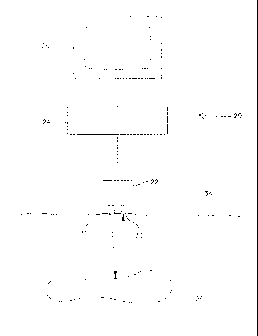
Brief Description of the Drawings
Fig. 1 is a schematic of apparatus of the present invention for ultrasonic
imaging;
Fig. 2 is a fragmentary schematic of a transducer of the apparatus;
Fig. 3 is a schematic of a digitized scan line and corresponding intensity
pixels and feature detection pixels; and
Fig. 4 is a graph showing an example of feature gain control as a function of
cluster index.
Corresponding reference characters indicate corresponding parts throughout
the several views of the drawings.
Detailed Description of the Preferred Embodiment
Referring now to the drawings and in particular to Fig. 1, apparatus of the
present invention for ultrasonic imaging is designated in its entirety by the
reference numeral
20. The apparatus 20 generally comprises a transducer 22 operatively connected
to a
control and processor unit 24 connected to a display 26. The transducer 22
includes
elements 30, 32 arranged in an array that emit and receive ultrasonic energy,
respectively,
5b
CA 02534561 2013-09-23
under the control of the control and processor unit 24. As will be appreciated
by those skilled
in the art, in most implementations of the present invention each element 30,
32 of the
transducer 22 both emits and receives energy. The elements are numbered 30 and
32 for
5c
CA 02534561 2006-02-02
WO 2005/016444 PCT/US2004/020431
convenience in describing the path of energy to and from the transducer 22.
Although the transducer 22 may include fewer or more transmitting and
receiving
elements 30, 32 without departing from the scope of the present invention, in
one
embodiment the transducer includes between about 20 and about 128 elements
30, 32 arranged in a line that both transmit and receive ultrasonic energy.
The transducer 22 is placed on or inside the body 34 of a patient.
Element 30 emits ultrasonic energy into the body 34 of the patient. The energy
is
reflected by tissue 36 in the body 34 of the patient. Some of the reflected
energy
returns to the transducer 22 where element 32 converts the ultrasonic energy
into
an electrical signal that is sent to the control and processor unit 24. The
electrical
signal is processed by the control and processor unit 24 and an image
corresponding to features detected by the apparatus 20 is displayed on the
display
26. With the exception of the hardware and software in the control and
processor
unit 24 that performs the method of the present invention, the previously
described
apparatus is conventional and will not be described in further detail.
Although only two elements 30, 32 are illustrated in Fig. 1, those of
ordinary skill the art will appreciate that the transducer 22 includes an
array of
elements as shown in Fig. 2 for emitting and receiving ultrasonic energy.
There
are a variety of conventional means to focus the emitted energy in a given
direction, referred to as an image scan line and to receive energy from that
image
scan line, while largely ignoring energy coming from other image scan lines.
One
such means, the phased array transducer, will be described below. A
conventional
planar ultrasound image is formed from a plurality of image scan lines
(usually
about 40 to about 120 image scan lines) arranged in a sector, usually
occupying
an angle from about 45 to about 90 degrees. In this case, ultrasound energy is
largely confined to a plane by the design of the transducer elements, and
further
focusing and detection described below take place largely within this plane.
A phased array transducer ignores energy coming from other image
scan lines by suitably timing ultrasound pulses emitted by each of the
plurality of
transmitting elements 30, under the control of control and processing unit 24,
so
that spherical waves generated by each of the ultrasound pulses constructively
interfere (i.e., add up) in the direction of the image scan line, and
destructively
interfere (i.e., substantially cancel out) in other directions. Ultrasound
energy is
6
CA 02534561 2006-02-02
WO 2005/016444 PCT/US2004/020431
thus effectively emitted along a given image scan line and reflected by
scatterers
located along this image scan line. The receiving process uses a similar
timing
process, under the control of control and processing unit 24, to combine
ultrasound
signals received by receiving elements 32, so as respond preferentially to
ultrasound energy reflected by scatterers along the given image scan line (so
the
signals received by the respective elements add to one another or
constructively
interfere), and to largely reject ultrasound energy reflected by scatterers
along
other image scan lines (so the signals received by the respective elements
largely
cancel each other or destructively interfere).
As will be appreciated by those skilled in the art, the received
ultrasonic energy varies in intensity according to the characteristics of the
tissue
from which the energy is reflected. The data received from each image scan
line
is arranged in a digitized scan line, generally designated by 40, as
illustrated in
Fig. 3. The scan line 40 comprises a plurality of linearly arranged signal
intensity
data points 42. In one preferred embodiment, conventional time gain
compensation is applied to adjust each of the signal intensity data points for
depth-
related attenuation.
The control and processor unit 24 includes hardware and/or software
that processes the data collected by each receiving element 32 to reduce
speckle
in the ultrasonic image sent to the display 26. The method comprises dividing
the
scan line 40 into a plurality of intensity pixels 44. Each of the intensity
pixels 44
includes at least one data point 42. Although each intensity pixel 44 may
include
fewer or more data points 42 without departing from the scope of the present
invention, in one embodiment each intensity pixel includes thirty-two (32)
contiguous data points 42. A raw intensity level and a feature gain factor are
determined for each of the intensity pixels 44. These values are multiplied to
calculate a corrected intensity level for each intensity pixel 44. The
corrected
intensity level of each of the intensity pixels 44 is selectively displayed on
the
display 26.
The step of determining the raw intensity level for each of the
intensity pixels 44 comprises rectifying a signal intensity obtained for each
data
point 42 within the respective intensity pixel and calculating an average of
the
rectified signal intensities of the data points within the intensity pixel.
Optionally,
7
CA 02534561 2006-02-02
WO 2005/016444 PCT/US2004/020431
the calculated average of the rectified signal intensities for the intensity
pixel may
be compressing using conventional procedures such as logarithmic compression.
The raw intensity level for each of the intensity pixels 44 is calculated over
time to
provide a digitized intensity level waveform for each of the intensity pixels.
The step of determining the feature gain factor for each intensity pixel
44 comprises selecting a feature detection pixel 46 corresponding to each
intensity
pixel. Each of the feature detection pixel 44 includes at least one data point
42.
Preferably, at least one of the data points included in the feature detection
pixel 44
is also included in the intensity pixel 42 to which the feature detection
pixel
corresponds. Although each feature detection pixel 46 may include fewer or
more
data points 42 without departing from the scope of the present invention, in
one
embodiment each feature detection pixel includes sixty-four (64) contiguous
data
points 42. Further, in the one embodiment the data points 42 included in the
intensity pixel 44 are centrally located in the feature detection pixel 46,
and each
intensity pixel and the corresponding feature detection pixel share a central
signal
intensity data point or a central pair of signal intensity data points. The
intensity
level waveform for each of the feature detection pixels is rectified and a
Fourier
analysis is performed on the rectified intensity level waveform to obtain
Fourier
coefficient amplitudes for each of the feature detection pixels. A power
spectrum
is computed from the Fourier coefficient amplitudes for each of the feature
detection pixels.
Once the power spectrum is computed for each feature detection
pixel, the values are scanned to determine a value of a peak power spectrum
for
the pixels. The power spectrum for each feature detection pixel is divided by
the
value of the peak power spectrum to calculate a cluster index for the feature
detection pixel. A feature gain factor is selected for each intensity pixel
based at
least in part upon the cluster index calculated for the feature detection
pixel
corresponding to the respective intensity pixel. Various methods (e.g., fuzzy
logic
or neural networks) may be used to select the feature gain factor for each
intensity
pixel.
Fig. 4 is a graph showing one example of a correlation used to select
a feature gain factor for a given cluster index. Line 50 is used to select the
feature
gain factor. Line 50 may be developed by assessing the likelihood that a
signal
8
CA 02534561 2006-02-02
WO 2005/016444 PCT/US2004/020431
received for a intensity pixel 44 is a result of energy reflected by a
structure (e.g.,
tissue 36) within the body 34 of the patient or adversely affected by
phenomena
such as speckle. Studies have shown that the probability a signal is caused by
a
structure within the body increases as the cluster index increases. Line 52
illustrates how the probability that a signal is a result of energy reflected
by a
structure (e.g., tissue 36) within the body 34 of the patient varies with
cluster index.
In contrast, the probability a signal is caused by speckle decreases as the
cluster
index increases. Line 54 illustrates how the probability that a signal is a
result of
speckle varies with cluster index.
If desired, the corrected intensity level of each of the intensity pixels
having a feature gain factor below a predetermined level may be smoothed to
further reduce speckle. Although other levels may be used without departing
from
the scope of the present invention, in one embodiment the predetermined level
below which the corrected intensity level is smoothed is about ten percent of
a
peak compressed intensity level. The corrected intensity level may be smoothed
using conventional techniques such as averaging the corrected intensity level
with
corrected intensity levels of at least one adjacent intensity pixel. In one
embodiment, the corrected intensity level is smoothed by averaging the
corrected
intensity level with corrected intensity levels of each of the immediately
adjacent
intensity pixels.
Further, if desired the corrected intensity level of each intensity pixel
having a feature gain factor above a predetermined level may be displayed in a
contrasting color. The predetermined level may be selected to display
structures
(e.g., organs within a body of a patient) in a contrasting color. Although
other
levels may be used without departing from the scope of the present invention,
in
one embodiment the predetermined level above which the corrected intensity
level
is displayed in a contrasting color is about fifty percent of a peak corrected
intensity level of the intensity pixels.
In addition to reducing speckle, those skilled in the art will appreciate
that the methods described herein may be used to enhance imaging of features
such as perfusion. In one embodiment believed to be particularly suited for
enhancing imaging of features such as local characteristics (e.g., perfusion),
the
cluster index is not smoothed prior to selecting a feature gain factor based
on the
9
CA 02534561 2006-02-02
WO 2005/016444 PCT/US2004/020431
cluster index. However, in another embodiment believed to particularly suited
for
speckle reduction, the cluster index is smoothed such as by averaging with
cluster
indices for adjacent pixels prior to selecting a feature gain factor based on
the
cluster index.
Although the scan line may be digitized at other rates without
departing from the scope of the present invention, in one embodiment the scan
line
is digitized at a rate equal to about four times a center frequency of the
ultrasonic
energy reflected by structures within the body. In one embodiment this rate
equals
about thirty megahertz and each of the signal intensity data points has a
length
equal to about 25.7 micrometers.
In contrast conventional methods, the most preferred embodiment of
the method described above does not include averaging. Thus, the method of the
present invention preserves small-scale relative intensity variations due to,
for
example, boundaries, while altering the display characteristics (e.g.,
intensity or
color) according to the source of the variability (e.g., tissue boundary
versus
speckle). By not using averaging, the method of the present invention uses all
spatial frequencies in the transform domain and preserves all of the collected
data.
In one embodiment, a brightness of the displayed image is reduced in regions
with
high speckle content but smaller scale variations in brightness within those
regions
are preserved. Thus, the method may be used to remove the harmful effects of
speckle in degrading the image, while accentuating boundaries.
As will be further appreciated by those skilled in the art, in the
described embodiment the software and/or hardware of the control and processor
unit 24 performs the steps of the method described above and therefore
embodies
means for dividing the scan line into a plurality of intensity pixels, means
for
determining a raw intensity level for each of the intensity pixels, means for
determining a feature gain factor for each pixel of the intensity pixels, and
means
for calculating a corrected intensity level for each of the intensity pixels.
When introducing elements of the present invention or the preferred
embodiment(s) thereof, the articles "a", "an", "the" and "said" are intended
to mean
that there are one or more of the elements. The terms "comprising",
"including"
and "having" are intended to be inclusive and mean that there may be
additional
elements other than the listed elements.
CA 02534561 2006-02-02
WO 2005/016444 PCT/US2004/020431
As various changes could be made in the above constructions
without departing from the scope of the invention, it is intended that all
matter
contained in the above description or shown in the accompanying drawings shall
be interpreted as illustrative and not in a limiting sense.
11