Note: Descriptions are shown in the official language in which they were submitted.
CA 02538321 2006-03-09
WO 2005/027752 PCT/US2004/029978
PFO CLOSURE DEVICE WITH FLEXIBLE THROMBOGENIC JOINT AND
IMPROVED DISLODGEMENT RESISTANCE
Related Applications
[0001] This application is a continuation-in-part of U.S. Application No.
10/326,535, filed December 19, 2002, which claims the benefit of U.S.
Provisional
Application No. 60/340,858, filed on December 19, 2001.
Field of the Invention
[0002] The present invention relates generally to an occlusion device for the
closure of physical anomalies, such as a patent foramen ovate.
Background of the Invention
[0003] A patent foramen ovate (PFO), illustrated in Figure 1, is a persistent,
one-way, usually flap-like opening in the wall between the right atrium 10 and
left
atrium 12 of the heart. Because left atrial (LA) pressure is normally higher
than right
atrial (RA) pressure, the flap usually stays closed. Under certain conditions,
however,
right atrial pressure can exceed left atrial pressure, creating the
possibility that blood
could pass from the right atrium 10 to the left atrium 12 and blood clots
could enter the
systemic circulation. It is desirable that this circumstance be eliminated.
[0004] The foramen ovate serves a desired purpose when a fetus is gestating in
utero. Because blood is oxygenated through the umbilical chord, and not
through the
developing lungs, the circulatory system of the fetal heart allows the blood
to flow
through the foramen ovate as a physiologic conduit for right-to-left shunting.
After
birth, with the establishment of pulmonary circulation, the increased left
atrial blood
flow and pressure results in functional closure of the foramen ovate. This
functional
closure is subsequently followed by anatomical closure of the two over-lapping
layers
of tissue: septum primum 14 and septum secundum 16. However, a PFO has been
shown to persist in a number of adults.
[0005] The presence of a PFO is generally considered to have no therapeutic
consequence in otherwise healthy adults. Paradoxical embolism via a PFO is
considered in the diagnosis for patients who have suffered a stroke or
transient
ischemic attack (TIA) in the presence of a PFO and without another identified
cause of
ischemic stroke. While there is currently no definitive proof of a cause-
effect
CA 02538321 2006-03-09
WO 2005/027752 PCT/US2004/029978
relationship, many studies have confirmed a strong association between the
presence
of a PFO and the risk for paradoxical embolism or stroke. In addition, there
is
significant evidence that patients with a PFO who have had a cerebral vascular
event
are at increased risk for future, recurrent cerebrovascular events.
[0006] Accordingly, patients at such an increased risk are considered for
prophylactic medical therapy to reduce the risk of a recurrent embolic event.
These
patients are commonly treated with oral anticoagulants, which potentially have
adverse
side effects, such as hemorrhaging, hematoma, and interactions with a variety
of other
drugs. The use of these drugs can alter a person's recovery and necessitate
adjustments in a person's daily living pattern.
[0007] In certain cases, such as when anticoagulation is contraindicated,
surgery may be necessary or desirable to close a PFO. The surgery would
typically
include suturing a PFO closed by attaching septum secundum to septum primum.
This
sutured attachment can be accomplished using either an interrupted or a
continuous
stitch and is a common way a surgeon shuts a PFO under direct visualization.
[0008] Umbrella devices and a variety of other similar mechanical closure
devices, developed initially for percutaneous closure of atrial septal defects
(ASDs),
have been used in some instances to close PFOs. These devices potentially
allow
patients to avoid the side effects often associated with anticoagulation
therapies and
the risks of invasive surgery. However, umbrella devices and the like that are
designed for ASDs are not optimally suited for use as PFO closure devices.
[0009] Currently available septal closure devices present drawbacks, including
technically complex implantation procedures. Additionally, there are not
insignificant
complications due to thrombus, fractures of the components, conduction system
disturbances, perforations of heart tissue, and residual leaks. Many devices
have high
septal profile and include large masses of foreign material, which may lead to
unfavorable body adaptation of a device. Given that ASD devices are designed
to
occlude holes, many lack anatomic conformability to the flap-like anatomy of
PFOs.
Thus, when inserting an ASD device to close a PFO, the narrow opening and the
thin
flap may form impediments to proper deployment. Even if an occlusive seal is
formed, the device may be deployed in the heart on an angle, leaving some
components insecurely seated against the septum and, thereby, risking thrombus
2
CA 02538321 2006-03-09
WO 2005/027752 PCT/US2004/029978
formation due to hemodynamic disturbances. Finally, some septal closure
devices are
complex to manufacture, which may result in inconsistent product performance.
[0010] The present invention is designed to address these and other
deficiencies of prior art septal closure devices.
Brief Summary of Embodiments of the Invention
[0011] Various embodiments of the present invention are directed to devices
for closing septal defects such as PFOs. The closure devices generally include
a
proximal anchor member, a distal anchor member, and a flexible center joint
connecting the two anchor members. The center joint may be one or more
sutures.
Alternatively, the center joint may be a flexible elastomeric layer, which may
promote
tissue ingrowth or deliver drugs. The flexible material may also be covered
with a
biocompatible material to promote adherence to tissue or with growth factors
to
accelerate tissue ingrowth.
[0012] In accordance with some embodiments of the invention, the closure
device is formed of bioresorbable components such that substantially no
permanent
foreign material remains in the body.
[0013] In accordance with other embodiments of the invention, the proximal
andlor distal anchor members of the closure device may include a generally
cylindrical
member split along the center portion of its length to form an elongate oval
when the
ends of the member are pressed together. Of course, a variety of cross section
shapes
in addition to a circular cross section may be used. Such proximal and/or
distal anchor
members may be two-dimensional or three-dimensional. Such proximal and/or
distal
anchor members may further include a tissue scaffold.
[0014] In accordance with further embodiments of the invention, mechanisms
are provided to collapse the closure device in order to facilitate device
delivery,
removal and/or repositioning.
[0015] These and other features will become readily apparent from the
following detailed description wherein embodiments of the invention are shown
and
described by way of illustration. As will be realized, the invention is
capable of other
and different embodiments and its several details may be capable of
modifications in
3
CA 02538321 2006-03-09
WO 2005/027752 PCT/US2004/029978
various respects, all without departing from the invention. Accordingly, the
drawings
and description are to be regarded as illustrative in nature and not in a
restrictive or
limiting sense.
Brief Description of the Drawings
[0016] FIGURE 1 is a cross-sectional view of a portion of the heart
illustrating
a PFO;
[0017] FIGURE 2 illustrates a deployed PFO closure device with bioresorbable
components in accordance with one or more embodiments of the invention;
[0018] FIGURE 3 illustrates the PFO closure device of FIGURE 2 in a
collapsed state for passage through a delivery catheter or sheath;
[0019] FIGURE 4 illustrates a closure device deployed to close a PFO in
accordance with one or more further embodiments of the invention;
[0020] FIGURE 5 illustrates a closure device deployed to close the PFO in
accordance with one or more further embodiments of the invention;
[0021] FIGURES 6A and 6B are front and side views, respectively, of a PFO
closure device in accordance with one or more further embodiments of the
invention;
[0022] FIGURES 7A and 7B are front and side views, respectively, of a PFO
closure device in accordance with one or more further embodiments of the
invention;
[0023] FIGURES 8A and 8B are side and front views, respectively, of the PFO
closure device of FIGURE 6 deployed to close a PFO;
[0024] FIGURES 9A illustrates a closure device having a retrieval mechanism
in accordance with one or more further embodiments of the invention in a
collapsed
state for passage through a catheter or sheath;
[0025] FIGURE 9B is a front view of the FIGURE 9A device;
[0026] FIGURES 9C-E illustrate deployment of the FIGURE 9A device;
[0027] FIGURES 9F-H illustrate removal of the FIGURE 9A device;
[0028] FIGURE l0A illustrates a closure device having a retrieval mechanism
in accordance with one or more further embodiments of the invention in a
collapsed
state for passage through a catheter or sheath;
[0029] FIGURE lOB is a front view of the FIGURE l0A device;
4
CA 02538321 2006-03-09
WO 2005/027752 PCT/US2004/029978
[0030] FIGURES 11A and 11B illustrate an anchor member with an elastic
hinge in accordance with one or more further embodiments of the invention;
[0031] FIGURE 12 illustrates a PFO closure device made from a single
material in accordance with one or more further embodiments of the invention;
[0032] FIGURE 13 illustrates a PFO closure device having inflatable anchor
members in accordance with one or more further embodiments of the invention;
[0033] FIGURE 14 illustrates a PFO closure device with a wire connecting the
proximal and distal anchor members in accordance with one or more further
embodiments of the invention;
[0034] FIGURE 15 illustrates a PFO closure device having a frame member in
accordance with one or more further embodiments of the invention;
[0035] FIGURE 16 illustrates a PFO closure device having frame anchor
members in accordance with one or more further embodiments of the invention;
[0036] FIGURE 17 illustrates a PFO closure device having frame anchor
members in accordance with one or more further embodiments of the invention;
[0037] FIGURE 18 illustrates the FIGURE 17 device in a collapsed state for
passage through a catheter or sheath;
[0038] FIGURE 19 illustrates a frame anchor member having metal and
polymer components in accorda~lce with one or more further embodiments of the
invention;
[0039] FIGURES 20A and 20B illustrate a PFO closure device having anchor
members formed from a rolled material in accordance with one or more further
embodiments of the invention in rolled and unrolled positions, respectively;
[0040] FIGURES 21A and 21B illustrate an alternate PFO closure device
having anchor members formed from a rolled material in accordance with one or
more
further embodiments of the invention in rolled and unrolled positions,
respectively;
[0041] FIGURE 22A illustrates a closure device having frame anchor members
and a generally "X" shaped joint member in accordance with one or more further
embodiments of the invention;
[0042] FIGURE 22B illustrates the proximal anchor member of the FIGURE
22A device;
[0043] FIGURE 22C illustrates the FIGURE 22A device in a deployed state;
5
CA 02538321 2006-03-09
WO 2005/027752 PCT/US2004/029978
[0044] FIGURE 23 illustrates a closure device having frame anchor members
having a generally "+" shaped frame structure in accordance with one or more
further
embodiments of the invention;
[0045] FIGURE 24 illustrates a closure device having frame anchor members
having a generally "G" shaped frame structure in accordance with one or more
further
embodiments of the invention;
[0046] FIGURE 25 is a perspective view of a two-dimensional closure device
with anchor members having an elongate oval configuration in accordance with
one or
more further embodiments of the invention;
[0047] FIGURE 26 is a cross-sectional end view taken along line 26-26 of the
two-dimensional closure device of FIGURE 25;
[0048] FIGURE 27 is a schematic view of the two-dimensional closure device
of FIGURE 25 deployed at a delivery site ih vivo;
[0049] FIGURE 28 is a schematic end view of a three-dimensional closure
device with anchor members having an elongate oval configuration in accordance
with
one or more further embodiments of the invention;
[0050] FIGURE 29 is a schematic end view of a three-dimensional closure
device with anchor members having an elongate oval configuration in accordance
with
one or more further embodiments of the invention;
[0051] FIGURE 30 is a schematic end view of a three-dimensional closure
device with anchor members having an elongate oval configuration in accordance
with
one or more further embodiments of the invention;
[0052] FIGURE 31 is a schematic end view of a three-dimensional closure
device with anchor members having an elongate oval configuration in accordance
with
one or more further embodiments of the invention;
[0053] FIGURE 32 is a schematic view of the three-dimensional closure
device of FIGURE 29 deployed at a delivery site in vivo;
[0054] FIGURE 33 is a perspective view of a two-dimensional closure device
in accordance with one or more further embodiments of the invention;
[0055] FIGURE 34 is a cross-sectional view taken along line 34-34 of the two-
dimensional closure device of FIGURE 33;
6
CA 02538321 2006-03-09
WO 2005/027752 PCT/US2004/029978
[0056] FIGURE 35 is a perspective view of a two-dimensional closure device
in accordance with one or more further embodiments of the invention;
[0057] FIGURE 36 is a cross-sectional end view taken along line 36-36 of the
two-dimensional closure device of FIGURE 35;
[0058] FIGURE 37 is a schematic perspective view of the two-dimensional
closure device of FIGURES 25 and 26 in a collapsed state and inserted into a
catheter;
[0059] FIGURES 38-41 are schematic views of a method for delivering a
closure device to an intended delivery site if2 vivo according to one or more
further
embodiments of the invention;
[0060] FIGURE 42 is a schematic view of a method for repositioning a closure
device at a delivery site iia vivo according to one or more further
embodiments of the
invention;
[0061] FIGURE 43-46 are schematic views of a method for retrieving a closure
device from a delivery site in vivo according to one or more further
embodiments of
the invention;
[0062] FIGURE 47 is a perspective view of a two-dimensional closure device
in accordance with one or more further embodiments of the invention;
[0063] FIGURES 48A and 48B are perspective views of a two-dimensional
closure device with anchor members having an elongate oval configuration in
accordance with one or more further embodiments of the invention;
[0064] FIGURE 49A is a perspective view of a two-dimensional closure
device with anchor members having and elongate oval configuration in
accordance
with one or more further embodiments of the invention; and
[0065] FIGURE 49B is a schematic view of the two-dimensional closure
device of FIGURE 48A deployed at a delivery site ita vivo.
[0066] FIGURE 50 is a perspective view of a two-dimensional closure device
with anchor members having an elongate oval configuration in accordance with
one or
more further embodiments of the invention; and
[0067] FIGURE 51 is a schematic view of the two-dimensional closure device
of FIGURE 49 deployed at a delivery site ifa vivo.
7
CA 02538321 2006-03-09
WO 2005/027752 PCT/US2004/029978
Detailed Description of Embodiments
[0068] Various embodiments of the present invention are directed to methods
and devices for closing septal defects such as PFOs, primarily by eliciting a
healing
response at the defect. The device may have various configurations that, in
general,
include an anchor member on each side of the septal defect with at least one
connecting member between the anchor members that joins the anchor members.
The
at least one connecting member may have one of several configurations that
promotes
a healing response in the defect.
[0069] As shown in FIGURE 2, a PFO closure device 18 in accordance with
one or more embodiments of the present invention includes a distal anchor
component
or member 20 (which can be placed on the left atrial side of the PFO), a
proximal
anchor member 22 (to fix the device in place), a proximal attachment point 24
(for
attachment and release from a catheter), and a central connecting member 26
(which
can, for example, be a simple suture in accordance with this embodiment).
[0070] In some embodiments, the distal anchor, the proximal anchor, and the
connecting member are bioresorbable. These components can be fabricated from
either a single bioresorbable polymer or by a laminated composite of two or
more
materials to provide a unique mix of properties such as, for example, anchor
members
having stiff centers and flexible edges, and blood contacting surfaces having
controlled
porosity or surface texture to promote fast and thorough endothelialization,
while
minimizing thrombosis. In addition, the tissue-contacting surface of the
anchors can
be designed to provide added stability by, for example, being roughened.
[0071] The distal anchor 20 is an elongated, preferably generally cylindrical,
thin bar-like member with rounded, arcuately shaped ends. The tissue
contacting
surface of the anchor can be generally flattened to increase tissue surface
contact. In
size, the distal anchor component might, for example, be 15-30 mm long and 2
mm in
diameter with a circular cross-section. The proximal anchor 22 can be of
similar
dimensions and shape, although it can be shorter in overall length.
[0072] Other distal and proximal anchor structures are also possible. For
example, the anchors can be formed of a generally flat material rolled to form
a
cylindrical shape as described below with respect to the embodiments of
FIGURES 20
and 21.
8
CA 02538321 2006-03-09
WO 2005/027752 PCT/US2004/029978
[0073] For delivery and deployment, the distal anchor 20 and proximal anchor
22 are positioned to be generally aligned in a longitudinal, end-to-end manner
within a
delivery sheath or catheter 28 as shown in FIGURE 3. These components, with
the
flexible connecting member 26, traverse the catheter or delivery sheath in
this
longitudinal orientation. The catheter or delivery sheath is inserted between
septum
primum and septum secundum into the left atrium 18, and the distal anchor
component
20 is ejected. Then, the catheter or delivery sheath 28 is withdrawn into the
right
atrium, and the proximal anchor 22 is ejected. The flexible central connecting
member
26 extends between septum primum and septum secundum to join the distal anchor
20
and the proximal anchor 22. Once ejected, the distal anchor and proximal
anchor
generally self-orientate to be essentially perpendicular to the axis of the
central
connecting member and in generally parallel planes to one another. The exact
orientation will be governed by the individual patient's anatomy. The terms
"withdrawn" and "ejected" are relative and are intended to generically
describe the
relative movement of the device with respect to the delivery catheter.
[0074] An alternate delivery method for this device can be to deploy it
directly
through the septum primum as opposed to through the PFO.
[0075] The method of attaching the central connecting member 26 to the
anchor and stop mechanism 22 to permit the distal anchor and the proximal
anchor to
be drawn together could be, for example, via a friction fit or via a slip knot
on the
central connecting member. If a slip knot is used, the flee end of the suture
proximal
to the knot can be held remotely and released after the knot has been placed
in the
appropriate location.
[0076] In one or more alternate embodiments of the invention shown in
FIGURE 4, the central connecting member 26 is mounted to permit free sliding
movement of the proximal anchor 22 relative to the central connecting member
26. A
biasing spring 30, which may be an expandable coil spring, can be formed at
the outer
end of the central connecting member 26 to bias the proximal anchor toward the
distal
anchor when both are deployed from the catheter or sheath.
[0077] In the embodiments illustrated in FIGURES 4 and 5, a metallic
component may be used as the central connecting member 26 in order to provide
an
appropriate stop and apply compression force to the proximal anchor 22. The
metallic
9
CA 02538321 2006-03-09
WO 2005/027752 PCT/US2004/029978
component could be a piece of shape memory wire that has one end molded or
laminated into the distal anchor component 20. In FIGURE 4, the proximal
anchor 22
slides on the central connecting member 26, and once it is deployed, the
biasing spring
30 formed on the end of the shape memory wire expands to bias the proximal
anchor
22 toward the distal anchor 20.
[0078] In the FIGURE 5 embodiment, a shape memory wire forms a hook type
anchor 32 made from two wires that exit through the center of the proximate
anchor
and curve in opposite directions when expanded to draw the proximate anchor
toward
the distal anchor.
[0079] While the embodiments of FIGURES 4 and 5 can leave a permanent
foreign body when the bioresorbable components dissolve (if, for example, a
metallic
component is used as the central connecting member 26), one advantage of these
devices is that no thrombogenic tissue scaffold (usually a vascular material)
is placed
on the left atrial side. Thrombus forming on the LA side of a PFO closure
device can
be released into the systemic circulation causing an embolic event within the
coronary
arteries, cerebral circulation, or distally in the vasculature, and most
vascular graft
materials utilized to close PFOs are highly thrombogenic.
[0080] The PFO closure devices may need to be capable of x-ray visualization
and use with radiopaque fillers or marker bands, which may be fabricated from
noble
metals such as platinum or gold. These markers can be attached using a variety
of
common methods such as, for example, adhesive bonding, lamination between two
layers of polymer, or vapor deposition.
[0081] FIGURES 6A and 6B illustrate a closure device 50 in accordance with
one or more further embodiments of the invention. The device 50 includes
proximal
and distal anchor members 52, 54 connected with a flexible (and preferably
stretchable
elastomeric) center joint or connecting element 56. The anchor members 52, 54
are
preferably cylindrical in shape with rounded ends. In size, the distal anchor
member 54
might, for example, be about 15-30 mm long and about 2 mm in diameter with a
circular cross-section. The proximal anchor 52 can be of similar dimensions
and
shape, although it can be shorter in overall length. The anchor members 52, 54
are
preferably made from a relatively rigid (preferably bioresorbable) polymer
(regular or
shape memory), or biological tissue. Biocompatible metal can also be used.
CA 02538321 2006-03-09
WO 2005/027752 PCT/US2004/029978
[0082] Other distal and proximal anchor structures are also possible. For
example, the anchors can be formed of a generally flat material rolled to form
a
cylindrical shape as described below with respect to the embodiments of
FIGURES 20
and 21.
[0083] The center joint 56 of the FIGURE 6 device (as well as the center
joints
of the devices shown in FIGURES 7-10, 12-18, and 21-24) are preferably
elastomeric
and resilient and are made from thrombogenic or inflammatory materials
including, for
example, polyester, biological tissue, bioresorbable polymer, small diameter
springs
(e.g., Nitinol springs), or spongy polymeric material. Alternatively, the
center joint
can be made of multiple strands of material 58 such as, for example, polymer
fibers as
shown in the closure device 60 of FIGURES 7A and 7B. The center joint can be
textured, porous or in a form of a single or double-sided hook material such
as Velcro.
These kinds of surfaces produce inflammatory responses and therefore, promote
faster
tissue ingrowth and faster defect closure. The entire device or parts of it
can be made
from bioresorbable polymers.
[0084] FIGURE 8A and 8B are front and side views, respectively, of the
device 50 in a PFO defect. The proximal and distal anchor members 54, 52 are
longer
than the defect width, thereby inhibiting the device from being embolized.
[0085] In accordance with further embodiments of the invention, a closure
device can include a deliverylremoval mechanism to facilitate device delivery,
removal or repositioning. A device 70 shown in FIGURES 9A and 9B includes a
removal string 72 and a delivery string 74. The removal string is movably
secured and
slides freely inside of the proximal anchor member 76. The string extends from
one
end of the proximal member 76 and is fixed to an opposite end of the distal
anchor
member 78. By pulling on the free end of the removal string 72, the whole
device 70
can be collapsed and pulled into the delivery sheath 79 as shown in FIGURE 9A.
The
strings can, for example, be sutures or wires such as Nitinol wire.
[0086] The delivery and removal strings are manipulated separately in order to
deploy or remove the device. FIGURES 9C-E illustrate device deployment using
the
delivery string 74, which is preferably attached generally to the center of
the proximal
anchor member 76. The delivery sheath 79 containing the device 70 is first
inserted
between the septum primum and septum secundum into the left atrium as shown in
11
CA 02538321 2006-03-09
WO 2005/027752 PCT/US2004/029978
FIGURE 9C. As shown in FIGURE 9D, the distal anchor 78 is then ejected from
the
delivery catheter 79. Tension is then applied to the delivery string 74, and
the delivery
sheath is withdrawn into the right atrium and the proximal anchor 76 is
ejected.
Applying tension to the delivery string enables the proximal anchor 76 to be
properly
deployed in the right atrium, and keeps the anchor 76 from being ejected into
the left
atrium. Upon successful deployment of the device 70, both strings are released
and
the delivery system is withdrawn. No tension is applied to the removal string
during
delivery.
[0087] FIGURES 9F-H illustrate removal of the device 70. As shown in
FIGURE 9F, tension is applied to the removal string, while the delivery sheath
79 is
moved toward the device 70. The applied tension causes the proximal anchor 76
to be
withdrawn into the delivery sheath as shown in FIGURE 9G. The distal anchor 78
is
also withdrawn into the delivery sheath as further tension is applied to the
removal
string. The device can then be redeployed if desired or removed.
[0088] Alternatively, the delivery string 74 can be omitted, and the removal
string 72 can be used for both device deployment and removal. The delivery
sheath 79
containing the closure device is first inserted between septum primum and
septum
secundum into the left atrium in a similar manner to that shown in FIGURE 9C.
The
distal anchor 78 is then ejected from the delivery catheter 79 in a similar
manner to
that shown in FIGURE 9D. Tension is applied to the removal string 72, and the
delivery sheath is withdrawn into the right atrium, and the proximal anchor 76
is
ejected. Applying tension to the removal string enables the proximal anchor 76
to be
properly deployed in the right atrium and keeps the proximal anchor 76 from
being
ejected into the left atrium. The elasticity of the center joint connecting
the anchor
members helps properly position the proximal anchor at the defect. Upon
successful
deployment of the closure device, the string 72 is released and the delivery
system is
withdrawn.
[0089] As shown in FIGURES l0A and lOB, in another embodiment, strings
80 (suture, Nitinol wire, etc.) are attached to both ends of the proximal
anchor member
82 of a closure device 84. Both anchor members are flexible and can fold as
shown in
FIGURE 10A in order to be delivered to or removed from the defect.
12
CA 02538321 2006-03-09
WO 2005/027752 PCT/US2004/029978
[0090] In accordance with a further embodiment of the invention, as shown in
FIGURES 11A and 11B, each of the proximal and distal anchor members can
include
two elements 90 separated by an elastic hinge 92. The elastic hinge 92 can
facilitate
folding of the members as shown in FIGURE 11B. The hinge 92 can be molded or
made from a material such as, for example, Nitinol or other shape memory
materials,
which can be a different material from the elements 90.
[0091] In accordance with some embodiments of the invention, an entire
closure device can be made from a single sheet of a material as shown, for
example, in
the closure device 100 of FIGURE 12. Two opposite ends of the sheet can be
rolled to
form the proximal and distal anchor members. Glue or heat bonding can be used
to
maintain the rolled-up configuration of the anchor members 102, 104.
[0092] As shown in FIGURE 13, in accordance with some further
embodiments of the invention, one or both anchor members 110, 112 of a closure
device 114 can be inflatable. The anchor members can be inflated with, for
example,
saline or other physiological fluid during or before the delivery of the
device. A tube
116 can communicate with cavities in the anchor members. An inlet 118 can be
provided at one of the members for introducing fluid therein.
[0093] In accordance with some further embodiments of the invention, a wire
120 such as, for example, an S-shaped wire, can be provided to connect the
proximal
and distal anchor members 122, 124 of a device 126 as shown in FIGURE 14. The
wire can be used to provide additional clamping force while the device is in a
PFO
defect. Other wire shapes are also possible.
(0094] In accordance with further embodiments of the invention, one or more
frame structures can be used as the anchor members of a closure device. For
example,
FIGURE 15 shows a closure device 130 having a frame structure 132. Also,
FIGURE
16 shows a closure device 136 having frames 138, 139. The frames can be, for
example, a metal (e.g., Nitinol wire) or polymer frame.
[0095] FIGURES 17-19 illustrate closure devices in accordance with some
further embodiments of the invention. A closure device 140 shown in FIGURE 17
includes anchor members 142, 144 having a frame structure. The frame shape can
be
polygonal as shown in the figure or it can alternatively be a circular shape.
Other
13
CA 02538321 2006-03-09
WO 2005/027752 PCT/US2004/029978
frame shapes are also possible as, for example, will be described below with
respect to
FIGURES 22-24.
[0096] A recovery suture can be attached to opposite ends of the proximate
anchor member 142 to collapse the anchors for delivery in a catheter 146 as
shown in
FIGURE 18 or for retrieval or repositioning. The anchor members can be made
from a
metal, preferably Nitinol, or polymers. Alternatively, as shown in FIGURE 19,
an
anchor member 148 can include both metal and polymer components.
[0097] In accordance with one or more further embodiments of the invention,
the distal and proximal anchors can be formed of a flat sheet-like member
rolled to
form a cylindrical shape as shown, for example, in the device 170 of FIGURE
20A.
The anchors 172, 174 can unroll to form sheet-like members when deployed, as
shown
generally in FIGURE 20B. The sheet-like member can be made of a material
having
shape memory properties such as, for example, shape memory polymeric
materials.
Alternately, the sheet-like member can include metal struts made of shape
memory
metals such as, for example, Nitinol or Nitinol alloys. The shape memory
materials
allow the device to be delivered in a delivery sheath or catheter with the
anchors in the
rolled configuration of FIGURE 20A. The anchors attain the sheet-like geometry
of
FIGURE 20B once deployed due to their shape memory properties. The anchor
members 172, 174 can be connected to each other with a connecting member 176,
which can, for example, be a suture similar to that used in the FIGURE .2
device.
[0098] FIGURES 21A and 21B illustrate a closure device 180 having rolled
anchor members 182, 184, which are similar to the anchor members 172, 174 of
the
device of FIGURES 20A and 20B. The anchors 182, 184 are connected to each
other
by a connecting member or joint 186, which can be a sheet of flexible material
similar
to the connecting members previously described with respect to FIGURES 6 and
7.
[0099] FIGURE 22A illustrates a closure device 200 in accordance with one or
more further embodiments of the invention. The device 200 includes distal and
proximal anchor members 202, 204, each of which has a polygonal or circular
frame
structure. The anchor members are connected by a connecting member 206, which
can
be made from a flexible material similar to that previously described in
connection
with FIGURES 6 and 7. The connecting member 206 can be made of two sheets of
flexible material connected at their centers, generally forming an "X" shape
in the side
14
CA 02538321 2006-03-09
WO 2005/027752 PCT/US2004/029978
view of the device. As shown in FIGURE 22B, the proximal anchor member 204 can
include one or more recovery wires or sutures attached to the frame structure
for use in
device deployment of recovery. FIGURE 22C illustrates the device 200 as
deployed.
[0100] FIGURES 23 and 24 illustrate closure devices 220, 230, respectively, in
accordance with further embodiments of the invention. Each device 220, 230
includes
distal and proximal anchor members having a frame structure. The anchor
members
are connected by a flexible joint 222, which can be made from a flexible
material
similar to that previously described in connection with FIGURES 6 and 7. The
FIGURE 23 device 220 includes distal and proximal anchor members 224, 226
generally having a "+" shape. The FIGURE 24 device 230 includes distal and
proximal anchor members 232, 234 generally having a "G" shape.
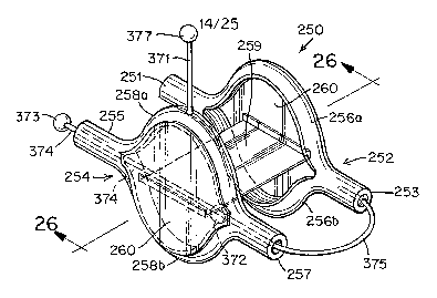
[0101] In still further embodiments of the closure device 250 according to the
present invention, the distal and/or proximal anchor members 252 and 254,
respectively, may be formed of cylindrical structures, split along the central
portion of
their length to provide elongate ovals (i.e., an "open-mouthed" configuration)
as
shown in FIGURES 25-27. In this elongate oval configuration, arcs 256 and 258
are
joined by ends 251, 253 and 255, 257, respectively (FIGURE 25). This
configuration
increases the size and surface area of the anchor member, thereby improving
the
dislodgement resistance of the closure device 250. As used herein,
"dislodgement
resistance" refers to the ability of a closure device to resist the tendency
of the force
applied by the unequal pressures between the right atrium 10 and the left
atrium 12
(i. e. the "dislodging force") to separate the closure device from the septal
tissue.
Generally, a high dislodgement resistance is desirable.
[0102] Distal andlor proximal anchor members 252 and 254 having this
elongate oval configuration may be either two-dimensional (FIGURES 25-27) or
three-dimensional (FIGURES 28-32). As shown in FIGURE 28, in the three-
dimensional configuration, the arcs 258a and 258b of proximal anchor member
254 are
predisposed to bend at an angle A from the plane A of the two-dimensional
proximal
anchor member 254. Arcs 258a and 258b may bend at an angle 0 either toward or
away from center joint 259 (FIGURES 29 and 30, respectively). In particular
embodiments, both distal anchor 252 and proximal anchor 254 are three-
dimensional.
In such embodiments, arcs 256a and 256b of distal anchor member 252 and arcs
258a
CA 02538321 2006-03-09
WO 2005/027752 PCT/US2004/029978
and 258b of proximal anchor member 254 may bend at the same angle 8 or at
different
angles edistal and eproximal, respectively. Further, arcs 256a, 256b and 258a,
258b may
bend toward center joint 259 (FIGURE 29), away from center joint 259 (FIGURE
30),
or in opposite directions (i.e., one toward center joint 259 and one away from
center
joint 259, as shown in FIGURE 31). As shown in FIGURES 28-32, arcs 256a, 256b
and 258a, 258b include a straight bend; however, arcs 256a, 256b and 258a,
258b may
also include a curved bend such that they are concave or convex. One skilled
in the art
will further recognize that, in a three-dimensional configuration, ends 251,
253 and
255, 257 may also be bent as described above for arcs 256a, 256b and 258a,
258b.
[0103] In some clinical applications, a three-dimensional configuration of
distal anchor member 252 andlor proximal anchor member 254 may be particularly
advantageous. For example, septum primum 14 and septum secundum 16 are
typically
of disparate thickness, as shown in FIGURE 32. Consequently, the septal tissue
in the
right atrium 10 is characterized by a step-like surface (indicated by line
LRA). The
septal tissue in the left atrium 12 may also be characterized by a similar
step-like
surface (indicated by line LLA). Insertion of a closure device including a two-
dimensional a~ichor into a PFO surrounded by such step-like septal tissue
often results
in undesirable seating of that anchor member against the septal tissue, in
that at least
one are of each anchor member does not contact the septal tissue, as shown in
FIGURE 27. However, the angled arcs of a three-dimensional anchor member may
more closely approximate the step-like surface of the septal tissue, as shown
in
FIGURE 32. Thus, in certain clinical applications, the use of a closure device
including a three-dimensional distal anchor member 252 and/or proximal anchor
member 254 may provide improved seating of the device 250 against the septal
tissue
and, correspondingly, a reduced profile of the device 250 and more effective
closure of
the PFO. As used herein, "profile" refers to the degree to which closure
device 250
extends away from the septal tissue (i.e., septum primum 14 and septum
secundum 16)
and is exposed in the atria. A device having a "low profile" is closely seated
against
the septal tissue and extends only slightly, if at all, into the atria. A
device having a
"high profile" extends away from the septal tissue and into the atria.
Generally, a
device having a low profile is desirable because it is less thrombogenic in
vivo. One
16
CA 02538321 2006-03-09
WO 2005/027752 PCT/US2004/029978
skilled in the art will be capable of determining those clinical applications
in which the
use of three-dimensional anchor members is appropriate.
[0104] Either or both of distal anchor member 252 and proximal anchor
member 254 having the above-described elongate oval configuration may include
a
tissue scaffold 260 extending between their two arcs 256a, 256b and 258a,
258b,
respectively, as shown in FIGURE 25. The inclusion of tissue scaffolds) 260
augments the area of septal tissue covered by the anchor members 252 and/or
254.
Consequently, device 250 provides improved closure of the PFO. Moreover,
tissue
scaffold 260 promotes encapsulation and endothelialization of the septal
tissue,
thereby further encouraging anatomical closure of the PFO. The tissue scaffold
260
may be formed of any flexible, biocompatible material capable of promoting
tissue
growth, including but not limited to, polyester fabrics, Teflon-based
materials, ePTFE,
polyurethanes, metallic materials, polyvinyl alcohol (PVA), extracellular
matrix
(ECM) or other bioengineered material, synthetic bioabsorbable polymeric
scaffolds,
other natural materials (e.g., collagen), or combinations of the foregoing
materials.
For example, the tissue scaffold 260 may be formed of a thin metallic film or
foil, e.g.,
a nitinol film or foil, as described in United States Patent Application No.
2003/0059640 (the entirety of which is incorporated herein by reference).
[0105] Distal anchor member 252 and proximal anchor member 254 may be
connected by a flexible center joint 259 (FIGURE 25). As previously described,
in at
least some embodiments, center joint 259 includes a stretchable elastomeric
material.
In at least some embodiments, center joint 259 includes a thrombogenic or
inflammatory material, such as polyester, biological tissue, bioresorbable
polymer,
small diameter springs, e.g., nitinol springs, spongy polymeric material, or
combinations of the foregoing materials. In at least some embodiments, center
joint
259 is textured, porous, or in the form of a single- or double-sided hook
material, such
as Velcro. These types of surfaces produce inflammatory responses and,
therefore,
promote faster tissue ingrowth and defect closure. In particular embodiments
and as
shown in FIGURE 25, center joint 259 is formed of a deformable or expandable
film,
such as those disclosed in United States Patent Application Nos. 2002/0165600
and
2002/0165576 (both of which are incorporated herein by reference). For
example,
center joint 259 may be formed of a shape memory film (e.g., nitinol film) or
a
17
CA 02538321 2006-03-09
WO 2005/027752 PCT/US2004/029978
polymeric film. Small openings 471, e.g., slits or holes, may be cut in the
film such
that, as the film expands upon deployment inz vivo, the openings 471 also
expand
(FIGURES 48A and 48B). In this manner, the center joint 259 is rendered more
flexible and capable of expanding significantly in length without placing
excessive
strain on the closure device (FIGURE 48B). In some embodiments, the closure
device
250 may include two flexible center joints 259a and 259b (FIGURE 33).
[0106] Center joint 259 may be of various shapes and sizes depending upon the
particular anatomy of the patient's septal tissue. For example, as shown in
FIGURE
25, center joint 259 may be generally rectangular. In other embodiments, and
as
shown in FIGURE 35, center joint 259 may be shaped generally as an "X" or
hourglass when in its relaxed configuration. Removing material from the sides
of
center joint 259 to form an hourglass shape increases its flexibility in vivo.
The
amount of material removed from the sides of a rectangular center joint 259 to
form an
hourglass shape will vary depending upon the particular application. According
to
some embodiments, between one-third and two-thirds of a rectangular center
joint 2.59
will be removed to form the corresponding hourglass center joint 259. In
particular
embodiments, approximately one-half of a rectangular center joint 259 will be
removed to form the corresponding hourglass center joint 259. In determining
the
precise amount of material to remove from the sides of a rectangular center
joint 259
to form an hourglass center joint 259, a sufficient portion of center joint
259 must be
retained to promote the healing response of the septal tissue that it contacts
in vivo.
One skilled in the art will be able to determine the precise amount of
material that may
be removed from a rectangular center joint 259 to form an hourglass center
joint 259
suitable to the patient's septal anatomy while sufficiently maintaining the
ability of
center joint 259 to promote the healing of the septal tissue.
[0107] Center joint 259 may be connected to distal and proximal anchor
members 252 and 254, respectively (FIGURES 35 and 36), or, if present, to
tissue
scaffolds 260 (FIGURE 25). Center joint 259 may connect to tissue scaffolds
260 at
their centers (FIGURE 25), at a location on their peripheries (FIGURES 33 and
34), or
somewhere in between (FIGURE 48A). In particular embodiments, center joint 259
is
connected at a location between the center and a periphery of tissue scaffold
260 on
distal anchor member 252 and at a location between the center and opposite
periphery
18
CA 02538321 2006-03-09
WO 2005/027752 PCT/US2004/029978
of tissue scaffold 260 on proximal anchor member 254 (FIGURE 48A) so as to
more
closely approximate the angled, tunnel-like anatomy of the PFO and reduce the
profile
of closure device 250 ifa vivo (FIGURE 48B). For example, as shown in FIGURES
48A and 48B, center joint 259 may be connected to the tissue scaffold 260 of
distal
anchor member 252 at a location between the center of the tissue scaffold 260
and the
arc 256a and connected to the tissue scaffold 260 of proximal anchor, member
254 at a
location between the center of tissue scaffold 260 and the arc 258b.
[0108] A closure device including a distal anchor member 252 andlor proximal
anchor member 254 having an elongate oval configuration may be deployed or
retrieved if arcs 256a, 256b and/or 258a, 258b, respectively, are collapsed to
reduce
the profile of closure device 250 such that it may be drawn into and contained
within a
delivery or retrieval catheter 370 (FIGURES 37-46). According to one
embodiment
and as shown in FIGURE 25, closure device 250 may include a delivery string
371.
As shown in FIGURE 25, delivery string 371 is permanently attached to arc 258a
of
proximal anchor member 254, although one of skill in the art will recognize
that
delivery string 371 may be attached anywhere on proximal anchor member 254.
Delivery string 371 may be attached in any suitable manner, for example,
through a
drilled hole, via glue, etc. Delivery string 371 is short (i.e., several
millimeters) and as
least thrombogenic as possible. As used herein, "string" includes various
materials,
which may be stiff or flexible. Delivery string 371 terminates in a ball 377
at its free
end. Closure device 250 further includes a recovery ball 373 attached to
recovery
string 374, which is threaded through ends 255 and 257 of proximal anchor
member
254 and subsequently attached to end 253 of distal anchor member 252. Slack
375
exists in recovery string 374 between end 253 of distal anchor member 252 and
end
257 of proximal anchor member 254. Closure device 250 still further includes a
ball
372 attached to recovery string 374 and contained between ends 255 and 257 of
proximal anchor member 254. Ends 255 and 257 of proximal anchor member 254
may have an inner diameter greater than that of ball 372 but are tapered such
that the
terminal segment of ends 255 and 257 have a diameter smaller than that of ball
372.
Thus, the movement of ball 372 is constrained between ends 255 and 257 of
proximal
anchor member 254.
19
CA 02538321 2006-03-09
WO 2005/027752 PCT/US2004/029978
[0109] Prior to deployment ira vivo, device 250 must be placed within delivery
catheter 370 (FIGURE 37). Device 250 may be loaded into catheter 370 in any
manner such that slack 375 is maintained in recovery string 374 between distal
anchor
member 252 and proximal anchor member 254, as shown in FIGURE 37. For
example, device 250 may be manually loaded into catheter 370. One skilled in
the art
will be capable of identifying suitable methods for loading device 250 into
catheter
370.
[0110] One of skill in the art will, of course, recognize that the maximum
amount of slack 375 in the recovery string 374 is dependent upon the distance
ball 372
may travel between ends 255 and 257 of proximal anchor member 254. Slack 375
increases as ball 372 travels closer toward the terminus of end 257. Thus, the
amount
of slack 375 may be adjusted by altering the tapering of the internal diameter
of ends
255 and 257. Additionally, the slit 480 splitting ends 255 and 257 of proximal
anchor
member 254 into arcs 258a and 258b may be extended toward the termini of ends
255
and 257 so as to maximize the distance ball 372 may travel within proximal
anchor
member 254 and, correspondingly, the slack 375 (FIGURE 47).
[0111] Device 250 may be delivered to its intended delivery site in vivo by
various methods, only one of which will be described herein. As shown in
FIGURE
38, the clinician holds both recovery ball 373 and delivery ball 377 by
suitable
devices, e.g., grips 376 and 401. As used herein, the terms "ball" and "grips"
are used
to generically describe the delivery mechanism. One skilled in the art will
recognize
that the precise structure of the delivery mechanism components may vary.
Grips 376
and 401 permit the clinician to apply tension or compression to delivery
string 371 or
recovery string 374 as desired to properly manipulate device 250. Generally,
during
delivery of device 250 by the method described herein, tension will be applied
only to
delivery string 371; recovery string 374 will be held in a relaxed
configuration such
that slack 375 is maintained. Once the clinician is properly holding both
recovery ball
373 and delivery ball 377, catheter 370 is delivered through the patient's
vasculature to
the right atrium 10 of the heart (FIGURE 38). Then, as shown in FIGURE 39,
catheter
370 is inserted between septum primum 14 and septum secundum 16 into the left
atrium 12. Distal anchor member 252 is ejected into the left atrium 12 by
pushing on
grips 401, and arcs 256a and 256b reassume their elongate oval configuration
CA 02538321 2006-03-09
WO 2005/027752 PCT/US2004/029978
(FIGURE 39). Catheter 370 is withdrawn between septum primum 14 and septum
secundum 16 and into the right atrium 10, such that proximal anchor member 254
is
deployed into the right atrium 10 and slack 375 extends through the PFO
(FIGURE
40). During this process, grips 401 are maintained on delivery ball 377, and
the
necessary tension is applied to delivery string 371 (FIGURE 40). As shown in
FIGURE 40, arcs 258a and 258b reassume their elongate oval configuration upon
deployment of proximal anchor member 254 into the right atrium 10, and
proximal
anchor member 254 may be positioned as desired against the septal tissue using
grips
401. Distal anchor member 252 and proximal anchor member 254 cooperate to
apply
a compressive force to septum primum 14 and septum secundum 16, thereby
closing
the PFO (FIGURE 41). If deployment of closure device 250 is satisfactory to
the
clinician, grips 401 release delivery ball 377, grips 376 release recovery
ball 373
(FIGURE 41), and catheter 370 is withdrawn from the right atrium 10 and
further
withdrawn through the patient's vasculature.
[0112] However, if, following deployment, the clinician is not satisfied with
the position of device 250, grips 376 and grips 401 may be maintained on balls
373
and 377, respectively, so that the device 250 may be repositioned and/or
retrieved.
Device 250 may be repositioned by further manipulating the tension applied to
delivery string 371 by grips 401 (FIGURE 42). To retrieve closure device 250,
catheter 370 is positioned against end 255 (FIGURE 43). Recovery ball 373 is
pulled
into the catheter 370, such that ball 372 moves to point B of end 255 and arcs
258a and
258b of proximal anchor member 254 are collapsed and withdrawn into catheter
360
(FIGURE 44). Upon nearing complete retrieval of proximal anchor member 254,
slack 375 in string 374 is eliminated, or nearly so, and end 257 of proximal
anchor
member 254 and end 253 of distal anchor member 252 are touching, or nearly
touching, such that proximal anchor member 254 and distal anchor member 252
are
aligned in a longitudinal, end-to-end manner (FIGURE 44). Grips 376 continue
to
apply tension to recovery string 374, pulling recovery ball 373 toward the
proximal
end of catheter 370, as shown in FIGURE 45. Arcs 256a and 256b of distal
anchor
member 252 are collapsed, and distal anchor member 252 is withdrawn into
catheter
370 (FIGURE 45). Catheter 370 is then withdrawn through the PFO, and into the
right
atrium 10 (FIGURE 46).
21
CA 02538321 2006-03-09
WO 2005/027752 PCT/US2004/029978
[0113] The delivery and recovery system of device 250 may be modified in
various ways, one of which is shown in the device 490 of FIGURES 50-51. String
374
may be extended from end 255 of proximal anchor member 254 toward arc 258a, be
attached to arc 258 at a point Y, further extend from arc 258a to form
delivery/recovery string 491, and terminate in delivery/recovery ball 492
(FIGURE
50). The device 490 may be deployed as described above, except that only grips
401
would be necessary hold delivery/recovery ball 492 and manipulate the tension
applied
to delivery/recovery string 491 during delivery. To retrieve device 490, grips
401
apply sufficient tension to delivery/recovery string 491 to break its
connection to arc
258a of proximal anchor member 254 at point Y (FIGURE 50). By applying further
tension to delivery/recovery string 374 by pulling delivery/recovery ball 492
towards
the proximal end of the catheter 370, device 490 orients in a longitudinal
manner and
may be withdrawn into the catheter 370 as described previously.
[0114] The closure devices described herein can optionally be used along with
suturing or stapling techniques where the anchors or flexible joints of the
devices can
be sewn or stapled to septum primum 14 and/or septum secundum 16 for better
dislodgment resistance. Also, the flexible joint can, if desired, be covered
with a
biocompatible adhesive to adhere to the tissue or can be loaded with drugs or
growth
factors to promote healing. The adhesive and also certain drugs can also
optionally be
stored in any cavities in the anchor members 252 and/or 254 (e.g., in the
cylindrical
members of FIGURES 6 and 7) and released after deployment. Radiopaque markers
can also be attached to the closure devices for better visualization during
the
implantation procedure. One skilled in the art will recognize that a variety
of
visualization techniques may be used, including fluoroscopy and magnetic
resonance
imaging (MRI).
[0115] The various closure devices described herein may further include a
number of advantageous features. The closure devices preferably have an
atraumatic
shape to reduce trauma during deployment or removal. In addition, the devices
can be
self orienting for ease of deployment. Furthermore, because of the flexible
center
joint, the devices generally conform to the anatomy instead of the anatomy
conforming
to the devices, which is especially useful in long tunnel defects. In
addition, the
devices can preferably be repositioned and/or removed during delivery. The
devices
22
CA 02538321 2006-03-09
WO 2005/027752 PCT/US2004/029978
also generally have a relatively low profile after deployment. The flexible
center joint
259 of the devices can encourage faster tissue ingrowth and therefore, faster
defect
closure. Furthermore, there are generally no exposed thrombogenic components
in the
left 12 and right 10 atria. Still further, the devices may advantageously
include
bioresorbable components, which will disappear from the body over time.
[0116] One skilled in the art will recognize that the features of any
embodiment described herein may be combined with those of any other embodiment
described herein.
[0117] Other benefits of the devices described herein include the possible use
of a relatively small diameter delivery sheath, use of a reduced amount, or
no, metal
mass in the device, ease of manufacturing, cost effectiveness, and overall
design
simplicity.
[0118] Having described preferred embodiments of the present invention, it
should be apparent that various modifications may be made without departing
from the
spirit and scope of the invention, which is defined in the claims below.
23