Note: Descriptions are shown in the official language in which they were submitted.
CA 02538400 2006-03-O1
BIOPSY DEVICE INCORPORATING AN ADJUSTABLE PROBE
SLEEVE
FIELD OF THE INVENTION
looox~ The present invention relates in general to biopsy devices, and more
particularly to
biopsy devices having a cutter for severing tissue.
BACKGROUND OF THE INVENTION
~aoo3~ The diagnosis and treatment of tissue is an ongoing area of
investigation. Medical
devices for obtaining tissue samples for subsequent sampling and/or testing
are know in
the art. For instance, a biopsy instrument now marketed under the tradename
MAMMOTOME is commercially available from Ethicon Endo-Surgery, Inc. for use in
obtaining breast biopsy samples.
looosl The following patent documents disclose various biopsy devices and are
incorporated
herein by reference in their entirety: US 6,273,862 issued August 14, 2001; US
6,231,522
issued May 1 S, 2001; US 6,228,055 issued May 8, 2001; US 6,120,462 issued
September
19, 2000; US 6,086,544 issued July 11, 2000; US 6,077,230 issued June 20,
2000; US
6,017,316. issued January 25, 2000; US 6,007,497 issued December 28, 1999; US
5,980,469 issued November 9, 1999; US 5,964,716 issued October 12, 1999; US
5,928,164 issued July 27, 1999; US 5,775,333 issued July 7, 1998; US 5,769,086
issued
June 23, 1998; US 5,649,547 issued July 22, 1997; US 5,526,822 issued June 18,
1996,
US Patent Application 2003/0199753 published October 23, 2003 to Hibner et
al.; US
Patent Application Ser. No. 10/676,944, "Biopsy Instrument with Internal
Specimen
Collection Mechanism" filed September 30, 2003 in the name of Hibner et al.;
and US
CA 02538400 2006-03-O1
Patent Application Ser. No. 10/732,843, "Biopsy Device with Sample Tube" filed
December 10, 2003 in the name of Cicenas et al.
)ooosl These generally-known vacuum assisted core biopsy devices include
desirable
features wherein larger samples are drawn in by vacuum assistance and severed
by a
cutter. These larger samples have benefits over needle biopsies in obtaining a
sample
more likely to include at least a portion of a suspicious lesion for
diagnostic purposes. In
addition, some of these known biopsy devices are capable of taking multiple
samples
without having to remove the probe. This shortens the duration and
inconvenience of the
procedure between taking samples. In addition, this facilitates taking
sufficient samples to
fully excise a suspicious lesion.
looobl Long side apertures of a probe of these biopsy devices in combination
with vacuum
assistance, especially with a separate vacuum lumen, have many desirable
features.
However, there are situations in which lesions near the skin are difficult to
biopsy with a
core biopsy probe. This is more often a challenge with a small breast,
especially when
compressed in a localization fixture that limits the choice in access
direction. If the side
aperture of the probe is partially exposed, then vacuum assist may be
ineffective as the
specimen bowl in the probe is exposed to atmospheric pressure. Further, skin
may
prolapse into the specimen bowl before the cutter advances into the tissue,
causing
gouging of the skin, increasing post-procedure pain and scarring.
tooo~l Consequently, a significant need exists for a care biopsy device that
is capable of
taking biopsies of a suspicious lesion that is proximate to the skin.
SUMMARY OF THE INVENTION
loooal The present invention addresses these and other problems of the prior
art by providing
a core biopsy device having a probe assembly with a side aperture that is
selectively
longitudinally sized for taking samples. A proximal blocking member may be
selectively
positioned proximate to the side aperture such that a proximal portion thereof
is blocked
when otherwise an outer layer of skin would prolapse into the side aperture
when a cutter
tube is retracted and then be gouged as the cutter is advanced to take a
tissue sample.
Thereby, discomfort and disfiguring scarring is avoided while still retaining
the ability to
take a tissue sample of a lesion near to a patient's skin.
2.
CA 02538400 2006-03-O1
10009 In one aspect consistent with other aspects of the invention, a device
for use with a
core biopsy includes a curved portion sized to correspond to a portion of the
probe
surrounding at least the proximal portion of the side aperture which is held
thereover by
an engaging structure attached to the curved portion and registered to at
least partially
encompass and engage the probe. A flange attached to the curved portion allows
for a
user to longitudinally position the curved portion over the proximal portion
of the side
aperture when desired. Thereby, an additional capability is provided for a
biopsy device
even when its operation requires that a cutter tube fully retract to remove a
tissue sample
before a subsequent translation for taking another sample.
looiol In another aspect of the invention, a transparent sleeve probe.is
attachable to a needle
of a biopsy device and includes a distal inner contour that helps to prevent
gouging by an
advancing cutter tube so that debris is not introduced into tissue or into the
side aperture
of the biopsy device.
~ooy In yet a further aspect of the invention, a core biopsy device has a
probe sleeve
including a curved portion sized to correspond to a portion of the probe of
the core biopsy
device surrounding at least the proximal portion of the side aperture which is
held
thereover by an engaging structure attached to the curved portion and
registered to at least
partially encompass and engage the probe. A flange attached to the curved
portion allows
a user to longitudinally position the curved portion over the proximal portion
of the side
aperture when desired. Thereby, an additional capability is provided for a
biopsy device
even when its operation requires that a cutter tube fully retract to remove a
tissue sample
before a subsequent translation for taking another sample.
~ooi21 These and other objects and advantages of the present invention shall
be made
apparent from the accompanying drawings and the description thereof.
BRIEF DESCRIPTION OF THE DRAWINGS
10031 While the specification concludes with claims particularly pointing out
and distinctly
claiming the present invention, it is believed the same will be better
understood by
reference to the following description, taken in conjunction with the
accompanying
drawings in which:
3.
CA 02538400 2006-03-O1
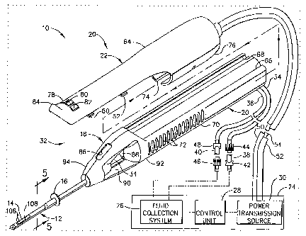
FIGURE 1 is a partial isometric and partial schematic view of a core sampling
biopsy
system with a handpiece having a long stroke cutter for the collection of soft
tissue
depicted with a holster separated from a probe assembly;
~oo~sl FIGURE 2 is an exploded isometric view of the probe assembly of FIG. 1;
~ooi6~ FIGURE 3 is an isometric view of the probe assembly with the left
handle shell
removed, showing the cutter in the first, fully retracted position, and a
tissue sample is
shown deposited onto a tissue sampling surface of the handle after the tissue
sample was
removed from the distal end of the cutter;
~ooi~~ FIGURE 4 is an exploded isometric view of the holster;
looisl FIG. 4A is a top view in section of the probe assembly aad a distal
portion of the
holster, revealing a cutter in a first, fully retracted position;
~oot9~ FIGURE 5 is a front view in elevation of the probe and probe sleeve
taken in cross
section along lines 5-5 of FIG. 1;
loozo~ FIGURE 6 is an isometric view of the probe sleeve of FIG. 1.
~oo2il FIGURE 7A is an isometric view of the probe of the biopsy system of
FIG. 1 with the
probe sleeve at a proximal position exposing a side aperture;
~oozz~ FIGURE 7B is an isometric view of the probe of the biopsy system of
FIG. 1 v~rith the
probe sleeve at a more distal position partially blocking the side aperture;
looz3l FIGURE ~C is an isometric view of the probe of the biopsy system of
FIG. 1 with the
probe sleeve at a most distal position completely blocking the side aperture;
loozal FIGURE 8A is a left side view in elevation of the probe and probe
sleeve of FIG. 7B
taken along a longitudinal centerline of lines 8-8 with vacuum assistance
being employed
to prolapse tissue into a bowl of the probe;
looz5~ FIGURE 8B is a left side view in elevation of the probe and probe
sleeve of FIG. 7B
taken along a longitudinal centerline of lines 8-8 with vacuum assistance
being employed
to prolapse tissue into a bowl and to draw severed tissue into a sample lumen
as a cutter
tube is rotated and translated in a cutter lumen of the probe;
4.
CA 02538400 2006-03-O1
~ooa6~ FIGURE 8C is a left side view in elevation of the probe and probe
sleeve of FIG. 7B
taken along a longitudinal centerline of lines 8-8 with a completely severed
tissue sample
captured in the sample lumen;
~ooZ~! FIGURE 9 is a perspective view of an alternative probe sleeve for the
core sampling
biopsy system of FIG. 1 incorporating a transparent body, measurement indicia
and a
protective insert to prevent cutter gouging;
~ooza~ FIGURE 10 is a perspective view of the transparent body of the
alternative probe
sleeve of FIG. 9;
~ooZS~ FIGURE 11 is a perspective view of the ribbed protective insert for the
alternative
probe sleeve of FIG. 9;
~0030~ FIGURE 12 is a perspective view of an alternative non-ribbed protective
insert for the
alternative probe sleeve of FIG. 9;
~oo3y FIGURE 13 is a perspective view of an alternative transparent body
having paired
transverse gripping flanges for a further alternative probe sleeve;
~oo3x~ FIGURE 14 is a perspective view of a further alternative transparent
body having
paired radial clip flanges and molded measurement indicia for a yet a further
alternative
probe sleeve;
Ioo33~ FIGURE 15 is a perspective view of an additional alternative
transparent body having
an increased diameter distal portion for an additional alternative probe
sleeve;
10034 FIGURE 16 is a left side view in elevation of the additional alternative
transparent
body of FIG. 15 taken in cross section along longitudinal centerline 16-16;
~oo3s~ FIGURE 17 is a partial isometric and partial schematic view of an
alternative biopsy
system that includes a handpiece with a short stroke cutter that is
advantageously
configured to perform a cutting stroke that blocks a proximal portion of a
side aperture of
a probe for taking biopsy samples near an external surface;
100361 FIGURE 18 is an isometric view of a probe assembly of the handpiece of
FIG. 17
with a holster removed;
S.
CA 02538400 2006-03-O1
~003~1 FIGURE 19A is a cross-sectional isometric view of the probe assembly of
FIG. 18
taken along line 19-19 with a cutter and carriage assembly positioned at a
proximal
position;
looasl FIGURE 19B is a cross-sectional isometric view of the pmbe assembly of
FIG. 18
taken along line 19-19 with the cutter and carriage assembly positioned
between proximal
and distal end positions;
100391 FIGURE 19C is a cross-sectional isometric view of the probe assembly of
FIG. 18
taken along line 19-19 with the cutter and carriage assembly positioned at the
distal end
position;
looeol FIGURE 20 is an exploded isometric view of the probe assembly of FIG.
18;
looail FIGURE 21A is a schematic left side view in elevation of a probe of the
probe
assembly of FIG. 18 taken along a longitudinal center line in cross section
with a cutter at
a fully retracted position just proximal to a side aperture of the probe;
looazl FIGURE 21B is a schematic left side view in elevation of the probe of
the probe
assembly of FIG. 18 taken along a longitudinal center line in cross section
with the cutter
at a partially blocking position within a bowl of the probe below the side
aperture, the
exposed cutter being used to seal an insertion point into tissue as vacuum
assist is used to
prolapse tissue into the distal portion of the side aperture;
looasl FIGURE 21 C is a schematic left side view in elevation of the probe of
the probe
assembly of FIG. 18 taken along a longitudinal center line in cross section
with the cutter
fully distally translated with vacuum assist being used to both push and pull
a severed
tissue sample in the cutter proximally; and
loo~al FIGURE 22 is a left side view in elevation of a distal end of a probe
for the probe
assemblies of FIGS. 1 and 17 with a reduced length piercing tip.
DETAILED DESCRIPTION OF THE INVENTION
looasl Core sampling biopsy devices are given additional flexibility to remove
tissue
samples that reside close to an insertion point by incorporating an ability to
block a
proximal portion of a side aperture an a probe, corresponding to where the
outer tissue
layers contact the probe when the distal portion of the side aperture is
placed beside a
suspicious lesion. This proximal blocking feature may be provided by a
separate member
6.
CA 02538400 2006-03-O1
attachable to generally-known biopsy devices, leveraging existing capital
investments in
an economical way. In the first illustrative version, a biopsy device that
includes a long
stroke cutter that retracts fully out of a probe between samples in order to
retrieve tissue
samples is thus adapted when a variable sized side aperture is desired.
Alternatively, in a
second illustrative version, a biopsy device that has tissue sample retrieval
that is
independent of cutter position is adapted to employ the cutter as the proximal
blocking
feature to achieve a variable sized side aperture.
Long Stroke Biopsy Device:
looas~ In FIGS. 1-3, a biopsy system 10, which is described in greater detail
in the
previously incorporated U.S. Pat. No. 6,273,862, performs a long cutting
stroke in
combination with vacuum assistance to obtain a plurality of consistently sized
core biopsy
samples suitable for diagnostic and therapeutic treatments without the
necessity of
removing a probe (a.k.a. needle, piercer) 12 to retrieve each sample. While
retaining a
long side aperture (port) 14 in the probe 12 is useful in many instances to
retrieve
relatively large samples, there are instances in which a suspicious lesion has
been imaged
proximate to the outer skin. Positioning the probe 12 for such a biopsy would
expose a
proximal portion of the side aperture 14 outside of the patient's body,
defeating
pneumatic features of the biopsy system 10. In addition, it should be
appreciated that
subsequent cutting strokes may gouge away portions of the skin that may
prolapse into
the side aperture 14, unnecessarily increasing discomfort and scarring at the
point of
insertion. Advantageously, a proximal aperture blocking member, depicted in
the
illustrative version of FIG. 1 as a probe sleeve 1~6, advantageously clips
onto the probe 12
and may be distally positioned to selectively cover a proximal portion of the
side aperture
14 when desired.
loo4yl The biopsy system 10 includes probe assembly 18 that includes a handle
20
proximally attached to the probe 12. The biopsy system 10 further includes a
detachable
holster 22 that serves as a manual user interface and a mechanical and
electrical
connection to a control module 24 that may be remotely positioned away from
diagnostic
systems (e.g., magnetic resonance imaging (MRI)) (not shown). The control
module 24
includes a fluid collection system 26, a control unit 28, and a power
transmission source
30. The handle~20 is detachably connected to the detachable holster 22.
Together they
constitute a lightweight, ergonomically shaped, hand manipulatable portion
referred to as
7.
CA 02538400 2006-03-O1
a biopsy device ("handpiece") 32. The handpiece 32 is fluidly connected to the
fluid
collection system 26 by a first vacuum tube 34 and a second vacuum tube 36.
The first
and second vacuum tubes 34, 36 are detachably connected to the fluid
collection system
26 by a first connector 38 and a second connector 40, respectively. The first
connector 38
has a male portion 42 and a female portion 44 attached to the first vacuum
tube 34. The
second connector 40 has a female portion 30 and a male portion 26 attached to
the second
vacuum tube 36. The connector male and female portions, 42-48, are attached in
this
manner to prevent the accidental switching of the first and second tubes 34,
36 to the fluid
collection system 26. The detachable holster 22 includes a first rotatable
shaft 50, a
second rotatable shaft 52, and a control cord 54. The first and second
rotatable shafts 50,
52 are advantageously flexible so that the operator may easily manipulate the
handpiece
32 with one hand. The control cord 54 operatively connects the handpiece 32 to
the power
transmission source 30 and control unit 28.
~oo4sl The detachable holster 22 and the handle 20 are separated in this
depiction for clarity,
although it would be appreciated that they would be assembled during
operation. A pair
of tabs 60, 62 project laterally from each side of a holster upper shell 64,
and insert into
left and right undercut ledges 66, 68 of a hollow handle housing 70 of the
handle 20. A
plurality of indentations 72 are provided on the hollow handle housing 70 to
improve the
operator's grip on the handpiece 32. A tube slot 74 in a lower shell 76 of the
holster 22
provides clearance for first and second vacuum tubes 34, 36. A first switch
78, a second
switch 80, and a third switch 82 are mounted in the distal portion of the
detachable holster
22 so that the physician can operate the handpiece 32 with a single hand while
having the
other hand free to operate an ultrasonic imaging device or the like. The
switches 78, 80,
and 82 are provided to operate the power transmission source 30 and the fluid
collection
system 26 in conjunction with the control unit 28. A ridge 84 on the distal
end of the
detachable holster 22 is provided to assist the operator in grasping the
handpiece 32 and
in operating the switches 78, 80, and 82. The ridge 84 further provides the
operator with a
tactile reference as to where to properly grasp the handpiece 32.
X00491 The handle 20 includes a window 86 so that a portion of the first
vacuum tube 34 may
be viewed. The first and second vacuum tubes 34, 36 are made from a flexible,
transparent or translucent material, such as silicone tubing. This enables
visualization of
the material flowing through the tubes 34, 36. By having the window 86 in the
handle 20,
the operator can see the flow in the first vacuum tube 34 without needing to
look away
8.
CA 02538400 2006-03-O1
from the tissue into which the probe 12 is inserted. A transverse opening 88
is provided in
the distal end of the hollow handle housing 70 which allows access from either
side to a
tissue sampling surface 90. The tissue extracted from the surgical patient is
retrieved by
the operator or an assistant from the tissue sampling surface 90.
loosol FIG. 2 is an exploded isometric view of the handle 20. The handle
housing ?0 is
formed from a left handle shell 92 and a right handle shell 94, each injection
molded from
a rigid, biocompatible plastic such as polycarbonate. Upon final assembly of
the handle
20, the left and right handle shells 92, 94 are joined together by ultrasonic
welding along
a joining edge 96, or joined by any of several other methods well known in the
art.
loony The probe 12 includes an elongated cutter tube 98, typically metallic,
defining a cutter
lumen 100. On the side of the distal end of the cutter tube 98 is the side
aperture 14 for
receiving the tissue to be extracted from the surgical patient. Joined
alongside the cutter
tube 98 is an elongated, tubular, metallic vacuum chamber tube 102 defining a
vacuum
lumen 104. Cutter lumen 100 is in fluid communication with vacuum lumen 104
via a
plurality of vacuum holes 106 located in the bottom of a "bowl" 108 defined by
the side
aperture 14. These holes 106 are small enough to remove the fluids but not
large enough
to allow excised tissue portions to be removed through the first vacuum tube
34, which is
fluidly connected to the vacuum chamber~tube 102. A sharpened, metallic distal
end 110
is attached to the distal end of the probe 12. It is designed to penetrate
soft tissue such as
the breast. In this embodiment, the sharpened distal end 110 is a three-sided,
pyramidal-
shaped point, although the tip configuration may also have other shapes.
loosz~ Still referring to FIG. 2, the proximal end of the probe 12 is attached
to a union sleeve
112 having a longitudinal bore 114 through it, a widened center portion 116,
and a
transverse opening 118 through the widened center portion 116. The union
sleeve 112 is
mounted between the left and right handle shells 92, 94 on a pair of union
sleeve ribs 120
projecting from each handle shell 92, 94. An elongated, metallic, tubular
cutter 122 is
axially aligned within the longitudinal bore 114 of the union sleeve 112 and
the cutter
lumen 100 of the probe 12 so that the cutter 122 may slide easily in both the
distal and
proximal directions. A pair of cutter guides 124 are integrally molded into
each of the
handle shells 92, 94 to slidably retain the cutter 122 in an coaxially aligned
position with
the proximal end of the cutter tube 98. Cutter 122 has a sample lumen 126
through the
entire length of the cutter 122. The distal end of the cutter 122 is sharpened
to form a
9.
CA 02538400 2006-03-O1
cutter blade 128 for cutting tissue held against the cutter blade 128 as the
cutter 122 is
rotated. The proximal end of the cutter 122 is attached to the inside of a
cutter gear bore
130 of a cutter gear 132. The cutter gear 132 may be metallic or polymeric,
and has a
plurality of cutter gear teeth 134, each tooth having a typical spur gear
tooth configuration
as is well known in the art.
loos3l Still in FIG. 2, the cutter gear 132 is driven by an elongated drive
gear 136 having a
plurality of drive gear teeth 106 designed to mesh with the cutter gear teeth
134. The
function of the drive gear 136 is to rotate the cutter gear 132 and the cutter
122 as they
translate in both longitudinal directions. The drive gear 136 may be made from
a metal
such as stainless steel for durability and strength or from a nonferrous
material for MRI
compatibility. A distal drive axle 138 projects from the distal end of the
drive gear 136
and mounts into an axle support rib 140 molded on the inside of the left
handle shell 92.
A gear shaft 142 projects from the proximal end of the drive gear 136 and is
supported by
a gear shaft support rib (not shown) also molded on the inside of the left
handle shell 92.
A left cross pin 146 is attached to the proximal end of the gear shaft 142 as
a means for
rotationally engaging the drive gear 136.
loosal Still refernng to FIG. 2, a carriage 148 is provided to hold the cutter
gear 132 and to
carry the cutter gear 132 as it is rotated in the distal and proximal
directions. In the
illustrative version, the carriage 148 is molded from a rigid polymer and is
cylindrically
shaped with a threaded bore 150 through it and with a carriage foot 152
extending from
its side. The foot 152 has a recess 154 formed into it for rotatably holding
the cutter gear
132 in the proper orientation for the cutter gear teeth 134 to mesh properly
with the drive
gear teeth 137. The carriage 148 is attached via the threaded bore 150 to an
elongated
screw 156 which is parallel to the drive gear 136. The screw 156 has a
plurality of
conventional lead screw threads 158 and may be made from a stainless steel.
The rotation
of the screw 156 in one direction causes the carriage 148 to move distally,
while the
reverse rotation of the screw 156 causes the carriage 148 to move proximally.
In turn, the
cutter gear 132 moves distally and proximally according to the direction of
the screw
rotation, and the cutter 122 is advanced or retracted. In this version, the
screw 156 is
shown with a right hand thread so that clockwise rotation (looking from the
proximal to
distal direction) causes the carriage 148 to translate in the distal
direction. It is also
possible to use a left hand thread for the screw 156 as long as provisions are
made to do
so in the control unit 28. A distal screw axle 160 and a proximal screw shaft
162 project
10.
CA 02538400 2006-03-O1
from the distal and proximal ends, respectively, of the screw 156. The distal
screw axle
160 mounts rotatably in a distal screw support 48 of the right handle shell 94
while the
proximal screw shaft 162 mounts rotatably in a proximal screw support 164,
also in the
right handle shell 94. A right cross pin 166 is attached to the proximal end
of the screw
shaft 162 as a rotational engagement means.
loossl FIGS. 2-3 also show the first and second vacuum tubes 34, 36 referred
to earlier. The
distal end of the first vacuum tube 34 is attached to a polymeric vacuum
fitting 168 that
inserts tightly into the transverse opening 118 of the union sleeve 112. This
allows the
communication of fluids in the cutter lumen 100 to the fluid collection system
26. The
first vacuum tube 34 is contained within the hollow handle housing 70 in an
open space
above the screw 156 and drive gear 136, and exits the distal end of the hollow
handle
housing 70 through an opening 170. The second vacuum tube 36 is fluidly
attached to the
proximal end of an elongated, metallic, tubular tissue remover 172. The second
vacuum
tube 36 exits the hollow handle housing 70 alongside the first vacuum tube 34
out the
opening 170. A strainer 174 is attached to the distal end of the tissue
remover 172 to
prevent the passage of fragmented tissue portions through it and into the
fluid collection
system 26. The tissue remover 172 inserts slideably into the tubular cutter
122. During
operation of the biopsy instrument, the tissue remover 172 is always
stationary and is
mounted between a pair of proximal supports 176 on the inside of the left and
right
handle shells 92, 94. When the cutter 122 is fully retracted to the first
position, the distal
end of the tissue remover 172 is approximately even with the distal end of the
cutter 122.
The distal end of the cutter 122 when at its first, fully retracted position,
is slightly distal
to a vertical wall 178 which is proximal and perpendicular to the tissue
sampling surface
90.
~oosb~ In FIG. 3, a right access hole 180 is shown in the proximal end of the
right handle
shell 43. The right access hole 180 provides access to the proximal end of the
screw 156
for operational engagement to the power transmission source 30. Similarly, a
left access
hole (not shown) is provided in the left handle shell 92 to provide access to
the proximal
end of the drive gear 136 for operational engagement with the power
transmission source
30.
~oos~l The tissue remover 172 has two functions. First, it helps to evacuate
fluids contained
in the cutter lumen 100. This is accomplished by the attachment of the second
vacuum
11.
CA 02538400 2006-03-O1
tube 36 to the proximal end of the tissue remover 172. Since the distal end of
the tissue
remover 172 is inserted into the cutter lumen 100, the cutter lumen 100 is
fluidly
connected to the fluid collection system 26. Second, the tissue remover 172
removes
tissue from the cutter 122 as follows. When a tissue sample is taken, the
cutter 122
advances to the fourth position just distal to the side aperture 14, and a
severed tissue
portion 184 is captured within the sample lumen 126 in the distal end of the
cutter 122.
Then the cutter 122 translates to the first position so that the cutter blade
128 is just distal
of vertical wall 178. At this position of the cutter 122, the distal end of
the tissue remover
172 (which is always stationary) is approximately even with the distal end of
the cutter
122. Therefore, any tissue portion of significant size contained within the
sample lumen
126 is pushed out of the sample lumen 126 and onto the tissue sampling surface
90. The
tissue portion 184 may then be retrieved by the operator or an assistant.
loosel With particular reference to FIG. 3, an isometric view of the handle 20
with the left
handle shell 92 removed reveals the placement of the components described for
FIG. 3.
Part of the first vacuum tube 34 has also been removed for clarity. The
carriage 148 is
shown in the fully retracted position so that the cutter 122 is also at the
fully retracted, or
first position. The cutter blade 128 is slightly distal to the vertical wall
178 on the handle
housing 70. The foot 152 of the carriage 148 is adapted to slide along a
carriage guide
surface 186 on the inside bottom of the hollow handle housing 70. A cutter
axial
transmission 188 includes the carriage 148, the screw 156, and the screw sham
162. A
cutter rotational transmission 190 includes the drive gear 136, the cutter
gear 132, and the
gear shaft 142.
~oos91 FIG. 4 is an exploded isometric view of the detachable holster 22. The
holster upper
shell 64 and a holster lower shell 76 are each injection molded from a rigid,
biocompatible plastic such as polycarbonate. Upon final assembly, the shells
64, 76 are
joined together by screws (not shown) or other types of fasteners well known
in the art,
into a plurality of alignment holes 192. A gear drive shaft 194 and a screw
drive shaft 196
are contained within the proximal, enclosed portion of the detachable holster
22. These
shafts extend from a grommet 198 which has a groove 200 for retainably
mounting onto a
shell edge 202 of both holster upper and lower shells 64, 76, respectively.
The grommet
198 rotatably attaches the first rotatable shaft 50 to the gear drive shaft
194 and the
second rotatable shaft 52 to the screw drive shaft 196. The first rotatable
shaft 50
rotatably inserts into a left bore 204 of the grommet 198. The second
rotatable shaft 52
12.
CA 02538400 2006-03-O1
rotatably inserts into a right bore 206. The grommet 198 also provides a
strain-relieved
attachment of the control cord 54 to the detachable holster 22.
~oosol Still referring to FIG. 4, the gear drive shaft 194 is supported
rotatably upon a pair of
gear drive mounts 208 formed into a first wall 210 and a second wall 212 of
the inside of
the upper and lower holster shells 64, 76. The screw drive shaft 196 is
likewise supported
rotatably on screw drive mounts 214. A left coupler 216 is attached to the
distal end of the
drive gear shaft 194 and has a left coupler mouth 218 for rotational
engagement with the
left cross pin 146 attached to the gear shaft 142. When the handle 20 shown in
FIG. 2 is
attached to the detachable holster 22, the gear shaft 142 becomes rotatably
engaged to the
gear drive shaft 194. Similarly, the screw drive shaft 196 has a right coupler
220 with a
right coupler mouth 221 which rotatably engages with the cross pin 166 of the
screw shaft
162. Each of the left and right couplers 216, 220 have a coupler flange 222,
224 that
rotatably insert into thrust slots 226 formed into the corresponding portions
of the drive
mounts 158, 160. These coupler flanges 222, 224 bear the axial loading of the
drive shafts
180, 182.
~oobil With reference to FIGS. 4-4A, the detachable holster 22 further
includes a screw
rotation sensor 228, available from Hewlett-Packard as part number HEDR-
81002P, for
providing an electronic signal to the control unit 28 to be described in more
detail later.
The rotation sensor 228 is mounted within the inside of the holster upper
shell 64 and in a
position directly above the screw drive shaft 196. A fluted wheel 230 is
attached to the
screw drive shaft 196 and extends in front of a light emitting diode (not
shown) contained
within the rotation sensor 228. As the fluted wheel 230 rotates, the
interrupted light
beams are electronically detected and transmitted back to the control unit 28
to provide
information about the rotational speed of the screw drive shaft (cutter tube
axial
advancement or retraction speed), and the number of screw rotations from the
beginning
of operation (instantaneous axial position of the cutter 122). Rotation sensor
leads 232
pass through the grommet 198 and are part of the bundle of conductors within
the control
cord 54.
loos2l The detachable holster 22 has the switches 78, 80, 82 mounted on the
inside of the
holster upper shell 64. The switches 78, 80, 82 are electronically connected
to a plurality
of conductors 234 contained in the control cord 54. The third switch 82
operates the fluid
communication between the handpiece 32 and the fluid collection system 26 and
also sets
13.
CA 02538400 2006-03-O1
the control unit 28 to respond to various commands; the second switch 80
operates the
movement of the cutter 122 in the proximal direction and sets the control unit
28 to
respond to various commands; and the first switch 78 operates the movement of
the cutter
122 in the distal direction and sets the control unit 28 to respond to various
commands.
The functions of the switches 78, 80, 82 are not restricted to what has been
described for
the first embodiment. Also, the physical locations of the switches 78, 80, 82
on the
handpiece 32 are not restricted to the locations depicted in FIG. 4.
Use Of Sleeve To Adjust Side Aperture Of Long Stroke Biopsy Device:
loos3l In FIGS. 5-6, the probe sleeve 16 is shown detached from the biopsy
system 10 and
advantageously is open along a lower longitudinal portion to allow for
snapping onto the
probe 14. In particular, a proximal collar 302 has an interrupted figure-eight
inner contour
304 (FIG. S) corresponding to a lateral cross section of the probe 12. A lower
opening
306 in the proximal collar 302 flares outwardly into an actuator for manual
positioning of
the probe sleeve 16. In particular, a finger flange 308 that has a wider
arcing opening 310
so that a right lower portion of the proximal collar 302 extends unsupported
as a flexible
locking lip 312. A distally projecting half tube 314 is attached to the
proximal collar 302
and overarches a top portion of the probe 12 with inwardly directed left and
right ridges
316, 318 running along each lateral lower edge of the half tube 314 to
longitudinally
slidingly engage a pinched lateral waist 320 of the probe 12. The half tube
314 distally
terminates in a beveled edge 322 (FIG. 6) to provide for smoother insertion at
the
insertion point into tissue, as illustrated in FIGS. 7A-7C wherein the probe
sleeve 16 is
first proximal to the side aperture 14 (FIG. 7A), then slid over a proximal
portion of the
side aperture 14 to advantageously enable a biopsy procedure to be performed
very close
to the surface, (FIG. 7B) and then slid further forward to completely block
the side
aperture 14 (FIG. 7C).
~oo6al In use, in FIG. 8A, the probe 12 has been inserted through skin 340
until the side
aperture 14 has been placed adjacent to a suspicious lesion 342. Vacuum
pressure as
indicated by arrows 344 flows proximally through sample lumen 126, through the
cutter
tube 122 and, as indicated by arrows 346, through vacuum holes 108 in the bowl
106 into
the vacuum lumen 104. The vacuum assistance causes a portion of the suspicious
lesion
342 to prolapse into the bowl 106 of the probe 12. In FIG. 8B, the cutter tube
122 is being
simultaneously rotated and distally translated to cut a biopsy sample. Vacuum
continues
14.
CA 02538400 2006-03-O1
to be drawn proximally through sample lumen 126 to assist in drawing in the
severed
tissue, as depicted by arrows 348, with vacuum also continuing to be drawn
from the
vacuum holes 108 in the bowl 106 through the vacuum lumen 104 to maintain the
prolapsed tissue in the bowl 106 for cutting. In FIG. 8C, the cutter tube 122
has reached
its most distal position. The tissue sample 184 is in the process of being
transported out of
the tissue by retracting the cutter tube 122 proximally just distal of
vertical wall 178 as
shown in FIG 3 until the tissue sample 184 is ejected onto sampling surface 90
via
strainer 174 as shown in FIG 2.
Transparent, Marked Probe Sleeve With Cutter Gouge Protection:
loossl In FIGS. 9-11, an alternative probe sleeve 350 includes a transparent
body 352
molded from an MRI compatible material. A half cylindrical tube portion 354 of
the
transparent body 352 distally terminates in an outer camped surface 356 for
atraumatic
insertion into an opening into tissue formed by the biopsy device (not shown
in FIGS. 9-
11). The half cylindrical tube portion 354 is shaped to encompass an upper
portion of a
needle of a biopsy device having a cross section that is a cylindrical, oval,
figure-eight
shape. It should be appreciated that various internal contours may be used to
correspond
to a selected needle. Distal and proximal relieved areas 358, 360 along a
lower right edge
of the half cylindrical tube portion 354 define there between a left curved
gripping flange
362 that wraps slightly farther than a half circle contour of the distal
portion of the half
cylindrical tube portion 354 to resiliently lock onto the needle. A
rectangular relieved area
364 along a distal lower right edge of the half cylindrical tube portion 354
defines with
the proximally spaced right-side recess 366, which is laterally across from
the distal
recess 385 on the left side. A right gripping flange 368 (shown in phantom)
also wraps
slightly under the right side of the needle for additional gripping.
loos6l Alignment and gripping of the half cylindrical tube portion 354 is
enhanced by
overmolding a transparent thermoplastic portion 370 onto an MRI compatible
spine
portion 372, which in the illustrative version is a flat non-ferromagnetic
metal (e.g.,
titanium) that is stamped and formed into a top spine 374, pairs of radiating
ribs 376 for
additional strength and grip and a distal half cylinder guide 378 that
underlies the outer
camped surface 356 and has sufficient longitudinal length to overlay a side
aperture. The
distal half cylinder guide 378 serves as protection against gouging of the
softer
transparent thermoplastic portion 370 by the cutter tube. Thereby,
introduction of debris
15.
CA 02538400 2006-03-O1
into the side aperture is avoided. The pairs of ribs 376 and/or molded or
applied
measurement maiks 380 into the thermoplastic portion 370 give a visual
indication
external to the patient as to how far the outer camped surface 356 has been
inserted along
the shaft of the needle into tissue. The user performs this adjustment by
grasping a half
circular disk flange 382 attached along distal edges of a pair of left and
right flanking
horizontal tabs 384, 386, each extending transverse to the proximal end of the
half
cylindrical tube portion 354. In FIG. 12, an alternative MRI compatible
reinforcement
portion 390 omits pairs of ribs for some applications.
~006~~ In FIG. 13, an additional alternative probe sleeve 400 is similar if
not identical to that
depicted in FIG. 10 with several exceptions. First, a reinforcement portion is
omitted for
clarity andlor to denote use of a material resistant to gouging from the
cutter tube. An aft
portion of a shortened half cylindrical tube portion 402 and the left and
right horizontal
tabs 384, 386 are omitted aft of the left curved gripping flange 362. A
heightened half
oval flange 404 replaces the half circular flange and is paired with a
distally spaced
second heightened half oval flange 406 for positioning the probe sleeve 400.
Both flanges
404, 406 are transverse to the half cylindrical tube portion 402 and
longitudinally flank
the left curved gripping flange 362.
~oo6s1 In FIG. 14, a fiuther alternative probe sleeve 420 formed of a
transparent MRI
compatible material includes a half cylindrical tube portion 422 with a distal
outer
camped portion 424. Marking indicia 426 are molded along lateral sides of the
half
cylindrical tube portion 422. Left and right recesses 428, 430 and a proximal
edge 432
define there between respective left and right curved gripping flanges 434,
436 that wrap
further around the needle to grip. Longitudinal positioning as well as
releasing the left
and right curved gripping flanges 434, 436 is facilitated by left and right
clip levers 438,
440 that flare upwardly and outwardly respectively from the gripping flanges
434, 436.
Depressing the clip levers 43 8, 440 toward each other pries the respective
gripping
flanges 434, 436 outwardly out of engagement with a needle (not shown).
X00691 In FIGS. 15-16, yet a further alternative probe sleeve 450 is similar
if not identical to
that depicted in FIG. 10 with a reinforcement portion omitted. To avoid
gouging of the
MRI compatible transparent material by the cutter tube, the inner and outer
contours of a
distal portion 452 of a half cylindrical tube portion 454 are heightened to
avoid contact.
Short Stroke Biopsy Device With Variable Aperture Implementation:
16.
CA 02538400 2006-03-O1
(oo~ol In the second illustrative version depicted in FIG. 17, a short stroke
core sampling
biopsy system 510 includes a handpiece 530 that may be held comfortably in a
single
hand, and may be manipulated with a single hand. Handpiece 530 may include a
probe
assembly 532 and a detachably connected holster 534. Probe assembly 532 may be
operatively connected to a vacuum source 536, such as by a first, lateral tube
538 and a
second, axial tube 540. First and second tubes 538, 540 may be made from a
flexible,
transparent or translucent material, such as silicon tubing, PVC tubing or
polyethylene
tubing. Using a transparent material enables visualization of the matter
flowing through
tubes 538, 540.
(oo~~l First tube 538 may includes a Y connector 542 for connecting to
multiple fluid
sources. A first proximal end of Y connector 542 may extend to a first
solenoid controlled
rotary valve 544 in a control module 546, while the second proximal end of the
Y
connector 542 may extend to a second solenoid controlled rotary valve 548 in
control
module 546. The first solenoid controlled rotary valve 544 in control module
546 may be
operable to connect either the vacuum source 536 or a compressed air source
550 to
lateral tube 538. It is understood within this specification that compressed
air means air
pressure at or above atmospheric pressure. In one configuration, when valve
544 is
activated, vacuum is supplied to tube 538 from vacuum source 536, and when
valve 544
is not activated, pressurized air from compressed air source 550 is supplied
through tube
538. The solenoid associated with valve 544 may be controlled by a
microprocessor 552
in control module 546, as indicated by dashed line 554. The microprocessor 552
may be
employed to adjust the position of valve 544 automatically based upon the
position of a
cutter 555 (as shown in FIG. 20) movably supported within probe assembly 532.
The
second solenoid controlled rotary valve 548 in control module 546 may be
employed to
either connect a saline supply 556 (such as a saline supply bag, or
alternatively, a
pressurized reservoir of saline) to a tube 558 or to seal offthe proximal end
of tube 558.
' For instance, rotary valve 548 may be activated by microprocessor 552 to
supply saline
when one of switches 560 on handpiece 530 is actuated. When rotary valve 548
is
activated, first rotary valve 544 may be automatically deactivated (such as by
microprocessor 552) to prevent the interaction of vacuum and saline within
lateral tube
538. A stopcock 561 may be included in lateral vacuum tube 538 to allow far a
syringe
injection of saline directly into the tube 538, if desired. For instance, a
syringe injection
17.
CA 02538400 2006-03-O1
may be employed to increase the saline pressure in the tube to dislodge any
clogs that
may occur, such as tissue clogging fluid passageways.
loo7zl In one version, axial vacuum tube 540 may be employed to communicate
vacuum
from source 536 to probe assembly 532 through a tissue storage assembly 562.
Axial tube
540 may provide vacuum through the cutter 555 within probe assembly 532 to
assist in
prolapsing tissue into a side aperture 564 prior to cutting. After cutting
occurs, the
vacuum in axial tube 540 may be employed to help draw a severed tissue sample
from
probe assembly 532 and into tissue storage assembly 562, as will be described
in further
detail below.
Holster 534 may include a control cord 566 for operationally connecting
handpiece
530 to control module 546, and a flexible rotatable shaft 568 connecting the
holster 534 to
a drive motor 570. A power source 572 may be employed to provide energy to
control
module 546 for powering holster 534 via control cord 566. Switches 560 are
mounted on
holster upper shell 574 to enable an operator to use handpiece 530 with a
single hand.
One-handed operation allows the operator's other hand to be free, for example,
to hold an
ultrasonic imaging device. Switches 560 may include'a two-position rocker
switch 576
for manually actuating the motion of the cutter 555 (e.g. forward movement of
the rocker
switch moves the cutter 555 in the forward (distal) direction for tissue
sampling and
rearward movement of the rocker switch 576 actuates the cutter 555 in the
reverse
(proximal) direction. Alternatively, the cutter 555 could be automatically
actuated by
control module 546. An additional switch 578 may be provided on holster 534
for
permitting the operator to activate saline flow on demand into lateral tube
538 (for
instance, switch 578 may be configured to operate valve 548 for providing
saline flow to
tube 538 when switch 578 is depressed by the user).
~oo~al FIG. 18 shows probe assembly 532 disconnected from holster 534. Probe
assembly
532 includes an upper shell 580 and a lower shell 582, each of which may be
injection
molded from a rigid, biocompatible plastic, such as a polycarbonate. Upon
final assembly
of probe assembly 532, upper and lower shells 580, 582 may be joined together
along a
joining edge 584 by any of a number of methods well- known for joining plastic
parts,
including, without limitation, ultrasonic welding, snap fasteners,
interference fit, and
adhesive joining.
18.
CA 02538400 2006-03-O1
loo7sl FIGS. 19A, 19B, 19C, and 20 illustrate probe assembly 532 in greater
detail. FIG 19A
depicts a cutter assembly and carriage 586 retracted proximally. FIG 19B
depicts the
cutter assembly and carriage 586 partially advanced. FIG 19C depicts the
cutter assembly
and carriage 586 advanced distally. With particular reference to FIG. 20, the
probe
assembly 532 may include a biopsy needle (probe) 588 located at a distal end
of a handle
589 of the probe assembly 532 for insertion into a patient's skin to obtain a
tissue sample.
Needle 588 comprises an elongated, metallic cannula 590, which may include an
upper
cutter lumen 592 for receiving the cutter 555 and a lower vacuum lumen 594 for
providing a fluid and pneumatic passageway. Cutter 555 may be disposed within
cannula
590, and may be coaxially disposed within cutter lumen 592.
100761 Cannula 590 may have any suitable cross-sectional shape, including a
circular or oval
shaped cross-section. Adjacent and proximal of the distal end of cannula 590
is the side
aperture 564 for receiving the tissue to be severed from the patient. The
sharpened tip of
needle 588 may be formed by a separate endpiece 596 attached to the distal end
of
cannula 590. The sharpened tip of endpiece 596 may be used to pierce the
patient's skin
so that the side tissue receiving port may be positioned in the tissue mass to
be sampled.
Endpiece 596 may have a two-sided, flat-shaped point as shown, or any number
of other
shapes suitable for penetrating the soft tissue of the patient.
100771 The proximal end of needle 588 may be attached to a union sleeve 598
having a
longitudinal bore 600 therethrough, and a transverse opening 602 into a
widened center
portion of the bore 600. The distal end of lateral tube 538 may be inserted to
fit tightly
into transverse opening 602 of union sleeve 598. This attachment allows the
communication of fluids (gas or liquid) between the lower vacuum lumen 594 and
the
lateral tube 538.
loo7sl The cutter 555, which may be an elongated, tubular cutter, may be
disposed at least
partially within upper cutter lumen 592, and may be supported for translation
and rotation
within cutter lumen 592. Cutter 555 may be supported within vacuum lumen 594
so as to
be translatable in both the distal and proximal directions. Cutter 555 may
have a
sharpened distal end 606 for cutting tissue received in upper cutter lumen 592
through
side aperture 564. The cutter 555 may be formed of any suitable material,
including
without limitation a metal, a polymer, a ceramic, or a combination of
materials. Cutter
555 may be translated within cutter lumen 592 by a suitable cutter drive
assembly 60?
19.
CA 02538400 2006-03-O1
such that distal end 606 travels from a position proximal of the side aperture
564
(illustrated in FIG. 19A) to a position distal of side aperture 564
(illustrated in FIG. 19C),
in order to cut tissue received in cutter lumen 592 through the side aperture
564. In an
alternative embodiment, an exterior cutter (not shown) may be employed, with
the
exterior cutter sliding coaxially with an inner cannular needle, and the inner
needle may
include a side tissue receiving port.
Iooi9l Union sleeve 598 is supported between probe upper and Iower shells 580,
582 to
ensure proper alignment between cutter S55 and the union sleeve 598. The
cutter 555 may
be a hollow tube, with a sample lumen 608 extending axially through the length
of cutter
555. The proximal end of cutter 555 may extend through an axial bore of a
cutter gear
610. Cutter gear 610 may be metallic or polymeric, and includes a plurality of
cutter gear
teeth 612. Cutter gear 610 may be driven by a rotary drive shaft 614 having a
plurality of
drive gear teeth 616 designed to mesh with cutter gear teeth 612. Drive gear
teeth 616
may extend along the length of drive shaft 614 so as to engage cutter gear
teeth 612 as the
cutter 555 translates from a proximal most position to a distal most position,
as illustrated
in FIGS. 19A-19C. Drive gear teeth 616 may be in continual engagement with
cutter gear
teeth 612 to rotate cutter 555 whenever drive shaft 614 is rotatably driven.
Drive shaft
614 rotates cutter 555 as the cutter advances distally through side aperture
564 for the
cutting of tissue. Drive shaft 614 may be injection molded from a rigid
engineered plastic
such as liquid crystal polymer material or, alternatively, could be
manufactured from a
metallic or non-metallic material. Drive shaft 614 includes a first axial end
620 extending
distally from the shaft 614. Axial end 612 is supported for rotation within
probe lower
shell 582, such as by a bearing surface feature 622 molded on the inside of
the probe
shells 580, 582. Similarly, a second axial end 624 extends proximally from
rotary drive
shaft 614 and is supported in a second bearing surface feature 626, which may
also be
molded on the inside of probe lower shell 582. An O-ring and bushing (not
shown) may
be provided on each axial end 620, 624 to provide rotational support and
audible noise
dampening of the shaft 614 when rotary drive shaft 614 is mounted in probe
lower shell
582.
~oosol As shown in FIGS. 19A, 19B, 19C, and 20, a drive carriage 634 is
provided in probe
assembly 532 to hold cutter gear 610, and carry the cutter gear and attached
cutter 555
during translation in both the distal and proximal directions. Drive carriage
634 may be
molded from a rigid polymer and has a cylindrically-shaped bore 636 extending
axially
20.
CA 02538400 2006-03-O1
therethrough. A pair of J-shaped hook extensions 640 extend from one side of
drive
carnage 634. Hook extensions 640 rotatably support cutter 555 on either side
of cutter
gear 610 to provide proximal and distal translation of the cutter gear 610 and
cutter 555
during proximal and distal translation of drive carriage 634. Hook extensions
640 align
cutter 555 and cutter gear 610 in the proper orientation for cutter gear teeth
612 to mesh
with drive gear teeth 616.
loosil Drive carriage 634 is supported on a translation shaft 642. Shaft 642
is supported
generally parallel to cutter 555 and rotary drive shaft 614. Rotation of the
translation shaft
642 provides translation of the drive carriage 634 (and so also cutter gear
610 and cutter
555) by employing a lead screw type drive. Shaft 642 includes an external lead
screw
thread feature, such as lead screw thread 644, on its outer surface. The screw
thread 644
extends into the hore 636 in drive carriage 634. The screw thread 644 engages
an internal
helical threaded surface feature(not shown) provided on the inner surface of
bore 636.
Accordingly, as shaft 642 is rotated, the drive carriage 634 translates along
the threaded
feature 644 of the shaft 642. The cutter gear 610 and the cutter 555 translate
with the
drive carriage 634. Reversing the direction of rotation of shaft 642 reverses
the direction
of translation of the drive carriage 634 and the cutter 555. Translation shaft
642 may be
injection molded from a rigid engineered plastic such as liquid crystal
polymer material
or, alternatively, could be manufactured from a metallic or non-metallic
material.
Translation shaft 642 with lead screw thread feature 644 may be molded,
machined, or
otherwise formed. Likewise, drive carriage 634 may be molded or machined to
include an
internal helical thread in bore 636. Rotation of shaft 642 drives the carriage
and cutter
gear 610 and cutter 555 in distal and proximal directions, depending upon the
direction of
rotation of shaft 642, so that cutter 555 translates within probe assembly
532. Cutter gear
610 is rigidly attached to cutter 555 so that the cutter translates in the
same direction and
at the same speed as drive carriage 634.
~ooaZl In one version, at the distal and proximal ends of lead screw thread
644, the helical
thread is cut short so that the effective pitch width of the thread is zero.
At the distal most
and proximal most positions of thread 644, translation of drive carriage 634
is no longer
positively driven by shaft 642 regardless of the continued rotation of shaft
642, as the
carriage effectively runs off thread 644. Biasing members, such as compression
coil
springs 650a and 650b (FIG. 20), are positioned on shaft 642 adjacent the
distal and
proximal ends of screw thread 644. Springs 650a1b bias drive carriage 634 back
into
21.
CA 02538400 2006-03-O1
engagement with lead screw thread 644 when the carriage runs offthread 644.
While
shaft 642 continues rotating in the same direction, the zero pitch width
thread in
combination with springs 650a/b cause drive carriage 634 and, therefore,
cutter 555 to
"freewheel" at the end of the shaft. At the proximal end of the threaded
portion of shaft
642, drive carriage 634 engages spring 650a. At the distal end of the threaded
portion of
shaft 642, drive carriage 634 engages spring 650b. When drive carriage 634
runs off
screw thread 644, spring 650a or 650b engages drive carriage 634 and biases
drive
carriage 634 back into engagement with screw thread 644 of shaft 642, at which
point
continued rotation of shaft 642 again causes drive carriage 634 to run off
screw thread
644. Accordingly, as long as rotation of shaft 642 is maintained in the same
direction,
drive carriage 634 (and cutter 555) will continue to "freewheel", with the
distal end of
cutter 555 translating a short distance proximally and distally as the
carriage is alternately
biased onto thread 644 by spring 650a or 650b and then run off screw thread
644 by
rotation of shaft 642. When the cutter is in the distal most position shown in
FIG. 19C,
with the distal end 606 of cutter 555 positioned distal of side aperture 564,
spring 650b
will engage drive carriage 634, and repeatedly urge drive carriage 634 back
into
engagement with screw thread 644 when drive carriage 634 runs off screw thread
644.
Accordingly, after cutter 555 is advanced such that the distal end 606 of
cutter 555
translates distally past side aperture 564 to cut tissue, to the position
shown in FIG. 19C,
continued rotation of shaft 642 will result in distal end 606 oscillating back
and forth,
translating a short distance proximally and distally, until the direction of
rotation of shaft
642 is reversed (such as to retract cutter 555 distally to the position shown
in FIG. 19A).
The slight movement of drive carriage 634 into engagement with screw thread
644 and
out of engagement with screw thread 644 against the biasing force of spring
650b, causes
the distal end 606 of cutter 555 to repetitively reciprocate a short distance
within cannula
590, which distance may be about equal to the pitch of threads 644, and which
distance is
shorter than the distance the cutter travels in crossing side aperture 564.
This reciprocal
movement of cutter 555 may provide alternate covering and uncovering of at
least one
fluid passageway disposed distally of side aperture 564, as described below.
looe3l The zero pitch width ends of lead screw thread 644 provide a defined
stop for the
axial translation of cutter 555, thereby eliminating the need to slow drive
carriage 634
(i.e. cutter 555) as it approaches the distal and proximal ends of the thread.
This defined
stop reduces the required positioning accuracy for drive carriage 634 relative
to shaft 642,
22.
CA 02538400 2006-03-O1
resulting in reduced calibration time at the initialization of a procedure.
The freewheeling
of drive carriage 634 at the distal and proximal most positions of translation
shaft 642
eliminates the need to rotate shaft 642 a precise number of turns during a
procedure.
Rather, translation shaft 642 only needs to translate at least a minimum
number of toms to
insure drive carriage 634 has translated the entire length of lead screw
thread 644 and into
the zero width thread. Additionally, the freewheeling of drive carriage 634
eliminates the
need to home the device, allowing probe assembly 532 to be inserted into the
patient's
tissue without first being attached to holster 534. After probe assembly 532
is inserted,
holster 534 is attached and sampling may be commenced.
~oosal As shown in FIG. 20, a non-rotating rear tube 652 may be provided in
which tube 652
may extend proximally from the proximal end of cutter 555 just proximal of
cutter gear
610. Rear tube 652 may be hollow, may have substantially the same inner
diameter as
cutter 555, and may be comprised of the same material as cutter 555. A seal
654 may be
positioned between cutter 555 and rear tube 652 to enable cutter 555 to rotate
relative to
rear tube 652 while providing a pneumatic seal between rear tube 652 and
cutter 555. A
rear lumen 656 may extend through the length of tube 652 and may be aligned
with
sample lumen 608 in cutter 555. Rear lumen 656 transports excised tissue
samples from
sample lumen 608 through probe assembly 532 to tissue storage assembly 562.
Sample
lumen 608 and rear lumen 656 are axially aligned to provide a continuous,
generally
straight lined, unobstructed passageway between side aperture 564 and tissue
storage
assembly 562 for the transport of tissue samples. The inner surfaces of cutter
555 and
tube 652 may be coated with a hydrolubricous material to aid in the proximal
transport of
the excised tissue samples.
lo~sl A lateral extension 658 may be provided and may be supported by and
extend distally
from rear tube 652 for securing the tube 652 to~drive carriage 634. The
extension 658
connects tube 652 to drive carnage 634 so that tube 652 translates with cutter
555, and
maintains lumens 608, 656 in continuous fluid-tight communication throughout
the
cutting cycle.
~oos61 FIGS. 21 A - 21 C provide simplified schematic views of the movement of
cutter 555
during a cutting cycle of the biopsy system 510. As shown in FIG. 21A, cutter
555 is
located at a distal-most position with distal cutting end 606 disposed
distally of the distal
23.
CA 02538400 2006-03-O1
most edge of the side aperture 564. This position is similar to when the probe
assembly
532 is being prepared for insertion.
~oos~~ With the probe assembly 532 thus positioned, the cutter 555 is
retracted a
preprogrammed amount, as shown in FIG 21 B. Thereby, the effective side
aperture
dimension is variably reduced as desired for taking a smaller length sample.
The probe
(needle) 588 has been inserted to point where an external surface 672 of body
tissue 673
encompasses a proximal blocked portion 674 of the side aperture 564 with a
distal
unblocked portion 675 of the side aperture adjacent to a suspicious lesion
676. As the
cutting cycle begins as depicted, a lateral vacuum force (indicated by arrow
677) may be
provided in lower vacuum' lumen 594. Vacuum force 677 may be transmitted from
vacuum source 536 through tube 538 to lower vacuum lumen 594 through a flow
path
provided by union sleeve 598 (not shown in FIG. 218). This vacuum force 677
maintains
a portion of the suspicious lesion 676 in a prolapsed position inside of the
bowl 671 for
cutting. Microprocessor 552 may be employed to activate valve 544 to supply
vacuum
force 682 when switch 576 is actuated by the user to begin moving cutter 555
distally
within needle 588. Lateral vacuum force 682 communicates with side aperture
564
through fluid passageways 678 disposed under side aperture 564, and through
one or
more fluid passageways 679 disposed distally of the side aperture 564.
loossl Lateral vacuum force 67? may be employed in combination with an axial
vacuum
force 680 through sample lumen 608 to draw a tissue sample 682 into side
aperture 564.
After tissue sample 682 is drawn into side aperture 564, cutter 555 may be
rotated and
simultaneously translated distally to sever the tissue sample 682 from the
surrounding
tissue. While cutter 555 advances, vacuum forces 677, 680 may be maintained
through
lower vacuum lumen 594 and sample lumen 608 to draw the tissue sample 682 into
the
sample lumen 608 as the sample 682 is severed. As shown in FIG. 21B, as cutter
555
advances the cutter 555 slides across fluid passageways 678, successively
blocking the
lateral vacuum 677 through fluid passageways 678.
~ooa91 When cutter 555 reaches the distal most position, as shown in FIG. 21
C, fluid
passageways 678 may be completely blocked by cutter 555. With passageway 679
open,
lower vacuum lumen 594 remains in fluid communication with sample lumen 608
through divider 670 despite the blocking of passageways 678.
24.
CA 02538400 2006-03-O1
100901 A predefined amount of time after cutter 555 reaches its distal most
position and
begins to freewheel, the solenoid on rotary valve 544 may be deenergized or
otherwise
controlled by microprocessor 552 to replace lateral vacuum force 677 with
forward
pressurized air (either atmospheric or greater) as shown by the arrows 682 in
FIG. 21 C.
The pressurized air is discharged through lateral tube 538 to vacuum lumen
594. With
port holes 678 closed off by cutter 555, the pressurized air communicates with
upper
cutter lumen 592 through fluid passageway 679 to apply a force against the
distal face of
sample 682. The "push" force acting on the distal face of sample 682 may act
in
combination with "pull" axial vacuum force 680 provided through sample lumen
608 of
cutter 555 to move sample 682 into and through sample lumen 608 of cutter 555,
as
shown in FIG. 21 C. Alternatively, instead of employing pressurized air to
provide a force
on the distal face of sample 682, a pressurized liquid, such as saline, may be
directed
through lower vacuum lumen 594 and fluid passageways 679 to provide the force
on the
distal face of sample 682. The cutter 555 closes side aperture 564 from the
flow of fluid
(gas or liquid) so that tissue surrounding the outer cannula and side aperture
564 is not
exposed to the fluid.
loo9tl As the tissue sample 682 translates proximally through probe assembly
532 towards
sample collection assembly 562, cutter 555 may be maintained in a distal most
position.
Alternatively, cutter 555 may be retracted back through side aperture 564
towards its
initial position in preparation for the next cutting cycle. After cutter 555
is retracted to its
partially blocking position, and the tissue sample is translated to tissue
storage assembly
562, lateral vacuum force 677 is again provided via vacuum lumen 594 to draw
the next
tissue sample into side aperture 564. During the translation of cutter 555,
cutter 555 may
operate in conjunction with divider 670 to separate cutter lumen 592 from
vacuum lumen
594.
loo9Z1 Doting the cutting cycle, cutter S55 translates from a point
selectively either just
proximal of side tissue receiving side aperture 564 or in the partially
blocking position to
a point just distal of side aperture 564. The severed tissue samples 682 are
directed
through the length of sample lumen 608 of cutter 555 and out of the proximal
end of
cutter 555, rather than translating with cutter 555 (with the samples carried
in the distal
end of the cutter) proximally through needle 588 to eject samples 682 with a
knock-out
pin, as in some prior devices. Accordingly, the cutting stroke length may be
reduced to be
just slightly longer than the length of the side aperture 564. With the
reduced stroke
25.
CA 02538400 2006-03-O1
length, the distal end of cutter 555 (as well as a length of cutter 555) may
remain within
needle 588 throughout the cutting cycle, eliminating the need to accommodate
the full
length of cutter 555 within probe housing (handle) 589 and proximal of needle
588. In
addition, the reduced cutting stroke length reduces the required length of
translation shaft
642, since the shaft need only translate cutter 555 a distance slightly longer
than the
length of side aperture 564. Reducing the translation shaft length, and
eliminating the
need to accommodate the cutter length within the probe housing (handle) 589,
enables the
length of handpiece 530 to be reduced. The time required to acquire each
tissue sample is
also reduced in the present invention, due to the shortened cutting stroke
reducing the
time required to advance and retract the cutter through cannula 590.
X00931 It should be appreciated that the biopsy system 510 advantageously
supports an
effectively reduced side aperture mode when desired. The reduced proximal
travel of
cutter 555 allows biopsy system 510 to be used on patients where the breast is
compressed to a thin cross-section. Under these circumstances, biopsy needle
588 is
inserted into the breast and the proximal end of side aperture 564 is not
within the breast.
The reduced cutter translation length effectively reduces the length of side
aperture 564
preventing the sharp distal edge 606 of cutter 555 from contacting the
patient's skin
during each sampling cycle. The reduced cutter translation length may be
preprogrammed
into the microprocessor 552 located in control module 546 by the user before
or during
the procedure.
Ioo9sl SHORTENED DISTAL PIERCING TIP: In FIG. 22, a probe 712 for the probe
assemblies 18, 532 advantageously includes a piercing tip 720 having a reduced
longitudinal length (e.g., approximately 2 mm shorter) than generally-known
piercing tips
so as to reduce the "dead space" to a distal end 722 of a side aperture 724 in
a cutter
lumen 726. Generally known dead spaces are often about 8 mm. Thereby, lesions
close to
the chest wall or the medial side of the breast may be sampled without
piercing as far
beyond the lesion. The piercing tip 720 may be a flat blade as depicted or a
pyramidal tip,
a rounded cone with needle point, orthogonally crossing flat blades or other
shapes.
~oo9sl As an alternative approach and apparatus, a piercing tip with a reduced
longitudinal
length may be incorporated into a obturator that extends out of a sleeve
having an open
distal end. Once the piercing tip reaches the surgical site, the obturator is
removed and
replaced with either a blunt ended stylus or a probe of a biopsy device. The
blunt distal
26.
CA 02538400 2006-03-O1
end thereof may be distally moved to occupy the location previously occupied
by the
piercing tip to closely approach a skin or chest wall barrier.
loo9sl While preferred embodiments of the present invention have been shown
and
described herein, it will be obvious to those skilled in the art that such
embodiments are
provided by way of example only. Numerous variations, changes, and
substitutions will
now occur to those skilled in the art without departing from the spirit and
scope of the
appended claims. Additionally, each element described in relation to the
invention may be
alternatively described as a means for performing that element's function.
~oos~~ For example, a proximal blocking feature for a side aperture of a core
sampling
biopsy device may be integral to a probe rather than being a detachable sleeve
nor a
selectable position of the cutter. For instance, a guillotine door may be
slidably attached
to the probe, either externally or internally to the probe and either manually
positioned or
remotely controlled from a handle of the device as desired to shorten the side
aperture.
loo9sl As another example, a sleeve may include a longitudinally stepped
recess such that
rotation presents either a blocking position or a nonblocking position, which
may be
particularly applicable to circular probes. A proximally placed turn wheel on
such a
sleeve may provide a visual indication of the current position and the
direction of rotation
to change the condition.
loo~l As another example, a frangibly-attached blocking member may be formed
across the
proximal portion of the side aperture. When a full-sized sample is planned,
this block
member may be detached and disposed of.
~ooiool As yet another example, a similar probe sleeve may be advantageously
used with a
biopsy system that does not rely upon vacuum assist (e.g., palpitating tissue
into the bowl
of the probe during ultrasonic imaging).
loo~o~l What is Claimed:
27.