Note: Descriptions are shown in the official language in which they were submitted.
CA 02540401 2012-11-16
SUBCUTANEOUS INJECTION PORT
WITH STABILIZING ELEMENTS
[0001] Field of the Invention
[0002] This invention relates generally to the field of medicine, and more
specifically to medical
devices that are surgically implanted in a patient, and is particularly
relevant to
implantable injection or infusion ports such as used for chemotherapy and
adjustable
gastric band procedures.
[0003] Background of the Invention
[0004] Surgeons routinely implant subcutaneous injection ports in patients
requiring periodic
fluid injections such as for chemotherapy and gastric band adjustments. The
injection
port connects to a flexible tube catheter to transport the fluid to the
affected area
(subclavian vein, etc.) or the gastric band. Current injection ports comprise
a rigid metal
or plastic housing, which is about 25mm in diameter and 15mm tall. A thick,
silicone
septum captured within the rigid housing covers an inner chamber that fluidly
communicates with the catheter. The surgeon uses a hypodermic needle to inject
fluid
into the chamber through the silicone septum.
[0005] Such injection ports are commonly use in conjunction with adjustable
gastric bands to
treat morbid obesity. Examples of an adjustable gastric band can be found in
U.S.
Patents 4,592,339 issued to Kuzmak; RE 36176 issued to Kuzmak; 5,226,429
issued to
Kuzmak; 6,102,922 issued to Jacobson and 5,601,604 issued to Vincent. In
accordance
with current practice, a gastric band is operatively placed to encircle the
stomach. This
divides the stomach into two parts with a stoma in-between. An upper portion,
or a
pouch, which is relatively small, and a lower portion which is relatively
large. The small
partitioned portion of the stomach effectively becomes the patient's new
stomach,
requiring very little food to make the patient feel full.
CA 02540401 2006-03-20
[00061 Once positioned around the stomach, the ends of the gastric band are
fastened to one
another and the band is held securely in place by folding a portion of the
gastric wall over
the band and closing the folded tissue with sutures placed therethrough
thereby
preventing the band from slipping and the encircled stoma from expanding.
Gastric
bands typically include a flexible substantially non-extensible portion having
an
expandable, inflatable portion attached thereto. The inflatable portion is in
fluid
communication with such an injection site, or port. Injection or removal of an
inflation
fluid into or from the interior of the inflatable portion is used to adjust
the size of the
stoma either during or following implantation. By enlarging the stoma, the
patient can
eat more food without feeling as full, but will not lose weight as fast. By
reducing the
size of the stoma, the opposite happens. Physicians regularly adjust the size
of stoma to
adjust the rate of weight loss.
[0007] Most commercially available injection ports have holes spaced around
the perimeter of
the housing for suturing the port to the tissue. Attaching the port to tissue
helps to prevent
the port from flipping over and/or migrating in the body. When implanting the
injection
port for a gastric band, the surgeon typically fastens the injection port with
four sutures to
the fascia covering the abdominal musculature and beneath the fat layer, which
may be
several centimeters thick for obese patients. Since for most commercially
available ports
the septum is accessible from only one side of the injection port, flipping
over may
require interventional surgery to right the port for subsequent injections.
[0008] Currently many surgeons implant the gastric band and catheter using
a laparoscopic
procedure to minimize patient pain, cost, and recovery time. However, once the
surgeon
has implanted the gastric band and catheter, the surgeon may externalize the
proximal
end of the catheter through a peritoneal incision, fluidly connect the
catheter to the
injection port, and then use an open procedure to attach the injection port to
the fascia
over the abdominal musculature. Placement of the band around the stomach is a
difficult
and important part of the surgical procedure. Implantation of the injection
port is no less
critical to the overall success of the gastric band, but many surgeons regard
this part of
2
CA 02540401 2012-11-16
the procedure as routine and are anxious to complete it. In addition, suturing
the
injection port to tissue requires a large enough surgical incision for
accessing the suturing
site with dissecting instruments and needle graspers. The associated wound and
tissue
trauma may result in significant post-operative pain and recovery time for the
patient.
What is needed, therefore, is a subcutaneously implantable injection port that
does not
require suture attachment to tissue to prevent migration of the port and/or
flipping over.
It is important that such an injection port be positionable into soft tissue
with minimal
trauma to surrounding tissue. The port should allow quick healing of the
surrounding
wound and be comfortable and cosmetically acceptable to the patient.
[0009] Summary of the Invention
[0010] The present invention is an implantable surgical injection port
including a housing having
a body, a closed distal end, an open proximal end and a fluid reservoir
therebetween. The
housing includes a needle penetrable septum attached to the housing about the
opening.
The injection port further includes at least one stabilizing element mounted
to the housing
for stabilizing the port within tissue. The stabilizing element is a member
having an
undeployed position and a deployed position, wherein the element extends
radially from
the body in the deployed position.
[0010a] In a further aspect, there is provided an implantable surgical
injection port comprising:
a. a housing having a body, a closed distal end, an open proximal end, and
a
fluid reservoir therebetween;
b. a needle penetrable septum retained in said open proximal end of said
housing; and
at least one stabilizing element mounted to said housing for stabilizing said
port within
tissue without the need for sutures, said stabilizing element comprising a
member having
an undeployed position and a deployed position, wherein said stability element
extends
radially from said body substantially coplanar with said closed distal end of
said housing.
3
CA 02540401 2012-11-16
[0011] Brief Description of the Figures
[0012] We present the specific, novel features of this invention in the
appended claims. The
reader may best understand, however, the organization and methods of operation
of this
invention by referring to the detailed description and the following drawings:
FIG. 1 is a side view of an injection port 2 of the prior art;
FIG. 2 is a top view of injection port 2 of the prior art;
FIG. 3 is a perspective view of injection port 2, a connector 16, a ferrule
18, and a
catheter 20, in general alignment for assembly and implantation through a
bodily incision
24;
3a
CA 02540401 2006-03-20
FIG. 4 is a perspective view of injection port 2 assembled to catheter 20 and
attached to a
tissue layer 26;
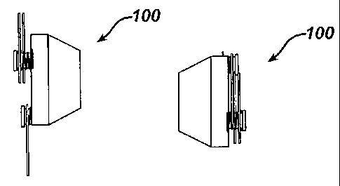
FIG. 5 is a side view of a first embodiment of an injection port 100 with
radially
extendable, stabilizing elements 102, shown in a deployed position;
FIG. 6 is a top view of injection port 100 in the deployed position;
FIG. 7 is a side view of injection port 100, shown in an undeployed position;
FIG. 8 is a top view of injection port 100, shown in the undeployed position;
FIG. 9 is a perspective, exploded view of the components of injection port
100;
FIG. 10 is a side view of a second embodiment of an injection port 200, shown
in a
deployed position;
FIG. 11 is a top view of injection port 200 in the deployed position;
FIG. 12 is a side view of injection port 200, shown in an undeployed position;
FIG. 13 is a top view of injection port 200 in the undeployed position;
FIG. 14 is a perspective, exploded view of injection port 200;
FIG. 15 is a side view of a third embodiment of an injection port 300, shown
in a
deployed position;
FIG. 16 is a top view of injection port 300 in the deployed position;
FIG. 17 is a top view of injection port 300 in an undeployed position;
FIG. 18 is a side view of injection port 300 in the undeployed position;
FIG. 19 is a perspective, exploded view of injection port 300;
FIG. 20 is a top view of a fourth embodiment of an injection port 400; shown
in a
deployed position;
FIG. 21 is a side view of injection port 400 in the deployed position;
FIG. 22 is a top view of a fifth embodiment of an injection port 500;
FIG. 23 is a side view of injection port 500;
FIG. 24 is a top view of a sixth embodiment of an injection port 600;
FIG. 25 is a side view of injection port 600;
FIG. 26 is a top view of a seventh embodiment of an injection port 700;
FIG. 27 is a side view of injection port 700;
4
CA 02540401 2006-03-20
FIG. 28 is a side view of an eighth embodiment of an injection port 800; and
FIG. 29 is a top view of injection port 800.
[0013] Detailed Description of the Invention
[0014] Referring now to the drawings, FIGS. 1 and 2 show an injection port
2 of the prior art.
Injection port 2 generally may have a truncated, conical configuration, and
comprises a
housing 14, a septum 4, and a catheter support 8. Injection port 2 further
comprises a
body 7 having a bottom surface, also called a distal closed end 13, and an
open proximal
end 5, which retains septum 4. Housing 14 is typically made of a
biocompatible,
corrosion resistant metal. Septum 4 may be made of an elastomeric material
such as
silicone rubber, which is easily penetrable by a hypodermic needle. Housing 14
and
septum 4 define a fluid reservoir 12 in injection port 2 for receiving and
containing a
fluid. Catheter support 8 extends through housing 14 to provide fluidic
communication
between fluid reservoir 20 and the exterior of injection port 2. A flange 6
extends from
housing 14 and contains a plurality of holes 10 for suturing injection port 2
to the tissue
of a patient.
[0015] FIG. 3 shows injection port 2 of the prior art as it may be
assembled to a catheter 20
during a surgical procedure. When using injection port 2 in a laparoscopic
procedure
such as implantation of a gastric band, it may be necessary for the surgeon to
assemble
injection port 2 to catheter 20 during the laparoscopic procedure. This may be
because
injection port 2 may be too large to pass through a standard size (12mm
diameter)
laparoscopic port, which may be used for access to the stomach inside the
abdominal
fluid reservoir. The surgeon may introduce the gastric band and catheter 20
into the
abdominal fluid reservoir without injection port 2 attached to the free end of
catheter 20.
Once the surgeon has secured the gastric band around the stomach, the surgeon
externalizes the free end of catheter 20 through the abdominal muscle and
fascia layers,
subcutaneous fat layer, and the skin to assemble injection port 20 to the free
end of
catheter 20. Then the surgeon implants the injection port subcutaneously at
the desired
CA 02540401 2014-06-16
6 of 16
location on the patient's abdomen. As shown in FIG. 3, a catheter element 16
fits over
catheter 20 and locks catheter 20 tightly over catheter support 8 of injection
port 2. A
catheter protector 18 also fits over catheter 20 and helps to prevent
accidental puncture of
catheter 20 when the surgeon accesses injection port 2 with a hypodermic
needle during
later injections of fluid. Once catheter 20 is fluidly connected to injection
port 2, the
surgeon attaches injection port 2 with a plurality of sutures 22 to the fascia
26 covering
the muscular layer of tissue. Typically the surgeon spends several minutes to
suture
injection port 2 to fascia 26, working with limited access through an incision
24 in the
patient. FIG. 4 shows injection port 2 attached to fascia 26 with four sutures
22 prior to
closure of incision 24.
[0016] The below embodiments describe an injection port that may be
configurable into a
collapsed or an undeployed position to facilitate placement into the tissue of
the patient,
and may be configurable, once positioned in the tissue of the patient, into an
extended or a
deployed position for long-term stability. The injection port resists
"flipping" over,
thereby allowing needle access to the septum for adding or withdrawing fluid,
and
provides sites for tissue in-growth for securing the injection port in the
tissue of the
patient. Furthermore, these embodiments eliminate the need to suture the
injection port to
tissue, thereby reducing surgery time and the tissue trauma associated with
suturing.
[0017] FIGS. 5, 6, 7, 8, and 9 show a first embodiment of an injection port
100, which includes a
housing having a body made of a rigid material such as titanium, stainless
steel, or a
biocompatible polymer. Housing 104 may be of a similar design as housing 14 of
the
prior art shown in FIG. 1, but without flange 6. A plurality of stability
elements 102
attach to housing 104. Each of stability elements 102 include a member that
may be made
of coiled, metallic wire, preferably a non-corroding, stainless steel or
titanium alloy spring
wire such as used for the manufacture of coiled springs. Each of stability
elements 102
have a torsion spring that attaches member to housing such that stability
elements 102
tend to spring from the undeployed position to the deployed position when not
sufficiently
restrained. FIG. 5 is a side view and FIG. 6 is a top view of injection port
100 while
CA 02540401 2014-06-16
7 of 16
stability elements 102 are in a deployed position. FIG. 7 is a side view and
FIG. 8 is a
bottom view of injection port 100 while stability elements 102 are in an
undeployed
position. The surgeon may hold stability elements 102 in the undeployed
position with a
surgical grasper or gloved hand and then place injection port 100 into the
incision of the
patient. Once the surgeon has placed injection port 100 in the desired implant
location of
the patient, the surgeon may release injection port 100 so that stability
elements 102 move
to the deployed position. The surgeon may use conventional surgical tools to
dissect
tissue around injection port 100 and facilitate the full extension of
stability elements 102.
[0018] FIG. 9 is an exploded, perspective view of injection port 100. Each
of stability elements
102 comprises member and torsion spring that springably attaches to housing
104 with a
pin pressed into a hole. The space inside of member allows the dissected
tissue planes to
heal together, thus helping to secure injection port 102 in the patient. Since
each of
stability elements 102 may be flexible and resiliently attached to housing,
the patient will
not experience significant discomfort while bending/twisting that portion of
his or her
body. A septum assembles into housing in a similar manner as shown in FIG. 1
of the
prior art. (Each of the embodiments of injection port disclosed herein include
a septum, a
fluid reservoir, and a catheter support having a basic design and function
similar to that of
the prior art injection port described for Fig. 1.)
[0019] FIGS. 10, 11, 12, 13, and 14 show a second embodiment of an
injection port 200. FIG. 10
is a side view, and FIG. 11 is a top view of injection port 200 while in a
deployed
position. FIG. 12 is a side view, and FIG. 13 is a top view of injection port
200 while in
an undeployed position. FIG. 14 is an exploded, perspective view of injection
port 200,
including a plurality of stability elements made of a metallic wire. Each of
stability
elements may have a pair of ends that pivotally attach to a housing in holes.
A septum
assembles into housing in a similar manner as shown in FIG. 1 of the prior
art. In this
embodiment, each of stability elements may be D-shaped. Initially, the surgeon
may hold
housing with a grasper or gloved hand while injection port may be in the
undeployed
position. As the surgeon pushes injection port 200 into the tissue of the
patient, stability
CA 02540401 2014-06-16
,
8 of 16
elements unfold into the deployed position while simultaneously penetrating
into tissue.
Therefore, the surgeon dissects the minimal amount of tissue to position
injection port
200, thus facilitating rapid healing and reducing the risk of infection. The
subcutaneous
fat layer and skin layers cover and hold injection port 200 while tissue heals
around
stability elements.
[0020] FIGS. 15, 16, 17, 18, and 19 show a third embodiment of an injection
port 300. FIG. 15 is
a side view, and FIG. 16 is a top view, of injection port 300 while in a
deployed position.
FIG. 17 is a top view, and FIG. 18 is a side view of injection port 300 while
in an
undeployed position. FIG. 19 is an exploded, perspective view of injection
port 300,
including a plurality of stability elements that are made of a spring metal
wire. Each of
stability elements may have a D-shape as in the previous embodiment, but may
be also
formed to have torsion springs that attach to a housing with a pin into holes
so that
stability element may be in the deployed position when unrestrained. The
surgeon may
place injection port into the tissue of a patient in a similar manner as
described for
injection port 200 of FIG. 14. A septum assembles into housing as described
for the prior
art of FIG. 1.
[0021] FIG. 20 is a top view and FIG. 21 is a side view of a fourth
embodiment of an injection
port 400, which includes a plurality of stability elements attached to a
housing. Stability
elements are made of a flexible wire, such as super elastic, nickel-titanium
memory metal,
also known in the art as Nitinol. The surgeon may hold stability elements in
the
undeployed position while positioning injection port into the tissue of the
patient, and then
use a surgical tool or fingertips to gently position stability elements in the
deployed
position. FIG. 20 also shows a phantom view of a catheter for fluid transfer
to a remote
portion of the body.
[0022] FIG. 22 is a top view and FIG. 23 is a side view of a fifth
embodiment of an injection port
500, that includes a stability element attached to a housing. Stability
element comprises a
support member that may be made of a flexible metal wire or plastic cord that
may be
attached to and forms the perimeter of a circular webbing. Webbing may be made
of a
CA 02540401 2014-06-16
. .
9 of 16
biocompatible, polymeric mesh material such as Prolene (Trademark, Ethicon,
Inc.) that
attaches to housing with a biocompatible adhesive. Webbing provides a site for
rapid
tissue in-growth and healing, and to comfortably secure injection port 500 in
the body.
[0023] FIG. 24 is a top view and FIG. 25 is a side view of a sixth
embodiment of an injection port
600, that includes a plurality of stability elements attached to a housing and
normally
extending radially. Each of stability elements is made of a flexible metal
wire material
such as super elastic nickel titanium alloy, and includes a curled end.
[0024] FIG. 26 is a top view and FIG. 27 is a side view of a seventh
embodiment of an injection
port 700, that includes a stability element attached to a housing. Stability
element
includes a flexible, star-shaped webbing that may be injection molded from a
plastic such
as polyethylene with a plurality of support members extending radially. An
annular
groove of housing retains stability element.
[0025] FIG. 28 is a side, sectional view and FIG. 29 is a top view of an
eighth embodiment of an
injection port 800, that includes a stability element. In this embodiment, the
surgeon or a
medical assistant may assemble injection port 2 of the prior art (FIG. 1) with
stability
element during the surgical procedure (but prior to placement in the body.)
Stability
element includes a webbing integrally molded from a flexible, biocompatible
plastic such
as polyethylene, with a support member that defines the perimeter of stability
element. A
retaining lip, also molded into stability element, snaps over and retains
flange 6 of housing
14. Therefore it may be possible for a surgeon to use a conventional injection
port that
comes with a particular medical implant device, together with stability
element, to avoid
the need to suture the injection port to tissue.
CA 02540401 2006-03-20
[0026] A surgeon may implant an injection port in accordance with the
present invention into the
tissue of a surgical patient, without the need for suturing. The surgeon may
create a
surgical incision through the skin and subcutaneous fat layers of the patient.
In the case
of a gastric band implant, this incision may be typically made in the abdomen
of the
patient. The surgeon dissects tissue in the surgical incision to create space
for a catheter
and the injection port between the subcutaneous fat layer and the fascia
tissue. The
surgeon may use conventional surgical tools for dissection and/or fingertips.
The surgeon
connects the injection port to the catheter using components such as described
for the
prior art in FIG. 1. The surgeon holds the injection port in an undeployed
position, and
then positions the injection port and the catheter through the incision. The
surgeon
manipulates the injection port into final position upon the fascia tissue
while allowing the
injection port to change into a deployed position. Finally, the surgeon or
medical
assistant closes the skin and subcutaneous fat layers over the injection port
and the
catheter. The method may also include an additional step of suturing the
stabilizing
elements to the tissue.
[0027] It will become readily apparent to those skilled in the art that the
above invention has
equally applicability to other types of implantable bands. For example, bands
are used
for the treatment of fecal incontinence. One such band is described in U.S.
Patent
6,461,292 which is hereby incorporated herein by reference. Bands can also be
used to
treat urinary incontinence. One such band is described in U.S. Patent
Application
2003/0105385 which is hereby incorporated herein by reference. Bands can also
be used
to treat heartburn and/or acid reflux. One such band is described in U.S.
Patent 6,470,892
which is hereby incorporated herein by reference. Bands can also be used to
treat
impotence. One such band is described in U.S. Patent Application 2003/0114729
which
is hereby incorporated herein by reference.
[0028] While preferred embodiments of the present invention have been shown
and described
herein, it will be obvious to those skilled in the art that such embodiments
are provided
by way of example only. Numerous variations, changes, and substitutions will
now occur
CA 02540401 2013-09-18
[0026] A surgeon may implant an injection port in accordance with the
present invention into the
tissue of a surgical patient, without the need for suturing. The surgeon may
create a
surgical incision through the skin and subcutaneous fat layers of the patient.
In the case
of a gastric band implant, this incision may be typically made in the abdomen
of the
patient. The surgeon dissects tissue in the surgical incision to create space
for a catheter
and the injection port between the subcutaneous fat layer and the fascia
tissue. The
surgeon may use conventional surgical tools for dissection and/or fingertips.
The surgeon
connects the injection port to the catheter using components such as described
for the
prior art in FIG. 1. The surgeon holds the injection port in an undeployed
position, and
then positions the injection port and the catheter through the incision. The
surgeon
manipulates the injection port into final position upon the fascia tissue
while allowing the
injection port to change into a deployed position.
Finally, the surgeon or medical
assistant closes the skin and subcutaneous fat layers over the injection port
and the
catheter. The method may also include an additional step of suturing the
stabilizing
elements to the tissue.
[0027] It will become readily apparent to those skilled in the art that the
above invention has
equally applicability to other types of implantable bands. For example, bands
are used for
the treatment of fecal incontinence. One such band is described in U.S. Patent
6,461,292.
Bands can also be used to treat urinary incontinence. One such band is
described in U.S.
Patent Application Publication No. 2003/0105385. Bands can also be used, to
treat
heartburn and/or acid reflux. One such band is described in U.S. Patent
6,470,892. Bands
can also be used to treat impotence. One such band is described in U.S. Patent
Application Publication No. 2003/0114729.
[0028] While preferred embodiments of the present invention have been shown
and described
herein, it will be obvious to those skilled in the art that such embodiments
are provided by
way of example only. Numerous variations, changes, and substitutions will now
occur
11
CA 02540401 2013-09-18
to those skilled in the art without departing from the invention. For example,
as would be
apparent to those skilled in the art, the disclosures herein have equal
application in
robotic-assisted surgery. In addition, it should be understood that every
structure
described above has a function and such structure can be referred to as a
means for
performing that function.
12