Note: Descriptions are shown in the official language in which they were submitted.
CA 02541392 2006-04-04
WO 2005/035056 PCT/US2004/033276
-1-
SECURE AND EFFICACIOUS THERAPY DELIVERY FOR AN EXTRA
SYSTOLIC STIMULATION PACING ENGINE
The present invention relates generally to the field of cardiac stimulation
devices
and more specifically to a device and method for secure and efficacious
delivery of an
extra-systolic stimulation (ESS) therapy to improve hemodynamic function in
the
treatment of cardiac mechanical insufficiency. In particular, implantable and
external
devices device and methods of therapy delivery according to the present
invention are
provided for adjusting the timing and delivery of extra-systolic stimulation.
Cardiac myocytes stimulated with so-called paired, coupled, bi-geminal ox
intercalated pacing stimulation produce enhanced mechanical function on
subsequent
I S depolarizations of the heart. Herein, this type of cardiac pacing therapy
is referred to as
extra-systolic stimulation (ESS) which refers to delivery of cardiac pacing
therapy soon
after either an intrinsic or pacing-induced systole. The magnitude of the
enhanced
mechanical function is strongly dependent on the timing of the extra systole
relative to the
preceding intrinsic or paced systole. When correctly timed, an ESS pulse
causes
depolarization of the heart but the attendant mechanical contraction is absent
or
substantially weakened. The contractility of the subsequent cardiac cycles,
referred to as
the post-extra-systolic beats, is increased as described in detail in commonly
assigned U.S.
Pat. No. 5,213,098 issued to Bennett et al., incorporated herein by reference
in its entirety.
The mechanism of ESS is thought to be related to the calcium cycling within
the
myocytes. The extra systole initiates a limited calcium release from the
sarcolasmic
reticulum (SR). The limited amount of calcium that is released in response to
the extra
systole is not enough to cause a normal mechanical contraction of the heart.
After the
extra systole, the SR continues to take up calcium with the result that
subsequent
depolarization(s) cause a large release of calcium from the SR, resulting in
vigorous
myocyte contraction.
As noted, the degree of mechanical augmentation on post-extra-systolic beats
depends strongly on the timing of the extra systole following a first
depolarization,
referred to as the extra-systolic interval (ESI). If the ESI is too long, the
ESS effects are
CA 02541392 2006-04-04
WO 2005/035056 PCT/US2004/033276
-2-
not achieved because a normal mechanical contraction takes place in response
to the extra-
systolic stimulus. As the ESI is shortened, a maximal effect is reached when
the ESI is
slightly longer than the physiologic refractory period. An electrical
depolarization occurs
without a mechanical contraction or with a substantially weakened contraction.
When the
ESI becomes too short, the stimulus falls within the absolute refractory
period and no
depolarization occurs.
The above-cited Bennett patent generally discloses a post-extra-systolic
potentiation stimulator for the treatment of congestive heart failure or other
cardiac
dysfunctions. A cardiac performance index is developed from a sensor employed
to
monitor the performance of the heart, and a cardiac stress index is developed
from a
sensor employed to monitor the cardiac muscle stress. Either or both the
cardiac
performance index and cardiac stress index may be used in controlling the
delivery of ESS
stimulation. Prior non-provisional U.S. patent application serial number
10/322,792 (Atty.
Dkt. P-9854.00) filed 28 August 2002 and corresponding PCT application
(publication no.
WO 02/053026) by to Deno et al., which is hereby incorporated herein by
reference in its
entirety, discloses an implantable medical device for delivering post extra-
systolic
potentiation stimulation. ESS stimulation is employed to strengthen the
cardiac
contraction when one or more parameters indicative of the state of heart
failure show that
the heart condition has progressed to benefit from increased contractility,
decreased
relaxation time, and increased cardiac output. PCT Publication WO 01/58518
issued to
Darwish et al., incorporated herein by reference in its entirety, generally
discloses an
electrical cardiac stimulator for improving the performance of the heart by
applying paired
pulses to a plurality of ventricular sites. Mufti-site paired pacing is
proposed to increase
stroke work without increasing oxygen consumption and, by synchronizing the
timing of
the electrical activity at a plurality of sites in the heart, decrease a
likelihood of
development of arrhythmia.
As indicated in the referenced '098 patent, one risk associated with ESS
stimulation is arrhythmia induction. If the extra-systolic pulse is delivered
to cardiac cells
during the vulnerable period, the risk of inducing tachycardia or fibrillation
in arrhythmia-
prone patients is higher. The vulnerable period encompasses the repolarization
phase of
the action potential, also referred to herein as the "recovery phase" and a
period
immediately following it. During the vulnerable period, the cardiac cell
membrane is
CA 02541392 2006-04-04
WO 2005/035056 PCT/US2004/033276
-3-
transiently hyper-excitable. Therefore, although the property of ESS has been
known of
for decades, the application of ESS in a cardiac stimulation therapy for
improving the
mechanical function of the heart has not been realized clinically because of
the perceived
risks.
In delivering extra-systolic stimulation for achieving mechanical enhancement
of
cardiac function on post-extra-systolic beats, therefore, it is important to
avoid certain
extra-systolic intervals that under certain circumstances, may cause
arrhythmias or other
deleterious effects. When securely delivered, the mechanical effects of ESS
therapy may
advantageously benefit a large number of patients suffering from cardiac
mechanical
insufficiency, such as patients in heart failure, among others. Hence, a
method for secure
and effective control of ESS therapy is needed that provides all the
advantages with little
or no of the potential disadvantages.
Extra-systolic stimulation (ESS) therapy is a means to treat cardiac
dysfunction
including heart failure that employs atrial and/or ventricular extra-systoles
via pacing like
stimulation of the heart. These extra-systoles must be timed correctly to
achieve
beneficial effects on myocardial mechanics (benefit) while maintaining an
extremely low
level of risk of arrhythmia induction and excellent ICD-like arrhythmia
sensing and
detection (security). This timing must adapt to variations in refractory
period such as
those resulting from intrinsic or physiologic rate changes and not compromise
security or
benefit. Further experience with ESS has led to improved implementation
methods that
depend on better blanking, ESS stimulation timing, and ESS delivery rules.
These
methods may be employed individually or in combinations in an external or
implantable
ESS device. A list of these improvements appears below:
The present invention pertains to a series of therapy delivery security
options for
the secure delivery of an ESS therapy. In one form of the present invention,
the inventive
therapy delivery options involve monitoring cardiac activity on a cycle-by-
cycle basis
during delivery of ESS therapy and based on the monitored activity determining
whether
or not ESS therapy delivery should commence and/or continue.
For example, therapy delivery could be inhibited in the event that a premature
beat (or
depolarization) occurs such as a premature atrial contraction (PAC) or a
premature
ventricular contraction (PVC).
CA 02541392 2006-04-04
WO 2005/035056 PCT/US2004/033276
-4-
In addition, the present invention maintains adequate arrhythmia detection and
in
the event that detection occurs, delivery of an ESS therapy is inhibited.
Maintaining
robust detection of ventricular tachycardia (VT) and ventricular fibrillation
(VF) is
deemed a prerequisite for secure and efficacious delivery of an ESS therapy.
Representative rules according to the present invention include
Delivery of an ESS therapy only at pacing rates (pacing per minute or PPM) and
a
corresponding heart rate (HR) range to enhance efficacy of the therapy.
Inhibit delivery of an ESS therapy in the event that a PVC occurs on a
preceding
cardiac cycle.
Ensure that the relatively short extra-systolic interval (ESI) typical of an
ESS
therapy do not inappropriately bias arrhythmia detection algorithms toward
erroneous
VT/VF detection.
Maintain adequate VT/VF detection in the presence of the additional blanking
periods typically imposed during delivery of an ESS therapy.
Withhold delivery of an ESS therapy during VT/VF episodes.
Avoid potential under-sensing of VT by withholding delivery of an ESS therapy
in the
event that an evidence counting-type VT detection algorithm has reached a
threshold
(prior to declaring positive detection of VT).
Maintain the ability to mode switch in the presence of a detected atrial
tachycardia
(AT) and suspend delivery of an ESS therapy or mode switch to a ventricular
coupled
pacing (VCP) modality in the event that an AT episode is detected.
Discontinue ESS therapy delivery if a tachycardia episode emerges during ESS
therapy
delivery and only commence ESS therapy delivery following clinician
intervention (e.g.,
remote or in-person device interrogation).
Accordingly, the present invention provides a system and method for securely
controlling the delivery of ESS therapy to effectively produce augmented
stroke volume
and cardiac output in the treatment of cardiac mechanical insufficiency.
According to one form of the present invention, ESS therapy delivery is
controlled
based on security rules that are preferably applied on a cycle-to-cycle basis.
As such, the
system includes an implantable medical device and associated lead system for
delivering
electrical stimulation pulses to the heart and receiving and processing
electrical cardiac
signals from the heart. The system includes arrhythmia detection and pacing
therapy
CA 02541392 2006-04-04
WO 2005/035056 PCT/US2004/033276
-S-
delivery capabilities and optionally, cardioversion and defibrillation
capabilities. In some
embodiments, the system further includes one or more physiological sensors for
measuring cardiac hemodynamic or contractile function in order to assess the
strength of
the myocardial contraction during extra systoles and/or during depolarizations
subsequent
to delivery of ESS therapy.
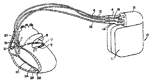
FIG. 1A is an illustration of an exemplary implantable medical device (IMD) in
which the present invention may be implemented.
FIG. 1B is an illustration of an alternative IMD including subcutaneous ECG
electrodes incorporated in the housing of the IMD.
FIG. 2A is a functional schematic diagram of the implantable medical device
shown in FIG. 1 A.
FIG. 2B is a functional schematic diagram of an alternative embodiment of the
IMD, with regard to the electrode configuration of FIG. 1B, which includes
dedicated
circuitry for measuring electrical restitution.
FIGS 3 - 9 depict timing sequences of several cardiac cycles during which an
ESS
therapy is applied or modified, as applicable, according to the present
invention.
The present invention is generally directed toward providing an implantable
system for delivering an electrical stimulation therapy to achieve augmented
stroke
volume (and, under certain conditions, cardiac output) by providing a
carefully timed
pacing stimulus to a chamber of a heart following an intrinsic or evoked
depolarization.
Herein the therapy is referred to herein as extra-systolic stimulation (ESS).
The timing of ESS therapy results in the device giving the patient a pacing
stimulus relatively close to what has historically been called the "vulnerable
zone." The
general consensus is that during the first few milliseconds after the
refractory period - and
depending to a degree on the magnitude of the ESS pulse delivered - the heart
may have
an increased vulnerability to a tachyarrhythmia and the risk of inducing a VT
or VF with a
pacing stimulus may be increased during this time.
The fact that ESS therapy pulses can be delivered at typical pacing amplitudes
greatly reduces the arrhythmia risk. Adaptive timing is also being explored to
position the
ESS therapy pulses some distance from the peak of the vulnerable zone. The
purpose of
CA 02541392 2006-04-04
WO 2005/035056 PCT/US2004/033276
-6-
the security rules discussed in this paper is to decide on a cycle-by-cycle
basis whether or
not to deliver ESS therapy. The security rules: 1) deliver ESS therapy only at
rates low
enough that efficacy can be ensured, 2) not deliver ESS therapy coupled to
premature
ventricular beats, 3) ensure that the short intervals associated with ESS
therapy do not
inappropriately bias detection algorithms towards VT/VF detection, 4) maintain
adequate
VT/VF detection in the presence of the additional blanking imposed by ESS
therapy
delivery, 5) allow for potential undersensing of a ventricular
tachyarrhythmia, 6) maintain
the ability to mode switch in the presence of an atrial tachyarrhythmia (and
suspend ESS
therapy or switch to Vcp only delivery if mode switch occurs), and 7) allow
ESS therapy
to be suspended if one or more ventricular tachyarrhythmias occur.
FIG. 1A is an illustration of an exemplary implantable medical device (IMD) in
which the present invention may be implemented. IMD 10 is coupled to a
patient's heart
by three cardiac leads 6,15,1 G. IMD 10 is capable of receiving and processing
cardiac
electrical signals and delivering electrical stimulation pulses for ESS and
may additionally
be capable of cardiac pacing, cardioversion and defibrillation. IMD 10
includes a
connector block 12 for receiving the proximal end of a right ventricular lead
16, a right
atrial lead 15 and a coronary sinus lead G, used for positioning electrodes
for sensing and
stimulating in three or four heart chambers.
In FIG. 1 A, the right ventricular lead 16 is positioned such that its distal
end is in
the right ventricle for sensing right ventricular cardiac signals and
delivering electrical
stimulation therapies in the right ventricle which includes at least ESS and
may include
cardiac bradycardia pacing, cardiac resynchronization therapy, cardioversion
and/or
defibrillation. For these purposes, right ventricular lead 16 is equipped with
a ring
electrode 24, a tip electrode 2G optionally mounted retractably within an
electrode head
28, and a coil electrode 20, each of which are connected to an insulated
conductor within
the body of lead 16. The proximal end of the insulated conductors are coupled
to
corresponding connectors carried by bifurcated connector 14 at the proximal
end of lead
16 for providing electrical connection to IMD 10.
The right atrial lead 15 is positioned such that its distal end is in the
vicinity of the
right atrium and the superior vena cava. Lead 15 is equipped with a ring
electrode 21, a
tip electrode 17, optionally mounted retractably within electrode head 19, and
a coil
electrode 23 for providing sensing and electrical stimulation therapies in the
right atrium,
CA 02541392 2006-04-04
WO 2005/035056 PCT/US2004/033276
_7-
which may include atrial ESS and/or other cardiac pacing therapies,
cardioversion and/or
defibrillation therapies. In one application of ESS, ESS is delivered to the
atria to improve
the atrial contribution to ventricular filling. The extra-systolic
depolarization resulting
from the atrial ESS stimulation pulse may be conducted to the ventricles for
achieving
ESS effects in both the atrial and ventricular chambers. The ring electrode
21, the tip
electrode 17 and the coil electrode 23 are each connected to an insulated
conductor with
the body of the right atrial lead 15. Each insulated conductor is coupled at
its proximal
end to a connector carried by bifurcated connector 13.
The coronary sinus lead 6 is advanced within the vasculature of the left side
of the
heart via the coronary sinus and great cardiac vein. 'The coronary sinus lead
6 is shown in
the embodiment of FIG. 1A as having a defibrillation coil electrode 8 that may
be used in
combination with either the coil electrode 20 or the coil electrode 23 for
delivering
electrical shocks for cardioversion and defibrillation therapies. Coronary
sinus lead 6 is
also equipped with a distal tip electrode 9 and ring electrode 7 for sensing
functions and
delivering ESS in the left ventricle of the heart as well as other cardiac
pacing therapies.
The coil electrode 8, tip electrode 9 and ring electrode 7 are each coupled to
insulated
conductors within the body of lead 6, which provides connection to the
proximal
bifurcated connector 4. In alternative embodiments, lead 6 may additionally
include ring
electrodes positioned for left atrial sensing and stimulation functions, which
may include
atrial ESS and/or other cardiac pacing therapies.
The electrodes 17 and 21, 24 and 26, and 7 and 9 may be used in sensing and
stimulation as bipolar pairs, commonly referred to as a "tip-to-ring"
configuration, or
individually in a unipolar configuration with the device housing 11 serving as
the
indifferent electrode, commonly referred to as the "can" or "case" electrode.
IMD 10 is
preferably capable of delivering high-voltage cardioversion and defibrillation
therapies.
As such, device housing 11 may also serve as a subcutaneous defibrillation
electrode in
combination with one or more of the defibrillation coil electrodes 8, 20 or 23
for
defibrillation of the atria or ventricles.
For the purposes of delivering ESS therapy in accordance with the present
invention, for at least one cardiac cycle during such therapy delivery various
timing
intervals or parameters are monitored. For example, a ventricular and/or
atrial
electrogram (EGM) may be derived by monitoring a bipolar "tip-to-ring" sensing
vector, a
CA 02541392 2006-04-04
WO 2005/035056 PCT/US2004/033276
_g_
unipolar tip-to-can sensing vector, a unipolar tip-to-coil or ring-to-coil
sensing vector, or a
relatively more global coil-to-can sensing vector.
It is recognized that alternate lead systems may be substituted for the three
lead
system illustrated in FIG. 1A. For example, lead systems including one or more
unipolar,
bipolar and/or mulitpolar leads may be configured for sensing cardiac
electrical signals
and/or delivering an ESS therapy according to the present invention. It is
contemplated
that extra-systolic stimuli may be delivered at one or more sites within the
heart.
Accordingly, lead systems may be adapted for sensing cardiac electrical
signals at multiple
cardiac sites and for delivering extra-systolic stimuli at the multiple sites,
which may be
located in one or more heart chambers. It is further contemplated that
subcutanteous ECG
electrodes could be included in the implantable system.
FIG. 1B is an illustration of an alternative IMD coupled to a set of leads
implanted
in a patient's heart. In FIG. 1B, IMD housing 11 is provided with an
insulative coating 35,
covering at least a portion of housing 11, with openings 30 and 32. The
uninsulated
openings 30 and 32 serve as subcutaneous electrodes for sensing global ECG
signals. An
implantable system having electrodes for subcutanteous measurement of an ECG
is
generally disclosed in commonly assigned U.S. Pat. No. 5,987,352 issued to
Klein,
incorporated herein by reference in its entirety. In alternative embodiments,
multiple
subcutaneous electrodes incorporated on the device housing 11 andlor
positioned on
subcutaneous leads extending from IMD 10 may be used to acquire multiple
subcutaneous
ECG sensing vectors. Multi-electrode ECG sensing in an implantable monitor is
described in U.S. Pat. No. 5,313,953 issued to Yomtov, et al., incorporated
herein by
reference in its entirety.
While a particular mufti-chamber IMD and lead system is illustrated in FIG.s
1A
and 1B, methodologies included in the present invention may be adapted for use
with
other single chamber, dual chamber, or multichamber IMDs that are capable of
sensing
and processing cardiac electrical signals and delivering electrical
stimulation pulses at
controlled time intervals relative to an intrinsic or paced heart rate. Such
IMDs optionally
include other electrical stimulation therapy delivery capabilities such as
bradycardia
pacing, cardiac resynchronization therapy, anti-tachycardia pacing, and
preferably include
arrhythmia detection and cardioversion, and/or defibrillation capabilities.
CA 02541392 2006-04-04
WO 2005/035056 PCT/US2004/033276
-9-
A functional schematic diagram of the IMD 10 is shown in FIG. 2A. This diagram
should be taken as exemplary of the type of device in which the invention may
be
embodied and not as limiting. The disclosed embodiment shown in FIG. 2A is a
microprocessor-controlled device, but the methods of the present invention may
also be
practiced in other types of devices such as those employing dedicated digital
circuitry.
With regard to the electrode system illustrated in FIG. 1A, the IMD 10 is
provided
with a number of connection terminals for achieving electrical connection to
the leads
6,15,16 and their respective electrodes. The connection terminal 311 provides
electrical
connection to the housing 11 for use as the indifferent electrode during
unipolar
stimulation or sensing. The connection terminals 320,310,318 provide
electrical
connection to coil electrodes 20,8,23 respectively. Each of these connection
terminals
311, 320,310,318 are coupled to the high voltage output circuit 234 to
facilitate the
delivery of high energy shocking pulses to the heart using one or more of the
coil
electrodes 8,20,23 and optionally the housing 11. Connection terminals
311,320,310,318
are further connected to switch matrix 208 such that the housing 11 and
respective coil
electrodes 20,8,23 may be selected in desired configurations for various
sensing and
stimulation functions of IMD 10.
The connection terminals 317,321 provide electrical connection to the tip
electrode
17 and the ring electrode 21 positioned in the right atrium. The connection
tern~inals
317,321 are further coupled to an atrial sense amplifier 204 for sensing
atrial signals such
as P-waves. The connection terminals 326,324 provide electrical connection to
the tip
electrode 26 and the ring electrode 24 positioned in the right ventricle. The
connection
terminals 307,309 provide electrical connection to tip electrode 9 and ring
electrode 7
positioned in the coronary sinus. The connection terminals 326,324 are further
coupled to
a right ventricular (RV) sense amplifier 200, and connection terminals 307,309
are further
coupled to a left ventricular (LV) sense amplifier 201 for sensing right and
left ventricular
signals, respectively.
The atrial sense amplifier 204 and the RV and LV sense amplifiers 200,201
preferably take the form of automatic gain controlled amplifiers with
adjustable sensing
thresholds. The general operation of RV and LV sense amplifiers 200,201and
atrial sense
amplifier 204 may correspond to that disclosed in U.S. Pat. No. 5,117,824, by
Keimel, et
al., incorporated herein by reference in its entirety. Generally, whenever a
signal received
CA 02541392 2006-04-04
WO 2005/035056 PCT/US2004/033276
-10-
by atrial sense amplifier 204 exceeds an atrial sensing threshold, a signal is
generated on
output signal line 206. P-waves are typically sensed based on a P-wave sensing
threshold
for use in detecting an atrial rate. Whenever a signal received by RV sense
amplifier 200
or LV sense amplifier 201 that exceeds an RV or LV sensing threshold,
respectively, a
signal is generated on the corresponding output signal line 202 or 203. R-
waves are
typically sensed based on an R-wave sensing threshold for use in detecting a
ventricular
rate.
In one embodiment of the present invention, ventricular sense amplifiers
200,201
may include separate, dedicated sense amplifiers for sensing R-waves and T-
waves, each
using adjustable sensing thresholds, for the detection of myocardial activity.
Myocardial
activity may be measured when a signal exceeding a threshold is received by an
R-wave
sense amplifier included in RV or LV sense amplifiers 200 or 201, causing a
corresponding signal to be generated on signal line 202 or 203, respectively.
Switch matrix 208 is used to select which of the available electrodes are
coupled to
a wide band amplifier 210 for use in digital signal analysis. Selection of the
electrodes is
controlled by the microprocessor 224 via data/address bus 218. The selected
electrode
configuration may be varied as desired for the various sensing, pacing,
cardioversion,
defibrillation and ESS functions of the IMD 10. Signals from the electrodes
selected for
coupling to bandpass amplifier 210 are provided to multiplexes 220, and
thereafter
converted to multi-bit digital signals by A/D converter 222, for storage in
random access
memory 226 under control of direct memory access circuit 228. Microprocessor
224 may
employ digital signal analysis techniques to characterize the digitized
signals stored in
random access memory 226 to recognize and classify the patient's heart rhythm
employing any of the numerous signal processing methodologies known in the
art. In
accordance with the present invention, digital signal analysis of a selected
EGM (or
subcutaneous ECG signals if available) is performed by microprocessor 224 to
derive
parameters related to cardiac activity and the ESS therapy pacing activity and
intervals
related thereto.
The telemetry circuit 330 receives downlink telemetry from and sends uplink
telemetry to an external programmer, as is conventional in implantable anti-
arrhythmia
devices, by means of an antenna 332. Data to be uplinked to the programmer and
control
signals for the telemetry circuit are provided by microprocessor 224 via
address/data bus
CA 02541392 2006-04-04
WO 2005/035056 PCT/US2004/033276
-11-
218. Received telemetry is provided to microprocessor 224 via multiplexer 220.
Numerous types of telemetry systems known for use in implantable devices may
be used.
The remainder of the circuitry illustrated in FIG. 2A is an exemplary
embodiment
of circuitry dedicated to providing ESS, cardiac pacing, cardioversion and
defibrillation
therapies. The timing and control circuitry 212 includes programmable digital
counters
which control the basic time intervals associated with ESS, various single,
dual or multi-
chamber pacing modes, or anti-tachycardia pacing therapies delivered in the
atria or
ventricles. Timing and control circuitry 212 also determines the amplitude of
the cardiac
stimulation pulses under the contxol of microprocessor 224.
During pacing, escape interval counters within timing and control circuitry
212 are
reset upon sensing of RV R-waves, LV R-waves or atrial P-waves as indicated by
signals
on lines 202,203,206, respectively. In accordance with the selected mode of
pacing,
pacing pulses are generated by atrial output circuit 214, right ventricular
output circuit
216, and left ventricular output circuit 215. The escape interval counters are
reset upon
1 S generation of pacing pulses, and thereby control the basic timing of
cardiac pacing
functions, which may include bradycardia pacing, cardiac resynchronization
therapy, and
anti-tachycardia pacing.
The durations of the escape intervals are determined by microprocessor 224 via
data/address bus 218. The value of the count present in the escape interval
counters when
reset by sensed R-waves or P-waves can be used to measure R-R intervals and P-
P
intervals for detecting the occurrence of a variety of arrhythmias.
Iri accordance with the present invention, timing and control 212 further
controls
the delivery of extra-systolic stimuli at selected extra-systolic intervals
(ESIs) following
either sensed intrinsic systoles or pacing evoked systoles. The ESIs used in
controlling the
delivery of extra-systolic stimuli by IMD 10 are preferably automatically
adjusted by IMD
10 based on measurements of electrical restitution as will be described in
greater detail
below. The output circuits 214,215,216 are coupled to the desired stimulation
electrodes
for delivering cardiac pacing therapies and ESS via switch matrix 208.
The microprocessor 224 includes associated ROM in which stored programs
controlling the operation of the microprocessor 224 reside. A portion of the
memory 226
may be configured as a number of recirculating buffers capable of holding a
series of
CA 02541392 2006-04-04
WO 2005/035056 PCT/US2004/033276
-12-
measured R-R or P-P intervals for analysis by the microprocessor 224 for
predicting or
diagnosing an arrhythmia.
In response to the detection of tachycardia, anti-tachycardia pacing (ATP)
therapy
can be delivered by loading a regimen from microcontroller 224 into the timing
and
control circuitry 212 according to the type of tachycardia detected. In the
event that
higher voltage cardioversion or defibrillation pulses are required,
microprocessor 224
activates the cardioversion and defibrillation control circuitry 230 to
initiate charging of
the high voltage capacitors 246,248 via charging circuit 236 under the control
of high
voltage charging control line 240. The voltage on the high voltage capacitors
is monitored
via a voltage capacitor (VCAP) line 244, which is passed through the
multiplexer 220.
When the voltage reaches a predetermined value set by microprocessor 224, a
logic signal
is generated on the capacitor full (CF) line 254, terminating charging. The
defibrillation
or cardioversion pulse is delivered to the heart under the control of the
timing and control
circuitry 212 by an output circuit 234 via a control bus 238. The output
circuit 234
determines the electrodes used for delivering the cardioversion or
defibrillation pulse and
the pulse wave shape.
In one embodiment, the implantable system may additionally include one or more
physiological sensors for monitoring hemodynamic or myocardial contractile
function or a
metabolic status. The physiological sensor may reside within or on the heart,
or endo- or
extra-arterially for sensing a signal proportional to the hemodynamic function
of the heart,
myocardial contraction or heart wall motion, and/or a metabolic parameter. As
such, IMD
10 is additionally equipped with sensor signal processing circuitry 331
coupled to a
terminal 333 for receiving an analog (or, optionally a digital) sensor signal.
A
physiological sensor included in the implanted system may be, but is not
limited to, a
sensor of flow, pressure, heart sounds, wall motion, cardiac chamber volumes
or metabolic
parameters such as oxygen saturation or pH. Sensor signal data is transferred
to
microprocessor 224 via data/address bus 218 such that an index of cardiac
hemodynamic
or contractile performance or a metabolic status may be determined according
to
algorithms stored in RAM 226. Sensors and methods for determining a cardiac
performance index as implemented in the previously-cited '098 patent to
Bennett may also
be used in conjunction with the present invention. As will be described in
greater detail
below, a mechanical or hemodynamic parameter of cardiac function or a
metabolic
CA 02541392 2006-04-04
WO 2005/035056 PCT/US2004/033276
-13-
parameter may be used in one embodiment of the present invention for
controlling the ESI
during ESS therapy delivery based on a safe and efficacious mechanical
enhancement of
the post-extra-systolic beats.
FIG. 3A-D illustrates normal sinus rhythm (NSR) in FIG. 3A and various forms
of
ESS therapy delivery (FIGS. 3B-D) referred to herein as "atrial coordinated
pacing." The
following brief introduction of atrial coordinated pacing (Acp) is intended to
help the
reader appreciate this aspect of the present invention and, in particular, the
timing
sequences depicted in FIGS. 3B-D.
According to one form of Acp, electrical stimulation temporally coordinated to
the
occurrence of various cardiac events (e.g., standard pacing events, sensing
events, Vth
events, etc.) is provided to the upper and/or lower chambers of the heart.
Such stimulation
may be delivered both during refractory and non-refractory periods to
coordinate atrial
contraction, stabilize the cardiac rhythm, and optimize cardiac output. This
Acp
stimulation is intended to be implemented according to the present invention
in a manner
that minimizes the chance of inducing an arrhythmia episode.
The inventors discovered that delivery of an ESS therapy may result in
intermittent
AV block condition believed largely due to the extended (or additional) period
of time that
the ventricles remain refractory following delivery of a ventricular extra-
systolic
stimulation pulse (referred to as "Vth" in FIGS. 3B-D). Unfortunately, such
2:1 (A:V)
conduction may produce a ventricular rate that is too slow to meet the
metabolic demand
of a patient, especially if based on physiologic atrial activity. In contrast,
if a patient's
intrinsic atrial activity produces 1:1 (A:V) conduction during ESS therapy
delivery a
ventricular rate can result that is too rapid for the patient. These rate
fluctuations
potentially offset some of the benefits provided by excitatory ESS therapy.
Thus, to
ameliorate these fluctuations, atrial pacing pulses can be delivered at an
interval shorter
than the intrinsic escape interval. In this form of Acp, the atria are AAI (or
AAI/R) paced
at a rate above (i.e., faster than) the intrinsic atrial rate, thus
establishing a regulated 2:1
AV block while the resulting intrinsic ventricular beats occur relatively more
frequently.
This type of ESS therapy delivery is termed Acp through "rapid" AAI atrial
pacing.
An alternative method of Acp exists wherein intrinsic or paced atrial events
are followed
by ventricular depolarizations (as in sinus or atrial paced rhythms) but
additional
stimulation pulses are provided to both the atria and ventricles at nearly the
same time.
CA 02541392 2006-04-04
WO 2005/035056 PCT/US2004/033276
-14-
This not only achieves enhanced atrial and ventricular function (via ESS
therapy triggered,
or "coupled," to the ventricular depolarizations) but also resets the sinus
node resulting in
an overall regular HR based on an intrinsic or physiologic A-A interval (i.e.,
interval
between successive P waves) and determined by the physiologic requirements of
the
patient. The Acp pulse associated with this form of therapy is sometimes
referred to
herein as "ACP" (all capital letters) to distinguish it as a special form of
atrial pacing.
The Acp and ACP concepts are best understood in reference to timing diagrams
such as
FIG. 3B-D. However, a first wavefonn (labeled "A") illustrates NSR (i.e.,
sinus rhythm
without pacing therapy intervention). Events sensed in the atrium 300 ("As"
events)
conduct through the AV node to the ventricle to cause an intrinsic
depolarization ("Vs"
events) 302. As noted above and as depicted by a second waveform (labeled
"B"), when
ESS therapy delivery begins, a 2:1 AV block typically occurs. This AV block
condition
can oftentimes consist of an unstable form of 2:1 AV block. In the case of the
second
waveform (B), every other intrinsic atrial beat 304 fails to conduct to the
ventricles
because of the AV block condition. This AV block causes an immediate HR
reduction
(typically on the order of a 50% reduction) due to the fact that the extra-
systole following
delivery of a Vth pulse increases the refractory period of the ventricles.
Yet another waveform "C" illustrates a particular pacing embodiment for ACP
(e.
g., AAI pacing). According to one form of the invention, atrial pacing
stimulation 303,310
occurs at a rate that is higher than the intrinsic rate. Even though 2:1
conduction is still
present, the intrinsic ventricular depolarizations 302 occur more frequently
because of the
increased atrial rate (clearly illustrated by comparison of the relative
timing of waveforms
B and C).
Yet another wavefonn "D" can be used to illustrate another form of ACP that
the
inventors consider a special case of ACP. In this special case, an atrial
coordinated pace
312 is delivered a relatively short time period (Tx) following a ventricular
depolarization
314 or a time Ty (not depicted) following an atrial depolarization 316.
Because of the AV
block and the refractory state of the ventricles, this ACP paced event 312
does not conduct
to the ventricles. Following this ACP paced beat 312 an intrinsic
depolarization is allowed
to occur in the atrium (As) 316. This intrinsic beat 316 conducts to the
ventricle, resulting
in a ventricular depolarization (Vs) 314. This aspect of the present invention
allows,
among other advantages, a patient's natural AV conduction and intrinsic rate
to emerge
CA 02541392 2006-04-04
WO 2005/035056 PCT/US2004/033276
-15-
during the cardiac cycle, providing better rate control during ESS therapy
delivery. At the
same time, the number of intrinsic ventricular beats occurnng in a
predetermined period of
time is greater than would otherwise occur without any atrial pacing. This
phenomenon is
referred to herein as physiologic atrial coordinated pacing ("ACP"). ACP can
be provided
by an implantable device as illustrated herein or can be provided by trans-
cutaneous
pacing (TCP) stimulation timed from the surface ECG's R-wave by stimuli of
sufficient
amplitude to capture both atria and ventricles.
In one form of the present invention, an ESS therapy can be delivered in a
DDD/R,
a DDI/R and/or a VVI/R pacing modality, among others (e.g., triple-chamber bi-
ventricular or resynchronization-type pacing therapies). Extra-systolic
stimulation can be
delivered to both the atrial and ventricular chambers (DDD/R or DDI/R modes)
or to one
ventricle only (VVI/R modes). An appropriate pacing mode selection can be
based on a
patient's bradycardia pacing indications (or lack thereof) and atrial
arrhythmia status.
According to one aspect of the present invention, the timing of the extra-
systolic
stimulation (i.e., the ESI) can be adjusted to occur earlier at higher HRs
(when the
refractory period of the heart is generally shorter than at lower heart
rates). The extra-
systolic ventricular stimulation can be monitored to assess whether or not it
captured (i.e.,
caused a depolarization), for diagnostic purposes and/or to adjust the timing
of the early
stimulation. In DDD/R or DDI/R modes, ESS therapy delivery can be applied to
both the
atrium and the ventricle at a designated interval after a ventricular pace or
sense. In
VVI/R pacing modes, ESS therapy delivery can be applied to the ventricle at a
designated
interval after a ventricular pace event (herein "paired pacing") or after a
ventricular sense
(herein "coupled pacing"). Following ESS therapy delivery, the prevailing
indicated
pacing interval (e.g., a programmed lower rate, a mode-switch rate, a
rise/fall rate, a
sensor-indicated rate, etc., expressed as an interval - also known as an
escape interval), is
applied to the pacing cycle following cessation of delivery of an ESS therapy.
In DDD/R
and DDI/R modes, a short escape interval is calculated using modified A-A
timing to
schedule the next atrial pace: escape interval - minimum value (A-Vcp,
Operating PAV).
To accommodate this, any operative atrial and ventricular rate limits are
defeated for a
pacing cycle scheduled to deliver ESS therapy. Then, the escape intervals
ending in ESS
therapy delivery are discarded except that they are stored together with the
pace events in
an episode record buffer. Optionally, an atrial therapy pace marker is
provided (e.g.,
CA 02541392 2006-04-04
WO 2005/035056 PCT/US2004/033276
-16-
counted, stored, uplinked via telemetry, etc.) for each atrial ESS therapy
pace. A
ventricular therapy pace marker is provided (e.g., counted, stored, uplinked
via telemetry,
etc.) for each ventricular ESS therapy pace. Atrial and/or ventricular
supplemental
markers are typically not provided (e.g., counted, stored, or uplinked)
following a cycle of
ESS therapy delivery.
In order to ensure that ESS therapy delivery is safe and effective, ESS
therapy
delivery is not applied after ventricular events that are deemed premature by
the rhythm
pattern. In DDDIR modes or when an atrial monitoring algorithm is enabled, an
intrinsic
ventricular event is considered premature if no atrial events have occurred
since the last
ventricular event or the atrial event occurs too close to a current event or
the atrial event
occurred too early in a given cardiac cycle. In DDI/R modes with such an
atrial
monitoring algorithm disabled, an intrinsic ventricular event is considered
premature if no
atrial events have occurred since the last ventricular event or the atrial
event is
(temporally) too near or too far from a ventricular event. In addition, a
scheduled ESS
therapy delivery is inhibited if an intrinsic ventricular event occurs prior
to delivery of an
ESS therapy pace(s). With DDD/R modes (when the atrial monitoring algorithm is
enabled), scheduled ESS therapy delivery is inhibited if an atrial event
occurs prior to
delivery of pacing stimulus.
In addition, an interval that elapsed since an immediately prior ventricular
event is
compared to a minimum value (e.g., a minimum ESS therapy interval) before
allowing
ESS therapy delivery to occur following a ventricular event. For example, when
tachycardia episode (i.e., VT/VF) detection is enabled, the interval elapsed
from a detected
ventricular event to an immediately previous ventricular event must be at
least 30 ms
longer than the longest VT/VF detection interval. For security, ESS therapy
delivery is
not enabled after a ventricular event if a combined count for a VT/VF
detection algorithm
is greater than a pre-set value (e.g., three detected sequential contractions
due to a
possible, or rapidly developing, tachycardia episode) or if a previously
detected VT/VF
episode is still in progress.
In addition, in order to preserve adequate VTNF detection, ESS therapy
delivery
can be inhibited periodically. For example, after a programmable number of
consecutive
cycles of ESS therapy delivery, ESS therapy is not delivered. If a ventricular
event
detected at the start of the dropped ESS therapy delivery cycle is a pace
event, the pacing
CA 02541392 2006-04-04
WO 2005/035056 PCT/US2004/033276
- 17-
interval for the dropped cycle must be at least as long as a predetermined
value (e.g., a
dropped interval >= a longest VT/VF detection interval + post-pace blanking +
a constant,
such as 30 ms). Continuing with this example, if a ventricular event is sensed
at an
interval less than the predetermined value (a dropped interval), an ESS
therapy is not
delivered during the subsequent cardiac cycle.
Further, the extra-systolic interval (ESI) can be adapted or modified based on
measurements of heart rate (HR). This adaptation appears linear in the
interval domain
and begins adapting at a programmable rate (Start Rate) and ending at a
programmable
rate (Stop Rate). The amount of adaptation is also programmable. According to
this
' aspect of the invention, at least two R-R intervals are measured and an
intermediate (e.g.,
average, mean, median, interpolated value, etc.) value calculated. An
operating ESI is
then implemented based on a set percentage (or ratio) of the intermediate
value. The
intermediate R-R value may be updated every N cardiac cycles (wherein a lower
value of
N provides more rapid response to physiologic changes in HR). Other methods of
deriving an ESI can be implemented such as employing a time-weighted constant
with the
HR metric (i.e., wherein more recent values are weighted more heavily than
less recent
events) and the like. As described elsewhere herein, the operating ESI should
be
maintained at less than half of the median R-R interval.
For capture detection of a post-extra-systolic pacing stimulus, a far-field
sensing
vector is preferred (e.g., a can-to-RVcoil EGM), so that capture of the
ventricular ESS
therapy pace (Vcp or Vth herein) can be assessed without involving same-
chamber
electrodes and the attendant blanking imposed thereon, the possibly
confounding polarity
of adjacent tissue, and to preserve operative sensing circuitry. With respect
to capture
detection, a capture counter is incremented if capture occurs (i.e., is
positively detected),
and a supplemental marker byte is provided (e.g., uplinked via telemetry) on a
next
ventricular event to indicate the temporal location (in the capture detection
window or
interval) wherein detection occurred. This capture detection mechanism can be
used to
periodically probe for the end of the refractory period, and, optionally,
adjust the ESI to
maintain a constant offset from the refractory period. For reference, a
supplemental
marker byte is provided (e.g., uplinked via telemetry) as a reflection of the
current
refractory period when ESS therapy is not delivered (e.g., for each non-
therapy ventricular
event).
CA 02541392 2006-04-04
WO 2005/035056 PCT/US2004/033276
-18-
ESS therapy delivery is preferably disabled in the event that a high voltage
therapy
(e.g., cardioversion or defibrillation therapy) is delivered or if a VTNF
episode is
detected. Counts of atrial and ventricular capture during ESS therapy delivery
are
maintained during therapy delivery. When ESS therapy delivery is inhibited, a
discrete
counter that identifies the reason for inhibiting the ESS therapy delivery is
incremented.
Such counters provide a handy reference providing a reference regarding the
ratio of ESS
therapy delivery to other therapy (or intrinsic sinus rhythm) to a clinician
and any counter
that previously incremented can be cleared at any time.
According to the present invention, both high- and low-resolution trends can
be
collected, such as HR, ESI, and refractory period information. These trends
can also be
provided to a clinician and/or may be cleared at any time.
The temporal timing diagrams depicted in FIGS. 4A and 4B provide a comparison
and
contrast of ESS therapy delivery with (FIG. 4A) and without Acp delivery (FIG.
4B).
This method of delivery is possible in VVI pacing modes, among others. In
FIGS. 4A and
4B a primary ventricular event (paced or sensed) is denoted by reference
numeral 400 and
a primary atrial event (paced or sensed) is denoted by reference numeral 402.
Also, Acp
403 refers to atrial extra-systolic pacing pulses and Vcp 404 refers to
ventricular extra-
systolic pacing pulses inserted to accomplish ESS therapy. An ESS therapy
pacing cycle
consists of an Extra-Systolic Interval (ESI) 406 and a Post Extra-Systolic
Interval (PESI)
408. The ESI 406 ends when the ventricular ESS therapy pace (Vcp) 404 is
delivered.
Referring now to FIG. 4A, an Acp 403 is delivered with temporal coordination
to a
subsequent Vcp 404. Such coordination may be accomplished by delivering the
Acp at a
set value decremented from the then-operative ESI. Thus, the Acp and Vcp will
trigger
from a primary ventricular depolarization 400. In one form of this embodiment
the set
value includes values of approximately 20 ms to about 40 ms, although other
values may
be utilized depending on a variety of factors (e.g., heart rate, activity
sensor input,
mechanical sensor input, metabolic sensor input, etc.). Although the range of
set values
may vary widely, a set value of 30 ms has shown positive empirical results.
Pacing when the HR is elevated may result in no efficacy or even worsened
hemodynamics (e.g., when the temporal length of the ESI 406 is approximately
equal to
the PESI 408 at high rates). Such a combination of ESI 406 and PESI 408
appears to
possibly be associated with an increased risk of arrthymia induction; in
particular if the
CA 02541392 2006-04-04
WO 2005/035056 PCT/US2004/033276
-19-
fast beats are part of an arrhythmia episode. For this reason, a principal ESS
therapy
delivery guideline involves withholding ESS therapy in the event that ESI is
equal to PESI
(or, stated another way delivering ESS therapy only if ESI < PESI).
An additional ESS therapy delivery guideline includes a limitation wherein ESS
therapy can be delivered only if the HR is below a programmable value. The HR
can be
measured on a cycle-by-cycle basis from a primary ventricular event 400 to
subsequent
primary ventricular event 400 (or from a Vcp event 404 to the non-ESS therapy
ventricular event 400 when ESS therapy is delivered).
In addition, the inventors have observed that pacing shortly after a premature
beat
should be more carefully considered compared to pacing shortly after a
normally
conducted beat. The rule described above will eliminate some premature beats,
because
they are "too fast." In order to further minimize the possibility of pacing
shortly after a
premature beat, ESS therapy can be withheld after a ventricular pace/sense
event in the
following situations:
1. If no non-refractory atrial event 402 occurred since a last ventricular
event 400.
This will eliminate situations where a premature beat occurred and may or may
not have
conducted retrograde and was followed by another premature event (i.e., the
current
ventricular event is a premature event, originating in either the atria or
ventricles) and
eliminate situations where the current ventricular event is premature and the
last
ventricular event had an accompanying far field R wave. This aspect of ESS
therapy
delivery guidance can also eliminate cases where the refractory atrial event
conducted to
produce the current ventricular event (i.e., the current ventricular event is
a conducted
beat), but basic pacemaker timing would typically not provide atrial tracking
of this beat
had it not conducted. Thus, precedent exists for not providing ESS therapy in
this
situation.
2. Non-refractory atrial sense 203 temporally too close to the ventricular
event
400. This aspect of ESS therapy guidance eliminates situations where the
current event is
a premature event with a preceding far-field R wave. One current
implementation of
temporally "too close" includes within about 60 ms (i.e., the same interval
used a typical
far-field R wave rejection rule).
3. Atrial pace event 402 temporally too close to the ventricular event 400.
This
will eliminate situations where a current event is a premature ventricular
contraction event,
CA 02541392 2006-04-04
WO 2005/035056 PCT/US2004/033276
-20-
and a scheduled atrial pace was to occur just ahead of the premature event. A
current
implementation of "too close" is within about 110 ms at slower rates and
within about 70
ms at faster rates (i.e., a common mechanism for determining a safety pacing
interval as
employed in a typical bradycardia pacemaker).
The Acp and Vcp 403,404 pulses are desirably delivered early in a given ESS
therapy delivery cycle, with the intent of capturing each chamber. Thus, if a
premature
atrial contraction (PAC) occurs prior to delivery of an Acp 403, the Acp 403
may not
capture the atria. Furthermore, the PAC wavefront could conduct to the
ventricle around
the time of delivery of the Vcp 404. While there are apparently no undesirable
effects of
the delivery of such Acp/Vcp in this case, no beneficial effects relate to
such delivery. On
the one hand, if the Vcp 404 captures, it may conduct retrograde and reset the
sinus node.
This atrial wavefront could be tracked by operative sensing circuitry and lead
to a
pacemaker mediated tachycardia (PMT). Another possible effect of such a
sequence of
events includes delivery of an atrial pace (with or without a non-competitive
atrial pacing,
or NCAP hold-off) because of the relatively late sinus node reset. Neither of
these results
are desirable. In addition, it would be desirable to withhold ESS therapy if
the PAC is the
start of a run of PACs or form part of an atrial tachyarrhythmia (i.e., atrial
tachycardia,
atrial flutter, atrial fibrillation). Therefore, if an atrial sense occurs and
is determined by
applicable far-field R wave criteria not to constitute a far-field R wave, ESS
therapy will
be withheld. If a premature ventricular contraction (PVC) occurs prior to
delivery of Vcp
404, the Vcp 404 should be aborted since the heart has in effect already
delivered an
intrinsic extra-systolic depolarization. In addition, if the PVC occurs in an
interval
bounded by an Acp and a Vcp, a safety pace should be delivered.
ESS therapy delivery differs markedly from a standard single-pacing stimulus
pacing modality. As a result, tachycardia detection modalities need to be
modified to
accommodate ESS therapy. For example, evaluation of V-V intervals is normally
done on
every ventricular event. Of course, ESS therapy typically (and intentionally)
requires
several relatively short intervals for each cardiac cycle. Such short
intervals should not
count towards an accumulative VT/VF detection mechanism. Otherwise, the short
intervals would inappropriately bias probabilistic VF counter algorithms
toward
inappropriately declaring tachycardia episodes. This is a security issue from
the
standpoint of potential delivery of inappropriate cardioversion and/or
defibrillation
CA 02541392 2006-04-04
WO 2005/035056 PCT/US2004/033276
-21 -
therapies. Therefore, ESS therapy delivery cycles that end with a Vcp 404 are
ignored by
the operative VT detection algorithm. That is, the V-V intervals used in the
detection
algorithm to compute R-R median values and determine cardiac rhythm pattern
codes will
start with a Vcp 404 and end at the next ventricular event 400 during ESS
therapy delivery
cycles. The A-A intervals used in a detection algorithm to compute P-P median
values
will be the most recent A-A interval at the time of a non-Vcp ventricular
event (400) for
ESS therapy pacing cycles. Another ESS therapy delivery option related to the
foregoing
involves withholding ESS therapy when an arrhythmia evidence-counting
mechanism
reaches a "combined count" greater than about three (as well as during a
confirmed
VT/VF episode).
As noted above, ESS therapy presents a unique challenge to ventricular
tachyarrhythmia (VT) sensing and detection, because it intentionally
introduces short
coupling intervals (V to Vcp) that may be less than a longest VT detection
interval. As
depicted in FIG. 5, ESS therapy delivery also introduces additional blanking
periods
502,506 following a primary ventricular pace event 500 into a single cardiac
cycle (in total
such blanking is denoted by arrow 508). In a worst case, where VP blanking 502
is
greater than or equal to ESI and while ESS therapy delivery occurs at an upper
rate, the
VT sensing window is open only about a third (~33%) of the time. For example,
with VP
blanking 502 of 300 ms, 300 ms of a 900 ms ESS therapy pacing cycle is
available for
sensing ventricular events. Thus, FIG. 5 represents a potentially worst-case
example of
blanking following ventricular pacing 500,504 wherein approximately 67% of the
ESS
therapy pacing cycle is blanked which might allows a relatively periodic VT to
continue
without detection.
FIG. 6 shows an example of a ventricular tachycardia where every other
tachyarrhythmia beat 602 occurs during a blanking period due to delivery of
Vcp 604.
The rhythm appears to consist of a sinus rhythm having an interval of 800 ms,
when in
fact the rhythm consists of a VT having an interval of 400 ms. This results
from VT
events 601, 602 that are sensed at half the actual rate and wherein every
other VT event
occurs during a blanking period. According to the present invention, one or
more cardiac
cycles wherein ESS therapy delivery is withheld allows detection of such VT
episodes that
otherwise would be effectively hidden due to the additional blanking periods
introduced
following delivery of a Vcp 604. The cardiac cycles wherein ESS therapy is
withheld can
CA 02541392 2006-04-04
WO 2005/035056 PCT/US2004/033276
- 22 -
include N cycles out of M cycles of ESS therapy delivery. If a ventricular
event occurring
at the start of a dropped cycle is a ventricular pacing event, the interval
from Vcp to the
next scheduled ventricular pace will be adjusted if necessary to ensure that
it is delivered
no sooner than the present Vcp blanking interval plus a maximum detection
interval. If a
ventricular sense occurs to end the dropped cycle, the V-V interval will be
checked to
ensure that it includes at least a Vcp blanking interval plus a maximum
detection interval.
If not, an additional ESS therapy cycle should be dropped. The escape interval
following
the Vcp will be set to a value normally used for pace timing, with interlocks
applied to
enforce the 50% sensing window and pacing withheld in the VT detection zone
for the
pacing cycle that starts with a Vcp. This approach offers several benefits;
namely: 1)
straightforward to implement within a cardiac pacing device whether
implantable or
external, 2) consistent with the empirical observations and theory that a
patient's response
to ESS therapy will be a relatively immediate lowering of HR, and 3) will
preserve more
of the ability to detect tachyarrhythmias quickly than would setting the
escape interval
based on the entire ESS therapy pacing cycle. This will be a desirable
operation especially
in the event that the Vcp-to-ventricular event interval more closely
approximates the
ventricular interval when ESS therapy is withheld for one or more cardiac
cycles than does
the ventricular interval associated with the "mechanical beats" (during ESS
therapy
delivery). Optionally, it may be desirable to have separate upper tracking
rates for pacing
cycles where ESS therapy is delivered and pacing cycles where ESS therapy is
withheld.
Such an approach allows for a higher predetermined maximum mechanical rate for
ESS
therapy delivery without requiring that the maximum rate during delivery of
other pacing
therapy to be programmed as high. For instance, with an ESI of 250 ms, then
the
maximum mechanical rate during ESS therapy delivery is 92 bpm with and upper
tracking
rate (UTR) programmed to 150 bpm. However, clinicians may not want the UTR
programmed that high when ESS therapy is not being delivered so the UTR should
continue as a clinician-programmable value.
The additional blanking introduced by ESS therapy delivery also impacts the
ability to detect atrial tachyarrhythmias (AT). FIG. 7 illustrates a 900 ms
cardiac cycle
showing an example of an atrial flutter episode characterized by relatively
periodic atrial
depolarizations wherein two of every three flutter waves 700 is blanked (see
arrows 702).
The blanking relates to blanking after a Vp 706 (optionally fixed at a
predetermined value,
CA 02541392 2006-04-04
WO 2005/035056 PCT/US2004/033276
- 23 -
such as 300 ms) and Acp post-pace blanking (nominally 200 ms). In this case to
enhance
security, the programmed sensed-AV (SAV) interval needs to be longer than
typical (i.e.,
slightly less than the atrial flutter cycle length). FIG. 7 shows Acp delivery
708
programmed to occur a preset time prior to Vp 710. Thus, FIG. 7 depicts a form
of Acp
delivery wherein delivery of Acp 708 and Vcp 710 are controlled in tandem
(e.g., with a
fixed temporal relationship for a given HR).
FIG. 8 illustrates an example of an atrial flutter where every other flutter
wave 800
is blanked (as denoted by arrow 802) due to blanking following Acp 804. This
case
requires a combination of a high programmed UTR (or 2:1 conduction) and a
short ESI.
At the right hand side of FIG. 8, a refractory atrial event (AR) 808 occurs
approximately at
the same time as delivery of a Vcp 812 and during PESI 810. In order to
provide adequate
detection of an AT during ESS therapy delivery and perform a mode switch
(e.g., suspend
or modify an ESS therapy delivery mode), in the event that an refractory
atrial event (AR)
808 occurs during a PESI 810. Far-field R wave criteria (e.g., using electrode
pairs
located outside the right ventricular chamber) can be used to determine
whether "actual"
atrial refractory events have occurred. In this mode of operation, ESS therapy
may be
suspended for three or more ventricular events. If Vcp-only (i.e., no Acp
delivery) is then
desired, it can be delivered until the mode switch termination criteria are
met. Note that if
more than one dropped cycle would be required in order for the PP Median to
reflect the
actual atrial cycle length.
It may be desirable to suspend delivery of ESS therapy in the event that VT/VF
episodes are detected during a period of time that ESS therapy is delivered;
particularly in
the event that a cause-effect relationship is suspected. This suspension could
occur after
one or a programmable number of episodes or high voltage therapy deliveries
and would
offer an opportunity to modify ESS therapy parameter values before allowing
ESS therapy
to continue to operate. ESS therapy may be suspended after a first VT/VF
detection and
may be enabled or not enabled following delivery of defibrillation therapy
delivery.
FIG. 9 illustrates ESS therapy delivery during an AT episode (a series of
rapid
atrial depolarizations as shown by the arrow and reference numeral 900).
Assuming that
ESS therapy is delivered in a VVI pacing mode or a DDI mode (or rate
responsive variant
thereof] a mode switch is performed. Thus, according to the present invention,
in the
event that an apparent AT episode 900 begins to occur (e.g., at least one
unscheduled
CA 02541392 2006-04-04
WO 2005/035056 PCT/US2004/033276
-24-
depolarization occurs in a cardiac chamber), or is detected during ESS therapy
delivery, a
pacing mode switch occur to a ventricular-only form of ESS therapy (Vcp 908).
In
addition, at any time that an atrial event is sensed during the ESI 902 (such
as atrial sense
event AS 906), ESS therapy delivery is modified to deliver only ventricular
extra-systolic
stimulation (Vcp 908). The ESS therapy delivery pacing cycle continues based
only on
ventricular events 910 and is still composed of an ESI 902 and a PESI 904.
Another
pacing mode switch may be performed from ventricular-only ESS therapy delivery
to a
non-ESS therapy delivery modality and/or a variety of AT suppression or AT
termination
techniques may be employed. In the event that the ventricular-only ESS therapy
delivery
modality continues the HR should continue at a relatively low value (albeit
with possibly
compromised hemodynamics due to a relative lack of atrial contribution to
ventricular
filling). In the event that a non-ESS therapy delivery modality is applied the
HR can be
expected to increase to approximately twice the level observed during ESS
therapy
delivery. In any event, any suitable technique for defeating the AT episode
may be
applied, such as atrial anti-tachycardia pacing (ATP), cardioversion therapy
delivery and,
if applicable, defibrillation therapy.
As noted, ventricular-only delivery of ESS therapy (Vcp 908) can be
implemented
during an episode of AT. This is desirable because an during ESS therapy
delivery,
especially a form of ESS therapy including atrial simulation (e.g., Acp
delivery) could be
expected not to capture the atrial chambers anyway but would nevertheless
insert
additional blanking periods. Such blanking periods may comprise a nominal
interval of
about 200 ms. Such blanking imposed on the atrial chamber sensing channel may
interfere with the ability to monitor for termination of the AT. 'Thus,
according to this
aspect of the present invention, delivery of Acp pacing stimulus is inhibited
when atrial
sense events occur during the ESI 902 thus promote atrial sensing and provide
for a pacing
mode switch (e.g., to a ventricular-only ESS therapy delivery regime).
Temporarily
suppressing the Acp pacing stimulus delivery also allows atrio-ventricular ESS
therapy
coordination to be resumed when the AT episode terminates (e.g., by simply
reinserting
the Acp pacing stimulus when an atrial sense event does not occur after the AT
ends).
When a patient is experiencing an AT episode, the timing of the atrial
depolarizations
cannot be used to discriminate premature ventricular events such as PVCs. Due
at least in
part to conduction-rate differences and undersensing of atrial events during
an AT episode,
CA 02541392 2006-04-04
WO 2005/035056 PCT/US2004/033276
- 25 -
non-refractory atrial events may or may not occur during a ventricular pacing
interval
(e.g., primary interval for a VP 910 or and ESI 902 for a Vcp 908). Since such
non-
refractory atrial events can conduct and cause a (premature) ventricular
depolarization, the
criteria for detecting AT episodes can be supplemented with a criterion that
includes the
relative pre-maturity of a ventricular sense event 910. If the ventricular
event 910 is
determined to occur early (as a percentage of the prevailing or then-present
ventricular
rate), the ESS therapy (Vcp 908) can be withheld or not initiated. Also, in
the event that a
PVC occurs prior to delivery of Vcp 908 (i.e., during the ESI 902), then the
Vcp 908 will
be withheld, at least for a then-present cardiac cycle. Furthermore, atrial
arrhythmia
detection techniques can be programmed to utilize or count atrial
depolarizations that
occur only during the PESI 904 (i.e., the interval between the Vcp 908 and the
VS/VP
910) during ESS therapy delivery. Thus, if a single unscheduled atrial
depolarization (of
the string of atrial sense events 900) occurs during the PESI 904, a PAC or
possibly the
beginning of an AT episode has occurred.
As previously described, delivery of an ESS therapy should be withheld (or not
initiated) in the event that a so-called "combined count" used in conjunction
with an
evidence accumulation-type arrthymia detection engine reaches a threshold
value. One
example of such an arrthymia detection engine is described in U.S. Pat. No.
5,545,186
entitled, "Prioritized Rule Based Method and Apparatus for Diagnosis and
Treatment of
Arrhythmias," the contents of which are incorporated by reference herein. In
the context
of the present invention, an exemplary threshold value of greater than three
for ventricular
tachycardia detection can be utilized, but other values may be employed.
In addition, for interval-based tachycardia detection engines, wherein slow
ventricular tachycardia (SVT) episodes can be distinguished from tachycardia
episodes
occurring at higher rates based simply on the differences in the interval of
time between
ventricular events can be employed in conjunction with ESS therapy delivery.
Thus, an
SVT detection zone can be defined as a range of relatively longer intervals as
compared to
the intervals that form a VT detection zone. According to this aspect of the
present
invention, a ventricular rate limit ensures that an ESS therapy is not
delivered when the
observed ventricular interval was close to or in a predefined SVT or VT
detection zone. In
the event that an observed ventricular interval impinges upon an SVT detection
zone an
output signal from an activity sensor (e.g., crystal oscillator,
accelerometer, etc.) or a
CA 02541392 2006-04-04
WO 2005/035056 PCT/US2004/033276
-26-
respiration rate can be used to help determine if an apparent SVT episode is
actually due
to NSR (from physical exertion and the like).
In addition, by dropping one or more cardiac cycles of ESS therapy delivery,
allows for a relatively unobstructed cardiac activity sensing opportunity due
to relative
lack of pacing and blanking compared to ESS therapy delivery. This aspect of
the
invention may include complete cessation of ESS therapy delivery for one or
mare cardiac
cycles, a periodic withholding of ESS therapy and/or periodic withholding of
ESS therapy.
During the time that ESS therapy is not delivered another pacing modality may
be applied
or, for a complete lack of blanking due to pacing stimulus delivery, all
pacing therapy is
withheld for at least one cardiac cycle. Thus, robust cardiac arrthymia
detection testing
occurs for a period of time or for several cardiac cycles. In addition to or
in lieu of the
foregoing, testing for the presence or emergence of an episode of ventricular
arrhythmia
can be implemented using an escape interval timed from Vcp 908 when ESS
therapy is
delivered.
Rather than suspending ESS therapy delivery in the event that a VT/VF episode
is
detected, ESS therapy delivery can be withheld following delivery of a
cardioversion or
defibrillation therapy is the episode was detected during ESS therapy
delivery. Preferably,
only after intervention by a clinician (e.g., interrogation of the ESS therapy
delivery
device) and manual re-programming of the device (either remotely or in-person)
can ESS
therapy delivery resume.
Thus, an implantable system and associated methods have been described for
securely controlling ESS therapy delivery. The methods presented herein
advantageously
allow for chronic ESS therapy delivery in an implantable medical device to a
patient
suffering from cardiac insufficiency.
The claimed methods according to the present invention may be embodied as
executable
instructions stored on a computer readable medium. Said instructions cause the
inventive
methods to be performed under processor control. Accordingly, the present
invention
expressly covers all suitable processors and computer readable media, as set
forth in the
appended claims.
The present invention as herein described and depicted may be modified
insubstantially by those of skill in the cardiac rhythm art for a given device
or patient
CA 02541392 2006-04-04
WO 2005/035056 PCT/US2004/033276
-27-
population. However, such insubstantial modifications are intended to be
covered by the
foregoing description as defined by the following claims.