Note: Descriptions are shown in the official language in which they were submitted.
CA 02541682 2006-04-05
WO 2005/037368 PCT/US2004/033999
MEDICAL LEAD FIXATION
The invention relates to medical devices and, more particularly, to
configurations
of distal of implantable medical leads facilitating fixation at an implant
site.
In the medical field, irnplantable leads are used with a wide variety of
medical
devices. For example, implantable leads are commonly used to form part of
implantable
cardiac pacemakers that provide therapeutic stimulation to the heart by
sensing electrical
activity of the heart and delivering pacing, cardioversion or defibrillation
pulses via
1 o electrodes disposed on the leads, e.g., typically near distal ends of the
leads. Leads may
also be used to deliver therapeutic agents. A number of challenges exist with
respect to
medical leads; in particular, as more advanced and complex therapeutic
techniques are
developed, new configurations are required to facilitate fixation of lead
electrodes at
alteniate implant sites within a patient.
The following drawings are illustrative of particular embodiments of the
invention
and therefore do not limit its scope, but are presented to assist in providing
a proper
understanding of the invention. The drawings are not to scale (unless so
stated) and are
intended for use in conjunction with the explanations in the following
detailed description.
2o The present invention will hereinafter be described iri conjunction with
the appended
drawings, wherein like numerals denote like elements, and:
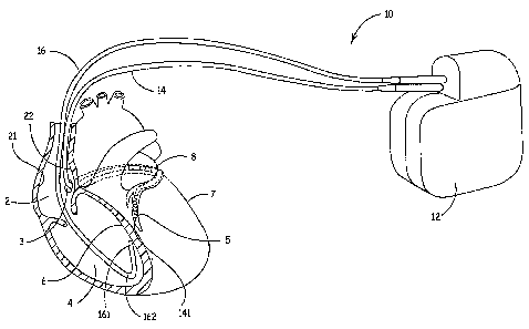
FIG. 1 is a conceptual overview of a system according to one embodiment of the
present invention;
FIG. 2A is cross-sectional side view of a distal portion of a medical lead
according
to one embodiment of the present invention;
FIG 2B is a plan view of the distal end of the lead shown in FIG 2A according
to
one embodiment;
FIG. 3 is an end view of a distal tip of a lead according to 'an embodiment of
the
present invention;
3o FIG 4 is a conceptual perspective view of the distal tip, shown in FIG. 3,
within a
vein of a heart;
CA 02541682 2006-04-05
WO 2005/037368 PCT/US2004/033999
2
FIGS. SA-B are section views of a helical fixation element according to
embodiments of the present invention;
FIG. 6 is a cross-sectional side view of a distal portion of a medical lead
according
to an alternate embodiment of the present invention; and
FIGs. 7 and 8 are cross-section side views of distal portions of leads
according to
additional alternate embodiments of the present invention.
The following detailed description is exemplary in nature and is not intended
to
limit the scope, applicability, or configuration of the invention in any way.
Rather, the
1 o following description provides a practical illustration for implementing
exemplary
embodiments of the invention.
FIG. 1 is a conceptual overview of a system according to one embodiment of the
present invention. FIG. 1 illustrates a system 10 including an implantable
medical device
(IMD) 12 and a first lead 14 and a second lead 16 electrically coupled to IMD
12, each
~ 5 lead including distal tips 141 and 161, respectively, configured to
facilitate fixation at
implant sites so that therapeutic stimulation pulses and/or agents can be
delivered through
leads 14, 16 to a heart from IMD 12. IMD 12 can deliver pacing, cardioversion
and/or
defibrillation therapy to a patient via electrodes disposed on leads 14, 16,
however,
embodiments of the present invention are not limited for use in therapy
delivery and leads
20 14, 16 may include physiological sensors gathering data for patient
monitoring devices or
for devices that integrate monitoring and therapy delivery features. Such
IMD's and
devices along with connection means for associated leads are well known to
those spilled
in the art.
As illustrated in FIG. 1, lead 14 is implanted in a cardiac vein 8, fixed to a
left
25 ventricular epicardial site 5 of the heart, while lead 16 is implanted in a
right ventricular
chamber 4, fixed to a septal wall 6. According to embodiments of the present
invention,
distal tips 141 and 161 of leads 14 and 16 include a fixation element, which
may extend
therefrom at an angle to facilitate fixation at the illustrated implant sites;
embodiments of
such a configuration will be described in detail below. Implant sites made
more viable by
3o embodiments of the present invention also include those in a right atrial
chamber 2, for
example sites on a atrial lateral wall 21 and on an airial septal wall 22, and
epicardial sites
accessed transthoracically, for example a left ventricular lateral site 7;
furthermore, ,
CA 02541682 2006-04-05
WO 2005/037368 PCT/US2004/033999
embodiments of the present invention are not limited to cardiac implantation
and may also
find use in other locations of a body, for example for neuro-stimulation or
drug delivery.
FIG. 2A is cross-sectional side view of a distal portion of a medical lead
according
to one embodiment of the present invention. FIG 2A illustrates a lead distal
tip 20
coupled to a distal end of a lead body 23 and including a canted passageway 32
and an
opening 35 positioned in proximity to a distal tip distal end 25 and
terminating
passageway 32; passageway 32 extends distally from a lumen 28 of body 23
through
which an elongated member 29 extends. Materials from which distal tip 20 may
be
formed include but are not limited to insulative polymers, one example of
which is
polyurethane. Figure 2A further illustrates elongated member 29 coupled to a
helical
fixation element 30 via a stud component 39, which bridges lumen 28 and
passageway 32
passing through a fluid tight seal 36; according the illustrated embodiment,
elongated
member 29 is formed as a coil and is adapted move helical element 30 through
passageway 32 and out opening 35 and to rotate helical element 30, guided by a
protrusion
15 38 formed within passageway 32, thereby affixing helical element 30 to an
implant site
(FIG. 1). Such a mechanism for moving a helical fixation element is well known
to those
skilled in the art and is typically activated at a proximal end of lead body
23 via a rotating
connector pin coupled to a proximal end of elongated element 29.
According to embodiments of the present invention, helical element 30 is
adapted
20 to deflect by flexing along its length so that it may be moved along canted
passageway 32
and out opening 35; materials forming helical element 30, which allow such
flexing,
comprise, but are not limited to, platinum, iridium, titanium, nickel,
polycarbonate, and
polypropylene. Appropriate materials may be selected by those skilled in the
art
depending upon functional requirements for helical element 30, for example, if
helical
25 element is only required for fixation, materials including synthetic resins
and super-elastic
metals, such as Nitinol, may be selected, while if helical element is further
required to
function as an electrode, a platinum-iridium alloy may be selected and coupled
to
elongated member 29, which also functions as a conductor, via a conductive
stud
component 39. According to additional embodiments, helical element 30 further
provides
3o a means for delivering a therapeutic agent, as will be further described in
conjunction with
FIG. 8. Referring back to FIG. 1, it can be seen that, according to the
present invention,
CA 02541682 2006-04-05
WO 2005/037368 PCT/US2004/033999
4
canted passageway 32 provides means to fix a lead to an implant site when that
site is in a
plane generally parallel with a longitudinal axis of the lead.
Referring back to FIG 1, according to embodiments of the present invention,
leads
14 and 16 further include pre-formed curvatures to facilitate orientation of
distal tips 141,
161, for example a curvature 162 is pre-formed, according to methods known to
those
skilled in the art, in a distal portion of lead 16 such that distal tip 161 is
positioned in
proximity to an implant site along septal wall 6 with an opening in tip 161,
similar to
opening 35(FIGs. 2A-B), facing toward septal wall 6. In another exemplary
embodiment,
lead 14 is formed with a curve in a manner corresponding to that described in
commonly
1o assigned U.S. Patent 6,144,882, the relevant teachings of which are herein
incorporated by
reference, wherein the curve serves to hold lead 14 in coronary vein 8 such
that an
opening, e.g. opening 35, faces toward epicardial site 5.
FIG. 2B is a plan view of the distal end of the lead shown in FIG 2A according
to
one embodiment. FIG. 2B illustrates distal tip 20 including a radiopaque
marker 92, which
includes a first indicator 93 and a second indicator 94, configured to
facilitate orientation
of opening 35 toward an implant site by means of fluoroscopic visualization in
a viewing
plane coinciding with that of the implant site. First indicator 93 is an
exemplary
embodiment of an indicator designed to indicate whether opening 35 is
generally directed
toward implant site, when located in a top position, as illustrated in FIG 2B,
or generally
2o directed away from implant site, when located in a bottom position,
indicated by dashed
lines, or visa versa depending on the direction of fluoroscopic viewing.
Second indicator
94, according to one embodiment, provides a means for further orienting
opening 35 in
that a true circular form visualized indicates that opening is aligned with
the viewing
plane, while an ovular forn indicates that opening 35 is skewed away from the
viewing
plane, and no view at all of second indicator 94 indicates that opening 35 is
approximately
perpendicular to viewing plane; an alternate embodiment of such an indicator
includes a
marker ring 95 formed about opening 35. Although marker 92 is illustrated
attached to a
base 26 of distal tip 20, in generally the same plane as opening 35, marker
may be
positioned elsewhere in distal tip 20.
3o FIG. 3 is an end view of distal tip 20 according to an embodiment of the
present
invention; and FIG 4 is a conceptual perspective view of distal tip 20 within
vein 8 (FIG.
1). FIGs. 3 and 4 illustrate distal tip 20 including an asymmetrical radial
section, which
CA 02541682 2006-04-05
WO 2005/037368 PCT/US2004/033999
facilitates orientation of base 26 toward epicardial surface 7 so that helical
fixation
element 30 may be fixed at an implant site therein. The section illustrated in
FIGS. 3 and 4
is generally bell-shaped according to one embodiment, however, any
asymmetrical shaped
radial section, which induces self alignment of tip 20 within a coronary vein,
i.e. vein 8, so
that helical element 30 extends out from opening 35 (FIGS. 2A-B) toward
epicardial
surface 7, is within the scope of the present invention.
FIGS. SA-B are section views of a helical fixation element according to
alternate
embodiments of the present invention. FIG SA illustrates a helical element 51
including a
first pitch along length A, a second pitch along length B and a third pitch
along length C;
second and third pitches, along lengths B and C, respectively, may be
substantially
equivalent. The pitch refers to the lateral distance associated with one
revolution of a
helical element. According to one embodiment of the present invention, pitch
along length
A is optimized for rotation into tissue in order to affix a lead at an implant
site, dimensions
of which have be established and are known to those skilled in the art, while
pitch along
length B is smaller than that along length A in order to facilitate deflection
through a
canted passageway of a distal tip, for example passageway 32 of tip 20
illustrated in FIG
2A. In particular, reduced helical pitch can cause helical element 51 to have
reduced
stiffness in area B relative to area A, making helical element 51 better
suited for forced
deflection. Area C defines a region for attachment to stud component 39, such
as via a
laser weld. FIG SB illustrates another embodiment in which helical element 51
includes a
pre-formed deflection along length B which would be held straight within a
straight
portion of the canted passageway and then resume the pre-formed shape upon
movement
of length B into a curved portion of the canted passageway.
FIG 6 is a cross-sectional side view of a distal portion of a medial lead
according
to an alternate embodiment of the present invention. FIG 6 illustrates helical
element 81
within a canted passageway 82 of a distal tip 80 and including a flexible
coupling 87
positioned in between a stud component 89 attached to elongated element 29;
according to
this alternate embodiment, flexible coupling 87 facilitates movement of
helical element
81, which may not bend along its length, through canted passageway 82.
According to
so one embodiment, flexible coupling 87 comprises a cable, and in another
embodiment a
spring; and, if helical element 81 further functions as an electrode, flexible
coupling 87
would comprise an electrically conductive material. In an alternate embodiment
flexible
CA 02541682 2006-04-05
WO 2005/037368 PCT/US2004/033999
coupling 87 comprises an elastic material and may be pre-formed to conform to
the
curvature of canted passageway 82, being held straight when retracted into a
straight
portion of passageway 82.
FIG. 7 is a cross-section side view of a distal portion of a lead according to
another
embodiment of the present invention. FIG. 7 illustrates a distal tip 70
coupled to a distal
end of a lead body 230 and including a canted passageway 72 extending from a
lumen 280
of lead body 230 to an opening 75 in proximity to a distal end 750 of distal
tip 70; a
deflectable helical fixation element 71 is within canted passageway 72 and
coupled to an
elongated member 79, which extends proximally within lumen 280 of lead body
230 and
1 o serves to move helical element 71 through passageway 72 an out opening 75
for fixation at
an implant site. FIG. 7 further illustrates distal tip 70 including a first
electrode 76
positioned distal to opening 75 and a second electrode 77 positioned proximal
to opening
75, each electrode coupled to a conductor, 73 and 74, respectively, which
extend within
lead body 230 to an electrical connector coupled to a proximal end of lead
body 230,
15 According to a first set of embodiments, helical element 71 does not
function as an
electrode; according to one of these embodiments, first and second electrodes
76, 77
function as a bipolar pair or as independent electrodes of the same polarity
and, according
to another of these embodiments, only first electrode 76 is included in tip 70
or only
second electrode 77 is included in tip 70. According to a second set of
embodiments
2o helical element 71 also functions as an electrode; this set also includes
alternate
embodiments wherein either one or both of electrode 76, 77 are included for
bipolar or
unipolar function. It can be seen in FIG. 7 that the positions of electrodes
76 and 77 with
respect to opening 75 facilitates good contact at an implant site in proximity
to fixation by
helical element 71.
25 FIG. 8 is a cross-section side view of a distal portion of a lead according
to yet
another embodiment of the present invention. FIG. 8 illustrates distal tip 20
coupled to
lead body 23, as in FIG. 2A, wherein elongated member 29 further includes a
fluid
delivery lumen 177 coupled to, or in fluid communication with a fluid
infiision lumen 178
of a deflectable helical fixation element 172, which terminates in an exit
port 179 at a
3o distal end of element 172. A stud component 139 serves to couple helical
element 172 to
elongated element 29 and to generally align fluid lumens 177, 178; according
to one
embodiment, as illustrated in FIG. 8~ helical element 172 is formed form a
hypo-tube
CA 02541682 2006-04-05
WO 2005/037368 PCT/US2004/033999
7
which is welded to a distal end of stud 139 and fluid delivery lumen 177 is
formed by a
tubing extending within elongated member 29 and through a bore of stud 139.
Fluid
lumens 177, 178 may used to deliver a contrast agent to help maneuver tip 20
to an
implant site or to deliver therapeutic agents for treating tissue, into which
helical member
is fixed, at an implant site, or for both. Although not shown, it is apparent
that fluid
delivery lumen 177 extends to a proximal end of lead body 23 where it is
coupled to a
fluid delivery reservoir including a means for pumping the fluid through
lumens 177, 178.
In the forgoing detailed description, the invention has been described with
reference to
specific embodiments. However, it may be appreciated that various
modifications and
changes can be made without departing from the scope of the invention as set
forth in the
appended claims.
