Note: Descriptions are shown in the official language in which they were submitted.
DEMANDES OU BREVETS VOLUMINEUX
LA PRESENTE PARTIE I)E CETTE DEMANDE OU CE BREVETS
COMPRI~:ND PLUS D'UN TOME.
CECI EST ~.E TOME 1 DE 2
NOTE: Pour les tomes additionels, veillez contacter 1e Bureau Canadien des
Brevets.
JUMBO APPLICATIONS / PATENTS
THIS SECTION OF THE APPLICATION / PATENT CONTAINS MORE
THAN ONE VOLUME.
THIS IS VOLUME 1 OF 2
NOTE: For additional vohxmes please contact the Canadian Patent Oi~ice.
CA 02541765 2006-04-05
WO 2005/038029 PCT/US2004/032705
LUCIFERASE BIOSENSOR
Cross-Reference to Related Applications
This application claims the benefit of the filing date of U.S. application
Serial No. 60/510,17, filed October 10, 2003, under 35 U.S.C. ~ 119(e), the
disclosure of which is incorporated by reference herein.
Field of the Invention
This invention relates to the field of biochemical assays and reagents.
More specifically, this invention relates to modified reporter proteins, e.g.,
luminescent reporter proteins, and to methods for their use.
Background
Luciferases are enzymes that catalyze the oxidation of a substrate (e.g.,
luciferin) with the concomitant release of photons of light. Luciferases have
been
isolated from numerous species, including Coleopteran arthropods and many sea
creatures. Because it is easily detectable and its activity can be quantified
with
high precision, Iuciferase/substrate pairs have been used widely to study gene
expression and protein localization. Unlike another reporter protein, green
fluorescent protein (GFP), which requires up to 30 minutes to form
chromophore, the products of luciferases can be detected immediately upon
completion of synthesis of the polypeptide chain (if substrate and oxygen are
also present). In addition, no post-translational modifications are required
for
enzymatic activity, and the enzyme contains no prosthetic groups, bound
cofactors, or disulfide bonds. Luciferase is a useful reporter in numerous
species
and in a wide variety of cells.
Luciferases possess additional features that render them particularly
useful as reporter molecules for biosensing, i.e., molecules which reveal
properties of a biological system. Signal transduction in biosensors (sensors
which comprise a biological component) generally involves a two-step process:
signal generation through a biological component, and signal transduction and
amplification through an electrical component. Signal generation is typically
achieved through binding or catalysis. Conversion of these biochemical events
into an electrical ignal is typically based on electrochemical or caloric
detection
CA 02541765 2006-04-05
WO 2005/038029 PCT/US2004/032705
methods, which are limited by the free energy change of the biochemical
reactions. For most reactions, this is less than the energy of hydrolysis for
two
molecules of ATP, or about 70 kJlmole. However, the luminescence elicited by
luciferases has a much higher energy content. For instance, the reaction
catalyzed by firefly luciferase (560 nm) emits 214 kJ/mole of energy.
Furthermore, the reaction catalyzed by luciferase is one of the most efficient
bioluminescent reactions known, having a quantum yield of nearly 0.9.
Luciferase is thus an extremely efficient transducer of chemical energy.
Luciferas.e biosensors have been described. For example, Sala-Newby et
al. (1991) disclose that a PhotifZUS pyralis luciferase cDNA was amplified in
vitf°o to generate cyclic AMP-dependent protein kinase phosphorylation
sites: In
particular, a valine at position 217 was mutated to arginine to generate a
site,
RRFS, and the heptapeptide kemptide, the phosphorylation site of the porcine
pyruvate kinase, was added at the N- or C-terminus of the luciferase. Sala-
Newby et al. relate that the proteins carrying phosphorylation sites were
characterized for their specific activity, pI, effect of pH on the color of
the light
emitted, and effect of the catalytic subunit of protein kinase A in the
presence of
ATP. They found that only one of the recombinant proteins (RRFS) was
significantly different from wild-type luciferase and that the RRFS mutant had
a
lower specific activity, lower pH optimum, emitted greener light at low pH
and,
when phosphorylated, decreased its activity by up to 80%. It is disclosed that
the latter effect was reversed by phosphatase.
Waud et al. (1996) engineered protein kinase recognition sequences and
proteinase sites into a Photiuus pyralis luciferase cDNA. Two domains of the
luciferase were modified by Waud et al.; one between amino acids 209 and 227
and the other at the C-terminus, between amino acids 537 and 550. Waud et al.
disclose that the mutation of amino acids between residues 209 and 227 reduced
bioluminescent activity to less than 1% of wild-type recombinant, while
engineering peptide sequences at the C-terminus resulted in specific
activities
ranging from 0.06%-120% of the wild-type recombinant luciferase. Waud et al.
also disclose that addition of a cyclic AMP dependent protein kinase catalytic
subunit to a variant luciferase incorporating the kinase recognition sequence,
LRR.ASLG (SEQ ID N0:107), with a serine at amino acid position 543, resulted
in a 30% reduction activity. Alkaline phosphatase treatment restored activity.
CA 02541765 2006-04-05
WO 2005/038029 PCT/US2004/032705
Waud et al. further disclose that the bioluminescent activity of a variant
luciferase containing a thrombin recognition sequence, LVPRES (SEQ ID
N0:108), with the cleavage site positioned between amino acids 542 and 543,
decreased by 50% when incubated in the presence ofthrombin.
Ozawa et al. (2001) describe a biosensor based on protein splicing-
induced complementation of rationally designed fragments of firefly
luciferase.
Protein splicing is a posttranslational protein modification through which
inteins
(internal proteins) are excised out from a precursor fusion protein, ligating
the
flanking exteins (external proteins) into a contiguous polypeptide. It is
disclosed
that the N- and C-terminal intein DnaE from Syraeclzocystis sp. PCC6803 were
each fused respectively to N- and C-terminal fragments of a luciferase.
Protein-
protein interactions trigger the folding of DnaE intein, resulting in protein
splicing, and thereby the extein of ligated luciferase recovers its enzymatic
activity. Ozawa et al. disclose that the interaction between known binding
partners, phosphorylated insulin receptor substrate 1 (IRS-1) and its target N-
tenninal SH2 domain of PI 3-kinase, was monitored using a split luciferase in
the presence insulin.
Paulmurugan et al. (2002) employed a split firefly luciferase-based assay
to monitor the interaction of two proteins, i.e., MyoD and Id, in cell
cultures and
in mice using both complementation strategy and an intein-mediated
reconstitution strategy. To retain reporter activity, in the complementation
strategy, fusion proteins need protein interaction, i.e., via the interaction
of the
protein partners MyoD and Id, while in the reconstitution strategy, the new
complete reporter protein formed via intein-mediated splicing maintains it
activity even in the absence of a continuing interaction between the protein
partners.
A protein fragment complementation assay is disclosed in Miclmick et al.
(U.S. Patent Nos. 6,270,964, 6,294,330 and 6,428,951). Specifically, Michnick
describe a split murine dihydrofolate reductase (DHFR) gene-based assay in
which an N-terminal fragment of DHFR and a C-terminal fragment of DHFR are
each fused to a GCN4 leucine zipper sequence. DHFR activity was detected in
cells which expressed both fusion proteins. Michnick et al. also describe
another
complementation approach in which nested sets of S1 nuclease generated
deletions in the aminoglycoside kinase (AID) gene are introduced into a
leucine
CA 02541765 2006-04-05
WO 2005/038029 PCT/US2004/032705
zipper construct, and the resulting sets of constructs introduced to cells and
screened for AID activity.
. What is needed is an improved recombinant reporter protein for use as a
biosensor, e.g., in detecting cellular events such as protein-protein
interactions,
with a high degree of specificity and a high quantum yield.
Summary of the Invention
The invention provides an improved gene product, e.g., a modified
reporter protein such as a modified beetle luciferase, which, in the presence
of
another molecule (one or more molecules of interest), or under certain
conditions, has one or more altered activities. In one embodiment, the amino
acid sequence of the modified reporter protein is different than the amino
acid
sequence of a corresponding mnnodified (native, wild-type or parental)
reporter
protein as a result of one or more modifications at a site (residue) or in a
region
which is tolerant to modification, e.g., tolerant to an insertion, a deletion,
circular
permutation, or any combination thereof. One or more modifications may be
internal to the N- or C-terminus of the unmodified reporter protein, and/or
may
be at the N- and/or C-.terminus of the unmodified reporter protein, e.g., a
deletion
and/or insertion of one or more amino acid residues, thereby yielding a
modified
reporter protein. The modifications) may include the introduction of one or
more discreet (isolated) amino acid sequences which directly or indirectly
interact with a molecule of interest and/or is/are otherwise sensitive to
changes
in conditions, and optionally may include the deletion of one or more amino
acids, e.g., at a site or in a region tolerant to modification including the N-
and/or
C-terminus of the unmodified reporter protein, so long as the resulting
modified
reporter protein has reporter activity before and/or after the interaction
with the
molecule of interest, such as an exogenous agent, or a change in conditions.
For
instance, the modified reporter protein may include deletions at the N- or C-
terminus of 1 to about 10 or 15 residues, or any integer in between, relative
to
the corresponding unmodified reporter protein. The modification may be the
absence of a peptide bond in the modified reporter protein between two amino
acids which are linked via a peptide bond in the corresponding umnodified
reporter protein, in conjunction with a peptide bond in the modified reporter
protein between residues found at or near the N-terminal and C-terminal
residues
CA 02541765 2006-04-05
WO 2005/038029 PCT/US2004/032705
of the corresponding unmodified reporter protein, yielding a circularly
permuted
reporter protein, which optionally includes an amino acid sequence which
directly or indirectly interacts with a molecule of interest or is otherwise
sensitive to changes in conditions. The modified reporter protein may thus be
employed to detect reversible interactions, e.g., binding of two or more
molecules, formation of disulfide bonds or other conformational changes or
changes in conditions, such as pH, temperature or solvent hydrophobicity, or
irreversible interactions, e.g., cleavage of a peptide bond, via an alteration
in the
activity of the modified reporter protein, such as an alteration in light
intensity,
color or kinetic profile.
As described hereinbelow, Tn5 was employed to prepare a library of
insertions of DNA encoding 19 amino acids into a click beetle luciferase
nucleic
acid sequence. Analysis of 416 clones with insertions showed that about 10%
(52) of the clones had partial activity, e.g., activities up to 2% of wild-
type. Of
the 52 clones, 27 clones had insertions in the luciferase open reading frame,
and
16 of those insertions were between residues 398 to 409 (the "hinge" region).
In
particular; in-frame insertions resulting in modified click beetle luciferases
with
detectable activity were at residue 21, 25, 117, 358, 376, 379, 398, 399, 400,
401, 402, 403, 405, 406, 407, 409 or 490 of click beetle luciferase, i.e.,
those
residues and/or regions near those residues are tolerant to modification
including
insertions. Thus, the invention includes a modified beetle luciferase with a
modification at a residue, for instance residue 21, 25, 117, 358, 376, 379,
398,
399, 400, 401, 402, 403, 405, 406, 407, 409 or 490, or in a region
corresponding
to residue 15 to 30, e.g., residue 21 or 25, residue 112 to 122, e.g., residue
117,
residue 352 to 362, for instance, residue 358, residue 371 to 384, e.g.,
residue
379, residue 393 to 414, or residue 485 to 495, of a click beetle luciferase.
Corresponding positions may be identified by aligning luciferase sequences. In
particular, the invention includes a modified beetle luciferase with a
modification in the hinge region of beetle luciferase, e.g., residues
corresponding
to residues 390 to 409 of click beetle luciferase, as well as other regions
which
can tolerate modification.
As also described herein, Tn7 was employed to prepare a library of
insertions into a firefly luciferase nucleic acid sequence. In-frame
insertions
resulting in modified firefly luciferases with detectable activity were at
residue
CA 02541765 2006-04-05
WO 2005/038029 PCT/US2004/032705
7, 121, 233 267, 294, 303, 361, 540 or 541 of firefly luciferase, i.e., those
residues and/or regions near those residues are tolerant to modifications
including insertions. Accordingly, the invention includes a modified beetle
luciferase with a modification at a residue or in a region corresponding to
residue
2 to 12, residue 116 to 126, residue 228 to 238, residue 262 to 272, residue
289
to 308, residue 356 to 366, or residue 535 to 546, of a firefly luciferase.
Corresponding positions may be identified by aligning luciferase sequences.
Thus, in one embodiment, the reporter protein is a beetle luciferase, and
the amino acid sequence of the modified beetle luciferase is different than
the
amino acid sequence of a corresponding unmodified beetle luciferase as a
result
of one or more modifications at a site or in a region which is tolerant to
modification. For example, in one embodiment, the modified beetle luciferase
has a detectable activity and includes an insertion of one or more amino acids
relative to a corresponding unmodified beetle luciferase at a site or in a
region
which is tolerant to modification, which insertion is internal to the N- and C-
terminus of the modified beetle luciferase. In one embodiment, a modified
beetle luciferase comprises an insertion of 2 or more, e.g., 3, 4, 5, 10, 20,
50,
100, 200, 300 or more, but less than about 500, or any integer in between,
amino
acid residues. In one embodiment, a modified beetle luciferase of the
invention
comprises an internal insertion of at least 4 amino acids at a residue or in a
region which is tolerant to modification, which insertion includes an amino
acid
sequence which directly interacts with a molecule of interest, e.g., an
insertion
which includes a recognition sequence for the molecule of interest, or
indirectly
acts with the molecule of interest, e.g., via another molecule. In one
embodiment, the modified beetle luciferase with an internal insertion fizrther
comprises an internal deletion of beetle luciferase sequences, e.g., a
deletion of 1
or more, but less than about 100, for instance less than 50, 40, 30, 20, 10 or
5, or
any integer in between, residues.
In one embodiment, the modified beetle luciferase has a deletion relative
to a corresponding unmodified beetle luciferase, at a site or in a region
which is
tolerant to modification. In one embodiment, a modified beetle luciferase of
the
invention comprises a deletion of at least 50, e.g., at least 100, contiguous
amino
acid residues relative to a corresponding unmodified beetle luciferase, i.e.,
the
modified beetle luciferase is a fragment of a full-length umnodified beetle
CA 02541765 2006-04-05
WO 2005/038029 PCT/US2004/032705
luciferase sequence, e.g., a fragment of at least 50, e.g., at least 100,
contiguous
amino acid residues, for instance, a fragment which has at least 5%, e.g.,
10%,
fewer residues than the corresponding full-length unmodified beetle
luciferase,
and an insertion of an amino acid sequence which directly or indirectly
interacts
with a molecule of interest or is otherwise sensitive to conditions. Such a
modified beetle luciferase may be employed in a protein complementation assay,
e.g., where a detectable activity of the luciferase increases in the presence
of ,
another fragment of the luciferase which is linked to a molecule of interest,
or in
a protein recombination assay, for instance, intein-mediated recombination. In
one embodiment, a beetle luciferase fragment (without one or more heterologous
sequences) has a detectable activity which is less than, e.g., about 0.001%,
0.01 %, 0.1 % or 1 %, the activity of the corresponding full-length unmodified
beetle luciferase and, when combined with a complementing fragment (without
one or more heterologous sequences), has an increase in activity relative to
either
fragment of greater than 3-fold, e.g., 10-, or 50- to 100-fold or more. For
instance, in one embodiment, the N-terminal beetle luciferase fragment has at
least 0.001 % but less than 1 %, and the C-terminal beetle luciferase fragment
has
at least 0.01 % but less than 5%, the activity of the corresponding full-
length
unmodified beetle luciferase. In another embodiment, a modified beetle
luciferase of the invention is a fragment which has a deletion of at least 50,
e.g.,
at least 100, contiguous amino acid residues 'relative to a corresponding
unmodified beetle luciferase, an insertion of an amino acid sequence which
directly or indirectly interacts with a molecule of interest or is otherwise
sensitive to conditions, and an insertion of heterologous, e.g., non-beetle
luciferase, sequences, which insertions preferably do not increase but may
individually or together decrease the activity of the beetle luciferase
fragment,
but which, once removed, result in a truncated beetle luciferase with
increased
activity relative to the modified beetle luciferase.
As further described herein, circularly permuted firefly and click beetle
luciferases, having a N-terminus at a residue or in a region which is tolerant
to
modification in the corresponding noncircularly permuted beetle luciferase,
and
optionally including an amino acid sequence which directly or indirectly
interacts with a molecule of interest, e.g., a protease recognition site or a
kinase
site, were prepared and shown to have detectable activity, which activity was
CA 02541765 2006-04-05
WO 2005/038029 PCT/US2004/032705
altered in the presence of the molecule of interest, for instance, a suitable
protease or kinase in constructs which encoded a protease recognition site or
a
kinase site, respectively, in the circularly permuted luciferase. Hence, in
one
embodiment, a modified beetle luciferase of the invention comprises an amino
acid sequence which is circularly permuted relative to the amino acid sequence
of a corresponding unmodified beetle luciferase, resulting in a new N- and C-
tenninus in the modified beetle luciferase, at least one of which is at a site
or in a
region which is tolerant to modification. In another embodiment, the
circularly
permuted beetle luciferase includes other modifications, including but not
limited to insertions andlor deletions internal to the N- or C-terminus of the
circularly permuted beetle luciferase, for instance, an insertion and/or
deletion,
e.g., at or near the N- and C-terminus of the corresponding unmodified beetle
luciferase such as at residues corresponding to residues 1 to about 10 or 15,
or
any integer in between, of the N-terminus and/or corresponding to the last
residue or about the last 15, or any integer in between 1 and 15, residues of
the
C-terminus of the corresponding umnodified beetle luciferase. Thus, the N- and
C-termini of a reporter protein can be altered via circular permutation, and
the
resulting permuted molecule may have one or more activities of t'he
nonpermuted reporter protein. Accordingly, a circularly permuted reporter
protein may be employed in a protein complementation assay or in a protein
recombination assay. Moreover, a circularly permuted reporter protein may be
engineered to have functionality by introducing an amino acid sequence which
directly or indirectly interacts with a molecule of interest or is otherwise
sensitive to changes in conditions. In one embodiment, a circularly permuted
reporter protein of the invention is a zymogen.
In one embodiment, in the absence of the molecule of interest, the
activity of a modified reporter protein such as a modified beetle luciferase
is less
than the activity of a corresponding unmodified reporter protein, e.g., the
reporter activity of the modified beetle luciferase is about 0.001%, 0.01%,
0.1%,
1%, 10%, 20%, 50%, 70% or more, but less than 100% that of a corresponding
unmodified beetle luciferase, the activity of which modified reporter protein
is
optionally detectable. In another embodiment, in the absence of the molecule
of
interest, the activity of a modified reporter protein such as a modified
beetle
luciferase is greater than the activity of a corresponding unmodified reporter
CA 02541765 2006-04-05
WO 2005/038029 PCT/US2004/032705
protein, e.g., the reporter activity of the modified beetle luciferase is
about 1.5-
fold, e.g., at least 2-, 3- or 5- fold or more, that of a corresponding
unmodified
beetle luciferase. In the presence of the molecule of interest, the activity
of the
modified reporter protein is detectably altered. For instance, a detectable
alteration in activity of a modified beetle luciferase in the presence of a
molecule
of interest is an alteration of at least 0.001 %, 0.01 %, 0.1 %, 1 %, 10%, or
100%,
and up to 2-fold, 4-fold, 10-fold, 100-fold, 1,000-fold, 10,000-fold or more,
relative to the activity of the modified beetle luciferase in the absence of
the
molecule of interest. Thus, the physical proximity of a molecule of interest
which interacts with a modification present in the modified reporter protein
but
not the corresponding unmodified reporter protein, alters, e.g., decreases,
eliminates or increases, the activity of the modified reporter protein. For
example, a modified beetle luciferase may comprise an internal insertion
relative
to a corresponding unmodified beetle luciferase, which insertion comprises a
protease recognition site, i.e., a site which is cleaved by a protease. The
luminescent signal of such a modified beetle luciferase in the presence of the
protease may be decreased, eliminated or increased relative to the luminescent
signal of the modified beetle luciferase in the absence of the protease or the
r
luminescent signal of the corresponding unmodified beetle luciferase in the
presence or absence of the molecule of interest. Alternatively, a modified
beetle
luciferase which comprises a deletion relative to a corresponding unmodified
beetle luciferase, may be fused to a ligand which interacts with a molecule of
interest. A complementing second fragment of a beetle luciferase is fused to
the
molecule of interest and the two fusions are allowed to interact, an
interaction
which alters, e.g., increases, the activity of the resulting complex relative
to the
activity of either fusion alone. W one embodiment, one fragment of a beetle
luciferase has residues corresponding to residues about 1 to 126, about 1 to
about
238, about 1 to about 272, about 1 to about 308, about 1 to about 366, about
116
to about 550, about 228 to about 550, about 262 to about 550, about 289 to
about
550, or about 356 to about 550, or any integer in between, of a firefly
luciferase,
or residues about 1 to about 122, about 1 to about 362, about 1 to about 384,
about 1 to about 414, about 352 to about 542, about 371 to about 542, or about
393 to about 542, or any integer in between, of a click beetle luciferase.
CA 02541765 2006-04-05
WO 2005/038029 PCT/US2004/032705
The invention also provides for a modified reporter protein which
includes heterologous sequences at the N-terminus and C-terminus of a reporter
protein, i.e., the modified protein is a fusion protein, which heterologous
sequences noncovalently interact, that is, the two heterologous sequences are
5 binding partners. In one embodiment, the modified reporter protein is a
circularly permuted beetle luciferase which includes heterologous sequences at
the N-terminus and C-terminus. In one embodiment, in the absence of one or
more exogenous agents (at least one of which may be a molecule of interest,
e.g.,
one which is to b'e detected or identified in a sample), a modified reporter
protein
10 which has both heterologous sequences, one at the N-terminus and the other
at
the C-terminus, has less, the same or greater activity than a corresponding
unmodified reporter protein. In one embodiment, the modified reporter protein
may also laclc one or more amino acids present at the N- and/or C-terminus of
the unmodified reporter protein, the absence of which does not substantially
alter
the reporter activity of the modified reporter protein, e.g., the activity of
the
reporter portion the modified reporter protein is at least 0.001 %, 0.01 %,
0.1 %,
1%, 10%, 50%, 100% or greater than the activity of a corresponding reporter
protein without the deletion(s). In one embodiment, in the presence of one or
more exogenous agents or under specified conditions, the activity of the
modified reporter protein having both heterologous sequences, but not the
corresponding reporter protein without the heterologous sequences (that is the
corresponding unmodified reporter protein), is detectably altered, e.g., by at
least
CA 02541765 2006-04-05
WO 2005/038029 PCT/US2004/032705
11
2-, 5-, or 10-fold or more. For instance, in the presence of rapamycin, a
luciferase fused to rapamycin binding protein (FRB) and FK506 binding protein
(FKBP), has reduced activity relative to a luciferase which lacks FRB and
FKBP. In one embodiment, in the absence of the exogenous agents) or under
different conditions, the modified reporter protein does not have detectable
activity, while in other embodiments it has detectable activity, which
activity
may be enhanced in the presence of at least one exogenous agent or under
specified conditions. For example, the modified reporter protein in the
absence
of an exogenous agent may have little or no activity, but, after addition of a
selected exogenous agent which enhances the noncovalent interaction of the two
heterologous sequences, the activity of the modified reporter protein is
enhanced. Alternatively, the activity of the modified reporter protein having
both heterologous sequences may be inhibited in the presence of at least one
exogenous agent or under specified conditions. W one embodiment, one
heterologous sequence includes a domain, e.g., 3 or more amino acid residues,
which optionally may be covalently modified, e.g., phosphorylated, that
noncovalently interacts with a domain in the other heterologous sequence.
Heterologous sequences useful as binding partners when fused to a beetle
luciferase include but are not limited to those which interact iya
vits°o and/or in.
vivo and optionally which, based on protein modeling for example, have linked
sequences that do not participate in binding but are an approximate selected
distance apart in the presence or absence of an exogenous agent which alters
the
interaction of the binding partners, such that their fusion to the ends of a
beetle
CA 02541765 2006-04-05
WO 2005/038029 PCT/US2004/032705
12
luciferase result in a modulatable beetle luciferase. Exemplary heterologous
sequences include but are not limited to sequences such as those in FRB and
FKBP, the regulatory subunit of protein kinase (PKa-R) and the catalytic
subunit
of protein kinase (PKa-C), a src homology region (SH2) and a sequence capable
of being phosphorylated, e.g., a tyrosine containing sequence, an isoform of
14-
3-3, e.g., 14-3-3t (see Mils et al., 2000), and a sequence capable of being
phosphorylated, a protein having a WW region (a sequence in a protein which
binds proline rich molecules (see Ilsley et al., 2002; and Einbond et al.,
1996)
and a heterologous sequence capable of being phosphorylated, e.g., a serine
and/or a threonine containing sequence, as well as sequences in dihydrofolate
reductase (DHFR) and gyrase B (GyrB).
In another embodiment, in the presence of one (first) exogenous agent, a
modified reporter protein which includes heterologous sequences at the N-
terminus and C-terminus which are binding partners, has an altered activity
relative to the activity in the absence of the exogenous agent, and in the
presence,
of a different (second) exogenous agent, the activity of the modified reporter
protein is altered relative to the activity in the presence of the first
exogenous
agent, e.g., the second exogenous agent competes with the first exogenous
agent.
W one embodiment, in the absence of the first exogenous agent, the modified
reporter protein has no or low detectable activity, and the addition of the
first
exogenous agent results in an increase in the activity of the modified
reporter
protein, which is reversible by the addition of a second exogenous agent. In
another embodiment, in the absence of the first exogenous agent, the modified
reporter protein has detectable activity, and the addition of the first
exogenous
agent results in reduced or a lack of detectable activity, or alternatively an
increase in detectable activity, which is reversible by the addition of a
second
exogenous agent. The modified reporter protein optionally may lack one or
more amino acids at the N- and/or C-terminus relative to the umnodified
reporter
protein, for instance a deletion of residue 1 or residues 1 to about 10 or 15,
or
CA 02541765 2006-04-05
WO 2005/038029 PCT/US2004/032705
13
any integer in between, of the N-terminus and/or. corresponding to the last
residue or about the last 15, or any integer in between 1 arid 15, residues of
the
C-terminus, of the corresponding unmodified reporter protein.
In yet another embodiment, a modified reporter protein includes a
heterologous sequence at the N-terminus or C-terminus which heterologous
sequence alters, e.g., inhibits, the activity of the modified reporter
protein, which
activity is modified, for instance, at least partially restored, by the
addition of a
first exogenous agent. Optionally, the effect of the first exogenous agent is
reversibly altered by a second exogenous agent. In one embodiment, the
heterologous sequence may inhibit substrate entry and the conformation'of the
heterologous sequence is substantially altered in the presence of the first
exogenous agent such that the modified reporter protein can interact with its
substrate. The modified reporter protein optionally may lack one or more amino
acids at the N- and/or C-terminus of the umnodified reporter protein such as
those that correspond to residues 1 to about 10 or 15, or any integer in
between,
of the N-terminus and/or corresponding to the last residue or about the last
15, or
any integer iri between 1 and 15, residues of the C-terminus, of the
corresponding unmodified reporter protein. A heterologous sequence useful in
this embodiment is calinodulin (CaM). .
Thus, a modified reporter protein may be employed to detect reversible
interactions of the binding partners, or reversible conformational changes of
a
heterologous sequence, which may be enhanced or inhibited by one or more
agents or changes in conditions, e.g., ionic strength or temperature.
Accordingly, a modified beetle luciferase of the invention may be
employed as a biosensor.
The invention also provides an isolated nucleic acid molecule
(polynucleotide) comprising a nucleic acid sequence encoding-a modified
reporter protein of the invention. Further provided is an isolated nucleic
acid
molecule comprising a nucleic acid sequence encoding fusion protein
comprising a modified reporter protein and one or more amino acid residues at
the N-terminus (a N-terminal fusion partner) and/or C-terminus (a C-terminal
fusion partner) of the modified reporter protein. Thus, as used herein, a
"fusion
protein" is a polypeptide which includes one or more amino acids at the N-
terminus and/or C-terminus of a modified reporter protein of the invention.
CA 02541765 2006-04-05
WO 2005/038029 PCT/US2004/032705
14
Preferably, the presence of one or more fusion partners in the fusion protein
does
not substantially alter the detectable activity of the fusion protein relative
to a
corresponding modified reporter protein. In one embodiment, the fusion protein
comprises at least two different fusion partners, one at the N-terminus and
another at the C-terminus of a modified reporter protein. The N- or C-terminal
fusion partner may be a sequence used for purification, e.g., a glutathione S-
transferase (GST) or a polyHis sequence, a sequence intended to alter a
property
of the modified reporter protein, e.g., a protein destabilization sequence or
a
kinase binding domain for a kinase site in the modified reporter protein at a
residue or in a region which is tolerant to modifications, or a sequence which
has
a property which is distinguishable from one or more properties of the
reporter
protein in the fusion protein. In one embodiment, the fusion protein comprises
a
modified beetle luciferase and a fusion partner which is a reporter protein
that is
different than the beetle luciferase, which reporter protein is useful as an
intrainolecular control, e.g., a fluorescent protein. In another embodiment,
the
invention includes a vector comprising a nucleic acid sequence encoding a
fusion protein comprising a modified beetle luciferase of the invention and a
nucleic acid fragment which encodes a reporter protein that is different than
the
beetle luciferase in the modified beetle luciferase. Optionally, optimized
nucleic
acid sequences, e.g., human codon optimized sequences, encoding at least the
beetle lu'ciferase, and preferably the modified beetle luciferase or a fusion
protein comprising a modified beetle luciferase, are employed in the nucleic
acid
molecules of the invention, as those optimized sequences can increase the
strength of the signal for beetle luciferase. The optimization of nucleic acid
sequences is known to the art, see, for example WO 02/16944.
The invention also includes a stable cell line that expresses a modified
reporter protein, e.g., a beetle luciferase, or fusion protein of the
invention, as
well as an expression cassette comprising a nucleic acid molecule encoding the
modified reporter protein or fusion protein of the invention, and a vector
capable
of expressing the nucleic acid molecule of the invention in a host cell.
Preferably, the expression cassette comprises a promoter, e.g., a constitutive
or
regulatable promoter, operably linked to the nucleic acid sequence. In one
embodiment, the expression cassette contains an inducible promoter. Also
provided is a host cell, e.g., a prokaryotic cell or an eukaryotic cell such
as a
CA 02541765 2006-04-05
WO 2005/038029 PCT/US2004/032705
plant or vertebrate cell, e.g., a mammalian cell, including but not limited to
a
human, non-human primate, canine, feline, bovine, equine, ovine or rodent
(e.g.,
rabbit, rat, ferret or mouse) cell, which comprises the expression cassette or
vector of the invention, and a kit which comprises the nucleic acid molecule,
5 expression cassette, vector, host cell or modified beetle luciferase or
fusion
protein of the invention.
A modified reporter protein of the invention may be employed in
applications where unmodified reporter proteins camlot, such as, as a
functional
reporter to measure or detect various conditions and/or molecules of interest.
10 For instance, a vector encoding a modified beetle luciferase comprising an
insertion of a protease cleavage recognition site, or the modified beetle
luciferase, is introduced to a cell, cell lysate, izz vitro
transcription/translation
mixture, or supernatant, and the activity of the modified beetle luciferase
detected or determined, e.g., at one or more time points and relative to a
15 corresponding unmodified beetle luciferase. An alteration in luminescent
activity in the cell, cell lysate, irz vitro transcription/translation
mixture, or
supernatant over time, and/or relative to a control, e.g., a cell having the
corresponding unmodified beetle luciferase, indicates the presence of the
protease. For instance, the invention includes a method to detect a virus
associated with severe acute respiratory syndrome. The method includes
contacting a biological, e.g., a physiological tissue or fluid, sample with a
modified reporter protein, e.g., a modified beetle luciferase, comprising an
internal insertion relative to a corresponding unmodified reporter protein,
which
modified reporter protein has a detectable activity. The insertion is at a
residue
or in a region in the reporter protein sequence which is tolerant to
modification
and comprises an amino acid recognition sequence for a protease of the virus.
It
is detected or determined whether the activity of the modified reporter
protein in
the presence of the sample is altered, thereby indicating whether the sample
contains the virus.
The invention also provides a method of detecting the presence of a
molecule of interest. For instance, a cell is contacted with a vector
comprising a
promoter, e.g., a regulatable promoter, and a nucleic acid sequence encoding a
modified reporter protein of the invention which comprises an insertion which
interacts with the molecule of interest. In one embodiment, a transfected cell
is
CA 02541765 2006-04-05
WO 2005/038029 PCT/US2004/032705
16
cultured under conditions in which the promoter induces transient expression
of
the modified reporter protein, and a detectable activity at the modified
reporter
protein determined.
Also provided is a method to prepare a selected mutated polynucleotide
encoding a modified reporter protein. The method includes mutating a parent
polynucleotide encoding a modified reporter protein with detectable activity
to
yield one or more mutated polynucleotides encoding a mutated modified reporter
protein. The parent polynucleotide comprises an open reading frame for the
modified reporter protein which is modified relative to a corresponding
uilmodified reporter protein at a residue or in a region which is tolerant to
modification. The modified reporter protein comprises an amino acid sequence
which directly or indirectly interacts with a molecule of interest or is
otherwise
sensitive to conditions relative to the corresponding unmodified reporter
protein.
One or more mutated polynucleotides are selected which encode mutated
modified reporter proteins that have an altered interaction with the molecule
of
interest or altered activity under certain conditions relative to the
interaction.or
activity of the modified reporter protein. In another embodiment, the
invention
provides a method which includes contacting a modified reporter protein of the
invention with a library of molecules, and detecting or determining whether
one
or more molecules interacts with the modification or a non-reporter protein
sequence in the modified reporter protein.
Brief Description of the Figures
Figure 1. Overview of the EZ::TN in frame linker insertion protocol.
Figure 2. Results for Tn5 insertion mutagenesis into the cbg69 gene.
The protein encoded by cbg69 has one amino acid substitution at position 409
(I409V) relative to a wild-type click beetle luciferase (see Figure 3).
Figure 3. Positions of Tn5 insertions (bolded) in a click beetle luciferase
(SEQ ID N0:89).
Figure 4. Activity of click beetle luciferases modified with a Tn5
insertion.
Figures SA-C. Activity of a click beetle luciferase modified with a
caspase-3 recognition site insertion (cbg69DEVD). A) Relative light units
(RLU) in a caspase assay with cbg69ss or cbg69DEVD. B) RLU in a caspase
CA 02541765 2006-04-05
WO 2005/038029 PCT/US2004/032705
17
assay with click beetle luciferases and a caspase inhibitor (Ac-DEVD-CHO). C)
RLU over time in an assay with varying amounts of caspase-3 and cbg69DEVD.
Figure 6A. Sequence and activity of click beetle luciferases with
modifications in the hinge region, including a protease recognition site, a
kinase
recognition site, an antibody binding site, and a metal binding site. 6 HIS =
6 x
His-tag; FLAG = DYKDDDDK (SEQ ID N0:4); DEVD (SEQ ID N0:106) _
site recognized by caspases 3/7, Pka = Pka kinase site (SEQ ID NOs. 90-96).
Insertions were introduced in the hinge region of CbgLuc (I409V) using SnaBI
and SaII.
Figure 6B. SARS virus 3CL protease activity in the presence of
modified click beetle luciferases having SARS virus protease recognition
sites.
Figure 6C. Sequence and activity of click beetle luciferases with SARS
virus protease recognition sites in the hinge region (SEQ ID Nos. 90-91 and 97-
102). Insertions were introduced in the hinge region of CbgLuc (I409V) using
SnaBI and SaII.
Figures 7A-D. Activity of firefly luciferases modified with an
enterokinase recognition site. A) Amino acid sequence of a parental
(unmodified) firefly luciferase (luc+) (SEQ ID N0:103). B) RLU in an
enterokinase assay with a modified firefly luciferase having a
Gly(3)Asp(4)LysGly(3) insertion after residue 233 or the parental firefly
luciferase (WT). C) RLU in an enterokinase assay with a modified firefly
luciferase having a ProGlyProGly(3)Asp(4)LysGly(3)ProGlyPro insertion after
residue 233 or the parental firefly luciferase (WT). D) RLU in an enterokinase
assay with a modified firefly luciferase having an insertion Asp(4)Lys after
residue 541 or the parental firefly luciferase (WT).
Figure 8. Enterokinase activation of a circularly permuted firefly
luciferase having an enterol~inase site.
Figure 9. Caspase-3 activation over time by a circularly permuted firefly
luciferase having a caspase-3 site.
Figure 10A. RLU in a caspase assay with various amounts of caspase-3
and a circularly permuted firefly luciferase having a caspase-3 recognition
site.
Figure l OB. RLU in a caspase assay with various amounts of caspase-3
and a circularly permuted firefly luciferase having a caspase-3 recognition
site.
CA 02541765 2006-04-05
WO 2005/038029 PCT/US2004/032705
18
Figure 11. Comparison of data for a circularly permuted firefly
luciferase having an enterokinase site or a caspase-3 site.
Figure 12. Graphs showing SARS virus 3CL protease activity with
circularly permuted click beetle (CP1: R = Asn401 and CP2: R=Arg223) and
firefly (CP: R = Asp234) luciferases having BARS virus protease recognition
sites.
Figure 13. RLIJ for a circularly permuted luciferase having a caspase-3
site, which was treated with TRAIL.
Figure 14. Schematic of vectors for a dual luciferase caspase assay.
Figure 15. Schematic of pBIND vector and control luciferase construct
and N- or C-terminal luciferase constructs for self assembly.
Figure 16A. SDS-PAGE analysis of full-length firefly luciferase, N-
terminal portion of firefly luciferase, C-terminal portion of firefly
luciferase or a
mixture of the N-terminal and C-terminal portions.
Figure 16B. Ifa vitro activity of full-length firefly luciferase, N-terminal
portion of firefly luciferase, C-terminal portion of firefly luciferase or a
mixture
of the N-terminal and C-terminal portions.
Figure 17. Iya vivo activity of luciferase proteins in CHO or 293
mammalian cell extracts.
Figure 1 ~. Cloning strategy for preparing constructs to express fusions
of luciferase with binding partners X or Y.
Figure 19. SDS-PAGE gel analysis of unmodified luciferase protein and
fusions of luciferase with one or more heterologous sequences generated using
an ira vitro transcription/translation reaction. ~
Figure 20A. Luciferase activity of unmodified luciferase (Luc2),
luciferase fused to FRB (rapamycin binding protein), luciferase fused to FKBP
(FK506 binding protein) and luciferase fused to FRB and FKBP, in the presence
or absence of rapamycin.
Figure 20B. Luciferase activity of a fusion of luciferase and FRB and
FKBP in the presence of increasing concentrations of FK506.
Figure 21A. SDS-PAGE analysis of fusions of firefly luciferase (Luc2),
click beetle luciferase (Cbg and Cbr) and Reh.illa (RLuc) luciferase with FRB
and FKBP.
CA 02541765 2006-04-05
WO 2005/038029 PCT/US2004/032705
19
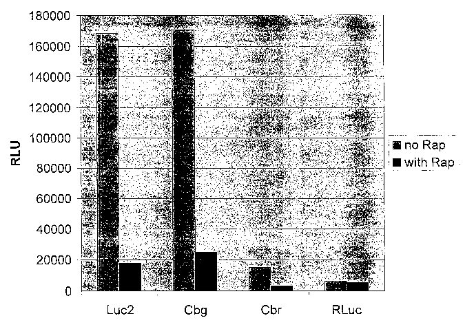
Figure 21B. Luciferase activity of FRB and FKBP fusions with firefly
luciferase, click beetle luciferases and Rehilla luciferase, in the presence
or
absence of rapamycin.
Figure 22. Construct for expressing luciferase from a TK promoter.
Figure 23. Titration of FK506 in the presence of rapamycin in D293
cells transfected with luciferase fused to FRB and FKBP (FRBI-luc2-FKBP),
demonstrating inhibition of rapamycin-mediated modulation by FK506.
Figure 24. Relative luminescence over time in D293 cells transfected
with a construct with a TK promoter and a coding region for a FRB-luciferase-
FKBP fusion, in the presence or absence of rapamycin.
Figure 25A-D. Relative luminescence over time in D293 cells
transfected with a construct with a CMV promoter linked to a coding region for
a FRB-luciferase-FKBP fusion (A), a FRB-luciferase fusion (B), luciferase (C),
or a luciferase-FKBP fusion (D), in the presence or absence of rapamycin.
Figure 26. Relative luminescence of a calmodulin-luciferase fusion in
the presence of EGTA or Ca2+
Detailed Description of the Invention
Definitions
The term "nucleic acid molecule", "polynucleotide", or "nucleic acid
sequence" as used herein, refers to nucleic acid, DNA or RNA, that comprises
coding sequences necessary for the production of a polypeptide or protein
precursor. The encoded polypeptide may be a full-length polypeptide, a
fragment
thereof (less than full-length), or a fusion of either the full-length
polypeptide or
fragment thereof with another polypeptide, yielding a fusion polypeptide.
A "nucleic acid", as used herein, is a covalently linked sequence of
nucleotides in which the 3' position of the pentose of one nucleotide is
joined by
a phosphodiester group to the 5' position of the pentose of the next, and in
which
the nucleotide residues (bases) are linked in specific sequence, i.e., a
linear order
of nucleotides. A "polynucleotide", as used herein, is a nucleic acid
containing a
sequence that is greater than about 100 nucleotides in length. An
"oligonucleotide" or "primer", as used herein, is a short polynucleotide or a
portion of a polynucleotide. An oligonucleotide typically contains a sequence
of
CA 02541765 2006-04-05
WO 2005/038029 PCT/US2004/032705
about two to about one hundred bases. The word "oligo" is sometimes used in
place of the word "oligonucleotide".
Nucleic acid molecules are said to have a "5'-terminus" (5' end) and a
"3'-terminus" (3' end) because nucleic acid phosphodiester linkages occur to
the
5 5' carbon and 3' carbon of the pentose~ring of the substituent
mononucleotides.
The end of a polynucleotide at which a new linkage would be to a 5' carbon is
its
5' terminal nucleotide. The end of a polynucleotide at which a new linkage
would be to a 3' carbon is its 3' terminal nucleotide. A terminal nucleotide,
as
used herein, is the nucleotide at the end position of the 3'- or 5'-terminus.
10 DNA molecules are said to have "5' ends" and "3' ends" because
mononucleotides are reacted to make oligonucleotides in a manner such that the
5' phosphate of one mononucleotide pentose ring is attached to the 3' oxygen
of
its neighbor in one direction via a phosphodiester linkage. Therefore, an end
of
an oligonucleotides referred to as the "5' end" if its 5' phosphate is not
linked to
15 the 3' oxygen of a mononucleotide pentose ring and as the "3' end" if its
3'
oxygen is not linked to a 5' phosphate of a subsequent mononucleotide pentose
ring.
As used herein, a nucleic acid sequence, even if internal to a larger
oligonucleotide or polynucleotide, also may be said to have 5' and 3' ends. In
20 either a linear or circular DNA molecule, discrete elements are referred to
as
being "upstream" or 5' of the "downstream" or 3' elements. This terminology
reflects the fact that transcription proceeds in a 5' to 3' fashion along the
DNA
strand. Typically, promoter and enhancer elements that direct transcription of
a
linked gene (e.g., open reading frame or coding region) are generally located
5'
or upstream of the coding region. However, enhancer elements can exert their
effect even when located 3' of the promoter element and the coding region.
Transcription termination and polyadenylation signals are located 3' or
downstream of the coding region.
The term "codon" as used herein, is a basic genetic coding unit,
consisting of a sequence of three nucleotides that specify a particular amino
acid
to be incorporated into a polypeptide chain, or a start or stop signal. The
term
"coding region" when used in reference to structural gene refers to the
nucleotide
sequences that encode the amino acids found in the nascent polypeptide as a
CA 02541765 2006-04-05
WO 2005/038029 PCT/US2004/032705
21
result of translation of a mRNA molecule. Typically, the coding region is
bounded on the 5' side by the nucleotide triplet "ATG" which encodes the
initiator methionine and on the 3' side by a stop codon (e.g., TAA, TAG, TGA).
In some cases the coding region is also known to initiate by a nucleotide
triplet
"TTG".
The term "gene" refers to a DNA sequence that comprises coding
sequences and optionally control sequences necessary for the production of a
polypeptide from the DNA sequence.
As used herein, the term "heterologous" nucleic acid sequence or protein
refers to a sequence that relative to a reference sequence has a different
source,
e.g., originates from a foreign species, or, if from the same species, it may
be
substantially modified from the original form.
Nucleic acid' are known to contain different types of mutations. A
"point" mutation refers to an alteration in the sequence of a nucleotide at a
single
base position from the wild-type sequence. Mutations may also refer to
insertion
or deletion of one or more bases, so that the nucleic acid sequence differs
from a
reference, e.g., a wild-type, sequence.
As used herein, the terms "hybridize" and "hybridization" refer to the
annealing of a complementary sequence to the target nucleic acid, i.e., the
ability
of two polymers of nucleic acid (polynucleotides) containing complementary
sequences to anneal through base pairing. The terms "annealed" and
"hybridized" are used interchangeably throughout, and are intended to
encompass any specific and reproducible interaction between a complementary
sequence and a target nucleic acid, including binding of regions having only
partial complementarity. Certain bases not commonly found in natural nucleic
acids may be included in the nucleic acids of the present invention and
include,
for example, inosine and 7-deazaguanine. Those skilled in the art of nucleic
acid
technology can determine duplex stability empirically considering a number of
variables including, for example, the length of the complementary sequence,
base composition and sequence of the oligonucleotide, ionic strength and
incidence of mismatched base pairs. The stability of a nucleic acid duplex is
measured by the melting temperature, or "Tm". The Tm of a particular nucleic
acid duplex under specified conditions is the temperature at which on average
half of the base pairs have disassociated.
CA 02541765 2006-04-05
WO 2005/038029 PCT/US2004/032705
22
The term "recombinant DNA molecule" means a hybrid DNA'sequence
comprising at least two nucleotide sequences not normally found together in
nature. The term "vector" is used in reference to nucleic acid molecules
into which fragments of DNA may be inserted or cloned and can be used to
transfer DNA segments) into a cell and capable of replication in a cell.
Vectors
may be derived from plasmids, bacteriophages, viruses, cosmids, and the like.
The terms "recombinant vector" and "expression vector" as used herein
refer to DNA or RNA sequences containing a desired coding sequence and
appropriate DNA or RNA sequences necessary for the expression of the
operably linked coding sequence in a particular host organism. Prokaryotic
expression vectors include a promoter, a ribosome binding site, an origin of
replication for autonomous replication in a host cell and possibly other
sequences, e.g. an optional operator sequence, optional restriction enzyme
sites.
A promoter is defined as a DNA sequence that directs RNA polymerase to bind
to DNA and to initiate RNA synthesis. Eukaryotic expression vectors include a
promoter, optionally a polyadenlyation signal and optionally an enhancer
sequence.
A polynucleotide having a nucleotide sequence encoding a protein or
polypeptide means a nucleic acid sequence comprising the coding region of a
gene, or in other words the nucleic acid sequence encodes a gene product. The
coding region may be present in either a cDNA, genomic DNA or RNA form.
When present in a DNA form, the oligonucleotide may be single-stranded (i.e.,
the sense strand) or double-stranded.' Suitable control elements such as
enhancers/promoters, splice junctions, polyadenylation signals, etc. may be
placed in close proximity to the coding region of the gene if needed to permit
proper initiation of transcription and/or correct processing of the primary
RNA
transcript. Alternatively, the coding region utilized in the expression
vectors of
the present invention may contain endogenous enhancers/promoters, splice
junctions, intervening sequences, polyadenylation signals, etc. In further
embodiments, the coding region may contain a combination of both endogenous
and exogenous control elements.
The term "transcription regulatory element" or "transcription regulatory
sequence" refers to a genetic element or sequence that controls some aspect of
the expression of nucleic acid sequence(s). For example, a promoter is a
CA 02541765 2006-04-05
WO 2005/038029 PCT/US2004/032705
23
regulatory element that facilitates~the initiation of transcription of an
operably
linked coding region. Other regulatory elements include, but are not limited
to,
transcription factor binding sites, splicing signals, polyadenylation signals,
termination signals and enhancer elements.
Transcriptional control signals in eukaryotes comprise "promoter" and
"enhancer" elements. Promoters and enhancers consist of short arrays of DNA
- sequences that interact specifically with cellular proteins involved in
transcription. Promoter and enhancer elements have been isolated from a
variety
of eukaryotic sources including genes in yeast, insect and mammalian cells.
Promoter and enhancer elements have also been isolated from viruses and
analogous control elements, such as promoters, are also found in prokaryotes.
The selection of a particular promoter and enhancer depends on the cell type
used to express the protein of interest. Some eukaryotic promoters and
enhancers have a broad host range wlule others are functional in a limited
subset
of cell types. For example, the SV40 early gene enhancer is very active in a
wide variety of cell types from many mammalian species and has been widely
used for the expression of proteins in mammalian cells. Two other examples of
promoter/enhancer elements active in a broad range of mammalian cell types are
those from the human elongation factor 1 gene and the long terminal repeats of
the Rous sarcoma virus; and the human cytomegalovirus.
The term "promoter/enhancer" denotes a segment of DNA containing
sequences capable of providing both promoter and enhancer functions (i.e., the
functions provided by a promoter element and an enhancer element as described
above). For example, the long terminal repeats of retroviruses contain both
promoter and enhancer functions. The enhancer/promoter may be "endogenous"
or "exogenous" or "heterologous." An "endogenous" enhancer/promoter is one
that is naturally linked with a given gene in the genome. An "exogenous" or
"heterologous" enhancer/promoter is one that is placed in juxtaposition to a
gene
by means of genetic manipulation (i.e., molecular biological techniques) such
that transcription of the gene is directed by the linked enhancer/promoter.
The presence of "splicing signals" on an expression vector often results
in higher levels of expression of the recombinant transcript in eukaryotic
host
cells. Splicing signals mediate the removal of introns from the primary RNA
CA 02541765 2006-04-05
WO 2005/038029 PCT/US2004/032705
24
transcript and consist of a splice donor and acceptor site. A commonly used
splice donor and acceptor site is the splice junction from the 16S RNA of
SV40.
Efficient expression of recombinant DNA sequences in eukaryotic cells
requires expression of signals directing the efficient termination and
polyadenylation of the resulting transcript. Transcription termination signals
are
generally found downstream of the polyadenylation signal and are a few hundred
nucleotides in length. The term "poly(A) site" or "poly(A) sequence" as used
herein denotes a DNA sequence which directs both the termination and
polyadenylation of the nascent RNA transcript. Efficient polyadenylation of
the
recombinant transcript is desirable, as transcripts lacking a poly(A) tail are
unstable and are rapidly degraded. The poly(A) signal utilized in an
expression
vector may be "heterologous" or "endogenous." An endogenous poly(A) signal
is one that is found naturally at the 3' end of the coding region of a given
gene in
the genome. A heterologous poly(A) signal is one which has been isolated from
one gene and positioned 3' to another gene. A commonly used heterologous
poly(A) signal is the SV40 poly(A) signal. The SV40 poly(A) signal is
contained on a 237 by BanaH IlBcl I restriction fragment and directs both
termination and polyadenylation.
Eukaryotic expression vectors may also contain "viral replicons "or "viral
origins of replication." Viral replicons are viral DNA sequences that allow
for
the extrachromosomal replication of a vector in a host cell expressing the
appropriate replication factors. Vectors containing either the SV40 or polyoma
virus origin of replication replicate to high copy number (up to 104
copies/cell)
in cells that express the appropriate viral T antigen. In contrast, vectors
containing the replicons from bovine papillomavirus or Epstein-Barr virus
replicate extrachromosomally at low copy number (about 100 copies/cell).
The term "in vitYO" refers to an artificial environment and to processes or
reactions that occur within an artificial environment. Ih vitro environments
include, but are not limited to, test tubes and cell lysates. The term "in
vivo"
refers to the natural environment (e.g., an animal or a cell) and to processes
or
reaction that occur within a natural environment.
The term "expression system" refers to any assay or system for
determining (e.g., detecting) the expression of a gene of interest. Those
skilled
in the field of molecular biology will understand that any of a wide variety
of
CA 02541765 2006-04-05
WO 2005/038029 PCT/US2004/032705
expression systems may be used. A wide range of suitable mammalian cells are
available from a wide range of source (e.g., the American Type Culture
Collection, Rockland, MD). The method of transformation or transfection and
the choice of expression vehicle will depend on the host system selected.
5 Transformation and transfection methods are well known to the art.
Expression
systems include in vitro gene expression assays where a gene of interest
(e.g., a
reporter gene) is linked to a regulatory sequence and the expression of the
gene
is monitored following treatment with an agent that inhibits or induces
expression of the gene. Detection of gene expression can be through any
10 suitable means including, but not limited to, detection of expressed mRNA
or
protein (e.g., a detectable product of a reporter gene) or through a
detectable
change in the phenotype of a cell expressing the gene of interest. Expression
systems may also comprise assays where a cleavage event or other nucleic acid
or cellular change is detected.
15 The term "wild-type" as used herein, refers to a gene or gene product that
has the characteristics of that gene or gene product isolated from a naturally
occurring source. A wild-type gene is that which is most frequently observed
in
a population and is thus arbitrarily designated the "wild-type" form of the
gene.
In contrast, the term "mutant" refers to a gene or gene product that displays
20 modifications in sequence and/or functional properties (i.e., altered
characteristics) when compared to the wild-type gene or gene product. It is
noted that naturally-occurring mutants can be isolated; these are identified
by the
fact that they have altered characteristics when compared to the wild-type
gene
or gene product.
25 The term "isolated" when used in relation to a nucleic acid, as in
"isolated oligonucleotide" or "isolated polynucleotide" refers to a nucleic
acid
sequence that is identified and separated from at least one contaminant with
which it is ordinarily associated in its source. Thus, an isolated nucleic
acid is
present in a form or setting that is different from that in which it is found
in
nature. In contrast, non-isolated nucleic acids (e.g., DNA and RNA) are found
,
in the state they exist in nature. For example, a given DNA sequence (e.g., a
gene) is found on the host cell chromosome in proximity to neighboring genes;
RNA sequences (e.g., a specific mRNA sequence encoding a specific protein),
are found in the cell as a mixture with numerous other mRNAs that encode a
CA 02541765 2006-04-05
WO 2005/038029 PCT/US2004/032705
26
multitude of proteins. However, isolated nucleic acid includes, by way of
example, such nucleic acid in cells ordinarily expressing that nucleic acid
where
the nucleic acid is in a chromosomal location different from that of natural
cells,
or is otherwise flanked by a different nucleic acid sequence than that found
in
nature. The isolated nucleic acid or oligonucleotide may be present in
single-stranded or double-stranded form. When an isolated nucleic acid or
oligonucleotide is to be utilized to express a protein, the oligonucleotide
contains
at a minimum, the sense or coding strand (i.e., the oligonucleotide may
single-stranded), but may contain both the sense and anti-sense strands (i.e.,
the
oligonucleotide may be double-stranded).
By "peptide," "protein" and "polypeptide" is meant any chain of amino
acids, regardless of length or post-translational modification (e.g.,
glycosylation
or phosphorylation). The nucleic acid molecules of the invention may also
encode a variant of a naturally-occurring protein or polypeptide fragment
thereof, which has an amino acid sequence that is at least 85%, 90%, 95% or
99% identical to the amino acid sequence of the naturally-occurnng (native or
wild-type) protein from which it is derived. The term "fusion polypeptide" or
"fusion protein" refers to a chimeric protein containing a reference protein
(e.g.,
luciferase) joined at the N- and/or C-terminus to one or more heterologous
sequences (e.g., a non-luciferase polypeptide). In some embodiments, a
modified polypeptide, fusion polypeptide or a portion of a full-length
polypeptide of the invention, may retain at least some of the activity of a
corresponding full-length functional (nonchimeric) polypeptide. In other
embodiments, in the absence of an exogenous agent or molecule of interest, a
modified polypeptide, fusion polypeptide or portion of a full-length
functional
polypeptide of the invention, may lack activity relative to a corresponding
full-
length functional polypeptide. In other embodiments, a modified polypeptide,
fusion polypeptide or portion of a full-length functional polypeptide of the
invention in the presence of an exogenous agent may retain at least some or
have
substantially the same activity, or alternatively lack activity, relative to a
corresponding full-length functional polypeptide.
Polypeptide molecules are said to have an "amino terminus"
(N-terminus) and a "carboxy terminus" (C-terminus) because peptide linkages
occur between the backbone amino group of a first amino acid residue and the
CA 02541765 2006-04-05
WO 2005/038029 PCT/US2004/032705
27
backbone carboxyl group of a second amino acid residue. The terms
"N-terminal" and "C-terminal" in reference to polypeptide sequences refer to'
regions of polypeptides including portions of the N-terminal and C-terminal
regions of the polypeptide, respectively. A sequence that includes a portion
of
the N-terminal region of polypeptide includes amino acids predominantly from
the N-terminal half of the polypeptide chain, but is not limited to such
sequences. For example, an N-terminal sequence may include an interior portion
of the polypeptide sequence including bases from both the N-terminal and
C-terminal halves of the polypeptide. The same applies to C-terminal regions.
N-terminal and C-terminal regions may, but need not, include the amino acid
defining the ultimate N-terminus and C-terminus of the polypeptide,
respectively.
The term "recombinant protein" or "recombinant polypeptide" as used
herein refers to a protein molecule expressed from a recombinant DNA
molecule. In contrast, the term "native protein" is used herein to indicate a
protein isolated from a naturally occurring (i.e., a nonrecombinant) source.
Molecular biological techniques may be used to produce a recombinant form of
a protein with identical properties as compared to the native form of the
protein.
The terns "cell," "cell line," "host cell," as used herein, are used
interchangeably, and all such designations include progeny or potential
progeny
of these designations. By "transformed cell" is meant a cell into which (or
into
an ancestor of which) has been introduced a nucleic acid molecule of the
invention. Optionally, a nucleic acid molecule of the invention may be
introduced into a suitable cell line so as to create a stably-transfected cell
line
capable of producing the protein or polypeptide encoded by the gene. Vectors,
cells, and methods for constructing such cell lines are well known in the art.
The
words "transformants" or "transformed cells" include the primary transformed
cells derived from the originally transformed cell without regard to the
number
of transfers. All progeny may not be precisely identical in DNA content, due
to
deliberate or inadvertent mutations. Nonetheless, mutant progeny that have the
same functionality as screened for in the originally transformed cell are
included
in the definition of transformants.
The term "homology" refers to a degree of complementarity between two
or more sequences. There may be partial homology or complete homology (i.e.,
CA 02541765 2006-04-05
WO 2005/038029 PCT/US2004/032705
28
identity). Homology is often measured using sequence analysis software (e.g.,
Sequence Analysis Software Package of the Genetics Computer Group.
University of Wisconsin Biotechnology Center. 1710 University Avenue.
Madison, WI 53705). Such software matches similar sequences by assigning
degrees of homology to various substitutions, deletions, insertions, and other
modifications. Conservative substitutions typically include substitutions
within
the following groups: glycine, alanine; valine, isoleucine, leucine; aspartic
acid,
glutamic acid, asparagine, glutamine; serine, threonine; lysine, arginine; and
phenylalanine, tyrosine.
The term "isolated" when used in relation to a polypeptide, as in "isolated
protein" or "isolated polypeptide" refers to a polypeptide that is identified
and
separated from at least one contaminant with which it is ordinarily associated
in
its source. Thus, an isolated polypeptide is present in a form or setting that
is
different from that in which it is found in nature. In contrast, non-isolated
polypeptides (e.g., proteins and enzymes) are found in the state they exist in
nature.
The term "purified" or "to purify" means the result of any process that
removes some of a contaminant from the component of interest, such as a
protein or nucleic acid. The percent of a purified component is thereby
increased in the sample.
As used herein, "pure" means an object species is the predominant
species present (i.e., on a molar basis it is more abundant than any other
individual species in the composition), and preferably a substantially
purified.
fraction is a composition wherein the object species comprises at least about
50
percent (on a molar basis) of all macromolecular species present. Generally, a
"substantially pure" composition will comprise more than about 80 percent of
all
macromolecular species present in the composition, more preferably more than
about 85%, about 90%, about 95%, and about 99%. Most preferably, the object
species is purified to essential homogeneity (contaminant species cannot be
detected in the composition by conventional detection methods) wherein the
composition consists essentially of a single macromolecular species.
The term "operably linked" as used herein refer to the linkage of nucleic
acid sequences in such a manner that a nucleic acid molecule capable of
directing the transcription of a given gene and/or the synthesis of a desired
CA 02541765 2006-04-05
WO 2005/038029 PCT/US2004/032705
29
protein molecule is produced. The term also refers to the linkage of sequences
encoding amino acids in such a manner that a functional (e.g., enzymatically
active, capable of binding to a binding partner, capable of inhibiting, etc.)
protein or polypeptide is produced.
As used herein, the term "poly-histidine tract" or (His tag) refers to a
molecule comprising two to ten histidine residues, e.g., a poly-histidine
tract of
five to ten residues. A poly-histidine tract allows the affinity purification
of a
covalently linked molecule on an immobilized metal, e.g., nickel, zinc, cobalt
or
copper, chelate column or through an interaction with another molecule (e.g.,
an
antibody reactive with the His tag).
A "protein destabilization sequence" includes, but is not limited to, a
PEST sequence, for example, a PEST sequence from cyclin, e.g., mitotic
cyclins,
uracil permease or ODC, a sequence from the C-terminal region of a short-lived
protein such as ODC, early response proteins such as cytokines, lymphokines,
protooncogenes, e.g., c-myc or c-fos, MyoD, HMG CoA reductase, or S-
adenosyl methionine decarboxylase, CL sequences, a cyclin destruction box, or
N-degron.
As used herein, a "marker gene" or "reporter gene" is a gene that imparts
a distinct phenotype to cells expressing the gene and thus permits cells
having
the gene to be distinguished from cells that do not have the gene. Such genes
may encode either a selectable or screenable marker, depending on whether the
marker confers a trait which one can 'select' for by chemical means, i.e.,
through
the use of a selective agent (e.g., a herbicide, antibiotic, or the like), or
whether it
is simply a "reporter" trait that one can identify through observation or
testing,
i.e., by 'screening'. Elements of the present disclosure are exemplified in
detail
through the use of particular marker genes. Of course, many examples of
suitable marker genes or reporter genes are known to the art and can be
employed in the practice of the invention. Therefore, it will be understood
that
the following discussion is exemplary rather than exhaustive. In light of the
techniques disclosed herein and the general recombinant techniques which are
known in the art, the present invention renders possible the alteration of any
gene. Exemplary modified reporter proteins are encoded by nucleic acid
molecules comprising modified reporter genes including, but are not limited
to,
modifications of a neo gene, a (3-gal gene, a gus gene, a cat gene, a gpt
gene, a
CA 02541765 2006-04-05
WO 2005/038029 PCT/US2004/032705
lZyg gene, a hisD gene, a ble gene, a mprt gene, a bar gene, a nitrilase gene,
a
galactopyranoside gene, a xylosidase gene, a thymidine kinase gene, an
arabinosidase gene, a mutant acetolactate synthase gene (ALS) or acetoacid
synthase gene (AAS), a methotrexate-resistant elhf ° gene, a dalapon
5 dehalogenase gene, a mutated anthranilate synthase gene that confers
resistance
to 5-methyl tryptophan (WO 97/26366), an R-locus gene, a (3-lactamase gene, a
xylE gene, an a,-amylase gene, a tyrosinase gene, a luciferase (luc) gene,
(e.g., a
Refzilla ~enifornais luciferase gene, a firefly luciferase gene, or a click
beetle .
luciferase (Pyr°ophoi°us plagiophthalamus) gene), an aequorin
gene, a red
10 fluorescent protein gene, or a green fluorescent protein gene.
All amino acid residues identified herein are in the natural
L-configuration. In keeping with standard polypeptide nomenclature,
abbreviations for amino acid residues are as shown in the following Table of
Correspondence.
TABLE OF CORRESPONDENCE
1-Letter AMINO ACID
3-Letter
Y Tyr L-tyrosine
G Gly L-glycine
F Phe L-phenylalanine
M Met L-methionine
A Ala L-alanine
S Ser L-serine
I Ile L-isoleucine
L Leu L-leucine
T Thr L-threonine
V Val L-valine
P Pro L-proline
K Lys L-lysine
H His L-histidine
Q Gln L-glutamine
E Glu L-glutamic
acid
W Trp L-tryptophan
R Arg L-arginine
CA 02541765 2006-04-05
WO 2005/038029 PCT/US2004/032705
31
D Asp L-aspartic
acid
N Asn L-asparagine
C Cys L-cysteine
I. Methods to Identify Residues or Regions of a Reporter Protein Which
Are Tolerant to Modification
Numerous methods are available to identify sites and/or regions in a
reporter protein gene wluch may be modified, e.g., disrupted, yet when
transcribed and translated, yield a desirable, for instance, a readily
detectable,
gene product. For instance, amplification reactions may be employed to delete
and/or insert nucleotides for one or more amino acid residues in a reporter
protein gene. Alternatively, transposons may be employed to prepare libraries
of
insertional mutations. Transposons are mobile DNA sequences found in the
genomes of prokaryotes and eukaryotes. Transposon tagging has long been
recognized as a powerful research tool for randomly distributing primer
binding
sites, creating gene "knockouts," and introducing a physical tag or a genetic
tag
into large target DNAs. Insertions in a reporter gene useful to prepare the
modified reporter proteins of the invention are those which are internal, in
frame
insertions in the coding region for the reporter protein. The following
examples,
which are for illustration only, describe the use of a Tn5-based system
(EZ::TNTM from Epicentre, Madison, WI) and a Tn7-based system (GPS-M
Mutagenesis System, New England Biolabs, Inc.) to identify regions in a
reportergene which are tolerant to insertions.
A. Tn-5 Insertional Muta_e
One frequently used transposition system is the Tn5 system isolated from
gram-negative bacteria. The Tn5 transposase is a small, single subunit enzyme
that has been cloned and purified to high specific activity, and carries out
transposition without the need for host cell factors. Moreover, Tn5 transposon
insertions into target DNA are highly random, and proceed by a simple process.
Tn5 transposase will transpose any DNA sequence contained between its short
19 basepair Mosaic End (ME) Tn5 transposase recognition sequences. An
overview of the EZ::TN in frame linker insertion protocol is shown in Figure
1.
CA 02541765 2006-04-05
WO 2005/038029 PCT/US2004/032705
32
i. Transposon Insertion Reaction
Target DNA Preparation. The target reporter DNA is selected as one which is
not encoded by a transposon gene, e.g., a kanamycin resistance gene. While the
transposon insertion reaction is not significantly inhibited by high levels of
RNA
contamination in target DNA preparations, if the target DNA is heavily
contaminated with chromosomal DNA, which is a direct competitor for target
transposition, the number of clones is reduced. Plasmid and cosmid clones can
be purified by standard minilysate procedures and used as target DNA in the
insertion reaction. Low copy-number vectors, for example, BAC or cosmid
clones, are often contaminated with a higher molar proportion of E. coli
chromosomal DNA, thus reducing the transposon insertion frequency.
Therefore, it is preferred BAC and cosmid DNA are purified, to remove the
chromosomal DNA prior to the insertion reaction.
Ih Vita°o Transposon Insertion Reaction. Reaction conditions are
optimized to
maximize the efficiency of the transposon insertion while minimizing multiple
insertion events. For example, an equimolar amount of the transposon is added
to the moles of target DNA.
1. Prepare the transposon insertion reaction mixture by adding in the
following order:
1 p,1 l OX reaction buffer
0.2 ~,g target DNA*
x p,1 molar equivalent transposon
x ~1 sterile water to a reaction volume of 9 ~.l
1 ~,l transposase
10 p.1 total reaction volume
2. Incubate the reaction mixture for 2 hours at 37°C.
3. Stop the reaction by adding ~,1 stop solution.
Mix and heat for 10 minutes at 70°C.
The reaction mixture may be stored at -20°C.
CA 02541765 2006-04-05
WO 2005/038029 PCT/US2004/032705
33
ii. Selection of Transposon Insertion Clones
Transformation and Recovery. The number of transposon insertion clones
obtained per reaction depends on, among other factors, the transformation
efficiency of the competent cells used. The greater the transformation
efficiency
of the competent. cells, the greater the number of insertion clones obtained.
A
y~ecA- strain of E. coli is preferred to eliminate the possibility of
generating
multimeric forms of the vector. Also, the host strain must not express any
antibiotic resistance marker, e.g., a kanamycin resistance marker, present in
the
transposon.
1. Using 1 ~.1 of the insertion reaction mixture, transform recA- E. coli,
e.g.,
electrocompetent cells.
2. Recover the electroporated cells by adding SOC medium to the
electroporation cuvette to 1 ml final volume immediately after
electroporation. Pipette the medium/cells gently to mix. Transfer to a
tube and incubate on a 37°C shaker for 30-60 minutes to facilitate cell
outgrowth.
Plating and Selecting Transformants. Transposon insertion clones are selected
on antibiotic-containing plates. For TnS, kanamycin-containing plates may be
used, however, the transposon can also confer resistance to neomycin and G41 ~
in E. coli.
1. Plate portions of cells onto LB plates containing 50 ~,g/ml kanamycin.
2. To determine the transposon insertion efficiency, plate identical dilutions
and dilution aliquots of the transformation reaction on a second plate
containing an antibiotic specific for selecting target DNA (e.g., 100
~.g/ml ampicillin for the control DNA). The transposition frequency is
given by the ratio of KanR/AmpR clones for the control DNA.
3. Grow plates overnight at 37°C. Assuming a transposon insertion
efficiency of 1 % and use of high purity target DNA (i.e., little or no
chromosomal DNA contamination), there are about 100-500 KanR clones
per plate.
iii. Generating An In Frame 19 Codon Insertion
Transposon Insertion Mapping. Tn5 randomly inserts into target DNA.
Therefore, the transposon insertion site in each clone should be determined
prior
CA 02541765 2006-04-05
WO 2005/038029 PCT/US2004/032705
34
to restriction endonuclease digestion, e.g., NotI digestion, by one of three
methods:
1. Insertion clones can be sequenced bidirectionally using forward and
reverse transposon-specific primers. The insertion site of each clone can also
be
mapped prior to sequencing.
2. Insertion sites can be mapped by size analysis of PCR products using
colony minilysate DNA as a template. To map the insertion sites, forward or
reverse transposon-specific primers and a vector-specific flanking primers may
be employed.
3. Alternatively, insertion sites can be mapped by restriction
endonuclease digest(s).
Once the transposon insertion site of the desired clones is determined, the
clones are individually digested with a restriction enzyme, e.g., NotI, to
linearize
the DNA. The linearized DNA is then purified (e.g., by agarose gel
electrophoresis, column purification, and the like).
Reli~ation and Transformation. The linearized clones are religated using T4
DNA ligase. Successful religation regenerates a single restriction site, e.g.,
NotI,
and creates the 57 nucleotide (19 codon) insertion into all three reading
frames.
The religated DNA is transformed into selected cells and recombinants selected
using an antibiotic marker present on the original cloning vector (e.g.,
ampicillin
for the control DNA).
Analysis of the 19 Codon Insertion Clones. Nine of the 57 nucleotides are the
result of a 9 by sequence duplication immediately flanking the transposon
insertion site. The amino acid sequence of the protein encoded by the target
DNA is conserved on both sides of the 19 codon insertion.
iv. DNA Sequencing of Transposon Insertion Clones
Primer Consideration. Primers should be constructed to minimize homology to
commonly used cloning vectors, and the sequence of each primer should be
compared to that of the user's specific cloning vector to ensure minimal
sequence homology to the vector.
Target Site Duplication. Tn5-catalyzed transposon insertion results in the
generation of a 9 by target site sequence duplication where one copy
immediately flanks each side of the inserted transposon.
CA 02541765 2006-04-05
WO 2005/038029 PCT/US2004/032705
Distinguishing Transposon Sequence for Insert Se uq ence. If the primers
anneal
to a region near the ends of the transposon, the first sequence data obtained
from
each sequencing reaction is that of Transposon DNA.
5 B. Tn7-Based Insertional Muta_eg nesis
The GPS-M Mutagenesis System uses TnsABC* Transposase to insert a
Tn7-based transposon randomly into a DNA target. Target DNA may be a
plasmid, cosmid, BAC or purified chromosomal DNA. If the insertion site is
within a translated gene segment, this will normally result in a null (loss of
10 function) mutation. There is minimal site preference for insertion, so
disruption
of any open reading frame is possible. Due to target immunity, only one
insertion occurs per DNA molecule ih vivo over a distance of about 190 kb.
Therefore, the ira vitf°o reaction produces a population of target DNA
molecules
each containing the transposable element at a different position.
15 The transposon donor can be modified by adding to or replacing the
antibiotic, e.g., kanamycin, resistance marker. The donor plasmid may be grown
in standard laboratory E. coli strains, and the vector backbone carries a
different
antibiotic marker, e.g., Ampr, than the transposon and an origin of
replication.
To destroy unreacted donor molecules and avoid undesirable reaction products,
20 the donor can be destroyed by digestion with a rare-cutting enzyme, for
instance,
PI-SceI (VDE). For applications in which the mutagenized DNA is transformed
into naturally-competent organisms (which take up single DNA strands), the
gaps are filled-in and ligated.
25 i. Reaction Protocol
1. Mix the following reagents (per 20 ~.1 reaction):
2 ~1 10 X buffer
1 ~.1 supercoiled custom donor (0.02 ~,g)
0.08 ~,g target DNA
30 dHzO
18 ~.1 Total Volume
Mix well by pipetting up and down a few times.
2. Add 1 ~,1 transposase to each tube. Mix again.
3. Incubate for 10 minutes at 37°C. This is the assembly reaction.
CA 02541765 2006-04-05
WO 2005/038029 PCT/US2004/032705
36
4. Add 1 ~,1 start solution to each tube. Mix well by pipetting up and
down a few times.
5. Incubate for 1 hour at 37°C. This is the strand transfer reaction.
6. Heat inactivate at 75°C for 10 minutes. Note: 65°C is not
adequate.
7. Optional gap repair.
8. Add 5 ~,1 l OX Pl-SceI Buffer
0.5 ~,1 BSA
18.5 ~,l dHzO
6 ~.1 Pl-SceI (VDE) (6 units)
9. Incubate for 1-2 hours at 37°C.
10. hzcubate for 10 minutes at 75°C.
11. Transform. For chemical transformation with subcloning
efficiency cells (107 per microgram of pUC), transform 1 ~.1 and 10 ~,l of
undiluted reaction. For electroporation (>109 per microgram of pUC),
dilute 10-fold in dHzO and transform 1 ~,1 and 10 ~,1. To outgrow, dilute
the transformation mixture into 1 ml LB or as directed by the
manufacturer, and incubate for 1 hour at 37°C with aeration. This
period
without selection is necessary for expression of drug resistance,
especially kanamycin.
ii. General Considerations
Amount of Tar-g-et. The recommended mass of target DNA (0.08 ~,g per
reaction) works well for plasmid targets. For cosmids and BACs, a molar ratio
of around 2:1 (donor to target) works well. Increasing the ratio to 4:1
decreases
the efficiency slightly.
Donor:Target Ratio. The recommended donoraarget mass ratio (1:4, 0.08 ~g
target per 20 ~,l reaction) is optimal. Small deviations produce only small
changes in the number of recovered products. However, saturating amounts of
donor inhibit the reaction and may lead to accumulation'of double insertions.
Order of Addition. Water, target DNA, buffer and donor plasmid should be
added first, followed by transposase. The start solution should be added only
after the assembly reaction.
Assembly Reaction. If this step is omitted, the proportion of complicated
CA 02541765 2006-04-05
WO 2005/038029 PCT/US2004/032705
37
products is increased.
Time of Incubation. The reaction is linear at 37°C for at least
one hour.
Extremely long incubation times may lead to accumulation of double insertions.
Temperature of Incubation. The reaction proceeds, but more slowly, at room
temperature and at 30°C. For reactions with BACs, 30°C is
recommended.
Heat Killing. Heating at 75°C for 10 minutes effectively disrupts the
reaction
complexes. Heating at 65°C for 20 minutes is not adequate.
Phenol/chloroform
extraction followed by alcohol precipitation is also effective.
Scaling the Procedure. Increase or reduce the final volume and the volume of
all
components by the same percentage; the relative concentrations of the two DNA
species and the proteins are very important, as are the buffer conditions.
Enzyme Names. Pl-SceI (VDE),is not the same as 1-SceI. Use Pl-SceI (VDE) to
digest the donor and Scel for mapping insertions obtained.
Gap Repair. This step is not required for transformation into E. coli and is
necessary only when the desired application involves transformation into
naturally competent bacteria. Naturally competent bacteria include members of
the genera Neisse~ia, HaenZOphilus, Bacillus, Pneuynococeus, Staphyloc~ccus,
and Sty~eptococcus. DNA uptake into these organisms involves degradation of
one strand, concomitant with internalization of the other strand. Without gap
repair, the 5-base gaps at the transposon insertion site will unlink the
transposon
insertion from flanking DNA on one side or the other. Organisms in which
competence is induced chemically or by electroporation (e.g., E. coli and
other
enteric bacteria tissue culture cells, etc.) take up both DNA strands. Gaps at
the
insertion site are efficiently repaired by the cellular machinery.
iii. GAP Repair Protocol
7. Phenol/chloroform extract (50 ~,l).
8. Ethanol precipitate:
6 ~,13M NaAcetate
100 ~,1 EtOH
Incubate for 20 minutes at -20°C
Centrifuge for 10 minutes in a microfuge
9. Resuspend in 15 w1 TE.
10. 1 ~,1 DNA Polymerase I (E. coli) (10 units)
CA 02541765 2006-04-05
WO 2005/038029 PCT/US2004/032705
38
3 ~,1 l OX EcoPol Buffer
9 ~.1 dNTP (at 100 wM each nucleotide; final concentration 33 ~,M
each)
11. Incubate for 15 minutes at room temperature.
12. Add 1 ~,l T4 DNA ligase (400 units) and ATP to a final .
concentration of 1 mM.
13. Incubate for 4 hours at 16°C.
14. Phenol/chloroform extract.
15. Alcohol precipitate.
16. Resuspend in 20 ~1 TE.
17. Add 5 ~,1 OX Pl-SceI Buffer
0.5 ~.1 BSA
18.5 ~,1 dH20
6 p.1 Pl-SceI (VDE) (6 units)
18. Incubate for 1-2 hours at 37°C.
19. Incubate for 10 minutes at 75°C.
20. Transform according to the appropriate method.
i
iv. Donor Manipulation
1. The transposon donor must be supercoiled. The efficiency of reaction
using a relaxed or linear donor is reduced by about 100-fold. The donor
preparation should be good quality, but CsCI-purification is not
necessary.
2. Essential recognition elements for the transposase are not dispensable.
There may be stop codons in all frames reading into the transposon.
Transcription can proceed into the dispensable region from outside
without difficulty.
3. Transposition efficiency may decline somewhat as the transposon
becomes longer.
4. For best results, ensure that your transposon donor plasmid is
monomenc.
5. The Pl-SceI digestion step may be omitted if the donor preparation is
monomeric and supercoiled and if the donor molecules will not replicate
in the host organism.
CA 02541765 2006-04-05
WO 2005/038029 PCT/US2004/032705
39
c
v. Target DNA Ree~uirements
Plasmid targets for sequencing should be in circular form to facilitate
recovery. Linear (e.g., chromosomal) DNA is an efficient substrate. A repaii
land
ligation step is required before transformation, when using naturally
transformable organisms. Large plasmids, such as cosmids and BACs, are
usable targets. Target DNA must be at least 5 wgl ml in a no-salt buffer such
as
1 X TE. The concentration can be estimated by comparison of agarose gel band
intensity with a DNA of known concentration or by absorbance at 260.
I
II. Exemplary Modifications y'
Once a site or region in a reporter protein is identified that is toler iiit
to
modification, that site or region may be modified by deletion of one or mar
residues, insertion of one or more residues and/or by circular permutation o~
any
combination thereof. In one embodiment, the modification may be the
introduction of a recognition site for a hydrolase including but not
limited~to
proteases, peptidases, esterases (e.g., cholesterol esterase), phosphatases
li~-.g.,
alkaline phosphatase) and the like. For instance, hydrolases include, but ,are
not
limited to, enzymes acting on peptide bonds (peptide hydrolases) such
aminopeptidases, dipeptidases, dipeptidyl-peptidases and tripeptidyl
peptidyl-dipeptidases, serine-type carboxypeptidases,
cysteine-type carboxypeptidases, omega peptidases, serine endopeptidase~
cysteine endopeptidases, aspartic endopeptidases, metalloendopeptidase,s,
threonine endopeptidases, and endopeptidases of unknown catalytic mechanism.
For example, a modified beetle luciferase of the invention may compri~~e an
c
enterokinase cleavage site, a caspase cleavage site, a coronavirus protease
site
(STLQ-SGLRKMA; SEQ ID NO:10), a kinase site, a HIV-1 protease site
(SQNY-PIVQ or KAVRL-AEAMS; SEQ ID NO:l 1 and SEQ ID N0:12,
respectively), a HCV protease site (AEDVVCC-SMSYS; SEQ ID N0:~13) (see,
e.g., Lee et al., 2003), a SARS vines protease site (e.g.,
QTSITSAVLQSGFRKMAFPS; SEQ ID N0:16, or
VRQCSGVTFQGKFKKIVKGT; SEQ ID N0:17), a rhinovirus protease site,
e.g., rhinovirus 3C protease site, a prohormone convertase site, an
interleukin-
16-converting enzyme site, a CMV assembling site, a leishmandysin site, B.
CA 02541765 2006-04-05
WO 2005/038029 PCT/US2004/032705
af~.thracis lethal factor, a botulinum neurotoxin light chain protease site, a
(i-
secretase site for amyloid precursor protein (VKM-DAEF; SEQ ID N0:14),
prostate specific antigen sequence, a thrombin site, a renin and angiotensin-
converting enzyme site, a cathepsin D site, a matrix metalloproteinase site, a
5 uPA site, a plasmin site, a binding site for a ration, such as a calcium
binding
domain, a calmodulin binding domain, a cellulose binding domain, a chitin
binding domain, a maltose binding protein domain, or a biotin binding domain.
In another embodiment, a modified reporter protein of the invention may
comprise a sequence recognized by a ligand such as an antibody or a metal such
10 as calcium.
III. Exemplary Polynucleotides and Proteins
The invention includes a modified reporter protein encompassing any
amino acid sequence which provides a polypeptide having a detectable activity,
15 e.g., luminescent activity, as well as protein fragments thereof, which are
recombinantly or synthetically synthesized. The reporter protein sequences of
a
modified reporter protein are the same or are substantially the same as the
amino
acid sequence of a corresponding unmodified reporter protein. A polypeptide or
peptide having substantially the same sequence means that an amino acid
20 sequence is largely, but may not entirely be, the same and retains a
functional
activity of the sequence to which it is related. In general, two amino acid
sequences are substantially the same or substantially homologous if they are
at
least 70% identical, e.g., have at least 80%, 90%, 95% or more identity.
Homology or identity is often measured using sequence analysis software
25 (e.g., Sequence Analysis Software Package of the Genetics Computer Group,
University of Wisconsin Biotechnology Center, 1710 University Avenue,
Madison, WI 53705). Such software matches similar sequences by assigning
degrees of homology to various deletions, substitutions and other
modifications.
The terms "homology" and "identity" in the context of two or more nucleic
acids
30 or polypeptide sequences, refer to two or more sequences or subsequences
that
are the same or have a specified percentage of amino acid residues or
nucleotides
that are the same when compared and aligned for maximum correspondence over
a comparison window or designated region as measured using any number of
sequence comparison algorithms or by manual alignment and visual inspection.
CA 02541765 2006-04-05
WO 2005/038029 PCT/US2004/032705
41
For sequence comparison, typically one sequence acts as a reference
sequence, to which test sequences are compared. When using a sequence
comparison algorithm, test and reference sequences are entered into a
computer,
subsequence coordinates are designated, if necessary, and sequence algorithm
program parameters are designated. Default program parameters can be used, or
alternative parameters can be designated. The sequence comparison algorithm
then calculates the percent sequence identities for the test sequences
relative to
the reference sequence, based on the program parameters.
Methods of alignment of sequence for comparison are well-known in the
art. Optimal alignment of sequences for comparison can be conducted by the
local homology algorithm of Smith et al. (1981), by the homology alignment
algorithm of Needleman et al. (1970), by the search for similarity method of
Person et al. (1988), by computerized implementations of these algorithms
(GAP, BESTFIT, FASTA, and TFASTA in the Wisconsin Genetics Software
Package, Genetics Computer Group, 575 Science Dr., Madison, WI), or by
manual alignment and visual inspection.
Computer implementations of these mathematical algorithms can be
utilized for comparison of sequences to determine sequence identity. Such
implementations include, but are not limited to: CLUSTAL in the PC/Gene
program (available from Intelligenetics, Mountain View, California); the ALIGN
program (Version 2.0) and GAP, BESTFIT, BLAST, FASTA, and TFASTA in
the Wisconsin Genetics Software Package, Version 8 (available from Genetics
Computer Group (GCG), 575 Science Drive, Madison, Wisconsin, USA).
Alignments using these programs can be performed using the default parameters.
The CLUSTAL program is well described by Higgins et al. (1988); Higgins et
al. (1989); Corpet et a1.(1988); Huang et al. (1992); and Pearson et al.
(1994).
The ALIGN program is based on the algoritlmn of Myers and Miller (1988). The
BLAST programs of Altschul et al. (1990), are based on the algorithm of Karlin
and Altschul (1990).
Software for performing BLAST analyses is publicly available through
the National Center for Biotechnology Information
(http://www.ncbi.nlm.nih.gov/). This algorithm involves first identifying high
scoring sequence pairs (HSPs) by identifying short words of length W in the
query sequence, which either match or satisfy some positive-valued threshold
CA 02541765 2006-04-05
WO 2005/038029 PCT/US2004/032705
42
score T when aligned with a word of the same length in a database sequence. T
is referred to as the neighborhood word score threshold (Altschul et al.,
1990).
These initial neighborhood word hits act as seeds for initiating searches to
find
longer HSPs containing them. The word hits are then extended in both
directions
along each sequence for as far as the cumulative alignment score can be
increased. Cumulative scores are calculated using, for nucleotide sequences,
the
parameters M (reward score for a pair of matching residues; always > 0) and N
(penalty score for mismatching residues; always < 0). For amino acid
sequences, a scoring matrix is used to calculate the cumulative score.
Extension
of the word hits in each direction are halted when the cumulative alignment
score falls off by the quantity X from its maximum achieved value, the
cumulative score goes to zero or below due to the accumulation of one or more
negative-scoring residue alignments, or the end of either sequence is reached.
In addition to calculating percent sequence identity, the BLAST
algoritlnn also performs a statistical analysis of the similarity between two
sequences (see, e.g., Marlin & Altschul (1993). One measure of similarity
provided by the BLAST algorithm is the smallest sum probability (P(N)), which
provides an indication of the probability by which a match between two
nucleotide or amino acid sequences would occur by chance. For example, a test
nucleic acid sequence is considered similar to a reference sequence if the
smallest sum probability in a comparison of the test nucleic acid sequence to
the
reference nucleic acid sequence is less than about 0.1, more preferably less
than
about 0.01, and most preferably less than about 0.001.
To obtain gapped alignments for comparison purposes, Gapped BLAST ,
(in BLAST 2.0) can be utilized as described in Altschul et al. (1997).
Alternatively, PSI-BLAST (in BLAST 2.0) can be used to perform an iterated
search that detects distant relationships between molecules. See Altschul et
al.,
supf°a. When utilizing BLAST, Gapped BLAST, PSI-BLAST, the default
parameters of the respective programs (e.g. BLASTN for nucleotide sequences,
BLASTX for proteins) can be used. The BLASTN program (for nucleotide
sequences) uses as defaults a wordlength (W) of 1 l, an expectation (E) of 10,
a
cutoff of 100, M=5, N=-4, and a comparison of both strands. For amino acid
sequences, the BLASTP program uses as defaults a wordlength (W) of 3, an
expectation (E) of 10, and the BLOSUM62 scoring matrix (see Henikoff &
CA 02541765 2006-04-05
WO 2005/038029 PCT/US2004/032705
43
gienikoff, 1989). See htt~//www ncbi nlm.nih.gov.
In particular, a polypeptide may be substantially related but for a
conservative variation. A conservative variation denotes the replacement of an
amino acid residue by another, biologically similar residue. Examples of
conservative variations include the substitution of one hydrophobic residue
such
as isoleucine, valine, leucine or methionine for another, or the substitution
of one
polar residue for another such as the substitution of arginine for lysine,
glutamic
for aspartic acids, or glutamine for asparagine, and the like. Other
illustrative
examples of conservative substitutions include the changes of: alanine to
serine;
arginine to lysine; asparagine to glutamine or histidine; aspartate to
glutamate;
cysteine to serine; glutamine to asparagine; glutamate to aspartate; glycine
to
proline; histidine, to asparagine or glutamine; isoleucine to leucine or
valine;
leucine to valine or isoleucine; lysine to arginine, glutamine, or glutamate;
methionine to leucine or isoleucine; phenylalanine to tyrosine, leucine or
methionine; serine to threonine; threonine to serine; tryptophan to tyrosine;
tyrosine to tryptophan or phenylalanine; valine to isoleucine to leucine.
In one embodiment, a polynucleotide of the invention is optimized for
expression in a particular host. As used herein, optimization includes codon
optimization as well as, in eukaryotic cells, introduction of a I~ozak
sequence,
and/or one or more introns. Thus, a nucleic acid molecule may have a codon
composition that differs from that of a wild-type nucleic acid sequence
encoding
an unmodified beetle luciferase at more than 30%, 35%, 40% or more than 45%,
e.g., 50%, 55%, 60% or more of the codons. Preferred codons for use in the
invention are those which are employed more frequently than at least one other
codon for the same amino acid in a particular organism and, more preferably,
are
also not low-usage codons in that organism and are not low-usage codons in the
organism used to clone or screen for the expression of the nucleic acid
molecule.
Moreover, preferred codons for certain amino acids (i.e., those amino acids
that
have three or more codons,), may include two or more codons that are employed
more frequently than the other (non-preferred) codon(s). The presence of
codons in the nucleic acid molecule that are employed more frequently in one
organism than in another organism results in a nucleic acid molecule which,
when introduced into the cells of the organism that employs those codons more
CA 02541765 2006-04-05
WO 2005/038029 PCT/US2004/032705
44
frequently, is expressed in those cells at a level that is greater than the
expression
of the wild-type or parent nucleic acid sequence in those cells.
In one embodiment of the invention, the colons that are different are
those employed more frequently in a mammal, while in another embodiment the
colons that are different are those employed more frequently in a plant. A
particular type of mammal, e.g., human, may have a different set of preferred
colons than another type of mammal. Likewise, a particular type of plant may
have a different set of preferred colons than another type of plant. In one
embodiment of the invention, the majority of the colons which differ are ones
that are preferred colons in a desired host cell. Preferred colons for mammals
(e.g., humans) and plants are known to the art (e.g., Wada et al., 1990). For
example, preferred human colons include, but are not limited to, CGC (Arg),
CTG (Leu), TCT (Ser), AGC (Ser), ACC (Thr), CCA (Pro), CCT (Pro), GCC
(Ala), GGC (Gly), GTG (Val), ATC (Ile), ATT (Ile), AAG (Lys), AAC (Asn),
CAG (Gln), CAC (His), GAG (Glu), GAC (Asp), TAC (Tyr), TGC (Cys) and
TTC (Phe) (Wada et al., 1990). Thus, preferred "humanized" synthetic nucleic
acid molecules of the invention have a colon composition which differs from a
wild type nucleic acid sequence by having an increased number of the preferred
human colons, e.g. CGC, CTG, TCT, AGC, ACC, CCA, CCT, GCC, GGC,
GTG, ATC, ATT, AAG, AAC, CAG, CAC, GAG, GAC, TAC, TGC, TTC, or
any combination thereof. For example, the nucleic acid molecule of the
invention,may have an increased number of CTG or TTG leucine-encoding
colons, GTG or GTC valine-encoding colons, GGC or GGT glycine-encoding
colons, ATC or ATT isoleucine-encoding colons, CCA or CCT proline-
encoding colons, CGC or CGT arginine-encoding colons, AGC or TCT serine-
encoding colons, ACC or ACT threonine-encoding colon, GCC or GCT
alanine-encoding colons, or any combination thereof, relative to the wild-type
nucleic acid sequence. Similarly, nucleic acid molecules having an increased
number of colons that are employed more frequently in plants, have a colon
composition which differs from a wild-type nucleic acid sequence by having an
increased number of the plant colons including, but not limited to, CGC (Arg),
CTT (Leu), TCT (Ser), TCC (Ser), ACC (Thr), CCA (Pro), CCT (Pro), GCT
(Ser), GGA (Gly), GTG (Val), ATC (Ile), ATT (Ile), AAG (Lys), AAC (Asn),
CAA (Gln), CAC (His), GAG (Glu), GAC (Asp), TAC (Tyr), TGC (Cys), TTC
CA 02541765 2006-04-05
WO 2005/038029 PCT/US2004/032705
(Phe), or any combination thereof (Murray et al., 1959). Preferred codons may
differ for different types of plants (Wada et al., 1990).
The modified beetle luciferase proteins or fusion proteins of the
invention may be prepared by recombinant methods or by solid phase chemical
5 peptide synthesis methods. Such methods have been known in the art since the
early 1960's (Merrifield, 1963) (See also Stewart et al., Solid Phase Peptide
Synthesis, 2 ed., Pierce Chemical Co., Rockford, Ill., pp. 11-12)) and have
recently been employed in commercially available laboratory peptide design and
synthesis kits (Cambridge Research Biochemicals). Such commercially available
10 laboratory kits have generally utilized the teachings of Geysen et al.
(1984) and
provide for synthesizing peptides upon the tips of a multitude of rods" or
"pins"
all of which are connected to a single plate. When such a system is utilized,
a
plate of rods or pins is inverted and inserted into a second plate of
corresponding
wells or reservoirs, which contain solutions for attaching or anchoring an
15 appropriate amino acid to the pin' s or rod' s tips. By repeating such a
process
step, e.g., inverting and inserting the rod' s and pin' s tips into
appropriate
solutions, amino acids are built into desired peptides. In addition, a number
of
available FMOC peptide synthesis systems are available. For example, assembly
of a polypeptide or fragment can be carried out on a solid support using an
20I Applied Biosystems, Inc. Model 431A automated peptide synthesizer. Such
equipment provides ready access to the peptides of the invention, either by
direct '
synthesis or by synthesis of a series of fragments that can be coupled using
other
known techniques.'
25 IV. Fusion Partners Useful with the Modified Reporter Protein of the
Invention
The polynucleotide of the invention which encodes a modified reporter
protein may be employed with other nucleic acid sequences, e.g., a native
sequence such as a cDNA or one which has been manipulated in vitro, e.g., to
30 prepare N-terminal, C-terminal, or N- and C-terminal fusion proteins, e.g.,
a
fusion with a protein encoded by a different reporter gene including a
selectable
marker. Many examples of suitable fusion partners are known to the art and can
be employed in the practice of the invention.
CA 02541765 2006-04-05
WO 2005/038029 PCT/US2004/032705
46
Fusion partners include but are not limited to affinity domains or other
functional protein sequences, such as those having an enzymatic activity. For
example, a functional protein sequence may encode a kinase catalytic domain
(Hanks and Hunter, 1995), producing a fusion protein that can enzymatically
add
phosphate moieties to particular amino acids, or may encode a Src Homology 2
(SH2) domain (Sadowski et al., 1986; Mayer and Baltimore,1993), producing a
fusion protein that specifically binds to phosphorylated tyrosines.
Affinity domains are generally peptide sequences that can interact with a
binding partner, e.g., such as one immobilized on a solid support. DNA
sequences encoding multiple consecutive single amino acids, such as histidine,
when fused to the expressed protein, may be used for one-step purification of
the
recombinant protein by high affinity binding to a resin column, such as nickel
sepharose. Sequences encoding peptides, such as the chitin binding domain
(which binds to chitin), glutathione-S-transferase (which binds to
glutathione),
biotin (which binds to avidin and strepavidin), and the like, can also be used
for
facilitating purification of the protein of interest. The affinity domain can
be
separated from the protein of interest by methods well known in the art,
including the use of inteins (protein self splicing elements (Chong et al.,
1997).
Exemplary affinity domains include HisVS (HI3HHH) (SEQ ID NO:l), HisX6
(HHHHHH) (SEQ ID NO:2), C-myc (EQKLISEEDL) (SEQ ID NO:3), Flag
(DYKDDDDK) (SEQ ID NO:4), SteptTag (WSHPQFEK) (SEQ ID NO:S),
hemagluttinin, e.g., HA Tag (YPYDVPDYA) (SEQ ID N0:6), GST,
thioredoxin, cellulose binding domain, RYIRS (SEQ ID N0:104), Phe-His-His-''
Thr (SEQ ID NO:105), chitin binding domain, S-peptide, T7 peptide, SH2
domain, C-end RNA tag, WEAAAREACCRECCARA (SEQ ID N0:8), metal
binding domains, e.g., zinc binding domains or calcium binding domains such as
those from calcium-binding proteins, e.g., calmodulin, troponin C, calcineurin
B,
myosin light chain, recoverin, S-modulin, visinin, VILIP, neurocalcin,
hippocalcin, frequenin, caltractin, calpain large-subunit, S 100 proteins,
parvalbumin, calbindin D~K, calbindin D28K, and calretinin, inteins, biotin,
streptavidin, MyoD, Id, leucine zipper sequences, and maltose binding protein.
In one embodiment, the fusion partner is a sequence useful to purify a fusion
protein, e.g., a His or GST tag, and in one embodiment the purification tag is
fused to the N- or C-terminus of a circularly permuted reporter protein.
CA 02541765 2006-04-05
WO 2005/038029 PCT/US2004/032705
47
Another class of fusion partners includes a protein encoded by a reporter
gene, including, but are not limited to, a neo gene, a [3-gal gene, a gus
gene, a eat
gene, a gpt gene, a hyg gene, a hisD gene, a ble gene, a rnprt gene, a bay
gene, a
nitrilase gene, a galactopyranoside gene, a xylosidase gene, a thymidine
kinase
gene, an arabinosidase gene, a mutant acetolactate synthase gene (ALS) or
acetoacid synthase gene (AAS), a methotrexate-resistant dhfi~ gene, a dalapon
dehalogenase gene, a mutated anthranilate synthase gene that confers
resistance
to 5-methyl tryptophan (WO 97/26366), an R-locus gene, a [3-lactamase gene, a
xylE gene, an a-amylase gene, a tyrosinase gene, an anthozoan luciferase (luc)
gene, (e.g., a Rehilla ren.ifornais luciferase gene), an aequorin gene, a red
fluorescent protein gene, or a green fluorescent protein gene. Included within
the terms selectable or screenable marker genes are also genes which encode a
"secretable marker" whose secretion can be detected as a means of identifying
or
selecting for transformed cells. Examples include markers which encode a
secretable antigen that can be identified by antibody interaction, or even
secretable enzymes which can be detected by their catalytic activity.
Secretable
proteins fall into a number of classes, including small, diffusible proteins
detectable, e.g., by ELISA, and proteins that are inserted or trapped in the
cell
membrane.
V. Vectors and Host Cells Encoding: the Modified Reporter Protein or
Fusions Thereof
Once a desirable nucleic acid molecule encoding a modified reporter
protein or a fusion thereof is prepared, an expression cassette encoding the
modified reporter protein or a fusion protein comprising the modified reporter
protein is prepared. For example, a nucleic acid molecule comprising a nucleic
acid sequence encoding a modified beetle luciferase is optionally operably
linked to transcription regulatory sequences, e.g., one or more enhancers, a
promoter, a transcription termination sequence or a combination thereof, to
form
an expression cassette. The nucleic acid molecule or expression cassette may
be
introduced to a vector, e.g., a plasmid or viral vector, which optionally
includes a
selectable marker gene, and the vector introduced to a cell of interest, for
example, a prolcaryotic cell such as E. coli, Streptomyces spp., Bacillus
spp.,
Staphylococcus spp. and the like, as well as eukaryotic cells including a
plant
CA 02541765 2006-04-05
WO 2005/038029 PCT/US2004/032705
48
(dicot or monocot), fungus, yeast, e.g., Pichia, Sacclzaromyces or
Sclzizosaccharomyces, or a mammalian cell. Preferred mammalian cells include
bovine, caprine, ovine, canine, feline, non-human primate, e.g., simian, and
human cells. Preferred mammalian cell lines include, but are not limited to,
CHO, COS, 293, Hela, CV-1, SH-SYSY, HEK293, and NIH3T3 cells.
The expression of an encoded modified reporter protein may be
controlled by any promoter capable of expression in prokaryotic cells or
eukaryotic cells. Preferred prokaryotic promoters include, but are not limited
to,
SP6, T7, T5, tac, bla, trap, gal, lac or maltose promoters. Preferred
eukaryotic
promoters include, but are not limited to, constitutive promoters, e.g., viral
promoters such as CMV, SV40 and RSV promoters, as well as regulatable
promoters, e.g., an inducible or repressible promoter such as the tet
promoter, the
hsp70 promoter and a synthetic promoter regulated by CRE. The nucleic acid
molecule, expression cassette and/or vector of the invention may be introduced
to a cell by any method including, but not limited to, calcium-mediated
transformation, electroporation, microinjection, lipofection and the like.
VI. Exemplar,
The modified reporter proteins or fusions thereof are useful for any
purpose including, but not limited to, detecting the amount or presence of a
particular molecule (a biosensor), isolating a particular molecule, detecting
conformational changes in a particular molecule, e.g., due to binding,
phosphorylation or ionization, detecting conditions, for instance, pH or
temperature, facilitating high or low throughput screening, detecting protein-
protein, protein-DNA or other protein-based interactions, or selecting or
evolving biosensors. For instance, a modified reporter protein or a fusion
thereof, is useful to detect e.g., in an in vitro or cell-based assay, the
amount,
presence or activity of a particular kinase (for example, by inserting a
kinase site
into a reporter protein), RNAi (e.g., by inserting a sequence suspected of
being
recognized by RNAi into a coding sequence for a reporter protein, then
monitoring reporter activity after addition of RNAi), or protease, such as one
to
detect the presence of a particular viral protease, which in turn is indicator
of the
presence of the virus, or an antibody; to screen for inhibitors, e.g.,
protease
inhibitors; to identify recognition sites or to detect substrate specificity,
e.g.,
CA 02541765 2006-04-05
WO 2005/038029 PCT/US2004/032705
49
using a modified luciferase with a selected recognition sequence or a library
of
modified luciferases having a plurality of different sequences with a single
molecule of interest or a plurality (for instance, a library) of molecules; to
select
or evolve biosensors or molecules of interest, e.g., proteases; or to detect
protein-
protein interactions via complementation or binding, e.g., in an ifz vitro or
cell-
based approach. In one embodiment, a modified beetle luciferase which
includes an insertion is contacted with a random library or mutated library of
molecules, and molecules identified which interact with the insertion. In
another
embodiment, a library of modified luciferases having a plurality insertions is
contacted with a molecule, and modified luciferases which interact with the
molecule identified.
The invention also provides methods to monitor the expression, location
and/or trafficking of molecules in a cell, as well as to monitor changes in
microenvironments within a cell, using a modified beetle luciferase or a
fusion
protein thereof. For example, in one embodiment, a modified beetle luciferase
comprises an internal insertion containing two domains which interact with
each
other under certain conditions. In one embodiment, one domain in the insertion
contains an amino acid which can be phosphorylated and the other domain is a
phosphoamino acid binding domain. In the presence of the appropriate kinase or
phosphatase, the two domains in the insertion interact and change the
conformation of the modified beetle luciferase resulting in an alteration in
the
detectable activity of the modified beetle luciferase. In another embodiment,
a
modified beetle luciferase comprises a recognition site for a molecule, and
when
the molecule interacts with the recognition site, results in an increase in
activity,
and so can be employed to detect or determine the presence of amount or the
other molecule.
Two-hybrid systems are extremely powerful methods for detecting
protein:protein interactions ih vivo as well as identifying residues/domains
involved in protein:protein interactions. The basis of two-hybrid systems is
the
modular domains found in some transcription factors: a DNA-binding domain,
which binds to a specific DNA sequence, and a transcriptional activation
domain, which interacts with the basal transcriptional machinery (Sadowski,
1988). A transcriptional activation domain in association with a DNA-binding
domain may promote the assembly of RNA polymerase II complexes at the
CA 02541765 2006-04-05
WO 2005/038029 PCT/US2004/032705
TATA box and increase transcription. In the CheckMateTM Mammalian Two-
Hybrid System (Promega Corp., Madison, WI), the DNA-binding domain and
the transcriptional activation domain, produced by separate plasmids, are
closely
associated when 'one protein ("X") fused to a DNA-binding domain interacts
5 with a second protein ("Y") fused to a transcriptional activation domain. In
this
system, interaction between proteins X and Y results in transcription of
either a
reporter gene or a selectable marker gene. In particular, the pBIND Vector
contains a yeast GAL4 DNA-binding domain upstream of a multiple cloning
region, and a pACT Vector contains the herpes simplex virus VP16 activation
10 domain upstream of a multiple cloning region. In addition, the pBIND Vector
expresses the Resailla ~efaifo~rrais luciferase. The two genes encoding the
two
potentially interactive proteins of interest are cloned into pBIND and pACT
Vectors to generate fusion proteins with the DNA-binding domain of GAL4 and
the activation domain of VP16, respectively. The pGSluc Vector contains five
15 GAL4 binding sites upstream of a minimal TATA box, which in turn, is
upstream of the firefly luciferase gene (luc+), The pGAL4 and pVPl6 fusion
constructs are transfected along with pGSluc Vector into mammalian cells. Two
to three days after transfection the cells are lysed, and the amount of
Rehilla
luciferase and firefly luciferase can be quantitated using the Dual-
Luciferase~
20 Reporter Assay System (Promega Cat.# E1910). Interaction between the two
test proteins, as GAL4 and VP16 fusion constructs, results in an increase in
firefly luciferase expression over the negative controls. A modified beetle
luciferase of the invention, e.g., one which is deleted at a site or region
which is
tolerant to modification (a N-terminal fragment), is fused to a DNA binding
25 domain while the remainder of the beetle luciferase (the C-terminal
fragment) is
fused to a transcriptional activator domain.
The invention also provides methods of screening for agents ("test"
agents) capable of modulating the activity of a molecule of interest.
"Modulation" refers to the capacity to either enhance or inhibit a functional
30 property of biological activity or process (e.g., enzyme activity); such
enhancement or inhibition may be contingent on the occurrence of a specific
event, such as activation of a signal transduction pathway, and/or may be
manifest only in particular cell types. A "modulator" refers to an agent
(naturally occurring or non-naturally occurnng), such as, for example, a
CA 02541765 2006-04-05
WO 2005/038029 PCT/US2004/032705
51
biological macromolecule (e.g., nucleic acid, protein, non-peptide, or organic
molecule), small molecules, or an extract made from biological materials such
as
bacteria, plants, fungi, or animal (particularly mammalian) cells or tissues.
Modulators are evaluated for potential activity as inhibitors or activators
(directly or indirectly) of a biological process or processes (e.g., agonist,
partial
antagonist, partial agonist, antagoust, antineoplastic agents, cytotoxic
agents,
inhibitors of neoplastic transformation or cell proliferation, cell
proliferation-
promoting agents, and the like) by inclusion in the screening assays described
herein. The activities (or activity) of a modulator may be known, unknown or
partially known. Such modulators can be screened using the methods of the
invention. The term "test agent" refers to an agent to be tested by one or
more
screening methods) of the invention as a putative modulator. Usually, various
predetermined concentrations are used for screening such as 0.01 p,M, 0.1 p.M,
1.0 p,M, and 10.0 p.M. Controls can include the measurement of a signal in the
absence of the test agent, comparison to an agent known to modulate the
target,
or comparison to a sample (e. a cell, tissue or organism) before, during
and/or
after contacting with the test agent.
In one embodiment, the method includes screening for agents that
modulate protease activity. For example, in one embodiment, a method of
identifying an agent capable of modulating apoptosis is provided. Caspase
family proteases have been associated with apoptosis. Thus, the method
includes
contacting a sample suspected of containing a caspase-family protease with an
agent suspected of modulating the caspase activity, and a modified reporter
protein having a cleavage site cleavable by the caspase. The activity of the
modified reporter protein is detected in the sample before and after
contacting
with the test agent. An increase in activity after contacting with the agent
is
indicative of an agent that inhibits apoptosis and a decrease is indicative of
an
agent that activates apoptosis.
' Accordingly, the invention provides a screening system useful for
identifying agents which modulate the cleavage of recognition sequence present
in a modified reporter protein of the invention and detecting its activity.
This
allows one to rapidly screen for protease activity modulators. Utilization of
the
screening system described herein provides a sensitive and rapid means to
identify agents which modulate (e.g., inhibit or activate) a protease, for
example,
CA 02541765 2006-04-05
WO 2005/038029 PCT/US2004/032705
52
a caspase family protease.
A modified reporter protein of the invention is thus useful as a substrate
to study agents or conditions that modulate an interaction between an
insertion in
the modified reporter protein and a molecule of interest. In particular, the
invention contemplates modified luciferase proteins in which the insertion
includes an amino acid sequence that is a cleavage site for an enzyme of
interest.
Thus, when the molecule of interest is a protease, the insertion comprises a
peptide containing a cleavage recognition sequence for the protease. A
cleavage
recognition sequence for a protease is a specific amino acid sequence
recognized
by the protease during proteolytic cleavage. Accordingly, the invention
provides
methods to determine the amount of a protease in a sample by contacting the
sample with a modified luciferase polypeptide of the invention and measuring
changes in luciferase activity. The modified luciferase protein of the
invention
can be used for, among other things, monitoring the activity of a protease
inside
a cell that expresses the modified luciferase.
The assays of the invention can be used to screen drugs to identify
compounds that alter the activity of a protease that cleaves the modified
reporter
protein. hl one embodiment, the assay is performed on a sample iya vitro
containing a protease. A sample containing a known amount of protease is mixed
with a modified reporter protein of the invention and with a test agent. The
amount of the protease activity in the sample is then determined as described
above. Then the amount of activity per mole of protease in the presence of the
test agent is compared with the activity per mole of protease in the absence
of
the test agent. A difference indicates that the test agent alters the activity
of the
protease. Accordingly, the alterations may be an increase in protease activity
resulting in a decrease in modified reporter protein activity or a decrease in
protease activity corresponding to an increase or maintenance of modified
reporter protein activity.
W one embodiment, the ability of an agent to alter protease activity is
determined. In this assay, cells are conditioned or contacted with an agent
suspected of modulating protease activity. The cell or cells in the culture
are
lysed and protease activity measured. For example, a lysed cell sample
containing a known or unknown amount of protease is mixed with a modified
reporter protein of the invention. The amount of the protease activity in the
CA 02541765 2006-04-05
WO 2005/038029 PCT/US2004/032705
53
sample is then determined as above by determining the degree of modified
reporter protein activity in a control or non-treated sample and the treated
lysed
cellular sample. The activity or inhibition can be calculated based on a per
microgram or milligram protein in the sample. Accordingly, the modulation in
protease activity includes an increase in protease activity resulting in a
decrease
in modified reporter protein activity or a decrease in protease activity
corresponding to an increase or maintenance of modified reporter protein
activity. Typically, the difference is calibrated against standard
measurements to
yield an absolute amount of protease activity. A test agent that inhibits or
blocks
the activity or expression of the protease can be detected by increased
modified
reporter protein activity in treated cells compared to untreated controls.
In another embodiment, the ability of an agent to alter protease activity in
vivo is determined. In an ih vivo assay, cells transfected with an expression
vector encoding a modified reporter protein of the invention are exposed to
different amounts of the test agent, and the effect on reporter protein
activity in a
cell can be determined. Typically, the difference is calibrated against
standard
measurements to yield an absolute amount of protease activity. A test agent
that
inhibits or blocks the activity or expression of the protease can be detected
by
increased modified reporter protein activity in treated cells compared to
untreated controls.
The materials and composition for use in the assay of the invention are
ideally suited for the preparation of a kit. Such a kit may comprise a carrier
means containing one or more container means such as vials, tubes, and the
like,
each of the container means comprising one of the separate elements to be used
in the method. One of the containers comprises a modified reporter protein or
polynucleotide (e.g., in the form of a vector) of the invention. A second
container may contain a substrate for the modified reporter protein.
The invention will be further described by the following non-limiting
examples.
Example I
Tn5 Insertional Muta~enesis of a Click Beetle Luciferase Gene
A. Transposon Insertion Reaction
CA 02541765 2006-04-05
WO 2005/038029 PCT/US2004/032705
54
T~et DNA Preparation. A click beetle luciferase gene (cbg69) was cloned into
an E. coli T7 expression vector and the resulting plasmid (pJLCl) was used as
target DNA for transposon mutagenesis reaction.
Ira Vitro Transposon Insertion Reaction. Reaction conditions were optimized to
maximize the efficiency of the transposon insertion while minimizing multiple
insertion events. For example, an equimolar amount of the transposon was
added to the moles of target DNA.
1. Prepare the transposon insertion reaction mixture by adding in the
following order:
1 ~,1 l OX reaction buffer
0.3 5 ~,g target DNA (pJLC 1 ) (7 ~1)
1 ~.1 molar equivalent transposon
1 ~,l transposase
10 ~.1 total reaction volume
2. Incubate the reaction mixture for 2 hours at 37°C.
3. Stop the reaction by adding 1 ~,1 stop solution.
Mix and heat for 15 minutes at 65°C.
The reaction mixture was stored at -20°C.
B. Selection of Transposon Insertion Clones
Transformation and Recovery. The number of transposon insertion clones
obtained per reaction depends on, among other factors, the transformation
efficiency of the competent cells used. The greater the transformation
efficiency
of the competent cells, the greater the number of insertion clones obtained. A
~ecA- strain of E. coli (EC100 competent cells from Epicentre) was used for
transformation.
3. Use 1 ~,1 of the insertion reaction mixture, transform into EC100
electrocompetent cells.
4. Recover the electroporated cells by adding SOC medium to the
electroporation cuvette to 1 ml final volume immediately after
electroporation. Pipette the medium/cells gently to mix. Transfer to a
tube and incubate on a 37°C shaker for 30-60 minutes to facilitate cell
outgrowth.
5. Plate portions of cells onto LB plates containing SO,~,g/ml kanamycin.
CA 02541765 2006-04-05
WO 2005/038029 PCT/US2004/032705
6. Grow plates overnight at 37°C.
C. Transposon~Insertion Mapping. Thousands of insertion colonies were
obtained. Twenty-seven insertion clones were selected and the click beetle luc
gene containing Tn5 transposon was PCR amplified using a primer set at the two
termini of the cbg69 gene. The PCR products were sequenced using the same
set of primer. The locations of the Tn5 insertion were shown to be random
(Figures 2-3).
10 D Generatin~~a Plasmid Library of luc Gene with Transposon Insertions.
Clones which had insertions in the luc gene need to be separated from the ones
with insertions in the plasmid backbone. To do this, all transformants were
pooled and plasmid DNA was purified. The resulting plasmid DNA was
digested with a pair of restriction enzymes (e.g., NdeI and EcoRI) to release
a
15 DNA fragment containing the cbg69 gene with transposon insertions. This DNA
fragment was recloned into the respective restriction enzyme sites of the E.
coli
T7-expression vector free of transposon insertions, yielding a plasmid library
containing luc gene with Tn5 insertions.
20 E Generating A Library of In Frame 19 Codon Insertions
Removal of Tn5 transposon. Once the plasmid library of luc gene with
transposon insertions was generated, the Tn5 transposon was removed by
digestion with a restriction enzyme, e.g., NotI. The linearized DNA was
separated from the DNA fragment containing Tn5 by agarose gel electrophoresis
25 and then purified.
Religation and Transformation. The linearized DNA was religated using T4
DNA ligase. Successful religation regenerated a single restriction site, e.g.,
NotI,
and created the 57 nucleotide (19 codon) insertion into one of the three
reading
frames. The religated DNA was transformed into EC100 cells and recombinants
30 were selected using an antibiotic marker present on the original cloning
vector
(e.g., ampicillin for the control DNA).
F. Screen for Active Linlcer Insertion Clones. Individual linker insertion
clones
were used to inoculate 1 ml of LB medium containing 100 ~,g/ml ampicillin and
CA 02541765 2006-04-05
WO 2005/038029 PCT/US2004/032705
56
grown at 37°C overnight. Luciferase activities were measured by mixing
100 ~,1
of overnight culture with 100 ~.1 Bright-Glo reagent from Promega Corp.
(Madison, WI). Luminescence was recorded on a luminometer after 5 minutes.
G. DNA Sequencing of the active Linker Insertion Clones. Over 400 clones
were screened. Linker insertion clones that had luciferase activities > 20-
fold
above background were selected. The location of the linker insertion was
determined by sequencing PCR products of the luc gene containing linker
insertion. The positions and the relative activities of each active linker
insertion
clone are shown in Figures 3-4.
Example II
Tn-7 Insertional Muta~enesis of a Firefl~Luciferase Gene
A commercial kit (GPSTM-M GPS-Mutagenesis System from New
England Biolabs (NEB)) was used to insert a Tn7-based transposon randomly
into firefly luciferase DNA. The major portion of this insert was then excised
by
restriction enzyme digestion and religation to yield a 5 amino acid insertion.
Initially, colonies were grown and screened pre-excision for loss of
luciferase
activity. Plasmids in those cultures which had luciferase activity were then
excised, transformed back into cells and colonies examined for a return of
luciferase activity. Later, a more efficient approach was used where a gel-
purified luciferase fragment containing the large insertion at random
locations
was cloned into a vector and mass-excision of the vector population was
performed. Here, colonies were chosen which expressed luciferase activity
following transformation with the excised vector. Because the transposon
carried lcanamycin resistance it was possible to eliminate vector molecules
which
did not contain insertions.
For the first approach, a reaction was assembled as follows:
2 ~,l lOX GPS buffer
1 x,120 ~,g/ml pGPSS
1 ~,l 80 ~,g/ml pSP-Luc+
14 ~,1 HZO
20 ~,1
CA 02541765 2006-04-05
WO 2005/038029 PCT/US2004/032705
57
pGPSS (NEB), which carries a kanamycin resistance gene, was the donor
plasmid, and pSP-Luc+ (Promega Corp.), which has an ampicillin resistance
gene, was the acceptor. Successful transposition resulted in the insertion of
the
kanamycin resistance cassette into the acceptor plasmid. The reaction was
mixed by pipetting up and down and then 1 ~,l of TnsABC Transposase was .
added and the reaction remixed. The reaction was incubated for 1 hour at
37°C,
heated for 10 minutes at 75°C, and then put on ice. 5 ~,l was then
transformed
into 100 ~.1 high efficiency competent E. coli JM109 (Promega Corp.).
Following a 10 minute incubation on ice, the cells were subjected to a 45
second
42°C heat shock, followed by a 2 minute incubation on ice. 1 ml of
Luria Broth
(LB) was then added and the cells were shaken at 37°C for 1 hour. 40
~,1
portions were then plated on LB agar plates containing 100 ~,g/ml ampicillin
and
25 ~,g/ml kanamycin.
The next day colonies were picked from those plates and individually
grown in 3 ml of LB/amp/kan + 0.5 mM IPTG. After overnight growth, these
cultures were assayed for luciferase activity by adding 10 ~.l of culture to
100 ~1
of 1 mM luciferin in 100 mM sodium citrate pH 5.5 and readings taken in a
Turner 20/20 luminometer.
Plasmid was prepared from the low activity cultures (Promega Wizard
Plus Minipreps kit), digested with restriction enzyme Pn2eI (NEB) to excise
the
majority of the insert, and then religated. Typically, these reactions were as
follows:
1 ~,l miniprep DNA
1 ~.l 10 U/~.1 PtneI
2 ~,1 l OX Buffer C (Promega)
16 ~1 HZO
20 ~,1
Incubation was for 1 hour at 37°C. Reactions were then heated at
65°C for 20
minutes to inactivate the restriction enzyme and the ligation reaction
assembled
as described below:
1 ~.1 above reaction
3 ~.1 l OX ligase buffer (Promega)
1 x,13 U/wl T4 DNA ligase (Promega)
25 ~.1 HZO
30 ~1
CA 02541765 2006-04-05
WO 2005/038029 PCT/US2004/032705
58
Ligations were incubated at 16°C for at least 2 hours and then 3
~.1 was
transformed into JM109 as described above. 50 ~,1 of each transformation was
plated on either LB/amp plates or on nitrocellulose filters overlayered on
these
plates. After overnight growth at 37°C, the filters were removed and
placed on
top of 1 ml of a solution of 1 mM luciferin in 100 mM sodium citrate pH 5.5 on
a slide warmer (Fisher Scientific) set to 40°C. The room was darkened
and the
filters observed for luminescence. Colonies from picks observed to glow were
grown up from the LB/amp plate, plasmid was isolated and then analyzed by
restriction enzyme cutting and sequencing. Following excision of the large
kanamycin insert, a single PmeI site remains at the site of insertion. Thus,
cutting with PfneI and another restriction enzyme allows mapping of the site
of
insertion.
In a second approach, a library of insertions was isolated in a gel-purified
luciferase fragment and cloned into a vector for excision and expression of
the
protein. Specifically, transposition into pSPLuc+ was accomplished as
described above and then 3 x 5 p.1 was transformed into 3 x 100 ~.1 high
efficiency JM109 as described above. 40 ~,1 from each tube was plated on
LB/amp/kan and the cells from the remainder of this tube as well as the other
tubes was added to 50 ml LB/amp/lcan and grown overnight at 37°C. The
plate
yielded 93 colonies corresponding to a library of about 7,000 different
plasmids,
of which about 1,400 insertions were expected to be within the luciferase
coding
sequence. Plasmid was isolated from 8 ml of the liquid culture. Digestion of
the
plasmid with KpnI and EcoRI, which flank the luciferase gene, resulted in 4
fragments, corresponding to vector backbone and luciferase coding sequence,
each either with or without the kanamycin insert. The band of interest was
3,438
by in length and corresponded to the transposed luciferase gene fragment.
About
2 p,g of plasmid from the library was digested with KpnI and EcoRI and
electrophoresed on a 1 % agarose gel containing 1 ~,g/ml ethidium bromide. The
3,438 by band was excised from the gel after visualization with UV
illumination
and purified from the agarose slice using Wizard PCR Preps (Promega Corp:).
This DNA was then cloned into KpnI and EcoRI digested pGEM-3Z (Promega
Corp.) following standard procedures. This places the luciferase gene under
the
control of the Lac promoter in the vector. The majority of the kanamycin
insert
was excised from the library by cutting with PrneI:
CA 02541765 2006-04-05
WO 2005/038029 PCT/US2004/032705
59
2 x,10.25 ~,g/~l pGEM-3Z-luc-kan library
2 ~1 10 X Buffer C (Promega)
1.5 ~.1 10 Uh,l PrrteI (NEB)
14.5 ~,l H20
20 ~,1
This reaction was incubated at 37°C for 1 hour, then heated for 20
minutes at
65°C and ligated as described below:
2 ~,1 above digest
3 ~.1 10 X ligase buffer (Promega)
1 x,13 U/~.1 T4 DNA ligase (Promega)
24 ~,1 H20
30 ~,1
The ligation reaction was incubated at 16°C overnight and then
transformed into
competent JM109 to obtain individual colonies. By plating on plates containing
only ampicillin or both ampicillin + kanamycin it was possible to infer that
approximately 90% of the transfonnants on ampicillin plates were sensitive to
kanamycin and thus had successfully excised the insert. Individual colonies
were cultured in 3 ml of LB +100 ~.g/ml ampicillin and the cultures assayed
for
luciferase activity.
Results
For the first approach, about 20% of the cultures had greatly reduced
luciferase activity, which is consistent with the transposon being inserted
into the
luciferase coding region in the pSP-Luc+ plasmid. For the second approach,
significant activity was observed in about 15% of the cultures from individual
colonies. Plasmid was prepared from cultures with activity and restriction .
mapping performed to identify the approximate location of the PmeI site
insert.
These samples were then subjected to standard dideoxy sequencing at the
University of Iowa DNA Sequencing Facility. About half of the active clones
contained the insert just outside of the luciferase coding region. The
remainder
had the insert at various places within the coding region. The combined
results
from the two different methods discussed above are presented below with the
position of the insertion and the approximate percent activity remaining
indicated:
CA 02541765 2006-04-05
WO 2005/038029 PCT/US2004/032705
Table 1
Inserted after Amino Acid % Activity
7 10
5 121 5-10
233 50-75
267 2
294 3
303 5-10
10 361 3-5
540 15
541 75
15 Example III
Modified Click Beetle Luciferases with Modifications In the Hin a Re ig-onon
In order to conveniently insert various sites of interest into the positions
identified by transposon mutagenesis study, a click beetle luciferase gene
(cbg69) was modified to generate two unique restriction enzyme sites, SnaBI
20 (TACGTA) and SaII (GTCGAC), flanking the sequence encoding the hinge
region. Specifically, two oligonucleotides: GGCTACGTAAACAATGTGGAG
(SEQ ID N0:9) and
GCCACTAAAGAAGCCCGTCGACGATGATGGCTGGCTC(SEQID
N0:18), were used to modify the cbg69 gene using GeneEditor (Promega). The
25 resulting click beetle luciferase, Cbg69ss, which has one amino acid
substitution
of I1e409 to Val, was shown to be twice as active as the wild-type Cbg69. The
plasmid harboring cbg69ss (pJLClss) was used as a template to generate other
luciferases with modifications in the hinge region. To that end, the following
pairs of oligonucleotides were synthesized:
GTGAACCATCACCATCACCATCACAATGTGGAGGCCACTAAA
6His-a GAAGCCG (SEQ ID N0:31)
6His-b TCGACGGCTTCTTTAGTGGCCTCCACATTGTGATGGTGATGGT
GATGGTTCAC (SEQ ID N0:32)
FLAG-a GTGAACGACTATAAGGACGACGACGACAAGAATGTGGAGGC
CACTAAAGAAGCCG (SEQ ID N0:33)
FLAG-b TCGACGGCTTCTTTAGTGGCCTCCACATTCTTGTCGTCGTCGT
CCTTATAGTCGTTCAC (SEQ ID N0:34)
GTGAACGACGAGGTCGACAATGTGGAGGCCACTAAAGAAGC
DEVD-a CG (SEQ ID N0:35)
TCGACGGCTTCTTTAGTGGCCTCCACATTGTCGACCTCGTCGT
DEVD-b TCAC (SEQ ID N0:36)
CA 02541765 2006-04-05
WO 2005/038029 PCT/US2004/032705
61
Pka-a GTGAACCTGCGCCGCGCCTCCCTGGGTAATGTGGAGGCCACT
AAAGAAGCCG (SEQ ID NO:37)
Pka-b TCGACGGCTTCTTTAGTGGCCTCCACATTACCCAGGGAGGCG
CGGCGCAGGTTCAC (SEQ ID NO:38)
SARS3-a gtaaacACTTCTGCTGTTCTGCAGAGTGGTTTTcgcAATGTGGAGG
CCACTAAAGAAGCCg (SEQ ID N0:39)
SARS3-b tcgacGGCTTCTTTAGTGGCCTCCACATTgcgAAAACCACTCTGC
AGAACAGCAGAAGTgtttac (SEQ ID N0:40)
SARS6-a gtaaacTCTGGTGTTACCTTCCAAGGTAAGTTCAAGAATGTGGA
GGCCACTAAAGAAGCCg (SEQ ID N0:41)
SARS6-b tcgacGGCTTCTTTAGTGGCCTCCACATTCTTGAACTTACCTTGG
AAGGTAACACCAGAgtttac (SEQ ID N0:42)
Each oligonucleotide was phosphorylated using the following reaction
conditions:
Oligonucleotide 30 pmol
l Ox T4 pol5niucleotide kinase2.5 ~,l
buffer
mM ATP 2.5 ~.l
T4 oligonucleotide~kinase (1 0.5 ~.1
~./~,1)
Water to 25 ~,l
Incubate at 37C for 30 minutes
and inactivate at 70C for 10
minutes.
10 For each linker, a pair of
phosphorylated oligonucleotides
(10 ~1 from
above reaction) were annealed by heating at 95°C for 5 minutes and
cooled down
to 37°C in 1 hour. Each linker was then cloned into the ShaBI and SaII
sites of
pJLC 1 ss.
Results
A. A click beetle luciferase was modified after residue 400 to contain a
caspase-3 recognition site (DEVD), yielding Cbg69DEVD. Cbg69ss and
Cbg69DEVD were expressed in a bacterial host. The bacterial lysates were
mixed with varying amounts of caspase-3 (0, 6.25, 12.5, 25, 50, 100 or 200 ng)
or 200 ng caspase-3 and 0.1 mM of a caspase inhibitor Ac-DEVD-CHO, and
luciferase activity monitored. Figure SA shows that as caspase-3 concentration
increased, the activity of Cbg69DEVD but not that of Cbg69ss, decreased.
Moreover, the decrease in activity was not observed when a caspase inhibitor
was present (Figure SB). Further, the luciferase activity decreased over time
(Figure SC).
B. SARS virus 3CL protease is a cysteine protease for SARS coronavirus,
and is a potential target for an anti-SARS virus drug. Two click beetle
CA 02541765 2006-04-05
WO 2005/038029 PCT/US2004/032705
62
luciferases were modified after residue 400 to contain one of two SARS
protease
recognition sites (Cbg69SARS3 and Cbg69SARS6). Cbg69ss, Cbg69SARS3
and Cbg69SARS6 were produced using lfa vitro translation systems such as a
rabbit reticulocyte lysate and/or a wheat germ extract (Promega). The SARS
protease was partially purified using a pMAL purification system from New
England Biolabs. The lysates containing click beetle luciferase were mixed
with
SARS protease and luciferase activity monitored. Figure 6C shows that after 1
hour of incubation at room temperature, Cbg69SARS, but not Cbg69ss, showed
decreased activity when treated with SARS protease (about 0.3 ~.g) as compared
to the untreated samples.
C. Modified click beetle luciferases which have various insertions sites after
Asn400 were all active, as shown in Figures 6A-C. These modified luciferases
had activities ranging from 12-64% as compared to Cbg69ss. Thus,
modifications in the hinge region of click beetle luciferase can yield a
modified
luciferase which retains activity.
Example IV
A Modified Firefly Luciferase with an Internal Enterokinase Site
Since the 5 amino acid insertion after amino acids 233 and 541 of firefly
luciferase retained the greatest fraction of enzyme activity (Example II),
those
sites were chosen for further analysis. The GeneEditorTM ih vitro Site-
Directed
Mutagenesis System (Promega Corp.) was used to perform i>z. vitro mutagenesis
to insert protease cleavage sites at these sites in order to examine the
effect on
luciferase activity after cleavage with the protease. First, the luciferase
gene was
cloned into the expression vector pRSET-B (Invitrogen) between the NcoI and
Hin.dIII sites using standard techniques. The luc+ gene (encoding the protein
sequence shown in Figure 7A) was excised on a NcoI-EcoRV fragment from
pSPLuc+ and cloned between the NcoI and HirldIII in pRSET-B after filling in
the HindIII site to create a blunt end. This construct fused luciferase amino
acid
sequence with an amino terminal 6XHis tag.
To insert an enterokinase protease cleavage site (Asp(4)Lys) into
pRSET-B-luc+ after Pro233 in luc+, an oligonucleotide of the sequence Pi-
CCTATTTTTGGCAATCAAATCATTCCGGATGATGACGACAAGGATAC
CA 02541765 2006-04-05
WO 2005/038029 PCT/US2004/032705
63
TGCGATTTTAAGTGTTGTTCC (SEQ ID NO:1) was used. The plasmid
template was first denatured as described below:
2 ~,l 1 mg/ml pRSET-B-luc+
2 ~.l 2M NaOH, 2 mM EDTA
16 ~,1 H20
20 ~.1
This mixture was incubated for 5 minutes at room temperature, then 2 ~.l 2 M
ammonium acetate and 75 x,195% ethanol was added, and the resulting mixture
incubated at -20°C for 30 minutes. The mixture was then centrifuged in
a
microcentrifuge at top speed for 5 minutes at 4°C. The pellet was then
washed
with 150 ~l -20°C 70% ethanol, subjected to centrifugation for 2
minutes,
vacuum dried and dissolved in 100 p,1 TE. The mutagenic oligonucleotide was
annealed to the denatured template in the following reaction:
10 ~,1 denatured template (above)
1 X12.9 ng/~.1 top strand selection oligonucleotide (0.25 pmole)
1 X128 ng/~,l above mutagenic oligonucleotide (1.25 pmole)
2 ~.1 annealing l OX buffer
6 ~,l H20
20 ~,1
This annealing reaction was put in a beaker containing 200 ml of water at
75°C
then allowed to cool in the water to 37°C. Then the following
components were
added:
5 ~,1 H20
3 ~,l 10 X synthesis buffer
1 ~.l 7.7 U/~.1 T4 DNA polymerase
1 x.13 U/~.1 T4 DNA ligase
p,1
This reaction was incubated at 37°C for 90 minutes after which 5 ~.1
of the
reaction was transformed into competent BMH 71-18 mutS as described in the
GeneEditorTM Technical Manual. The next day plasmid was prepared from the
resulting culture and retransformed into JM109. The resulting individual
colonies were picked, grown up, and plasmid prepared. Screening for mutants
was accomplished by digesting the plasmids with BanII and SphI and
electrophoresing the products on a 6% polyacrylamide gel (Novex, Invitrogen)
which was stained with ethidium bromide. The digest produces a 361 by
fragment in the case of a wild-type gene (WT) and a 376 by fragment for the
insertion mutants containing the enterokinase site. Mutants identified in this
CA 02541765 2006-04-05
WO 2005/038029 PCT/US2004/032705
64
fashion were then confirmed by sequencing. In this experiment, 7/8 clones
contained the desired insertion.
Plasmids encoding either the WT luc+ gene or the enterokinase site
insertion were transformed into BL21(DE3)pLysS (Novagen). Transformed
cultures were grown at 37°C to an A6oo of about 0.5 and then induced
with IPTG
at 1 mM and growth continued at 37°C for an additional 3-4 hours. Cells
were
then pelleted and enzyme purified using MagneHis resin (Promega Corp.).
Typically, 2 ml of cells were pelleted by centrifugation for 2 minutes in a
microcentrifuge. The pellet was resuspended in 100 p1 of MagneHis
Wash/Binding buffer and then 10 p.1 of lOX MLR (product # V583A) was added
to lyse the cells. 5 ~.1 of 1 U/~1 RQI DNase (Promega Corp.) and 3 ~,1 of 7
U/~,1
RNase One (Promega Corp.) were added to the lysed cells and following a 10
minute incubation on ice with occasional mixing, the lysate was spun for 5
minutes in a microcentrifuge at 4°C. 40 ~,1 of MagneHis resin was added
to the
supernatant and the resulting mixture incubated for 5 minutes at room
temperature with occasional mixing. The resin was then concentrated on the
tube wall by application of a magnet and washed through three cycles of
resuspension and magnetization in MagneHis Wash/Binding buffer. The protein
was finally eluted with 100 ~,1 of 500 mM imidazole in 100 mM HEPES pH 7.5.
This procedure yielded about 5 ~,g of either WT or modified proteins.
Although the modified protein incorporated the enterokinase site, the
corresponding protease had no effect on enzyme activity and did not cut the
mutant protein after Pro233. Both WT and mutant proteins also contained
another enterokinase site at the amino terminus which permits removal of the
6X
His tag from the protein. Gel analysis indicated that this site was utilized
by
enterokinase in both proteins.
Another modified protein was prepared which had a
Gly(3)Asp(4)LysGly(3) site inserted after Pro233 which potentially makes the
enterokinase site more accessible. The mutagenesis was performed as above
utilizing a mutagenic oligonucleotide having the sequence Pi-
CCTATTTTTGGCAATCAAATCATTCCGGGTGGCGGTGATGATGACGA
CAAGGGTGGCGGTGATACTGCGATTTTAAGTGTTGTTCC (SEQ ID
N0:2).
Digestion reactions were assembled as follows:
CA 02541765 2006-04-05
WO 2005/038029 PCT/US2004/032705
_1 _2 _3 _4
10 ~.l l OX EI~MMax 10 p.1 l OX EKMax 10 p,1 l OX EI~MMax 10 p1 l OX
EI~Max
2 p,1 WT Enzyme 2 ~,1 WT Enzyme 1 ~,1 Mutant Enzyme 1 ~,l Mutant
5 Enzyme
- 1 ~,1 1 U/p.l EKMax - 1 p1 1 Uh,l
EKMax
83 ~.1 H20 82 ~1 H20 83 ~,1 H20 82 p1 H20
100 ~,l 100 p,1 10100 p1 10100 ~.1
Enterokinase (EI~MMax) and its l OX Buffer were from Invitrogen. Reactions
were incubated at room temperature and at 15 and 30 minutes, 1 p,1 of the
reaction was added to 100 ~,1 of Luciferase Assay Reagent (Promega Corp.).
Each sample was then read in a Turner 20/20 luminometer.
This yielded the following data:
_1 _2 _3 _4
0 minutes 2517 2561 4090 3914
15 minutes 2855 2905 3460 6108
30 minutes 2987 3190 3301 5717
When the modified protein with the Gly(3)Asp(4)LysGly(3) site was treated
with enterolcinase, luciferase activity was found to increase by 50-100%
(Figure
7B). In contrast, enterokinase had no effect on the activity of the WT enzyme.
Thus, nicking of the modified luciferase backbone did not destroy enzymatic
activity. Moreover, the amino acid sequence of the insert may cause a stress
on
the modified protein which is relieved by nicking with the protease, resulting
in
an increase in the activity of the enzyme.
A larger insert containing an enterokinase site, i.e.,
ProGlyProGly(3)Asp(4)LysGly(3)ProGlyPro, was inserted after Pro233 in Luc+.
ProGlyPro was included to further increase the torsional stress on the
protein.
The oligonucleotide used to create this insertion was Pi-
CCTATTTTTGGCAATCAAATCATTCCGCCTGGCCCGGTGGCGGTGATG
ATGACGACAAGGGTGGCGGTCCTGGCCCGGATACTGCGATTTTAAGT
GTTGTTCC (SEQ ID N0:3). The mutagenesis was performed as above using
pRSET-B-Luc+ as the starting plasmid. In this case, the resulting mutant
plasmid was translated irz vitro in a rabbit reticulocyte (Promega TnT~
Coupled
Reticulocyte Lysate System) in reactions such as those below:
_1 _2
25 ~,l TnT lysate 25 ~,l TnT lysate
2 ~,1 TnT reaction buffer 2 ~1 TnT reaction buffer
CA 02541765 2006-04-05
WO 2005/038029 PCT/US2004/032705
66
1 ~,l T7 RNA Polymerase 1 w1 T7 RNA Polymerase
1 p1 amino acid mix 1 ~.1 amino acid mix
1 x,140 U/wl rRNasin 1 p,1 40 U/p,l rRNasin
1 ~.1 WT plasmid 1 w1 Mutant plasmid
19 ~1 H20 19 ~,1 Ha0
50 ~,l 50 ~,1
Reactions were incubated for 1 hour at 30°C and then treated with
enterokinase
(EKMax, hlvitrogen) as below:
_1 _2 _3 _4
2~,1 10 X EKMax 2~,1 10 X EKMax2~,1 10 X 2~,1 10 X
EKMax EKMax
1 p,1 rxn 1 1 ~.l rxn 1 1 p1 rxn 2 1 ~.l rxn
2
- 1 x,11 U/p,l - 1 ~,1 EKMax
EKMax
17 ~,1 H20 16 p,1 HZO 17 ~,1 H20 16 ~,1 H20
20 ~.1 20 ~,l 20 ~.l 20 p,1
1 ~,1 was assayed in 100 ~1 Luciferase Assay Reagent (LAR) prior to adding the
enterokinase, then at various times at room temperature after protease
addition.
The resulting data is shown in Figure 7C. The activity of the WT enzyme was
not affected by the protease whereas the modified enzyme was inactivated by
treatment with the protease.
The effect of an enterokinase site insertion after Lys541 in Luc+ was also
determined. In this case the oligonucleotide Pi-
GCAAGAAAAATCAGAGAGATCCTCATAAAGGATGATGACGACAAGG
CCAAGAAGGGCGGAAAGATCGC (SEQ ID N0:4) was used with the
pRSET-B-luc+ plasmid and the GeneEditor kit as described above to introduce
the enterokinase site after Lys541, which is the ninth amino acid from.the
carboxyl end. The mutant plasmid, along with the WT as a control, was
transcribed and translated in reactions similar to those described above, and
then
digested with enterokinase (Figure 7D). Treatment of the modified enzyme with
enterokinase reduced its activity by about 75% while the activity of the WT
enzyme was not altered.
Example V
A Modified Firefly Luciferase with a Deletion and Heterolo~ous Insertion
To prepare a luciferase zymogen useful in in vitro or in vivo protease
assays and in monitoring cellular events that are caused by or dependent on
specific proteolysis, e.g., apoptosis, a firefly luciferase mutant was
constructed
which had 9 amino acids inserted after Lys541 (out of 550 amino acids). The 9
CA 02541765 2006-04-05
WO 2005/038029 PCT/US2004/032705
67
amino acids encoded a 5 residue enterokinase protease site followed by two
glycines, and then 2 amino acids encoding an EcoRV site for cloning
(DDDDKGGDI; SEQ ID NO:S~). The vector also had an EcoRI site outside the
3' end of the gene which was used as a cloning site. When the protein
specified
by this base construct was cut with enterokinase, the carboxy terminal 9 amino
acids were removed, generating an enzyme which had about 10% the activity of
the WT enzyme. A library of EcoRV and EcoRI fragments of E. coli DNA was
cloned between these sites in the base vector. 100 colonies were picked and
assayed for luciferase activity. 7 colonies were found to have activity that
was
reduced by 100-1000 fold relative to WT. The 7 colonies were cultured and
plasmid prepared. The plasmids were each found to contain an insert of E. coli
DNA ranging in size from about 0.2 to 3 kb. These plasmids were translated in
a
TNT rabbit reticulocyte lysate and found to encode luciferases of higher
molecular weight. Enterolcinase cleavage of one of the proteins was found to
increase luciferase activity by up to 40-fold. The modified protein showing
the
greatest activation had a molecular weight of about 68 kD, indication that
about
60 residues had been appended to luciferase to generate the zymogen.
Examule VI
A Modified Firefl~erase which is Circularly Permuted
Plainkum et al. (2003) reported that circularly permuted forms of
ribonuclease A having new N- and C-termini and a peptide linker containing a
protease recognition site linking the original N- and C-termini had reduced
ribonuclease activity due to steric occlusion of the active site. Plainlcum et
al.
found that cleavage of the circularly permuted ribonuclease A with the
protease
increased the activity of the protein, presumably by removing the block to the
active site.
In the case of luciferase, the N- and C-termini are separated by about 40
angstroms, a distance equivalent to 5-6 amino acids. The linking the N- and C
termini of luciferase with a peptide tether may disrupt its activity by
preventing
the closure of the "lid" domain formed by the carboxyl terminal domain of the
protein. Thus, a head to tail dimer of the firefly luciferase luc+ gene was
constructed. PCR primers were designed so that the upstream primer amplified
beginning at Asp(234) and the downstream primer amplified beginning at
CA 02541765 2006-04-05
WO 2005/038029 PCT/US2004/032705
68
Pro(233). The upstream primer contained an ATG codon for a methionine just
prior to Asp(234), and the downstream primer contained a stop codon. In vitro
mutagenesis was used to remove the stop codon between the original C- and N-
termini, linking these termini with a sequence encoding a protease recognition
site. For purposes of cloning the resulting PCR product, both the upstream and
downstream primers also encoded a restriction enzyme site.
Methods
The head to tail luc+ dimer was constructed as follows. The vector
pSPLuc+ (Promega Corp.) was digested with NcoI, the ends filled using T4
DNA polymerase, and the blunt end linearized vector digested with EcoRI. To
serve as the accepting vehicle, pSPLuc+ was digested with XbaI, the ends
filled
using T4 DNA polymerise, then digested with EcoRI. The luciferase fragment
from the first digest was cloned into this vector, resulting in a head to tail
arrangement of two luc+ genes in the same vector. Specifically, pSPLuc+ was
digested in a reaction as follows:
1 ~,1 1 mg/ml pSPLuc+
5 ~,l 10 X Buffer D (Promega)
2 ~,l 10 U/wl NcoI
42 ~,l H20
50 ~,1
The reaction was incubated for 1 hour at 37°C, heated 15 minutes at
65°C, and
then chilled briefly on ice. Then 5 w1 10 mM dNTP and 1 x,19 U/~,l T4 DNA
polymerise (Promega Corp.) were added and the reaction incubated for 20
minutes at 37°C. The reaction was purified using a Wizard Clean-Up kit
(Promega Corp.). Following elution at 65°C in 50 ~,1 from the Clean-Up
resin,
the mixture was cooled, and the DNA was digested by adding 5 ~,l 10 X Buffer
H (Promega Corp.) and 1 ~1 12 U/~1 EcoRI (Promega Corp.). The reaction was
incubated for 1 hour at 37°C and then heated at 65°C for 15
minutes. The
accepting vector was then prepared as follows:
1 ~,1 1 mg/ml pSPLuc+
5 ~,1 10 X Buffer D
1.5 ~,1 10 U/~,1 XbaI
42.5 ~.l H20
50 ~.1
CA 02541765 2006-04-05
WO 2005/038029 PCT/US2004/032705
69
The above reaction was incubated at 37°C for 1 hour then purified
using the
Promega Wizard Clean-Up Kit with elution in 50 ~l at 65°C. The
following was
added to the purified DNA:
w1 lOX Buffer C (Promega Corp.)
5 5 ~.1 10 mM dNTP
1 x.19 Uh,l T4 DNA Polymerase
The reaction was incubated for 20 minutes at 37°C and then purified as
described
above. 5 ~,1 lOX Buffer H and 1 w1 12 U/~.1 EcoRI was added to the eluate from
the Clean-Up Resin. The reaction was incubated for 1 hour at 37°C and
then
heated at 65°C for 15 minutes to inactivate the restriction enzyme.
This DNA
was then mixed with the above digested DNA as below:
~,1 XbaI cut, filled EcoRI cut, heated pSPLuc+
~,1 lVcoI cut, filled, EcoRI cut, heated pSPLuc+
15 ~ 5 ~1 lOX ligase buffer (Promega Corp.)
2 x,13 Ul~,l T4 DNA ligase
After ligation overnight at 16°C, 1 ~.l was transformed into high
efficiency
competent E. coli JM109 (Promega Corp.) and the cells plated on LB/amp
20 plates. Transfonnants were identified which contained the correct sized
plasmid.
Those transformants were expanded, plasmid isolated therefrom and the identity
of the plasmid confirmed by restriction enzyme digestion.
The head to tail dimer Luc+ DNA constructed above was used as a
template for the PCR amplification of a permuted luciferase with a new N-
25 terminus at Asp(234) and a new C-terminus at Pro(233). The primers used in
this amplification had the sequence:
Upstream primer=
AGCTACATATGGATACTGCGATTTTAAGTGTTGTTC (SEQ ID N0:109)
Downstream primer=
AGCTAGGATCCTTACGGAATGATTTGATTGCCAA.AAATAG (SEQ ID
NO:110)
The amplification reaction was as follows:
CA 02541765 2006-04-05
WO 2005/038029 PCT/US2004/032705
5 ~,1 10 X PfuUltra buffer (Stratagene)
1 ~,1 10 mM dNTP
1 x.15 ng/~.l above Luc+ dimer construct DNA
1 ~,1 100 ng/~1 upstream primer
5 - 1 ~,1 100 ng/~,l downstream primer
40 ~,l H20
49 ~,1
The reaction was mixed, overlayed with mineral oil and placed into a PE480
10 thermal cycler at 95°C. After 2 minutes at this temperature, 1 ~,1
of 2.5 U/~.l
PfuUltra DNA polymerase (Stratagene) was added and 20 cycles of
95°C 30
seconds, 50°C 30 seconds, 72°C 1 minute were performed, after
which the block
was brought to 4°C. The completed reaction was then purified using
Promega's
Wizard PCR Preps kit and subsequent elution from the Wizard resin in 50 ~,1 of
15 HZO. The PCR primers incorporated into the product have a site for NdeI
(upstream primer) or BanaHI (downstream primer). The PCR product was
digested with these enzymes and cloned into the T7 expression vector pET-3a
(Novagen) as below:
1 2
20 5 ~,1 10 X Buffer D (Promega) 5 ~,1 10 X Buffer D (Promega)
20 ~,1 above PCR 1 x.10.38 ~g/~,1 pET-3a
1 ~,1 10 U/ ~,1 NdeI 1 ~,1 10 U/ ~,1 NdeI
1 ~,1 10 U/~,1 Ba»zHI 1 ~,1 10 U/~,1 BamHI
23 ~,1 H20 42 ~1 H20
25 50 ~.1 50 ~,1,
The above reactions were incubated at 37°C for 1 hour, then each
was
purified using the Promega Wizard Clean Up kit and DNA eluted in 50 ~,1 of TE
at 65°C. The two purified DNAs were mixed and ligated as below:
30 5 ~1 lOX ligase buffer
20 ~,1 eluted 1
10 ~,l eluted 2
2 x,13 U/~,1 T4 DNA ligase
13 ~,1 H20
35 50 ~,1
The ligation reaction was incubated at 16°C for 2 hours, then 5
~,1 was
transformed into competent JM109 and the cells plated on LB/amp. Colonies
containing the appropriately sized plasmid were expanded, plasmid prepared and
40 each preparation checked for the correct insertion by restriction
digestion.
CA 02541765 2006-04-05
WO 2005/038029 PCT/US2004/032705
71
Plasmid was found containing the insertion of the PCR product and this was
used as the base vector for an iya vitro mutagenesis which eliminated the stop
codon and linked the C- and N-termini at the junction separating the two
pieces
of the luciferase gene.
The initial mutagenesis was performed using the Gene Editor kit from
Promega Corp. utilizing a mutagenic oligonucleotide containing a recognition
site for the protease enterokinase which cleaves on the carboxyl terminal side
of
Asp(4)Lys. This oligonucleotide had the sequence:
Pi-
GAAGGGCGGAAAGATCGCCGTGGATGATGACGACAAGATGGAAGAC
GCCAAAAACATAAAG (SEQ ID N0:7)
Six colonies from the second transformation round in the mutagenesis procedure
were grown up individually and plasmid prepared therefrom. These plasmids
were screened for having incorporated the mutagenic oligonucleotide by coupled
transcription/translation in a TnT rabbit reticulocyte lysate (Promega Corp.).
The correct mutants have fused the C- and N-termini of the luciferase domains
and produce a full length luciferase protein. Translation reactions were
performed as follows:
25 ~,l TnT Rabbit reticulocyte lysate
2 ~1 TnT reaction buffer
1 ~,1 T7 RNA polymerase
1 ~.1 complete amino acid mix
1 ~.1 Fluorotect Lys tRNA
1 w1 40 U/~,1 rRNasin
5 ~.1 mini prep DNA
- 14 w1
50 ~,l
The translation reactions were incubated for 60 minutes at 30°C and
then treated
(or not) with enterolcinase (EK) (EKMax, Invitrogen) as below:
2 ~,1 10 X EKMax buffer
5 ~,1 above translation reactions
+/- 1 ~,1 1 U/~,1 EKIVIax
12 ~.l H20
20 ~.1
CA 02541765 2006-04-05
WO 2005/038029 PCT/US2004/032705
72
These digestions were performed at room temperature for 30 minutes then 1 ~,1
was assayed by addition to 100 ~l luciferase assay reagent (Promega Corp.).
Data collection was performed in a Turner 20/20 luminometer. 5 ~,1 of 4X SDS
sample buffer was added to the remainder of each reaction and the samples
heated for 2 minutes at 65°C. The samples were then electrophoresed on
a 4-
20% Novex Tris-glycine gel and the gel scanned at high sensitivity in
Molecular
Dynamics FluorImager. The results indicated that the fused full-length protein
was made in two of the six clones, indicating that the mutagenesis was
successful. Moreover, the activity of the fused mutant proteins was increased
about 150-fold by treatment with enterokinase. Furthermore, the gel showed
that
the protease digested the full length protein into its pieces.
To examine the effect of EK treatment on the activity of mutant
luciferases which had not been labeled by incorporation of the fluorescent
lysine
derivatives, translation reactions were performed as above but the Fluorotect
Lys
tRNA was omitted from the reactions. In this case, about a 90-fold activation
of
luciferase activity was observed when the enzyme was treated with EK (Figure
7). Following activation, the mutant enzyme regained about 0.5% of the WT
activity.
Another mutagenesis was performed to insert a caspase-3 DEVD
cleavage site between the two luciferase domains. The Promega Gene Editor' kit
was used with the following mutagenic oligonucleotide:
Pi-
GAAGGGCGGAAAGATCGCCGTGGACGAAGTTGACGGTATGGAAGAC
GCCAAAAACATAAAG (SEQ ID NO:111 )
In this case the desired mutant was found in Sl8 clones, and screened by
in vitYO transcriptionltranslation. It was found that the fold activation by
caspase-3 was higher than the fold activation previously observed for
enterokinase. Also, the percent of activity restored by cleavage was also
greater.
hr vitro translations were done in Promega TnT rabbit reticulocyte lysate
in reactions containing either plasmid encoding permuted luciferase containing
a
caspase-3 DEVD cleavage site or WT luciferase. Portions of these reactions
were then digested with caspase-3 (100 units, BioMol) to generate the data
shown in Figures 9-11). The activity of the WT enzyme was not affected by the
protease. In contrast, the activity of the mutant enzyme was greatly increased
by
CA 02541765 2006-04-05
WO 2005/038029 PCT/US2004/032705
73
treatment with caspase-3. The fold activation in this case was about 500-fold
and the activated enzyme had about 17% the activity of the WT.
The ability of the permuted enzyme to detect caspase-3 activity was also
examined in luminescent protease assays. Caspase reactions were performed in:
10 X12 X Caspase buffer
5 ~,l ifi vitro translated proteins
1 ~.1 diluted caspase-3
_4 ~,l H20
20 ~,1
Reactions contained from between 9.6 to 2333 pg of caspase-3 and were
incubated at room temperature for 90 minutes then 1 ~,l was removed and added
to 100 ~,1 luciferase assay reagent for reading in a Turner 20/20 luminometer.
Figure 9A shows the data obtained. Replotting the lower protease amount points
(Figure 9B) shows that the assay is capable of detecting low picogram amounts
of caspase-3. Moreover, increasing the time of incubation from 90 minutes to
overnight increased the sensitivity of the assay by an additional 4-fold (data
not
shown).
The synthesis and activation of the permuted luciferases was also
examined in TnT Wheat Germ extracts (Promega Corp.). Reactions contained
the following:
~.1 TnT T7 WG extract
2 ~1 TnT reaction buffer
1 ~.1 T7 RNA polymerase
25 1 ~,1 amino acid mix
1 x,140 U/~1 rRNasin
5 X150 ng/~,1 luciferase plasmids
15 ~,1 HZO
50 ~,1
Reactions were incubated at 30°C for 90 minutes then digested with
proteases as
below:
10 x,12 X buffer (100 mM HEPES pH 7.5, 200 mM NaCI,
0.2% CHAPS, 2 mM EDTA, 20%
glycerol, 20 mM DTT)
10 w1 ifz. vitro translation reactions
1 ~1 1 U/~l EI~MMax or 1 ~.l 100 U/~1 Caspase-3
Protease digestions were incubated at room temperature and at various
times 1 ~,l was added to 100 ~,1 luciferase assay reagent for reading in the
Turner
CA 02541765 2006-04-05
WO 2005/038029 PCT/US2004/032705
74
20/20 luminometer. In this experiment caspase-3 increased the activity of the
permuted caspase-luciferase by about 3000-fold to about one quarter that of
WT,
and EK increased the activity of the EK-luciferase by about 300-fold to about
1.1% of WT (Figures ~ and 11). Note that both inserts are the same size,
DEVDG in the one case and DDDDG in the other. Thus, longer sequences may
be incorporated by replacing the three N-terminal amino acids of luciferase
and
the six C-terminal amino acids, respectively, from the original termini of the
protein. This should permit a site of at least 14-15 amino acids to be
incorporated between the two luciferase domains. Note that the 9 residues
mentioned above do not appear in the corresponding crystal structure and thus
are highly flexible and likely replaceable without incurring a deleterious
effect
on the enzyme activity.
Example VII
Additional Circularly Permuted Constructs
A. PSA is a protease which cleaves Semenogelin I between Gln and Ser in
the sequence Ala-Asn-Lys-Ile-Ser-Tyr-Gln-Ser-Ser-Ser-Thr-Glu (SEQ ID
N0:21). To generate a modified luciferase with a cleavage substrate for PSA,
an
oligonucleotide for the related l2mer peptide Ala-Asn-Lys-Ala-Ser-Tyr-Gln-
Ser-Ala-Ser-Thr-Glu (SEQ ID NO:22) was cloned between the XIZOI and NcoI
sites in the plasmid construct described in Example VI. An oligonucleotide
having the sequence
TCGAAGCTAACAAAGCTTCCTACCAGTCTGCGTCCACCGAAC(SEQID
NO:23) was hybridized to an oligonucleotide having the sequence
CATGGTTCGGTGGACGCAGACTGGTAGGAAGCTTTGTTAGCT (SEQ ID
N0:24). The hybridized oligonucleotides produce a double-stranded fragment
having XhoI and NcoI compatible ends, although the NcoI site is reformed while
the XlzoI site is destroyed. A vector was digested with XhoI and NcoI and
ligated
to the annealed oligonucleotides, followed by transformation into E. coli.
Mini-
prep DNA was prepared from individual colonies and plasmids were screened
for digestion with NcoI but not with XhoI, indicating incorporation of the
oligonucleotide containing the protease site. The desired construct was
translated in vitro in either a wheat germ (WG) translation extract or a
rabbit
CA 02541765 2006-04-05
WO 2005/038029 PCT/US2004/032705
reticulocyte lysate and the resulting protein treated with purified PSA
(Sigma).
Translations were performed. Cleavage reactions were performed as below:
1 2 3 4
5 ~l rabbit retic translation ----------~ 5 ~1 WG translation --
- 1 ~l 400 ng/ul PSA - 1 X1400 ngl~l PSA
5 ~l 2X Buffer ____________-~ _____________~ ___________
5 2X Buffet=100 mM Tris-HCl pH 7.5
0.3 M NaCI
0.2% Tween-20
The reactions were incubated at room temperature for 20 or 40 minutes. 1 ~,1
of
10 each reaction was added to 100 p,1 of luciferase assay reagent (LAR) and
the
light output recorded in a Turner 20/20 luminometer. The following data was
obtained:
20 minutes 40 minutes
20
(1) 3.131 LU (1) 3.696 LU
(2) 2061 LU (2) 2149 LU
(3) 0.516 LU (3) 0.649 LU
(4) 573.1 LU (4) 564.6 LU
The addition of PSA resulted in substantially increased light output. At 20
minutes, the fold activation of the modified luciferase was 65~X for the
modified
luciferase synthesized in the rabbit reticulocyte lysate, and 1,110X for the
modified luciferase synthesize in the wheat germ extract.
B. PreScission protease is a fusion protein composed of GST (glutathione S-
transferase) and Rhinovirus 3C protease (Amersham). The protease can cleave
between the Gln and Gly residues in the sequence Leu-Glu-Val-Leu-Phe-Gln-
Gly-Pro (SEQ ID N0:25). Oligonucleotides specifying this sequence were
' designed and had the sequence (top strand)
TCGAGCTGGAAGTTCTGTTCCAGGGTCCGG (SEQ ID N0:26) and
(bottom strand) CATGCCGGACCCTGGAACAGAACTTCCAGC (SEQ ID
N0:27). The annealing of these oligonucleotides resulted in a double-stranded
fragment having .~hoI and NcoI compatible ends, in which the ~YhoI site is
retained while the NcoI site is destroyed. As in the above example, the
annealed
CA 02541765 2006-04-05
WO 2005/038029 PCT/US2004/032705
76
oligonucleotides were cloned into a vector which was cut with~hol and NcoI.
To enrich for the desired clones, the ligation mix was recut with NcoI prior
to
transformation. The desired plasmid was selected and subjected to ih
vitf°o
translation in a rabbit reticulocyte lysate as above. A digestion reaction was
prepared as below: .
1 2
5 ~,1 translation reaction -----------------~
5 ~12X Buffer -__________________________~
- 0.5 X12 ~/wl PreScission Protease
The reactions were incubated at room temperature and at various times, 1 ~,1
was
added to 100 ~1 LAR and samples read in a Turner 20/20 luminometer. The
following data was generated:
20 minutes 40 minutes 60 minutes
(1) 0.610 LU
(1) 0.556 LU (1) 0.595 LU
(2) 2242 LU (2) 2500 LU (2) 2447 LU
Activation of the luciferase with PreScission protease occurred quickly and
resulted in a greater than 4,000 fold increase in luminescence in the presence
of
the protease.
C. While a high degree of activation was observed by proteolytic treatment
of permuted luciferases synthesized in eukaryotic cell-free lysates, a much
smaller degree of activation was observed when the unfused proteins were
synthesized in E. coli. Interestingly, partial purification of the E. coli .
preparations produced proteins with an increased ability to be activated by
protease. To efficiently purify the circularly permuted luciferases from
bacterial
cells, a vector was prepared in which a circularly permuted luciferase having
a
caspase-3 site was fused to GST in the vector pGEX-6P3 (Amersham). The
PCR reaction contained:
5 ~.1 10 X PfuUltra buffer
3 5 1 ~.l 10 mM dNTP
1 x,15 ng/~,1 caspase-3 site plasmid
CA 02541765 2006-04-05
WO 2005/038029 PCT/US2004/032705
77
1 ~,1 100 ng/~,1 upstream oligonucleotide
1 ~,l 100 ng/~,1 downstream oligonucleotide
40 ~,1 H20
50 ~1~
The PCR was initiated by the addition of 1 x,12.5 ~/~,1 PfuUltra DNA
polymerase
(Stratagene) and was cycled at 95°C for 30 seconds, 50°C for 30
seconds, and
72°C for 1 minute, for 20 cycles, then brought to 4°C.
The upstream oligonucleotide contains a BanaHl and has the sequence
AGCTAGGATCCGATACTGCGATTTTAAGTGTTGTTC (SEQ ID N0:28)
and the downstream oligonucleotide contains an EcoRI site and has the sequence
AGCTAGAATTCTTACGGAATGATTTGATTGCCAAAAATAG (SEQ ID
N0:29). The resulting PCR product was digested with EcoR1 and BamHl and
cloned between these sites in the vector, which results in an in-frame fusion
of
luciferase to GST. The desired plasmid was identified and transfornied into
the
E. coli strain Rosetta (Novagen). Cells were grown in LB medium and induced
by the addition of IPTG to 1 mM. The best growth conditions were found to be
an overnight induction at 25-26°C. Cells were collected and lysed by
sonication.
Following clearing by centrifugation, the supernatant was applied to a column
containing immobilized glutathione and eluted with a buffer containing free
glutathione. The yield of fusion protein was about one milligram per liter of
initial culture. Activation with caspase-3 was no less than about 1,200 fold
and,
depending on the conditions of the activation reaction, up to 50,000 fold
(with
activation overnight on ice).
D. Three circularly permuted luciferases containing the SARS virus protease
site TSAVLQSGFR (SEQ ID N0:19) were generated: two for click beetle
luciferase (CP 1: R = Asn401 and CP2: R=Arg223) and one for a firefly (CP: R =
Asp234) luciferase. CP2 has an insertion at a position in click beetle
luciferase
which corresponds to position 234 in firefly luciferase.
The circular permuted click beetle luciferases with a SARS virus
protease site were constructed as follows. A plasmid, pJLC33, which contains
an insertion mutant cbg69SARS3 gene between NdeI and BanaHI sites and a
sequence encoding a SARS virus protease site between SnaBI and SaII as
described above, was used as a starting vector. The following primer sets were
used to amplify PCR fragments from pJLCl containing wild-type cbg69:
CA 02541765 2006-04-05
WO 2005/038029 PCT/US2004/032705
78
For CP1, CP1-a: atgcgtcgacGTGAAACGCGAAAAGAACGTGATC
(SEQ ID N0:43) and atgcggatccttaGTTCACGTAGCCTTTAGAGACCATA
(SEQ ID N0:44); CP1-b:
atgccatatgAATGTGGAGGCCACTAAAGAAGCCATTG (SEQ ID N0:45) and
agtctacgtaGCCGCCAGCTTTTTCGAGGAG (SEQ ID NO:46);
For CP2, CP2-a: atgcgtcgacGTGAAACGCGAAAAGAACGTGATC
(SEQ ID N0:47) and atgcggatccttaAGGGTCGAGAGCGTGGATCAAACG
(SEQ ID N0:48); CP2-b: atgccatatgCGTGTGGGTACTCAATTGATCCC (SEQ
ID N0:49) and agtctacgtaGCCGCCAGCTTTTTCGAGGAG (SEQ ID NO:50).
The PCR product of CP1-a (or CP2-a) was digested with SaII and
Ba~aHI, and cloned into the respective sites in pJLC33, yielding pJLC-cpla (or
pJLC-cp2a). The PCR product of CP1-b (or CP2-b) was digested with NdeI and
SrzaBI and cloned into the respective sites in pJLC-cpla (or pJLC-cp2a). The
resulting plasmid, pJLC47 (or pJLC48), contains the circular permuted mutant 1
(or 2) of click beetle luciferase with the SARS virus protease site.
For the permuted firefly luciferase, the permuted vectors were modified
to incorporate a linker with XhoI and NcoI sites separating the DNA for the
original N- and C-termini. The linker was Pi -
GAGATCCTCATAAGGCCAAGAAGCTCGAGATGGTTCCATGGGCCAAA
AACATAAAGAAAGGCCCG (SEQ ID N0:20), which removes 6 amino acids
from the C-terminus in the first domain and 3 amino acids from the N-terminus
of the second domain. The SARS virus N-terminal autocleavage site is
SITSAVLQSGFRI~MA (SEQ ID N0:53). Oligonucleotides specifying this
sequence were designed as follows:
TCGAATCCATCACCTCTGCTGTTCTGCAGTCCGGTTTCCGTAAAATGG
CTC (top strand, SEQ ID NO:51) and
CATGGAGCCATTTTACGGAAACCGGACTGCAGAACAGCAGAGGTGAT
GGAT (bottom strand, SEQ ID N0:52). The annealed oligonucleotides retain
the NcoI site and lack the XhoI site. The annealed and digested
oligonucleotides
were cloned into the base vector as above.
All three circular permuted luciferases with SARS virus protease sites,
Cbg69CP1, Cbg69CP2 and FfCP, were produced using in vztro translation
systems such as a rabbit reticulocyte lysate and/or a wheat germ extract
(Promega). The SARS virus protease was partially purified using a pMAL
CA 02541765 2006-04-05
WO 2005/038029 PCT/US2004/032705
79
purification system from New England Biolabs. The lysates containing mutant
luciferase were mixed with SARS virus protease and luciferase activity
monitored. Cbg69CP2 and FfCP were activated 20-30-fold and 60-200-fold,
respectively (Figure 12), whereas Cbg69CPl was not activated (data not shown),
S after 1 hour of incubation with about 0.3 ~.g of SARS virus protease at room
temperature.
E. Activation of procaspase-3 to produce active caspase-3 is a proxy for the
induction of apotosis in living cells. To ascertain whether a modified
luciferase
could be used to monitor apotosis in cells, a circularly permuted luciferase
containing a caspase-3 cleavage site was cloned into a mammalian expression
vector under control of the CMV promoter and introduced into Hela cells via
transient transfection. Cells were then treated with the protein TRAIL to
induce
apoptosis via activation of the death receptor to form active caspase-~, which
in
turn activates procaspase-3 to caspase-3. Thus, the appearance of active
1 S caspase-3 should be accompanied by an increase in luminescence as the
luciferase substrate is cleaved and activated by the enzyme.
A PCR fragment of permuted luciferase containing the caspase-3
cleavage site was generated using primers containing sites for NheI and Ec~Rl
and cloned into the vector pCI-neo (Promega) between these sites. The
amplification was performed as above with the upstream primer
GACTAGCTAGCATGGATACTGCGATTTTAAGTGTTGTTC (SEQ ID
N0:30). The resulting construct had an optimum I~ozak sequence of the general
form ANNATGG. DNA was transfected into Hela cells using TransFast
transfection reagent (Promega) and apoptosis was initiated by adding TRAIL
protein (Biomol) at 1 ~,g/~l in DMEM +10% Cosmic Calf Serum. Some wells
were transfected with the plasmid pGL3-control which carries the natural
firefly
luciferase gene (non-permuted) under the control of the SV40 early
promoter/enhancer. At the indicated times, 100 ~1 of Bright-Glo reagent were
added to the wells and luminescence recorded in an Orion luminometer (0.5
second reads).
CA 02541765 2006-04-05
WO 2005/038029 PCT/US2004/032705
Minutes (-)TRAIL (+)TRAIL (-)TRAIL (+)TRAIL
0 172 134 2734 2232
30 150 186 3288 2448
60 164 330 2906 3198
294 1442 4058 3636
120 462 1508 3880 3946
150 398 940 2972 2856
pCaspasepCaspase pGL3-ctlpGL3-ctl
The data in Figure 13 show that for cells receiving the permuted
luciferase-caspase-3 plasmid, TRAIL protein induces luminescent activity
between 1-2 hours after which luminescence fell off. This effect is not seen
in
5 wells containing medium alone. Wells containing pGL3-control plasmid
showed no difference between medium alone and medium + TRAIL. The
general increase in LU seen upon changing the medium may be due to the
induction of protein synthesis, which for pGL3-control luciferase is in an
active
form, while for the pCI-neo-caspase plasmid, the luciferase is in an inactive
10 form. The small increase seen in this case with medium alone may be due to
the
accumulation of dead cells over the course of the assay, as a dead cell
background is observed due to the stress of the transfection.
Examule VIII
15 Cell-Based Assays with Modified Luciferases
To provide a vector which encodes an intramolecular control and detects
caspase-3 activity, vectors which encoded a fusion protein of the invention
were
prepared. Renilla luciferase (control) was fused to either the N-terminus or
the
C-terminus of a modified click beetle luciferase containing DEVD after residue
20 400 (Cbg69DEVD). The linker sequence of
(Gly(2)SerGly(4)SerGly(4)SerGly(2)) was placed between the two proteins.
To make a rLuc-linker-Cbg69DEVD fusion, a pair of oligonucleotides,
atgcatatCATATGGCTTCCAAGGTGTACGACCCC (SEQ ID N0:54) and
atgcATTAATgccaccggaaccgccgccaccgctaccgccgccaccgctgccCTGCTCGTTCTT
25 CAGCACGCGCTCCACG (SEQ ID NO:55), was used to amplify a full length
Renilla luciferase gene (rLuc) from plasmid pJLC6. The resulting PCR
fragment was digested with NdeI and AseI, and cloned into the NdeI site of
pJLC23, which encodes Cbg69DEVD.
CA 02541765 2006-04-05
WO 2005/038029 PCT/US2004/032705
81
To make a Cbg69DEVD-linker-rLuc fusion, a pair of oligonucleotides,
atgcatatCATATGGTGAAACGCGAAAAGAACGT (SEQ ID N0:56) and
atgcATTAATgccaccggaaccgccgccaccgctaccgccgccaccgctGCCGCCAGCTTTTT
CGAGGAGTTGCTTCAG (SEQ ID NO:57), was used to amplify a full length
Cbg69DEVD gene from plasmid pJLC23. The resulting PCR fragment was
digested with NdeI and AseI, and cloned into the NdeI site of pJLC6, which
contains the rLuc.
Figure 14 shows that each fusion protein had Renilla luciferase as well as
click beetle luciferase activities.
Example IX
Evalulation of Ability of Luciferase Fragments to
Associate and Form Functional Luciferase
In one embodiment, the invention provides a system where two
independent fragments of luciferase can complement each other to produce a
functional protein.
Materials and Methods
Three constructs were designed to evaluate the ability of N- and C-
terminal fragments of luciferase to associate and form a functional luciferase
protein in vitro and in vivo (Figure 15). The N-terminal 699 nucleotides of
the
firefly luciferase gene (amino acids 1-233) were amplified from pSP-luc+
(Promega Corporation) using forward primer
5'ATGCGCTAGCCCGGGGATATCGCCACCATGGAAGACGCCAAAAAC
ATAAAG3' (SEQ ID N0:60) and reverse primer
5'GATAAA.AACCGTTAGTTTAGTAAGGCATTCCTAGGATCGA3' (SEQ
ID N0:61) under the following conditions: 95°C for 2 minutes, 25
cycles of
95°C for 30 seconds, 50°C for 30 seconds, and 72°C for 2
minutes, followed by
72°C for 10 minutes on a Perkin Elmer 2400 ThermalCycler. A NheI
restriction
site was engineered onto the 5' end of the forward primer and a BamHI
restriction. site was engineered onto the 5' end of the reverse primer. The
resultant N-terminal luciferase fragment was subsequently cloned into the NheI
and BarnHI restriction sites of the pBIND vector using established techniques
(Sambrook et al., 199), yielding expression vector pJLC 62 (n luc).
CA 02541765 2006-04-05
WO 2005/038029 PCT/US2004/032705
82
Similarly, the C-terminal 951 nucleotides of the firefly luciferase gene
(amino acids 234-550) were amplified from pSP-luc+ using forward primer
5'ATGCGCTAGCCCGGGATATCGCCACCATGGATACTGCGATTTTAA3'
(SEQ ID N0:62) and reverse primer
5'TTGGCGCGCCGGATCCTTACACGGCGATCTTTCCGCCCTTCTTG3'
(SEQ ID N0:63) using the same PCR conditions described above for the N-
terminal cloning. NlaeI and BamHI restriction sites were engineered into the
primers as described above for the N-terminal primers, and the C-terminal
luciferase fragment was cloned into the NheI and BamHI of the pBIND vector,
yielding expression vector pJLC 63 (c luc).
The whole luciferase gene (1650 nucleotides, 550 amino acids) was
cloned into the pBIND vector in the same manner as that used for the N- and C-
terminal clones, using,forward primer
5'ATGCGCTAGCCCGGGATATCGCCACCATGGAAGACGCCAAAAACA3
' (SEQ ID N0:64) and reverse primer
5'TTGGCGCGCCGGATCCTTACACGGCGATCTTTCCGCCCTTCTTG3'
(SEQ ID N0:65) using the same PCR conditions described above. The resultant
expression vector, pJLC64 (full length FF), was used as a control for the
protein
complementation experiments.
All constructs were verified for correct protein size using the TnT~
Coupled Wheat Germ Extract System in conjunction with the FluoroTectTM
GreenLYs in vitro Translation Labeling System (Promega Corporation) following
the manufacturer's protocol.
Ih vitro protein complementation experiments were performed using the
TnT~ Coupled Wheat Germ Extract System in conjunction with the
FluoroTectTM GreenLYs in vitro Translation Labeling System (Promega
Corporation) following the manufacturer's protocol. After translation, 2 ~1 of
each sample were added to 100 ~1 of Luciferase Assay Reagent and
luminescence was measured using a Veritas Luminometer.
Ire. vivo complementation experiments were performed in Chinese
Hamster Ovary (CHO) and 293 human embryonic kidney tissue culture cells.
Tissue culture cells, either CHO or 293 cells, were seeded into 6-well tissue
culture plates, allowed to grow overnight at 37°C and 5% CO2, and
transfected at
80% confluency the following day. Transfection was performed using
CA 02541765 2006-04-05
WO 2005/038029 PCT/US2004/032705
83
TransFastTM Transfection Reagent (Promega Corporation) according to the
manufacturer's recommendations. Briefly, for control reactions, 1 ~,g of
either
pJLC 62, pJLC 63, or pJLC 64 was transfected (3 ~1 TransFastTM Reagent/~,g
DNA) with 1 ~g of pBIND control plasmid (original vector with no firefly
luciferase gene) so that the final concentration for each transfection was 2
~g
total DNA. For the protein complementation test, 1 ~g of pJLC62 and 1 ~g of
pJLC63 were transfected following the same protocol. Twenty-four hours post-
transfection, cells were trypsinized and divided into two groups for each
transfection condition. 250 ~1 of 1X Passive Lysis Buffer (Promega
Corporation, PLB) was added to one group and 250 ~,1 of 1 Phosphate Buffered
Saline (PBS) was added to the other group. Groups with PLB were subjected to
one freeze thaw cycle at -80°C to ensure lysis of the tissue culture
cells, whereas
the groups with PBS were not subjected to freeze thaw thereby maintaining non-
lysed cells. Luminescence from all groups was measured using the Dual-
Luciferase~ Reporter Assay System according to the manufacturer's
recommendation. Basically, 20 ~l from each group was added to a white, 96-
well plate in triplicate and the assay was performed on a Veritas Luminometer.
All firefly luciferase data was normalized to Ren.illa luciferase signal.
Results
All 3 constructs shown in Figure 15 yielded a protein of the correct size
(Figure 16A). The activation of a circularly permuted firefly luciferase upon
protease cleavage described hereinabove suggested that fragments of luciferase
could complement and reconstitute enzyme activity. As can be seen in Figure
16B (N- and C-fragments of luciferase in the same TnT reaction), in vitro
protein complementation of the N- and C-terminal luciferase fragments yielded
a
functional protein when compared to the full-length luciferase protein.
Moreover, in. vivo protein complementation occured in both CHO and 293 tissue
culture cells (Figure 17). Similar trends were seen even if the tissue culture
cells
were not lysed (PBS; data not shown).
Example X
Detection of Non-Covalent Association of Luciferase Fusion
Proteins in a Modulator System
CA 02541765 2006-04-05
WO 2005/038029 PCT/US2004/032705
84
In one embodiment, the invention provides a modulator system with an
exogenous agent (effector A) that induces or enhances, or alternatively
inhibits,
binding of two moieties, and optionally another exogenous agent (effector B)
that dissociates, or alternatively enhances, respectively, binding the two
moieties. For instance, such a system may employ rapamycin as an inducer of
binding, and FI~506 as a dissociator of binding, of FKBP and FRB which are
fused to a luciferase.
A. In vits°o Experiments Demonstrating a Luciferase Modulator
System
Materials and Methods
A human codon optimized firefly luciferase gene (luc2.0) was amplified
by polymerase chain reaction (PCR) from pGL4.10[luc2] (Promega Corporation)
(SEQ ID NO:66) using the forward primer
5'ATGCAAGCTTGGATCCGTTTAAACGCCACCATGGATATCGCCAAAA
ACATTAAGAAGGGCCCAG3' (SEQ ID N0:67) and reverse primer
5'GAGCTCGCGGCCGCCTCGAGTTATACGTAGATCTTGCCGCCCTTC3'
(SEQ ID N0:68) under the following conditions: 95°C for 2 minutes
followed
by 25 cycles of 95°C for 30 seconds, 50°C for 30 seconds and
72°C for 2
minutes, with a final extension of 72°C for 10 minutes. NcoI and EcoRV
restriction endonuclease sites were engineered on the 5' end of the forward
primer to facilitate the generation of a N-terminal fusion with the luciferase
protein. SnaBI, NotI, and SacI restriction endonuclease sites were engineered
on
the 5' of the reverse primer to facilitate generation of a C-terminal fusion
with
the luciferase protein. The amplified luciferase gene with additional cloning
sites
on the 5' and 3' ends was cloned into a HindIIIlSacI site of the Luciferase T7
Control Vector (Promega Corp., Cat No # L4821) replacing the luciferase gene
normally present in the Control Vector. The resulting vector was called pJLC
65.
A general scheme for cloning into the ifa vitro expression Luciferase T7
Control
Vector can be seen in Figure 18.
Several expression constructs were created using the pJLC 65 vector; a
N-terminal fusion of FRB to the firefly luciferase (pJLC 66), a C-terminal
fusion
of FKBP to firefly luciferase (pJLC 67), and a double fusion of FRB (N-
terminus) and FKBP (C-terminus) to firefly luciferase (pJLC 68). FRB was
obtained from a plasmid from Blue Heron containing a synthetic gene for FRB
CA 02541765 2006-04-05
WO 2005/038029 PCT/US2004/032705
(CCATGGTGGCCATCCTCTGGCATGAGATGTGGCATGAAGGCCTGGA
AGAGGCATCTCGTTTGTACTTTGGGGAAAGGAACGTGAAAGGCATGT
TTGAGGTGCTGGAGCCCTTGCATGCTATGATGGAACGGGGCCCCCAG
ACTCTGAAGGAAACATCCTTTAATCAGGCCTATGGTCGAGATTTAAT
5 GGAGGCCCAAGAGTGGTGCAGGAAGTACATGAAATCAGGGAATGTC
AAGGACCTCACCCAAGCCTGGGACCTCTATTATCATGTGTTCCGACG
AATCTCAGGTGGCGGAGATATC; SEQ ID NO:69). FRB was cut from the
Blue Heron vector using a NcoI restriction endonuclease site on the 5' end and
an EcoRV restriction site on the 3' end, and was cloned into the N-terminus of
10 the luciferase gene using known molecular biological techniques (Sambrook
et
al., 1989).
FKBP was obtained from a plasmid from Blue Heron containing a
synthetic gene for FKBP
(TACGTAGGTGGAGTGCAGGTGGAAACCATCTCCCCAGGAGACGGGC
15 GCACCTTCCCCAAGCGCGGCCAGACCTGCGTGGTGCACTACACCGGG
ATGCTTGAAGATGGAAAGAAATTTGATTCCTCCCGGGACAGAAACAA
GCCCTTTAAGTTTATGCTAGGCAAGCAGGAGGTGATCCGAGGCTGGG
AAGAAGGGGTTGCCCAGATGAGTGTGGGTCAGAGAGCCAAACTGAC
TATATCTCCAGATTATGCCTATGGTGCCACTGGGCACCCAGGCATCAT
20 CCCACCACATGCCACTCTCGTCTTCGATGTGGAGCTTCTAAA.ACTGGA
ATGACTCGAGGCGGCCGC; SEQ ID N0:70). FKBP was cut from the Blue
Heron vector using Sn.aBI restriction endonuclease site on the 5' end and a
NotI
restriction endonuclease site on the 3' end of the gene so that the FKBP
fragment
could be cloned into the C-terminus of the luciferase gene. The double fusion
25 included FRB and FKBP on the N-terminus and C-terminus (respectively).
The four luciferase constructs were evaluated for correct expressed
protein size using the TnT~ Coupled Wheat Germ Extract System in
conjunction with the FluoroTectTM GreenLys isZ vitro Translation Labeling
System (Promega Corporation) following the manufacturer's protocol. Briefly,
30 in each of four reactions 1 ~g of the appropriate DNA was added to a 50 ~,1
reaction including the FluoroTectTM GreenLys tRNA. A sample from each
reaction (5 ~1) was run on a 10% NuPAGE~ Novex Pre-Cast Bis-Tris gel
(Invitrogen Corporation) using 1X NuPAGE~ MES SDS running buffer as
CA 02541765 2006-04-05
WO 2005/038029 PCT/US2004/032705
86
described in the NuPAGE~ Technical Guide (Version E, IM-1001). Gels were
imaged using the FluorImager SI (Molecular Dynamics).
For the ifZ vitro assay, 5 ~,l from each TnT~ reaction described above
were separately added to 95 p1 of 1X Passive Lysis Buffer (Promega
Corporation) with or without 0.2 pM rapamycin (BioMol). After addition of
rapamycin, 10 p1 from each sample were added to 100 p.1 of Luciferase Assay
Reagent (furnished with the TnT~ System) and luminescence was measured
using a Turner 20/20 Luminometer (Turner BioSystems).
To study whether the interaction between FRB and FKBP could be
modulated, FK506, which is known to compete with rapamycin and inhibit the
interaction between the fusion partners, was used in in vitf°o
experiments. The
double fusion FRB-luc2-FKBP was transcribed and translated as described
above. After translation, 4 ~,1 of sample was mixed with 5 ~,12X FLICE buffer
(100 mM HEPES, pH 7.5, 200 mM NaCI, 0.2% CHAPS, 2 mM EDTA, 20%
glycerol, 20 mM DTT) and 1 ~1 rapamycin (10 nM) with varying concentrations
of FK506 (Tacrolimus, Antibioticplus.com) of 0, l, 2, 5, 10, 20 and 40 nM
(equivalence of 0, 0.82, 1.64, 4.1, 8.2, 16.4 and 32.8 ng/ml Tacrolimus). The
samples were incubated at room temperature for 15 minutes, after which 5 p1 of
sample was diluted in 100 ~1 of Luciferase Assay Reagent and luminescence was
measured on a Turner 20/20 Luminometer.
Results
Four constructs were prepared: luc2 (encoding a firefly luciferase; 550
amino acids), FRB-luc2 (encoding a fusion of FRB and a firefly luciferase; 644
amino acids), luc2-FKBP (encoding a fusion of a firefly luciferase and FKBP;
657 amino acids), and FRB-luc2-FKBP (encoding a double fusion; 771 amino
acids). The four constructs (three controls and one double fusion) were
evaluated for correct expressed protein size using the TnT~ Coupled Wheat
Germ Extract System in conjunction with the FluoroTectTM GreenLys ira vitro
Translation Labeling System. All four constructs yielded a protein of the
correct
size (Figure 19).
The constructs were then used in experiments to detect an interaction
between the two fusion partners FRB and FKBP in an ifa vitro system. In the
presence of the inducer rapamycin, the two fusion partners should associate
resulting in a decrease in luminescence. The addition of rapamycin resulted in
a
CA 02541765 2006-04-05
WO 2005/038029 PCT/US2004/032705
87
20-fold reduction in relative luminescence with the double fusion FRB-luc2-
FKBP when compared to the control reactions (Figure 20A). Therefore, in the
presence of rapamycin, the two binding partners in the double fusion with
luciferase associated, thereby restricting the luciferase enzyme so that it
could
not interact efficiently with the luciferin substrate. Conversely, with
addition of
increasing amounts of FK506, luminescence of the double fusion reaction
increased in response to increasing amounts of FK506 (Figure 20B).
B. Another Luciferase Modulator System
Materials and Methods
Cloning was performed as described above with the following
exceptions. The red click beetle gene (cbr) was amplified out of pCBR-Basic
(Promega Corporation) using the forward primer
5'ATGCGATATCGTGAAACGCGAAAAGAACG3' (SEQ ID NO:71) and
reverse primer 5'GCATAGATCTTACCGCCGGCCTTCACCAAC3' (SEQ ID
N0:72). An EcoRV site was engineered into the 5' end of the forward primer
and a BgIII was engineered into the 5' end of the reverse primer, and the
corresponding amplified fragment subsequently cloned into the corresponding
sites in pJLC 68. The green click beetle gene (cbg) was amplified out of
pCBG68-Basic (Promega Corporation) using the forward primer
5'ATGCGATATCGTGAAACGCGAAAAGAACG3' (SEQ ID N0:73) and the
reverse primer 5'GCATAGATCTTGCCGCCAGCTTTTTCGAGGAGTTG3'
(SEQ ID NO:74). The same restriction sites were engineered into these primers
as for the red cliclc beetle for cloning into the pJLC 68 vector. The Reyailla
luciferase gene (Rluc) was amplified from phRL-null (Promega Corporation)
using the forward primer
5'ATGCTACGTAGCTTCCAAGGTGTACGACCCCG3' (SEQ ID N0:75) and
the reverse primer 5'GCATAGATCTTCTGCTCGTTCTTCAGCACGCG3'
(SEQ ID N0:76). A SfZaBI site was engineered into the 5' end of the forward
primer and a BgIII site was engineered into the 5' end of the reverse primer
for
cloning into the pJLC 68 vector on the EcoRV (blunt end ligation with ShaBI)
and BgIII. The cloning of cbg, cbr, and Rluc into pJLC 68 resulted in double
fusions of the type FRB-luciferase-FKBP. Clones were verified for correct
protein size (Figure 21A). Double fusions were transcribed and translated and
CA 02541765 2006-04-05
WO 2005/038029 PCT/US2004/032705
88
luminescence measured as described above, with the exception that for the
rapamycin experiments only 0.2 ~,M rapamycin was used.
Results
To determine whether a similar modulation of the FRB and FKBP
system that was seen with the firefly luciferase protein could also be seen
with
other species of luciferase, the firefly luciferase gene was replaced with two
modified click beetle genes, red and green, from Pyr~oph~rus plagioplaalam,
and
the luciferase gene from Renilla ~ehiformis. The cloning of cbg, cbr, and Rluc
into pJLC 68 resulted in double fusions of FRB-luciferase-FI~BP. Clones were
verified for correct protein size (Figure 21A). Double fusions were
transcribed
and translated, and luminescence measured, as described above.
As seen in Figure 21B, both green and red click beetle double fusions
showed the same relative effect when rapamycin was present when compared to
the control (Luc2); luminescence decreased in response to rapamycin, whereas
the Renilla luciferase did not respond to rapamycin.
C. Ira vivo Demonstration of a Luciferase Modulator System
Using pJLC 68 from Example X.A as a template, the fragment for the N-
terminal fusion of FRB with luciferase (FRB-Luc2), C-terminal fusion of
luciferase with FKBP (Luc2-FKBP), or double fusion (FRB-Luc2-FI~BP) were
amplified following the PCR program of 95°C for 2 minutes followed by
25
cycles of 95°C for 30 seconds, 50°C for 30 seconds and
72°C for 2 minutes, with
a final extension of 72°C for 1'0 minutes. All forward primers for
amplification
were engineered to contain a NZzeI restriction endonuclease on the 5' end of
the
primer and all reverse primers were engineered to contain a BamHI restriction
endocuclease site on the 5' end of the primer, thereby creating amplification
fragments flanked on the 5' end by a NheI site and a BamHI site of the 3' end
for
cloning into the pBIND vector. Primers for amplification are as follows: '
Luc2:
Forward Primer:
ATGCGCTAGCCCGGGATATCGCCACCATGGATATCGCCAAAA
ACATTAAG (SEQ ID N0:77)
Reverse Primer:
GCATGGATCCTTATACGTAGATCTTGCCG (SEQ m N0:78)
FRB-Luc2:
CA 02541765 2006-04-05
WO 2005/038029 PCT/US2004/032705
89
Forward Primer:
TGCGCTAGCCCGGGATATCGCCACCATGGTGGCCATCCTCTGG
CATGAG (SEQ ID N0:79) '
Reverse Primer:
GCATGGATCCTTATACGTAGATCTTGCCG (SEQ m N0:80)
Luc 2-FKBP:
Forward Primer:
ATGCGCTAGCCCGGGATATCGCCACCATGGATATCGCCAAAA
ACATTAAG (SEQ ID N0:81)
Reverse Primer:
GCATGGATCCTTATCATTCCAGTTTTAGAAGCTCCACATC(SEQ
ID NO:82)
FRP-Luc2-FKBP:
Forward Primer:
TGCGCTAGCCCGGGATATCGCCACCATGGTGGCCATCCTCTGG
CATGAG (SEQ ID N0:83)
Reverse Primer:
GCATGGATCCTTATCATTCCAGTTTTAGAAGCTCCACATC(SEQ
ID N0:84)
Using the phRL-TK vector (Promega Corporation) as the source of the
TK promoter and vector backbone (Figure 22A), the Rerailla luciferase found in
the vector was removed and replaced with the FRB-Luc2-FKBP sequence
amplified from the pBIND-FRB-Luc2-FKBP to generate a NheI restriction , .
endonuclease on the 5' end of the fragment and a ~'baI site on the 3' end of
the
fragment. The following primers generated the product for subsequent insertion
into the phRL-TK vector using established molecular biological techniques
(Sambrook et al., 1989).
TK FRP-Luc2-FKBP:
Forward Primer:
TGCGCTAGCCCGGGATATCGCCACCATGGTGGCCATCCTCTGG
CATGAG (SEQ ID N0:85)
Reverse Primer
GCATTCTAGATTAATTCCAGTTTTAGAAGCTCC (SEQ ID N0:86)
CA 02541765 2006-04-05
WO 2005/038029 PCT/US2004/032705
The in vivo response of the FRB-FKBP interaction to rapamycin was
studied using D293 cells (a subpopulation of the parent ATCC CRL-1573
HEK293 cells that were previously selected for their increased response to
cAMP stimulation). For all izz vivo experiments, D293 cells were seeded onto
96-
5 well tissue culture plates at 5,000 cells/well prior to transfection and
incubated at
37°C and 10% COZ for at least ~ hours. The pBIND constructs and the TK
double fusion construct were transfected into D293 cells using TYa>zsIT~ LT1
Transfection Reagent (Mirus Corporation) as described in the protocol using
0.1
~g DNA/0.3 ~l transfection reagent per/well of a 96-well plate. Approximately
10 24 hours after transfection (Figures 24-25), 10 ~,l of 50 xnM of Luciferin
EF
(Promega Corporation, endotoxin free) was added to each well and cells were
equilibrated for at least 15 minutes. An initial luminescent reading was
measured from each sample (time point 0 minutes) and then 10 ~,1 of a 0.2 ~,M
rapamycin stock diluted in OptiMEM tissue culture media (Invitrogen) was
15 added to the rapamycin tests wells, leaving control wells free of
rapamycin.
Plates were read following addition of rapamycin and approximately every 15
minutes after the addition of rapamycin up to one hour. For data shown in
Figure 24, FK506 was titered into reactions from 0-50 ~,M, and rapamycin was
present at 1 ~,M. ~ Luminescence was measured directly using the Veritas
20 Luminometer.
Results
Rapamycin-mediated modulation of FRB-luciferase-FKBP was observed
in vivo (Figure 23-25). Up to 5-fold and 2-fold decreases of luminescent
signal
were observed using the TK or CMV promoter systems, respectively. Control
25 constructs did not show a response to rapamycin (Figures 25 B-D). Moreover,
FK506, which competes with rapamycin for binding to FKBP, counteracts the
effect of rapamycin in a titratable manner (Figure 24).
Example XI
30 In Vitro Exberiments with a C-terminal Modulated Luciferase Fusion System
In one embodiment, luciferase activity may be modulated by a fusion at
either the N- or C-terminus of luciferase. For instance, a luciferase C-
terminal
fusion to calmodulin may be modulated by agents that modulate calmodulin.
Materials and Methods
CA 02541765 2006-04-05
WO 2005/038029 PCT/US2004/032705
91
The human calmodulin gene (CaM) was amplified from vector pOTB7
(ATCC~ Global Resource Center, MGC-1447) using the forward primer
5'ATGCTACGTAGCTGACCAGCTGACTGAGGAGCAG3' (SEQ ID N0:87)
and reverse primer 5'ATGCCTCGAGTCACTTTGCAGTCATCATCTGTAC3'
(SEQ ID NO:88) following the program: 95°C for 5 minutes followed by 20
cycles of 95°C for 30 seconds, 60°C for 30 seconds, 72°C
for 1 minute and 10
seconds. A SnaBI site was engineered onto the 5' end of the forward primer and
a ~'hoI site was engineered onto the 5' end of the reverse primer. The CaM
gene
was cloned into the C-terminal end of the Luciferase T7 Control Vector with
the
Luc2 gene (as described above) on the ShaBIlXhoI sites, thereby creating the
Luc2-CaM fusion construct. The fusion protein was expressed in vitro using the
TnT~ Coupled Reticulocyte Lysate System (Promega Corp.) according to the
manufacturer's protocol. Luminescence was measured on a Turner 20/20
luminometer.
To assay modulation of the luciferase protein by the attached CaM
protein, EGTA and CaCl2 were sequentially added to the in vitro Luc2-CaM
fusion protein lysate. Initially, 1 ~l of the Luc2-CaM lysate from the TnT~
reaction was added to 100 ~,1 of Luciferase Assay Reagent (LAR, Promega
Corp.) and 25 ~l of the mixture was used to define baseline luminescence prior
to addition of EGTA,and CaCla. After initial luminescence was determined, 1 ~1
of a 75 mM EGTA solution (final concentration of 3 mM) was added to the
lysate/LAR and luminescence was determined. Once luminescence in response
to the addition of EGTA was determined, 1 ~,1 of a 100 mM CaCl2 solution was
added to the lysate/LAR and luminescence was then determined. Therefore,
there were three luminescent measurements of the Luc2-CaM fusion construct;
1) baseline, without addition of EGTA or Ca+2, 2) after addition of EGTA, and
3)
after addition of Ca+2.
CA 02541765 2006-04-05
WO 2005/038029 PCT/US2004/032705
92
Results
The calmodulin protein undergoes large structural changes in response to
calcium and thereby provides another possibility to modulate luciferase
activity
through a C-terminal fusion. Without the presence of either EGTA or Ca+Z, CaM
limits the interaction between luciferase and its substrate (Figure 27,
"sample").
However, upon addition of EGTA this limitation is relieved (Figure 27,
"EGTA") and luminescence increases about 9-fold. This increase in
luminescence can be reversed by the addition of Ca 2 (Figure 27, "CaCl2").
Therefore, the conformation of CaM appeared to affect the luciferase activity
in
the Luc2-CaM fusion.
References
Altschul et al., J. Mol. Biol., 215:403 (1990).
Altschul et al., Nuc. Acids Res., 25:3389 (1977).
Chong et al., Gene, 192:271 (1997).
Corpet et al., Nucl. Acids Res., 16:1088 (1988).
Einbond et al., FEBS Lett., 384:1 (1996).
Geysen et al., Proc. Natl. Acad. Sci. USA, 3998 (1984).
Hanks and Hunter, FASEB J, 9:576-595 (1995).
Henikoff & Henikoff, Proc. Natl. Acad. Sci. USA, 89:10915 (1989).
Higgins et al., Gene, 73:237 (1988).
Higgins et al., LABIOS, 5:157 (1989).
Huang et al., LABIOS, 8:155 (1992).
Ilsley et al., Cell Signaling, 14:183 (2002).
Karlin & Altschul, Proc. Natl. Acad. Sci. USA, 90:5873 (1993).
Lee et al., Anal. Biochem., 316:162 (2003).
Liu et al., Gene, 237:153 (1999).
Mayer and Baltimore, Trends Cell. Biol., 3:8 (1993).
Merrifield, J. Am. Chem. Soc., 2149 (1963).
Mils et al., Onco ene, 19:1257 (2000).
Myers and Miller, LABIOS, 4:11 (1988).
Ozawa et al, Analytical Chemistry, 73:2516 (2001).
Paulmurugan et al., PNAS, 24:15603 (1999).
CA 02541765 2006-04-05
WO 2005/038029 PCT/US2004/032705
93
Pearson et al., Methods Mol. Biol., 24:307 (1994).
Plainkum et al., Nat. Struct. Biol., 10:115 (2003).
Sadowski, et al., Mol. Cell. Bio., 6:4396 (1986).
Sadowski et al., Nature, 335:563 (1988).
Sala-Newby et al., Biochem J., 279:727 (1991).
Sala-Newby et al., FEBS,~307:241 (1992).
Sambrook et al., In: Molecular Cloning: A Laboratory Manual, Cold Spring
Harbor (1989).
Stewart et al., Solid Phase Peptide Synthesis, 2 ed., Pierce Chemical Co.,
Rockford, Ill., pp. 11-12).
Wang et al., BBRC, 282:28 (2001).
Waud et al, BBA, 1292:89 (1996).
All publications, patents and patent applications are incorporated herein
by reference. While in the foregoing specification, this invention has been
described in relation to certain preferred embodiments thereof, and many
details
have been set forth for purposes of illustration, it will be apparent to those
skilled
in the art that the invention is susceptible to additional embodiments and
that
certain of the details herein may be varied considerably without departing
from
the basic principles of the invention.
DEMANDES OU BREVETS VOLUMINEUX
LA PRESENTE PARTIE DE CETTE DEMANDE OU CE BREVETS
COMPRI~:ND PLUS D'UN TOME.
CECI EST L,E TOME 1 DE 2
NOTE: Pour les tomes additionels, veillez contacter 1e Bureau Canadien des
Brevets.
JUMBO APPLICATIONS / PATENTS
THIS SECTION OF THE APPLICATION / PATENT CONTAINS MORE
THAN ONE VOLUME.
THIS IS VOLUME 1 OF 2
NOTE: For additional valumes please contact the Canadian Patent Office.