Note: Descriptions are shown in the official language in which they were submitted.
CA 02547489 2006-05-18
CTD-004-CA
FLUID CONTAMINATION ANALYZER AND A SAMPLE CELL THEREFOR
RELATED APPLICATIONS
This application claims benefit of U.S. Provisional Patent
Application to Hollebone et al, Serial No. 60/681,714 filed on
18 May 2005.
FIELD OF THE INVENTION
This invention relates to fluid contamination analyzers, and
more specifically, to an apparatus and method, which perform a
dual function of entrapping contaminants present in the
aqueous fluid to be analyzed, and analyzing the identity and
concentration thereof by using optical methods.
BACKGROUND OF THE INVENTION
It is known that water may contain impurities and
contaminants, either soluble or non-soluble, e.g. in
suspension, that may be harmful to human health even if
present at ultra-trace levels. This concern has already given
rise to numerous methods and systems of trace level water
analysis. The contaminants, which are also called "analytes",
in the context of trace contaminant analysis media, apparatus
and procedures, e.g. heavy metals, certain organic compounds,
organic microorganisms, may be present at levels of parts per
billion (ppb) or trillion (ppt), or less.
In the US Patent 5,512,491, a trapping medium of a micro-
porous absorbent material is described, which provides
entrapping of colloidal and other suspended matter present in
water flowing through the medium. After a sufficient amount of
the suspended matter has been entrapped in the medium, ultra-
trace analysis of the entrapped analytes is carried out either
by optical methods (photometry, fluoroscopy, spectroscopy or
other) or by extraction.
1
CA 02547489 2006-05-18
CTD-004-CA
Optical methods are usually more efficient and accurate for
the trace analysis purposes. They typically require a source
of excitation light for illuminating a sample containing
analytes, causing it to emit a secondary light signal, e.g.
transmitted, reflected, fluorescent, luminescent, scattered
light or other, indicative of the presence and amount of
analytes in the sample, and a detector for receiving the
secondary light signal and interpreting it as a measure of
fluid contamination.
Typically, the intensity of the secondary light signal is very
low, as explained e.g. in the US Patent 4,245,910 (Kallander),
where a scattered secondary light has been measured, which
also varies strongly in various directions. Typically, samples
containing analytes are unoriented emitters, which emit
secondary light in the full 4~ steradian angle. In addition,
the level of the secondary light may be as low as individual
photon count.
It is thus advisable to collect as much as possible of the
secondary light signal at the detector to obtain a reliable
contamination reading.
Such means have been known in the art . In early days, it has
been suggested to use an integrating sphere for an improved
light collection. However, it presents two practical problems,
first, the optimum emission and detection foci are coincident
at the centre of the integrating sphere, meaning the two
optical intensities could not be discriminated. Secondly, the
optimum positions of sample and detector are likewise
coincidental. Thus, the mechanical requirements of locating
these components are mutually exclusive.
2
CA 02547489 2006-05-18
CTD-004-CA
In practice, one of the two optical functions of emitting or
detecting light can be removed to the outside, being replaced,
e.g. by a beam entering or exiting through a small opening in
the integrating sphere. However, this immediately means that
the sphere is degraded to a monofunctional optical component,
rather than serving as a complete optical system.A description
of single and double integrating spheres is provided, e.g. in
the article by John W. Pickering, Scott A. Prahl, Niek van
Wieringen, Johan F. Beek, Henricus J. C. M. Sterenborg, and
Martin J. C. van Gemert, "Double-integrating-sphere system for
measuring the optical properties of tissue", APPLIED OPTICS,
Vol. 32, No. 4, 1 February (1993).
Other examples of efficient collection of light are described
in the above mentioned US Patent No. 4,245,910, and also Nos.
4,188,543 issued to Brunsting et al.; 4,808,825 to Miyatake et
al.; 4,200,802 and 3,946,239 to Salzman et al.; 4,861,163 to
Bach; and 4,577,603 to Oehler et al. These references describe
various types of reflective shells of an ellipsoidal or semi
ellipsoidal shape, which have two foci spaced from each other,
and where the sample is disposed at one focal point, while the
detector is placed at the other focal point to collect the
secondary light emitted by the sample and reflected by the
shell .
Certain other prior art applications using elliptical geometry
include shock wave experiments, which focus an emission from
one focus onto another focus, thereby creating a compressed
liquid jet, see Gustafsson G., "Experiments on Shock-wave
Focusing in an Elliptical Cavity", J. Appl. Phys. 61, 1 June
(1987), and elliptical flash lamp setups for pumping solid
state lasers, where the two-dimensional ellipsoidal geometry
3
CA 02547489 2006-05-18
CTD-004-CA
is used to deliver as much of the excitation energy to the
lasing media as possible, see e.g. various laser cavity
products manufactured by Directed Light Inc. in San Jose,
California, USA as described in detail at
http://www.directedlight.com/components/cavities.html(~ 2004).
It is therefore necessary to provide effective entrapping of
contaminants present in the fluid to be analyzed, effective
illumination of the entrapped contaminants to generate the
secondary light of sufficient intensity, and to provide
effective collection of the secondary light on the detector to
ensure reliable measurements of the fluid contamination level.
In spite of the certain progress being made in the field of
fluid contamination analysis, the need still exists in the
industry for developing an improved apparatus for analyzing
contaminants suspended in water or other fluids, which would
be compact, portable, multi-functional, and have sufficient
sensitivity for measuring trace amounts of contaminants.
SUGARY OF THE INVENTION
It is therefore an object of the present invention to provide
an improved fluid contamination analyzer and a sample cell
therefor.
According to one aspect of the invention, there is provided a
fluid contamination analyzer comprising:
(a) a sample cell, comprising:
(i) a trapping medium for entrapping a contaminant from
an aqueous fluid flowing through the trapping medium; and
(ii) a radiation source illuminating the trapping medium
with excitation radiation to cause the entrapped
contaminant to generate a secondary radiation indicative
4
CA 02547489 2006-05-18
CTD-004-CA
of the identity, or the identity and concentration, of
the contaminant;
(b) a detector for detecting the secondary radiation; and
(c) a reflective shell at least partly encompassing the sample
cell and the detector, the shell having a shape defining two
focal points so that radiation generated at one of the focal
points is substantially reflected by the reflective shell to
the other focal point, the sample cell being disposed at or in
close proximity to one of the focal points, and the detector
being disposed at or in close proximity to the other focal
point to receive the secondary radiation generated by the
entrapped contaminant.
Conveniently, the radiation source is illuminating the
trapping medium and the detector is detecting the secondary
radiation at the same time as the fluid is flowing through the
trapping medium. Alternatively, these functions can be
performed sequentially.
Preferably, the reflective shell has a shape of ellipsoid, or
a truncated ellipsoid.
Alternatively, the reflective shell may have one of the
following shapes: a hyperboliod; a truncated hyperboliod; a
paraboloid; a truncated paraboloid.
Preferably, the secondary radiation is a fluorescent radiation
generated by the entrapped contaminant. Alternatively, the
secondary radiation may be one of the following: Magnetic
Circular Dichroism (MCD) radiation; Scattering radiation, e.g.
Raman scattering radiation; Scintillation radiation; Photo-
Acoustic radiation; Fluorescence radiation; Phosphorescence
radiation, Luminescence radiation or other. Yet alternatively,
5
CA 02547489 2006-05-18
CTD-004-CA
the excitation radiation illuminating the trapping medium may
be partially or completely absorbed by the entrapped
contaminants. In this case a transmittance radiation passing
through the trapping medium that has not been absorbed by the
entrapped contaminants will be considered as a secondary
radiation indicative of the presence of contaminants.
In the embodiment of the invention, the detector comprises two
back-to-back photo-detectors, having their detecting windows
facing in substantially opposite directions. Alternatively,
the detector may comprise a semiconductor ball, which is used
as a detecting component in the detector.
In the fluid contamination analyzer of the embodiment of the
invention, the trapping medium is translucent, and the light
source is disposed to illuminate the trapping medium from
inside thereof outwards.
Conveniently, the trapping medium has an essentially closed
form with a cavity formed inside thereof, and the radiation
source is placed inside the cavity.
Preferably, the sample cell has a substantially spherical
shape, and the trapping medium comprises a concentric layer of
the trapping medium.
The radiation source comprises a light source generating
excitation light at the excitation line of the contaminant of
interest, and a diffuser for dispersing the excitation light
substantially in a 4~t steradian angle.
The sample cell has a fluid inlet communicating with the
trapping medium, and a fluid outlet for discharging the fluid
that has passed through the trapping medium.
6
CA 02547489 2006-05-18
CTD-004-CA
Additionally, the sample cell comprises an outer transparent
shell, which blocks radiation from the radiation source and
passes through the secondary radiation. The outer transparent
shell may further incorporate a scintillation material for
detecting radioactive elements in the fluid to be analyzed.
As an alternative, the trapping medium is translucent and has
a shape resembling a disk or a slab, having a side surface and
respective top and bottom surfaces, the radiation source being
disposed to illuminate the trapping medium through the side
surface, or alternatively through at least one of the top or
bottom surfaces, causing the contaminant to generate the
secondary light through at least one of the top and bottom
surfaces.
According to another aspect of the invention, there is
provided a sample cell, comprising:
a trapping medium for entrapping a contaminant from an
aqueous fluid flowing through the trapping medium; and
a radiation source illuminating the trapping medium with
excitation radiation to cause the entrapped contaminant
to generate a secondary radiation indicative of the
identity, or both the identity and concentration, of the
contaminant.
Conveniently, the radiation source is illuminating the
trapping medium at the same time as the aqueous fluid is
flowing through the trapping medium. Alternatively, these
steps can be performed sequentially.
The trapping medium used in the sample cell is translucent,
and the light source is disposed to illuminate the trapping
medium from inside thereof outwards.
7
CA 02547489 2006-05-18
CTD-004-CA
Preferably, the trapping medium has an essentially closed form
with a cavity formed inside thereof, with the radiation source
being placed inside the cavity. For example, the sample cell
may have a substantially spherical shape, and the trapping
medium may comprise a concentric layer of the trapping medium.
The radiation source used in the sample cell comprises a light
source generating excitation light at the excitation line of
the contaminant of interest, and a diffuser for dispersing the
excitation light substantially in a 4~ steradian angle.
The sample cell has a fluid inlet communicating with the
trapping medium, and a fluid outlet for discharging the
aqueous fluid that has passed through the trapping medium.
Additionally, the sample cell may comprise an outer
transparent shell, which blocks radiation from the radiation
source and passes through the secondary radiation. The outer
transparent shell may further incorporate a scintillation
material for detecting radioactive elements in the aqueous
fluid to be analyzed.
In an alternative implementation of the sample cell, the
trapping medium is translucent and has a shape resembling a
disk or a slab, having a side surface and respective top and
bottom surfaces, the radiation source being disposed to
illuminate the trapping medium through the side surface, or
alternatively, through at least one of the top surface or
bottom surface, causing the contaminant to generate the
secondary light through at least one of the top and bottom
surfaces.
8
CA 02547489 2006-05-18
CTD-004-CA
In the fluid contamination analyzer and the sample cell
described above, the trapping medium preferably comprises a
three-dimensional matrix of micro-porous adsorbent support
material, whose surface has been chemically reconstructed with
a surface reconstruction reagent to bear active, hydrated
hydroxyl groups, which provide irreversible binding sites,
providing absorption and entrapment of colloids and entrained
analytes by immobilizing said colloids on said surface through
the release of hydronium/hydrogen ions from the hydroxyl
groups .
Preferably, the hydroxyl groups are chosen to match the range
of contaminant acid constant values, Ka, with an appropriate
range of base constant values Kb.
Preferably, said micro-porous support material comprises
diatomaceous earth, and the surface reconstruction reagent
comprises a metal hydroxide.
A method for analyzing an aqueous fluid containing a
contaminant is also provided. It comprises the steps of (a)
providing the fluid contamination analyzer as described above;
and (b) illuminating the trapping medium with excitation
radiation and detecting the secondary radiation at the
detector at the same time as the aqueous fluid is flowing
through the trapping medium.
BRIEF DESCRIPTION OF THE DRAWINGS
The invention will be described in more detail with the
reference to the attached drawings, in which:
Figure 1 is a schematic cross-sectional view of one form of a
sample cell;
9
CA 02547489 2006-05-18
CTD-004-CA
Figure 1A shows the trapping medium and the radiation source
of the sample cell of Figure 1 in more detail;
Figure 2 is a schematic cross-sectional view of another form
of the sample cell;
Figure 3 shows a schematic cross-sectional view of an
exemplary fluid contamination analyzer according to the
embodiment of the invention; and
Figure 4 illustrates one form of a detector for use in the
fluid contamination analyzer of Figure 3.
DETAILED DESCRIPTION OF PREFERRED EMBODIMENTS
Sample cell
Figure 1 illustrates one form of a sample cell 10 for the
fluid contamination analyzer of the embodiment of the present
invention, the sample cell 10 being suitable for an optical
fluorescent analysis of contaminants. The cell 10 has a
substantially spherical shape and includes a concentric layer
of the trapping medium 12 for trapping contaminants from a
fluid flowing therethrough, the trapping medium having an
internal cavity 11, where a radiation source (light source) 13
disposed to illuminate the trapping medium 12 from inside
thereof outwards. In the embodiment of the invention, the
trapping medium 12 is a gel previously patented by the
Applicant and described in detail in the US patent No.
5,512,491 to Mehkeri et al. entitled "METHOD FOR ULTRA-TRACE
LEVEL ANALYSIS OF WATER" issued April 30, 1996 and Canadian
patent No. 2,160,235 to Mehkeri et al entitled "A SYSTEM FOR
ULTRA-TRACE LEVEL ANALYSIS OF WATER AND A TRAPPING MEDIUM
THEREFOR" issued July 05, 2005. For further clarity, the
CA 02547489 2006-05-18
CTD-004-CA
trapping medium 12 having the internal cavity 11 and the light
source 13 of Figure 1 are also illustrated in Figure 1A.
For convenience, a short description of the properties of the
trapping medium is reproduced below.
The trapping medium may comprise a variety of microporous
materials that present "active" hydroxyl groups over the
surface of such material. "Active" hydroxyl groups are those
capable of forming new bonds with the hydroxyl-bridges found
within the colloidal carriers. This is effected through the
release or elimination of a hydrogen ion.
Such hydroxyl groups may be formed on the surfaces of both
organic and inorganic materials. An inorganic example would be
a micro-porous support coated with freshly-prepared aluminum
hydroxide. Suitable supports include zeolites, kieselguhr,
fuller's or diatomaceous earth, alumina and silica gel. A
calcined diatomaceous earth product produced by John Mansville
Corporation and sold under the trade mark CELITE~ is
moderately directly effective in this procedure as it contains
active hydroxyl groups in its natural form when hydrated and
has a high internal surface area with voids that readily
accommodate colloidal material. CELITE~, as with the other
referenced micro-porous inorganic materials, will perform in a
superior manner if specifically treated to add hydroxyl
groups, which are chosen to match the range of contaminant
acid constant values, Ka, with an appropriate range of base
constant values Kb.
An organic example of a suitable trapping media is the range
of porous materials originating from Pharmacia Incorporated of
New Jersey and sold under the trade mark SEPHADEX~. This
11
CA 02547489 2006-05-18
CTD-004-CA
material is a polymerized polysaccharide in the form of beads.
Specified pore-sizes can be prepared as required, ranging from
100 to 1 million Daltons. This material contains naturally
"active" hydroxyl groups as part of the sugar structure with
an appropriate range of Kb values for trapping contaminants.
Trapping media provided with the appropriate range of active
hydroxyl groups have the valuable feature that the colloidal
carriers become irreversibly bound in the medium. It is
believed that this occurs due to a chemical reconstruction
process on the surfaces of the medium, in which they become
bound by an esterification reaction to the hydroxyl groups.
This is suggested by the fact that it has been found that for
each ion of the colloid, which is bound, a hydrogen ion is
released in its typically hydrated form known as a "hydronium
ion". Under electron microscopy, the immobilized colloidal gel
can actually be seen accumulated within the pores of the
trapping media.
Tt appears, therefore, that the dissociation constant for the
colloidal gels, once absorbed, has been reduced by many orders
of magnitude by establishing the conditions of matching Ka and
Kb values to achieve complete reaction, called metathesis,
compared to trapping on conventional adsorber materials such
as AMBERLITE~ resins.
The efficiency of the trapping of the metals within
heavy
trapping media can be influenced, as pH
well, by adjusting the
of the water sample being fed to the trapping media. The pH
may be adjusted to the optimum values for effecting the
precipitation, as hydroxides, of the metal, or groups of
metals being isolated.
Such metathetical trapping media make possible the ultra-trace
12
CA 02547489 2006-05-18
CTD-004-CA
analysis of contaminants of greatest concern to society, e.g.
the detection of hydrophobic organic substances and insoluble
hydroxides of heavy metals. Examples include polychlorinated
biphenyls (PCB's), dioxins, furans, polycyclic aromatic
hydrocarbons (PAH's), lead, chromium, cadmium, mercury, etc.
The metathetical trapping media may also be capable to
accumulate and concentrate bacterial, protozoa, diatoms and
other microbiota.
Referring back to Figures 1 and 1A, the light source 13 is
formed within the internal cavity 11 of the trapping medium
12. The light source 13 has a diffuser 15, comprising a
plurality of dispersing elements 19, e.g. in the form of small
glass or plastic balls (beads) or similar objects, which
scatter light in various directions, and an optical fiber 14
supplying the excitation light at the excitation line of the
fluorescence for the analyte of interest to illuminate the
dispersion elements 19. The optical fiber 14 illuminates the
diffuser 15 approximately at the centre thereof, causing the
dispersion elements 19 to scatter the excitation light in
substantially 4~ steradian and illuminate the trapping medium
12 substantially isotropically.
Other components of the sample cell 10 are as follows:
A water feed in passage 16 for supplying water to the sample
cell 10, which is facilitated by a inlet tube or capillary
connected to a pump;
An outer transparent shell 18, which is made of a transparent
plastic or similar material, and serves as a suitable emission
band pass optical filter for the excitation light;
13
CA 02547489 2006-05-18
CTD-004-CA
An input water channel 20 in the form of a concentric passage,
which conducts the water symmetrically around the trapping
medium 12;
An outer porous surface 22, which is adjacent to or deposited
on the outer surface of the trapping medium 12 and provides
structural support against the influx of water from the input
water channel 20 to prevent the trapping medium 12 from being
washed away;
An inner perforated membrane 24, such as aluminized Mylar~,
which is adjacent to or deposited on the inner surface of the
trapping medium 12 and provides structural support for the
trapping medium 12 so as to prevent wash through and maintain
the position of the trapped trace contaminants. Preferably,
the inner perforated membrane 24 is also reflective to the
fluorescence line of the secondary light emitted by the
contaminants entrapped in the trapping medium 12;
An output water channel 26 in the form of a concentric passage
between the trapping medium 12 and the light source 13, which
conducts the water symmetrically around the light source 13
and outside of the sample cell 10; and
A water feed output passage 28 for removing water from the
sample cell 10, which is facilitated through the holes in the
inner perforated membrane 24 past the diffuser 15 and out
through an outlet tube or capillary. Conveniently, the outlet
pipe may also serve to deliver the optical fiber 14 to
illuminate the diffuser 15.
In operation, the contaminated water or any other aqueous
fluid to be analyzed is passed through the sample cell 10 via
14
CA 02547489 2006-05-18
CTD-004-CA
an inlet tube 16 and then through the trapping medium 12,
which collects contaminants present in the water flowing
therethrough. The water is withdrawn through an outlet tube
28. The light source 13 provides substantially isotropical
illumination of the trapping medium 12 from inside outwards.
When the trapping medium is illuminated by the light source
13, the contaminants entrapped in the trapping medium 12 emit
secondary fluorescent light (secondary radiation), which is
collected and analyzed in the fluid contamination analyzer of
the embodiment of the present invention as will be described
in detail below.
The water path in the sample cell 10 is as follows. The water
containing the analyte of interest is pumped into the sample
cell via an inlet tube or capillary 16 where it travels into
the input water channel 20. Then the water travels through the
outer porous surface 22 and through the trapping medium 12.
The water flow then continues through the inner perforated
membrane 24 to the water output channel 26 past the light
source 13, and then out of the sample cell 10 through the
water feed output 28.
The light path in the sample cell 10 is as follows. The
excitation light is guided from a source (not shown), e.g. a
laser, through the optical fiber 14 into the diffuser 15. The
optical fiber 14 may be separate or conveniently contained in
the water feed output 28. The excitation light is scattered by
the dispersion elements 19 within the diffuser 15 and then
propagates through the inner perforated membrane 24 and
illuminates the analyte of interest within the trapping medium
12. The optically excited analyte then emits secondary
(fluorescent) light sending it substantially in all
directions. Part of this secondary fluorescent light
CA 02547489 2006-05-18
CTD-004-CA
propagates through the trapping medium 12, the water input
channel 20, the outer porous surface 22, and further through
the outer transparent surface 18. To facilitate propagation of
the other part of the secondary fluorescent light, a
reflective coating is preferably placed on the surfaces of the
inner perforated membrane 24, the outer transparent surface 18
and possibly on the outer porous surface 22 should the need
arise.
In general, the sample cell 10 described above satisfies the
following requirements:
the trapping medium has an essentially closed form with a
cavity inside the trapping medium, e.g. the trapping medium 12
may have a form of a spherical layer as described above;
the trapping medium is illuminated from inside thereof and
outwards, e.g. the trapping medium is illuminated from inside
the cavity 11 by the light source 13 as described above;
an excitation light source is used to excite fluorescent
molecules entrapped in the trapping medium;
an optical filtering mechanism is used to filter excitation
photons while being transparent to the fluorescent photons
emitted from the entrapped contaminants;
the water flow around the trapping medium has to remain low
enough in turbidity in order to prevent the clogging of the
porous surfaces and not to obstruct light propagation;
the trapping medium is made sufficiently translucent by the
surface activation reaction to allow sufficient propagation of
the excitation light inside the volume to cause the
excitation of the entrapped contaminants.
16
CA 02547489 2006-05-18
CTD-004-CA
The spherical structure of the sample cell 10 should be
amenable to disassembling for loading and removal of the
trapping medium 12, the dispersing elements 19 and the optical
fiber 14. To this end, the sample cell 10 may be constructed
S of two halves, with their division plane approximately
coextensive with, or parallel to the axis of the optical fiber
14. The two halves may be assembled using waterproof seals.
Alternatively, the sample cell 10 may be constructed of a
number of symmetrical or asymmetrical sectors instead of the
two halves, which can be removed separately, and when
assembled, would form the sample cell 10 of Figure 1.
The overall structure of the sample cell 10 has preferred
dimensions in the range of a few centimeters in diameter, e.g.
about 2 cm diameter. Larger dimensions of the sample cell 10
are also possible, e.g. in the range of a few decimeters or
larger, provided the sample cell is to be used in a stationary
fluid contamination analyzer, which does not have to be
portable. In this case, the weight and dimensions of the
sample cell 10 and fluid contamination analyzer are not of
utmost importance.
The sample cell 10 may also be altered to incorporate a
scintillating material in the outer transparent shell 18.
This would allow for the detection of radioactive elements in
fluids by observing the radiation emitted through an
interaction with the radiation produced by the decay of the
analyte and the scintillator.
Another form of sample cell
Fig. 2 illustrates another form of the sample cell 50 for the
fluid contamination analyzer of the embodiment of the present
invention. The sample cell 50 has a substantially planar
17
CA 02547489 2006-05-18
CTD-004-CA
geometry and holds a disk-shaped trapping medium 52 (mounting
means are omitted for simplicity), having a side surface 54
and top and bottom surfaces 56 and 58 respectively. A fluid is
supplied into the sample cell 50 through a fluid inlet 60,
enters the trapping medium 12 through its top surface 56,
flows through the trapping medium 52, and exits the trapping
medium through its bottom surface 58, being removed from the
sample cell 50 via fluid outlet 62. The light source is
implemented in the form of at least one or more optical fibers
14, which illuminate the side surface 54 of the trapping
medium 52 with the excitation light, the optical fibers being
arranged preferably symmetrically so as to illuminate the side
surface 54 substantially uniformly. The secondary fluorescent
radiation indicative of the presence and concentration of
fluid contaminants present in the fluid is stimulated by the
excitation light and radiated through the top and bottom
surfaces 56 and 58 of the trapping medium 52, and through
respective top and bottom windows 66 and 68 of the sample cell
50. Thus, in the sample cell 50, the secondary fluorescent
radiation is emitted substantially into a 2~ steradian angle
through the top surface 56 of the trapping medium 50, and
substantially into a complementary 2~c steradian angle
through the bottom surface 58 of the trapping medium 50.
It is contemplated that various modifications are possible to
the design of the sample cell 50. The sample cell 50 may
comprise more than one disk-shaped trapping medium 52, the
trapping medium 52 itself may have a different shape, e.g.
slab like or other, the orientation of the trapping medium 52
within the sample cell and the respective fluid flow through
the trapping medium 12 may be changed, e.g. the trapping
medium 52 may be rotated at an angle, e.g. at approximately 90
18
CA 02547489 2006-05-18
CTD-004-CA
degrees, compared to its current position shown in Figure 2.
Illumination of the trapping medium 52 may be performed
differently, e.g. the primary radiation may illuminate one of
the top or bottom surfaces 56 or 58 of the medium 52, or,
alternatively, both top and bottom surfaces 56 and 58. The
form of the light source may be also different as long as it
supplies sufficient energy to illuminate the trapping medium
52 at the excitation line of the analyte of interest to
generate enough secondary fluorescent radiation for detection
purposes.
Thus, improved sample cells 10 and 50 for fluid contamination
analysis have been provided.
Fluid Contamination Analyzer
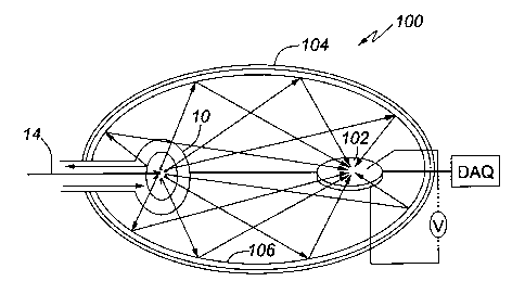
Figure 3 illustrates the fluid contamination analyzer 100 of
the embodiment of the present invention. The fluid
contamination analyzer 100 comprises the sample cell 10
described above, a detector 102 for detecting the secondary
radiation; and a reflective shell 104 in the form of an
ellipsoid, at least partly encompassing the sample cell 10 and
the detector 102, the reflective shell 104 defining two focal
points so that radiation generated at one of the focal points
is reflected by the reflective shell to the other focal point,
wherein the sample cell 10 is disposed at or in close
proximity to one of the focal points, and the detector 102 is
disposed at or in close proximity to the other focal point to
receive the secondary fluorescent radiation generated by the
entrapped contaminant. The reflective shell 104 is preferably
made of aluminum metal and has an internal reflective surface
106 made of gold or another suitable reflective material.
19
CA 02547489 2006-05-18
CTD-004-CA
The shape of the reflective shell can be different, for
example, non-continuous in the form, e.g. of a truncated
ellipsoid, or resembling a hyperboloid or paraboloid as long
as it serves the purpose of focusing the secondary fluorescent
light emitted from the sample cell 10 onto the detector 102,
which is spaced from the sample cell 10.
One form of the detector 102 is illustrated in Figure 4. It
comprises two back-to-back silicon photo avalanche diodes 110,
with their detecting windows 112 facing in opposite directions
and capable of collecting light from substantially
complementary 2~t steradian angles 61 and 02 as illustrated in
Figure 4. Accordingly this form of the detector 102 will be
referred to as a "2~ detector".
Another form of the detector 102 includes an optimized solid
state device as its detecting component, which has a spherical
shape and is preferably made of silicon. It is operated via
radio frequency or hard wired to the detector. Currently, a
prototype for such solid state device is available from Ball
Semiconductor Inc. of Allen, Texas, USA, which manufacturers
small spherical chips of about 1mm in size.
Preferred Requirements for the fluid contamination analyzer
100:
(1) The dimensions for the reflective shell are preferably of
the order of 1 foot to 2 foot length by one foot in diameter
(or smaller). These dimensions will be optimized for light
capturing efficiency depending on the dimensions of the sample
cell 10 and the detector 102, however they illustrate that the
fluid contamination analyzer is intended to be portable;
CA 02547489 2006-05-18
CTD-004-CA
(2) As mentioned previously, the turbidity of the fluid in the
sample cell 10 should be kept low enough to prevent fouling of
the flow apparatus. This will be characterized by the size of
the porous surfaces and flow rate;
(3) There is also a possible requirement of in an inert
atmosphere, e.g. nitrogen as certain flat chip detectors may
have to be operated in such an environment due to
manufacturer's specifications. This may also help hinder the
growth of oxides on the coatings found on the optics or
internal reflective surface 106. If this is necessary, then
vacuum seals will be employed anywhere there are joints to the
outside;
(4) As discussed above, the trapping medium 12 is to be
disposable and hence removable so as to make the sample cell
10 or 50 reusable; and
(5) The detector, 102, itself may require an anti-reflection
(AR) coating to limit the reflection of grazing angle
radiation.
As already mentioned, the contaminants are usually present in
water, or any other aqueous fluid, in trace amounts.
Therefore, in order to accumulate the amount of the
contaminant in the trapping medium 12, which would be
sufficient to provide a reliable reading at the detector 102,
it is understood that the volume of water may have to be
passed through the cell for a required period of time.
The fluid contamination analyzer 10 can function in two modes
of operation.
21
CA 02547489 2006-05-18
CTD-004-CA
In a sequential mode of operation, the fluid to be analyzed is
passed through the trapping medium for a predetermined period
of time to allow the accumulation of the sufficient amount of
the contaminant, and after that the secondary light
fluorescent analysis of the entrapped contaminant is performed
in the manner described above.
In a parallel mode of operation, the light source 13 is
illuminating the trapping medium, and the detector 102 is
detecting the secondary fluorescent light at the same time as
the fluid is flowing through the trapping medium 12. This
allows monitoring of the dynamics of accumulation of the
contaminant in the trapping medium and, in certain occasions,
to reduce the time required for the contamination analysis,
e.g. when only the presence of the contaminant has to be
detected.
It is understood that, in the fluid contamination analyzer
100, the sample cell 50 described above can be also used
instead of the sample cell 10. Other designs of sample cells
are also possible as long as they provide a dual function of
entrapping contaminants present in the fluid to be analyzed,
and analyzing the presence and concentration thereof by using
optical methods.
Although a fluorescence optical analysis of contaminants has
been used in the preferred embodiment of the invention, it is
contemplated that other spectroscopic techniques, which
generate the measurable presence or absence of the secondary
light indicative of the identity and concentration of
contaminants, can be also employed within the spirit of the
present invention.
22
CA 02547489 2006-05-18
CTD-004-CA
The fluid contamination analyzer 100 of the embodiment of the
present invention has the following advantages.
The fluid contamination analyzer 100 would not only provide a
device that is small, rugged, field-portable and in-situ tool,
but will also provide an improved detection sensitivity by a
minimum of two orders of magnitude compared to currently
commercial available analyzers. It is suitable for
environmental analysis of micro-organisms, organic and
inorganic substances found in various fluids, e.g. freshwater
sources.
Furthermore, the fluid contamination analyzer 100 allows a
simplified installation and removal of the trapping medium 12,
which enables users to conduct sampling more frequently. This
will empower plant managers with the ability to manage water
in a real time, i.e. in a prevention mode as opposed to the
historical monitoring and remediation mode used currently.
Thus, although particular embodiments of the invention have
been described in detail, it can be appreciated that
alternatives, such as those mentioned above and numerous other
changes, variations, and adaptations may be made without
departing from the scope of the invention as defined in the
following claims.
23