Note: Descriptions are shown in the official language in which they were submitted.
CA 02548661 2013-04-09
COMPRESSED HIGH DENSITY FIBROUS POLYMERS SUITABLE FOR IMPLANT
SPECIFICATION
TECHNICAL FIELD
[0001] The invention generally relates to medical devices and procedures. The
invention
more particularly concerns a mixture of fibers and suspension fluid that has
undergone
compression, and facilitated by lubrication, causing the fibers to migrate and
align, thereby
creating a strong fibrous implantable material.
BACKGROUND ART
[0002] Despite the growing sophistication of medical technology, repairing and
replacing
damaged tissues remains a costly, and serious problem in health care.
Currently,
implantable prostheses for repairing tissues are made from a wide number of
synthetic
and natural materials. Ideally, these prosthetic materials should be
chemically inert,
biocompatible, noncarcinogenic, capable of being secured at the desired site,
suitably
strong to resist mechanical stress, capable of being fabricated in large
quantities in the
form required, sterilizable, and free of viruses or other contaminating
agents. Examples of
tissue that can be treated with implantable prostheses include dura mater,
tendon (e.g.,
rotator cuff, anterior cruciate, etc.) and rectic abdominus muscle due to
hemiation.
[0003] A wide variety of prosthetic materials have been used, including
tantalum, stainless
steel, Dacron, nylon, polypropylene (e.g., MarlexTm), microporous expanded-
polytetrafluoroethylene (e.g., Gore-Tex'), dacron reinforced silicone rubber
(e.g.,
SilasticTm), polyglactin 910 (e.g., VicrylTm), polyester (e.g., MersileneTm),
polyglycohc add
(e.g., DexonTm), and cross-linked bovine pericardium (e.g., Peri-Guard Tm). To
date, no
single prosthetic material has gained universal acceptance.
[0004] Metallic meshes, for example, are generally inert and resistant to
infection, but
they are permanent, do not generally adapt in shape as a skeletal structure
grows, and
they shield the healing tissues from the stresses that may be necessary to
generate fully
CA 02548661 2013-04-09
functioning tissue. Non-resorbable synthetic meshes have the advantage of
being easily
molded and, except for nylon, retain their tensile strength in the body. Their
major
disadvantages are their lack of inertness to infection, the occasional
interference with
wound healing, and that they are often long-term implants. Absorbable meshes
have the
advantage of facilitating tissue in-growth and remodeling at the site of
implantation, but
often do not have the short-term or long-term mechanical strength necessary
for the
application.
[0005] Both U.S. Patent No. 4,948,540, granted to Nigam and U.S. Patent No.
5,206,028
granted to Li, disclose a collagen membrane suitable for medical uses. In the
case of Li,
the membrane is constructed in a fashion to make it easier for implantation,
by ensuring
the membrane is not transparent, and not slippery. Both patents begin by
providing a
solution of collagen, which is freeze-dried, cross-linked, and then
compressed. Li then
utilizes a second cross-linking, freeze-drying and compression step. The
initial cross-
linking step locks the fibers into a specific orientation. The compression
step merely
reduces the porosity within the sheet without inducing fiber migration that
would
substantially improve the strength of the composition. A second cross-linking
step is
necessary to hold the sheet in its compressed conformation. What is needed is
a sheet
with improved strength, capable of maintaining its structural competence
without the need
of multiple freeze-drying and cross-linking steps.
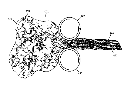
[0006] In U.S. Patent No. 6,599,524 granted to Li, there is disclosed a
membrane sheet
having oriented biopolymeric fibers. The membrane is manufactured with
oriented parallel '
fibers formed around a rotating mandrel. The rotations of the mandrel as the
fibers are
added results in the orientation of the fibers. The membrane is then
compressed to drive
out excess liquid, and cross-linked, resulting in a membrane with
directionally oriented
fibers. This material is only aligned in a single direction and must be
laminated with
binding agents in order to create a functional device. Additionally, such a
device does not
provide gradients such as those seen in natural tissues. What is needed is a
method that
allows for layering that occurs at the microscopic as well as the macroscopic
level as part
of a one step process and more closely represents the layered structure of
natural
connective tissues.
2
CA 02548661 2013-04-09
[0007] Prosthetic devices are used in the repair, augmentation, or replacement
of
articulating organs. For example, the rotator cuff (i.e., shoulder joint) is
made up by a
combination of the distal tendinous portion of four muscles: the
supraspinatus,
subspinatus, subscapularis and the teres minor. Proper functioning of this
tendonous cuff,
depends on the fundamental centering and stabilizing role of the humeral head
with
respect to sliding action during lifting and rotation movements of the arm. A
tear in the
rotator cuff tendons is a common injury that can be caused by constant
friction from
repetitive overhead motion, trauma, or age-related degeneration that can
narrow the
space between the clavicle and the top of the scapula.
[0008] To repair large tears of the rotator cuff, it is desirable to use a
scaffold or graft
material to help support the damaged tissue and guide its repair. Several
types of
materials have been used for such procedures. Wright Medical (Memphis, TN)
markets a
product known as GraftJacketTm, which is manufactured by Lifecell Corporation
(Branchburg, NJ) from human cadaver skin. Human cadaverous tissue products can
be
difficult to obtain and have the potential for disease transmission. Tissue
Sciences
(Covington, GA) markets a product known as Permacolni, which is comprised of
cross-
linked porcine dermis. DePuy (Warsaw, LN) markets the Restore Patch, which is
fabricated from porcine small intestine submucosa. Biomet (Warsaw, IN) markets
a
product known as CuffPatchTm another porcine small intestine product. The
CuffPatchTm
and the Restore Patch TM products provide biocompatible scaffolds for wound
repair but
they are complicated to manufacture, as they require the lamination of
multiple layers of
submucosal tissues to gain the strength needed for these applications.
Fabrication of such
patches from porcine small intestine submucosa are described in U.S. Patent
No.'s
4,902,508 Badylak et al. and 5,573,784 Badylak et at.
[0009] Additional applications for prosthetic devices exist in the form of
membrane
patches. The spinal cord and brain are covered with a protective membrane that
is known
as the dura mater. The integrity of the dura mater is critical to the normal
operation of the
central nervous system. When this integrity is intentionally or accidentally
compromised
(e.g., ruptured, severed, damaged, etc.), serious consequences may ensue,
unless the
membrane can be repaired. Typically, dura tissue is slow to heal. To enhance
the healing
3
CA 02548661 2013-04-09
process, graft materials can be utilized to guide the regeneration of the
tissue. Repairing
damaged membranes has largely focused on implantable materials known as dural
substitutes, which are grafted over the damaged dura mater and are designed to
replace
and/or regenerate the damaged tissue.
[0010] Thus, there is a need for an effective dura substitute that would be
biocompatible,
sufficiently noninfectious (e.g., purified, etc.) to prevent the transmission
of disease,
conformable, available in a variety of sizes, high in tensile strength, inert,
suturable, and
optionally capable of forming a water-tight seal.
[0011] Researchers have experimented with a wide variety of substances to act
as dura
substitutes. Autologous grafts of tissue, such as pericardium, can be
effective as a dura
substitutes; however, autologous tissue is not always available and it posses
additional
costs and risks for the patient. Cadaverous dura mater has also been used but
like
autologous tissues, cadaverous tissues can be difficult to obtain. Tutogen
Medical Inc.
(West Paterson, NJ) markets a product known as Tutoplast dura mater, which is
obtained
from human cadavers. Processed human cadaveric dura mater has been implicated
in the
transmission of cases of the fatal Creutzfeldt- Jakob disease. Other products
overcome
this shortcoming by using alternate materials. The Preclude Dura TM
substitute,
manufactured by W. L. Gore (Newark, DE), is an inert elastomeric fluoropolymer
material.
The material is biocompatible but is a permanent implant and does not resorb
over time.
DuralTm substitutes comprising collagen have been also been explored as
described in US
Patent No. 5,997,895 (Narotam et al.). Integra Lifesciences Corporations
(Plainsboro, NJ)
distributes a product known as DuraGenTM. The product is manufactured from
bovine
achilles tendon and is a pliable porous sheet. Although the material is
resorbable and
biocompatible, the integrity of the material is not sufficient enough to
withstand suturing to
the wound site.
[0012] The present invention overcomes these suturing and other difficulties
of the
materials currently available and provides a structure capable of being
adapted to a wide
variety of surgical applications.
4
CA 02548661 2013-04-09
[0013] Other applications for the implantable prosthesis of this invention, in
the form of a
surgical mesh, include pelvic floor disorders such uterine and vaginal vault
prolapse.
These disorders typically result from weakness or damage to normal pelvic
support
systems. The most common etiologies include childbearing, removal of the
uterus,
connective tissue defects, prolonged heavy physical labor and postmenopausal
atrophy.
Many patients suffering from vaginal vault prolapse also require a surgical
procedure to
correct stress urinary incontinence that is either symptomatic or latent.
[0014] Another embodiment of the present invention is directed to devices
useful as
prosthetic menisci, and in vivo or ex vivo scaffolds for regeneration of
meniscal tissue.
[0015] The medial and lateral menisci are a pair of cartilaginous structures
in the knee
joint which together act as a stabilizer, a force distributor, and a lubricant
in the area of
contact between the tibia and femur. Damaged or degraded menisci can cause
stress
concentrations in the knee thereby creating abnormal joint mechanics and
leading to
premature development of arthritic changes.
[0016] In the prior art, treatment of injured or diseased menisci has
generally been both by
surgical repair and by tissue removal (i.e., excision). With excision,
regeneration of
meniscal tissue may not always occur. Allografting or meniscal transplantation
is another
method of replacement, which has been previously tried.
[0017] This approach has been only partially successful over the long term due
to the
host's immunologic response to the graft and to failures in cryopreservation
and other
processes. Alternately, menisci have been replaced with permanent artificial
prostheses
such as Teflon TM and polyurethane. Such prostheses have been selected to be
inert,
biocompatible, and structurally sound to withstand the high loads which are
encountered
in the knee joint. Typically, these permanent implants do little to encourage
the
regeneration of the damaged host tissue. Therefore, what is needed is an
improved
prosthetic meniscus composed of biocompatible materials, which are
biocompatible,
compliant, durable, and suitable to acts as a temporary scaffold for meniscal
fibrocartilage
infiltration and regeneration of the host tissue.
CA 02548661 2013-04-09
[0018] In U.S. Patent 5,184,574 granted to Stone and U.S. Patent 6,042,610
granted to Li,
there is disclosed a meniscus replacement material, manufactured by shape
molding
collagen fibers within a mold via application of low pressure by a piston
prior to or after
drying. Stone requires the step of applying freezing cycles to the material.
The fibrous
materials achieve densities of 0.07 - 0.5 9/cc. Hydrated fibers at these
density range from
a free flowing liquid slurry to a loose dough-like material unable to maintain
a shape.
Freezing and possibly lyophilizing of the material is necessary to remove it
from the mold
and cross-linking solutions are applied to it while still in the frozen or
lyophilized state so
that it does not warp. Fiber orientation may be obtained by applying a
rotating force to the
piston in order to form a circumferential orientation. However, this
orientation occurs only
in areas directly in contact with the rotating piston. What is necessary is a
fibrous
construct with sufficient integrity to be handled without the necessity of
freezing and/or
lyophilizing and that can be implanted without the requirement of cross-
linking, if desired.
Additionally, this construct lacks any consistency throughout the thickness of
its structure,
being able to create oriented fibers only at the periphery.
[0019] Another embodiment of the present invention is directed to devices
useful as
prosthetic ligament, and in vivo or ex vivo scaffold for regeneration of
ligament tissue and
to methods for their fabrication.
[0020] The anterior cruciate ligament (ACL) of the knee functions to resist
anterior
displacement of the tibia from the femur during flexure. The ACL also resists
hyperextension and serves to stabilize the fully extended knee during internal
and external
tibial rotation. Partial or complete tears of the ACL are common. The
preferred treatment
of the torn ACL is ligament reconstruction, using a bone-ligament-bone
autograft (e.g.,
from the patient's patellar tendon or hamstring tendon). Cruciate ligament
reconstruction
generally provides immediate stability and a potential for immediate vigorous
rehabilitation. However, ACL reconstruction is not ideal; the placement of
intraarticular
hardware is required for ligament fixation; anterior knee pain frequently
occurs, and there
is an increased risk of degenerative arthritis with intraarticular ACL
reconstruction.
Another method of treating ACL injuries involves suturing the torn structure
back into
place. This repair method has the potential advantages of a limited
arthroscopic approach
6
CA 02548661 2013-04-09
and minimal disruption of normal anatomy. A disadvantage of this type of
repair is that
there is generally not a high success rate for regeneration of the damaged
tissues due to
the lack of a scaffold or other cellular inductive implant.
[0021] Another embodiment of the present invention relates to devices useful
as a
prosthetic intervertebral disc. The intervertebral disc plays an important
role in stabilizing
the spine and distributing the forces between the vertebral bodies. In the
case of a
damaged, degenerated, or removed disc, the intervertebral space collapses over
time and
leads to abnormal joint mechanics and premature development of arthritis.
[0022] In the prior art, discs have been replaced with prostheses composed of
artificial
materials. The use of purely artificial materials in the spine minimizes the
possibility of an
immunological response. Such materials must withstand high and repeated loads
seen by
the spinal vertebral joints, early attempts focused upon metallic disc
implants. These
efforts met with failure due to continued collapse of the disc space and or
erosion of the
metal prosthesis into the adjacent bone.
DISCLOSURE OF THE INVENTION
[0023] The current invention is directed to a general prosthesis, which, when
implanted
into a mammalian host, undergoes controlled biodegradation accompanied by
adequate
living cell replacement, such that the original implanted prosthesis is
remodeled by the
host's cells before it is degraded by the host's enzymes and/or by hydrolysis.
The device
of the subject invention is structurally stable, pliable, semi-permeable, and
suturable.
[0024] Embodiments of this invention can be utilized to repair, augment, or
replace
diseased or damaged organs, such as rotator cuff injuries, dura defects,
abdominal wall
defects, pericardium, hernias, and various other organs and structures
including, but not
limited to, bone, periosteum, perichondrium, intervertebral disc, articular
cartilage, dermis,
epidermis, bowel, ligaments, tendon, vascular or intra-cardiac patch, or as a
replacement
heart valve.
7
CA 02548661 2013-04-09
[0025] The device if this invention could be used for sling procedures (e.g.,
surgical
methods that place a sling to stabilize or support the bladder neck or
urethra). Slings are
typically used to treat incontinence. Additionally, in the form of a surgical
mesh, the device
can be used for such applications as hernia and dura repair.
[0026] In another embodiment, this invention provides a ligament repair or
replacement
prosthesis that is biocompatible, is able to withstand ACL forces, and
promotes healing of
the injured tissues by acting as a scaffold for cellular infiltration. Another
embodiment of
this invention is to provide an improved disc replacement or prosthesis that
is
biocompatible, does not interfere with normal vertebral segment motion, is
able to
withstand normal spinal column forces, does not wear into the surrounding
bone,
promotes regrowth of intervertebral disc material and acts as a scaffold for
fibrocartilage
infiltration.
[0027] The tissue repair implant of this invention, functioning as a
substitute body part,
may be flat, tubular, hollow, solid, or of complex geometry depending upon the
intended
use. Thus, when forming the structure of the prosthesis of this invention, a
mold or plate
can be fashioned to accommodate the desired shape.
[0028] Flat sheets may be used, for example, to support prolapsed or
hypermobile organs
by using the sheet as a sling for those organs or tissues (e.g., bladder or
uterus). Tubular
grafts may be used, for example, to replace cross sections of tubular organs
such as
esophagus, trachea, intestine, and fallopian tubes. These organs have a basic
tubular
shape with an outer surface and a luminal surface. In addition, flat sheets
and tubular
structures can be formed together to form a complex structure to replace or
augment
cardiac or venous valves and other biological tissue structures.
[0029] The tissue repair implant of the present invention may be rendered
porous to
permit the in-growth of host cells for remodeling or for deposition of the
collagenous layer.
The device can be rendered "non-porous" to prevent the passage of fluids if
necessary or
the porosity can be adjusted to create a membrane capable of selective
permeability. The
degree of porosity will affect mechanical properties of the implant, and these
properties
are also affected by processing (as will be discussed).
8
CA 02548661 2013-04-09
[0030] The mechanical properties include mechanical integrity such that the
tissue repair
implant resists creep for the necessary period of time, and additionally is
pliable (e.g., has
good handling properties) and suturable. The term "suturable" means that the
mechanical
properties of the layer include suture retention, which permits needles and
suture
materials to pass through the prosthesis material at the time of suturing of
the prosthesis
to sections of native tissue. During suturing, such prostheses must not tear
as a result of
the tensile forces applied to them by the suture, nor should they tear when
the suture is
knotted. Suturability of tissue repair implant, i.e., the ability of
prostheses to resist tearing
while being sutured, is related to the intrinsic mechanical strength of the
prosthesis
material, the thickness of the prosthesis, and the tension applied to the
suture. The
mechanical integrity of the prosthesis of this invention is also in its
ability to be draped or
folded, as well as the ability to cut or trim or otherwise shape the
prosthesis.
[0031] hi another embodiment of the invention, reinforcing elements (e.g.,
threads, fibers,
whiskers, textiles, etc.) are incorporated into the tissue repair implant for
reinforcement or
for different rates of remodeling. Thus, the properties of the tissue repair
device can be
varied by the geometry of the thread used for the reinforcement. Additionally,
thread
constructs such as a felt, a flat knitted or woven fabric, or a three-
dimensional knitted,
woven or braided fabric may be incorporated between layers or on the surface
of the
construct. Porous, non-fibrous sheets of polymer foam may also be incorporated
between
layers or on the surface of the construct. Such polymer foams can be made by
methods
known in the art such as particulate leaching or solvent freeze-drying
methods.
[0032] An embodiment of the present invention may be made by the following
steps:
providing a mixture comprising a plurality of fibers, a lubricant, and a
suspension fluid,
with the suspension fluid filling a void space between the fibers and
subjecting the mixture
to at least one compressive force. The compressive force causes the migration
and
alignment of the fibers; and may remove substantially all of the suspension
fluid from the
mixture. The mixture may further comprise a biologically active agent, or a
reinforcing
agent.
9
CA 02548661 2013-04-09
[0033] Additionally, the compressive forces may reduce the void space between
the
fibers, and the lubricant may assist fiber movement during compression, and be
in the
form of a liquid or a solid, and may be provided in a carrier fluid. The
suspension fluid flow
may also cause the alignment of the fibers to take the form of plates (e.g.,
plates of
oriented fibers).
[0034] The compressive force may be applied by a molding surface, thereby
creating a
shaped fibrous member in the mold. In a preferred embodiment, the molding
surfaces
may be such as to provide one or more penetrating holes in the shaped fibrous
member,
specifically by pushing aside fibers such that they tend to align themselves
circumferentially around each hole. Additionally, or alternatively, the
material may be
machined, allowing the fabrication of complicated shapes.
[0035] In a preferred embodiment, at least a portion of the compressed mixture
may be
cross-linked by exposure to a cross-linking agent. This process will affect
the strength and
resorption rate of the implant. Additionally, the strength may be tailored by
a reinforcing
element, such as particulates, threads, fibers, whiskers, textiles, rods,
meshes, or
combinations thereof. The function or properties of the implant may also be
affected by
additives, such as ceramics, polymers, cells, biologically active agents,
liquids,
surfactants, plasticizers, and combinations thereof.
BRIEF DESCRIPTION OF THE DRAWINGS
[0036] FIGS. 1 A and IB depict fibrous dough prior to (A) and after (B)
compression.
[0037] FIGS. 2A-2E depict a change in fiber orientation and inter-fiber void
space as the
fibrous dough is compressed.
[0038] FIGS. 3A and 3B depict fibrous dough prior to and after compression.
[0039] FIG. 4 depicts compression of fibrous dough as it passes through
rollers.
[0040] FIGS. 5 A and 5B depicts three-dimensional compression of fibrous
dough.
'
CA 02548661 2013-04-09
[0041] FIGS. 6 A and 6B depict compression of a cylindrical mass of fibrous
dough.
[0042] FIGS. 7 A and 7B depict incorporation if reinforcing materials within
compressed
fibers.
[0043] FIG. 8 depicts incorporation of particulates, biologies within the
compressed fibrous
matrix.
[0044] FIGS. 9A and 9B depict incorporation of microstructures within the
compressed
fibrous matrix.
[0045] FIG. 10 depicts a hemostatic tract plug of compressed fibrous matrix.
[0046] FIGS. 11A and 1 IB depict hemispherical cups of compressed fibrous
matrix.
[0047] FIGS. 12A and 12B depict a selectively compressed ring of fibrous
matrix
surrounding a non-compressed fibrous matrix.
[0048] FIG. 13 depicts selective compression of a fibrous matrix.
[0049] FIGS. 14 A and 14B depict compressed fibrous constructs useful for
surgical
applications.
[0050] FIG. 15 depicts the surgical application of a compressed fibrous
construct.
[0051] FIGS. 16A-16C depict compression of fibrous dough utilizing a
compression plate
having protrusions.
MODES FOR CARRYING OUT THE INVENTION
[0052] Embodiments of this invention include compressed, biodegradable,
fibrous
compositions for application to a tissue site in order to support, promote or
facilitate new
tissue growth. One aspect of this invention is a fibrous component (e.g.,
collagen, elastin,
chitosan, alginate, hyaluronic acid, polyglycohc acid, polyurethane, silk,
etc.; see table 1)
that provides unique mechanical and physical properties, as will be discussed.
Such
11
CA 02548661 2013-04-09
fibrous components in slurry form may be pre-processed into a fibrous dough or
paste by
removal of a portion of suspension fluid, as known in the art, prior to
formation into a
compressed conformation, as will be disclosed. An example is in the form of an
interlaced
matrix described in U.S. Patent No. 6,974,862. The material is a natively
cross-linked
collagen such as Semed Frg produced by Kensey Nash Corporation (Exton, PA).
[0053] The fibrous dough embodiment is dehydrated/desolvated by applying a
compressive force in such a manner as to reduce the inter fiber space by
removing at
least a portion of the suspension fluid. In a preferred embodiment
substantially all of the
suspension fluid is removed. Unlike unaltered or natural matrices (e.g.,
dermis, small
intestine submucosa, etc.), the thickness, porosity, fiber-density, fiber-
orientation, fiber-
length, fiber composition and component-ratio (e.g., Collagen TM to Elastin TM
ratio), as a
non-limiting example, can be controlled with the current invention.
[0054] To improve the migration of fibers and prevent clumping during the
compressive
process it is preferred to incorporate a percentage (e.g., 0%-50% by mass of
fibers) of
one or more lubricants (e.g., biocompatible oils, hydrogels, liquid polymers,
low-molecular
weight polymers, glycosaminoglycans, surfactants, waxes, fatty acids, fatty
acid amines
and metallic stearates such as zinc, calcium, magnesium, lead and lithium
stearate, etc.)
into the fibrous dough suspension. A lubricant is defined as a substance,
which is capable
of making surfaces smooth or slippery. These characteristics are due to a
reduction in
friction between the polymers to improve flow characteristics and enhance the
knitting and
wetting properties of compounds. The lubricant may be liquid or solid and may
be
suspended or dissolved in a carrier solvent (e.g., water, alcohol, acetone,
etc.).
Additionally the lubricant may only become lubricious under compressive force
or change
in temperature. The lubricant may remain in its entirety in the final product
of the
invention; may be partially removed in the dehydration/desolvation process;
or, may be
washed out or removed by methods known in the art during further processing.
Lubricants
that remain in the final product may be biologically active agents or may form
microstructures. Preferred lubricants include Tween-80, hyaluronic acid,
alginate,
glycerin or soluble collagen with the most preferred being acid soluble
collagen such as
Semed S TM produced by Kensey Nash Corporation (Exton, PA).
12
CA 02548661 2013-04-09
[0055] Additional ways in which to add lubricity to various embodiments of
this disclosure,
include physically or chemically altering the surface of the fibers making up
the
composition. Such alterations can be achieved through chemical or physical
attachment of
a lubricious substance to the fibers, temperature induced phase changes to the
surfaces
of the fibers or partial solubilization of the fibers through alteration of
the pH and/or
conductivity of the free fluid or use of a percentage of solvent for the
fibers within the free
fluid. Other methods of creating lubricity are known to those skilled in the
art, and are
embraced by this disclosure.
[0056] During the compression step of a preferred fibrous dough embodiment,
the fibers
align themselves into layered or plate-like structures. As the inter fiber
void space is
collapsed, the displaced fluid is forced outward and begins to flow out of the
device. The
flow may play a role in aligning the fibers in that direction. The rate of
flow is directly
affected by rate and duration at which compression occurs. This phenomenon
occurs
throughout the structure and results in aligned fibrous layers or plates
separated by fluid
planes. These planes facilitate migration within the structure, allowing the
fibers within a
single layer to move without interference from fibers in a different layer.
[0057] The compression induced fluid migration may occur three-dimensionally,
thereby
dissecting planes in the structure as it runs into resistance. Additionally
the fluid may be
forced through narrow passageways in the fibrous mats and begin creation of a
new plane
at a different level within the construct. Thus it is possible to create a
structure wherein the
planes do not traverse the entire length of the device, but instead exist as
multiple fissures
located randomly within the construct and each fissure can be defined by
fibrous plates
having an aligned fiber orientation unique from that of neighboring fissures.
The plates
themselves may be organized in a random, oriented, or aligned fashion. As
compression
continues, the lubricant reduces the friction, allowing the aligned fibers
within the plates or
planes to slide across each other and nest in the most compact orientation.
Additional
compression brings the plates of fibers in closer contact, allowing them to
become locked
into a compact anisotropic structure, although the material may be isotropic
in two
dimensions.
13
CA 02548661 2013-04-09
[0058] Unlike existing state of the art sheets, this layering occurs at the
microscopic as
well as the macroscopic level as part of a one step process and more closely
represents
the layered structure of natural connective tissues. Additionally, the amount
of fiber
compaction within a plate or layer and the spacing between the plates or
layers can be
controlled by the force applied and the amount of time allowed for
equilibration at a
specific force. The preferred force applied is from 0.01 tons/square inch
(0.14 MPa) to 100
tons/square inch (1400 MPa) with the most preferred force being in the range
of 0.2
tons/square inch (2.8 MPa) to 2.0 tons/square inch (28 MPa). This amount of
force is in
excess of state of the art methods used merely to extract fluid and
concentrate the fibers
into workable dough-like material. Existing methods do not induce fiber
migration or layer
formation. Devices created under such conditions as described above do not
require
additional steps such as freezing and/or cross-linking within molds to be
handled. The
preferred amount of equilibration time is in the range of about less than one
minute to
more than 500 minutes with a more preferred range of about 1 minute to 60
minutes.
[0059] The use of wicking materials such as paper towels/sponges or fluid
removal
systems such as screens or vacuum systems prevent excessive pooling of fluid
in any
single area of the structure during compression. If fluid is allowed to
accumulate, it can
create craters or voids within the structure. If the fibers surround these
pools of fluid
succumb to the compressive forces, a rip or discontinuity in the structure
will form as the
fluid is forcibly expelled. Strategic location of fluid exit pores within a
mold can be used to
create unique directional flows that in turn align the fibers within a layer
or plate. In this
way the fibers forming plates at each level can be oriented in the same
direction or turned
at any conceivable angle to each other. Although orientation of fibers from
plate to plate
may be organized or random, fiber orientation within a plate is organized with
the fibers
running predominantly parallel to each other. Molds with fluid vacuum assist
further
improve control of fiber orientation. Additionally, materials such as threads
and screens
provide avenues for fluid escape. As the fluid flows along the length of the
threads and
screens, the fibers adjacent to them are aligned parallel to them. Use of
porous rods or
porous hollow tubes that can be extracted or left in place as reinforcement
can also be
used to facilitate uniform fluid removal. If the fluid extraction tubes are
removed, long
channels will be left that can be utilized for purposes such as suture line
conduits.
14
CA 02548661 2013-04-09
=
[0060] As the inter fiber space is reduced and the free fluid within the dough
is expelled,
the overall porosity of the compressed composition is reduced towards a
theoretical zero
point. The amount of porosity as well as the size of the pores dictates
whether the device
functions as a tissue matrix or barrier. Additionally, the physical/mechanical
properties are
highly influenced by the amount of inter fiber space. Another factor affecting
the
mechanical and physical properties of the composition is the use of additives
(e.g.,
surfactants, plasticizers, particulates, porosifiers, meshes, etc.).
[0061] In a preferred embodiment, the method of preparing the high-density
fibrous matrix
involves: providing a fibrous material; contacting the fibrous material with a
suspension
fluid and a lubricant; applying a compressive force within one or more
dimensions that
partially dehydrates/desolvates the fibrous material. Subsequently, the
fibrous material
may be cross-linked. It may be further desirable to provide a directed means
of egress for
the suspension fluid during compression, as previously discussed. Additionally
use of a
fibrous suspension having interlaced, interlocked fibers may be desirable.
[0062] In another embodiment, the partially dehydrated fibrous matrix is fully
dried (e.g.
vacuum dried, freeze-dried, air-dried.) after which it may be cross-linked. It
may be further
desirable to rehydrate/resolvate the fibrous matrix to facilitate
incorporation of cross-
linking agents, plasticizers, surfactants, biologically active agents,
microstructures, cells or
other materials. If desired the sheet may again be dried.
[0063] Any method of compression known by those skilled in the art is
conceivable for this
invention, including, but not limited to, using hydraulically or pneumatically
powered
platens or pistons to compress the fibrous matrix material. Other methods
include but are
not limited to using a screw or an arbor press to compress the material, using
centrifugation to extract fluid and compress the fibers, or forcing the
material between
rollers.
[0064] The structure of the fibrous matrix material is also influenced by the
amount of
compressive force applied to the material. The amount of compression may
change the
porosity of the fibrous matrix material. The pore size distribution will also
be affected by,
the amount of compression as the fibrous matrix material may be compressed so
that only
CA 02548661 2013-04-09
certain areas have collapsed, or so that all areas collapse. The direction of
compression in
relationship to the original structure of the fibrous matrix material will
also affect the
structure of the compressed fibrous matrix material. For example, if the
initial fibrous
matrix material has long parallel fibers, a force applied could be used to
force the fibers
together in a parallel fashion or bunch up the fibers as the force attempts to
shorten the
length of the fibrous composition.
[0065] Compression of the fibrous matrix material can be controlled to create
various
structural patterns within the material; likewise, the mechanical properties
of the material
may be altered to meet specific requirements. The amount of compression is
directly
related to the tear strength of the material. If a medical device fabricated
from the
compressed material is not in the form of a sheet, the compressed material can
be
compressed three-dimensionally to form the desired shape. If the medical
device is axially
loaded, the compressed material may be compressed in one direction to optimize
the
mechanical properties of the material in that direction.
[00661 If not compressed initially into the final shape, after being
compressed and
removed from the compression device, the fibrous matrix material may be
machined into a
new shape or design with various features. Machining processes are well known
to those
skilled in the art. (e.g., punching, coring, milling, sawing, lathing, etc.)
Additionally, the
compressed fibrous matrix may function as a component of a larger device and
if not
attached during the compression step, may be attached to components by methods
known to those in the art (e.g., gluing, stapling, sewing, etc.).
[0067] The inventors have discovered that after the compressive
dehydration/desolvation
process the resultant material has mechanical properties, including tear
strength, superior
to those of non-compressed materials that have been cross-linked. Not being
confined to
a single theory, it is believed that the high compressive forces will create
weak chemical
linkages aside from the physical interaction of the fibers. This permits the
current invention
to be utilized in applications that initially require specific tear strength
but where it is
desirable for the device to be quickly degraded away after fulfilling its
initial function such
as dura repair. The current invention can be cross-linked, either chemically
(e.g., EDC) or
16
CA 02548661 2013-04-09
by non-chemical methods (e.g., dehydrothermal (DHT)) know to those skilled in
the art, for
applications requiring strength for an extended period of time, such as hernia
repair.
[0068] The inventors have further discovered that a non-compressed or mildly
compressed sheet can be cross-linked, completely or only at the surface, by a
first
method after which it is fully compressed and cross-linked by a second method.
The first
cross-linking restricts motion of the fibers during the compression step,
retarding an
increase in the footprint of the sheet. Even though the sheet is cross-linked
in the non-
compressed state, the addition of a lubricant facilitates migration and
shifting of the
partitions making up the sheet. This allows thick sheets to achieve the same
fiber density
per unit volume as thin sheets.
[0069] Highly compressed sheet embodiments consisting of collagen fibers
placed into
cross-linking solutions have formed a tough cross-linked skin around a
minimally cross-
linked to non-cross-linked center. The center of such sheets are easily
separated forming
a shell, pocket or bladder. The permeability of the bladders varied depending
upon the
initial compression. Low compression produced bladders that slowly allowed
dyed fluid to
exude. Moderate compression allowed water to pass through but filtered out the
larger
dye molecules. High compression created a barrier to fluid water but slowly
allowed the
escape of water vapor. Such a phenomenon was not evident in DHT cross-linked
sheets.
The lack of bladder formation in DHT cross-linked sheets is believed to be the
result of
uniform cross-linking throughout the thickness of the sheet.
[0070] Such devices may be useful for tissue engineering applications
associated with
bladder, intestine, tendons, ligaments and vessels, as well as the creation of
rotator cuff
patches, hernia repair sheets, orbital implant coverings, graft wraps and the
formation of
anti-adhesion devices. The shell of material may be filled with ceramics or
polymers useful
in bone repair or used as containment devices for injection of settable
polymers or
ceramics. Additionally, the center may be filled with fluids prior to or after
implantation for
applications such as controlled drug delivery or the creation of shock
absorbing vessels
useful for breast implants, fat pad replacement or meniscus and disc repair or
replacement.
17
CA 02548661 2013-04-09
[0071] Restricted contact of cross-linking solutions with the surfaces of
collagen devices
control the degree of cross-linking in fibrous, non-fibrous, compressed and
non-
compressed materials. For example, restricted contact can be achieved by
placing
shaped, fully hydrated, collagen dough into a cross-linking solution. The
cross-linking
solution slowly displaces hydration fluid at the periphery but does not
immediately come
into contact with the hydrated material in the center. As the material
continues to sit in the
cross-linking solution a gradient begins to form with a greater amount of
cross-linking
occurring at the surface and lesser amounts of cross-linking occurring toward
the center.
[0072] Additionally, a second type of cross-linking may be introduced after
drying to create
a bi-phasic cross-linking (e.g., DHT, chemical vapors, radiation). Devices
having such
unique cross-linkings may be useful in tissue-engineering applications
involving multi-
phasic tissues such as cartilage and skin or could function as in-vivo cell
culture vessels
capable of protecting foreign cells, such as islet cells from a different
person or animal,
from attack by the recipients' immune system.
[0073] The central portion of units cut from compressed collagen sheet
embodiments
having only the surface cross-linked swell when in contact with excess aqueous
fluids. A
small amount of fluid hydrates the sheet and creates thin flexible units. Only
after being
placed in contact with excess fluid does the sheet begin to swell. The
swelling can be
delayed by minutes to hours depending upon the initial thickness, magnitude of
compression, and the amount of cross-linking at the surface. This creates a
large central
porosity suitable for cell migration and/or delayed drug or biologies
delivery, centered
between two low-porosity protective sheets. Such a device may also be suitable
in
applications requiring implantation through a small opening that will swell to
full size after
becoming fully hydrated by body fluids.
[0074] The fibrous matrix material may be compression molded into an initial
or final
design of a medical device. If the device has complicated geometry, various
features may
be machined after compression molding. The material and mechanical properties
of the
final device can be altered by the temperature of the molds, the amount of
overall
compression, the design of the mold, etc. The fibrous matrix material may be
compressed
18
CA 02548661 2013-04-09
before molding, or all the compression may occur during the molding process.
The
direction of compression before or during compression molding will also affect
the
mechanical properties of the device. For example, a cylinder of fibrous dough
material
may be three-dimensionally compressed to improve the mechanical properties and
then
compression molded into a threaded bone screw. Additionally, the cylinder of
fibrous
material may be compressed into a cone shape providing a gradient of
compression.
Such gradients may be useful for multi-phasic tissue or multi-phasic drug
delivery.
[0075] The implantable prosthesis of the various embodiments of the present
invention
may be sterilized by any method known in the art. (e.g., exposure to ethylene
oxide,
hydrogen peroxide gas plasma, e-beam irradiation, gamma irradiation, etc.) The
sterilization minimizes the opportunity of infection to occur as a result of
the implant.
[0076] hi a prefened embodiment of the invention, the fibrous prosthesis is
manufactured
from a resorbable material, although this is not meant to exclude the use of
non-
resorbable polymers, minerals and metals within the final structure.
[0077] Different polymers, molecular weights, additives, processing methods,
cross-linking
methods and sterilization methods can be used to control the resorption rates
of
resorbable polymers and is well know by those skilled in the art. For example,
reconstituted collagen fibers degrade faster than natively cross-linked
collagen fibers and
collagen that has not been cross-linked degrades faster than cross-linked
collagen.
Additives such as ceramics capable of increasing the localized pH also
increase the rate
of degradation, as do chemotactic ground substances that attract cells to the
localized
area. Resorption rates can be adjusted to be shorter for applications that
require
mechanical strength for only a short period of time or longer for applications
that require
mechanical strength to be present for a longer duration. Examples of
resorbable polymers
that can be formed into fibers and used to form the prosthesis are shown in
Table I.
These materials are only representative of the materials and combinations of
materials
that can be used as prosthetic material and this table is not meant to be
limiting in any
way.
19
CA 02548661 2013-04-09
[0078] For the purposes of promoting an understanding of the principles of
this invention,
reference will now be made to the embodiments illustrated in the drawings and
specific
language will be used to describe the embodiments and elements of the
embodiments. It
must be understood that no limitation of the scope or applications of the
invention is
thereby intended. For ease of understanding, fibers are represented in the
drawings by
simple crossed lines, by no way does this indicate that they may not be
interconnected,
interwoven, interlaced or entangled, or that the final structure is porous or
non-porous,
organized or random, and/or reticulated, except as otherwise noted. In theory,
the
compressed fibrous structure could in fact be produced through the compression
of a
single continuous fiber.
[0079] Referring now to the drawings, FIG. 1 shows the fibrous matrix material
before and
after compression. Before compression, shown in FIG 1 A, the fibrous matrix
material 100
comprises a large percentage of void space surrounding the fibers 110. The
fibers 110
form a structure composed mostly of inter fiber void space 120. After being
compressed,
shown in FIG IB the compressed porous matrix material 130 contains the same
amount of
fibrous material 140; however, the sacrificed, inter fiber void space 150 has
resulted in a
reduced porosity in the material. It should be noted that the inter fiber void
space in this
figure and all other figures may contain a lubricant as has been discussed.
[0080] In another embodiment depicted in FIG. 2A, the fibrous matrix material
200 is
placed between two compressive devices 210 (e.g., platens, pistons, etc.),
which may or
may not be heated or cooled. Heating can be used for such purposes as to
modify the
fibers (e.g.,-denature, soften, melt), increase the rate of fluid evaporation,
fuse the fibers
once compressed, or improve the activity of any lubricant. Cooling can be used
for such
purposes as protecting the fibers from excessive heat during compression or to
induce
phase change or thickening of the suspension fluid and/or lubricant. The
fibers 220 and
the inter fiber void space 230 define the structure of the fibrous matrix
material. In FIG.
2B, the top compressive device 215 is lowered to compress the fibrous matrix
material
240 while the compressive device 210 remains stationary. A gradient is formed
starting at
the top of the material where fibers 250 are forced together, reducing the
interfiber void
space 230, while the fibers 220 in the lower part of the material retain their
conformation.
CA 02548661 2013-04-09
This can be employed to create an implant for biphasic tissues such as bone or
cartilage. .
Two gradients can be formed by compressing the fibrous matrix material 260
with both
compressive devices 215 at the same time, as shown in FIG. 2C. The top and
bottom
surfaces have a majority of compressed fibers 250. The next top and bottom
layer of
fibers 280 will be mildly compressed and have a reduced inter fiber void space
270. The
middle of the material 260 will have fibers 220 that maintain their original
inter fiber void
space 230. This can be employed to create an implant for a triphasic tissue
such as the
skull that transitions from cortical bone to cancellous bone and back to
cortical bone. As
shown in FIG. 2D, if compressive devices 215 continue to exert force the
material 290
maybe evenly compressed with no gradients. The compressed fibers 250 and inter
fiber
void space 295 will be evenly distributed, or nearly so, through the material
290.
Continued compression by compressive devices 215, as shown in FIG. 2E
initiates
migration of fully compressed fibers 296 in the material 297. This further
reduces the inter
fiber void space 298. This is useful in the creation of sheet implants having
superior
strength and finely controlled porosity to replace those currently
manufactured for such
applications as dura, tendon and hernia repair.
[0081] It is envisioned that desired percentages of porosity or desired pore
distribution can
be controlled based on the amount and method of compression. Specific pore
volumes or
densities may promote different types of tissue ingrowth (e.g., bone or
vascular tissue
ingrowth). Based on desired porosity or density, the fibrous matrix material
may act as a
cellular scaffold for various uses in tissue engineering.
[0082] In another embodiment as illustrated in FIG. 3 A, an amorphous mass of
fibrous
dough 300 containing fibers 310 and inter fiber void space 320 is compressed
to form an
anisotropic sheet material 330 shown in FIG. 3B. The fibers 340 begin to align
in the radial
direction as force 350 induces migration of the fibers 340 collapsing the
inter fiber void
space 360.
[0083] Another form of compression is illustrated in FIG. 4. An amorphous mass
400
containing fibers 410 and inter fiber void space 420 is drawn through rollers
430. This
drawing motion compresses and aligns fibers 440 while simultaneously reducing
the inter
21
CA 02548661 2013-04-09
fiber void space 450. The rollers may also be aligned circumferentially around
the mass
and used to draw the material into an elongated cylinder (not shown).
[0084] Another form of compression utilizes centrifugal force to compress
fibers in an
outward direction onto a porous structure. For example, the fibers may be
forced out
against a spinning porous drum creating a cylinder of compressed fibrous
material (not
shown). The drum may contain any number of contours or structures that would
then form
conesponding negatives and positives in the fibrous material. Such a method
may be
used to create detailed anatomical structures such as the cheek, nose or ear.
Additionally,
this process may be used to create multi-layered constructs or embed materials
such as
sutures, particulates or meshes into the fibrous constructs. In another
preferred
embodiment, the above-formed multi-layer construct is placed over a mandrel
and further
compressed creating a structure useful for tissue engineering applications
such as
vascular grafts, where each layer corresponds to the individual layers within
an artery. In
another embodiment, the above mandrel is replaced by a series of fibers or
threads,
which may or may not be woven or spun together, wherein the compressed fibrous
material
interpenetrates/interdigitates the series of fibers or threads, locking them
into a
conformation suitable for tendon, ligament or muscle repair.
[0085] In another embodiment as illustrated in FIGS. 5 A and 5B, a sphere 500
of fibrous
matrix material is three-dimensionally compressed by force 510. Fibers 520
separated by
inter fiber void space 530 create the sphere's 500 structure. After being
compressed, the
porosity, inter fiber void space and size of the sphere 540 are decreased.
Unlike two-
dimensional compression, the fibers 550 have not collapsed into thin layers.
The three-
dimensional compression caused each fiber 550 to fold or coil as the inter
void space 560
was reduced. This embodiment may be used as a device to promote staged
delivery of
biologically active agents or it may be split in half to create a chin or
cheek implant, for
example. A polymeric material may be placed in the center to release a
biologically active
agent (not shown). This embodiment may also be used to create a cell based
implant
wherein the cells supported in the non-compressed center of the device are
protected
from the body's immune system by the collapsed porous exterior. The center may
also be
22
CA 02548661 2013-04-09
hollowed out by using a central core material (e.g., ice, polymer, salt, etc)
that could
function similar to a porosifying agent and be removed after compression and
replaced
with cells (not shown). This may be particularly useful in supporting and
protecting
transplanted tissue (autograft or xenograft) such as islet cells capable of
producing insulin.
While the compressed fibers 550 would prevent immune cells from entering the
sphere
540 and destroying the islet cells, oxygen and nutrients may readily pass
through the
compressed inter fiber space 560. In turn, waste product and insulin would
pass out of the
sphere.
[0086] A modified three-dimensional compression is illustrated on a cylinder
600 of fibrous
matrix material in FIGS. 6A and 6B. Like the sphere 500, the cylinder 600 is
composed of
fibers 610 separated by inter fiber void space 620. Compression can be applied
to the
cylinder 600 by applying force around the circumference of the cylinder 600
while
restricting elongation (or increasing) of its height. This type of three-
dimensional
compression may cause the compressed cylinder's 630 fibers 640 to pack
together as the
inter fiber space 650 is reduced. If elongation is encouraged, the fibers
would draw out as
the inter fiber space is reduced (not shown). Depending on the amount of
compression,
and direction of fiber migration, the fibers 640 may define thin channels
running parallel to
each other throughout the height or width of the cylinder 630. Devices like
this would be
useful as orthopedic rods or nerve guides. Placement of one or more removable
solid rods
in the center of the mass may allow for the formation of one or more lumen
within the
cylinder. Uses would include tissue engineering of vessel and nerves as well
as any other
tubular tissue.
[0087] In another embodiment, the compressed fibrous material contains
reinforcing
materials such as long threads, meshes, rods, and other fibers. The migration
of the fibers
under the compressive force may confine, and lock the reinforcing material
within a spatial
conformation. This may retard the reinforcing material from migrating within,
or dissecting
from, the compressed fibrous material. This phenomenon can be used to alter
mechanical
properties (e.g., tear strength) of the construct. Additionally, the
compressed fibrous
material may improve the biocompatibility of the reinforcing material (e.g.,
improved
23
CA 02548661 2013-04-09
cellular migration within or adhesion to a mesh). FIG. 7A shows a construct
700
comprised of an embedded mesh/screen 710 embedded/entangled within the fibers
720.
[0088] The reinforcing material may be centered within the construct, located
on or just
below one or more surfaces or interspersed throughout the entire construct. As
an
example shown in FIG. 7B, the fibrous material 730 may be compressed over a
bone
screw 740 creating a coating 750 approximating the shape of the screw that is
used to
temporarily or permanently hide the material of the screw from the body's
immune system.
The coated implant 760 is useful as an improved interference screw.
Additionally, the
reinforcing material may be porous and permit interdigitation of the fibers.
This porosity
also assists in the removal of fluid/lubricant during compression. If desired,
vacuum can
be used to facilitate drawing of fluid and fibers into the porosity. The
lubricant may itself
function as a bridging agent locking the fibrous coating to the porous
reinforcing material.
[0089] In another embodiment, as seen in FIG. 8, a device 800 containing
compressed
fibers 810 are used to control the location and delivery of biologically
active agents 820
(e.g., growth factors, cytokines, genes, hormones, BMP, drugs, cells, viruses,
etc., see
Table 2). The unique compressive forces used to create the device can be used
to control
flow of fluid (e.g., blood, interstitial fluid, etc.) within the device during
processing, allowing
for tailored release properties. The biologically active agents 820 may be
located within or
supported between the compressed fibers 810 making up the device 800.
Additionally, the biologically active agents 820 may be physically or
chemically attached or
bonded to the fibers 810 or suspended within a hydration fluid that is
supported within the
inter fiber void space 830. This hydration fluid may contain a soluble polymer
that
suspends or binds the biologically active agent. Additionally, the hydration
fluid containing
the soluble polymer may be removed leaving the soluble polymer as a coating on
the
compressed fibers or microstructure suspended within the inter fiber void
space between
the compressed fibers.
[0090] In another embodiment, also shown in FIG. 8, the compressed fibers 810
are used
to control the location and orientation of reinforcing and/or biologically
active particulate
components 840 compounded into the fibrous material (e.g., tricalcium
phosphate,
24
CA 02548661 2013-04-09
hydroxyapatite, calcium sulfate, autologous bone graft, allograft bone matrix,
DBM,
polymers, microspheres, etc; additionally, see Table 3). The compressed fibers
810 may
confine, and lock the particulate components 840 within the inter fiber void
space 830.
This retards the particulate from migrating within or disassociating from the
compressed
fibrous device/construct 800. When adding particulate, the addition of a
lubricant
facilitates movement of the particulate within the construct during the
compression step
preventing stratification or clumping of the particulate in the final product.
Additionally, the
lubricant can be left within the polymer as a velour or coating entrapping the
particulate.
Such devices would useful as bone graft substitutes, bone void fillers,
spacers (e.g.
osteotomy wedge, segmental defect filler, pelvic graft site filler, etc.),
graft containment
reservoirs, graft overlays, etc.
[0091] It should also be noted that the use of reinforcing materials (e.g.,
polymer mesh,
titanium screens, TCP, etc.) or addition of biologically active agents (e.g.,
growth factors,
DBM, cells, drugs, etc.) may be employed as or in a fiber, rod, thread, wire,
particulate,
microsphere, fragment, suspension, emulsion or other addition. These materials
can be
uniformly distributed throughout the compressed fibrous construct, or if
desired, stratified
or concentrated to specific areas of the construct. This can be easily
achieved by placing
depots of materials between two or more layers of fibrous material prior to
compression,
as well as by the methods previously discussed.
[0092] It is also conceived that in one embodiment of this invention the
material can
contain an additive that can be used to help deliver or retain the previously
described
biologically active agents. As an example shown in FIG. 9A, the inter fiber
void space 910
of the gross compressed fibrous structure 900 may be invested with a
chemotactic ground
substance 920, such as the velour of hyaluronic acid suspended between the
compressed
fibers 930. A velour may serve to accomplish several biochemical and
biomechanical
functions essential for wound repair. For example, since hyaluronic acid is
extremely
hydrophilic, it may be valuable for drawing body fluid (e.g., blood, bone
marrow) or other
fluid-based biologically active agents into the fibrous device. Upon
hydration, the
hyaluronic acid can become an ideal carrier for pharmacological or
biologically active
agents (e.g., osteoinductive or osteogenic agents such as the bone
morphogenetic protein
CA 02548661 2013-04-09
(BMP) and other bone-derived growth factors (BDGF)) by providing for chemical
binding
sites, as well as by providing for mechanical entrapment of the agent as the
velour forms
a hydrogel. It is further conceived and shown in FIG. 9B that the velour 940
extend
beyond the boundaries of the compressed fibers 950, creating a layer of
microstructure
attached to the compressed fibrous structure. This bi-phasic device 960 is
useful as an
adhesive bandage when the microstructure is a tissue adhesive agent.
[0093] In another embodiment, the material may be cross-linked to impart
improved
characteristics such as: mechanical strength (e.g., suturablity, compression,
tension, etc.),
and biodurability (e.g., resistant to enzymatic and hydrolytic degradation).
This may be
accomplished using several different cross-linking agents, or techniques known
to those
skilled in the art (e.g., thermal dehydration, radiation, EDC, aldehydes
(e.g.,
formaldehyde, glutaraldehyde, etc.), natural agents such as genipin or
proanthocyanidin,
and
combinations thereof).
[0094] In another embodiment, a sheet produced by methods previous described
may be
rolled, contoured or shaped prior to cross-linking to lock the sheet into a
unique spatial
configuration, for example, a spiral configuration may be created having a
plane
separating each successive revolution of the sheet. The plane provides unique
compressive qualities, that when combined with the compressive qualities of
the cross-
linked compressed fibers, is ideal for applications receiving directional
compressive loads.
These applications include but are not limited to joint meniscus,
intervertebral disk and
articular cartilage. In another embodiment, the plane formed by the spiral
configuration
can be filled with materials to enhance its mechanical or biologic
characteristics (e.g.,
reinforcing materials, particulates, biologically active agents, natural and
synthetic
polymers).
[0095] In another embodiment, fibers can be compressed directly into a mold
that
approximates the gross anatomy of a tissue or organ (e.g., blood vessel, heart
valve, ear,
nose, breast, finger-bones, etc.) after which the construct may be cross-
linked. The
reduced inter fiber void space of the compressed fiber provides superior shape
holding
26
CA 02548661 2013-04-09
characteristics due to the unique resistance to fiber disassociation. A star-
shaped
structure 1000 shown in FIG. 10 illustrates a possible design for a hemostatic
tract plug
made possible by the superior shape-holding characteristics of the present
invention.
Preferably such a device is not cross-linked to provide the shortest
resorption time post
implantation. Upon exposure to body fluids the construct swells, creating a
tampanode
effect. Due to the compressive forces used during fabrication, the fibers do
not readily
disassociate from the unit. If cross-linking is desired, it is preferable to
cross-link the outer
surface only so that the interior fibers are able to swell. As the center of
the device swells,
the star's concave portions are pushed out creating a cylinder that seals the
wound site.
[0096] Such a swellable device has applications which include the occluding of
other
openings, ducts or lumens in the body (both natural and artificial) and that
it can be
utilized to deliver biologically active agents and drugs. Additionally, those
skilled in the art
will recognize other useful shapes (e.g., threaded, oval, square, circle,
etc.) for specific
applications (e.g., bone plug, plastic or cosmetic surgery, oviducts, etc.).
Such constructs
can be delivered through cannulas or by syringe-like devices.
[0097] In another embodiment, shown in FIG. 11 A, a hollow hemi-spherical
device 1100
depicts circumferentially aligned and compressed fibers 1110 and conesponding
inter
fiber void space 1120. Methods of producing the construct include compressing
masses of
fibrous dough-like material around spherical and hemi-spherical molds with and
without
rotation of the compression device and formation of bladders as previously
described.
FIG. 1 IB illustrates a cross section of a hollow device 1130 that contains a
material 1140.
This material 1140 (e.g., cells, particulate, gel or fluid-like material,
settable materials,
etc.) may have been placed in the hollow device prior to or after
implantation. Hollow
structures as described above are useful for tissue engineering applications
such as in-
vivo cell reservoirs, drug delivery systems, plastic and reconstructive
surgery implants,
and shock absorbing indications as previously described. For example, bladders
could be
formed to receive autologous fat cells, which could be relocated within the
body for
cosmetic augmentation.
27
CA 02548661 2013-04-09
[0098] In another embodiment, a hollow hemisphere formed by above methods may
be
used in reconstructive surgery of the acetabulum. For this application, as
well as other
orthopedic applications, it may be desirable to incorporate ceramics, such as
hydroxyapatite, tricalcium phosphate or bioglass, to reinforce the construct.
[0099] In another embodiment, hollow constructs formed by similar methods as
those
described above may be used to carry and maintain graft material. In long bone
applications these constructs may be formed into complete tubes or half tubes.
Additionally the material may be formed over anatomically specific molds to
create
devices for facial reconstruction of the chin or cheek.
[0100] In another embodiment, a bladder manufactured by above methods may be
used
to reduce and repair a fractured vertebral body by inserting the bladder into
the injury site
and inflating (e.g., gel or fluid, settable fluid, etc.) to realign the spinal
column by returning
the vertebra superior and inferior of the injury site to their appropriate
location.
[0101] In another embodiment, shown in FIG. 12A (top view) and FIG. 12 B (side
view), a
ring of material 1200 is selectively compressed sunounding a minimally-
compressed to
non-compressed fibrous region 1210. The device 1230 is useful in such
applications such
as a hernia patch or where a sponge-like material is needed with the
additional
requirement of suturability around the periphery of the device. Similar to
FIGS. 12A and
12B, FIG. 13 depicts a device 1300 that contains a preferentially compressed
region 1310
adjacent to a minimally-compressed to non-compressed region 1320. Such a
device may
be useful in the repair of transitional zones between tissues such as tendon
to muscle or
ligament to bone.
[0102] It is believed that the high compressive forces will create chemical
linkages aside
from the physical interaction of the fibers. In the case of collagen, it is
believed that the
compressive force re-establishes non-covalent forces such as hydrogen bonding,
hydrophobic/hydrophilic interactions, and electrostatic interactions, that the
individual
fibers and fibrils previously embodied in the native, pre-extracted tissues.
These additional
chemical linkages may act to create a pseudo-molecular weight increase to the
matrix,
providing improved mechanical properties prior to cross-linking, thereby
providing for
28
CA 02548661 2013-04-09
highly detailed crisp margins within the compressed fibrous construct that are
locked in
place with cross-linking. Constructs made using fibrous materials defined in
the prior art
do not hold crisp margins. Therefore, material in this embodiment would be
useful as, but
not limited to, devices for cosmetic and reconstructive surgery,
intervertebral disks, joint
meniscus and hollow tissues and organs (e.g., intestine, esophagus, ureter,
etc.).
[0103] In another embodiment, a fibrous material can be compressed into a mold
containing a structure or component (e.g., ring, mesh, particulate, screw,
rod, etc.) to
which the fibers attach, after which cross-linking may occur. The compressed
fibers
support, confine, and lock the structure or component within a spatial
conformation.
Additionally, the structure or construct may be porous and permit
interdigitation of the
fibers. This porosity also assists in the removal of fluid/lubricant during
compression. If
desired, vacuum can be used to facilitate drawing of fluid and fibers into the
porosity. The
lubricant may itself function as a bridging agent locking the fibrous coating
to the porous
reinforcing material.
[0104] Additionally, the compressed fibrous material may contain reinforcing
materials
such as long polymer threads or mesh(es) or may include particulates or
biologically
active agents, (e.g., growth factors, hormones, bmp, drags, cells, viruses,
etc.)
Additionally, the biologically active agents may be located within fibers
making up the
compressed fibrous material, mechanically or chemically attached to the fibers
making up
the compressed material, between the fibers in the inter fiber void space, or
suspended
within a hydration fluid or second soluble polymer suspended in the inter
fiber void space.
The biologically active agents and/or soluble polymer may be added prior to or
after fiber
compression and prior to or after cross-linking.
[0105] In various embodiments, the fibrous matrix material may be composed of
layers of
the same or different types of polymers. It is envisioned that this invention
may be useful
for medical devices that require specific abilities, material or mechanical
properties, or
biological conditions to function optimally in the body. For example, devices
may undergo
changes in loading over time, require specific degradation rates, may be
loaded differently
across the surface of the implant, etc. To accommodate the special
requirements of some
29
CA 02548661 2013-04-09
devices, layers of different compressed fibrous matrix material may be layered
with two or
more different polymers comprising one device. The layers of compressed
fibrous material
may increase or decrease degradation, provide controlled drag delivery to
specific
locations, etc. The layers may be stacked on one another or side-by-side. The
layers may
be fused together and may be separated by layers of biologies, particulates,
or reinforcing
materials. The layers will provide the device the ability to be multi-
functional. For example,
one or more layers will perform one function (e.g., provide structurally
integrity, maintain
shape, etc.) for the device while one or more other layers perform another
function (e.g.,
drug delivery, allow tissue ingrowth). Another way to modify the device is by
compressing
the layers by different methods or by different amounts of compression.
[0106] In another embodiment, two or more pre-compressed fibrous masses of
dough-like
material may be layered and compressed to create a laminated structure. The
fibrous
mass may or may not consist of different polymers. Depending on the starting
material
composition and compressive forces used, resultant constructs range in
composition from
a single homogeneous structure to a multi-layered laminate. Gradients and/or
laminates
may also be created in a similar fashion by layering multiple sheets of
varying
compressions and composition before applying a final compression to laminate
them into
a single unit. In another embodiment, reinforcing materials, foamed polymer
sheets,
biologically active agents, sheets of microstructure, particulates, etc. may
be placed
between the layers before compression. In another embodiment, a pre-compressed
sheet
is roll-compressed, radially creating a spiral laminate suitable for
controlled drag delivery
and creation of nerve guides when wrapped around a removable central core
material.
[0107] In another embodiment, compressed porous matrix material can be
machined or
molded into distinctive geometric shapes useful as internal fixation devices
used for
surgical repair, replacement, or reconstruction of damaged bone or soft tissue
in any area
of the body. Internal repair devices may be successfully employed for many
conditions,
such as orthopedic, spinal, maxiofacial, craniofacial, etc. Compressed fibrous
matrix
material can be machined or molded into any configuration. In various
embodiments
illustrated in FIGS. 14A and 14B, internal fixation, trauma, or sport medicine
devices may
be fabricated into any configuration from the compressed fibrous matrix
material. For
CA 02548661 2013-04-09
example, the device 1400 shown in FIG. 14A is a T-shaped compressed fibrous
construct
intended for implantation into an osteoarthritic joint. Tab 1410 separates the
damaged
joint surfaces and functions as a cushion while wings 1420 provide anchorage
points to
prevent migration of the device. Device 1430 shown in FIG. 14B is a Y-shaped
compressed fibrous construct intended for repair and reinforcement of damaged
ligaments
and tendons. In a ligament application/procedure the damaged tissue is placed
in
between tabs 1440 and secured in place with tacks, staples or sutures.
Extension 1450 is
then approximated to the original insertion point on the long bone and secured
by
methods such as interference screws, tacks or staples. Additional
applications, such as an
augmentation device for the anterior craciate ligament (ACL), for constructs
illustrated in
FIGS. 14A and 14B or similar constracts will be obvious to those skilled in
the art.
[0108] One embodiment of the device can be used to aid in the repair of muscle
and
tendon sunounding a joint. In FIG. 15, a glenohumeral joint 1500 in which
damaged tissue
1510 encompassing the rotator cuff is shown along with the device 1520. The
rotator cuff
is made up of the confluent tendons of four muscles (i.e. supraspinatus,
infraspinatus,
subscapularis, teres minor) originating on the scapula 1530, and is also
associated with
tendon from the long end of the bicep. These muscles control the proximal end
of the
humerus 1540, which is inserted into the glenoid cavity of the scapula. The
damage to the
rotator cuff may be a tear in one of the tendon insertions (for example a
crescent or an
acute L-shaped tear of the supraspinatus). In this case, the invention can be
used as a
reinforcement patch. The tear is repaired by normal suturing, and is then
protected and
reinforced by overlaying the repair with the invention. The muscle will be
able to function,
but while it is healing, the reinforcement patch takes on some of the load.
Additionally,
tissue will become integrated within the pores of the overlay graft and the
implant will add
bulk mass and strength to the repaired muscle tissue. In the situation where
the torn
muscle and tendon cannot be fixed by suture alone, an alternate use for the
invention is to
act as an artificial tendon. In the example of a torn infraspinatus tendon,
the invention is
sutured to a secure area of the torn infraspinatus. The implant material can
then bridge
the necessary distance and be sutured to the posterior aspect of the greater
tuberosity of
the humerus.
31
CA 02548661 2013-04-09
[0109] In another embodiment, with reference to FIG. 16A, fibrous mass 1600 is
compressed between top plate 1610 and bottom plate 1620, wherein either, or
both, of the
plates may have one or more protrusions (depicted here as spikes 1630) and a
conesponding matching plate (depicted here with conesponding holes). As
depicted here
in FIG. 16A, only bottom plate 1620 has spikes 1630, while top plate 1610 has
conesponding holes. As a compressive force is applied to the plates, spikes
1630
penetrate through fibrous mass 1640 (shown in FIG. 16B) individual fibers are
pushed
aside wherein they align themselves circumferentially around spikes 1630.
After
separation of plates 1610 and 1620 (shown in FIG. 16C), the compressed fibrous
mass
1650, now having one or more penetrating holes 1660 (conesponding in
orientation to
spikes 1630), can be removed. A high-density fibrous sheet formed in such a
way is
useful in hernia repair wherein it is advantageous to have opening to allow
for rapid fluid
transfer and cellular migration from one surface of the sheet to the other.
Although such
opening could easily be made by cutting, and is encompassed in the cunent
invention, it is
advantageous to avoid creation of discontinuities, such as may be caused by
cutting or
other alteration methods, in sheets that may be exposed to high forces. It is
also
recognized that the protrusion of the mold need not penetrate entirely through
the mass of
fibers, and may serve instead to create a dimpled surface or other uneven
surface
features in the fibrous sheet, in a manner similar to that just described.
[0110] Table 1: Examples of Biodegradable Polymers
for Construction of the Fibrous Device
Aliphatic polyesters
Cellulose
Chitin
Collagen
Copolymers of glycolide
Copolymers of lactide
Elastin TM
Fibrin TM
Glycolide/1-lactide copolymers (PGA PLLA)
32
CA 02548661 2013-04-09
Glycolide/trimethylene carbonate copolymers (PGATTMC)
Hydrogel
Lactide/tetramethylglycolide copolymers
Lactide/trimethylene carbonate copolymers
Lactide/E-caprolactone copolymers
Lactide/a-valerolactone copolymers
L-lactide/d1-lactide copolymers
Methyl methacrylate-N-vinyl pyrrolidone copolymers
Modified proteins
Nylon-21M
PHBA y-hydroxyvalerate copolymers (PHBA/HVA)
PLA/polyethylene oxide copolymers
PLA-polyethylene oxide (PELA)
Poly (amino acids) Poly (trimethylene carbonates)
Poly hydroxyalkanoate polymers (PHA) Poly(alklyene oxalates)
Poly(butylene diglycolate)
Poly(hydroxybutyrate) (PHB)
Poly(n-vinyl pynolidone)
Poly(ortho esters)
Polyalky1-2-cyanoacrylates
Polyanhydrides
Polycyanoacrylates
Polydepsipeptides
Polydihydropyrans
Poly-dl-lactide (PDLLA)
Polyesteramides
Polyesters of oxalic acid
Polyglycolide (PGA)
Polyiminocarbonates
Polylactides (PLA)
Poly-1-lactide (PLLA)
Polyorthoesters
33
CA 02548661 2013-04-09
Poly-p-dioxanone (PDO)
Polypeptides
Polyphosphazenes
Polysaccharides
Polyurethanes (PU)
Polyvinyl alcohol (PVA)
Poly-I3- hydroxypropionate (PHP A)
Poly-13-hydroxybutyrate (PBA)
Poly-a-valerolactone
Poly-I3-alkanoic acids
Poly-I3-malic acid (PMLA) Poly-E-caprolactone (PCL)
Pseudo-Poly(Amino Acids)
Starch
Trimethylene carbonate (TMC)
Tyrosine based polymers
[0111] Table 2: Examples of Biologically Active Agents
Deliverable via the Present Invention
Adenoviras with or without genetic material
Alcohol
Amino Acids
L-Arginine
Angiogenic agents
Angiotensin Converting Enzyme Inhibitors (ACE inhibitors) Angiotensin IT
antagonists
Anti-angiogenic agents
Antianhythmics
Anti-bacterial agents
Antibiotics
Erythromycin
Penicillin
34
CA 02548661 2013-04-09
Anti-coagulants
Heparin TM
Anti-growth factors
Anti-inflammatory agents
Dexamethasone
Aspirin TM
Hydrocortisone
Antioxidants
Anti-platelet agents
Forskolin TM
GPIlb-Illa inhibitors
eptiflbatide
Anti-proliferation agents
Rho Kinase Inhibitors
(+)-trans-4-(l-aminoethy04-(4-pyridylcarbamoyl)
cyclohexane
Anti-rejection agents
Rapamycin
Anti-restenosis agents
Adenosine A2A receptor agonists
Antisense
Antispasm agents
Lidocaine
Nitroglycerin
Nicarpidine
Anti-thrombogenic agents
Argatroban
Fondaparinux
Hirudin
GP Ilb/Illa inhibitors
Anti-viral drags
Arteriogenesis agents
CA 02548661 2013-04-09
acidic fibroblast growth factor (aFGF)
angiogenin
angiotropin
basic fibroblast growth factor (bFGF)
Bone morphogenic proteins (BMP)
epidermal growth factor (EGF)
fibrin
granulocyte-macrophage colony stimulating factor (GM- CSF)
hepatocyte growth factor (HGF)
HIF-V''
insulin growth factor- I (IGF-1)
interleukin-8 (IL-8)
MAC-1Tm
nicotinamide
platelet-derived endothelial cell growth factor (PD-ECGF)
platelet-derived growth factor (PDGF)
transforming growth factors alpha & beta (TGF-.alpha., TGF-beta.)
tumor necrosis factor alpha (TNF-.alpha)
vascular endothelial growth factor (VEGF)
vascular permeability factor (VPF)
Bacteria
Beta blocker
Blood clotting factor
Bone morphogenic proteins (BMP)
Calcium channel blockers
Carcinogens
Cells and cellular material
Adipose cells
Blood cells
Bone manow
Cells with altered receptors or binding sites
Endothelial Cells
36
,
CA 02548661 2013-04-09
Epithelial cells
Fibroblasts
Genetically altered cells
Glycoproteins
Growth factors
Lipids
Liposomes
Macrophages Mesenchymal stem cells
Progenitor cells
Reticulocytes
Skeletal muscle cells
Smooth muscle cells
Stem cells
Vesicles
Chemotherapeutic agents
Ceramidem
Taxol TM
Cisplatin TM
Cholesterol reducers
Chondroitin
Collagen Inhibitors
Colony stimulating factors
Coumadin
Cytokines prostaglandins
Dentin
Etretinate
Genetic material
Glucosamine TM
Glycosaminoglycans
GP IJb/ma inhibitors
L-703,081 TM
Granulocyte-macrophage colony stimulating factor (GM-CSF) Growth
37
CA 02548661 2013-04-09
factor antagonists or inhibitors
Growth factors
Bone morphogenic proteins (BMPs)
Core binding factor A
Endothelial Cell Growth Factor (ECGF)
Epidermal growth factor (EGF)
Fibroblast Growth Factors (FGF)
Hepatocyte growth factor (HGF)
Insulin-like Growth Factors (e.g. IGF-I)
Nerve growth factor (NGF)
Platelet Derived Growth Factor (PDGF)
Recombinant NGF (rhNGF)
Tissue necrosis factor (TNF)
Transforming growth factors alpha (TGF-alpha)
Transforming growth factors beta (TGF-beta)
Vascular Endothelial Growth Factor (VEGF)
Vascular permeability factor (UPF)
Acidic fibroblast growth factor (aFGF)
Basic fibroblast growth factor (bFGF)
Epidermal growth factor (EGF)
Hepatocyte growth factor (HGF)
Insulin growth factor- 1 (IGF-1)
Platelet-derived endothelial cell growth factor (PD-ECGF)
Tumor necrosis factor alpha (TNF-.alpha.)
Growth hormones
Heparin sulfate proteoglycan
HMC-CoA reductase inhibitors (statins)
Hormones
Erythropoietin
fmmoxidal
hnmunosuppressant agents
Inflammatory mediator
38
CA 02548661 2013-04-09
Insulin
Interleukins
Interlukin-8 (1L-8)
Interlukins
Lipid lowering agents
Lipo-proteins
Low-molecular weight heparin Lymphocites
Lysine
MAC-1 TM
Methylation inhibitors
Morphogens
Nitric oxide (NO)
Nucleotides
Peptides
Polyphenol
PR39TM
Proteins
Prostaglandins
Proteoglycans
Perlecan
Radioactive materials
Iodine - 125
Iodine - 131
Iridium - 192
Palladium 103
Radio-pharmaceuticals
Secondary Messengers
Ceramide
Somatomedins
Statins
Stem Cells
Steroids
39
CA 02548661 2013-04-09
Thrombin
Thrombin inhibitor Thrombolytics
Ticlid
Tyrosine kinase Inhibitors
ST638Tm
AG-17Tm
Vasodilators
Histamine
Forskolin
Nitroglycerin
Vitamins
Yeast
Ziyphi fractus
[0112] The inclusion of groups and subgroups in Table 2 is exemplary and for
convenience only. The grouping does not indicate a prefened use or limitation
on use of
any drug therein. That is, the groupings are for reference only and not meant
to be limiting
in any way (e.g., it is recognized that the Tax()In' formulations are used for
chemotherapeutic applications as well as for anti-restenotic coatings).
Additionally, the
table is not exhaustive, as many other drags and drug groups are contemplated
for use in
the cunent embodiments. There are naturally occurring and synthesized forms of
many
therapies, both existing and under development, and the table is meant to
include both
forms.
[0113] Table 3: Examples of Reinforcing and/or
Biologically Active Particulates
Alginate
Bioglasem
Calcium Compounds
CA 02548661 2013-04-09
Calcium Phosphate
Ceramics
Chitosan
Cyanoacrylate
Collagen
Dacron TM Demineralized bone
Elastin
Fibrin
Gelatin
Glass
Gold
Hyaluronic acid
Hydrogels
Hydroxy apatite
Hydroxyethyl methacrylate
Hyaluronic Acid
Liposomes
Mesenchymal cells
Nitinol
Osteoblasts
Oxidized regenerated cellulose
Phosphate glasses
Polyethylene glycol
Polyester
Polysaccharides
Polyvinyl alcohol
Platelets, blood cells
Radiopacifiers
Salts
Silicone
Silk
Steel (e.g. Stainless Steel)
41
CA 02548661 2013-04-09
Synthetic polymers
Thrombin
Titanium
[0114] The following examples are given for purposes of illustration to aid in
understanding the invention and it is to be understood that the invention is
not restricted to
the particular conditions, proportion, and methods set forth therein.
Example 1:
[0115] Starting with a dough-like material (90: 10 ratio of fibrous collagen
(Semed F TM,
supplied by Kensey Nash Corporation) to soluble collagen (Semed SnA, supplied
by
Kensey Nash Corporation)) (approximately 20% solids), the composition was
rolled into a
flat sheet approximately 5 mm thick. This was then sandwiched between two
sheets of
wicking material, such as a paper towel. This entire anangement was placed in
a 30 ton
hydraulic press at 60,000 ibf (270 kN). The product was left until equilibrium
was achieved
and no additional water was being expelled from the product at the given
pressure. The
press was opened and the product was removed as an approximately 1 mm sheet.
An
expansion of approximately 30-40% was noted in a radial direction. The sheet
was cross-
linked using 50 mM EDC (pH 5.4) in water. The sheet was soaked overnight in
the
solution and then serially rinsed 3X for 2 hours with agitation in water. Tear
strengths in
excess of 120 N were achieved.
Example 2:
[0116] Starting with a fibrous dough-like material (90:10 ratio of fibrous
collagen (Semed
F Tm, supplied by Kensey Nash Corporation) to soluble collagen (Semed STM,
supplied by
Kensey Nash Corporation)) (approximately 20% solids), the composition was
rolled into a
flat sheet approximately 5 mm thick. This was then sandwiched between two
sheets of
wicking material, such as a paper towel. The product was then wrung through a
set of
high compression rollers allowing the wicking material to remove a large
portion of the
available water. It was noted the material expanded in both the lengthwise and
widthwise
42
CA 02548661 2013-04-09
directions unless constrained in one direction. The sheet was then freeze
dried to
preserve the small amount of porosity that was still remaining within the
sample.
Example 3: [0117] Starting with a fibrous dough-like material (85:15 ratio of
fibrous
collagen (Semed FTM, supplied by Kensey Nash Corporation) to soluble collagen
(Semed
STm, supplied by Kensey Nash Corporation)) (approximately 12% solids), the
composition
was spread into a flat sheet approximately 3 mm thick. This was then
sandwiched
between two sheets of wicking material, such as a paper towel. The entire
composition
was then placed in a 30 ton hydraulic press and subjected to 60,000 lbf (270
kN) for 15
minutes. The sheet was removed and a thickness of approximately 0.2 mm was
noted.
Additionally, the material had expanded radially 200-300%. The material was
crosslinked
using 50 mM EDC (pH 5.4) in water. The sheet was soaked overnight in the
solution and
then serially rinsed 3X for 2 hours with agitation in water. This was then
allowed to air dry.
43