Note: Descriptions are shown in the official language in which they were submitted.
CA 02553025 2006-07-07
WO 2005/068971 PCT/US2005/001866
METHODS AND SYSTEMS FOR DYNAMIC RANGE EXPANSION
BACKGROUND OF THE INVENTION
Field of the Invention
This invention generally relates to methods and systems for dynamic range
expansion. Certain
embodiments relate to methods and systems for dynamic range expansion in flow
cytometry applications.
2. Description of the Related Art
The following descriptions and examples are not admitted to be prior art by
virtue of their inclusion within
this section.
Generally, flow cytometers can be used to provide measurements of the
intensity of fluorescent light
emitted by polystyrene beads, human cells, or other discrete substances due to
exposure to an excitation source such
as a laser as they pass linearly through a flow chamber. In some systems,
there are four measurements that are
performed: the level of light scattered by a particle at 90 degrees to the
excitation source, two or more measurements
of fluorescence used to determine the particle "identity," and an additional
fluorescence measurement typically used
to determine and/or quantify a surface chemical reaction of interest. Each of
the fluorescent measurements is
typically made at a different wavelength.
The fluorescence measurement of the surface chemical reaction is typically
quantified by optically
?0 projecting an image of the particle as it passes through an illumination
zone of the excitation source on the
photosensitive area of a photomultiplier tube (PMT) or another photosensitive
detector. The output of the detector
is a current pulse, which is then conditioned by analog electronics and
digitized by an analog to digital (A/D)
converter. The resultant digital values obtained from the A/D converter may be
further conditioned in the digital
domain by a digital signal processing (DSP) algorithm. The end product per
particle is a single integer value, which
'S is proportional to the chemical reaction on the surface of the particle.
The fluorescent measurements) related to the
particle identity may be performed in a similar manner. Alternatively, the
integer values of the fluorescence emitted
by a particle corresponding to the particle identity may be used in a
different manner to determine the particle
identity (e.g., by a ratio of the integer values, etc.).
The dynamic range (DR) of a flow cytometry system as described above may be
generally defined as the
.0 ratio of the measurable maximum fluorescence level to measurable minimum
fluorescence level. In this manner, the
higher the DR, the more useful the system is at discriminating the level of
chemical reaction and/or the particle
identity.
The DR of currently available flow cytometers is limited by the DR of each
individual element in the
system (e.g., the major components including the photosensitive detector,
analog electronics, and A/D converter).
.5 Typically, the photonic nature of light and noise inherent to the
detector's amplification method define the detection
limit at the low end of the scale, and the analog electronics and A/D
converter constrain the maximum measurable
fluorescence level. With commonly available off the-shelf linear components,
the useful dynamic range of flow
cytometers is limited to approximately 4 decades ( 1 to 10,000). Usually a
flow cytometry system is designed and
calibrated to discern the smallest possible fluorescent signal level from the
particles thereby sacrificing the ability to
measure the very brightest levels of fluorescence due to the DR limits of the
system.
CA 02553025 2006-07-07
WO 2005/068971 PCT/US2005/001866
In U.S. Patent No. 5,367,474 to Auer et al., which is incorporated by
reference as if fully set forth herein, a
method to increase the DR of a flow cytometer is shown, which uses an
electrical gain stage inserted between the
first electrical amplifier and subsequent processing circuitry. A bypass path
around the amplifier is also provided.
For small signal inputs, the additional amplifier stage is used to increase
the small signal, while the bypass path can
be selected for signals that are already large.
Tlus technique, while seemingly adequate to cover both small and large signal
ranges, is disadvantageous
in that the electrical gain stage, when inserted in the signal path, adds
noise to the small signal level. It is known to
those skilled in the art of flow cytometer design that the best signal-to-
noise ratio occurs when the maximum
electrical system gain occurs in the first circuitry stages. Thus, the bias on
the photomultiplier tube, which
determines its photon to electron gain factor, and is the actual first gain
stage, should be maximized, and subsequent
gain stages minimized.
Accordingly, it would be desirable to increase the dynamic range of a
measurement system such as a flow
cytometer in the first gain stage to produce the maximum signal-to-noise ratio
without adding noise to small signal
levels.
SUMMARY OF THE INVENTION
The following description of various embodiments of methods and systems for
dynamic range expansion is
not to be construed in any way as limiting the subject matter of the appended
claims.
One embodiment relates to a method for expanding a dynamic range of a system
that includes splitting
~0 fluorescent light emitted by a particle into multiple light paths. The
fluorescent light in the multiple light paths has
different intensities. The method also includes detecting the fluorescent
light in the multiple light paths with
different channels to generate multiple signals. Each of the multiple signals
represents the fluorescent light in one of
the multiple light paths. In addition, the method includes determining which
of the different channels is operating in
a linear range based on the multiple signals. The method further includes
altering the signal generated by the
~5 channel determined to be operating in the linear range to compensate for
the different intensities.
In one embodiment, the fluorescent light emitted by the particle corresponds
to an identity of the particle.
In a different embodiment, the fluorescent light emitted by the particle
corresponds to a molecule reacted with an
additional molecule attached to the particle. In some embodiments, the system
may be confligured as a flow
cytometer. In another embodiment, the method includes determining an intensity
of the fluorescent light emitted by
30 the particle from the altered signal. In an additional embodiment, altering
the signal increases the dynamic range for
the system.
In a further embodiment, the fluorescent light in a first of the multiple
light paths is lower in intensity than
the fluorescent light in a second of the multiple light paths. In such an
embodiment, the method may include prior to
the detecting step, decreasing the intensity of the fluorescent light in the
first of the multiple light paths. Each of the
35 embodiments of the method described above may include any other steps)
described herein.
Another embodiment relates to a system configured to have an expanded dynamic
range. The system
includes an optical component configured to split fluorescent light emitted by
a particle into multiple light paths.
The fluorescent light in the multiple light paths has different intensities.
The system also includes different channels
configured to separately detect the fluorescent light in the multiple light
paths and to generate multiple signals. Each
~0 of the multiple signals represents the fluorescent light in one of the
multiple light paths. In addition, the system
CA 02553025 2006-07-07
WO 2005/068971 PCT/US2005/001866
includes a processor configured to determine which of the different channels
is operating in a linear range based on
the multiple signals and to alter the signal generated by the channel
determined to be operating in the linear range to
compensate for the different intensities.
The fluorescent light emitted by the particle may correspond to an identity of
the particle. Alternatively,
the fluorescent light emitted by the particle may correspond to a molecule
reacted with an additional molecule
attached to the particle. In some embodiments, the system may be configured as
a flow cytometer. In an additional
embodiment, the processor may be configured to determine an intensity of the
fluorescent light emitted by the
particle from the altered signal. Altering of the signal preferably increases
the dynamic range of the system.
In one embodiment, each of the different channels includes a photomultiplier
tube, a photodiode, an
avalanche photodiode, a charge coupled device (CCD), or a complementary metal-
oxide-semiconductor (CMOS)
detector. In an additional embodiment, each of the different channels includes
any type of diode detector known in
the art or any type of linear array type detector. In another embodiment, the
fluorescent light in a first of the
multiple light paths is lower in intensity than the fluorescent light in a
second of the multiple light paths. In one such
embodiment, the system includes an additional optical component positioned in
the first of the multiple light paths
between the optical component and one of the different channels. The
additional optical component may be
configured to decrease the intensity of the fluorescent light in the first of
the multiple light paths. Each of the
embodiments of the system described above may be further configured as
described herein.
An additional embodiment relates to a different method for expanding a dynamic
range of a system. This
method includes illuminating a particle in multiple illumination zones with
light having different intensities. The
ZO method also includes separately detecting fluorescent light emitted by the
particle while the particle is located in the
multiple illumination zones to generate multiple signals. Each of the multiple
signals is representative of the
fluorescent light emitted by the particle while located in one of the multiple
illumination zones. In addition, the
method includes determining which of the multiple signals is located in a
linear range. The method further includes
altering the signal located in the linear range to compensate for the
different intensities.
Z5 In one embodiment, the multiple illumination zones are spaced apart along a
flow path of the particle. A
first of the multiple illumination zones in which the particle is first
located is lower in intensity than a second of the
multiple illumination zones in wluch the particle is subsequently located. In
some embodiments, the fluorescent
light emitted by the particle corresponds to an identity of the particle. In
other embodiments, the fluorescent light
emitted by the particle corresponds to a molecule reacted with an additional
molecule attached to the particle. Each
30 of the embodiments of the method described above may include any other
steps) described herein.
A further embodiment relates to a different system configured to have an
expanded dynamic range. The
system includes an illumination subsystem configured to illuminate a particle
in multiple illumination zones with
light having different intensities. The system also includes a detection
subsystem configured to separately detect
fluorescent light emitted by the particle while the particle is located in the
multiple illumination zones and to
35 generate multiple signals. Each of the multiple signals represents the
fluorescent light emitted by the particle while
the particle is located in one of the multiple illumination zones. In
addition, the system includes a processor
configured to determine which of the multiple signals is located in a linear
range and to alter the signal located in the
linear range to compensate for the different intensities.
CA 02553025 2006-07-07
WO 2005/068971 PCT/US2005/001866
In one embodiment, the multiple illumination zones are spaced apart along a
flow path of the particle. A
first of the multiple illumination zones in which the particle is first
located is lower in intensity than a second of the
multiple illumination zones in which the particle is subsequently located.
In some embodiments, the illumination subsystem includes a single light
source. In one such embodiment,
the illumination subsystem also includes a glass slide arranged in a path of a
light beam emitted by the single light
source and fizrther arranged at an angle with respect to the light beam. In a
different embodiment, the illumination
subsystem includes a wedge of glass with non-parallel surfaces arranged in a
path of a light beam emitted by the
single light source. In other embodiments, the illumination subsystem includes
multiple fiber optic cables coupled
to the single light source. In yet another embodiment, the illumination
subsystem includes one or more
demultiplexers coupled to the single light source. In still another
embodiment, the illumination subsystem includes a
diffraction grating arranged in a path of a light beam emitted by the single
light source. In another embodiment, the
illumination subsystem may include two or more light sources.
In some embodiments, the detection subsystem includes a single detector. The
single detector may include
a photomultiplier tube or any other suitable detector known in the art such as
a photodiode, an avalanche
photodiode, a CCD, a CMOS detector, or any other suitable type of diode or
linear array detector known in the art.
In a different embodiment, the detection subsystem includes multiple
detectors. Each of the multiple detectors may
include a photomultiplier tube or any other suitable detector known in the art
such as a photodiode, an avalanche
photodiode, a CCD, a CMOS detector, or any other suitable type of diode or
linear array detector known in the art.
Each of the embodiments of the system described above may be further
configured as described herein.
BRIEF DESCRIPTION OF THE DRAWINGS
Other objects and advantages of the invention will become apparent upon
reading the following detailed
description and upon reference to the accompanying drawings in which:
Fig. 1 is a schematic diagram illustrating a cross-sectional view of one
embodiment of a system configured
to have an expanded dynamic range (DR) that includes an optical component
configured to split fluorescent light
into multiple light paths and different channels configured to separately
detect the fluorescent light in the multiple
light paths;
Fig. 2 is a schematic diagram illustrating a cross-sectional view of an
embodiment of a system configured
to have an expanded DR that includes an illumination subsystem configured to
illuminate a particle in multiple
illumination zones with light having different intensities;
Fig. 3 is a graph illustrating examples of multiple signals that may be
generated by separately detecting
fluorescent light emitted by a particle while the particle is located in
multiple illumination zones; and
Figs. 4-8 are schematic diagrams illustrating cross-sectional views of
different embodiments of an
illumination subsystem configured to illuminate a particle in multiple
illumination zones with light having different
intensities that may be included in a system configured to have an expanded
DR.
While the invention is susceptible to various modifications and alternative
forms, specific embodiments
thereof are shown by way of example in the drawings and will herein be
described in detail. It should be
understood, however, that the drawings and detailed description thereto are
not intended to limit the invention to the
particular form disclosed, but on the contrary, the intention is to cover all
modifications, equivalents and alternatives
falling within the spirit and scope of the present invention as defined by the
appended claims.
CA 02553025 2006-07-07
WO 2005/068971 PCT/US2005/001866
DETAILED DESCRIPTION OF THE PREFERRED EMBODIMENTS
Although embodiments are described herein with respect to particles, it is to
be understood that the systems
and methods described herein may also be used with microspheres, polystyrene
beads, microparticles, gold
nanoparticles, quantum dots, nanodots, nanoparticles, nanoshells, beads,
microbeads, latex particles, latex beads,
fluorescent beads, fluorescent particles, colored particles, colored beads,
tissue, cells, micro-organisms, organic
matter, non-organic matter, or any otlier discrete substances known in the
art. The particles may serve as vehicles
for molecular reactions. Examples of appropriate particles are illustrated in
U.S. Patent Nos. 5,736,330 to Fulton,
5,981,180 to Chandler et al., 6,057,107 to Fulton, 6,268,222 to Chandler et
al., 6,449,562 to Chandler et al.,
6,514,295 to Chandler et al., 6,524,793 to Chandler et al., and 6,528,165 to
Chandler, which are incorporated by
l0 reference as if fully set forth herein. The systems and methods described
herein may be used with any of the
particles described in these patents. In addition, particles for use in flow
cytometry may be obtained from
manufacturers such as Luminex Corp., Austin, Texas. The terms "particles" and
"microspheres" are used
interchangeably herein.
In addition, the types of particles that are compatible with the systems and
methods described herein
l5 include particles with fluorescent materials attached to, or associated
with, the surface of the particle. These types
of particles, in which fluorescent dyes or fluorescent particles are coupled
directly to the surface of the particles in
order to provide the classification fluorescence, are illustrated in U.S.
Patent Nos. 6,268,222 to Chandler et al. and
6,649,414 to Chandler et al., which are incorporated by reference as if fully
set forth herein. The types of particles
that can be used in the methods and systems described herein may also include
particles having one or more
?0 fluorochromes incorporated into the core of the particles. Particles that
can be used in the methods and systems
described herein also include particles that in of themselves will exhibit one
or more fluorescent signals upon
exposure to one or more appropriate light sources. Furthermore, particles may
be manufactured such that upon
excitation the particles exhibit multiple fluorescent signals, each of which
may be used separately or in combination
to determine an identity of the particles.
?5 Although the methods and systems are described herein with respect to
"fluorescent light emitted by a
particle," it is to be understood that this fluorescent light can include any
fluorescent light emitted as a result of
illumination of the particle by an excitation source. For example, as
described above, the fluorescent light emitted
by a particle can be light emitted by one or more fluorochromes attached to or
incorporated into the particles or light
emitted by the particles themselves. In this manner, the fluorescent light
emitted by the particle may correspond to
30 an identity of the particle. Alternatively, the fluorescent light emitted
by the particle may correspond to a molecule
that has reacted with an additional molecule attached to the particle. In
other words, the fluorescent light emitted by
the particle may be representative of one or more materials associated with
the particle, which may include, for
example, fluorescent biomolecules or other biomolecules attached to the
surface of the particle (e.g., via one or
more other biomolecules). In one particular example, an antigen may be coupled
to the surface of the particle,
35 which is then allowed to react with an antibody from a sample, which may
also be allowed to react with a
fluorescently labeled antibody. Therefore, the fluorescently labeled antibody
is three molecules removed from the
particle, but the fluorescently labeled antibody is associated with the
particle through the reactions. Therefore, the
methods and systems described herein may be used for measurement of
fluorescence from surface bound, labeled
biomolecules in one application. Additional examples of biomolecules that may
be associated with the particle in a
similar manner include, but are not limited to, nucleotides, polynucleotides,
oligonucleotides, enzymes, etc.
CA 02553025 2006-07-07
WO 2005/068971 PCT/US2005/001866
To address the disadvantages of the currently available systems and methods
for dynamic range expansion,
which as discussed above in the description of the related art section include
increasing the gain in later stages of the
electrical system and adding noise to small signal levels, superior methods
and systems are described herein that
keep the high gain stages close to the front of the signal processing chain.
Turning now to the drawings, it is noted that the figures described herein are
not drawn to scale. In
particular, the scale of some of the elements of the figures are greatly
exaggerated to emphasize characteristics of the
elements. Some elements of the systems have not been included in the figures
for the sake of clarity.
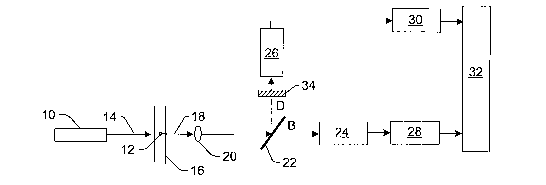
Fig. 1 illustrates one embodiment of a system configured to have an expanded
or extended dynamic range
(DR) that can be used for accurate measurement of the intensity of fluorescent
light emitted from the brightest
particles. The system may be configured as a flow cytometer. However, the
system may be configured as any othez
measurement system that will benefit from having an expanded or extended DR.
The system includes light source
10. Light source 10 is configured to illuminate particle 12 with light 14 as
the particle flows through cuvette 16.
Cuvette 16 may include any appropriate cuvette or other flow channel 1Q10Wn in
the art. Light source 10 may
include any appropriate light source known in the art such as a laser, a laser
diode, or a light emitting diode (LED).
Preferably, the light source includes an excitation source. In other words,
light source 10 is preferably configured to
generate light 14 having one or more wavelengths such that upon illumination
by the light, particle 12 will emit
fluorescent light 18.
Fluorescent light 18 may be collected by lens 20. Lens 20 may include any
appropriate lens known in the
art. In addition, although lens 20 is shown in Fig. 1 to be a refractive
optical component, it is to be understood that a
reflective optical component may be used in place of lens 20 to collect the
fluorescent light emitted by the particle.
In addition, although lens 20 is shown in Fig. 1 to be a single lens, it is to
be understood that lens 20 may be
replaced with a multi-lens system. Furthermore, the system may optionally not
include lens 20 or any other
fluorescent light collector. Additionally, it is to be understood that the
system shown in Fig. 1 may include more
than one lens and/or other lenses. For example, the system may include a
focusing lens (not shown) that is
configured to focus light 14 onto particle 12.
The system also includes optical component 22, which is configured to split
fluorescent light emitted by the
particle into multiple light paths. In the embodiment shown in Fig. 1, optical
component 22 is configured to split the
fluorescent light collected by lens 20 into multiple light paths. The
fluorescent light in the multiple paths has
different intensities. The optical component may include, in one embodiment, a
partially reflecting beam splitter or
any other appropriate optical component known in the art. In one particular
example, the optical component may
include an uncoated beam sputter. An uncoated beam sputter will reflect about
4% of the incident energy (in this
case, fluorescent light 18), and transmit the remaining portion of the
incident energy (i.e., approximately 96% of the
fluorescent light).
Note that while the intensity of the light transmitted by the optical
component is lower than in standard flow
cytometer configurations, assuming that the fluorescence saturation level on
the particle has not been reached, this
reduction in fluorescent light intensity can optionally and easily be
compensated for by increasing the power (or
intensity) of light source 10 in proportion to the intensity reduction.
As shown in Fig. 1, the optical component is configured to split the
fluorescent light into two light paths.
However, it is to be understood that the optical component may be configured
to split the emitted fluorescent light
into more than two light paths. For example, the optical component may include
a wedge of glass having non-
CA 02553025 2006-07-07
WO 2005/068971 PCT/US2005/001866
parallel surfaces, which may be configured as described further herein. In
addition, optical component 22 may
include any of the other optical components described herein which can be used
to split a light beam into multiple
light beams. Furthermore, it is to be understood that the system may include
more than one optical component that
is configured to split the fluorescent light into multiple light paths. For
example, the system may include more than
one partially reflecting beam splitter. The number of light paths into which
the fluorescent light is split may vary
depending on, for example, the number of different operating ranges of a
detector and/or the different intensities that
can be achieved in each of the multiple light paths.
The system also includes different channels configured to separately detect
the fluorescent light in the
multiple light paths. The different channels are also configured to generate
multiple signals. Each of the multiple
signals represents the fluorescent light in one of the multiple light paths.
For example, as shown in Fig. 1, the
system includes detectors 24 and 26, each of which constitutes at least a
portion of one of the different channels.
Detector 24 is configured to detect the fluorescent light transmitted by
optical component 22. Detector 24 is also
configured to generate a signal that represents the intensity of the
fluorescent light in light path B (B for bright).
Detector 24 may be a photomultiplier tube (PMT) or any other appropriate
detector known in the art. For example,
detector 24 may also be a photodiode, an avalanche photodiode, a charge
coupled device (CCD), or a
complementary metal-oxide-semiconductor (CMOS) detector. In addition, detector
24 may be any type of diode
detector known in the art or any type of linear array type detector. Detector
26 is configured to detect the
fluorescent light reflected by optical component 22. Detector 26 is also
configured to generate a signal that
represents the intensity of the fluorescent light in light path D (D for dim).
Detector 26 may be a PMT or any other
appropriate detector known in the art. For example, detector 26 may also be a
photodiode, an avalanche
photodiode, a CCD, or a CMOS detector. In addition, detector 26 may be any
type of diode detector known in the
art or any type of linear array type detector. Typically, detectors 24 and 26
will be the same type of detector, but
will operate in different ranges (e.g., linear and non-linear) due to the
different intensities of the fluorescent light in
the different light paths.
As shown in Fig. 1, detectors 24 and 26 are coupled to electronic components
28 and 30, respectively.
Electronic components 28 and 30 may include, for example, analog-to-digital
(A/D) converters or any other suitable
electronic components. In addition, electronic components 28 and 30 may form
only a portion of the entire
electronic chains of detectors 24 and 26, respectfully. For example, analog
components (not shown) may be
interposed between the detectors and the AJD converters. In addition, or
alternatively, the electronics chains may
include digital components (not shown) coupled to the output of the A/D
converters. These optional analog and
digital components may include any such suitable electronic components known
in the art. In addition, the
electronic chains coupled to detectors 24 and 26 may be similarly or
differently configured.
The system shown in Fig. 1 also includes processor 32. Processor 32 is
configured to determine which of
the different channels is operating in a linear range based on the signals
generated by the different channels. In
addition, processor 32 is configured to alter the signal generated by the
channel determined to be operating in the
linear range to compensate for the different intensities. Processor 32 may
include, for example, a digital signal
processor (DSP) or any other suitable component that can be used to execute
one or more program instructions to
perform at least the functions described herein.
CA 02553025 2006-07-07
WO 2005/068971 PCT/US2005/001866
In one particular example based on the uncoated beam splatter optical
component described above, the
processor examines output integer values (B and D) from the A/D converters
coupled to each of the detectors.
Based on the signal levels, the processor determines which channel is
operating in a linear range for the detectors.
The processor also appropriately alters or scales the value corresponding to
the channel operating in the linear range
to compensate for the actual light level entering that photosensitive
detector. For this simple example, the processor
would multiply the D (dim) A/D output by 1/0.04 = 25 (if chosen), or
alternately, the B (bright) A/D output by
1/0.96 = 1.041 (if chosen) to properly scale the result based on the division
of light between the photosensitive
detectors created by optical component 22. Such altering or scaling of the
signal corresponding to the channel
operating in the linear range effectively increases the dynamic range of the
system by IoglO(25/1.041) = 1.38
decades. In this manner, altering of the signal by the processor increases the
dynamic range of the system.
The processor may also be configured to perform a number of additional
functions. For example, the
processor may be configured to determine an intensity of the fluorescent light
emitted by the particle from the
altered signal. In addition, the processor may be configured to determine an
identity of the particle from the
intensity of the fluorescent light, possibly in combination with one or more
other output signals generated by the
system. Alternatively, the processor may be configured to determine an
identity of a molecule attached to the
surface of the particle or a reaction that has taken place on the surface of
the particle from the intensity of the
fluorescent light. In addition, the processor may be configured to determine a
quantity of a molecule attached to the
surface of the particle or a reaction that has taken place on the surface of
the particle from the intensity of the
fluorescent light. The processor may also be configured to perform any other
function typically performed in flow
cytometer data analysis.
The dynamic range improvement described above could be fiu~ther increased by
increasing the difference
between the intensities of the fluorescent light in the multiple light paths.
In one such embodiment, the intensity of
the light reflected by optical component 22 may be reduced by applying an anti-
reflective coating (not shown) to the
input face of the optical component. In another embodiment, as shown in Fig.
1, the system may include additional
optical component 34 positioned in the light path of the lower intensity
fluorescent light between optical component
22 and detector 26. The additional optical component is configured to decrease
the intensity of the fluorescent light
in this light path. Additional optical component 34 may include any optical
component that can be used to reduce
the intensity of the fluorescent light such as a neutral density filter. Each
of these configurations effectively reduces
the intensity of the fluorescent light detected by detector 26, thus further
expanding or extending the DR of the
system.
The system shown in Fig. 1 may be further configured as described herein. In
addition, although the
system shown in Fig. 1 is configured to have an expanded or extended DR for
only one fluorescent measurement, it
is to be understood that if multiple fluorescent measurements are performed by
the system, the DR of the system
may be expanded for each, or more than one, of the fluorescent measurements in
a manner as described above. For
example, the fluorescent light emitted by the particle may be separated by
wavelength using, in one example, one or
more dichroic beam splitters (not shown). In this manner, the fluorescent
light may be separated based on the
fluorochrome that emitted the fluorescent light. As such, fluorescent light
corresponding to an identity of the
particle may be separated from fluorescent light corresponding to a molecule
attached to the particle (e.g., via a
reaction with another molecule attached to the surface of the particle). In
addition, two or more of the fluorescent
light paths generated by the one or more dichroic beam splitters may be split
into multiple light paths as described
CA 02553025 2006-07-07
WO 2005/068971 PCT/US2005/001866
above such that the fluorescent light in the multiple light paths has
different intensities. The fluorescent light in the
multiple light paths may then be detected and processed as described above.
One method for expanding a dynamic range of a system, which can be performed
by the system shown in
Fig. 1, includes splitting fluorescent light into multiple light paths. The
fluorescent light in the multiple light paths
has different intensities. The method also includes detecting the fluorescent
light in the multiple light paths with
different channels to generate multiple signals. Each of the multiple signals
represents the fluorescent light in one of
the multiple light paths. In addition, the method includes determining which
of the different channels is operating in
a linear range based on the multiple signals. The method fiu~ther includes
altering the signal generated by the
channel determined to be operating in the linear range to compensate for the
different intensities.
l0 In one embodiment, the system may be configured as a flow cytometer or
another measurement system that
will benefit from having an extended or expanded DR. In another embodiment,
the method may include
determining an intensity of the fluorescent light emitted by the particle from
the altered signal. Altering the signal as
described above increases the dynamic range for the system.
In some embodiments, the fluorescent light in a first of the multiple light
paths is lower in intensity than the
fluorescent light in a second of the multiple light paths. In one such
embodiment, the method includes prior to the
detecting step, decreasing the intensity of the fluorescent light in the first
of the multiple light paths. Each of the
embodiments of the method described above may include any other steps)
described herein.
While the system and method embodiments described above advantageously keep
the high gain at the
beginning of the processing chain for small signals, the system and method
described above is more expensive due
:0 to the added expense of the second photosensitive detector, analog
electronics, and A/D conversion circuitry.
Fortunately, because of the geometry of a flow cytometer, there is another way
to accomplish this multiple light
level measurement with far fewer components.
For example, another method and system involves using multiple illumination
zones spatially separated
along the flow path of a particle. In a flow cytometer as described above, a
particle being measured travels along a
generally straight path through a cuvette, passing through an illumination
zone which results in excitation of one or
more fluorochromes associated with the particle. The resulting fluorescent
light is projected, focused, and/or
imaged on a portion of a detector's photosensitive area, while the particle is
illuminated in the illumination zone,
and a single current pulse is generated by the detector as a result. The
collector or pickup lens magnification may be
selected such that the florescent light emitted by the particle fills only a
portion of the photosensitive area of the
0 detector. In other words, the photosensitive area of the detector may be
relatively long in comparison to the cross-
sectional area of the fluorescent light beam projected, focused, and/or imaged
onto the detector. Therefore,
theoretically, the particle can still be "seen" by the detector when the
particle is not located within the illumination
zone. However, no signal will be produced by the detector when the particle is
not located in the illumination zone
since the fluorescence will extinguish quickly without excitation of the
fluorochrome(s) associated with the particle.
5 Thus, the width of the pulse generated by the detector is generally
proportional to the length of time that the particle
is located in the illumination zone.
It is possible, therefore, to take advantage of the spatially "long"
photosensitive area of the detector for the
dynamic range extension methods and systems described herein. For example, a
second, but less bright illumination
zone, can be added to the system, which is spatially separated from the
primary illumination zone. As such,
fluorescent light emitted by a particle as a result of illumination in both of
these illumination zones can be directed
9
CA 02553025 2006-07-07
WO 2005/068971 PCT/US2005/001866
to different portions of the photosensitive area of the detector. In this
manner, the detector will generate two time-
separated current pulses, and one pulse would be much larger than the other
due to the different intensities of the
light in the illumination zones.
A processor such as a DSP can be configured to measure the amplitude of each
current pulse and, similar to
the methods described above, determine which signal is located in a linear
range of the detector. The processor may
also be configured to alter or scale the signal located in the linear range in
proportion to the ratio of the illumination
zone excitation energies. Thus, only a single detector and associated
electronics may be included in the system. As
such, the systems described below avoid the costs associated with multiple
detectors and corresponding electronics.
However, the challenge now becomes to provide a cost effective way to create
the additional, dimmer illumination
zone(s). To address this challenge, several embodiments of a system have been
identified and are described further
lierein, which provide cost effective ways to illuminate a particle in
multiple illumination zones with light having
different intensities.
It is important to note that the order of the bright and dim illumination
zones may be important. In
particular, preferably, the particle travels through the dim illumination zone
first, then through the bright
illumination zone. In contrast, if the particle travels through the bright
illumination zone before the dim illumination
zone, the analog electronics may not settle in time to accurately reproduce
the pulse resulting from illumination of
the particle in the dim illumination zone. Another factor in selecting the
order in which the particle travels through
the illumination zones is the potential for photo-degradation of the
fluorochrome(s) associated with the particle due
to illumination in the bright illumination zone. Therefore, it may be
preferable to illuminate the particle in the dim
illumination zone first, then in brighter illumination zones.
Fig. 2 illustrates one embodiment of a system configured to have an expanded
DR. The system includes an
illumination subsystem, which includes light sources 35 and 37. Light sources
35 and 37 are configured to
illuminate particle 36 in multiple illumination zones 38 and 40, respectively,
with light that has different intensities.
Although the illumination subsystem is shown in Fig. 2 to include two light
sources, it is to be understood that the
system shown in Fig. 2 may include two or more light sources. In an
alternative, the illumination subsystem may
include a single light source coupled to a beam multiplier, which may include
any of the beam multipliers as
described herein.
As shown in Fig. 2, illumination zones 38 and 40 are spaced apart along flow
path 42 of particle 36. In this
manner, as particle 36 moves through cuvette 44, which may be configured as
described above, the particle is
located in illumination zone 38, then in illumination zone 40. In this manner,
for the reasons described above, the
illumination subsystem is preferably configured such that the light directed
to the particle in illumination zone 38
has a lower intensity than the light directed to the particle in illumination
zone 40. The illumination subsystem may
be further configured as described herein. In addition, although two
illumination zones are shown in Fig. 2, it is to
be understood that the illumination subsystem may be configured to illuminate
the particle in more than two
illumination zones, each of which is spatially separated along the flow path
of the particle.
As further shown in Fig. 2, fluorescent light emitted by the particle in the
different illumination zones may
be collected by lens 46. Lens 46 may be configured as described above. In
addition, it is to be understood that lens
46 riiay optionally not be included in the system.
CA 02553025 2006-07-07
WO 2005/068971 PCT/US2005/001866
The system shown in Fig. 2 also includes a detection subsystem that is
configured to separately detect
fluorescent light emitted by the particle while the particle is located in the
multiple illumination zones. The
detection subsystem is also configured to generate multiple signals, each of
which represents the fluorescent light
emitted by the particle while the particle is located in one of the multiple
illumination zones. For example, in this
embodiment, the detection subsystem includes detector 48 having photosensitive
area 50. In this manner, the
detection subsystem includes a single detector. Detector 48 may be a PMT or
any other suitable detector known in
the art such as a photodiode, an avalanche photodiode, a CCD, a CMOS detector,
or any other suitable type of diode
or linear array detector known in the art.
As shown in Fig. 2, photosensitive area 50 is larger than an area of
fluorescent light 52 emitted by the
particle while the particle is located in illumination zone 38. Photosensitive
area 50 is also larger than an area of
fluorescent light 54 emitted by the particle while the particle is located in
illumination zone 40. Furthermore,
photosensitive area 50 is larger than a combined area of fluorescent light 52
and 54. In this manner, fluorescent
light 52 and 54 may be directed to spatially separated portions of
photosensitive area 50. In an alternative
embodiment, the detection subsystem may include multiple detectors (not shown)
in place of detector 48.
Fluorescent light emitted by the particle due to illumination in each of the
illumination zones may be directed to a
different detector. In some such embodiments, each of the multiple detectors
may be a PMT or any other suitable
detector known in the art such as a photodiode, an avalanche photodiode, a
CCD, a CMOS detector, or any other
suitable type of diode or linear array detector known in the art.
When particle 36 is located in illumination zone 38, detector 48 will generate
a signal that represents
ZO fluorescent light 52. When particle is located in illumination zone 40,
detector 48 will generate a signal that
represents fluorescent light 54. Fig. 3 is a plot showing examples of signals
that can be generated by the detection
subsystem of Fig. 2 and other embodiments described herein. As shown in Fig.
3, the signal generated by the
detection subsystem representing light emitted by the particle while the
particle is located in the first illumination
zone (e.g., illumination zone 38) has a much lower value than the signal
generated by the detection system
ZS representing fluorescent light emitted by the particle while the particle
is located in the second illumination zone
(e.g., illumination zone 40). The difference in the values of the signals is a
direct result of the intensity of the light
illuminating the particle in the different illumination zones. Preferably, the
intensities of the light illuminating the
particle in the different illumination zones is selected such that one of the
signals will be generated in the linear
operating range of the detector.
30 The system also includes a processor (not shown), which may be coupled to
the detector as described
above (e.g., via one or more analog and/or digital electronic components). The
processor is configured to determine
which of the multiple signals generated by detector 48 due to illumination of
the particle in one of the multiple
illumination zones is produced in a linear range of the detector. The
processor is also configured to alter or scale the
signal located in the linear range of the detector to compensate for the
different intensities. For example, the
35 processor may be configured to scale the signal located in the linear range
of the detector in proportion to the
different intensities of the light illuminating the particle in the different
illumination zones. The processor may be
further configured as described above. In addition, the system shown in Fig. 2
may be further configured as
described herein.
11
CA 02553025 2006-07-07
WO 2005/068971 PCT/US2005/001866
The illumination subsystem described in Fig. 2 may include two or more light
sources such as lasers. Each
of the light sources may be configured to provide illumination for one of the
multiple illumination zones. Adding
one or more additional light sources such as lasers to the system increases
the cost of the system. Since the intensity
of the light in one illumination zone is preferably much less than that of the
other, an inexpensive LED and relatively
narrow band pass filter could be employed as the secondary light source. In
this manner, an additional light source
may be added to the system without substantially increasing the cost of the
system. However, in other embodiments
described herein, the system may include a single light source coupled to one
or more optical components that are
configured to split the light generated by the single light source into
multiple light beams having different intensities.
Fig. 4 illustrates one embodiment of an illumination subsystem that may be
included in a system configured
to have an expanded DR. As shown in Fig. 4, the illumination subsystem
includes single light source 56 such as a
laser, which is coupled to glass slide 58 arranged in a path of light beam 60
emitted by single light source 56. In
addition, as shown in Fig. 4, glass slide 58 is arranged at an angle, Ol, with
respect to light beam 60. In this manner,
the glass slide may be configured as a beam multiplier. For example, light
beam 60 enters glass slide 58 and at the
boundary of the uncoated air to glass interface of glass slide 58, a portion
of light beam 60 is reflected back towards
the light source. Therefore, light beam 62 exiting glass slide 58 has an
intensity that is lower than an intensity of the
light beam generated by the single light source. A portion of this reflected
light once again partially reflects off of
the first surface of glass slide 58, and then exits the second surface as
light beam 64. Since a relatively small portion
of light beam 60 is reflected back toward the light source, and a portion of
the light beam reflected off of the first
surface of glass slide 58 will again be reflected by the second surface of the
glass slide, light beam 64 will have a
lower intensity than light beam 62. Additional light beams such as light beam
66 may be generated in this manner.
Each of the additional light beams will have a lower intensity than the light
beams that previously exited the glass
slide.
Since the glass slide is canted at an angle with respect to the single light
source, there is a physical
separation between light beams 62, 64, and 66. As such, light beams 62, 64,
and 66 xnay be directed to illumination
zones 70, 72, and 74, respectively, which are spaced apart along a flow path
of the particle (not shown). As the
particle travels through cuvette 68, the particle will travel through
illumination zone 74. After traveling through
illumination zone 74, the particle will travel through illumination zone 72,
which has an intensity that is greater than
that of illumination zone 74. After traveling through illumination zone 72,
the particle will travel through
illumination zone 70, which has an intensity that is greater than that of
illumination zone 72. In this manner, as
described above, the particle will be illuminated in the illumination zones
having progressively higher intensities.
The distance between the light beams is generally proportional to this angle
O~.
The angle of the glass slide can be selected such that light beams 62, 64, and
66 are substantially parallel to
one another and have substantially the same diameters. As such, a single
focusing lens (not shown) may be
configured to focus the light beams exiting the glass slide onto the path
along which the particle travels in the
cuvette. Alternatively, or in addition, a focusing lens (not shown) may be
positioned in the path of the light beam
generated by single light source 56. In addition, a single collection lens
(not shown) may be configured to collect
the fluorescent light emitted by the particle. The focusing and collecting
lenses may be further configured as
described above.
12
CA 02553025 2006-07-07
WO 2005/068971 PCT/US2005/001866
Furthermore, the intensity of each relatively low intensity beam that exits
the glass slide will be reduced
significantly. For example, in the case where the reflection coefficient at
the air-glass interface is 2%, the reduction
can be calculated by comparing the relative intensities of beams 62 and 64 as
1og10(0.02 x 0.02 x 0.98) = 3.4
decades. Therefore, the first two beams of the illumination subsystem shown in
Fig. 4 can add more than three
decades of DR between the illumination zones. Note that an additional 3.4
decade reduced beam (e.g., light beam
66) (-6.8 decades from the primary) will be present, and could potentially be
used as a third illumination zone. This
pattern of reduced intensity light beams repeats with decreasing magnitude,
but it is expected that the dynamic range
of the particle and chemistry, or instrument electronics, may limit the useful
light beams to two or three. The
separation distance between each parallel ray is preferably, at a minimum,
equivalent to the diameter of the laser
1'0 beam. Since the beam diameter is typically focused to a spot having a
diameter of tens of microns at cuvette 68, the
necessary thickness t of the glass slide will decrease as the glass slide is
placed closer to the cuvette.
A system that includes the illumination subsystem shown in Fig. 4 may be
further configured as described
herein. For example, such a system also includes a detection subsystem that is
configured to separately detect
fluorescent light emitted by the particle while the particle is located in the
multiple illumination zones. The
15 detection subsystem is also configured to generate multiple signals, each
of which represents the fluorescent light
emitted by the particle while the particle is located in one of the multiple
illumination zones. The detection
subsystem may be further configured as described herein. In addition, such a
system includes a processor that is
configured to determine which of the multiple signals is located in a linear
range and to alter the signal located in the
linear range to compensate for the different intensities. The processor may be
further configured as described
20 herein.
Fig. 5 illustrates another embodiment of an illumination subsystem that may be
included in a system
configured to have an expanded DR. As shown in Fig. 5, the illumination
subsystem includes single light source 76.
Single light source 76 may include a laser or any other appropriate light
source known in the art. The illumination
subsystem also includes wedge of glass 78. Wedge of glass 78 has non-parallel
surfaces (e.g., surfaces 78a and 78b)
ZS arranged in a path of light beam 80 emitted by single light source 76.
Light beam 80 enters the wedge of glass, and
like the glass slide described above, a portion of the light beam will be
reflected back towards the single light
source. The portion of the light transmitted through the wedge of glass forms
light beam 82. Since a relatively large
portion of light beam 80 will be transmitted by wedge of glass 78, light beam
82 will be the brightest light beam
exiting wedge of glass 78. In other words, of the light beams that exit the
wedge of glass, light beam 82 will have
30 the highest intensity.
The portion of light beam 80 reflected back toward the single light source
will again be reflected (at least
partially) by the entrance face (e.g., surface 78a) of the wedge of glass. A
portion of this light beam will exit the
wedge of glass as light beam 84. Light beam 84 obviously will have a lower
intensity than light beam 82. A portion
of the original light beam will be reflected again within the wedge of glass
and may exit the wedge of glass as light
35 beam 86. Light beam 86 will have the lowest intensity of light beams 82,
84, and 86. Obviously, additional light
beams may also exit the wedge of glass, and if the additional light beams have
sufficient intensity, these light beams
may also used for illumination of a particle.
Light beams 82, 84, and 86 are directed to cuvette 88 through which a particle
(not shown) will move
during measurements. In particular, light beams 82, 84, and 86 are directed to
the cuvette in multiple illumination
EO zones 90, 92, and 94, respectively, arranged one after another along a flow
path of the particle. The particle will
13
CA 02553025 2006-07-07
WO 2005/068971 PCT/US2005/001866
preferably move through the illumination zone having the lowest intensity
(e.g., illumination zone 94) first and the
highest intensity (e.g., illumination zone 90) last for the reasons set forth
above. Due to the non-parallel surfaces of
the wedge of glass, light beams 82, 84, and 86 will be directed to spaced
apart locations along the flow path of the
particle. In addition, the distance between beams 82, 84, and 86 will be
larger than that between the beams
produced by the glass slide shown in Fig. 4 for the same (average glass
thickness). The distance between beams 82,
84, and 86 can also be altered by changing the angle, O, between the entrance
face and exit face of the wedge of
glass.
The illumination subsystem may also include a focusing lens (not shown). In
one example, the focusing
lens may be placed in a path of light beam 80 between single light source 76
and wedge of glass 78. However, if a
wedge of glass with non-parallel surfaces is used as the beam multiplier, the
distance between beams 82, 84, and 86
is larger than that between the beams produced by the glass slide shown in
Fig. 4 for the same (average) glass
thickness, making it more feasible to place the beam multiplier between the
laser and focusing lens, where the beam
width is widest. In this manner, the focusing lens may be positioned in the
optical paths of light beams 82, 84, and
86 between the wedge of glass and cuvette 88. The focusing lens may be further
configured as described above. A
system that includes an illumination subsystem as shown in Fig. 5 may be
further configured as described herein.
Fig. 6 illustrates another embodiment of an illumination subsystem that may be
included in a system
configured to have an expanded DR. As shown in Fig. 6, the illumination
subsystem includes single light source 96.
Single light source 96 may include a laser or any other appropriate light
source known in the art. This illumination
subsystem also includes beam expander 98. Beam expander 98 is configured to
increase a cross-sectional area of
light beam 100. Light beam 102 exiting the beam expander is directed to the
input faces of multiple fiber optic
cables 104, 106, and 108. Fiber optic cables 104, 106, and 108 may include any
appropriate fiber optic cables
known in the art. As shown in Fig. 6, fiber optic cables 104, 106, and 108 are
arranged such that each of the fiber
optic cables directs light to cuvette 110 in a different illumination zone. In
particular, fiber optic cables 104, 106,
and 108 direct light to multiple illumination zones 112, 114, and 116,
respectively, which are spaced apart along a
flow path of a particle (not shown). In addition, although the illumination
subsystem is shown in Fig. 6 to include
three fiber optic cables, it is to be understood that the illumination
subsystem may include two or more fiber optic
cables arranged in such an optical configuration.
Since beam expander 98 will produce an expanded light beam that has a
relatively constant intensity across
the cross-sectional area of the light beam, each of the fiber optic cables may
receive a substantially equal amount of
light. In this manner, the illumination subsystem may also include one or more
optical components (not shown)
such as neutral density filters either at an input face or an output face of
one or more of the fiber optic cables. The
one or more optical components may be configured to alter the intensity of the
light exiting one or more of the fiber
optic cables such that the particle is illuminated in the multiple
illumination zones with light having different
intensities. Preferably, the particle is illuminated as it travels along the
flow path with light having relatively low
intensity then light having higher intensity for the reasons set forth above.
In other embodiments, optical components other than a beam expander may be
used to direct light from a
single light source to multiple fiber optic cables substantially
simultaneously. For example, an uncoated beam
splitter such as that shown in Fig. 1 may be used to split the light from the
single light source into multiple light
beams having different intensities. Each of the multiple light beams may then
be directed to one of the multiple
fiber optic cables. Obviously, many other optical configurations may be used
to deliver light from a single light
14
CA 02553025 2006-07-07
WO 2005/068971 PCT/US2005/001866
source to multiple fiber optic cables, and the description provided herein is
not intended to limit the'optical
configurations that can be used in this system embodiment.
The illumination subsystem shown in Fig. 6 may also include a focusing lens
(not shown). In one example,
the focusing lens may be placed in a path of light beam 100 between single
light source 96 and beam expander 98.
Alternatively, the focusing lens may be positioned in the optical path of
light beam 120 between the beam expander
and fiber optic cables 104, 106, and 108. In yet another alternative, the
focusing lens may be positioned in the
optical path of light exiting fiber optic cables 104, 106, and 108 between the
fiber optic cables and cuvette 110. The
focusing lens may be fiu-ther configured as described above. A system that
includes the illumination subsystem
shown in Fig. 6 may be further configured as described herein.
Fig. 7 illustrates another embodiment of an illumination subsystem that may be
included in a system
configured to have an expanded DR. As shown in Fig. 7, the illumination
subsystem includes single light source
118. Single light source 118 may include a laser or any other appropriate
light source known in the art. The
illumination subsystem also includes demultiplexer 120. Demultiplexer 120 is
configured to receive light beam 122
generated by light source 118 and to separate light beam 122 into individual
light beams (e.g., light beams 124, 126,
and 128). The demultiplexer is preferably configured to separate light beam
122 into individual light beams having
different intensities. However, if the demultiplexer is not configured to
generate individual light beams having
different intensities, the illumination subsystem may include one or more
components (not shown) positioned in a
path of one or more of the individual light beams such as a neutral density
filter or any other components that can
alter the intensity of the individual light beams. Demultiplexer 120 may
include any appropriate demultiplexer
known in the art. In addition, although demultiplexer 120 is shown in Fig. 7
to generate three individual light
beams, it is to be understood that the demultiplexer may be configured to
generate two or more individual light
beams.
Individual light beams 124, 126, and 128 are directed from the demultiplexer
to cuvette 130 through wluch
a particle (not shown) will move during measurement. In particular, individual
light beams 124, 126, and 128 are
directed to multiple illumination zones 132, 134, and 136, respectively, which
are spaced apart along a flow path
through cuvette 130. Preferably, the intensity of the light in illumination
zone 136 is lower than that in illumination
zones 134 and 132, and the intensity of the light in illumination zone 134 is
lower than that in illumination zone 132.
In this manner, as the particle travels along the flow path, it will be
illuminated first with low intensity light then
increasingly higher intensity light as it moves through the different
illumination zones. As further shown in Fig. 7,
the individual light beams exiting the demultiplexer may be spaced apart such
that the spacing between the
individual light beams exiting the demultiplexer defines the spacing between
the multiple illumination zones.
However, the spacing between the multiple illumination zones may be altered
using one or more optical components
(not shown) positioned in the path of the individual light beams and
configured to alter a direction of the individual
light beams (e.g., by refraction).
The illumination subsystem may also include one or more demultiplexers, which
may be configured as
described above, coupled to the single light source. The illumination
subsystem shown in Fig. 7 may further include
a focusing lens (not shown). In one example, the focusing lens may be placed
in a path of light beam 122 between
single light source 118 and demultiplexer 120. Alternatively, the focusing
lens may be positioned in the optical
paths of light beams 124, 126, and 128 between the demultiplexer and cuvette
130. The focusing lens may be
CA 02553025 2006-07-07
WO 2005/068971 PCT/US2005/001866
fiufiher configured as described above. A system that includes an illumination
subsystem as shown in Fig. 7 may be
fixrther configured as described herein.
Fig. 8 illustrates another embodiment of an illumination subsystem that may be
included in a system
configured to have an expanded DR. As shown in Fig. 8, the illumination
subsystem includes single light source
138. Single light source 138 may include a laser or any other appropriate
light source known in the art. The
illumination subsystem also includes diffraction grating 140 arranged in a
path of light beam 142 emitted by light
source 138. Diffraction grating 140 is configured to reflect light beam 142
generated by light source 138 into
multiple light beams 144, 146, and 148.
Each of the multiple light beams may include one of the different orders of
light that is reflected from the
diffraction grating. In particular, when a monochromatic source such as a
laser is directed to a lined diffraction
grating, multiple beams are reflected at different orders, and the intensity
of each light beam is reduced as the
"order" increases (e.g., the zero order reflected beam will be brighter than
the first order reflected beam, and so on).
In this manner, light beam 144 may include the zero order light reflected from
diffraction grating 140. Therefore,
light beam 144 will have the highest intensity of the light beams reflected
from the diffraction grating. Light beam
146 may include the first order light reflected from the diffraction grating.
In this manner, light beam 146 will have
an intensity that is lower than light beam 144. Likewise, light beam 148 may
include the second order light reflected
from diffraction grating 140 and, therefore, will have the lowest intensity of
the three light beams. The diffraction
grating is also configured to reflect the different orders of light in
different directions. Therefore, the diffraction
grating may generate individual light beams that have both different
intensities and adequate spacing for the multiple
illumination zone configuration. In addition, the line spacing of the
diffraction grating could be varied to change the
angle of each order, and the intensity of the light beam reflected at each
order. Furthermore, more than three orders
of light may be reflected from the diffraction grating, and the number of
orders which are directed to cuvette 150
may vary depending on how many of the different orders have sufficient
intensity for particle measurements.
As shown in Fig. 8, the illumination subsystem may also include focusing lens
152. Focusing lens 152 may
be configured to receive light reflected from the diffraction grating and to
direct the different orders of light to
different fiber optic cables. In particular, light beams 144, 146, and 148 may
be directed to fiber optic cables 154,
156, and 158, respectively. Fiber optic cables 154, 156, and 158 may be
configured to direct the different orders of
light to multiple illumination zones 160, 162, and 164, respectively, which
are spaced apart along the flow path of
the particle (not shown) within cuvette 150. Light in illumination zone 164
has a lower intensity than the light in
illumination zones 162 and 160, and light in illumination zone 162 is lower in
intensity than the light in illumination
zone 160. In this manner, as the particle travels through the illumination
zones, it is illuminated with lower intensity
light first, and then with increasingly higher intensities of light. Focusing
lens 152 may be further configured as
described above. In addition, focusing lens may be located in a different
position within the illumination subsystem
(e.g., between light source 138 and diffraction grating 140). Fiber optic
cables 154, 156, and 158 may include any
appropriate fiber optic cables known in the art. In addition, the illumination
subsystem shown in Fig. 8 may not
include the fiber optic cables. In one such alternative, light from lens 152
may be directly focused to cuvette 150. A
system that includes the illumination subsystem shown in Fig. 8 may be further
configured as described herein.
One method for expanding a dynamic range of a system, which can be performed
by the system shown in
Fig. 2 and a system, which includes an illumination subsystem as shown in
Figs. 4-8, includes illuminating a particle
in multiple illumination zones with light having different intensities. The
method also includes separately detecting
16
CA 02553025 2006-07-07
WO 2005/068971 PCT/US2005/001866
fluorescent light emitted by the particle while the particle is located in the
multiple illumination zones to generate
multiple signals. Each of the multiple signals is representative of the
fluorescent light emitted by the particle in one
of the multiple illumination zones. In addition, the method includes
deternzining which of the multiple signals is
located in a linear range. The method further includes altering the signal
located in the linear range to compensate
for the different intensities.
In one embodiment, the multiple illumination zones are spaced apart along a
flow path of the particle, and a
first of the multiple illumination zones in which the particle is first
located is lower in intensity than a second of the
multiple illumination zones in which the particle is subsequently located. The
fluorescent light emitted by the
particle may correspond to an identity of the particle. Alternatively, the
fluorescent light emitted by the particle
corresponds to a molecule reacted with an additional molecule attached to the
particle. Each of the embodiments of
the method described above may include any other steps) described herein.
Although the above methods and systems are described with respect to flow
cytometry applications, it is to
be understood that the methods and systems described herein may be used in
other applications. For example, the
methods and systems described herein may be used for absorption spectroscopy
applications. In absorption
techniques, such as infrared (IR) and ultraviolet-visible (IJV-Vis), light is
focused on a sample, and a detector
measures the light not absorbed by the sample, which is known as the
transmitted light. Obviously, in highly
absorbing or concentrated samples, the transmittance is low, and in dilute, or
low absorbing samples transmittance is
high. Therefore, in some instances, increasing the dynamic range as described
herein in absorption spectroscopy
applications may be advantageous.
It will be appreciated to those skilled in the art having the benefit of this
disclosure that this invention is
believed to provide methods and systems for dynamic range expansion. Further
modifications and alternative
embodiments of various aspects of the invention will be apparent to those
skilled in the art in view of this
description. Accordingly, this description is to be construed as illustrative
only and is for the purpose of teaching
those skilled in the art the general manner of carrying out the invention. It
is to be understood that the forms of the
invention shown and described herein are to be taken as the presently
preferred embodiments. Elements and
materials may be substituted for those illustrated and described herein, parts
and processes may be reversed, and
certain features of the invention may be utilized independently, all as would
be apparent to one skilled in the art
after having the benefit of this description of the invention. Changes may be
made in the elements described herein
without departing from the spirit and scope of the invention as described in
the following claims.
17