Note: Descriptions are shown in the official language in which they were submitted.
CA 02553532 2006-07-17
WO 2005/072659 PCT/US2005/001478
SYSTEMS AND TECHNIQUES FOR RESTORING AND MAINTAINING
INTERVERTEBRAL ANATOMY
Cross-Reference to Related Application:
This application is a continuation-in-part of U.S. Patent Application No.
10/274,856
filed on October 21, 2002, which is incorporated herein by reference in its
entirety.
BACKGROUND
Various surgical instruments and methods have been devised for the
implantation of
devices into the disc space between adjacent vertebrae of the spinal column.
For example,
spinal fusion procedures can require sequential distraction to restore the
disc space height
prior to inserting a pair of fusion devices in the disc space in side-by-side
relation. To
implant these devices, an initial opening or openings are made in the disc
space at the
locations tlmough which the devices are to be inserted. A first distracter is
inserted in the disc
space at one of the device locations. A second larger distracter is inserted
in the disc space at
the other of the device locations. Sequential distraction in alternate disc
space locations is
continued until the desired disc space height is achieved. The next to last
inserted distracter
is then removed. The disc space is 'prepared for insertion of one fusion
device in the location
previously occupied by the withdrawn distracter while the other distracter
maintains the
restored disc space height.
In another technique, a spinal disc space is accessed and distracted for
insertion of an
implant. Distraction of the disc space is maintained by applying a distraction
force to bone
screws engaged in the vertebrae on each side of the disc space.
While the above procedure can be effective for some techniques, there are
disadvantages. For example, dissection and retraction of tissue, vasculature
and nervature is
required to accommodate the pair of distracters inserted in the disc space, or
to accommodate
the external distracters. Alternating sequential distraction can be time-
consuming and
requires many steps to complete the surgical procedure. Engagement of bone
screws to the
vertebrae and application of a distraction force to the engaged bone screws
also requires
additional time and steps in the surgical procedure.
There remains a need for instruments and techniques for restoring and
maintaining a
spinal disc space anatomy that minimizes dissection and retraction and of
tissue, vasculature
CA 02553532 2006-07-17
WO 2005/072659 PCT/US2005/001478
2
r
and nervature. There further remains a need for instruments and techniques for
restoring and
maintaining a spinal disc space anatomy that minimizes the steps and
complexity of the
procedure during surgery.
SUMMARY
Implants are provided that can be sequentially inserted and withdrawn from a
spinal disc space to restore the disc space to a desired disc space height and
to post-
operatively maintain the desired spinal disc space height when a selected
implant is left in
the spinal disc space.
Instruments are provided for deternzining the desired disc space height and
for
selecting an implant providing the desired disc space height when inserted in
the collapsed
disc space.
Implants are provided that can have the same height and leading end portion
configuration of at least some trial instruments of a set of trial
instruments. Each trial
instrument of the set has a trial body providing a restored disc space height
and a leading
end portion configured to distract the disc space to the restored disc space
height.
Implants are provided that have a self distracting lead end configuration.
f
Instruments for inserting implants are also provided.
Related aspects, forms, and embodiments will be apparent from the following
description.
BRIEF DESCRIPTION OF THE DRAWINGS
Figs. 1A and 1B are elevation views of a self distracting trial instrument and
a pair of
adjacent vertebrae before and after insertion of the trial instrument.
Figs. 2A and 2B are elevation views of a self distracting implant and a pair
of vertebrae
before and after insertion of the implant.
Figs. 3A and 3B are elevation views of a distal portion of another embodiment
self
distracting trial instrument and a pair of adjacent vertebrae before and after
insertion of the
trial instrument.
Figs. 4A and 4B are elevation views of another embodiment self distracting
implant
and a pair of adjacent vertebrae before and after insertion of the implant.
Fig. 5 shows a set of trial instruments.
Fig. G shows a set of implants and implant insertion instruments.
CA 02553532 2006-07-17
WO 2005/072659 PCT/US2005/001478
3
Fig. 7 is a perspective view of another embodiment implant and implant
insertion
instrument.
Fig. 8 is a perspective view of an embodiment of the implant of Fig. 7.
Fig. 9 is an exploded perspective view of the implant of Fig. 8.
Fig. 10A is an elevation view of a proximal end of the implant of Fig. 7
coupled to the
insertion instrument.
Fig. l OB is an elevation view of the proximal end of the implant of Fig. 7
uncoupled
from the insertion instrument.
Figs. 1 1A and 11B are a plan view and side view, respectively, of a distal
portion of
another embodiment trial instrument.
Figs. 12A and 12B are a plan view and side view, respectively, of a distal
portion of
another embodiment trial instrument.
Figs. 13A and 13B are a plan view and side view, respectively, of a distal
portion of
another embodiment trial instrument.
Figs. 14A and 14B are a plan view and side view; respectively, of a distal
portion of
another embodiment trial instrument.
Figs. 15A and 15B are a plan view and side view, respectively, of a distal
portion of
another embodiment trial instrument.
Figs. 16A and 16B are a plan view and side view, respectively, of a distal
portion of
another embodiment trial instrument.
Figs. 17A and 17B are a plan view and side view, respectively, of a distal
portion of
another embodiment trial instrument.
Figs. 18A and 18B are a plan view and side view, respectively, of a distal
portion of
another embodiment trial instrument.
Figs. 19A and 19B are a plan view and side view, respectively, of a distal
portion of
another embodiment trial instrument.
Figs. 20A and 20B are a plan view and side view, respectively, of a distal
portion of
another embodiment trial instrument.
Figs. 21A and 21B are a plan view and side view, respectively, of a distal
portion of
another embodiment trial instrument.
CA 02553532 2006-07-17
WO 2005/072659 PCT/US2005/001478
4
Figs. 22A and 22B are a plan view and side view, respectively, of a distal
portion of
another embodiment trial instrument.
Figs. 23A and 23B are a plan view and side view, respectively, of a distal
portion of ,
another embodiment trial instrument.
Figs. 24A and 24B are a plan view and side view, respectively, of a distal
portion of
another embodiment trial instrument.
Figs. 25A and 25B are a plan view and side view, respectively, of a distal
portion of
another embodiment trial instrument.
Figs. 26A and 26B are a plan view and side view, respectively, of a distal
portion of
another embodiment trial instrument.
Figs. 27A and 27B are a plan view and side view, respectively, of a distal
portion of
another embodiment trial instrument.
Figs. 28A and 28B are a plan view and side view, respectively, of a distal
portion of
another embodiment trial instrument.
Figs. 29A and 29B are a plan view and side view, respectively, of a distal
portion of
another embodiment trial instrument.
Fig. 30 is an elevation view of another embodiment implant.
Fig. 31 is an end view of the implant of Fig. 30.
Fig. 32 is a section view through line 32-32 of Fig. 30.
Fig. 33 is an enlarged view of a portion of the implant of Fig. 30.
Fig. 34 is a perspective view of another embodiment implant.
Fig. 35 is a perspective view of another embodiment implant.
Fig. 36 is a perspective view of another embodiment insertion instrument.
Fig. 37 is an enlarged view of a distal end of the insertion instrument of
Fig. 36.
Fig. 38 is a view of the distal end of the insertion instrument engaged to a
trailing end
of an implant.
Fig. 39 is a perspective view of another embodiment insertion instrument.
CA 02553532 2006-07-17
WO 2005/072659 PCT/US2005/001478
DESCRIPTION OF THE ILLUSTRATED EMBODIMENTS
For the purposes of promoting an understanding of the principles of the
invention,
reference will now be made to the embodiments illustrated in the drawings and
specific
language will be used to describe the same. It will nevertheless be understood
that no
limitation of the scope of the invention is thereby intended. Any such
alterations and further
modifications in the illustrated device, and any such further applications of
the principles of
the invention as illustrated therein being contemplated as would normally
occur to one skilled
in the art to which the invention relates.
Methods, techniques, instrumentation and implants are provided to restore
andlor
maintain a collapsed spinal disc space at a desired disc space height. The
instruments and
implants may be used in techniques employing minimally invasive instruments
and
technology to access the disc space. Access to the collapsed disc space can be
uni-portal, bi-
portal, or mufti-portal. The instruments and implants may also be employed in
open surgical
procedures in which slcin and tissue is dissected and retracted to access the
collapsed spinal
disc space. The methods, techniques, instruments and implants may also be
employed in any
surgical approach to the spine, including lateral, antero-lateral, postern-
lateral, posterior, and
anterior approaches. Also, the surgical methods, techniques, instruments and
implants may
find application at all vertebral segments of the spine, including the lumbar,
thoracic and
cervical spinal regions.
Referring now to Fig. lA, there is shown an implant trial instrument 20 having
a
proximal handle 22, a shaft 24 extending distally from handle 22, and a trial
body 26. Trial
body 26 includes a proximal end 28 connected with or formed with a distal end
of shaft 24
and a leading insertion end 30. Trial body 26 further includes an upper
surface 26a and an
opposite lower surface 26b. Trial body has a height Hl between upper surface
26a and lower
surface 26b. Proximal end 28 can be tapered or otherwise configured to provide
a gradual
transition between surfaces 26a, 26b to facilitate withdrawal of trial body 26
from the spinal
disc space.
Trial instrument 20 is insertable into a collapsed disc space D between
adjacent
vertebrae V 1 and V2. Leading end portion 30 can be provided with a rounded
nose-like
shape that allows at least a portion of leading end portion 30 to be inserted
into a collapsed,
undistracted disc space D. As trial body 26 is advanced into disc space D, the
edges of
CA 02553532 2006-07-17
WO 2005/072659 PCT/US2005/001478
6
vertebrae Vl and V2 ride upwardly and downwardly, respectively, along the
rounded nose
portion of leading end portion 30. Once leading end portion 30 is completely
inserted,
collapsed disc space D is distracted to restore disc space D', as shown in
Fig. 1B. Restored
disc space D' has a height between the endplates of the adjacent vertebrae Vl,
V2 which
corresponds to height Hl of trial body 26 between upper surface 26a and lower
surface 26b.
With trial body 26 inserted in disc space D, the surgeon can determine whether
disc
space D has been adequately distracted or positioned to a desired disc space
height by the
tactile feel and visual inspection of trial instrument 20 in the disc space.
For example, if trial
instrument 20 is easily moved, or does not provide a snug fit, then a trial
instrument 20 may
be withdrawn and a second trial instrument having a trial body with a greater
height H1 is
inserted. Alternatively, if the fit of trial body 26 is too tight or won't
fit, it can be withdrawn
and another trial instrument having a trial body with a smaller height Hl can
be inserted in
disc space D. The particular trial instrument 20 providing a restored disc
space height that
corresponds to a desired disc space height is noted by the surgeon for
selection of an implant.
~ In Fig. 2A there is shown an implant 40 having a body 42. Body 42 includes a
proximal end 44 and a leading insertion end 46. Body 42 further includes an
upper surface
42a and an opposite lower surface 42b. Body 42 has a height H1 between
surfaces 42a,~42b.
Leading insertion end 46 is the same size and shape as leading end portion 30
of trial body
26. Height H1 between surfaces 42a, 42b of implant body 42 is also the same of
height Hl
between surfaces 26a, 26b of trial body 26.
In use, implant 40 can be selected from a set of implants corresponding in
size and
shape with a set of trial instrument bodies 26. The selected implant
corresponds in size and
shape with the trial body 26 providing the desired fit and desired disc space
height for
collapsed disc space D. Once implant 40 is selected, trial body 26 is
withdrawn from restored
disc space D', and restored disc space D' at least partially collapses.
Implant 40 has a leading
end portion 46 that is the same size and shape as that of trial body 26, and
implant 40 will be
insertable into the collapsed disc space D since trial body 26 was insertable
in collapsed disc
space D. Implant 40 restores and post-operatively maintains the collapsed disc
space D at a
desired disc space height Hl between vertebrae Vl and V2, as shown in Fig. 2B.
Referring to Fig. 3A, an alternate embodiment trial instrument 20' is shown.
Trial
instrument 20' can include a proximal handle (not shown), a shaft 24 extending
distally from
CA 02553532 2006-07-17
WO 2005/072659 PCT/US2005/001478
7
the handle, and a trial body 26'. Trial body 26' includes a proximal end 28'
connected with
or formed with a distal end of shaft 24' and a leading insertion end 30'.
Trial body 26'
further includes an upper surface 26a' and an opposite lower surface 26b'.
Trial body has a
height Hl between upper surface 26a' and lower surface 26b'. Proximal end 28'
can be
tapered or otherwise configured to provide a gradual transition between
surfaces 26a', 26b' to
facilitate withdrawal of trial body 26' from the spinal disc space.
Trial instrument 20' is insertable into a collapsed disc space D between
adjacent
vertebrae Vl and V2. Leading end portion 30' can be provided with an
aggressively tapered
nose portion as compared to leading end portion 30, which is overlaid on
leading end portion
30' in Fig. 3A for comparison. Leading end portion 30' can have a pointed or
blunt end nose
portion 30a'. Nose portion 30a' can be relatively small in height for
insertion into a severely
collapsed disc space D. For example, the height of nose portion 30a' can be in
the range
from 3 millimeters or less to about 5 or 6 millimeters. Leading end portion
30' further
includes an upper transition surface 30b' and a lower transition surface 30c'.
Transition
surfaces 30b', 30c' extend from nose portion 30a' to respective ones of the
upper surface
26a' and lower surface 26b'. Transition surfaces 30b', 30c' provide a smooth
and gradual
transition for separation of collapsed vertebrae V1 and V2 as trial body 26'
is advanced into
collapsed disc space D. As shown in Fig. 3B, once leading end portion 30' is
completely
inserted, collapsed disc space D is distracted or restored by body 26'.
Vertebrae Vl' and V2'
can be separated by height H1 to provide restored disc space D' having a
height between the
endplates of the adjacent vertebrae which corresponds to height Hl of trial
body 26' between
upper surface 26a' and lower surface 26b'.
In Fig. 4A there is shown an implant 40' having a body 42'. Body 42' includes
a
proximal end 44' and a leading insertion end 46'. Body 42' further includes an
upper surface
42a' and an opposite lower surface 42b'. Body 42' has a height H1 between
surfaces 42a',
42b'. Leading insertion end 46' is the same size and shape as leading end
portion 30' of trial
body 26'. Height Hl between surfaces 42a', 42b' of implant body 42' has height
H1 between
surfaces 26a', 26b' of trial body 26'.
Leading end portion 46' can be provided with an aggressively tapered nose
portion
such as that provided with leading end portion 30' of trial instrument 20'.
Leading end
portion 46' can have a pointed or blunt nose portion 46a'. Nose portion 46a'
can be
CA 02553532 2006-07-17
WO 2005/072659 PCT/US2005/001478
relatively small in height for insertion into a severely collapsed disc space
D. For example,
the height of nose portion 46a' can range from 3 millimeters or less to about
5 to 6
millimeters. Leading end portion 46' further includes an upper transition
surface 46b' and a
lower transition surface 46c'. Transition surfaces 46b', 46c' extend from nose
portion 46a' to
5, respective ones of the upper surface 42a' and lower surface 42b'.
Transition surfaces 46b',
46c' provide a smooth and gradual transition for separation of collapsed
vertebrae V1 and V2
as implant body 42' is advanced into collapsed disc space D. Leading end
portion 46' is
completely inserted to restore collapsed disc space D. As shown in Fig. 4B,
the distracted or
restored disc space D' between vertebrae V 1' and V2' has a height between the
endplates of
the adjacent vertebrae Vl', V2' which corresponds to the height Hl of implant
body 42'
between upper surface 42a' and lower surface 42b'.
In use, implant 40' can be selected from a set of implants having similar
configurations
but different heights H1. Implant 40' can be selected to correspond in height
with the trial
body 26' providing the desired fit and desired disc space height for collapsed
disc space D.
Once implant 40' is selected, the last inserted trial body 26' is withdrawn
from restored disc
space D', and restored disc space D' collapses. However, since leading end
portion 46' of
implant 40' is the same as that of leading end portion 30' of trial body 26',
and the last
inserted trial body 26' was insertable in the collapsed disc space D, the
selected implant 40'
will also be insertable in the collapsed disc space D. Implant 40' thus
provides a restored
disc space D' corresponding to the desired disc space height indicated by
trial body 26', and
the selected and inserted implant 40' post-operatively maintains the restored
disc space D' at
a desired disc space height Hl.
In the embodiments of Figs. lA-4B, it is contemplated that implants 40, 40'
could be
releasably attachable to the distal end of shaft 24 for insertion into
collapsed disc space D. It
is further contemplated that, rather than providing separate trial
instruments, a series of
implants 40, 40' could be provided of increasing height Hl . The surgeon could
insert and, if
necessary, withdraw various ones of the implants 40, 40' to determine which of
the various
height implants provide a desired disc space height. The implant providing the
desired disc
space height can be left in the disc space to post-operatively maintain the
desired disc space
height. The number of steps in the surgical procedure and time required for
surgery can be
further reduced by providing such self distracting implants that do not
require pre-distraction
CA 02553532 2006-07-17
WO 2005/072659 PCT/US2005/001478
9
of the collapsed disc space for insertion. However, providing trial
instruments can be
advantageous for implants made from some types of bone material or other
material that may
not withstand impaction into a collapsed disc space since the trial
instruments provide an
indication that the implant will fit before it is impacted into the disc
space, reducing the
chance of damaging the implant during withdrawal or during insertion.
It is further contemplated that implants 40' can be provided in a set of
implants having
increasing heights Hl. The height at leading end portion 46' can be the same
for each
implant 40' of the set so that any of the implants of the set could be
selected for insertion into
the collapsed disc space when it is initially accessed. Sequential distraction
with the implants
40' may not be needed or can be minimized if one of the first selected
implants provides the
desired disc space height and fit. For example, each of the various height
implants 40' of the
set can include transition surfaces 46b', 46c' that taper from the same height
nose portion
46a' provided on each implant to the differing heights Hl between upper and
lower surfaces
42a', 42b' provided on each implant.
In Fig. 5 there is shown a trial instrument set 50 having a number of trial
instruments
52, 54, 56, 58, 60, 62, 64, 66, 68 and 70. Trial instrument 52 includes a
handle 52a, a shaft
52b extending distally from handle 52a, and a trial body 52c. Each of the
other trial
instruments also includes a handle, a shaft and a trial body. It is
contemplated that each trial
body of the trial instruments provides a different height between an upper and
a lower contact
surface thereof for restoring a collapsed disc space. For example, trial
instrument 52 can be
provided with a trial body having the smallest height H of the instrument set
50, and trial
instrument 70 can be provided with a trial body having the largest height H'
of the instrument
set 50. The remaining trial instruments can provide a number of different
height trial
instruments ranging in height between H and H'. In one particular embodiment
of instrument
set 50, the height of the trial instruments in the set increase in one
millimeter increments. In
another particular embodiment, the heights range from 6 millimeters to 15
millimeters in one
millimeter increments. Other increments and other ranges of heights are also
contemplated.
In Fig. 6 there is shown a set 80 of implant insertion instruments 82, 84, 86.
Implant
insertion instrument 82 includes a handle 82a, a shaft 82b, and an implant 82c
releasably
coupled to the distal end of shaft 82b. Implant 82c can have a height H"'
between its upper
and lower vertebral contacting surfaces. Implant insertion instrument 84
includes a handle
CA 02553532 2006-07-17
WO 2005/072659 PCT/US2005/001478
84a, a shaft 84b, and an implant 84c releasably coupled to the distal end of
shaft 84b.
Implant 84c can have a height H" between its upper and lower vertebral
contacting surfaces.
Implant insertion instrument 86 includes a handle 86a, a shaft 86b, and an
implant 86c
releasably coupled to the distal end of shaft 86b. Implant 86c can have a
height H' between
its upper and lower vertebral contacting surfaces. As further shown in Fig. 6,
each of the
implants 82c, 84c, 86c is releasable from its insertion instrument so that any
one of implants
82c, 84c, 86c can be selected for insertion and post-operative implantation in
the disc space.
It is contemplated that implant insertion instrument set 80 can be provided
with trial
instrument set 50. Each of the implants can be preloaded on an instrument
shaft to save time
10 during surgery. However, each of the implants could also be provided
separated with a single
instrument shaft and then, when the desired implant height is determined, the
appropriate
implant coupled to the instrument shaft. Heights H"', H", and H' of implants
82c, 84c, 86c
correspond to the heights H"', H", H' of the trial bodies of trial instruments
66, 68, and 70,
respectively. Accordingly, the surgeon determines which of the trial bodies of
trial
instruments 66, 68 or 70 has a height providing the desired fit in the disc
space by alternately
inserting selected ones of the trial bodies in the disc space. The trial body
providing the
desired disc space height is removed and the implant insertion instrument
providing an
implant with the same height is selected, and the implant is inserted into the
disc space to
restore and maintain the desired disc space height.
It is contemplated that more than three implant insertion instruments 82, 84,
86 could
be provided with implant insertion instrument set 80. For example, a set of
implant insertion
instruments could be provided with implants each having a height corresponding
to the height
of one of the trial bodies of trial instrument set 50. It is further
contemplated that, rather than
providing any trial instniment set 50, an implant insertion instrument set 80
can be provided
with a number of implants providing the desired range of heights. The implants
of the
implant insertion instrument set are sequentially inserted and, if necessary,
withdrawn from
the collapsed disc space. The implant providing the desired fit and desired
disc space height
is left in the disc space to post-operatively maintain the disc space height.
In Fig. 7 there is shown an implant 110 coupled to the distal end of an
insertion
instrument 90. Insertion instrument 90 includes a proximal shaft 92 and a
proximal end cap
94. An intermediate hub 96 is located at the distal end of proximal shaft 92.
A slap hammer
CA 02553532 2006-07-17
WO 2005/072659 PCT/US2005/001478
11
or other instrument for assisting in impacting implant 110 into a disc space
can be secured
about proximal shaft 92 and impacted against end cap 94 andlor hub 96.
Extending distally from hub 96 is an actuator assembly including a first
member 98 and
a second member 100. First member 98 includes a coupling portion 108 at its
distal end, and
second member 100 includes a coupling portion 104 at its distal end. First and
second
members 98, 100 are pivotally coupled at pin 106 so that at least one of the
coupling portions
104, 108 is movable relative to the other coupling portion about pin 106. In
the illustrated
embodiment, coupling portion 104 is movable about pin 106 in the directions of
arrow P 1 by
moving handle 102 in the directions of arrows P2 to engage and release implant
110 between
coupling members 104, 108.
In one embodiment, implant 110 is comprised of two or more pieces of material
that
can be temporarily or permanently joined together, and can be held together by
insertion
instrument 90 during insertion into the disc space. Implant 110 includes a
self distracting
leading end portion 116 to facilitate insertion in a collapsed disc space. In
another
embodiment, implant 110 is comprised of a single piece of material. The
material comprising
implant 110 can be solid, porous, multiply drilled, perforated, open and/or
spongy, for
example.
Further details regarding one embodiment of implant 110 are shown in Fig. 8.
Implant
110 includes a body 112 with an upper surface 114 and an opposite lower
surface 117. The
upper and lower surfaces 114, 117 can be provided with grooves, recesses,
ridges, serrations,
knurlings, spikes, roughened surfaces, or smooth surfaces for engaging the
endplates of the
adjacent vertebrae. Body 112 includes a leading end portion 116 that is
rounded or tapered
configured so that body 112 distracts the adjacent vertebrae as it is inserted
in a collapsed
disc space. Body 112 also includes a proximal end wall 111, and sidewalls 113,
115
extending between proximal end wall 111 and leading end portion 116. As shown
in Figs.
10A and l OB, a first notch 124a in lateral wall 113 and a second notch 124b
in lateral wall
115 each extend distally from and open at proximal end wall 111. First notch
124a can be
provided with an indent 126a therein, and second notch 124 can be provided
with an indent
126b therein.
In Fig. 9, implant 112 is shown in an exploded view. Body 112 can be provided
in a
first lateral section 112a and a second lateral section 112b. Lateral sections
112a, 112b each
CA 02553532 2006-07-17
WO 2005/072659 PCT/US2005/001478
12
include a corresponding portion of the upper surface 114a, 114b, the lower
surface, and
leading end portion 116a, 116b. One of the lateral sections, such as lateral
section 112a, can
be provided with a bore 120, and the other of the lateral sections, such as
lateral section 112b
can be provided with a pin 118. Pin 118 is insertable into bore 120 to secure
lateral sections
112a, 112b to one another. Lateral section 112a includes a medial surface 122a
and lateral
section 112b includes a medial surface 122b. Medial surfaces 122a, 122b are
positioned
adjacent one another when lateral sections 112a, 122b are assembled. Medial
surfaces 122a,
122b can each be provided with peaks and valleys that interdigitate with peaks
and valleys of
the other medial surface to assist in holding lateral sections 112a, 112b
together and prevent
relative movement there between. In the illustrated embodiment, the peaks and
valleys
extend in the direction between uppei surface 114 and lower surface 117. Other
orientations
for the peaks and valleys are also contemplated, such as extending between
leading end
portion 116 and proximal end 111, or extending diagonally.
In the embodiment of implant 110 discussed above, it is contemplated that
implant 110
can be made of cortical bone cut so that the longitudinal axes of lateral
sections 112a, 112b
between leading end portions 116a, 116b and proximal end 111 are parallel to
the
longitudinal axis of the host bone from which the sections are cut. By cutting
through the
host bone longitudinally to obtain the implant sections, leading end portion
116 of implant
110 is provided with maximum 'strength and durability to withstand impaction
of implant 110
into the disc space. Other embodiments of implant 110 contemplate that implant
110 is
provided as an integral unit, and can be made from a single piece of bone
material, 'or made
from non-bone material.
As shown in Figs. 10A and l OB, the coupling portions 104, 108 are
positionable in
notches 124a, 124b to engage implant 110 to insertion instrument 90. Coupling
portion 104
can include a protrusion 105 positionable in detent 126b, and coupling portion
108 can
include a protrusion 109 positionable in detent 126a. In Fig. 10A, coupling
portions 104, 108
define a width W2 between the lateral outside edges thereof that is less than
a width W 1
between lateral walls 113, 115 of implant 110. Thus, coupling portions 104,
108 and
insertion instrument 90 do not protrude laterally from implant 110 during
insertion. As
shown in Fig. 10B, coupling portions 104, 108 are moved away from one another
to
disengage implant 110 and to remove protrusions 105, 109 from detents 126b,
126a,
CA 02553532 2006-07-17
WO 2005/072659 PCT/US2005/001478
13
respectively so that insertion instrument 90 can be longitudinally withdrawn
from implant
110. The width between the lateral outside edges of coupling members 104, 108
can be
limited in the uncoupled position to be the same as or less than width W 1 of
implant 110. In
this manner, insertion instrument 90 can be uncoupled from implant 110 while
maintaining a
low profile that does not protrude or project laterally beyond lateral walls
113, 115. As a
result, the pathway through which implant 110 is positioned to the collapsed
disc space need
only be large enough to accommodate implant 110.
Refernng now to Figs. 11A-11B, there is shown an embodiment of a distal
portion,140
of a trial instrument attachable to an insertion instrument. Other embodiment
distal portions
140 for trial instruments are shov~m Figs. 12A-20B that are similar to the
distal portion of Fig.
1 1A but with differing geometrical properties for determining a desired disc
space height.
However, as discussed further below, the distal portions of Figs. 12A-20B have
geometrical
properties which differ from distal portion 140, providing a set of distal
portions 140 which
can be sequentially inserted and withdrawn from a collapsed spinal disc space
to determine
an appropriate implant for insertion therein. In addition, it is contemplated
that implants
could be provided having the same size and shape of each of the trial bodies
of distal portions
140 shown in Figs. 11 A-20B.
Distal portion 140 includes a trial body 142 and a shaft coupling portion 144
extending
proximally from trial body 142. Shaft coupling portion 144 can be coupled to
an insertion
instrument. Other embodiments contemplate that trial body 142 can be integral
with the
insertion instrument. Contemplated coupling arrangements between trial body
142 and the
insertion instrument include clamping connections, frictional connections, set
screw
connections, tlueaded connections, bayonet connections, and ball-detent
connections, for
example. Trial body 142 includes an upper surface 142a and a lower surface
142b for
contacting the endplate of the adjacent vertebra. Trial body 142 also includes
lateral surfaces
142c and 142d. Rounded or tapered lateral transition surfaces extend between
upper and
lower surfaces 142a, 142b and the respective lateral surfaces 142c, 142d.
Trial body 142
further includes a leading end portion 146 and a proximal end 148. Proximal
end 148 can be
tapered to facilitate withdrawal of trial body 142 from the disc space.
Leading end portion
146 includes a nose portion 146a and rounded portions transitioning to the
upper and lower
surfaces 142a, 142b.
CA 02553532 2006-07-17
WO 2005/072659 PCT/US2005/001478
14
Distal portion 140 includes an overall length L1, and trial body 142 includes
a length
L2. Upper and lower surfaces 142a, 142b can be curved along a radius R2 to
generally mate
with the vertebral endplate geometry. The upper and lower transition surfaces
of leading end
portion 146 can be curved along radius R2'. Trial body 142 includes an overall
maximum
height H2 between upper and lower surfaces 142a, 142b. Upper and lower
surfaces 142a,
142b can be curved to provide a height H2' at leading end 146. Height H2' is
less than
height H2 to facilitate insertion of leading end portion 146 into the spinal
disc space. Trial
body 142 can be provided with an overall width W3 between lateral surfaces
142c and 142d.
In Figs. 12A and 12B, distal portion 140 is provided with a body 142 having
upper and
lower surfaces 142a, 142b curved along radius R3. The upper and lower
transition surfaces
of leading end portion 146 are curved along radius R3'. Trial body 142 has an
overall
maximum height H3 between upper and lower surfaces 142a, 142b. Upper and lower
surfaces 142a, 142b are curved to provide a height H3' at leading end portion
146. Height
H3' is less than height H3 to facilitate insertion of leading end portion 146
into the spinal disc
space.
In Figs. 13A and 13B, distal portion 140 is provided with a body 142 having
upper and
lower surfaces 142a, 142b curved along radius R4. The upper and lower
transition surfaces
of leading end portion 146 are curved along radius R4'. Trial body 142 has an
overall
maximum height H4 between upper and lower surfaces 142a, 142b. Upper and lower
surfaces 142a, 142b are curved to provide a height H4' at leading end portion
146. Height
H4' is less than height H4 to facilitate insertion of leading end portion 146
into the spinal disc
space.
In Figs. 14A and 14B, distal portion 140 is provided with a body 142 having
upper and
lower surfaces 142a, 142b curved along radius R5. The upper and lower
transition surfaces
of leading end portion 146 are curved along radius RS'. Trial body 142 has an
overall
maximum height HS between upper and lower surfaces 142a, 142b. Upper and lower
surfaces 142a, 142b are curved to provide a height HS' at leading end portion
146. Height
HS' is less than height H5 to facilitate insertion of leading end portion 146
into the spinal disc
space.
In Figs. 15A and 15B, distal portion 140 is provided with a body 142 having
upper and
lower surfaces 142a, 142b curved along radius R6. The upper and lower
transition surfaces
CA 02553532 2006-07-17
WO 2005/072659 PCT/US2005/001478
of leading end portion 146 are curved along radius R6'. Trial body 142 has an
overall
maximum height H6 between upper and lower surfaces 142a, 142b. Upper and lower
surfaces 142a, 142b are curved to provide a height H6' at leading end portion
146. Height
H6' is less than height H6 to facilitate insertion of leading end portion 146
into the spinal disc
5 space.
In Figs. 16A and 16B, distal portion 140 is provided with a body 142 having
upper and
lower surfaces 142a, 142b curved along radius R7. The upper and lower
transition surfaces
of leading end portion 146 are curved along radius R7'. Trial body 142 has an
overall
maximum height H7 between upper and lower surfaces 142a, 142b. Upper and lower
10 surfaces 142a, 142b are curved to provide a height H7' at leading end
portion 146. Height
H7' is less than height H7 to facilitate insertion of leading end portion 146
into the spinal disc
space.
In Figs. 17A and 17B, distal portion 140 is provided with a body 142 having
upper and
lower surfaces 142a, 142b curved along radius R8. The upper and lower
transition surfaces
15 of leading end portion 146 are curved along radius R8'. Trial body 142 has
an overall
maximum height H8 between upper and lower surfaces 142a, 142b. Upper and lower
surfaces 142a, 142b are curved to provide a height H8' at leading end portion
146. Height
H8' is less than height H8 to facilitate insertion of leading end portion 146
into the spinal disc
space. Upper and lower surfaces 142a, 142b further taper along proximal end
148 to form
angle a with the central axis of the insertion instrument. Angle a provides a
smooth
transition between coupling portion 144 and body 142 to prevent body 142 from
hanging up
or catching on the vertebral endplates as it is withdrawn.
In Figs. 18A and 18B, distal portion 140 is provided with a body 142 having
upper and
lower surfaces 142a, 142b curved along radius R9. The upper and lower
transition surfaces
of leading end portion 146 are curved along radius R9'. Trial body 142 has an
overall
maximum height H9 between upper and lower surfaces 142a, 142b. Upper and lower
surfaces 142a, 142b are curved to provide a height H9' at leading end portion
146. Height
H9' is less than height H9 to facilitate insertion of leading end portion 146
into the spinal disc
space. Upper and lower surfaces 142a, 142b further taper along proximal end
148 to form
angle a with the central axis of the insertion instrument.
CA 02553532 2006-07-17
WO 2005/072659 PCT/US2005/001478
16
In Figs. 19A and 19B, distal portion 140 is provided with a body 142 having
upper and
lower surfaces 142a, 142b curved along radius R10. The upper and lower
transition surfaces
of leading end portion 146 are curved along radius R10'. Trial body 142 has an
overall
maximum height H10 between upper and lower surfaces 142a, 142b. Upper and
lower
surfaces 142a, 142b are curved to provide a height H10' at leading end portion
146. Height
H10' is less than height H10 to facilitate insertion of leading end portion
146 into the spinal
disc space. Upper and lower surfaces 142a, 142b further taper along proximal
end 148 to
form angle a with the central axis of the insertion instrument.
In Figs. 20A and 20B, distal portion 140 is provided with a body 142 having
upper and
lower surfaces 142a, 142b curved along radius Rl 1. The upper and lower
transition surfaces
of leading end portion 146 are curved along radius Rl 1'. Trial body 142 has
an overall
maximum height H11 between upper and lower surfaces 142a, 142b. Upper and
lower
surfaces 142a, 142b are curved to provide a height H11' at leading end portion
146. Height
H11' is less than height H11 to facilitate insertion of leading end portion
146 into the spinal
disc space. Upper and lower surfaces 142a, 142b further taper along proximal
end 148 to
form angle a with the central axis of the insertion instrument.
It is contemplated that a set of self distracting implants could be provided
by modifying
each of the distal portions 140 of Figs. 1 lA-20B so that between its distal
and proximal ends
the implant has a length that fits within a spinal disc space. For example,
shaft coupling
portion 144 could be removed, or trial body 142 could be truncated at a
proximal end wall
150. The proximal end of the implant could includes a threaded hole in the
proximal end
wall, notches in the lateral walls, or other suitable configuration for
releasable engagement
with an insertion instrument.
In one specific embodiment of a trial instrument set employing the distal
portions of
Figs. 11A-20B, each of the bodies 142 can be provided with a width W3 of about
10
millimeters and a length L1 of about 42 millimeters. Each of the distal
portions 140 can be
provided with an overall length L2 of about 60 millimeters. Leading end
portion 146 can be
provided with a radius R of 5 millimeters between lateral surfaces 142c, 142d,
and angle a
can be about 25 degrees.
In the specific embodiment, height H2 of the Fig. 1 1A embodiment is 6
millimeters.
Each of the heights H3 through H11 can increase in one millimeter increments
from height
CA 02553532 2006-07-17
WO 2005/072659 PCT/US2005/001478
17
H2 to height H11. Thus, height Hl 1 is 15 millimeters. Furthermore, the
reduced height at
each of the leading end portions, such as height H2' can be 4 millimeters, or
2 millimeters
less than height H2. Similarly, each of the heights H3' through H11' can be 2
millimeters
less than the corresponding heights H3 through H11. The radii R2' through RS'
transitioning
between the nose portion 146a and upper and lower surfaces 142a, 142b can each
be 2
millimeters. Radii R6' and R7' can each be 3 millimeters, and radii R8'
through Rl l' can
each be 4 millimeters.
The specific embodiment further contemplates that upper and lower surface
142a, 142b
have a different curvature for each of the bodies 142 to conforni to an
adjacent vertebral
endplate associated with the particular distraction height provided by the
particular body 142.
For example, radius R2 can about 221 millimeters, radius R3 can be about 179
millimeters,
radius R4 can be about 152 millimeters, radius RS can be about 133
millimeters, radius R6
can be about 119 millimeters, radius R7 can be about 108 millimeters, radius
R8 can be about
100 millimeters, radius R9 can be about 92 millimeters, radius R10 can be
about 86
millimeters, and radius Rl l can be about 81 millimeters.
While specific dimensional and geometrical features have been provided for one
particular embodiment of a set of distal portions 140, it should be understood
however, that
such dimensional and geometrical attributes are provided for a specific
embodiment, and
other embodiments contemplate other dimensions than those provided herein.
Referring now to Figs. 21A-21B, there is shown an embodiment of a distal
portion 240
of a trial instrument attachable to an insertion instrument. Other embodiment
distal portions
240 for trial instruments are shown Figs. 22A-29B that are similar to the
distal portion of Fig.
21A but with differing geometric properties for determining a desired disc
space height.
However, as discussed further below, the distal portions of Figs. 22A-29B have
geometrical
properties which differ from the distal portion 240, providing a set of distal
portions 240
which can be sequentially inserted and withdrawn from a collapsed spinal disc
space to
determine an appropriate implant for insertion therein. In addition, it is
contemplated that
implants could be provided having the same size and shape of each of the trial
bodies of the
distal portions 240 shown in Figs. 21A-29B.
Distal portion 240 includes a trial body 242 and a shaft coupling portion 244
extending
proximally therefrom. Shaft coupling portion 244 can be coupled to an
insertion instrument.
CA 02553532 2006-07-17
WO 2005/072659 PCT/US2005/001478
18
Other embodiments contemplate that trial body 242 can be integral with the
insertion
instrument. Contemplated coupling arrangements between trial body 242 and the
insertion
instrument include clamping connections, frictional connections, set screw
connections,
threaded connections, bayonet connections, and ball-detent connections, for
example. Trial
body 242 includes an upper surface 242a and a lower surface 242b for
contacting the
endplate of the adjacent vertebra. Trial body 242 also includes lateral
surfaces 242c and
242d. Rounded or tapered lateral transition surfaces extend between upper and
lower
surfaces 242a, 242b and the respective lateral surfaces 242c, 242d. Trial body
242 further
includes a leading end portion 246 and a proximal end 248. Proximal end 248
can tapered to
facilitate withdrawal of trial body 242 from the disc space. Leading end
portion 246 includes
a flat or slightly rounded nose portion 246a and upper and lower transition
surfaces 246b,
246c extending therefrom. Upper and lower transition surfaces 246b, 246c
provide a
gradually increasing distraction height extending from nose portion 246a to
facilitate
distraction of the adj acent vertebrae.
Distal portion 240 includes an overall length L1, and trial body 242 includes
a length
L2. Upper and lower surfaces 242a, 242b can be curved along a radius R3 to
generally mate
with the vertebral endplate geometry. The upper and lower transition surfaces
246b, 246c of
leading end portion 246 can be tapered along angle A1 relative to a central
axis extending
longitudinally through body 242. Trial body 242 includes an overall maximum
height H3
between upper and lower surfaces 242a, 242b. Upper and lower surfaces 242a,
242b are
tapered from height H3 to height H12 at nose portion 246a. A radius R12 can
provide a
smooth transition between transition surfaces 246b, 246c and nose portion
246a. Height H12
is less than height H3 to facilitate insertion of leading end portion 246 into
the spinal disc
space. Trial body 242 can be provided with an overall width W3 between lateral
surfaces
242c and 242d.
In Figs. 22A and 22B, distal portion 240 is provided with a body 242 having
upper and
lower surfaces 242a, 242b curved along radius R4. The upper and lower
transition surfaces
246b, 246c of leading end portion 246 can be tapered along angle A1 relative
to central axis
C extending longitudinally through body 242. Trial body 242 includes an
overall maximum
height H4 between upper and lower surfaces 242a, 242b. Upper and lower
surfaces 242a,
242b are tapered from height H4 to height H12 at nose portion 246a. Radius R12
can provide
CA 02553532 2006-07-17
WO 2005/072659 PCT/US2005/001478
19
a smooth transition between transition surfaces 246b, 246c and nose portion
246a. Height
H12 is less than height H4 to facilitate insertion of leading end portion 246
into the spinal
disc space.
In Figs. 23A and 23B, distal portion 240 is provided with a body 242 having
upper and
lower surfaces 242a, 242b curved along radius R5. The upper and lower
transition surfaces
246b, 246c of leading end portion 246 can be tapered along angle A2 relative
to central axis
C extending longitudinally through body 242. Trial body 242 includes an
overall maximum
height H5 between upper and lower surfaces 242a, 242b. Upper and lower
surfaces 242a,
242b are tapered from height HS to height H12 at nose portion 246a. Radius R12
can provide
a smooth transition between transition surfaces 246b, 246c and nose portion
246a. Height
H12 is less than height HS to facilitate insertion of leading end portion 246
into the spinal
disc space.
In Figs. 24A and 24B, distal portion 240 is provided with a body 242 having
upper and
lower surfaces 242a, 242b curved along radius R6. The upper and lower
transition surfaces
246b, 246c of leading end portion 246 can be tapered along angle A2 relative
to central axis
C extending longitudinally through body 242. Trial body 242 includes an
overall maximum
height H6 between upper and lower surfaces 242a, 242b. Upper and lower
surfaces 242a,
242b are tapered from height H6 to height H12 at nose portion 246a. Radius R12
can provide
a smooth transition between transition surfaces 246b, 246c and nose portion
246a. Height
H12 is less than height H6 to facilitate insertion of leading end portion 246
into the spinal
disc space.
In Figs. 25A and 25B, distal portion 240 is provided with a body 242 having
upper and
lower surfaces 242a, 242b curved along radius R7. The upper and lower
transition surfaces
246b, 246c of leading end portion 246 can be tapered along angle A3 relative
to central axis
C extending longitudinally through body 242. Trial body 242 includes an
overall maximum
height H7 between upper and lower surfaces 242a, 242b. Upper and lower
surfaces 242a,
242b are tapered from height H7 to height H12 at nose portion 246a. Radius R12
can provide
a smooth transition between transition surfaces 246b, 246c and nose portion
246a. Height
H12 is less than height H7 to facilitate insertion of leading end portion 246
into the spinal
disc space.
CA 02553532 2006-07-17
WO 2005/072659 PCT/US2005/001478
In Figs. 26A and 26B, distal portion 240 is provided with a body 242 having
upper and
lower surfaces 242a, 242b curved along radius R8. The upper and lower
transition surfaces
246b, 246c of leading end portion 246 can be tapered along angle A4 relative
to central axis
C extending longitudinally through body 242. Trial body 242 includes an
overall maximum
height H8 between upper and lower surfaces 242a, 242b. Upper and lower
surfaces 242a,
242b are tapered from height H8 to height H12 at nose portion 246a. Radius R12
can provide
a smooth transition between transition surfaces 246b, 246c and nose portion
246a. Height
H12 is less than height H8 to facilitate insertion of leading end portion 246
into the spinal
disc space. Upper and lower surfaces 242a, 242b further taper along proximal
end 248 to
10 form angle a with the central axis of the insertion instrument to provide a
smooth transition
between coupling portion 244 and body 242 to prevent body 242 from hanging up
or catching
on the vertebral endplates as it is withdrawn.
In Figs. 27A and 278, distal portion 240 is provided with a body 242 having
upper and
lower surfaces 242a, 242b curved along radius R9. The upper and lower
transition surfaces
15 246b, 246c of leading end portion 246 can be tapered along angle A4
relative to central axis
C extending longitudinally through body 242. Trial body 242 includes an
overall maximum
height H9 between upper and lower surfaces 242a, 242b. Upper and lower
surfaces 242a,
242b are tapered from height H9 to height H12 at nose portion 246a. Radius R12
can provide
a smooth transition between transition surfaces 246b, 246c and nose portion
246a. Height
20 H12 is less than height H9 to facilitate insertion of leading end portion
246 into the spinal
disc space. Upper and lower surfaces 242a, 242b further taper along proximal
end 248 to
form angle a with the central axis of the insertion instrument to provide a
smooth transition
between coupling portion 244 and body 242 to prevent body 242 from hanging up
or catching
on the vertebral endplates as it is withdrawn.
In Figs. 28A and 28B, distal portion 240 is provided with a body 242 having
upper and
lower surfaces 242a, 242b curved along radius R10. The upper and lower
transition surfaces
246b, 246c of leading end portion 246 can be tapered along angle A4 relative
to central axis
C extending longitudinally through body 242. Trial body 242 includes an
overall maximum
height H10 between upper and lower surfaces 242a, 242b. Upper and lower
surfaces 242a,
242b are tapered from height H10 to height H12 at nose portion 246a. Radius
R12 can
provide a smooth transition between transition surfaces 246b, 246c and nose
portion 246a.
CA 02553532 2006-07-17
WO 2005/072659 PCT/US2005/001478
21
Height H12 is less than height H10 to facilitate insertion of leading end
portion 246 into the
spinal disc space. Upper and lower surfaces 242a, 242b further taper along
proximal end 248
to form angle a with the central axis of the insertion instrument.
In Figs. 29A and 29B, distal portion 240 is provided with a body 242 having
upper and
lower surfaces 242a, 242b curved along radius Rl 1. The upper and lower
transition surfaces
246b, 246c of leading end portion 246 can be tapered along angle AS relative
to central axis
C extending longitudinally through body 242. Trial body 242 includes an
overall maximum
height Hl 1 between upper and lower surfaces 242a, 242b. Upper and lower
surfaces 242a,
242b are tapered from height Hl 1 to height H12 at nose portion 246a. Radius
R12 can
provide a smooth transition between transition surfaces 246b, 246c and nose
portion 246a.
Height H12 is less than height H11 to facilitate insertion of leading end
portion 246 into the
spinal disc space. Upper and lower surfaces 242a, 242b further taper along
proximal end 248
to form angle a with the central axis of the insertion instrument.
It is contemplated that a set of self distracting implants could be provided
by modifying
each of the distal portions 240 of Figs. 21A-29B so that between its distal
and proximal ends
the implant has a length that fits within a spinal disc space. For example,
shaft coupling
portion 244 could be removed, or trial body 242 could be truncated at a
proximal end wall
250. The proximal end of the implant could includes a threaded hole in the
proximal end
wall, notches in the lateral walls, or configuration for releasable engagement
with an insertion
instrument.
In one specific embodiment of a trial instrument set employing the distal
portions of
Figs. 21 A-29B, each of the bodies 242 can be provided with a width W3 of
about 10
millimeters and a length L1 of about 42 millimeters. Each of the distal
portions 240 can be
provided with an overall length L2 of about 60 millimeters. Leading end
portion 246 can be
provided with a radius R of 5 millimeters between lateral surfaces 242c, 242d.
In the specific embodiment, height H3 of the Fig. 21A embodiment is 7
millimeters.
Each of the heights H4 through H11 increase in one millimeter increments from
height H3 to
height H11. Thus, height H11 is 15 millimeters. Height H12 at nose portion
246a is 3
millimeters for each of the bodies 242. The radii Rl2 transitioning between
nose portion
246a and upper and lower transition surfaces 246b, 246c can be about 1.5
millimeters.
CA 02553532 2006-07-17
WO 2005/072659 PCT/US2005/001478
22
Transition surfaces 246b, 246c extend between radius R12 and the adjacent
upper and
lower surface 242a, 242b. The angular orientation of transition surfaces 246b,
246c relative
to the central axis of the body 242 can range from angle A1 to angle AS for
various ones of
the embodiments shown. In one specific embodiment trial instrument set, angle
A1 is about
15 degrees, angle A2 is about 20 degrees, angle A3 is about 25 degrees, angle
A4 is about 30
degrees, and angle AS is about 35 degrees. The specific embodiment further
contemplates
that upper and lower surface 242a, 242b can be provided with a different
curvature for each
of the bodies 242. For example, radius R3 can be about 179 millimeters, radius
R4 can be
about 152 millimeters, radius RS can be about 133 millimeters, radius R6 can
be about 119
millimeters, radius R7 can be about 108 millimeters, radius R8 can be about
100 millimeters,
radius R9 can be about 92 millimeters, radius R10 can be about 86 millimeters,
and radius
Rl l can be about 81 millimeters.
While specific dimensional and geometrical features have been provided for one
particular embodiment of a set of distal portions 240, it should be understood
however, that
such dimensional and geometrical attributes are provided for a specific
embodiment, and
other embodiments contemplate other dimensions than those provided herein.
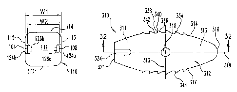
Referring now to Figs. 30-33, there is shown another embodiment implant 310.
Implant 310 includes a self distracting leading end portion,316 to facilitate
insertion in a
collapsed disc space. Implant 310 can be comprised of a single piece of
material or multiple
pieces of material as discussed above. Other examples of assembled implants
are provided in
U.S. Patent Application Serial No. 10/669,779, which is incorporated herein by
reference in
its entirety. The material comprising implant 310 can be solid, porous,
multiply drilled,
perforated, open and/or spongy, for example. Implant 310 can be fabricated
from one or
more pieces of bone material, non-bone material, or combinations thereof.
Implant 310 includes a body 312 extending along a longitudinal axis 319
between a
leading end portion 316 and a trailing end portion 311. Sidewalk 313, 315
extend along axis
319 between leading end portion 316 and trailing end portion 311. Body 312
includes an
upper surface 314 and an opposite lower surface 317. The upper and lower
surfaces 314, 317
can be provided with engagement members 334, which can be comprised of any one
or
combination of grooves, recesses, ridges, serrations, knurlings, spikes, or
roughened surfaces
for engaging the endplates of the adjacent vertebrae. Leading end portion 316
can include a
CA 02553532 2006-07-17
WO 2005/072659 PCT/US2005/001478
23
rounded or tapered configuration so that body 312 is provided with a leading
end nose that
distracts the adjacent vertebrae as it is inserted in a collapsed disc space.
Body 312 also
includes a proximal trailing end wall 321, and sidewalls 313, 315 extending
between
proximal end wall ,321 and leading end portion 316. A first notch 324 in
lateral wall 313 and
a second notch 326 in lateral wall 315 open proximally at proximal end wall
321, and extend
distally into trailing end portion 311.
Proximal end wall 321 includes a bore 328 formed therein to facilitate
engagement with
an insertion tool. Bore 328 can be a circular bore, and can be threaded
therealong for
engagement with a threaded post of an insertion instrument. Bore 328 includes
a flared
proximal end opening 330 to facilitate placement of the insertion instrument
into bore 328.
Other embodiments contemplate that bore 328 can be smooth and unthreaded.
Still further
embodiments contemplate bore 328 is non-circular. Also contemplated are
implants 310
without bore 328, or with multiple bores 328.
Implant 310 includes a cavity 332 extending between and opening at upper
surface 314
and lower surface 317. Cavity 332 is enclosed by sidewalls 313, 315, leading
end portion
316, and trailing end portion 311. Sidewalls 313, 315 include holes 318, 320,
respectively,
extending therethrough and in communication with cavity 332. Holes 318, 320
include a
circular shape, although other shapes and numbers of holes are contemplated in
sidewalls
313, 315. Cavity 332 includes an oval or racetrack shape when viewed from one
of the upper
and lower surfaces 314, 317 as shown in Fig. 32. The inner wall surfaces of
sidewalls 313,
315 extend parallel to one another, and the inner wall surfaces of leading end
portion 316 and
trailing end portion 311 are radiused and extend between the inner surfaces of
sidewalls 313,
315.
Engagement members 334, 344 extend along the sidewalk 313, 315, and project
from
respective ones of the upper and lower surfaces 314, 317. Engagement surfaces
334, 344
extend along the portions of sidewalls 313, 315 extending along cavity 332.
Upper and lower
surfaces 314, 317 include a smooth surface profile along leading end portion
316 and trailing
end portion 311. Accordingly, engagement surfaces 334, 344 are optimally
positioned along
the convexly curved upper and lower implant surfaces for engagement with the
softer bony
material near the center of the vertebral endplates to resist backout of
implant 310. Also, the
smooth surface profiles at the leading and trailing ends maximize the bearing
surface area of
CA 02553532 2006-07-17
WO 2005/072659 PCT/US2005/001478
24
implant 310 at the portions of the upper and lower surfaces 314, 317 adjacent
the harder bone
material at the cortical rim. This enhances the maintenance of distraction and
resistance to
post-operative settling of the adjacent vertebra when implant 310 is
positioned in the spinal
disc space.
In one form, engagement members 334, 344 are in the form of teeth, and include
a
sloped leading end wall 316, a generally vertical trailing end wall 338, and a
transition
surface 340 extending therebetween, as shown in Fig. 33. A groove or recess
342 extends
between adjacent ones of the teeth. The teeth project a distance 350 from the
respective
upper or lower surface 314, 317. In one form, upper and lower surface 314, 317
extend along
an arc defined by radius 350, and engagement surfaces 334, 344 extend along an
arc defined
by radius 352, with radius 352 being greater than radius 350. The adjacent
teeth are
separated by a spacing 354 measured between adjacent trailing end walls 338.
To maintain
the orthogonal orientation of the teeth spaced along the arc formed by radius
350, trailing 'end
walls 338 is oriented at an angle 356 relative to one another. Trailing end
walls 338 are
perpendicular to the arc formed by radius 350, and leading end wall 336 forms
an angle 358
with trailing end wall 338. In one specific embodiment, radius 352 is 1
millimeter greater
than radius 350, and spacing 354 is 3 millimeters. Angle 358 can range from 0
degrees to 90
degrees in one embodiment; in another embodiment angle 358 ranges from 30
degrees to 80
degrees; in a further embodiment angle 350 ranges from 50 degrees to 80
degrees; and in still
another embodiment angle 358 is 65 degrees. It should be understood however
that other
radii, spacing and angles are contemplated.
Implant 310 can be provided in a kit with a number of implants having various
heights
and/or lengths from which a surgeon can select during surgery to provide a
desired fit of the
implant in the spinal disc space. It is contemplated that each implant is
provided with a width
360. In one form, the width 360 is the same for each implant in the lcit. In a
further form,
each implant 310 includes a leading end nose that includes a distally
oriented, rounded or
radiused leading end profile to facilitate insertion in a non-distracted or
partially distracted
disc space. In still another embodiment, a number of implants 310 are provided
in a lcit with
a number of distracters that include a head corresponding in size and shape to
corresponding
ones of the implant 310. As discussed,above, the distracters can include a
head integrally
attached to a shaft or removably attached to a shaft.
CA 02553532 2006-07-17
WO 2005/072659 PCT/US2005/001478
It is further contemplated that implant 310 includes cavity 332 that provides
a
maximum surface area opening through implant 310 for bone growth through the
implant. In
one embodiment, cavity 332 includes a width that extends across more than 50
percent of the
width 260 of implant 310. In another embodiment, cavity 332 includes a width
that extends
across more than about 60 percent of width 360 of implant 310. In still
another form, cavity
332 extends along more than 50 percent of the length of implant 310 along axis
319.
Any suitable osteogenetic or osteoinductive material or composition is
contemplated for
placement within cavities of any of the implant embodiments discussed herein.
Such material
includes, for example, autograft, allograft, xenograft, demineralized bone,
synthetic and natural
10 bone graft substitutes, such as bioceramics and polymers, and
osteoinductive factors. Where
bony material is placed within the implant cavity, the material can be pre-
packed into the cavity
before the device is implanted. A separate carrier to hold the materials
within the cavities of the
implants can also be used. These carriers can include collagen-based carriers,
bioceramic
materials, such as BIOGLASS~, hydroxyapatite and calcium phosphate
compositions. The
15 Garner material can be provided in the form of a sponge, a block, folded
sheet, putty, paste, graft
material or other suitable form. Moreover, the osteogenetic compositions
contained within the
implants can comprise an effective amount of a bone morphogenetic protein,
transforming
growth factor (31, insulin-like growth factor l, platelet-derived growth
factor, fibroblast
growth factor, LIM mineralization protein (LMP), and combinations thereof or
other
20 therapeutic or infection resistant agent, held within a suitable carrier
material.
Referring to Fig. 34, there is shown another embodiment implant 410. Implant
410 is
similar to implant 310, and includes a leading end portion 416 and an opposite
trailing end
portion 411. Implant 410 includes convexly curved upper surface 414 and an
opposite
convexly curved lower surface. A first sidewall 413 and opposite parallel
sidewall extend
25 between leading end portion 416 and trailing end portion 411. The trailing
or proximal end
wall can include a bore as discussed above with respect to implant 310.
Implant 410 can also
be provided without a bore in its trailing end wall. A pair of opposite
notches (only notch
424 is shown) are provided in sidewalk 313, 315 to facilitate engagement with
an insertion
instrument as discussed above with respect to implant 310. In contrast to the
illustrated
implant 310, implant 410 includes a solid body and smooth surface profile
along its upper
and lower surfaces to maximize the bearing surface area support. In addition,
leading end
CA 02553532 2006-07-17
WO 2005/072659 PCT/US2005/001478
26
portion 416 includes a nose with a leading end surface profile rounded between
the sidewalk
along an arc defined by a radius 420. The surface profile of the nose is also
rounded between
the upper and lower surfaces of implant 410. The leading end nose surface
profiles can be
formed by multiple curved segments having differing radii, and also include
one or more
linear segments.
The rounding of leading end portion 416 medially-laterally between sidewalls
413, 415
facilitates placement of implant 410 through the tissue in the approach to the
disc space. For
example, the rounded nose eliminates any abrupt comers at the leading end of
the implant.
Neural structures and other tissue can be pushed out of the insertion path of
the implant due
to the smooth surface profile. The rounding of the leading end nose bi-
directionally, i.e.
between the sidewalls and between the upper and lower surfaces of the implant,
facilitates
placement of implant 410 in a small opening in the annulus tissue by both
distracting the
adjacent vertebrae and separating the annulus tissue to form an opening of
sufficient yet
minimized size to accommodate placement of implant 410. Still further, when
the implant is
positioned in the disc space, the leading end nose can more easily be
positioned at the cortical
rim of the endplates at the far end of the disc space. There are no abrupt
edges or transitions
which may embed or catch on the cortical rim at its transition with the
concavely curved
region of the vertebral endplates, facilitating final positioning of implant
410 in the disc
space.
Implant 410 can be provided with holes in its sidewalk, such as is shown with
holes
420, 422. The sidewall holes provide an avenue for bone ingrowth into implant
410,
enhancing its anchoring in the disc space during fusion. In the illustrated
embodiment, two
holes are provided, although more than two holes or less than two holes are
contemplated.
The holes can include a blind end in implant 410, or can extend completely
through implant
410 in communication with an opening in the opposite sidewall. Other
embodiments
contemplate that the sidewalk are provided without holes.
Referring to Fig. 35, there is shown another embodiment implant 510. Implant
510 is
similar to implant 410, and includes a leading end portion 516 and an opposite
trailing end
portion 511. Implant 510 includes convexly curved upper surface 514 and an
opposite
convexly curved lower surface. A first sidewall 526 and opposite parallel
sidewall 528
extend between leading end portion 516 and trailing end portion 511. The
trailing or
CA 02553532 2006-07-17
WO 2005/072659 PCT/US2005/001478
27
proximal end wall can include a bore and/or notches 524 to facilitate
engagement with an
insertion instrument. Implant 510 further includes a cavity 532 extending
between and
opening at the upper and lower surfaces thereof. Sidewalls 526, 528 include
holes 518, 520,
respectively, in communication with central cavity 532.
In contrast to the illustrated implant 310, implant 510 includes a smooth
surface profile
along its upper and lower surfaces, including the portions along cavity 532.
In addition,
leading end porEion 516 includes a nose rounded about a radius 512 between the
sidewalk, ,
and also rounded between the upper and lower surfaces, as discussed above
,with respect to
implant 410.
Referring to Fig. 36, there is shown an insertion instrument 550. Insertion
instrument
550 includes an elongated shaft 552 extending between a proximal portion 554
and a distal
gripping portion 556 which serves as a coupling member to couple the implant
to shaft 552.
Proximal portion 554 includes a handle 558 extending transversely to shaft
552. In one
embodiment, handle 558 is obliquely oriented to shaft 552 to facilitate
manipulation and
~ gesturing with insertion instrument 550. Shaft 552 projects proximally from
handle 558 to a
housing portion 560. Housing portion 560 includes an adjustment member 562
housed
therein. An inner shaft 564 (Fig. 37) extends distally from adjustment member
562 and
through shaft 552 to distal gripping portion 556. Adjustment member 562
provides a
thumbwheel or other suitable gripping element to facilitate the surgeon
rotating inner shaft
564 within outer shaft 552 for engagement of the distal end of inner shaft 564
with an
implant.
Distal gripping portion 556 includes body member 566 and a pair of finger 568,
570
extending distally from opposite sides of body member 566. The distal end of
inner shaft 564
projects distally from body member 566 and is centrally located between
forgers 568, 570.
As shown in Fig. 38, forgers 568, 570 are positionable in respective ones of
the notches of the
implant to which insertion instrument 550 is engaged such as implant 310 in
the illustrated
embodiment. Inner shaft 564 is engageable in the bore in the proximal end wall
of implant,
such as bore 328 of implant 310. The outer surfaces of fingers 568, 570 are
flush or recessed
relative to the outer lateral surfaces of sidewalk of the implant such that
fingers 568, 570 do
not protrude therefrom. When engaged to the implant, fingers 568, 570 define
an overall
width that is less than the width of the implant between the outer lateral
surfaces of its
CA 02553532 2006-07-17
WO 2005/072659 PCT/US2005/001478
28
sidewalk. This minimizes the insertion profile of the implant and instrument
assembly, and
facilitates a less invasive approach to the spinal disc space.
Referring to Fig. 39, there is shown another embodiment insertion instl-ument
580.
Insertion instrument 580 includes an outer shaft 582 longitudinally movable
about an inner
shaft 584. Insertion instrument 580 includes a distal gripping portion 586
which forms a
coupling member for coupling an implant to inner shaft 584. Distal gripping
portion 586
includes a body member at a distal end of inner shaft 584 that includes a base
portion 598 and
a pair of biasing members 588, 590 separated by a central slot 596. Biasing
members 588,
590 are coupled to one another about a living or integral hinge formed at base
portion 598.
Fingers 592, 594 extend distally from respective ones of the biasing members
588, 590. Base
portion 598 includes a proximally tapered outer surface profile. Outer shaft
582 is movable
distally relative to inner shaft 584 and along the outer surface profile of at
least base portion
598 to move biasing members 588, 590 and thus fingers 592, 594 toward one
another to grip
an implant therebetween. The implant can be released by proximally displacing
outer shaft
582 relative to inner shaft 584 to allow biasing members 588, 590 and thus
fingers 592, 594
to move away from one another toward their normal state.
Various mechanisms for moving outer shaft 582, 584 are contemplated. For
example,
shafts 582, 584 can be threadingly engaged to one another and outer shaft 584
is rotated
about inner shaft 582 to effect proximal and distal movement therebetween. In
another
example, proximal handle actuators are coupled to inner and outer shaft 582,
584, and the
handles effect proximal and distal linear movement between the shafts as the
handles are
manipulated. Other suitable mechanisms for moving the inner and outer shafts
longitudinally
relative to one another are also contemplated.
The present invention contemplates various procedures and instrument sets. For
example, the surgeon can determine whether a trial body or implant provides a
desired disc
space height by tactile feedback of the inserted trial body or implant, and
also by visual
inspection. The inserted trial body or implant body should sufficiently
stretch the remaining
annulus tissue to provide firm engagement between the upper and lower surfaces
of the trial
or implant body and the adjacent vertebral endplates. Sufficient surface area
contact should
be present to prevent or minimize post-operative movement of the adjacent
vertebrae relative
to the implant. By providing the trial bodies and implant bodies with
correspondingly sized
CA 02553532 2006-07-17
WO 2005/072659 PCT/US2005/001478
29
and shaped leading end portions, and by inserting the trial bodies and implant
bodies in a
non-distracted disc space, the inserted trial or implant body provides
immediate feedback to
the surgeon of the desirability of the fit. If distraction were maintained by,
for example, a
second distracter, feedback to the surgeon of the post-operative fit of the
implant would not
be reliable or available, if at all, until distraction were removed. As such,
the trial bodies and
implants can be employed without utilization of external distraction or
distraction maintained
in another disc space location during trial body and implant insertion.
However, secondary
distraction can be used to at least partially maintain disc space distraction
upon withdrawal of
the implants and trial bodies can be employed. For example, pedicle screws and
a rod can be
employed on the contralateral side to at least partially maintain distraction
obtained with a
particular implant or trial body; however, use of the same is not required.
Further, the trial bodies provide an indication of the fit of the implant into
the disc
space location. Since the implant includes a leading end portion and height
that corresponds
to that of the trial body, there is an immediate confirmation to the surgeon
that the
corresponding implant will fit into the space occupied by the trial body. If
distraction were
maintained at another location in the disc space or externally, there is no
indication that the
implant will fit properly until the implant is inserted and distraction
removed. As a result, the
implant may wedge too tightly in the disc space when distraction is removed,
making
subsequent removal of the implant difficult if an appropriate fit is not
obtained.
Alternatively, the implant may be too loose when the distraction is removed
due to over
distraction of the disc space.
The implants can be impacted or pushed into the disc space. As a result,
disruption to
the annulus tissue and tissue approaching the collapsed disc space is
minimized since the
lateral and vertical footprint of the implant in the disc space can be the
same as the lateral and
vertical footprint occupied in the implant's approach to the disc space. Also,
by providing
the implant with the same footprint as the trial body laterally and
vertically, and by
performing distraction and implant insertion through the same portal or
pathway, no
additional tissue dissection and/or retraction is required to accommodate
distraction of the
disc space during implant insertion.
The trial bodies and implants can be inserted into the disc space with minimal
disc
space preparation. According to one method, the collapsed disc space is
accessed, and an
CA 02553532 2006-07-17
WO 2005/072659 PCT/US2005/001478
opening is formed in the annulus having a width corresponding to the width of
the trial 'bodies
and/or implants. Disc material is removed through the annulus opening, and, if
desired by the
surgeon, manual roughening of the endplates is performed with a scraper or
other suitable
endplate roughening instrument. The trial bodies and/or implant bodies are
then sequentially
inserted and, if necessary, withdrawn through the annulus opening and into the
disc space.
Since the implants are self distracting, it is not necessary to chisel, drill
or otherwise form the
vertebral endplates to receive the implant, although such steps are not
precluded.
Consequently, fewer steps in the surgical procedure are necessary since
requirements for
bilateral distraction, external distraction, chiseling, drilling and reaming
are eliminated. In
10 addition, the lack of other instruments or devices in the disc space
facilitates visualization of
the disc space preparation, trial body insertion, and/or implant insertion.
Elimination of
cutting instruments in the disc space also theoretically improves the safety
of the procedure.
Minimally invasive techniques employing the trial instruments and implants are
contemplated. In any particular patient, the implants can be inserted via any
one or
15 combination of posterior, postero-lateral, antero-lateral, transforaminal,
far lateral and/or
anterior approaches. Implant insertion can occur through a single pathway to a
collapsed
spinal disc space, or through multiple pathways to the collapsed disc space,
or through
multiple pathways to multiple levels of collapsed discs of the spinal 'column.
Since the
implant, and trial instruments if employed, are inserted into the same disc
space location from
20 the same approach, the entire procedure for inserting an implant can be
completed through
one pathway. If a multiple pathway procedure is to be employed, the surgeon
can complete
implant insertion through one pathway before creating and moving to work in a
second
pathway.
Since distraction and implant insertion occur along the same pathway to the
collapsed
25 disc space, the implants and trial instruments are suited for use in
minimally invasive
procedures which employ a retractor sleeve to provide a pathway to the
collapsed disc space.
Such retractor sleeves can employ any one or combination of an endoscopic
viewing element
in the working channel, a microscopic viewing system over the proximal end of
the retractor
sleeve, fluoroscopic viewing, loupes, naked eye and/or image guidance.
30 The trial bodies of the trial instruments and the implant bodies can be
made from any
biocompatible material, including synthetic or natural autograft, allograft or
xenograft tissues,
CA 02553532 2006-07-17
WO 2005/072659 PCT/US2005/001478
31
and can be resorbable or non-resorbable in nature. Examples of tissue
materials include hard
tissues, connective tissues, demineralized bone matrix and combinations
thereof. Further
examples of resorbable materials are polylactide, polyglycolide, tyrosine-
derived
polycarbonate, polyanhydride, polyorthoester, polyphosphazene, calcium
phosphate,
hydroxyapatite, bioactive glass, and combinations thereof. Further examples of
non-
resorbable materials are non-reinforced polymers, carbon-reinforced polymer
composites,
PEEK and PEEK composites, shape-memory alloys, titanium, titanium alloys,
cobalt chrome
alloys, stainless steel, ceramics and combinations thereof and others as well.
If the trial body
or implant is made from radiolucent material, radiographic markers can be
located on the trial
body or implant to provide the ability to monitor and determine
radiographically or
fluoroscopically the location of the body in the spinal disc space. The
material comprising
the trial bodies can be solid, porous, spongy, perforated, drilled, and/or
open.
There is contemplated an implant for insertion into a spinal disc space
between adjacent
vertebrae. The implant can be impacted or pushed into the disc space. The
implant can be
provided with a distal end or leading insertion end that is sized for
insertion into the collapsed
disc space. As the implant is inserted, the implant can restore the collapsed
disc space to a
desired disc space height. The desired disc space height corresponds to the
height of the
implant proximal the distal end. Once inserted, the implant can maintain the
disc space at the
desired disc space height.
There is further contemplated an implant that, when inserted, restores and
maintains a desired disc space height of a collapsed disc space between an
upper vertebra
and a lower vertebra. The implant includes a body with a distal end, a
proximal end, an
upper surface orientable toward an endplate of the upper vertebra and a lower
surface
orientable toward an endplate of the lower vertebra. The body of the implant
has a first
height between the upper and lower surfaces corresponding to the desired disc
space
height. The body of the implant also has a second height at its distal end
that is less than a
height of the collapsed disc space.
It is contemplated that the implants can be provided with bi-convex curvature
of
the upper and lower surfaces, allowing the implants to center in the endplates
of the disc
space. It is further contemplated that the upper and lower surfaces of the
implant can be
planar or include compound geometry. The upper and lower surfaces of the
implant can
CA 02553532 2006-07-17
WO 2005/072659 PCT/US2005/001478
32
also be configured to establish lordotic or kyphotic angulation between the
adjacent
vertebral bodies.
Also contemplated is a set of implants having two or more implants of
increasing
height. The height of each implant corresponds to a restored disc space
height. The leading
insertion end of each implant is sized for insertion into a collapsed disc
space. As each
implant is inserted, the implant restores the collapsed disc space to the
restored disc space
height provided by the inserted implant. If the restored disc space height
does not correspond
to the desired disc space height, the inserted implant is withdrawn and a
larger height implant
is inserted. Sequential insertion and withdrawal of increasing height implants
is continued
until the restored disc space height provided by an implant of the set of
implants corresponds
to the desired disc space height. The implant providing the desired disc space
height is
positioned in the disc space to restore and poshoperatively maintain the
desired disc space
height.
There is further contemplated an instrument set having two or more self
distracting trial
instruments and at least one implant. The two or more trial instruments each
have a body
with a leading insertion end sized for insertion into a collapsed disc space.
The leading
insertion ends of each trial body are substantially the same in size and
shape. Each trial body
has a height proximal the leading insertion end that restores the collapsed
disc space height to
a height different than that of the other trial bodies. The at least one
implant has a leading
insertion end that is substantially the same in size and shape as the leading
insertion end of at
least one of the trial bodies of the trial instruments. The implant has a
height proximal its
leading insertion end that corresponds to the desired restored disc space
height provided by
the at least one trial body.
Also contemplated is a kit including a set of trial instruments, each having a
trial body
at a distal end thereof. The trial bodies have a self distracting leading end
portion insertable
in a collapsed spinal disc space. The kit further includes a set of implants
positionable in the
collapsed spinal disc space. Each implant has a body sized and shaped to
correspond in size
and shape to a respective trial body of the trial instruments. The fit of each
implant body in
the spinal disc space is indicated to the surgeon by the fit of the
corresponding trial body of
the trial instruments. When a trial body provides a desired fit, the trial
body is removed and
CA 02553532 2006-07-17
WO 2005/072659 PCT/US2005/001478
33
the implant corresponding to the trial body is inserted into the collapsed
disc space in the
location previously occupied by the withdrawn trial body.
It is contemplated that an insertion instrument can be engaged to lateral
walls of an
intervertebral implant. The insertion instrument includes a distal coupling
portion
positionable in notches formed in corresponding ones of the lateral walls of
the implant. The
coupling portion has a first position engaging the implant in the notches and
a second
position disengaged from the implant in the notches. The width of the coupling
portion in
each of its first and second positions is less than the width of the implant
between the lateral
walls of the implant.
Methods for inserting an intervertebral implant into a collapsed spinal disc
space are
also contemplated. A number of implants are sequentially inserted into the
collapsed disc
space to restore the disc space. If a particular implant does not restore the
disc space to a
desired disc space height, the implant is withdrawn from the disc space. When
an inserted
implant is withdrawn, the disc space is non-distracted and allowed to
collapse. The implant
providing the desired disc space height remains in the disc space to post-
operatively maintain
the desired disc space height.
A method is contemplated for inserting an intervertebral implant that includes
accessing a collapsed spinal disc space from an uni-portal approach. A first
implant is
inserted through the portal into the disc space to restore the disc space
height. If the restored
disc space height does not correspond to a desired disc space height, the
inserted implant is
removed from the disc space and portal, and the disc space is allowed to
collapse. A second
implant of different height is inserted into the undistracted, collapsed disc
space to provide
another restored disc space height. When an inserted implant provides a
restored disc space
height that corresponds to a desired disc space height, the inserted implant
remains in the disc
space to post-operatively maintain the desired disc space height.
Also contemplated is a method for inserting an intervertebral implant is
provided that
includes accessing a collapsed spinal disc space. A number of trial bodies are
provided with
leading end portions sized for insertion into a non-distracted disc space. The
trial bodies are
sequentially inserted into and removed from the disc space. The trial body
providing the
desired disc space height is used to select an implant having a height and a
self distracting
leading end portion corresponding to the height and leading end portion of the
last inserted
CA 02553532 2006-07-17
WO 2005/072659 PCT/US2005/001478
34
trial body. The implant is then inserted into the non-distracted disc space to
restore the disc
space and post-operatively maintain the desixed disc space height.
While the invention has been illustrated and described in detail in the
drawings and
foregoing description, the same is to be considered as illustrative and not
restrictive in
character, and that all changes and modifications that come within the spirit
of the invention
are desired to be protected.