Note: Descriptions are shown in the official language in which they were submitted.
CA 02554077 2006-07-19
WO 2005/077274 PCT/US2005/003621
1
METHOD AND APPARATUS FOR
MEASURING AN ANALYTE IN A BODY FLUID
FIELD OF THE INVENTION
The present invention relates generally to testing systems for determining the
concentration of an analyte in a fluid sample, and more particularly, to a
system for
lancing a test subject's skin, harvesting a body fluid sample, and determining
the
concentration of an analyte in the body fluid sample.
BACKGROUND OF THE INVENTION
It is often necessary to quiclcly obtain a sample of blood and perform an
analysis of the blood sample. One example of a need for obtaining a sample of
blood
is in connection with a blood glucose monitoring system, which a user must
frequently use to monitor the user's blood glucose level.
One method of obtaining a blood sample and analyzing the sample for
determining the glucose level is with a lancing device and a separate blood
collection
device. In obtaining a blood sample, a drop of blood is obtained from the
fingertip
using the lancing device, and the blood is harvested using a test strip, which
is then
analyzed by a test unit to determine the glucose concentration in the blood,
often
using an electrochemical- or colorimetric-based analysis. Test strips are also
used for
determining the concentration or presence of various other analytes (e.g.,
fructosamine, hemoglobin, cholesterol, glucose, alcohol, drugs including
illegal drugs,
etc. ) in a variety of body fluids (e.g., blood, interstitial fluid, saliva,
urine, etc.).
A drawback associated with using physically separate lancing and collection
devices is that a patient/user must manipulate two different instnunents
requiring the
user/patient to bring the collection device (e.g., the test strip) to the area
of skin that
has been lanced to collect the sample. Because the user must align the
collection
device with the sample to be collected, a larger than necessary sample amount
is often
produced and collected to ensure an accurate analysis. In other situations,
not enough
sample is collected for accurate analysis because the collection device is not
properly
positioned. This problem can be further compounded if the user has impaired
vision
or poor dexterity. Because test systems are requiring smaller volumes of blood
for
CA 02554077 2006-07-19
WO 2005/077274 PCT/US2005/003621
2
analysis, it becomes more difficult to position a collection instrlunent for
proper
collection. Further impacting the self testing process is that some users are
adverse to
the pain associated with repeated lancings.
SUMMARY OF THE INVENTION
An apparatus and method for analyzing an analyte in a body fluid sample
using a lancing device having a hollow lancet are disclosed. According to one
embodiment of the present invention, the method comprises the acts of lancing
the
shin of a test subject with the hollow lancet having an interior of the hollow
lancet
that forms a capillary channel, collecting a body fluid sample from the lanced
skin in
the capillary channel of the hollow lancet, and analyzing the body fluid
sample for
determining the analyte concentration in the body fluid sample while the
collected
body fluid sample remains in the lancet.
The above summary of the present invention is not intended to represent each
embodiment, or every aspect, of the present invention. Additional features and
benefits of the present invention will become apparent from the detailed
description,
figures, and claims set forth below.
BRIEF DESCRIPTION OF THE FIGURES
FIG. 1 is a side view of a lancing device according to one embodiment of the
present invention.
FIG. 2 is an enlarged cross-sectional view of the forward end of the lancing
device of FIG. 1.
FIG. 3 is an enlarged cross-sectional view of the forward end of the lancing
device of FIG. 1 shown while lancing a test subject's skin.
FIG. 4 is an enlarged cross-sectional view of the forward end of the lancing
device of FIG. 1 shown while harvesting a body fluid sample.
FIG. 5 is a side view of a lancing device according to an another embodiment
of the present invention.
FIG. 6 is a side view of a vacuum-assisted lancing device according to another
embodiment of the present invention.
CA 02554077 2006-07-19
WO 2005/077274 PCT/US2005/003621
3
While the invention is susceptible to various modifications and alternative
forms, specific embodiments are shown by way of example in the drawings and
are
described in detail herein. It shoed be understood, however, that the
invention is not
intended to be limited to the particular forms disclosed. Rather, the
invention is to
cover all modifications, equivalents, and alternatives falling within the
spirit and
scope of the invention as defined by the appended claims.
DETAILED DESCRIPTION OF THE ILLUSTRATED EMBODIMENTS
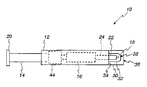
Turning now to the drawings and initially to FIGS. 1 and 2, a lancing device
10 according to one embodiment of the present invention is shown. In the
illustrated
embodiment of the present invention, the lancing device 10 is vacuum as listed
as is
described in detail below and as is known in the art. The device 10 includes a
body
12 that houses a plunger 14 and a lancing mechanism 16 for driving a lancet
18. A
top end 20 of the plunger 14 extends beyond the body 12. In using the lancet
18 to
punctl~re a test subject's skin, a user grasps the device 10 by the body 12
and
depresses the top end 20 of the plunger 14-moving the plunger 14 into the body
12
of the device 10-to downwardly advance the lancet 18 into a test subject's
skin. The
lancet 18, one end of which is embedded in a base 22, is removably attached to
a
lancet holder 24, which is coupled to the plunger 14 through the lancing
mechanism
16 within the body 12.
An end cap including an outer end cap 30 and an inner-locating end cap 32 are
removably attached to a forward end 34 of the device 10 opposite the plunger
14. The
inner- locating end cap 32 is located within the outer end cap 30. Generally,
as is
described below, the outer end cap 30 contacts a test subject's skin, and the
test
subject's skin is pulled against the inner end cap 32 during the ensuing
lancing
operation for puncturing the test subject's skin and collecting the sample
produced at
the lance site. Both the outer end cap 30 and the inner end cap 32 have open
ends 36,
38 though which the lancet 18 passes to puncture a test subject's skin during
the
lancing operation. The end caps are removably attached to the lancing device
10 so
that a used lancet can be replaced with a new lancet after a lancing
procedure.
Further, the end caps, which may come into contact with a sample during
testing, may
also be disposable in some embodiments of the present invention. According to
one
CA 02554077 2006-07-19
WO 2005/077274 PCT/US2005/003621
4
embodiment of the present invention the outer and inner end caps 30, 32 are
integrally
formed such that detaching the outer end cap 30 from the forward end 34 of the
device 10 also removes the Timer end cap 32.
The lancet 18 is constructed of a substantially optically clear material and
includes a micro-capillary channel according to one embodiment of the present
invention. The lancet 18 has a hollow interior, which forms the micro-
capillary
channel. The micro-capillary channel includes a reagent or enzymatic indicator
system disposed along its inner walls. In operation, as is described in detail
below,
the lancet 18 is used to both puncture a test subject's skin and then to
harvest the body
fluid sample produced at the puncture site. The analyte of interest (e.g.,
glucose) in
the collected body fluid sample (e.g., blood) reacts with the reagent disposed
within
the lancet 18 to produce a colorimetric reaction indicative of the
concentration of the
analyte in the sample. This reaction is then measured by an optical readhead
such as a
light detector. The lancet 18 is used for puncturing the test subject's skin,
harvesting
a sample produced at the punctured area of the test subject's skin, and for
providing
an area within the lancet 18 that the harvested sample reacts with the
reagent. Finally,
an optical transmission measurement is used to read the colorimetric reaction
within
the capillary channel of the lancet 18, and an analysis of the transmitted
light is
performed for determining the analyte concentration.
According to one embodiment of the present invention, the lancet 18 is a
microcapillary tube constructed of fused silica and has a polygonal cross
section (e.g.,
rectangular, square, hexagonal, etc.) In other embodiments of the present
invention,
the lancet 18 is constructed of another substantially optically clear material
such as,
for example, pyrex, quartz, acrylic, polycarbonate, or polyester. The
puncturing end
or tip 40 of the microcapillary tube lancet 18 is cleaved as shown in FIG. 2
at an acute
angle with respect to the longitudinal axis of the lancet 18 to form a sharp
point. The
sharp-puncturing end 40 of the lancet 18 cleanly punctures the test subject's
skin to
produce a consistently sized sample on the test subject's slcin.
According to one embodiment of the present invention, the lancet 18 has a
square cross section having an outer dimension of about 300 microns, which is
smaller than a 360 micron diameter of a typical 28-gauge steel lancet,
resulting in a
small puncture site on a test subject's stein. A smaller laceration is
desirable because
CA 02554077 2006-07-19
WO 2005/077274 PCT/US2005/003621
it translates to less pain for the test subject. The fused silica
microcapillary tubing f'or
use in constructing the lancet 18 is commercially available having interior
channel
widths of about 50, 75, or 100 microns, with corresponding volumes of about
13, 29,
and 50 nanoliters ("nl"), respectively, for a lancet 18 having a length of
about 5 mm,
5 which can used in alternative embodiments of the present invention. The
fused silica
microcapillary tubing for use in constructing the lancet 18 according to one
embodiment of the present invention is commercially available from Polymicro
Technologies, LLC of Phoenix, Arizona.
The flat surfaces of the lancet 18 provide a substantially optically clear
window for transmitting light through the sample. As is described below,
transmission spectroscopy may be used to analyze the sample. The absorbance of
the
sample reacted with the analyte in the lancet 18 is used to determine analyte
concentration. The transmission of light through fused silica, for example, is
spectrally flat from the ultra-violet region (e.g., wavelengths ranging from
about 3 50
run to about 2000 mn) into the infrared region. The square fused
microcapillary
lancet 18 reduces the path length error associated with transmission
spectroscopy
measurements. For example, the path length error is limited to one tolerance.
inside
the square fused silica microcapillary lancet 18. As an example, a fused
silica
microcapillary tube with a path length of 100 microns has a path length
tolerance of
~5 qm, which reduces errors occurring in the analyte concentration analysis.
Another advantage of the lancet 18 having a square cross section is that
square
shape provides a two-fold increase in transverse optical interaction path
length when
compared to round capillaries. Thus, the square lancet 18 can be smaller than
round
capillaries used in a optical transmission environment, resulting in a smaller
sample
(e.g., as low as about 8 r~l) for filing the square lancet 18 and a smaller
puncture on a
test subject's skin.
Referring to FIGS. 1-3, during the lancing of the test subject's skin S, the
open
end 36 of the outer end cap 30 is placed on an area of the test subject's skin
(e.g_ , a
forearm or forger). The plunger 14 is depressed to advance the lancet 18 from
a
retracted position (FIG. 2), wherein the lancet 18 is completely contained
within rthe
end caps 30, 32, to a lancing position (FIG. 3), wherein the lancet 18 extends
through
the open ends 36, 28 of the end caps 30, 32 and, into the test subject's skin
S.
CA 02554077 2006-07-19
WO 2005/077274 PCT/US2005/003621
6
Movement of the ph~nger 14 by the user triggers a drive spring within the
lancing
mechanism 16 that advances the lancet 18 into a test subject's skin S. A
rebound
spring within the lancing mechanism 16 then retracts the tip 40 of the lancet
18 from
the test subject's skin S.
According to one embodiment of the present invention, the lancing device 10
is vacuum-assisted to facilitate the production of a blood sample at the
puncture site
on the test subject's skin. In such an embodiment, the outer end cap 30 forms
a
substantially airtight seal with the forward end 34 of the device 10. The
placement of
the open end 36 of the outer end cap 30 against a test subject's skin S, aided
by
pressing against the skin, forms the substantially airtight seal. The laalcing
device 10
includes a vaculun member 44 such as a diapluagm or bellows that displaces air
within the lancing device 10 and the end cap 30. Release of the plunger 14. by
the
user triggers the vacuum member 30, which evacuates air from the inner and
outer
end caps 14, 18.
When the vacuum member 44 is activated, the test subject's skin S is drawn
inside the outer end cap 14 to the inner-locating end cap 32 as is depicted in
FIG. 3.
As the created vacuum pulls the test subject's skin S into the device 10, the
test
subject's skin S bulges around the locating end cap 32. The test subject's
skin S is
stretched flat across the open end 38 of the inner end cap 32. This stretched,
flat skin
facilitates sample formation and collection. The vacuum holds the skin and
puncture
sight in a fixed position while the sample harvesting occurs.
Referring now to FIG. 4, after the lancet 18 punctures the test subject's skin
S,
a body fluid sample B (e.~., blood) forms on the skin S at the puncture site.
As
discussed above, the lancet 18 is hollow for harvesting the body fluid sample
produced at the lance site. The lancing mechanism 16 holds the skin under
vacuum
and positions the hollow tip 40 of the lancet 18 in a collection position
adjacent the
lance site for collecting the produced body fluid sample B. The sample B
contacts the
hollow lancet 18 and the sample moves into the lancet 18 via capillary action.
If the
tip 40 of the microcapillary lancet 18 rests too far from the skin S, the
sample B will
not be drawn into the microcapillary channel. And if the tip 40 of the
microcapillary
lancet 18 rests on or below the puncture site, it may cause discomfort to the
user, and
a sample may not be drawn into the tip 40 of the lancet 18.
CA 02554077 2006-07-19
WO 2005/077274 PCT/US2005/003621
7
A reagent or enzymatic indicator system is disposed within the lancet 18 far
reacting with the analyte of interest in the harvested sample for producing a
colorimetric reaction indicative of the analyte concentration in the body
fluid sample.
The colorimetric reaction is read by optical instrmnents as it described below
in
connection with FIG. 5. Colorimetric testing is described in detail in U.S.
Patents
Nos. 6,181,417 B1 (entitled "Photometric Readhead with Light Shaping Plate");
5,518,689 (entitled "Diffuse Light Reflectance Readhead"); and 5,611,999
(entitled
"Diffuse Light Reflectance Readhead"); each of which is incorporated herein by
reference in its entirety.
Referring now to FIG. 5, the lancing mechanism 16 retracts the lancet 18 away
from the slcin S (i.e., into the lancing device 10) after the sample B is
collected from
the lance site on the skin S for analyzing the blood according to one
embodiment of
the present invention. Alternatively, the lancing device 10 may maintain the
lancet 18
in the collection position for analyzing the analyte concentration in the
blood sample.
The lancing device 10 includes an illumination unit 60, which may include a
light
source such as an LED, illumination optics for directing and collimating
light, or both.
Alternatively, the illumination unit 60 may comprise the output end of a fiber
optic
cable that pipes in light from a light source.
The colorimetric reaction within the substantially optically clear lancet 18
between the reagent and the analyte of interest in the harvested body fluid
sample is
measured using transmission spectroscopy. The illumination unit 60 outputs a
monochromatic collimated beam of light 62 onto the microcapillary lancet 18.
Light
transmitted through the microcapillary lancet 18-referred to with reference
number
64-is detected by a light detector 66 that outputs a signal indicative of the
received
light. The detected transmitted light is then compared to a reference sample
(e.g.,
light from the source directly detected by the detector without the sample or
lancet 18
present). The difference in light absorption between the two is used to
determine the
analyte concentration in the blood sample. The results of the analysis axe
communicated to the user via a user interface including a display (not shown)
of the
lancing device 10.
According to an alternative embodiment of the present invention, the amount
of light transmitted through the sample is used to determine the time at which
to begin
CA 02554077 2006-07-19
WO 2005/077274 PCT/US2005/003621
8
analyzing the reaction between the reagent and the analyte of interest. For
example,
the detector 66 may constantly detect light transmitted through the lancet 18
upon
retracting the lancet 18 to analyze the sample. Once the detector 66 detects
that the
light transmitted through the lancet 18 is consistent with a sample being
contained
within the lancet 18, the processor waits a predetermined about of time after
the
expiration of which the transmitted light detected by the detector 66 is used
by the
processor to determine the analyte concentration in the fluid sample. B ecause
the
colorimetric reaction requires a predetermined about of time to develop, only
transmitted light detected after the expiration of the predetermined time are
used in
the analysis. Waiting for the reaction to develop guards against an inaccurate
analysis
according to one embodiment of the present invention.
Referring now to FIG. 6, a vacuum-assisted lancing device 100 is shown,
which may be adapted for use as the lancing device 10 according to an
alternative
embodiment of the present invention. A vacuum member, such as a diaphragm 138,
within the lancing device 100 is activated when the plunger 112 is depressed
by the
user and travels toward the open end of the lancing device 100. As the plunger
112 is
depressed, a rebound spring 132 captured between a return 134 and a release
136 is
expanded and extended. This action displaces the rolling diaphragm 138 toward
the
end cap 114. A central portion of the rolling diaphragm 138 is secured to the
stem of
the phmger 112 and a piston 140 such that the central portion moves with the
plunger
112. The interfaces between the rolling diaphragm 138 and the stem of the
plunger
112 and a housing 124 of the device 100 are air tight. The displacement of the
rolling
diaphragm 138 displaces air in the housing 124 creating a vacuum. Further
details of
the vacuum-assisted lancing device 100 illustrated in FIG. 4, which may be
used in
connection with alternative embodiments of the present invention, are
described in
U.S. Patent No. 6,152,942, entitled "Vacuum Assisted Lancing Device," which is
incorporated herein by reference in its entirety.
While the invention is susceptible to various modifications and alternative
forms, specific embodiments thereof have been shown by way of example in the
drawings and herein described in detail. It should be understood, however,
that it is
not intended to limit the invention to the particular forms disclosed, but on
the
CA 02554077 2006-07-19
WO 2005/077274 PCT/US2005/003621
9
contrary, the intention is to cover all modifications, equivalents, and
alternatives
falling within the spirit and scope of the invention as defined by the
appended claims.