Note: Descriptions are shown in the official language in which they were submitted.
DEMANDES OU BREVETS VOLUMINEUX
LA PRESENTE PARTIE I)E CETTE DEMANDE OU CE BREVETS
COMPRI~:ND PLUS D'UN TOME.
CECI EST ~.E TOME 1 DE 2
NOTE: Pour les tomes additionels, veillez contacter le Bureau Canadien des
Brevets.
JUMBO APPLICATIONS / PATENTS
THIS SECTION OF THE APPLICATION / PATENT CONTAINS MORE
THAN ONE VOLUME.
THIS IS VOLUME 1 OF 2
NOTE: For additional vohxmes please contact the Canadian Patent Oi~ice.
CA 02554431 2006-07-26
WO 2005/074483 PCT/US2005/001474
Title: Genetic Markers for Skatole Metabolism
This application claims benefit from United States application 10/206,118
filed July
29 2402, which is a divisional of United States application 09/672,039, filed
September
29,2000 now patent 6,448,028 which is a continuation of United States
provisional
application No. 60/156,935, filed September 30, 1999 all of which are
incorporated herein
by reference.
FIELD OF THE INVENTION
This invention relates generally to the detection of genetic differences among
animals. More particularly, the invention relates to polymorphisms that affect
enzyme
efficiency and are indicative of heritable phenotypes associated with boar
taint in porcine.
Methods and compositions for use of these genetic differences in genotyping of
animals
and selection are also disclosed as well as novel sequences.
BACKGROUND OF THE INVENTION
Male pigs that are raised for meat production are usually castrated shortly
after birth
to prevent the development of off odors and off flavors (boar taint) in the
carcass. Boar
taint is primarily due to high levels of either the 16-androstene steroids
(especially S.alpha.
(-androst-16-en-3-one)) or skatole in the fat. S~atole is produced by bacteria
in the hindgut
which degrade tryptophan that is available,from undigested feed or from the
turnover of
cells lining the gut of the pig (Jensen and Jensen, 1995). Skatole is absorbed
from the gut
and metabolized primarily in the liver (Jensen and Jensen, 1995). High levels
of skatole can
accumulate in the fat, particularly in male pig, and the presence of a
recessive gene
Ska<sup>l</sup>, which results in decreased metabolism and clearance of skatole has
been
proposed (Lundstrom et al., 1994; Friis, 1995). Skatole metabolism has been
studied
extensively in ruminants (Smith, et al., 1993), where it can be produced in
large amounts
by ruminal bacteria and results in toxic effects on the lungs (reviewed in
Yost, 1989). The
metabolic pathways involving skatole have not been well described in pigs. In
particular,
the reasons why only some intact male pigs have high concentrations of skatole
in the fat
are not clear. Environmental and dietary factors are important (Kjeldsen,
1993; Hansen et
al., 1995) but do not sufficiently explain the reasons for the variation in
fat skatole
concentrations in pigs. Claus et al. (1994) proposed high fat skatole
concentrations are a
result of an increased intestinal skatole production due to the action of
androgens and
CA 02554431 2006-07-26
WO 2005/074483 PCT/US2005/001474
glucocorticoids. Lundstrom et al. (1994) reported a genetic influence on the
concentrations
of skatole in the fat, which may be due to the genetic control of the
enzymatic clearance of
skatole. The liver is the primary site of metabolism of skatole and liver
enzymatic activities
could be the controlling factor of skatole deposition in the fat. Baebuttedk
et al. (1995)
described several liver metabolites of skatole found in blood and urine with
the major
being MII and MIII. MII, which is a sulfate conjugate of 6-hydroxyskatole (pro-
MII), was
only found in high concentrations in plasma of pigs which were able to rapidly
clear
skatole from the body, whereas high MIII concentrations were related to slow
clearance of
skatole. Thus the capability of synthesis of MII could be a major step in a
rapid metabolic
clearance of skatole resulting in low concentrations of skatole in fat and
consequently low
levels of boar taint.
In view of the foregoing, further work is needed to fully understand the
metabolism
of skatole in pig liver and to identify the key enzymes involved.
Understanding the
biochemical events involved in skatole metabolism can lead to novel strategies
for treating,
reducing or preventing boar taint. In addition, polyrnorphisms in these
candidate genes may
be useful as possible markers for low boar taint pigs.
SUMMARY OF THE INVENTION
This invention relates to the discovery of genetic variation associated with
quantitative trait loci or linkage equilibrium analysis that may be used to
predict phenotypic
traits in animals. According) to the invention, major affect genes have been
identified
which are related to phenotypic variation in animals. According to the
invention,
phenotypic variation in skatole metabolism and concomitant boax taint are
correlated to
major effect alleles linked to variation in sulfotransferase genes. To the
extent that this
family of genes are conserved among species and animals, and it is expected
that the
different alleles disclosed herein will also correlate with variability in
these genes) in other
economic or meat-producing animals such as cattle, sheep, chicken, etc with
concomitant
effects on sulfotransferase activity related to other traits in lieu of or in
addition to boar
taint.
To achieve the objects and in accordance with the purpose of the invention, as
embodied and broadly described herein, the present invention provides the
discovery of
alternate genotypes which provide a method for genetically typing animals and
screening
animals to determine those with favorable allelic forms of genes resulting in
skatole
enzymes with increased or decreased activity and concomitant effects on
reduced boar taint
or to select against animals which have alleles indicating less favorable
characteristics. As
2
CA 02554431 2006-07-26
WO 2005/074483 PCT/US2005/001474
used herein a "favorable" or "desired" or "improved" with respect to a trait
means a
significant improvement (increase or decrease) in one of any measurable
indicia of boar
taint or other sulfotransferase-related phenotype above the mean of a given
group, species
line or population, so that this information can be used in breeding to
achieve a uniform
population which is optimized for these traits. This may include an increase
in some traits
or a decrease in others depending on the desired characteristics. Traits may
also be
observed at the molecular level by assaying for activity of enzymes involved
in skatole
metabolism.
Methods for assaying for these traits generally comprises the steps 1)
obtaining a
biological sample from a animal; and 2) analyzing the genomic DNA or protein
obtained in
1) to determine which alleles) is/are present. Haplotype data which allows for
a series of
linked polymorphisms to be combined in a selection or identification protocol
to maximize
the benefits of each of these markers may also be used.
Since several of the polymorphisms may involve changes in amino acid
composition of the respective protein or will be indicative of the presence of
this change,
assay methods may even involve ascertaining the amino acid composition of the
protein of
the major effect genes of the invention. Methods for this type or purification
and analysis
typically involve isolation of the protein through means including
fluorescence tagging
with antibodies, separation and purification of the protein (i.e. through
reverse phase HPLC
system), and use of an automated protein sequencer to identify the amino acid
sequence
present. Protocols for this assay are standard and known in the art and are
disclosed in
Ausubel et. al.(eds.), Short Protocols in Molecular Biology Fourth ed. John
Wiley and Sons
1999.
In another embodiment, the invention comprises a method for identifying
genetic
markers for boar taint. Once a major effect gene has been identified, it is
expected that
other variation present in the same gene, allele or in related family of gene
sequences in
useful linkage disequilibrium therewith may be used to identify similar
effects on these
traits. The identification of other such genetic variation, once a major
effect gene has been
discovered, represents more than routine screening and optimization of
parameters well
known to those of skill in the art and is intended to be within the scope of
this invention.
The following terms are used to describe the sequence relationships between
two or
more nucleic acids or polynucleotides: (a) "reference sequence", (b)
"comparison window",
(c) "sequence identity", (d) "percentage of sequence identity", and (e)
"substantial identity".
(a) As used herein, "reference sequence" is a defined sequence used as a basis
for
sequence comparison. In this case the Reference sequences. A reference
sequence may be
a subset or the entirety of a specified sequence; for example, as a segment of
a full-length
cDNA or gene sequence, or the complete cDNA or gene sequence.
3
CA 02554431 2006-07-26
WO 2005/074483 PCT/US2005/001474
(b) As used herein, "comparison window" includes reference to a contiguous and
specified segment of a polynucleotide sequence, wherein the polynucleotide
sequence may
be compared to a reference sequence and wherein the portion of the
polynucleotide
sequence in the comparison window may comprise additions or deletions (i.e.,
gaps)
compared to the reference sequence (which does not comprise additions or
deletions) for
optimal alignment of the two sequences. Generally, the comparison window is at
least 20
contiguous nucleotides in length, and optionally can be 30, 40, 50, 100, or
longer. Those of
skill in the art understand that to avoid a high similarity to a reference
sequence due to
inclusion of gaps in the polynucleotide sequence, a gap penalty is typically
introduced and
is subtracted from the number of matches.
Methods of alignment of sequences for comparison are well-known in the art.
Optimal alignment of sequences for comparison may be conducted by the local
homology
algorithm of Smith and Waterman, Adv. Appl. Math. 2:482 (1981); by the
homology
aligmnent algorithm of Needleman and Wunsch, J. Mol. Biol. 48:443 (1970); by
the search
for similarity method of Pearson and Lipman, PYOG. Natl. Acad. Sci. 85:2444
(1988); by
computerized implementations of these algorithms, including, but not limited
to:
CLUSTAL in the PC/Gene program by Intelligenetics, Mountain View, California;
GAP,
BESTFIT, BLAST, FASTA, and TFASTA in the Wisconsin Genetics Software Package,
Genetics Computer Group (GCG), 575 Science Dr., Madison, Wisconsin, USA; the
CLUSTAL program is well described by Higgins and Sharp, Gene 73:237-244
(1988);
Higgins and Sharp, CABIOS 5:151-153 (1989); Corpet, et al., Nucleic Acids
Research
16:10881-90 (1988); Huang, et al., Computes' Applications iu the Biosciertces
8:155-65
(1992), and Pearson, et al., Methods ih Molecular Biology 24:307-331 (1994).
The
BLAST family of programs which can be used for database similarity searches
includes:
BLASTN for nucleotide query sequences against nucleotide database sequences;
BLASTX
for nucleotide query sequences against protein database sequences; BLASTP for
protein
query sequences against protein database sequences; TBLASTN for protein query
sequences against nucleotide database sequences; and TBLASTX for nucleotide
query
sequences against nucleotide database sequences. See, Cu~~eut Protocols in
Molecula~~
Biology, Chapter 19, Ausubel, et al., Eds., Greene Publishing and Wiley-
Interscience, New
York (1995).
Unless otherwise stated, sequence identity/similarity values provided herein
refer to
the value obtained using the BLAST 2.0 suite of programs using default
parameters.
Altschul et a., Nucleic Acids Res. 25:3389-3402 (1997). Software for
performing BLAST
analyses is publicly available, e.g., through the National Center for
Biotechnology-
Information (http://www.hcbi.nlm.nih.~ov/).
4
CA 02554431 2006-07-26
WO 2005/074483 PCT/US2005/001474
This algorithm involves first identifying high scoring sequence pairs (HSPs)
by
identifying short words of length W in the query sequence, which either match
or satisfy
some positive-valued threshold score T when aligned with a word of the same
length in a
database sequence. T is referred to as the neighborhood word score threshold
(Altschul et
al., supra). These initial neighborhood word hits act as seeds for initiating
searches to find
longer HSPs containing them. The word hits are then extended in both
directions along
each sequence for as far as the cumulative alignment score can be increased.
Cumulative
scores are calculated using, for nucleotide sequences, the parameters M
(reward score for a
pair of matching residues; always > 0) and N (penalty score for mismatching
residues;
always < 0). For amino acid sequences, a scoring matrix is used to calculate
the
cumulative score. Extension of the word hits in each direction are halted
when: the
cumulative alignment score falls off by the quantity X from its maximum
achieved value;
the cumulative score goes to zero or below, due to the accumulation of one or
more
negative-scoring residue alignments; or the end of either sequence is reached.
The BLAST
algorithm parameters W, T, and X determine the sensitivity and speed of the
alignment.
The BLASTN program (for nucleotide sequences) uses as defaults a wordlength
(W) of 11,
an expectation (E) of 10, a cutoff of 100, M=5, N=-4, and a comparison of both
strands.
For amino acid sequences, the BLASTP program uses as defaults a wordlength (W)
of 3,
an expectation (E) of 10, and the BLOSUM62 scoring matrix (see Henikoff &
Henikoff
(1989) Proc. Natl. Acad. Sci. USA 89:10915).
In addition to calculating percent sequence identity, the BLAST algorithm also
performs a statistical analysis of the similarity between two sequences (see,
e.g., Marlin &
Altschul, Proc. Natl. Acad. Sci. USA 90:5873-5787 (1993)). One measure of
similarity
provided by the BLAST algorithm is the smallest sum probability (P(N)), which
provides
an indication of the probability by which a match between two nucleotide or
amino acid
sequences would occur by chance.
BLAST searches assume that proteins can be modeled as random sequences.
However, many real proteins comprise regions of nonrandom sequences which may
be
homopolyrneric tracts, short-period repeats, or regions enriched in one or
more amino
acids. Such low-complexity regions may be aligned between unrelated proteins
even
though other regions of the protein are entirely dissimilar. A number of low-
complexity
filter programs can be employed to reduce such low-complexity alignments. For
example,
the SEG (Wooten and Federhen, Comput. Chern., 17:149-163 (1993)) and XNLJ
(Claverie
and States, Comput. Chena., 17:191-201 (1993)) low-complexity filters can be
employed
alone or in combination.
(c) As used herein, "sequence identity" or "identity" in the context of two
nucleic
acid or polypeptide sequences includes reference to the residues in the two
sequences
5
CA 02554431 2006-07-26
WO 2005/074483 PCT/US2005/001474
which are the same when aligned for maximum correspondence over a specified
comparison window. When percentage of sequence identity is used in reference
to proteins
it is recognized that residue positions which are not identical often differ
by conservative
amino acid substitutions, where amino acid residues are substituted for other
amino acid
residues with similar chemical properties (e.g. charge or hydrophobicity) and
therefore do
not change the functional properties of the molecule. Where sequences differ
in
conservative substitutions, the percent sequence identity may be adjusted
upwards to
correct for the conservative nature of the substitution. Sequences which
differ by such
conservative substitutions are said to have "sequence similarity" or
"similarity". Means for
making this adjustment are well-known to those of skill in the art. Typically
this involves
scoring a conservative substitution as a partial rather than a full mismatch,
thereby
increasing the percentage sequence identity. 'Thus, for example, where an
identical amino
acid is given a score of 1 and a non-conservative substitution is given a
score of zero, a
conservative substitution is given a score between zero and 1. The scoring of
conservative
substitutions is calculated, e.g., according to the algorithm of Meyers and
Miller, Conaputer
Applic. Biol. Sci., 4:11-17 (1988) e.g., as implemented in the program PC/GENE
(Intelligenetics, Mountain View, California, USA).
(d) As used herein, "percentage of sequence identity" means the value
determined
by comparing two optimally aligned sequences over a comparison window, wherein
the
portion of the polynucleotide sequence in the comparison window may comprise
additions
or deletions (i.e., gaps) as compared to the reference sequence (which does
not comprise
additions or deletions) for optimal alignment of the two sequences. The
percentage is
calculated by determining the number of positions at which the identical
nucleic acid base
or amino acid residue occurs in both sequences to yield the number of matched
positions,
dividing the number of matched positions by the total number of positions in
the window
of comparison and multiplying the result by 100 to yield the percentage of
sequence
identity.
(e)(I) The term "substantial identity" of polynucleotide sequences means that
a
polynucleotide comprises a sequence that has at least 70% sequence identity,
preferably at
least 80%, more preferably at least 90% and most preferably at least 95%,
compared to a
reference sequence using one of the alignment programs described using
standard
parameters. One of skill will recognize that these values can be appropriately
adjusted to
determine corresponding identity of proteins encoded by two nucleotide
sequences by
taking into account codon degeneracy, amino acid similarity, reading frame
positioning and
the like. Substantial identity of amino acid sequences for these purposes
normally means
sequence identity of at least 60%, or preferably at least 70%, 80%, 90%, and
most
preferably at least 95%.
6
CA 02554431 2006-07-26
WO 2005/074483 PCT/US2005/001474
These programs and algorithms can ascertain the analogy of a particular
polymorphism in a target gene to those disclosed herein. It is expected that
this
polymorphism will exist in other animals and use of the same in other animals
than
disclosed herein involved no more than routine optimization of parameters
using the
teachings herein.
It is also possible to establish linkage between specific alleles of
alternative DNA
markers and alleles of DNA markers known to be associated with a particular
gene (e.g. the
genes discussed herein), which have previously been shown to be associated
with a
particular trait. Thus, in the present situation, taking one or both of the
genes, it would be
possible, at least in the short term, to select for animals likely to produce
desired traits, or
alternatively against animals likely to produce less desirable traits
indirectly, by selecting
for certain alleles of an associated marker through the selection of specific
alleles of
alternative chromosome markers. As used herein the term "genetic marker" shall
include
not only the nucleotide polymorphisms disclosed by any means of assaying for
the protein
changes associated with the polymorphism, be they linked markers, use of
microsatellites,
or even other means of assaying for the causative protein changes indicated by
the marker
and the use of the same to influence traits of an animal.
As used herein, often the designation of a particular polymorphism is made by
the
name of a particular restriction enzyme. This is not intended to imply that
the only way
that the site can be identified is by the use of that restriction enzyme.
There are numerous
databases and resources available to those of skill in the art to identify
other restriction
enzymes which can be used to identify a particular polymorphism, for example
http://darwin.bio.geneseo.edu which can give restriction enzymes upon analysis
of a
sequence and the polymorphism to be identified. In fact as disclosed in the
teachings
herein there are numerous ways of identifying a particular polymorphism or
allele with
alternate methods which may not even include a restriction enzyme, but which
assay for the
same genetic or proteomic alternative form.
The accompanying figures, which are incorporated herein and which constitute a
part of this specification, illustrates one embodiment of the invention and,
together with the
description, serve to explain the principles of the invention.
Other features and advantages of the present invention will become apparent
from
the following detailed description. It should be understood, however, that the
detailed
7
CA 02554431 2006-07-26
WO 2005/074483 PCT/US2005/001474
description and the specific examples while indicating preferred embodiments
of the
invention are given by way of illustration only, since various changes and
modifications
within the spirit and scope of the invention will become apparent to those
skilled in the art
from this detailed description.
ERIEF DESCRIPTION OF THE DRAWINGS
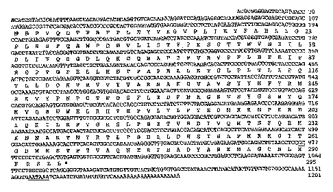
Figure 1 shows the cDNA sequence that was isolated from a pig liver cDNA
library
and the predicted amino acid sequence. SULT1A1 cDNA was isolated from a pig
liver
cDNA library. The nucleotide sequence has been registered in GenBank
(accession
number, AY193893). The predicted amino acid sequence is indicated below the
corresponding nucleotide sequence. The numbers of nucleotides and amino acids
are
indicated at the right. Polyadenylation signal (AATAAA) is underlined.
Figure 2shows an amino acid sequence comparison between pig phenol
sulfotransferase and human SULT1A1, SULT1A2 and SULT1A3. G1u83, Asp134 and
Asp263 are reported to be active sites for human SULT1A1. G1n121, Thr185, and
Thr267
axe common residues in phenol sulfotransferase. The asterisk indicates
residues for the
active sites between human and pig. The common residues of phenol
sulfotransferase
between human and pig are in bold.
Figure 3 shows the sequence of the genetic polymorphism, in vivo microsomal
sulfation activity, and skatole level in fat. Liver micosomal sulfation
activity and skatole
level in fat for both substitution and wild type samples.
DETAILED DESCRIPTION OF THE INVENTION
Reference will now be made in detail to the presently referred embodiments of
the
invention, which together with the following examples, serve to explain the
principles of
the invention.
The invention relates to genetic markers and methods of identifying those
markers
in an animal of a particular breed, strain, population, or group, whereby the
animal is more
likely to yield desired boar taint traits.
According to the invention, the genes encoding sulfotransferase enzymes which
are
involved in skatole metabolism have been identified as major effect genes.
Variation in
these genes has a measurable effect on boar taint in pigs. Thus screening
methods may be
8
CA 02554431 2006-07-26
WO 2005/074483 PCT/US2005/001474
developed for variation within or linked to these genes that is predictive of
phenotypic
variation.
In pigs, it has been found that a plasma concentration of 6-sulfatoxyskatole,
the
sulfoconjugate of 6-hydroxyskatole produced by phase II metabolism by
sulfotransferase, is
positively correlated to clearing skatole. (Babol et al., 1998). The
capability of synthesis of
6-sulfatoxyskatole is a major step in a rapid metabolic clearance of skatole,
resulting in low
concentrations of skatole in fat and further low level of boar taint.
Therefore,
sulfotransferase plays an important role in the metabolism and clearance of
skatole from
the body in pigs.
Sulfation is one of the major conjugation reactions involved in the metabolism
of
many hormones, neurotransmitters, drugs, and xenobiotic compounds (Winshilboum
et al.,
1997; Her et al, 1996; Dooley, 1998). Phenol sulfortransferase is considered
to be the most
important enzyme that catalyzes sulfate conjugation (Dooley, 1998). In humans,
phenol
sulfotransferase is expressed in many tissues including liver, spleen, lung,
testis, kidney,
skin, brain, adrenal gland, olfactory epithelium, and platelets. The
expression of this gene
in many tissues shows its importance in life process in vivo.
The molecular biology of phenol sulfotransferase has advanced rapidly. The
phenol
sulfotransferase genes in human (Her et al 1996), mouse (Sakakibara et al,
1998), rat
(access number: AF394783) and bovine (Henry et al., 1996) have been isolated
and
characterized.
Functionally significant genetic polymorphisms for phenol sulfotransferase
enzymes have been reported in humans, and other molecular genetic mechanisms
that
might be involved in the regulation of the expression of these enzymes have
been explored
(Chen et al, 2000; Seth, et al, 2000; Dooley, 1998). In humans, knowledge of
the molecular
biology of phenol sulfotransferase enzymes promises to significantly improve
the
understanding of the regulation of the sulfate conjugation of hormones,
neurotransmitters,
drugs, and xenobiotic compounds, in order to diagnose lung cancer, protect
against
colorectal cancers and breast cancers (Wang et al. 2002; Bamber et al, 2001;
Seth et al,
2000). In pigs, it has been reported that phenol sulfotransferase is
negatively correlated
with skatole accumulation in fat (Babol et al, 1998, Diaz and squires, 2003).
Pigs with high
sulfation activity have low level of skatole in fat, vice verse. Thus changes
in the actrivity
9
CA 02554431 2006-07-26
WO 2005/074483 PCT/US2005/001474
of the sulfation metabolic pathway could be used as genetic marker to select
for skatole
metabolism in pigs. However, the information about phenol sulfotransferase
gene, its
expression and how a genetic variation in this enzyme translates into
interindividual
variation in skatole level in pigs is unknown.
According to the invention a cDNA library was constructed from pig liver by
rapid
amplification of cDNA ends (RACE) and the sequence of porcine SULTlAl cDNA was
determined. The expression pattern of the SULT1A1 mRNA species was examined in
different tissues in pigs by RT-PCR. The polymerase chain reaction technique
combined
with single strand conformational polymorphism (PCR-SSCP) was used to scan for
polymorphisms in the SULTIAl coding region from porcine liver tissues, which
may alter
the metabolic capacities of the enzyme. We have identified a substitution
mutation A? G
in the coding region of the SULT1A1 gene that codes for a Lysla7 Glula7.
Functional
characterization of this mutant was carried out by transfection into a COS-7
cell line.
According to the invention, the association of alternate forms of
sulfotransferase
enzymes may be used to identify and select pigs with differences in boar
taint. For
example, according to the invention, an allele of the sulfotransferase gene
has been
identified that results in a protein change and increase activity of the
sulfotransferase
enzyme, which leads to lower skatole levels in the pig.
Further according to the invention, other polymorphisms sulfotransferase
genes in the pig may be identified to genetically type and select pigs based
upon their
proclivity to boar taint. Many factors can influence a metabolic pathway, some
products
are the result of rate limiting substrates or enzymes and it is unpredictable
which enzymes
may have variability that will result in an actual increase of a reaction
product and thus a
phenotypic trait. Once an association between a particular gene or gene
product in the
pathway and and protein activity that affects the resultant trait is made,
genes encoding
these proteins may be screened for other polymorphisms or markers which may be
used to
indicate differences in these animals with respect to the trait. The active
sites of thse
enzymes are the most susceptible to variability that will cause a significnat
affect in the
metabolic products. These polymorphisms with these genes enables genetic
markers to be
identified for specific breeds or genetic lines or animals, boar taint
potential early in the
animal's life.
CA 02554431 2006-07-26
WO 2005/074483 PCT/US2005/001474
An alternate form of sulfotransferase has been identified according to the
invention
which results in an amino acid change and decreased enzyme activity causing
higher
skatole levels in the pig. Tests for the presence of this alternate form may
be developed
using the novel sequence for sulfotransferase as disclosed herein. These tests
include but
are not limited to PCR, SSCP, and the like.
Thus, the invention relates to genetic markers and methods of identifying
those
markers in an animal of a particular animal, breed, strain, population, or
group, whereby
the animal is has increased, decreased or otherwise altered skatole
metabolism, and thus
boar taint.
Any method of identifying the presence or absence of these markers may be
used,
including, for example, single-strand conformation polymorphism (SSCP)
analysis, base
excision sequence scanning (BESS), RFLP analysis, heteroduplex analysis,
denaturing
gradient gel electrophoresis, and temperature gradient electrophoresis,
allelic PCR, ligase
chain reaction direct sequencing, mini sequencing, nucleic acid hybridization,
micro-array-
type detection of genes encoding enzymes involved in skatole metabolism. Also
within the
scope of the invention includes assaying for protein conformational or
sequences changes
which occur in the presence of this polymorphism. The polymorphism may or may
not be
the causative mutation but will be indicative of the presence of this change
and one may
assay for the genetic or protein bases for the phenotypic difference.
The following is a general overview of techniques which can be used to assay
for
the genetic marker of the invention.
In the present invention, a sample of genetic material is obtained from an
animal.
Samples can be obtained from blood, tissue, semen, etc. Generally, peripheral
blood cells
are used as the source, and the genetic material is DNA. A sufficient amount
of cells are
obtained to provide a sufficient amount of DNA for analysis. 'This amount will
be known
or readily determinable by those skilled in the art. The DNA is isolated from
the blood
cells by techniques known to those skilled in the art.
Isolation and Amplification of Nucleic Acid
Samples of genomic DNA are isolated from any convenient source including
saliva,
buccal cells, hair roots, blood, cord blood, amniotic fluid, interstitial
fluid, peritoneal fluid,
chorionic villus, and any other suitable cell or tissue sample with intact
interphase nuclei or
11
CA 02554431 2006-07-26
WO 2005/074483 PCT/US2005/001474
metaphase cells. The cells can be obtained from solid tissue as from a fresh
or preserved
organ or from a tissue sample or biopsy. The sample can contain compounds
which are not
naturally intermixed with the biological material such as preservatives,
anticoagulants,
buffers, fixatives, nutrients, antibiotics, or the like.
Methods for isolation of genomic DNA from these various sources are described
in,
for example, Kirby, DNA Fihge~prifzting, Au hctroductioh, W.H. Freeman & Co.
New
York (1992). Genomic DNA can also be isolated from cultured primary or
secondary cell
cultures or from transformed cell lines derived from any of the aforementioned
tissue
samples.
Samples of animal RNA can also be used. RNA can be isolated from tissues
expressing the gene as described in Sambrook et al., supra. RNA can be total
cellular
RNA, mRNA, poly A+ RNA, or any combination thereof. For best results, the RNA
is
purified, but can also be unpurified cytoplasmic RNA. RNA can be reverse
transcribed to
form DNA which is then used as the amplification template, such that the PCR
indirectly
amplifies a specific population of RNA transcripts. See, e.g., Sambrook,
supra, Kawasaki
et al., Chapter 8 in PCR Teclanology, (1992) supra, and Berg et al., Hum.
Genet. 85:655-
658 (1990).
PCR Amplification
The most common means for amplification is polymerase chain reaction (PCR), as
described in U.S. Pat. Nos. 4,683,195; 4,683,202; and 4,965,188 each of which
is hereby
incorporated by reference. If PCR is used to amplify the target regions in
blood cells,
heparinized whole blood should be drawn in a sealed vacuum tube kept separated
from
other samples and handled with clean gloves. For best results, blood should be
processed
immediately after collection; if this is impossible, it should be kept in a
sealed container at
4°C until use. Cells in other physiological fluids may also be assayed.
When using any of
these fluids, the cells in the fluid should be separated from the fluid
component by
centrifugation.
Tissues should be roughly minced using a sterile, disposable scalpel and a
sterile
needle (or two scalpels) in a 5 mm Petri dish. Procedures for removing
paraffin from tissue
sections are described in a variety of specialized handbooks well known to
those skilled in
the art.
12
CA 02554431 2006-07-26
WO 2005/074483 PCT/US2005/001474
To amplify a target nucleic acid sequence in a sample by PCR, the sequence
must
be accessible to the components of the amplification system. One method of
isolating
target DNA is crude extraction which is useful for relatively large samples.
Briefly,
mononuclear cells from samples of blood, amniocytes from amniotic fluid,
cultured
chorionic villus cells, or the like are isolated by layering on a sterile
Ficoll-Hypaque
gradient by standard procedures. Interphase cells are collected and washed
three times in
sterile phosphate buffered saline before DNA extraction. If testing DNA from
peripheral
blood lymphocytes, an osmotic shock (treatment of the pellet for 10 sec with
distilled
water) is suggested, followed by two additional washings if residual red blood
cells are
visible following the initial washes. This will prevent the inhibitory effect
of the heme
group carried by hemoglobin on the PCR reaction. If PCR testing is not
performed
immediately after sample collection, aliquots of 106 cells can be pelleted in
sterile
Eppendorf tubes and the dry pellet frozen at -20°C until use.
The cells are resuspended (106 nucleated cells per 100 ~.1) in a buffer of 50
mM
Tris-HC1 (pH 8.3), 50 mM KC1 1.5 mM MgCl2, 0.5% Tween 20, and 0.5% NP40
supplemented with 100 ~.g/ml of proteinase K. After incubating at 56°C
for 2 hr. the cells
are heated to 95°C for 10 min to inactivate the proteinase K and
immediately moved to wet
ice (snap-cool). If gross aggregates are present, another cycle of digestion
in the same
buffer should be undertaken. Ten ~,l of this extract is used for
amplification.
When extracting DNA from tissues, e.g., chorionic villus cells or confluent
cultured
cells, the amount of the above mentioned buffer with proteinase K may vary
according to
the size of the tissue sample. The extract is incubated for 4-10 hrs at
50°-60°C and then at
95°C for 10 minutes to inactivate the proteinase. During longer
incubations, fresh
proteinase K should be added after about 4 hr at the original concentration.
When the sample contains a small number of cells, extraction may be
accomplished
by methods as described in Higuchi, "Simple and Rapid Preparation of Samples
for PCR",
in PCR Technology, Ehrlich, H.A. (ed.), Stockton Press, New York, which is
incorporated
herein by reference. PCR can be employed to amplify target regions in very
small numbers
of cells (1000-5000) derived from individual colonies from bone marrow and
peripheral
blood cultures. The cells in the sample are suspended in 20 ~,1 of PCR lysis
buffer (10 mM
Tris-HC1 (pH 8.3), 50 mM KC1, 2.5 mM MgCl2, 0.1 mg/ml gelatin, 0.45% NP40,
0.45%
13
CA 02554431 2006-07-26
WO 2005/074483 PCT/US2005/001474
Tween 20) and frozen until use. When PCR is to be performed, 0.6 ~.1 of
proteinase K (2
mg/ml) is added to the cells in the PCR lysis buffer. The sample is then
heated to about
60°C and incubated for 1 hr. Digestion is stopped through inactivation
of the proteinase K
by heating the samples to 95°C for 10 min and then cooling on ice.
A relatively easy procedure for extracting DNA for PCR is a salting out
procedure
adapted from the method described by Miller et al., Nucleic Aciels Res.
16:1215 (1988),
which is incorporated herein by reference. Mononuclear cells are separated on
a Ficoll-
Hypaque gradient. The cells are resuspended in 3 ml of lysis buffer (10 mM
Tris-HC1, 400
mM NaC 1, 2 mM Na2 EDTA, pH 8.2). Fifty l.~l of a 20 mg/ml solution of
proteinase K and
150 (.tl of a 20% SDS solution are added to the cells and then incubated at
37°C overnight.
Rocking the tubes during incubation will improve the digestion of the sample.
If the
proteinase K digestion is incomplete after overnight incubation (fragments are
still visible),
an additional 50 ~.1 of the 20 mg/ml proteinase K solution is mixed in the
solution and
incubated for another night at 37°C on a gently rocking or rotating
platform. Following
adequate digestion, one ml of a 6M NaCl solution is added to the sample and
vigorously
mixed. The resulting solution is centrifuged for 15 minutes at 3000 rpm. The
pellet
contains the precipitated cellular proteins, while the supernatant contains
the DNA. The
supernatant is removed to a 15 ml tube that contains 4 ml of isopropanol. The
contents of
the tube are mixed gently until the water and the alcohol phases have mixed
and a white
DNA precipitate has formed. The DNA precipitate is removed and dipped in a
solution of
70% ethanol and gently mixed. The DNA precipitate is removed from the ethanol
and air-
dried. The precipitate is placed in distilled water and dissolved.
Kits for the extraction of high-molecular weight DNA for PCR include a Genomic
Isolation Kit A.S.A.P. (Boehringer Mannheim, Indianapolis, Ind.), Genomic DNA
Isolation
System (GIBCO BRL, Gaithersburg, Md.), Elu-Quilc DNA Purification Kit
(Schleicher &
Schuell, Keene, N.H.), DNA Extraction Kit (Stratagene, LaJolla, Calif.),
TurboGen
Isolation Kit (Invitrogen, San Diego, Calif.), and the like. Use of these kits
according to
the manufacturer's instructions is generally acceptable for purification of
DNA prior to
practicing the methods of the present invention.
The concentration and purity of the extracted DNA can be determined by
spectrophotometric analysis of the absorbance of a diluted aliquot at 260 nm
and 280 nm.
14
CA 02554431 2006-07-26
WO 2005/074483 PCT/US2005/001474
After extraction of the DNA, PCR amplification may proceed. The first step of
each cycle
of the PCR involves the separation of the nucleic acid duplex formed by the
primer
extension. Once the strands are separated, the next step in PCR involves
hybridizing the
separated strands with primers that flank the target sequence. The primers are
then
extended to form complementary copies of the target strands. For successful
PCR
amplification, the primers are designed so that the position at which each
primer hybridizes
along a duplex sequence is such that an extension product synthesized from one
primer,
when separated from the template (complement), serves as a template for the
extension of
the other primer. The cycle of denaturation, hybridization, and extension is
repeated as
many times as necessary to obtain the desired amount of amplified nucleic
acid.
In a particularly useful embodiment of PCR amplification, strand separation is
achieved by heating the reaction to a sufficiently high temperature for a
sufficient time to
cause the denaturation of the duplex but not to cause an irreversible
denaturation of the
polyrnerase (see U.S. Pat. No. 4,965,188, incorporated herein by reference).
Typical heat
denaturation involves temperatures ranging from about 80°C to
105°C for times ranging
from seconds to minutes. Strand separation, however, can be accomplished by
any suitable
denaturing method including physical, chemical, or enzymatic means. Strand
separation
may be induced by a helicase, for example, or an enzyme capable of exhibiting
helicase
activity. For example, the enzyme RecA has helicase activity in the presence
of ATP. The
reaction conditions suitable for strand separation by helicases are known in
the art (see
Kuhn Hoffinan-Berling, 1978, CSH Quantitative Biology, 43:63-67; and Radding,
1982,
Aran. Rev. Genetics 16:405-436, each of which is incorporated herein by
reference).
Template-dependent extension of primers in PCR is catalyzed by a polymerizing
agent in the presence of adequate amounts of four deoxyribonucleotide
triphosphates
(typically dATP, dGTP, dCTP, and dTTP) in a reaction medium comprised of the
appropriate salts, metal cations, and pH buffering systems. Suitable
polymerizing agents
are enzymes known to catalyze template-dependent DNA synthesis. In some cases,
the
target regions may encode at least a portion of a protein expressed by the
cell. In this
instance, mRNA may be used for amplification of the target region.
Alternatively, PCR
can be used to generate a cDNA library from RNA for further amplification, the
initial
template for primer extension is RNA. Polymerizing agents suitable for
synthesizing a
CA 02554431 2006-07-26
WO 2005/074483 PCT/US2005/001474
complementary, copy-DNA (cDNA) sequence from the RNA template are reverse
transcriptase (RT), such as avian myeloblastosis virus RT, Moloney murine
leukemia virus
RT, or Thermus they~mophilus (Tth) DNA polymerase, a thermostable DNA
polymerase
with reverse transcriptase activity marketed by Perkin Elmer Cetus, Inc.
Typically, the
genomic RNA template is heat degraded during the first denaturation step after
the initial
reverse transcription step leaving only DNA template. Suitable polymerases for
use with a
DNA template include, for example, E. coli DNA polymerase I or its Klenow
fragment, T4
DNA polymerase, Tth polymerase, and Taq polymerase, a heat-stable DNA
polyrnerase
isolated from The~mus aquaticus and commercially available from Perkin Elmer
Cetus,
Inc. The latter enzyme is widely used in the amplification and sequencing of
nucleic acids.
The reaction conditions for using Taq polymerase are known in the art and are
described in
Gelfand, 1989, PCR Technology, supra.
Allele Specific PCR
Allele-specific PCR differentiates between target regions differing in the
presence
of absence of a variation or polymorphism. PCR amplification primers are
chosen which
bind only to certain alleles of the target sequence. This method is described
by Gibbs,
Nucleic Acid Res. 17:12427-2448 (1989).
Allele Specific Oli~onucleotide Screening Methods
Further diagnostic screening methods employ the allele-specific
oligonucleotide
(ASO) screening methods, as described by Saiki et al., Nature 324:163-166
(1986).
Oligonucleotides with one or more base pair mismatches are generated for any
particular
allele. ASO screening methods detect mismatches between variant target genomic
or PCR
amplified DNA and non-mutant oligonucleotides, showing decreased binding of
the
oligonucleotide relative to a mutant oligonucleotide. Oligonucleotide probes
can be
designed so that under low stringency, they will bind to both polymorphic
forms of the
allele, but at high stringency, bind to the allele to which they correspond.
Alternatively,
stringency conditions can be devised in which an essentially binary response
is obtained,
i.e., an ASO corresponding to a variant form of the target gene will hybridize
to that allele,
and not to the wild-type allele.
Ligase Mediated Allele Detection Method
16
CA 02554431 2006-07-26
WO 2005/074483 PCT/US2005/001474
Target regions of a test subject's DNA can be compared with target regions in
unaffected and affected family members by ligase-mediated allele detection.
See
Landegren et al., Science 241:107-1080 (1988). Ligase may also be used to
detect point
mutations in the ligation amplification reaction described in Wu et al.,
Genonaics 4:560-569
(1989). The ligation amplification reaction (LAR) utilizes amplification of
specific DNA
sequence using sequential rounds of template dependent ligation as described
in Wu,
supra, and Barany, Proc. Nat. Acad. Sci. 88:189-193 (1990).
Denaturing Gradient Gel Electrobhoresis
Amplification products generated using the polymerase chain reaction can be
analyzed by the use of denaturing gradient gel electrophoresis. Different
alleles can be
identified based on the different sequence-dependent melting properties and
electrophoretic
migration of DNA in solution. DNA molecules melt in segments, termed melting
domains,
under conditions of increased temperature or denaturation. Each melting domain
melts
cooperatively at a distinct, base-specific melting temperature (T~,). Melting
domains are at
least 20 base pairs in length, and may be up to several hundred base pairs in
length.
Differentiation between alleles based on sequence specific melting domain
differences can be assessed using polyacrylamide gel electrophoresis, as
described in
Chapter 7 of Erlich, ed., PCR Technology, "Principles and Applications for DNA
Amplification", W.H. Freeman and Co., New York (1992), the contents of which
are
hereby incorporated by reference.
Generally, a target region to be analyzed by denaturing gradient gel
electrophoresis
is amplified using PCR primers flanking the target region. The amplified PCR
product is
applied to a polyacrylamide gel with a linear denaturing gradient as described
in Myers et
al., Meth. EnzynZOl. 155:501-527 (1986), and Myers et al., in Genomic
Analysis, A
Practical Approach, K. Davies Ed. IRL Press Limited, Oxford, pp. 95-139
(1988), the
contents of which are hereby incorporated by reference. The electrophoresis
system is
maintained at a temperature slightly below the Tm of the melting domains of
the target
sequences.
In an alternative method of denaturing gradient gel electrophoresis, the
target
sequences may be initially attached to a stretch of GC nucleotides, termed a
GC clamp, as
described in Chapter 7 of Erlich, supra. Preferably, at least 80% of the
nucleotides in the
17
CA 02554431 2006-07-26
WO 2005/074483 PCT/US2005/001474
GC clamp are either guanine or cytosine. Preferably, the GC clamp is at least
30 bases
long. This method is particularly suited to target sequences with high Tm s.
Generally, the target region is amplified by the polymerase chain reaction as
described above. One of the oligonucleotide PCR primers carries at its 5' end,
the GC
clamp region, at least 30 bases of the GC rich sequence, which is incorporated
into the 5'
end of the target region during amplification. The resulting amplified target
region is run
on an electrophoresis gel under denaturing gradient conditions as described
above. DNA
fragments differing by a single base change will migrate through the gel to
different
positions, which may be visualized by ethidium bromide staining.
Temperature Gradient Gel Electrophoresis
Temperature gradient gel electrophoresis (TGGE) is based on the same
underlying
principles as denaturing gradient gel electrophoresis, except the denaturing
gradient is
produced by differences in temperature instead of differences in the
concentration of a
chemical denaturant. Standard TGGE utilizes an electrophoresis apparatus with
a
temperature gradient running along the electrophoresis path. As samples
migrate through a
gel with a uniform concentration of a chemical denaturant, they encounter
increasing
temperatures. An alternative method of TGGE, temporal temperature gradient gel
electrophoresis (TTGE or tTGGE) uses a steadily increasing temperature of the
entire
electrophoresis gel to achieve the same result. As the samples migrate through
the gel the
temperature of the entire gel increases, leading the samples to encounter
increasing
temperature as they migrate through the gel. Preparation of samples, including
PCR
amplification with incorporation of a GC clamp, and visualization of products
are the same
as for denaturing gradient gel electrophoresis.
Single-Strand Conformation Polymorphism Analysis
Target sequences or alleles at the chosen boar taint loci can be
differentiated using
single-strand conformation polymorphism analysis, which identifies base
differences by
alteration in electrophoretic migration of single-stranded PCR products, as
described in
Orita et al., P~oc. Nat. Acad. Sci. 85:2766-2770 (1989). Amplified PCR
products can be
generated as described above, and heated or otherwise denatured, to form
single-stranded
amplification products. Single-stranded nucleic acids may refold or form
secondary
structures which are partially dependent on the base sequence. Thus,
electrophoretic
18
CA 02554431 2006-07-26
WO 2005/074483 PCT/US2005/001474
mobility of single-stranded amplification products can detect base-sequence
difference
between alleles or target sequences.
Chemical or Enzymatic Cleavage of Mismatches
Differences between target sequences can also be detected by differential
chemical
cleavage of mismatched base pairs, as described in Grompe et al., Am. J. Hum.
Genet.
48:212-222 (1991). In another method, differences between target sequences can
be
detected by enzymatic cleavage of mismatched base pairs, as described in
Nelson et al.,
Natuy~e Gehetics 4:11-18 (1993). Briefly, genetic material from an animal and
an affected
family member may be used to generate mismatch free heterohybrid DNA duplexes.
As
used herein, "heterohybrid" means a DNA duplex strand comprising one strand of
DNA
from one animal, and a second DNA strand from another animal, usually an
animal
differing in the phenotype for the trait of interest. Positive selection for
heterohybrids free
of mismatches allows determination of small insertions, deletions or other
polymorphisms
that may be associated with polymorphisms.
Non-~e1 Systems
Other possible techniques include non-gel systems such as TAQMANTM (Perkin
Elmer). In this system, oligonucleotide PCR primers are designed that flank
the mutation
in question and allow PCR amplification of the region. A third oligonucleotide
probe is
then designed to hybridize to the region containing the base subject to change
between
different alleles of the gene. This probe is labeled with fluorescent dyes at
both the 5' and
3' ends. These dyes are chosen such that while in this proximity to each other
the
fluorescence of one of them is quenched by the other and cannot be detected.
Extension by
Taq DNA polymerase from the PCR primer positioned 5' on the template relative
to the
probe leads to the cleavage of the dye attached to the 5' end of the annealed
probe through
the 5' nuclease activity of the Tay DNA polymerase. This removes the quenching
effect
allowing detection of the fluorescence from the dye at the 3' end of the
probe. The
discrimination between different DNA sequences arises through the fact that if
the
hybridization of the probe to the template molecule is not complete, i.e.,
there is a
mismatch of some form, the cleavage of the dye does not take place. Thus, only
if the
nucleotide sequence of the oligonucleotide probe is completely complimentary
to the
template molecule to which it is bound will quenching be removed. A reaction
mix can
19
CA 02554431 2006-07-26
WO 2005/074483 PCT/US2005/001474
contain two different probe sequences each designed against different alleles
that might be
present thus allowing the detection of both alleles in one reaction.
Yet another technique includes an Invader Assay, which includes isothermic
amplification that relies on a catalytic release of fluorescence. See Third
Wave Technology
at www.twt.com.
Non-PCR Based DNA Diagnostics
The identification of a DNA sequence linked to sequences encoding enzymes
involved in skatole metabolism can be made without an amplification step,
based on
polyrnorphisms including restriction fragment length polymorphisms in an
animal and a
family member. Hybridization probes are generally oligonucleotides which bind
through
complementary base pairing to all or part of a target nucleic acid. Probes
typically bind
target sequences lacking complete complementarity with the probe sequence
depending on
the stringency of the hybridization conditions. The probes are preferably
labeled directly or
indirectly, such that by assaying for the presence or absence of the probe,
one can detect the
presence or absence of the target sequence. Direct labeling methods include
radioisotope
labeling, such as with P32 or 535. Indirect labeling methods include
fluorescent tags, biotin
complexes which may be bound to avidin or streptavidin, or peptide or protein
tags. Visual
detection methods include photoluminescents, Texas red, rhodamine and its
derivatives,
red leuco dye and 3,3',5,5'-tetramethylbenzidine (TMB), fluorescein, and its
derivatives,
dansyl, umbelliferone and the like or with horse radish peroxidase, alkaline
phosphatase
and the like.
Hybridization probes include any nucleotide sequence capable of hybridizing to
the
porcine chromosome where the sulfotransferase gene or other gene involved in
skatole
metabolism resides, and thus defining a genetic marker linked to the gene,
including a
restriction fragment length polymorphism, a hypervariable region, repetitive
element, or a
variable number tandem repeat. Hybridization probes can be any gene or a
suitable analog.
Further suitable hybridization probes include exon fragments or portions of
cDNAs or
genes known to map to the relevant region of the chromosome.
Preferred tandem repeat hybridization probes for use according to the present
invention are those that recognize a small number of fragments at a specific
locus at high
CA 02554431 2006-07-26
WO 2005/074483 PCT/US2005/001474
stringency hybridization conditions, or that recognize a larger number of
fragments at that
locus when the stringency conditions are lowered.
One or more additional restriction enzymes and/or probes and/or primers can be
used. Additional mzyines, constructed probes, and primers can be determined by
routine
experimentation by those of ordinary skill in the art and are intended to be
within the scope
of the invention.
According to the invention, polymorphisms in genes encoding enzymes involved
in
skatole metabolism have been identified which have an association with boar
taint. The
presence or absence of the markers, in one embodiment may be assayed by PCR-
RFLP
analysis using the restriction endonucleases and amplification primers may be
designed
using analogous human, pig or other sequences due to the high homology in the
region
surrounding the polymorphisms, or may be designed using known gene sequence
data as
exemplified in GenBank or even designed from sequences obtained from linkage
data from
closely surrounding genes based upon the teachings and references herein. The
sequences
surrounding the polymorphism will facilitate the development of alternate PCR
tests in
which a primer of about 4-30 contiguous bases taken from the sequence
immediately
adjacent to the polymorphism is used in connection with a polymerase chain
reaction to
greatly amplify the region before treatment with the desired restriction
enzyme. The
primers need not be the exact complement; substantially equivalent sequences
are
acceptable. The design of primers for amplification by PCR is known to those
of skill in
the art and is discussed in detail in Ausubel (ed.), Short Protocols iu
Molecular Biology,
4th Edition, John Wiley and Sons (1999).
The following is a brief description of primer design. Generally the primers
used
for the assays of the invention will flank nt 546 on each side, one forward
and one reverse.
Primer Desi~xi StrateQ;y
Increased use of polyrnerase chain reaction (PCR) methods has stimulated the
development of many programs to aid in the design or selection of
oligonucleotides used as
primers for PCR. Four examples of such programs that are freely available via
the Internet
are: PRIMER by Mark Daly and Steve Lincoln of the Whitehead Institute (UNIX,
VMS,
DOS, and Macintosh), Oligonucleotide Selection Program (OSP) by Phil Green and
LaDeana Hiller of Washington University in St. Louis (UNIX, VMS, DOS, and
21
CA 02554431 2006-07-26
WO 2005/074483 PCT/US2005/001474
Macintosh), PGEN by Yoshi (DOS only), and Amplify by Bill Engels of the
University of
Wisconsin (Macintosh only). Generally these programs help in the design of PCR
primers
by searching for bits of known repeated-sequence elements and then optimizing
the Tm by
analyzing the length and GC content of a putative primer. Commercial software
is also
available and primer selection procedures are rapidly being included in most
general
sequence analysis packages.
Seguencing and PCR Primers
Designing oligonucleotides for use as either sequencing or PCR primers
requires
selection of an appropriate sequence that specifically recognizes the target,
and then testing
the sequence to eliminate the possibility that the oligonucleotide will have a
stable
secondary structure. Inverted repeats in the sequence can be identified using
a repeat-
identification or RNA-folding program such as those described above. If a
possible stem
structure is observed, the sequence of the primer can be shifted a few
nucleotides in either
direction to minimize the predicted secondary structure. The sequence of the
oligonucleotide should also be compared with the sequences of both strands of
the
appropriate vector and insert DNA. Obviously, a sequencing primer should only
have a
single match to the target DNA. It is also advisable to exclude primers that
have ouy a
single mismatch with an undesired target DNA sequence. For PCR primers used to
amplify genomic DNA, the primer sequence should be compared to the sequences
in the
GenBank database to determine if any significant matches occur. If the
oligonucleotide
sequence is present in any known DNA sequence or, more importantly, in any
known
repetitive elements, the primer sequence should be changed.
The methods and materials of the invention may also be used more generally to
evaluate pig DNA, genetically type individual pigs, and detect genetic
differences in pigs.
In particular, a sample of pig genomic DNA may be evaluated by reference to
one or more
controls to determine if a polymorphism in the particular gene is present.
Preferably, RFLP
analysis is performed with respect to the pig gene, and the results are
compared with a
control. The control is the result of a RFLP analysis of the pig gene of a
different pig
where the polymorphism(s) of the pig gene is/are known. Similarly, the
genotype of a pig
may be determined by obtaining a sample of its genomic DNA, conducting RFLP
analysis
of the gene in the DNA, and comparing the results with a control. Again, the
control is the
22
CA 02554431 2006-07-26
WO 2005/074483 PCT/US2005/001474
result of RFLP analysis of the gene of a different pig. The results
genetically type the pig
by specifying the polyrnorphism(s) in its genes. Finally, genetic differences
among pigs
can be detected by obtaining samples of the genomic DNA from at least two
pigs,
identifying the presence or absence of a polymorphism in the gene, and
comparing the
results.
These assays are useful for identifying the genetic markers relating to boar
taint, , as
discussed above, for identifying other polymorphisms in the genes encoding
enzymes
involved in skatole metabolism and for the general scientific analysis of pig
genotypes and
phenotypes.
The examples and methods herein disclose certain genes) which has been
identified to have a polymorphism(s) which is associated either positively or
negatively
with a beneficial trait that will have an effect on boar taint for animals
carrying this
polymorphism. The identification of the existence of a polymorphism within a
gene is
often made by a single base alternative that results in a restriction site in
certain allelic
forms. A certain allele, however, as demonstrated and discussed herein, may
have a
number of base changes associated with it that could be assayed for which are
indicative of
the same polymorphism (allele). Further, other genetic markers or genes may be
linked to
the polymorphisms disclosed herein so that assays may involve identification
of other
genes or gene fragments, but which ultimately rely upon genetic
characterization of animals
for the same polymorphism. Any assay which sorts and identifies animals based
upon the
allelic differences disclosed herein are intended to be included within the
scope of this
invention.
One of skill in the art, once a polymorphism has been identified and a
correlation to
a particular trait established will understand that there are many ways to
genotype animals
for this polymorphism. The design of such alternative tests merely represents
optimization
of parameters known to those of skill in the art and is intended to be within
the scope of
this invention as fully described herein.
The following non-limiting examples are illustrative of the present invention:
23
CA 02554431 2006-07-26
WO 2005/074483 PCT/US2005/001474
EXAMPLES
Tissue samples
A liver tissue was obtained from a male pig for construction of cDNA library.
To
identify genetic polymorphisms in SULT1A1 gene, liver tissues were obtained
from sixty
nine intact male pigs from a variety of breeds, including Yorkshire, Duroc,
Landrace, and
Pietrain, as well as crosses between Landrace and Duroc, Large White and
Duroc, and
Large White and Pertain. The animals were slaughtered at an average live
weight of 144 ~
33 kg. A sample of liver was taken immediately following exsanguination,
frozen in liquid
nitrogen and stored at -70°C before use. For measuring the expression
profile of SULT1A1
mRNA, tissues including spleen, thymus, liver, lung, muscle, kidney, small
intestine, heart,
ovaries and testis were collected from one Landrace boar and one Landrace
female that
weighed approximately 100 kg.
Measurement of skatole level in fat
A backfat sample was collected at the midline point of l lth rib and frozen at
-20°C
until assayed for skatole. The skatole content was measured with a HPLC assay,
according
to the method described by Diaz and Squires (2000).
Isolation of total RNA
One hundred milligrams of each tissue sample was homogenized in 1 ml of Tri-
Reagent (Sigma, ST. Louis, MO) and incubated for 10 minutes at room
temperature. After
incubation, 0.2 ml of chloroform was added and the samples were vortexed and
then
centrifuged at 12,OOOXg for 10 minutes at 4°C.The aqueous phase was
transferred into a
sterile tube and mixed with 0.5 ml of isopropanol and incubated at room
temperature for 10
minutes. The samples were centrifuged at 12,OOOXg for 10 minutes at 4°C
to precipitate the
RNA. The pellet was washed with 75% ethanol and then suspended into 50 ~,1 of
DEPC
water.
Construction and Screening of a pig cDNA RACE library
5' and 3' rapid amplification of cDNAs (RACE) were constructed from 1 ~,g of
total RNA from liver with the use of Smart RACE cDNA Amplification kit (BD
Biosciences, Palo Alto, CA), and used as templates in the subsequent PCR
screening of
porcine phenol sulfotransferase cDNA. The 5'RACE was performed by synthesizing
the
24
CA 02554431 2006-07-26
WO 2005/074483 PCT/US2005/001474
first strand cDNA with a modified lock-docking oligo (dT) primer and then
tailing the
product 5'AAG CAG TGG TAT CAA CGC AGA GTA CGC GGG 3' (anchor primer) in
the 5'end via terminal transferase. 'The 3' RACE was performed with oligo (dT)
primer but
including the same lock-docking nucleotide positions as in the 5'RACE. The
cDNA
fragments of porcine phenol sulfotransferase were amplified with anchor primer
and the
primers (A and B) designed from human SULT1A1 and SULTlA2 cDNA sequences.
Primer A was 5' CAC AGC TCA GAG CGG AAG C3' and primer B was 5' AGT GGT
GGG AGC TGC GTC ACA C 3'. To obtain the full-length porcine phenol
sulfotransferase
cDNA, the following primers were used in the subsequent PCR-based screening:
primer A
and anchor primer with 5'Race as a template (annealing 61 °C); primer B
and anchor primer
with 3'Race as a template (annealing 63°C). The PCR consisted of 30
cycles of denaturing
for 1 minute at 94°C, optimal annealing for 1 minute, and. extending
for 1 minute, with a
final 10 minute extension step at 72°C. Ten microliters of the PCR
products were analyzed
by electrophoresis on a 1 % agarose gel.
Colony hybridization
When multiple bands were amplified from both 3'and 5'Race templates, the PCR
products were cloned into pGEM-T Easy Vector System (Promega, Madison, WI),
and
subjected to colony hybridization to confirm the specificity of amplified
fragment prior to
DNA sequencing. Colonies were lifted from the positively charged nylon
membrane
(Roche, Indianapolis,1N)), and subjected to lysis and fixation in O.SM NaCI
for 5 minutes,
followed by rinsing in SxSSC for 1 minute, and allowed to air dried. Colony
hybridization
was performed with the ECL nucleotide DNA labeling and detection kit (Amersham
Biosciences, Piscataway, NJ). The probe used in the hybridization was the
fragment
amplified by primer A and primer B designed from the human SULT1A1 and SULTlA2
cDNAs. Thermal cycling consisted of (1) 5 cycles of 94°C for 30 sec and
72°C for 3 min;
(2) 5 cycles of 94°C for 30 sec, 70°C for 30 sec, and
72°C for 3 min; (3) 25 cycles of 94°C
for 30 sec, 61 °C for 30 sec, and 72°C for 3 min, with a final
72°C extension for 10 min.
After hybridization overnight at 42°C, the membrane was washed twice
with O.lSxSSC for
20 minutes and exposed to x-ray film. The colony that gave the strongest
signal was
selected for sequencing.
CA 02554431 2006-07-26
WO 2005/074483 PCT/US2005/001474
Isolation of full-length porcine phenol sulfotransferase cDNA
To obtain a full-length porcine phenol sulfotransferase sequence, the forward
primer 5' ATG GAG CCG GTC CAG GAC A 3' ' and reverse primer 5' TCA CAG CTC
AGA GCG GAA GC 3' were designed based on the sequence obtained from the 5' and
3'
RACE. They were used to amplify the full-length porcine phenol
sulfotransferase with
either 5' or 3' RACE cDNA as a template. PCR profile was 3 min at 94°C,
followed by 30
cycles of 1 min at 94°C, 1 min 30 sec at 63°C, 1 min at
72°C and final extension of 10 min
at 72°C. The PCR fragment was cloned into T-Easy vector (Promega,
Madison, WI) and
subjected to sequence analysis.
Expression of phenol sulfotransferase gene (SULT1A1) in tissues
The tissue distribution of SULTlAI mRNA was determined by RT-PCR. Total
RNAs were isolated from 100 mg of porcine spleen, thymus, liver, lung, muscle,
ovary,
kidney, small intestine, heart, and testis tissues with Tri-Reagent (Sigma).
Total RNAs
were treated with DNase I (Ambion) for 20 minutes at 37°C according to
the product
manual prior to RT-PCR. One microgram of treated total RNA from liver samples
was
used to synthesize the first strand cDNA by using Superscript reverse
transcriptase
(Invitrogen) and oligo (dT) primer (Sigma). RT-PCR was carried out based on
the method
described below. The forward primer (5' ATG GAG CCG GTC CAG GAC A 3') and
reverse primer (5' TCA CAG CTC AGA GCG GAA GC 3') were designed to amplify the
entire coding region of porcine SULT1A1 gene. It corresponds to the product
from the
transcription start site (nucleotide position 108) to transcription stop site
(nucleotide
position 995), spanning 888 bp. Ten microliters of the PCR products were
analyzed by
electrophoresis on a 1 % agarose gel.
Sequencing analysis
The PCR fragments were ligated into pGEM-T Easy Vector System (Promega,
Madison, WI), and then transformed into competent DHSoc cells. DNAs were
purified and
subject to sequencing using an Applied Biosystems model ABI 377 DNA sequencer.
RT-PCR
To scan for genetic polymorphisms in the SULT1A1 gene, RT-PCR products that
cover the whole coding region were amplified and then subjected to SSCP
analysis. One to
26
CA 02554431 2006-07-26
WO 2005/074483 PCT/US2005/001474
five micrograms of total RNA from liver samples were used to synthesize first
strand
cDNA using Superscript reverse transcriptase (Invitrogen, Carlsbad, CA) and
oligo (dT)
primer (Sigma, ST. Louis, MO). Following the reverse transcription, 2.5,1 of
the first
strand cDNA was used as the template for PCR. The PCR mixtures (50 ul)
contained
1 ~PCR buffer (100 mM Tris-HCI, pH 8.3; 500 mM KCl, 11 mM MgCl2, 0.1 %
gelatin), 0.2
mM dNTP, 0.4 mM primers (forward and reverse primer) and 2.5 U of Red Taq
polymerase (Sigma, ST. Louis, MO). The forward primer (5' ATG GAG CCG GTC CAG
GAC A 3') and reverse primer (5' TCA CAG CTC AGA GCG GAA GC 3') were designed
to amplify the entire coding region of SULT1A1 gene, which was based on our
isolated
SULT1A1 (GenBank accession number AY193893). The PCR profile was 3 minutes at
94°C, followed by 35 cycles of 1 minute at 94°C, 1 minute at
63°C, 1 minute at 72°C and
final extension of 10 minutes at 72°C.
Single-strand conformational polymorphism (SSCP) analysis
PCR products were first cut into fragments with KpnI enzyme, and then resolved
by
SSCP analysis. Ten microliters of amplified PCR product was digested with KpnI
in a 25
~,l reaction at 37°C for 3 hours. A total of 7~,1 of digested fragments
were then diluted
with 13,1 of loading buffer (10% sucrose, 0.01% bromophenol blue and 0.01%
xylene
cyanol FF). Each digestion reaction was denatured at 100°C for 5
minutes, chilled on ice
and resolved on a 10% polyacrylamide gel. The electrophoresis was carried out
in a
130x160x1.Omm vertical unit (Bio-Rad Laboratories, Hercules, CA), in 0.6xTBE
buffer
for 17 hours at 15°C at 160 V. The gels were then silver stained.
Expression of the phenol sulfotransferase cDNA in COs-7 cells
The expression vector, pcDNA3.1/VS-His TOPO TA Expression vector
(Invitrogen), was used. The whole coding region of phenol sulfotransferase
cDNA was
amplified from the cDNA library with the following primers, forward: 5' ATG
GAG CCG
GTC CAG GAC A 3' (start codon bolded); reverse: 5' TCA CAG CTC AGA GCG GAA
GC 3'(stop codon bolded). The PCR reaction was performed under the following
conditions: 3 minutes at 94°C, followed by 30 cycles of 1 minute at
94°C, 1 minute at 63°C,
1 minute at 72°C, with a final 10 min extension step at 72°C.
Following amplification, 50.1
of PCR product was purified by a QIAquick Nucleotide Removal kit (QIAGEN) and
suspended in 30,1 of distilled water. Four microliters of purified PCR product
was ligated
27
CA 02554431 2006-07-26
WO 2005/074483 PCT/US2005/001474
to 1 ~1 (10 ng) of expression vector and incubated at room temperature for 30
minutes. The
recombinant DNA was then transformed into TOP 10 competent cells (Invitrogen),
purified,
and subjected to sequencing to confirm its orientation.
COS-7 cells, routinely maintained in Dulbecco's modified Eagle's medium
(DMEM) containing 10% fetal bovine serum and 1 % antibiotics, were used as the
host
cells for the expression of the recombinant protein. Dishes (150 mm) of COS-7
cells were
individually transfected with 54 ~.g of recombinant DNA containing mutant (A?
G at
nucleotide 546 bp) and wild type porcine SULT1A1 cDNA using the Lipofectamine
2000
mediated procedure (Invitrogen), while COS-7 cells only and expression vector
only were
used as negative control. After transfection, the cells were incubated at
37°C, 5%C02 for
the first 18 hours without serum and antibiotics, and then incubated at
37°C, 5%C02 in
DMEM containing 10% fetal bovine serum, 1 % antibiotics for 48 hours. At the
end of
incubation, the cells were rinsed twice with phosphate buffered saline and
precipitated at
500 g for 5 minutes at 4°C. After discarding the supernatant, the
precipitate was stored at -
80°C before assay for sulfotransferase activity.
Sulfotransdrase activity assay
?-nitrophenol was used as a substrate for the SULT1A1 enzymatic activity assay
according to the method previously described (Diaz and Squires, 2003). The COS-
7 cell
pellets were lysed in buffer (50 mM Tris-HCI, lOmM MgCl2, 0.1 mM EDTA, pH 7.4)
and
sonicated for 20 sec. The protein concentrations were measured by Bio-Rad
Protein assay.
The reaction was run in a mixture of 4 mg/ml protein, 8 mM p-nitrophenol, 2 mM
PAPS
(Sigma) for 30 min at 37°C, terminated by adding an equal volume of ice-
cold acetonitrile,
vortexed and centrifuged to remove protein. One hundred microliters of
supernatant was
used to measure the formation of p-nitrophenyl sulfate by HPLC.
Sequence characterization of phenol sulfotransferase (SULT1A1) cDNA
Porcine SULT1A1 cDNA was isolated by PCR screening of the liver cDNA library
constructed with the RACE method. The nucleotide was 1201 by long and
contained a 888
bp-long open reading frame (ORF) encoding 296 amino acids and 206 by long 3'
untranslated region including one polyadenylation signal, AATAAA (Figure 1).
Porcine
SULT1A1 cDNA sequence was submitted to Genbank database under the accession
number AY193893.
28
CA 02554431 2006-07-26
WO 2005/074483 PCT/US2005/001474
In humans, there are three highly homologous phenol sulfotransferases (PSTs)
and
three highly homologous (over 94%) PST genes, SULT1A1, SULTlA2, and SULT1A3
are
located on chromosome 16p12.1. When we compared the pig phenol
sulfotransferase
coding region to the human genes, it showed 86% homology to SULT1A1 and
SULT1A2,
and 85% to SULT1A3. The deduced amino acid sequence for pig phenol
sulfotransferase
showed 86.7% homology to SULT1A1, 86.5% to SULT1A2, and 85.4% to SULT1A3
(Figure 2). In humans, SULT1A1, G1u83, Asp134 and Asp263 are reported to be
the active
site for SULT1A1, and especially G1u83 and Asp134 are essential amino acids
for
SULTlAl catalytic activity (Chen et al, 2000). G1n121, Thr185, and Thr267 are
common
residues in human phenol sulfotransferase (Honma et al, 2001). All the above
active sites
are conserved in the putative pig phenol sulfotransferase. To further
characterize this gene,
the recombinant protein encoded by this gene was expressed in COS-7 cells, and
the
enzyme activity of the expressed protein was assayed using ?-nitrophenol as a
substrate.
These results indicate that this gene isolated from pig liver clearly
represents phenol
sulfotransferase.
Expression of phenol sulfotransferase mRNA in various tissues
The expression patterns of phenol sulfotransferase mRNA in spleen, thymus,
liver,
lung, muscle, ovary, kidney, small intestine, heaxt, and testis tissues of
pigs were
investigated by RT-PCR. To determine the mRNA level in tissues, the total RNA
samples
were treated with DNAse I to remove possible contamination with genomic DNA
prior to
RT-PCR. The result showed that phenol sulfotransferase (about 900 by PCR
products) was
expressed in all of the 10 tissues examined except the small intestine (Figure
3). This
suggests that phenol sulfotransferase plays an important role in the life
process in vivo in
pigs.
Phenol sulfotransferase genetic polymorphism
In order to identify any genetic polymorphism of phenol sulfotransferase that
may
alter the metabolic capacities of the enzyme, a polymerase chain reaction
technique
combined with single strand conformational polymorphism (PCR-SSCP) was used to
scan
the phenol sulfotransferase coding region from porcine liver tissues. The
phenol
sulfotransferase full-length cDNA was amplified by PCR with the primer pair:
forward
primer 5' ATG GAG CCG GTC CAG GAC A 3'; reverse primers: 5' TCA CAG CTC
29
CA 02554431 2006-07-26
WO 2005/074483 PCT/US2005/001474
AGA GCG GAA GC 3'. The resulting PCR products were about 900 by in size and
were
digested with KpnI and subjected to SSCP analysis using our optimized system.
We found
that there are several different polymorphisms present in the phenol
sulfotransferase coding
region (data not shown). One substitution (Figure 4-B) of Lys147 (AAA) to
Glul47 (GAA)
at nucleotide 546 by was of particular interest because of the big difference
in the skatole
level between wild type and mutant samples (Figure 4-A. We proposed that the
substitution
might result in decreased phenol sulfotransferase activity for this individual
and that the
skatole level would be higher due to decreased activity of this enzyme
important in clearing
skatole from the body.
To evaluate the above hypothesis and investigate the association of this
genetic
polymorphism to phenol sulfotransferase activity, recombinant DNA containing
the
substitute mutant (A? G) and wild type of pig phenol sulfotransferase cDNA
were used to
transfect marmnalian cells, the activities of recombinant proteins produced
were assayed
using ?-nitrophenol as a substrate (Fig 5). For the wild type, sulfation
activity was
211.2475.57 pmol/min/mg, whereas for the Lys147 to G1u147 mutation, the
activity was
15.977.18 pmol/min/mg, showing a significant difference between the mutant and
wild
type (P<0.05). This result indicates that Lysl47 is crucial for the catalytic
activity of phenol
sulfotransferase. The results strongly support our suggestion that the Lys147
to G1u147
mutation caused a decrease in the catalytic activity of phenol
sulfotransferase and hence
result in a higher level skatole in the pig.
Phenol sulfotransferase genes have been extensively investigated in humans. In
pigs, it has been reported that phenol sulfotransferase is negatively
correlated with skatole
accumulation in fat (Babol et al, 1998; Diaz and Squires, 2003). However, the
information
about the phenol sulfotransferase gene, its expression in different tissues
and how a genetic
variant of it affects sulfation activity, hence skatole level in pig has not
been previously
reported. In humans, three members of the phenol sulfotransferase family,
SULT1A1,
SULTlA2, and SULT1A3 have been cloned and characterized. DNA sequences and the
structure of these three enzymes are highly homologous, and all three genes
are localized
on chromosome 16p12.1 (Dooley et al, 1993; Gaedigk et al, 1997; Aksoy et al,
1994). Both
SULT1A1 and SULT1A2 catalyze the sulfation of ?-nitrophenol (Raftogianis et
al, 1997),
while SULT1A3 shows a trivial activity for ?-nitrophenol (Veronese et al,
1994).
CA 02554431 2006-07-26
WO 2005/074483 PCT/US2005/001474
Therefore, SULT1A1 and SULT12A are considered the main enzymes that catalyze
sulfation in humans. We designed the first primer pair based on human SULT1A1
and
SULT1A2 cDNA sequences. Therefore, by using the designed primers, we screened
out the
first fragment, and subsequently the whole sequence of pig phenol
sulfotransferase cDNA.
To further character this gene, this pig putative phenol sulfotransferase cDNA
was
subcloned into the expression vector and used to transfect COS-7 cells. The
expressed
enzyme showed high catalytic activity towards the ?-nitrophenol substrate. The
results
demonstrate that this cDNA is indeed pig phenol sulfotransferase, and is one
of isoforms of
SULT1A1 or SULTlA2 rather than SULTlA3. In humans, SULTlAl has up to 10-fold
higher phenol sulfotransferase activity compared with that of SULT1A2
(Raftogianis et al,
1997). It is also suggested that SULT1A2 does not contribute substantially to
the sulfation
of endogenous or xenobiotic agents in vivo (Dooley, 1998). Due to the high
identity (96%)
between human SULT1A1 and SULT1A2 cDNAs, the pig phenol sulfotransferase cDNA
and its deduced amino acid sequence showed the same homology (86%) with human
SULT1A1 and SULT1A2 cDNA and amino acid sequences. SULTlAl and SULT1A2
genes in human have been mapped to chromosome 16p12.1. When we searched
against the
human genomic database with the pig phenol sulfotransferase cDNA sequence, we
found
that this cDNA hit a human genomic clone (NT 010393.13), which contains both
SULT1A1 and SULT1A2 from chromosome 16p12.1. The hit scores showed that pig
cDNA sequence has 91 % identity with human SULT1A1 and 88% identity with human
SULTlA2. All these finding taken together suggest that the cDNA we isolated
from pig
liver is SULTlAl .
Applicants isolated pig phenol sulfotransferase from liver tissue using the
RACE
method, then performed PCR-SSCP analysis to scan its coding region. A
substitution from
A to G at nucleotide 546 bp, which caused a change in amino acid sequence from
Lys147 to
G1u147 was identified. To help clarify possible genotype-phenotype correlation
for the
genetic mutation, we next determined the sulfation activity of the protein
encoded by
SULT1A1 and SULT1A1 Lys147 to G1u14~ mutant expressed in COS-7 cells. The
result
showed that the transition from A to G significantly reduced enzymatic
activity.
31
CA 02554431 2006-07-26
WO 2005/074483 PCT/US2005/001474
References
Aksoy IA, Callen DF, Apostolou, S, Her C, Weinshilboum RM (1994)
Thermolabile phenol sulfotransferase gene (STM): Localization to human
chromsome
16p11.2. Genomics 23, 275-277
Babol J, Squires EJ, Lundstrom K (1998) Relationship between oxidation and
conjugation metabolism of skatole in pig liver and concentrations of skatole
in fat. J. Anim.
Sci. 76, 829-838
Bamber DE, Fryer AA, Strange, RC, Elder JB, Deakin M, Rajagopal R, Fawole A,
Gilissen R, Campbell FC, Coughtrie WH (2001) Phenol sulphotransferase SUL1A1*1
genotype is associated with reduced risk of colorectal cancer.
Pharmacogenetics 11, 679-
685
Chen G, Rabjohn PA, York JL, Wooldridge C, Zhang D, Falany CN, Radominska-
Pandya A (2000) Carboxyl Residues in the active site of human phenol
sulfotransferase
(SULT1A1). Biochemistry 39, 16000-16007
Diaz, G.J. and Squires, E.J. (2000). Metabolism of 3-Methylindole by Porcine
Liver
Microsomes: Responsible Cytochrome P450 Enzyme. Toxicological Science 55, 284-
292.
Diaz, GJ and Squires EJ (2003) Phase II in vitro metabolism of 3-methylindole
metabolites in porcine liver. Xenobiotica 33, 485-498.
Dooley TP (1998) Molecular biology of the human phenol sulferasferase gene
family. The Journal of experimental zoology 282, 223-230
Dooley TP, Obermoeller RD, Leiter EH, Chapman HD, Falany CN, Deng Z,
Siciliano MJ (1993) Mapping of the phenol sulfotransferase gene (STP) to human
chromosome 16p12.1-p11.2 and to mouse chromosome 7. Genomics 18, 440-443
Henry T, Kliewer B, Palmatier R, Ulphani JS, Beckmann JD (1996) Isolation and
characterization of a bovine gene encoding phenol sulfotransferase. Gene 174,
221-224
Her C, Raftogianis R, Weinshilboum RM (1996) Human phenol sulfotransferase
STP2 gene: Molecular cloning, structural characterization, and chromosome
localization.
Genomics 33, 409-420
Honma W, Kamiyama Y, Yoshinari K, Sasano H, Shimada M, Nagata K, Yamazoe
Y (2001) Enzymatic characterization and interspecies difference ofphenol
sulfotransferases, ST1A forms. Drug Metabolism and Disposition 29, 274-281
32
CA 02554431 2006-07-26
WO 2005/074483 PCT/US2005/001474
Gaedigk A, Beatty BG, Grant DM (1997) Cloning, structural organization, and
chromosomal mapping of the human phenol sulfotransferase STP2 gene. Genomics
40,
242-246
Raftogianis RB, Wood TC, Otterness WD, Loon JA, Weinshilboum RM (1997)
Phenol sulfotransferase pharmacogenetics in human: association of cormnon
SULTIAl
alleles with TS PST phenotype. Biochemical and Biophysical Research
Communications
239, 298-304
Sakakibara Y, Yanagisawa K, Takami Y, Nakayasna T, Suiko M, Liu MC (1998)
Molecular cloning, expression, and functional characterization of novel mouse
sulfotransferases. Biochemical and Biophysical research communications 247,
681-686
Seth P, Lunetta KL, Bell DW et al (2000) Phenol sulfotransferases: Hormonal
regulation, polymorphism, and age of onset of breast cancer. Cancer Research
60, 6859-
6863
Veronese ME, Burgess W, Zhu X, McManus ME (1994) Functional
characterization of two human sulphotransferase cDNAs that encode monoamine-
and
phenol-sulphating forms oh phenol sulfotransferase: substrate kinetics,
thermal-stability
and inhibitor-sensitivity studies. Bichemical Journal 302, 497-502
Wang Y, Spitz MR, Tsou AM, Zhang K, Makan N, Wu X (2002) Sulfotransferase
(SULT) 1A1 polymorphism as a predisposition factor for lung cancer: a case-
control
analysis. Lung Cancer 35, 137-142
Weinshilboum RM, Otterness DM, Aksoy IA, Wood TC, Her C, Raftogianis RB
(1997) Sulfation and sulfotransferases 1: Sulforansferase molecular biology:
cDNAs and
genes. The FASEB Journal 11, 3-14
While the present invention has been described with reference to what are
presently
considered to be the preferred examples, it is to be understood that the
invention is not
limited to the disclosed examples. To the contrary, the invention is intended
to cover
various modifications and equivalent arrangements included within the spirit
and scope of
the appended claims.
All publications, patents and patent applications are herein incorporated by
reference in their entirety to the same extent as if each individual
publication, patent or
33
CA 02554431 2006-07-26
WO 2005/074483 PCT/US2005/001474
patent application was specifically and individually indicated to be
incorporated by
reference in its entirety.
34
DEMANDES OU BREVETS VOLUMINEUX
LA PRESENTE PARTIE DE CETTE DEMANDE OU CE BREVETS
COMPRI~:ND PLUS D'UN TOME.
CECI EST L,E TOME 1 DE 2
NOTE: Pour les tomes additionels, veillez contacter le Bureau Canadien des
Brevets.
JUMBO APPLICATIONS / PATENTS
THIS SECTION OF THE APPLICATION / PATENT CONTAINS MORE
THAN ONE VOLUME.
THIS IS VOLUME 1 OF 2
NOTE: For additional valumes please contact the Canadian Patent Office.