Note: Descriptions are shown in the official language in which they were submitted.
CA 02555141 2006-08-03
WO 2005/076868 PCT/US2005/003246
DEVICES AND METHODS FOR INSERTING A SPINAL FIXATION
ELEMENT
BACKGROUND
[0001 ] This application relates to tools for use in spinal surgery, and in
particular to
minimally invasive methods and devices for introducing a spinal fixation
element to one or
more spinal anchor sites within a patient's spine.
[0002] For a number of known reasons, spinal fixation devices are used in
orthopedic surgery
to align and/or fix a desired relationship between adjacent vertebral bodies.
Such devices
typically include a spinal fixation element, such as a relatively rigid
fixation rod, that is
coupled to'adjacent vertebrae by attaching the element to various anchoring
devices, such as
hooks, bolts, wires, or screws. The fixation elements can have a predetermined
contour that
has been designed according to the properties of the target implantation site,
and once
installed, the instrument holds the vertebrae in a desired spatial
relationship, either until
desired healing or spinal fusion has taken place, or for some longer period of
time.
[0003] Spinal fixation elements can be anchored to specific portions of the
vertebrae. Since
each vertebra varies in shape and size, a variety of anchoring devices have
been developed to
facilitate engagement of a particular portion of the bone. Pedicle screw
assemblies, for
example, have a shape and size that is configured to engage pedicle bone. Such
screws
typically include a threaded shank that is adapted to be threaded into a
vertebra, and a head
portion having a rod-receiving element, usually in the form of a U-shaped slot
formed in the
head. A set-screw, plug, or similar type of fastening mechanism is used to
lock the fixation
element, e.g., a spinal rod, into the rod-receiving head of the pedicle screw.
In use, the shank
portion of each screw is threaded into a vertebra, and once properly
positioned, a rod is seated
through the rod-receiving member of each screw and the rod is locked in place
by tightening
a cap or other fastener mechanism to securely interconnect each screw and the
fixation rod.
[0004] Recently, the trend in spinal surgery has been moving toward providing
minimally
invasive devices and methods for implanting spinal fixation devices. One such
method, for
example, is disclosed in U.S. Patent No. 6,530,929 of Justis et al. and it
utilizes two
percutaneous access devices for implanting an anchoring device, such as a
spinal screw, into
adjacent vertebrae. A spinal rod is then introduced through a third incision a
distance apart
1
CA 02555141 2006-08-03
WO 2005/076868 PCT/US2005/003246
from the percutaneous access sites, and the rod is transversely moved into the
rod-engaging
portion of each spinal screw. The percutaneous access devices can then be used
to apply
closure mechanisms to the rod-engaging heads to lock the rod therein. While
this procedure
offers advantages over prior art invasive techniques, the transverse
introduction of the rod can
cause significant damage to surrounding tissue and muscle. Moreover, the use
of three
separate access sites can undesirably lengthen the surgical procedure, and
increase patient
trauma and recovery time.
[0005] Accordingly, there remains a need for improved minimally invasive
devices and
methods for introducing a spinal fixation element into a patient's spine.
SUMMARY
[0006] Disclosed herein are minimally invasive methods and devices for
delivering a spinal
fixation element to one or more spinal anchor sites in a patient's spinal
column. In one
exemplary embodiment, a method for introducing a spinal fixation element into
a patient's
spinal column may comprise providing at least two percutaneous access devices,
engaging a
spinal fixation element to a shaft of a manipulator instrument, positioning
the shaft of the
manipulator instrument through the at least one sidewall opening of the at
least two
percutaneous access devices such that the spinal fixation element extends in
an orientation
substantially parallel to the longitudinal axis of each percutaneous access
device, and rotating
the manipulator instrument to change the orientation of the spinal fixation
element to a
substantially transverse orientation to seat the spinal fixation element in
the receiver head of
at least two adjacent spinal anchors.
[0007] In another exemplary embodiment, a percutaneous access system for
introducing a
spinal fixation element into a patient's body may comprise a plurality of
spinal anchors that
are adapted to be implanted in bone, a plurality of elongate, generally
cylindrical hollow
tubes, a manipulator instrument adapted to engage a spinal fixation element,
and a spinal
fixation element that is adapted to be engaged by the manipulator instrument
and positioned
in relation to at least two spinal anchors disposed within adjacent vertebra.
In the exemplary
embodiment, the tubes may have a proximal end, a distal end that is adapted to
mate to a
spinal anchor, and at least one sidewall opening extending from the distal end
of the hollow
tube and terminating at a position distal to the proximal end.
2
CA 02555141 2006-08-03
WO 2005/076868 PCT/US2005/003246
[0008] In a further exemplary embodiment, an instrument for positioning a
spinal rod
through a lumen of a cannula may comprise a shaft having a proximal end, a
distal end and a
longitudinal axis extending therebetween, and a rod engaging mechanism
disposed at the
distal end of the shaft. In the exemplary embodiment, the shaft may have an
extent in a
direction transverse to the longitudinal axis that is less than an extent of
the lumen of the
cannula and the rod engaging mechanism may have a rod engaging surface. The
rod
engaging mechanism, in the exemplary embodiment, may be movable between a
first
position, in which the rod engaging surface engages the rod, and a second
position, in which
the rod engaging surface is displaced from the rod.
[0009] In another exemplary embodiment, a method for determining the length of
a spinal
fixation element for insertion between two bone anchors may comprise inserting
a first arm
of a measuring instrument through a first percutaneous access device into
proximity to a first
bone anchor connected to the first percutaneous access device, inserting a
second arm of the
measuring instrument through a second percutaneous access device into
proximity to a
second bone anchor connected to the second percutaneous access device,
determining the
distance between a distal end of the first arm and distal end of a second arm,
and selecting a
spinal fixation element based on the determined distance.
[0010] In a further exemplary embodiment, a method for introducing a spinal
fixation
element between two bone anchors may comprise engaging a spinal fixation
element to a
shaft of an instrument, positioning the shaft of the instrument through a
sidewall opening of a
first percutaneous access device connected to a first bone anchor and through
a side wall
opening of a second percutaneous access device connected to a second bone
anchor, the
spinal fixation element extending in an orientation substantially parallel to
the longitudinal
axis of at least one of the first percutaneous access device and the second
percutaneous access
device, and pivoting the instrument to change the orientation of the spinal
fixation element
and position the spinal fixation element in proximity to the first bone anchor
and in proximity
to the second bone anchor.
3
CA 02555141 2006-08-03
WO 2005/076868 PCT/US2005/003246
BRIEF DESCRIPTION OF THE DRAWINGS
[0011] These and other features and advantages of the methods and devices
disclosed herein
will be more fully understood by reference to the following detailed
description in
conjunction with the attached drawings in which like reference numerals refer
to like
elements through the different views. The drawings illustrate principles of
the methods and
devices disclosed herein and, although not to scale, show relative dimensions.
[0012] FIG. 1 is a perspective view of an exemplary embodiment of a
percutaneous access
device coupled to a spinal anchor;
[0013] FIG. 2 is a side elevational view taken along the longitudinal axis L
of the
percutaneous access device shown in FIG. 1;
[0014] FIG. 3 is a side view of an exemplary embodiment of a percutaneous
access device;
[0015] FIG. 4 is a perspective view of an exemplary embodiment of a
percutaneous access
device;
[0016] FIGS. 5A and B are cutaway perspective views of an exemplary embodiment
of a
percutaneous access device having a guide member;
[0017] FIG. 6A is a sideview of an exemplary embodiment of a percutaneous
access device
having an external guide member;
[0018] FIG. 6B is cutaway view showing a spinal fixation element moving
through the
percutaneous access device of FIG. 6A;
[0019] FIG. 7A is front view of an exemplary embodiment of an instrument for
engaging a
spinal fixation element;
[0020] FIG. 7B is a side view of the instrument of FIG. 7A;
[0021] FIG. 7C is an bottom view of the instrument of FIG. 7C;
[0022] FIG. 8A is a front view of the instrument of FIG. 7A, illustrating a
spinal fixation
element connected to a distal end of the instrument;
4
CA 02555141 2006-08-03
WO 2005/076868 PCT/US2005/003246
[0023] FIG. 8B is a side view of the instrument of FIG. 7A, illustrating a
spinal fixation
element connected to a distal end of the instrument;
[0024] FIG. 8C is a bottom view of the instrument of FIG. 7A, illustrating a
spinal fixation
element connected to a distal end of the instrument;
[0025] FIG. 8D is a perspective view of the distal end of the instrument of
FIG. 7A,
illustrating the connection of a spinal fixation element to the instrument;
[0026] FIG. 9A is a perspective view of a distal end of an instrument for
engaging a spinal
fixation element, the exemplary instrument having a clamping mechanism with a
clamp jaw;
[0027] FIG. 9B is a perspective view of the instrument of FIG. 9A,
illustrating the instrument
connected to a spinal fixation element;
[0028] FIG. 10A is a perspective view of an instrument for engaging a spinal
fixation
element, the exemplary instrument having a collet designed to engage a spinal
fixation
element;
[0029] FIG. l OB is a perspective view of the collet of the instrument of FIG.
10A;
[0030] FIG. 1OC is a perspective view of the collet of the instrument of FIG.
10A, illustrating
the collet engaging a spinal fixation element;
[0031] FIG. 11A is a perspective view of the distal end of another exemplary
embodiment of
an instrument for engaging a spinal fixation element, illustrating the
instrument connected to
a spinal rod;
[0032] FIG. 11B is a perspective view of the distal end of the instrument of
FIG. 1 1A,
illustrating the instrument engaged to a spinal fixation element;
[0033] FIG. 11C is a partially cut away, perspective view of the distal end of
the instrument
of FIG. 1 1A, illustrating the instrument disengaged from a spinal fixation
element;
[0034] FIGS. 12-17 illustrate a method of inserting a spinal fixation element
through the
percutaneous access devices shown in FIGS. 1-4;
CA 02555141 2006-08-03
WO 2005/076868 PCT/US2005/003246
[0035] FIGS. 18-19 illustrate an instrument for determining the position of a
spinal fixation
element relative to a spinal anchor;
[0036] FIGS. 20-25 illustrate a method of inserting a spinal fixation element
through the
percutaneous access devices shown in FIG. 4;
[0037] FIGS. 26-28 illustrate a method of inserting a spinal fixation element
through the
percutaneous access devices shown in FIGS. 6A-6B;
[0038] FIGS 29-31 illustrate another exemplary embodiment of an instrument for
engaging a
spinal fixation element, the exemplary instrument engaging the spinal fixation
element to
facilitate articulation of the spinal fixation element;
[0039] .FIG. 32A is a perspective view of another exemplary embodiment of an
instrument
for engaging a spinal fixation element, illustrating the instrument connected
to a spinal rod;
[0040] FIG. 32B is an exploded perspective view of the instrument of FIG. 32A;
[0041] FIG. 32C is a rear elevation view of the instrument of FIG. 32A;
[0042] FIG. 33A is a front perspective view of the handle of the instrument of
FIG. 32A;
[0043] FIG. 33B is a rear perspective view of the handle of the instrument of
FIG. 32A;
[0044] FIG. 33C is a side elevational view of the handle of the instrument of
FIG. 32A;
[0045] FIG. 34A is a perspective view of the shaft of the instrument of FIG.
32A;
[0046] FIG. 34B is a side elevation view in cross section of the shaft of the
instrument of
FIG. 32A;
[0047] FIG. 35A is a side elevational view of the elongated pin of the
instrument of FIG.
32A;
[0048] FIG. 35B is a side elevational view in cross section of the proximal
end of the
elongated pin of the instrument of FIG. 32A;
6
CA 02555141 2006-08-03
WO 2005/076868 PCT/US2005/003246
[0049] FIG. 36 is a perspective view of the rod engagement mechanism of the
instrument of
FIG. 32A;
[0050] FIG. 37 is a side elevational view in cross section of the shaft of the
instrument of
FIG. 32A, illustrating the operation of the instrument;
[0051] FIG. 3 8A is a side elevational view in cross section of the distal end
of the shaft of the
instrument of FIG. 32A, illustrating the operation of the instrument;
[0052] FIGS. 38B and 38C are a side elevational views in cross section of an
alternate
embodiment of a distal end of the shaft of the instrument of FIG. 32A,
illustrating the
instrument connected to a spinal fixation element;
[0053] FIG. 38D is a perspective view of an instrument having the distal end
illustrated in
FIGS. 38B and 38C;
[0054] FIG. 39 is a perspective view of an exemplary embodiment of a spinal
fixation
element;
[0055] FIG. 40 is a perspective view of another exemplary embodiment of an
instrument for
engaging a spinal fixation element, illustrating the instrument connected to a
spinal fixation
element;
[0056] FIG. 41 is a side elevational view of the instrument of FIG. 40;
[0057] FIG 42A is a side elevational view in cross section of the instrument
of FIG. 40,
illustrating the instrument in a first, disengaged position;
[0058] FIG 42B is a side elevational view in cross section of the instrument
of FIG. 40,
illustrating the instrument in a second, engaged position;
[0059] FIG. 43 is a side elevational view in cross section of the distal end
instrument of FIG.
40, illustrating operation of the rod engaging mechanism of the instrument;
[0060] FIGS. 44A and 44B are perspective views of exemplary embodiments of an
instrument for determining the distance between two bone anchors;
7
CA 02555141 2006-08-03
WO 2005/076868 PCT/US2005/003246
[0061] FIG. 45 is a perspective view of the instrument of FIG. 44A,
illustrating the
instrument inserted through two percutaneous access devices;
[0062] FIG. 46 is a perspective view of the instrument of FIG. 44A,
illustrating the
instrument positioned within a template block;
[0063] FIG. 47A is a perspective view of an exemplary embodiment of a sleeve
for use with
a percutaneous access device to facilitate manipulation of the percutaneous
access device;
[0064] FIG. 47B is a cut away view of the sleeve of FIG. 47A;
[0065] FIG. 47C is an end view of the sleeve of FIG. 47A; and
[0066] FIG. 48 is a perspective view of the sleeve of FIG. 47A, illustrating
the sleeve being
positioned over a percutaneous access device.
DETAILED DESCRIPTION
[0067] Certain exemplary embodiments will now be described to provide an
overall
understanding of the principles of the structure, function, manufacture, and
use of the devices
and methods disclosed herein. One or more examples of these embodiments are
illustrated in
the accompanying drawings. Those of ordinary skill in the art will understand
that the devices
and methods specifically described herein and illustrated in the accompanying
drawings are
non-limiting exemplary embodiments and that the scope of the present invention
is defined
solely by the claims. The features illustrated or described in connection with
one exemplary
embodiment may be combined with the features of other embodiments. Such
modifications
and variations are intended to be included within the scope of the present
invention.
[0068] The articles "a" and "an" are used herein to refer to one or to more
than one (i.e. to at
least one) of the grammatical object of the article. By way of example, "an
element" means
one element or more than one element.
[0069] The terms "comprise," "include," and "have," and the derivatives
thereof, are used
herein interchangeably as comprehensive, open-ended terms. For example, use of
"comprising," "including," or "having" means that whatever element is
comprised, had, or
8
CA 02555141 2006-08-03
WO 2005/076868 PCT/US2005/003246
[0070] Disclosed herein are minimally invasive methods and devices for
introducing a spinal
fixation element into a surgical site in a patient's spinal column. In
general, the methods
disclosed herein involve advancing a spinal fixation element in a lengthwise
orientation along
a minimally invasive pathway that extends from a minimally invasive
percutaneous incision
to a spinal anchor site. In one exemplary embodiment, a percutaneous access
device is used
to create the minimally invasive pathway for receiving the spinal fixation
element and for
delivering the fixation element to a spinal anchor site. The spinal fixation
element is
preferably inserted through a lumen in the percutaneous access device in a
lengthwise
orientation, such that the spinal fixation element is oriented substantially
parallel to a
longitudinal axis of the percutaneous access device. As the spinal fixation
element
approaches or reaches the distal end of the pathway, the spinal fixation
element can be
manipulated to orient it at a desired angle with respect to the percutaneous
access device,
preferably such that the spinal fixation element is substantially parallel to
the patient's spinal
column. The spinal fixation element can then optionally be positioned to
couple the spinal
fixation element, either directly or indirectly, to one or more spinal
anchors. A fastening
element or other closure mechanism, if necessary, can then be introduced into
the spinal
anchor site to fixedly mate the spinal fixation element to the anchor(s).
[0071] The methods and devices disclosed herein are particularly advantageous
in that they
can be achieved using one or more minimally invasive percutaneous incisions
for accessing
the spinal column. Such incisions minimize damage to intervening tissues, and
reduce
recovery time and post-operative pain. The methods and devices disclosed
herein may
advantageously provide techniques for delivering spinal fixation elements and
anchors along
a minimally invasive pathway, thus eliminating the need to create a large
working area at the
surgical site.
[0072] While a variety of devices can be used to perform the methods disclosed
herein,
FIGS. 1 and 2 illustrate an exemplary embodiment of a percutaneous access
device 12 that is
mated to a spinal anchor 50 (FIG. 1) to form a spinal implant assembly 10. As
shown, the
device 12 is in the form of a generally elongate, cylindrical tube having an
inner lumen 12c
formed therein and defining a longitudinal axis L that extends between
proximal and distal
ends 12a, 12b. The size of the access device 12 can vary depending on the
intended use. In
certain exemplary embodiments, for example, the percutaneous access device 12
may have a
9
CA 02555141 2006-08-03
WO 2005/076868 PCT/US2005/003246
length la that allows the proximal end 12a of the access device 12 to be
positioned outside the
patient's body, while the distal end 12b of the access device 12 is coupled
to, or positioned
adjacent to, a spinal anchor, e.g., anchor 50, that is disposed in a vertebra
in a patient's spine.
The illustrated exemplary percutaneous access device 12 provides a minimally
invasive
pathway for the delivery of a spinal fixation element, such as a spinal rod.
The exemplary
percutaneous access device 12 may be implanted through a minimally invasive
percutaneous
incision, which is a relatively small incision that typically has a length
that is less than a
diameter or width of the device being inserted therethrough. For example, a
minimally
invasive percutaneous incision may be a stab or point incision through which
the
percutaneous access device is positioned.
[0073] In an exemplary embodiment, the device 12 has an inner diameter d1 that
is sufficient
to allow a spinal fixation element to be introduced therethrough, preferably
in a lengthwise
orientation. The inner diameter dl can also optionally be configured to allow
a driver
mechanism to be introduced therethrough for applying a closure mechanism to
lock the spinal
fixation element in relation to a spinal anchor. The outer diameter do of the
access device 12
can also vary, and it can be the same as, less than, or greater than an outer
diameter d, of the
spinal anchor. In the illustrated embodiment, the access device 12 has an
outer diameter do
that is substantially the same as an outer diameter of the spinal anchor,
which, in the
illustrated exemplary embodiment, is the outer diameter of the receiver head
or member 52 of
the exemplary spinal screw 50. This is particularly advantageous in that the
size of the
incision does not need to be any larger than necessary. The matching outer
diameters of the
access device 12 and the anchor 50 also allow the access device 12 and/or the
anchor 50 to be
introduced through a cannula. If the access device 12 is mated to the anchor
50, the matching
outer diameters also allow a sleeve or other device to be slidably disposed
therearound to
prevent disengagement between the access device 12 and the anchor 50. In
another,
exemplary embodiment, the outer diameter do of the access device 12 can be
slightly greater
than the outer diameter of the spinal anchor. By way of non-limiting example,
where a
receiver head of the spinal anchor has an outer diameter that is about 13 mm,
the access
device 12 preferably has an outer diameter d,, that is about 15 mm.
[0074] The percutaneous access device 12 may also include a pair of opposed
sidewall
openings or slots 14a formed therein and extending proximally from the distal
end 12b
CA 02555141 2006-08-03
WO 2005/076868 PCT/US2005/003246
thereof. In an alternate exemplary embodiment of a percutaneous access device
212 shown
in FIG. 3, an additional pair of opposed proximal sidewall openings 14b are
also formed in
alignment with the first pair of distal sidewall openings 14a and extend
distally from the
proximal end 12a of the device. A web 16 is formed in the middle portion of
the device
separating the proximal and distal sidewall openings 14a, 14b. The sidewall
openings 14a,b
provide access to the lumen of the device 212 for an instrument holding a
spinal fixation
element and the spinal fixation element.
[0075] A spinal fixation element, such as, for example, a spinal rod, may be
introduced
through a sidewall opening, such as a proximal sidewall opening 14b of the
embodiment
illustrated in FIG 3, into the lumen of the device 212 in a first, lengthwise
orientation, in
which the spinal fixation element is substantially parallel to the
longitudinal axis L of the
access device 212. The spinal fixation element can then to be manipulated to
extend at an
angle with respect to the first orientation, such that the fixation element
extends in a direction
substantially transverse to the longitudinal axis L of the access device 212,
for example, in a
direction that is substantially parallel to the patient's spine. Since the
length L of the spinal
fixation element will necessarily be greater than the inner diameter d1 of the
access device
212, the openings 14 allow the spinal fixation element to pass therethrough
while being
transitioned from the first, lengthwise orientation to the second orientation.
A person skilled
in the art will appreciate that the exact position of the spinal fixation
element with respect to
the longitudinal axis L will of course vary depending on the configuration of
the spinal
fixation element.
[0076] As shown in FIGS. 3 and 4, the shape and size of each sidewall opening
14a,b can
vary, but the opening(s) 14a,b may be effective to allow movement of the
spinal fixation
element from the first orientation to the second orientation. The relationship
of the length of
the sidewall openings can vary. For example in the embodiment illustrated in
FIG.4, each
pair of sidewall openings 14a,b extend over about less than half of the length
of the
percutaneous access device 212. In this exemplary embodiment, the device
exhibits a
generally H-shape when viewed facing the openings 14. In the exemplary
embodiment
illustrated in FIG.3, the length of the proximal sidewall openings extends
over more than half
the length of the device and is longer than the length of the distal sidewall
openings, however,
one skilled in the art will appreciate that in other embodiments the length of
the distal
11
CA 02555141 2006-08-03
WO 2005/076868 PCT/US2005/003246
sidewall openings may be greater than the length of the proximal sidewall
openings,
depending for example, on the surgical approach, e.g., posterior, anterior, or
lateral, and the
region of the spine treated. In addition, the length lp of the sidewall
openings 14b at the
proximal end of the device may depend on, for example, the size of the patient
and the design
of the instrument to hold the spinal fixation element.
[0077] The proximal sidewall openings 14b of the device, in the exemplary
embodiment, are
open at the proximal end 12a of the device. The proximal sidewall openings 14b
terminate at
the distal end thereof at the web 16. Leaving the proximal sidewall openings
14b open at the
proximal end 12a of the device allows for the instrument holding the spinal
fixation element
to pass through unobstructed as the instrument manipulates the spinal fixation
element from
one orientation to another orientation.
[0078] In the exemplary embodiment, the distal sidewall openings 14a in the
distal end of the
device may be open at the distal end 12b. The distal sidewall openings 14a
terminate at the
proximal end thereof at the web 16. The web 16, in the exemplary embodiment,
provides
strength and rigidity to the device 212 and provides a bearing surface to
facilitate
manipulation of the spinal fixation element with an instrument, as discussed
below. The
length Id of the distal sidewall openings 14a can be, for example, a function
of the distance
between the spinal anchors, the length of the spinal fixation element, the
surgical approach,
the region of the spine being treated, and/or the patient anatomy. The length
of the sidewall
openings 14a, 14b may determine the placement of the web 16, which can be used
as a guide
to facilitate rotation of the instrument holding the spinal fixation element
when manipulating
the fixation element from one orientation to a second orientation. The shape
of the sidewall
openings 14a, b can be generally elongate, and may have a width w that is
sufficient to
accommodate the diameter of the spinal fixation element and the shaft of the
instrument
holding the spinal fixation element. Another function of the length of the
access device is to
enable the shaft of the manipulator instrument to maintain contact with the
device as it
manipulates the spinal fixation element from the first orientation to the
second.
[0079] A person skilled in the art will appreciate that the percutaneous
access device 12 can
include any number of sidewall openings or slots having any shape that is
sufficient to allow
a spinal fixation element to be moved from the first orientation to the second
orientation.
Other embodiments of percutaneous access devices are described in commonly
owned U.S.
12
CA 02555141 2012-01-11
Patent Application Publication No. 2005/0131421, filed December 16, 2003,
entitled "Methods and
Devices for Minimally Invasive Spinal Fixation Element Placement" and U.S.
Patent
Application Publication No. 2005/0131422, filed December 16, 2003, entitled
"Methods and Devices
for Spinal Fixation Element Placement," .
[0080] FIGS. 5-6B, illustrate another exemplary embodiment of a percutaneous
access device
112 that includes an optional guide member 120 formed within the distal end
112b of the
lumen 11 2c to facilitate guiding the spinal fixation element from a first
orientation to a
second orientation. The guide member 120 can have a variety of configurations,
but it
preferably is effective to guide the spinal fixation element from a first
orientation toward the
anchor 50 attached to, or positioned adjacent to, the access device 112, and
optionally toward
anchor(s) implanted in adjacent vertebrae. In an exemplary embodiment, as
shown in FIGS.
5A-5B, the guide member 120 is in the form of a sloped shelf formed within the
inner lumen
1 12c of the access device 112 and preferably positioned opposite to a single
sidewall slot 114
formed in the access device 112. In an alternate embodiment, shown in FIGS.6A-
6B the
sloped shelf can be externally attached to the proximal end of the access
device 112 and enter
the lumen from the sidewall opening 14. The sloped shelf can be adjustable to
any position
within the sidewall opening depending on where the user wants the spinal
fixation element to
begin changing its orientation. The sloped shelf 120 can vary in shape and
size depending on
the type of fixation element being used and/or the geometry of the access
device. In use, as
the leading end of a spinal fixation element, such as a spinal rod, contacts
the shelf 120, the
shelf 120 begins to direct the spinal fixation element into the second
orientation, thereby
causing the spinal fixation element to extend in a direction that is
substantially transverse to
the axis L of the device 112, and that is preferably substantially parallel to
the patient's spinal
column. The spinal fixation element can then be manipulated to position it in
relation to one
or more spinal anchors, as will be discussed in more detail below.
[0081] Referring back to FIG. 1, in use, the percutaneous access device 12 can
be adapted to
attach to a spinal anchor 50. Accordingly, the distal end 12b of the
percutaneous access
device 12 can include one or more mating elements 18 formed thereon or therein
for
engaging the anchor 50. Suitable mating elements include, for example,
threads, a twist-lock
engagement, a snap-on engagement, or any other technique known in the art, and
in an
13
CA 02555141 2012-01-11
exemplary embodiment the mating elements are formed on opposed inner surfaces
of the
distal end 12b of the access device 12. A sleeve 100 (partially shown in FIG.
5B) or other
device, preferably having sidewall openings (not shown) that correspond with
the sidewall
openings 14 formed in the percutaneous access device 12, can also be placed
over the
percutaneous access device 12, and optionally over the implant 50 as well, to
prevent
disengagement of the access device 12 from the implant 50 during use.
Exemplary
techniques for mating the percutaneous access device 12 to an anchor are
disclosed in
commonly owned U.S. Patent Application Publication No. 2005/0131408, filed
December 16, 2003
entitled "percutaneous Access Devices and Bone Anchor Assemblies,"
A person skilled in the art will appreciate that a variety
of other techniques can be used to removably mate the percutaneous access
device to an
anchor.
[0082] FIGS. 7-8D illustrate an exemplary instrument 80 for holding a spinal
fixation
element, such as, for example, a spinal rod, and manipulating the spinal
fixation element into
position relative to a spinal anchor through a cannula, such a percutaneous
access device
described above. The exemplary instrument 80 has a generally elongate shaft 82
defining a
longitudinal axis L that extends between proximal 82a and distal 82b ends. The
distal end
82b is adapted to engage a spinal fixation element. The width w5 of the shaft
82 is sized to fit
within the lumen of the cannula through which the spinal fixation element is
to be introduced.
In embodiments in which a percutaneous access device is employed, for example,
the width
ws of the shaft 82 is sized to fit within the sidewall openings and lumen of
the percutaneous
access device. The length l,, of the shaft 82 can vary depending on the
cannula with which it
is designed to be used. In embodiments in which a percutaneous access device
is employed,
for example, the length 1, may vary depending on, for example, the length of
the percutaneous
access device to be used and the sidewall configurations. In the exemplary
embodiment, the
shaft 82 may have an inner lumen 91 formed therein to provide access to the
spinal fixation
element and, in certain exemplary embodiments, such as the illustrated
embodiment, to
accommodate at least a portion of a spinal fixation element engagement
mechanism. As
discussed in more detail below, the spinal fixation element engagement
mechanism allows
the instrument to be connected to a spinal fixation element and permits the
spinal fixation
element to be released from the instrument when, for example, the spinal
fixation element is
in a final position relative to a spinal anchor.
14
CA 02555141 2006-08-03
WO 2005/076868 PCT/US2005/003246
[0083] In the illustrated exemplary embodiment, the proximal end 82a of the
shaft 82
connects to a handle 86 having a U-shaped configuration adapted to fit around
or cup the
proximal end of a cannula, such as, for example, a percutaneous access device.
The proximal
end 87 of the handle 86 has a through-hole 88 to allow an instrument to be
inserted
therethrough to access the spinal fixation element engagement mechanism and/or
the spinal
fixation element through the inner lumen 91.
[0084] In certain exemplary embodiment, the spinal fixation element engagement
mechanism
of the instrument may rigidly engage the spinal fixation element to maintain
the spinal
fixation element in a fixed position during the entire procedure. Preferably,
the spinal
fixation element engagement mechanism, in such exemplary embodiments, orients
the
longitudinal axis of the instrument shaft 82 perpendicular to the spinal
fixation element to
facilitate entry into the percutaneous access device. For example, the
illustrated instrument
includes a spinal fixation element engaging mechanism comprising an elongated
pin 84
having a threaded distal end 93 for engaging a spinal fixation element. For
example, in the
illustrated embodiment, the spinal fixation element is a rod having an
internally threaded hole
87 positioned thereon for receiving the threaded distal end 93 of the
elongated pin 84. The
proximal end 95 of the elongated pin 84 includes a drive feature 85 that is
accessible at the
proximal end 82a of the shaft 82. The drive feature 85, and the pin 84, is
retained in position
by a retaining pin 97 that limits axial motion of the drive feature 85
relative to the instrument
shaft 82 but permits relative rotation. In the exemplary embodiment, the drive
feature 85 is
generally spool-shaped and includes a hexagonal or other suitable shaped
socket 99 for
receiving an instrument, such as a screw driver or the like, for rotating the
pin 84. Such an
instrument, for example, a screwdriver, may be positioned through the through
hole 88 in the
proximal end 87 of the instrument handle 86 to engage the drive feature 85. In
operation,
rotation of the pin 84 one direction cause the distal end 93 of the pin 84
advance into to the
hole 87 in the exemplary rod 70 and rotation in the opposite direction causes
the distal end 93
to retreat from the hole 87.
[0085] One skilled in the art will appreciate that the threaded hole 87 may be
provided at any
position on the rod 70. In the illustrated exemplary embodiment, for example,
the threaded
hole 87 is positioned at an end of the rod 70. In certain embodiments, such as
the illustrated
embodiment, the rod 70 may have a bullet-shaped tip 71 to facilitate
advancement of rod 70
CA 02555141 2006-08-03
WO 2005/076868 PCT/US2005/003246
through tissue. In such embodiments, the threaded hole 87 may be positioned at
an end of the
rod 70 opposite the tip 71.
[0086] In certain exemplary embodiments, the shaft 82 of the instrument 80 may
have an
extent, at least the distal end 82b of the shaft 82, in a direction transverse
to the longitudinal
axis L of the shaft 82, that is less than or equal to the extent of the spinal
fixation element in a
direction transverse to the longitudinal axis of the spinal fixation element.
For example, in
the illustrated embodiment, the width ws is less that or equal to the diameter
of the spinal rod
70. In the illustrated exemplary embodiment, the shaft 82 has a generally
circular cross
section such that width w,3 is the diameter of the shaft 82. In other
exemplary embodiments,
the shaft 82 may have a non-circular cross section, including for example,
oblong, elliptical,
polygonal, and/or rectilinear. In the case of a non-circular cross section,
the width w,s can be
measured in a direction transverse to the longitudinal axis L of the shaft 82.
[0087] FIGS. 11A-i 1C illustrate another exemplary embodiment of instrument
300 for
positioning a spinal fixation element through a lumen of a cannula. In the
illustrated
embodiment, the instrument 300 includes instrument shaft 302 having a distal
end 310 that is
configured to threadingly engage a spinal fixation element, such as, for
example, a spinal rod
70. The instrument shaft 302 includes a lumen 304 through which an actuation
mechanism is
positioned. In the illustrated exemplary embodiment, the actuation mechanism
is an
elongated pin 306 that is rotatable within the lumen 304 and includes a distal
end 308 having
a first gear 312A formed thereon. The distal end 310 of the instrument shaft
302 is generally
L-shaped and includes a laterally offset housing 314 that extends in a
direction transverse to
the longitudinal axis of the instrument shaft 302. The laterally offset
housing 314 may
include a spinal fixation element engaging mechanism, which in the exemplary
embodiment
comprises one or more gears 312 for translating the rotational motion of the
pin 306 to a
threaded shaft 318 that is configured to engage an internally threaded hole 87
in the spinal
fixation element. In the illustrated exemplary embodiment, three adjacent
gears 312A,B,C
are provided, although any number of gears may be provided depending on the
application.
In the illustrated exemplary embodiment, the threaded shaft 318 is connected
to one of the
gears 312A. In operation, rotation of the shaft 306 causes the first gears
312A to rotate and,
through engagement of the gear teeth of the second and third gears 312B,C, the
rotational
movement is translated to threaded shaft 318.
16
CA 02555141 2006-08-03
WO 2005/076868 PCT/US2005/003246
[0088] In another embodiment, the instrument rigidly engages the spinal
fixation element by
a clamping mechanism. The clamping mechanism at the distal end of the
instrument shaft
can be a j aw clamp 180 having one arm 181 biased in an open position to allow
the fixation
element to be inserted. The arm 181 has a projection 182 adapted to mate with
a groove 183
on the spinal fixation element to secure it within the clamp. An example of a
jaw clamp is
shown in FIGS. 9A-B. The arm is spring loaded in the open position and movable
to a closed
position by using a screwdriver. The clamping arm can mate with any projection
such as a
lip or tab or indention such as a groove, channel or detent of the fixation
element to hold the
element in place during manipulation through the percutaneous access device
into final
position with the spinal anchors. Instead of a jaw clamp, a collet style clamp
can be used
where two fingers 181, 183 of the collet are squeezed together by an outer
sleeve to clamp a
projection 282 on the spinal fixation element. An example of a collet style
clamp 280 is
shown in FIGS. 1 0A-C.
[0089] FIGS. 32A-38D illustrate other exemplary embodiments of an instrument
400 for
engaging a spinal fixation element, such as a spinal rod 470, and manipulating
the spinal
fixation element through a cannula, such as a percutaneous access device
described above. In
the illustrated exemplary embodiment, the instrument 400 includes a handle 402
and an
instrument shaft 404. The handle 402 may be configured in a manner analogous
to the
instrument 80 described above and may be connected to the instrument shaft 404
by one or
more fasteners 406. In the illustrated exemplary embodiment, for example, two
threaded
bolts 406 connect the handle 402 to the instrument shaft 404. The bolts are
received in
threaded holes 408 provided in the instrument shaft 404. In alternative
exemplary
embodiments, such as that shown in FIG. 3 8D the handle 402 and shaft 404
maybe of
unitary construction.
[0090] The instrument shaft 404 of the exemplary instrument 400 may include a
lumen 414
through which an actuation mechanism is positioned. In the illustrated
exemplary
embodiment, the actuation mechanism is elongated pin 416 positioned in the
lumen 414. The
pin 416 is rotatable within the lumen 414 and includes a proximal end 418 that
includes
external threads 420 for matingly engaging internal threads 424 provided in
the lumen 414 at
the proximal end 422 of the instrument shaft 404. Rotation of the elongated
pin 416 in a first
direction causes the distal end 426 of the elongate pin 416 to advance toward
the distal end
17
CA 02555141 2006-08-03
WO 2005/076868 PCT/US2005/003246
428 of the instrument shaft 404. Rotation of the elongate pin 416 in a second
direction,
opposite the first direction, causes distal end 426 of the elongate pin 416 to
retreat from the
distal end 428 of the instrument shaft 404.
[0091] The distal end 428 of the instrument shaft 404 is configured to house a
spinal fixation
element engaging mechanism and to seat the spinal fixation element, which in
the illustrated
embodiment is a spinal rod 470. In the illustrated exemplary embodiment, the
distal end 428
of the instrument shaft 404 is oriented at an angle to the longitudinal axis
and includes an
angled lumen 430 that houses and defines a path of motion for the rod engaging
mechanism.
In the illustrated exemplary embodiment, the path defined by the angled lumen
430 is
generally linear and can be oriented between approximately 40 and
approximately 60 to the
longitudinal axis of the instrument shaft 404, although, one skilled in art
will appreciate that
the other shapes and orientations of the path, including, for example,
arcuate, are possible. In
the exemplary embodiment illustrated in FIG. 3 8A, for example, the path is
oriented at 45 to
the longitudinal axis of the instrument shaft 404. In the exemplary embodiment
illustrated in
FIG. 38B, for example, the path is oriented at 55 to the longitudinal axis of
the instrument
shaft 404. The distal end 428 of the instrument shaft 404, in the illustrated
exemplary
embodiment, includes a generally hook shaped rod seat 432 positioned distal to
and at the
terminus of the angled lumen 430. The rod engaging mechanism, in the
illustrated exemplary
embodiment, is a cylindrically shaped component 434 that is seated in and
movable within
the path defined by the angled lumen 430. The cylindrical component 434
includes a
proximal surface 436 and a distal, rod engaging surface 438.
[0092] In operation, rotation of the elongate pin 416 in the first direction
causes the distal end
426 of the elongate pin 416 to engage the proximal end 436 of the cylindrical
component 434
and advance the distal, rod engaging surface 438 of the cylindrical component
434 into
engagement with the rod, thereby fixing the rod 470 between the rod engaging
surface 438 of
the cylindrical component 434 and the rod seat 432, as illustrated in FIG.
38A. Rotation of
the elongate pin 416 in the second direction causes the rod engaging surface
438 to be
displaced away from the rod 470, to facilitate removal of the rod. In certain
exemplary
embodiments, a spring or other biasing mechanism may be provided to bias the
cylindrical
component 434, and/or the elongate pin 416, in a proximal or distal
orientation. In the
illustrated embodiment, for example, a spring may be provided to bias the rod
engaging
18
CA 02555141 2006-08-03
WO 2005/076868 PCT/US2005/003246
surface 438 of the cylindrical component 434 distally into engagement with the
rod 470.
Alternately, the elongated pin 416 maybe rotatably connected to the
cylindrical component
434 so that rotation of the pin 416 retracts the cylindrical component 434
away from the rod.
[0093] In certain exemplary embodiments, the spinal fixation element may
include one or
more features to facilitate connection with the instrument. In the illustrated
exemplary
embodiment, for example, the exemplary spinal rod 470 includes a generally V-
shaped notch
472 at the distal end 474 thereof to facilitate engagement of the cylindrical
component 434
with the rod 470, as illustrated in FIG. 39. An alternate embodiment of an
instrument 400'
illustrated in FIGS. 38B-38D, the spinal rod 470 includes a generally W-shaped
notch 492 at
the distal end 494 thereof to facilitate engagement with the complementary W-
shaped distal
end 488 of the cylindrical component 434.
[0094] FIGS. 40-43 illustrate another exemplary embodiment of an instrument
500 for
engaging a spinal fixation element, such as a spinal rod 570, and manipulating
the spinal
fixation element through a cannula, such as a percutaneous access device
described above. In
the illustrated exemplary embodiment, the instrument 500 includes a handle 502
and an
instrument shaft 504. The handle 502 may be configured in a manner analogous
to the
instrument 80 described above and may be connected to the instrument shaft 504
by one or
more fasteners. In alternative exemplary embodiments, the handle and shaft may
be of
unitary construction.
[0095] The instrument shaft 504 of the exemplary instrument 500 may include a
lumen 514
through which an actuation mechanism is positioned. In the illustrated
exemplary
embodiment, the actuation mechanism is linkage 550 positioned in the lumen
414. The
linkage 550 comprises multiple links 552 positioned within the lumen 514 and a
proximal
handle 554 connected to a proximal link 552A. In the illustrated exemplary
embodiment, the
linkage 550 includes three pivotably connected links - first link 552A, second
link 552B, and
third link 552C. The links 552 cooperate to move within the lumen 514 to
adjust the position
of the rod engaging mechanism described below. Pivoting the linkage handle 554
from a first
position, illustrated in FIG. 42A, in which the linkage handle 554 is oriented
generally
perpendicular to the longitudinal axis of the instrument shaft 504, to a
second position,
illustrated in FIG. 42B, causes the third link 552C to advance distally. One
skilled in the art
19
CA 02555141 2012-01-11
will appreciate that the number of links provided may be varied depending on,
for example,
the length of instrument shaft 504.
[0096] The distal end 528 of the instrument shaft 504 is configured to house a
spinal fixation
element engaging mechanism and to seat the spinal fixation element, which in
the illustrated
embodiment is a spinal rod 570. In the illustrated exemplary embodiment, the
distal end 528
of the instrument shaft 504 is oriented generally transverse to the
longitudinal axis and
includes a housing for the rod engaging mechanism. In the illustrated
exemplary
embodiment, the distal end 528 of the instrument shaft 504 includes a rod seat
532. The rod
engaging mechanism, in the illustrated exemplary embodiment, is a generally
block shaped
component 534 that is pivotable about a pivot axis defined by a pivot pin 536.
The block
shaped component 534 includes a first surface 538 and a second, rod engaging
surface 540.
[0097] In operation, pivoting of the linkage handle 554 from the first
position to the second
position causes the third link 552C of the linkage 550 to advance distally in
the lumen 514
and engage the first surface 538 of the component 534. As the link 552C is
advanced the
distally, the component 534 pivots causing the rod engaging surface 534 of the
component
550 to engage the rod, thereby fixing the rod 570 between the rod engaging
surface 540 and
the rod seat 532, as illustrated in FIG. 40. In certain exemplary embodiments,
a spring or
other biasing mechanism may be provided to bias the component 534, and/or the
linkage 550,
in a particular orientation. In the illustrated embodiment, for example, a
spring 560 may be
provided to bias the rod engaging surface 538 of the component 534 away from
the rod seat
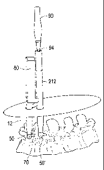
532.
[0098] In an alternate embodiment, it may be desirable for the engagement
between the
instrument and the spinal fixation element to change the orientation of the
spinal fixation
element with respect to the instrument shaft during the procedure. This
embodiment of the
instrument has an articulating engagement that allows for manipulation of the
fixation
element from an orientation parallel with the instrument shaft to an
orientation perpendicular
to the shaft during the procedure. An example of an instrument allowing these
movements is
disclosed U.S. Patent Application Publication No. 2005/0131419, filed December
16, 2003 entitled
"Pivoting Implant Holder" and U.S. Patent Application Publication No.
2005/0131420, filed
December 16, 2003, entitled "Pivoting Implant Holder,"
Another embodiment of an instrument 800 having an
CA 02555141 2012-01-11
articulating engagement with the spinal fixation element is shown in FIGS. 29-
30. The
instrument 800 engages a projection 872 on a spinal fixation element, shown as
a spinal rod
870, by a collet 860 extending by a spring 862 from the distal end of the
instrument shaft 803.
A locking sleeve 864 locks the projection 872 of the rod 870 within the collet
860.
Articulation of the rod 870 is provided by two linking arms 866 extending from
the shaft 803
to the collet 860, which allow the rod 870 to rotate or pivot.
[0099] For reference purposes, FIG. 1 illustrates an exemplary spinal anchor
for use with the
methods and devices of the present invention. A person skilled in the art will
appreciate that
a variety of anchors can be used with the devices and methods of the present
invention,
including, for example, spinal screws, hooks, bolts, and wires. FIG. 1
illustrates a spinal
screw that includes a distal, bone-engaging portion, e.g., a threaded shank
54, and a proximal,
U-shaped, receiver member head 52 that is adapted to seat a spinal fixation
element, for
example a spinal rod. The threaded shank 54 can be fixedly attached to the
receiver head 52
to form a monoaxial screw, or alternatively the shank 54 can be configured as
a polyaxial
screw, as shown, that is rotatably disposed through an opening formed in the
distal end of the
receiver head 52 to allow rotation of the shank 54 with respect to the
receiver head 52. A
variety of techniques can be used to allow rotation of the head 52 with
respect to the shank
54.
[00100] FIGS. 12-17 show a minimally invasive method of implanting a spinal
fixation
element. While the method is shown and described in connection with the
percutaneous
access device 12 (FIG. 1), percutaneous access device 212 (FIG. 3), and spinal
screw 50
disclosed herein, a person skilled in the art will appreciate that the method
is not limited to
use with such devices, and that a variety of other devices described herein
and known in the
art can be used. Moreover, while only two access devices 12, 212 and two
anchors 50, 50'
are shown in FIGS. 12-17, the method of the present invention can be performed
using any
number of access devices and anchors. The method can also be performed using
only some
of the method steps disclosed herein, and/or using other methods known in the
art.
[00101] An example of a procedure for placing the spinal anchors and
percutaneous
access devices is disclosed in U.S. Patent Application Publication No. 2005/0
1 3 1 42 1, filed December
16, 2003, entitled "Methods and Devices for Minimally Invasive Spinal Fixation
Element
Placement, After the anchors 50, 50' are
21
CA 02555141 2006-08-03
WO 2005/076868 PCT/US2005/003246
implanted with the percutaneous access devices attached, a spinal fixation
element 70 may be
delivered to the anchor site as described below.
[00102] In accordance with one exemplary method, an instrument for engaging
and
manipulating a spinal fixation element, such as the instrument 80 described
above, may be
connected to a spinal fixation element, e.g., a spinal rod 70, as illustrated
in FIG. 12. The
shaft 82 of the instrument 80, with the spinal rod 70 engaged at the distal
end of the shaft 82,
may be positioned through the side wall openings 14b of the percutaneous
access device 212
attached to a second bone anchor 50' and through the sidewall opening 14b of
the
percutaneous access device 12 attached to a first bone anchor 50, as
illustrated in FIGS. 13
and 14. The spinal rod 70 may be introduced into percutaneous access device 12
in a first,
lengthwise orientation, such that the spinal rod 70 is oriented substantially
parallel to the
longitudinal axis L of the access device 12. Where the spinal fixation element
has a curved
orientation or it has some other configuration, it is understood that the
fixation element is in
the "substantially parallel" orientation when it is positioned lengthwise
through the
percutaneous access device.
[00103] The spinal rod 70 may be moved through the lumen of the percutaneous
access device 12 toward the distal end 12b, by moving the handle 86 of the
instrument 80
distally, as shown in FIG. 15. Referring now to FIGS. 16 and 17, as the spinal
rod 70
approaches the distal end 12b of the access device 12, the orientation of the
spinal fixation
element 70 can be manipulated to direct it towards the spinal anchor 50' by
rotating the
handle 86 of the instrument 80 from a position parallel to the patient's spine
to a position
parallel to the percutaneous access device 12 such that the handle straddles
the proximal end
of the percutaneous access device 12. Rotating the handle causes the spinal
fixation element
70 to assume a second orientation that is different from the first
orientation, and that is
substantially parallel to the patient's spinal column and/or transverse to the
first orientation.
As the handle is rotated to straddle the percutaneous access device 12, the
shaft 82 of the
instrument moves through the proximal sidewall opening 14 of device 212 and
exits through
the proximal end of the device 212. The shaft 82 of the instrument 80
maintains contact with
percutaneous access device 12 until the spinal fixation element 70 has
established contact
with the distal sidewall opening of percutaneous access device 212. The sizing
of the shaft
82 of the instrument 80 aids in keeping the sidewall openings 14 of the
percutaneous access
22
CA 02555141 2006-08-03
WO 2005/076868 PCT/US2005/003246
devices 12, 212 in alignment while the spinal fixation element 70 is being
manipulated into
position in relation to the spinal anchors 50, 50'.
[00104] It is understood that the angle of the fixation element 70 in the
second
orientation will vary depending on the type of fixation device being
implanted, as well as the
orientation of the access device 12, which can vary throughout the surgical
procedure since
the access device 12 can be positioned at several angles with respect to the
patient's spinal
column.
[00105] During transition of the spinal fixation element 70 from the first
orientation to
the second orientation, a leading end of the spinal fixation element 70 may be
positioned
below the fascia layer. Referring to FIGS. 16-17, manipulation of the spinal
fixation element
70 is continued until the spinal fixation element 70 is positioned in relation
to one or more
spinal anchors. Depending on the type of spinal anchor used, the fixation
element can be
positioned to be directly or indirectly mated to the spinal anchor. As shown
in FIG. 17, the
fixation element 70 is fully seated in the receiver heads 52, 52' of the
adjacent spinal anchors
50, 50'.
[00106] A person skilled in the art will appreciate that the spinal fixation
element 70
does not need to be directly attached to each anchor 50, 50', and that it can
be indirectly
attached to the anchors 50, 50' using, for example, a band clamp, or slotted
or offset
connectors.
[00107] To verify that the spinal fixation element is fully seated in the
receiver head of
the spinal anchor an instrument 90 can be inserted through the proximal end of
the
percutaneous access device 12 until it can not be advanced any further, as
illustrated in
FIGURES 18-19. The proximal end 90a of the instrument has a marker 94 to
indicate the
depth from the proximal end of the percutaneous access device to the top of a
spinal fixation
element fully seated in a spinal anchor. If the marker 94 is aligned with the
proximal end 12a
of the access device, when the instrument is placed down the lumen of the
access device, then
the spinal fixation element is in the proper position within the spinal anchor
and the closure
mechanism can be applied, through the access device. The spinal fixation
element 70 can
then be disengaged from the instrument 80, which can be removed from the
access device 12.
If the marker is not visible above the proximal end of the access device, the
fixation element
23
CA 02555141 2006-08-03
WO 2005/076868 PCT/US2005/003246
is in not in the proper position and should be repositioned. In an alternate
embodiment of the
instrument 90, a closure mechanism may be attached to the instrument 90 and
the marker
may be employed to indicate if the spinal fixation element is fully seated and
if closure
mechanism is properly inserted.
[00108] Once the fixation element 70 is secured in relation to the implants
50, 50', the
access devices 12, 212 can be removed from the implants 50, 50', leaving only
minimally
invasive percutaneous incisions in the patient where each access device 12,
212 was
introduced. This is particularly advantageous in that it reduces the amount of
trauma caused
to the patient, and it minimizes the damage to muscle surrounding the surgical
site.
[00109] An alternative embodiment of delivering a spinal fixation element,
spinal rod
70 to a first bone anchor 50 and a second bone anchor 50' is illustrated in
FIGS. 20-25. In
the illustrated embodiment, a first percutaneous access device 212 (FIG. 3)
and a second
percutaneous access device 212' are connected to a first bone anchor 50 and a
second bone
anchor 50'. An instrument 80 is connected to the spinal rod 80 (FIG. 20) and
may be
employed to position the spinal rod 70 in the proximal side wall openings 14b
of the first and
a second percutaneous access devices 212, 212' (FIGS. 21-23) and to manipulate
the spinal
rod 70 into proximity to the first bone anchor 50 and the second bone anchor
50' (FIGS. 24-
25).
[00110] In another embodiment, the percutaneous access device 112 shown in
FIGS. 5
-6B can be used to facilitate introduction of a spinal fixation element into a
surgical anchor
site. As previously stated, access device 112 includes a guide member 120 to
direct the
spinal fixation element 70 from the first orientation to the second
orientation. This is
illustrated in FIGS. 26-28. As shown, as the spinal fixation element 70 is
moved distally to
come into contact with the guide member 120, the guide member 120 causes the
spinal
fixation element 70 to rotate and extend toward the opening 114 in the
percutaneous access
device 112. As a result, the spinal fixation element 70 is directed into the
second orientation,
whereby it can be positioned in or adjacent to the receiver heads 52, 52' of
the adjacent spinal
implants 50, 50'. The guide member 120 can be adjusted along the longitudinal
axis of the
access device to position the guide at the desired location to contact the
spinal fixation
element and begin changing its orientation.
24
CA 02555141 2006-08-03
WO 2005/076868 PCT/US2005/003246
[00111] As previously stated, a person skilled in the art will appreciate that
the
exemplary methods described herein can be performed in any sequence using some
or any of
the steps. Moreover, the percutaneous access devices, instruments, and methods
described
can be used in any combination to deliver multiple spinal fixation elements
simultaneously or
sequentially, and/or to perform a variety of other surgical procedures not
illustrated or
described herein.
[00112] FIGS. 47A-48 illustrate an instrument 600 for aiding in the insertion
and
manipulation of a percutaneous access device, such one of the exemplary
percutaneous
access devices described above. The instrument 600, as discussed below, is
particularly
suited to facilitate the delivery and manipulation of a percutaneous device
having one or more
sidewall openings, such as the exemplary percutaneous access device
illustrated in FIG. 3.
The instrument 600 is in the form of a cylindrically-shaped sleeve having a
proximal end
612a, a distal end 612b, and an inner lumen 612c formed therein that extends
between
proximal and distal ends 612a, 612b. The length of the instrument 600 may vary
depending
on, for example, the length of the percutaneous access device. In the
illustrated exemplary
embodiment, for example, the instrument 600 is approximately equal to or less
than the
length of the percutaneous access device 212. The distal end 612b of the
instrument 600 may
have a chamfer 615 to ease insertion. The outer surface of the instrument 600
may have
surface features to facilitate gripping of the instrument 600. For example, in
the illustrated
exemplary embodiment, the outer surface of the instrument 600 includes a
plurality of
dimples 618 arranged about the circumference of the instrument 600 proximate
the distal end
612b and the proximal end 612a. The inner surface of the instrument 600, which
defines the
lumen 612c, may have one or more projections extending inwardly therefrom to
engage a
sidewall opening in the percutaneous access device and inhibit rotation of the
instrument 600
relative to the percutaneous access device. In the illustrated exemplary
embodiment, for
example, in the inner surface of the instrument includes a pair of projections
620 proximate
the distal end 612b and a pair of projections 620 at the proximal end 612. The
projections
620 are each sized to fit within a sidewall opening of a percutaneous access
device, for
example, the sidewall openings 14b of the percutaneous access device 212, as
illustrated in
FIG. 48. In the illustrated embodiment, each projection 620 in a pair of
projections is
positioned diametrically opposite the other projection 620 in the pair. In
use, the instrument
600 provides rigidity to the percutaneous access device to aid in insertion of
the percutaneous
CA 02555141 2006-08-03
WO 2005/076868 PCT/US2005/003246
access device and bone anchor assembly. Moreover, the instrument 200 may be
employed
after insertion to facilitate manipulation of the percutaneous access device.
For example, the
instrument 600 may be used to provide counter-torque and/or for compression or
distraction
of the bone anchors. The instrument 600 may be removed prior to insertion of
the spinal
fixation element.
[00113] To facilitate insertion of the spinal fixation element having the
proper length, a
measuring instrument 700 may be used to determine the length of the spinal
fixation element
for insertion between two bone anchors. The measuring instrument 700, in the
illustrated
exemplary embodiment, may have a first arm 710a and second arm 710b that are
connected
proximate the proximate end 712a of the measuring instrument 700. In the
illustrated
exemplary embodiment, the two arms 710a, 710b pivot around a pivot point 730
relative to
one another. The first arm 710a and the second arm 710b may be connected by a
spring 720
that biases the arms 710 away from each other. In the illustrated embodiment,
each arm 710
may have a generally cylindrical shape and may tapers along the length from a
first diameter
at the proximal end 712a to a second, reduced diameter at the distal end 712b.
In the
illustrated exemplary embodiment, the diameter of each arm 710 may be less
than the inner
diameter of a percutaneous access device to permit the arm 710 to be inserted
through the
percutaneous access device, distally, into proximity with a bone anchor
connected to the
percutaneous access device and engaged to a vertebra. The distal end 712b of
each arm 710
may have a spherical tip 715 having a size analogous to a size of a spinal
fixation element to
facilitate placement of the spherical tip 715 into a bone anchor, for example
into the receiver
head of the bone anchor. A centering ball 750 maybe located along each arm 710
near the
distal end 712b to center the arm 710 within the percutaneous access device
and facilitate
proper measurement of the distance between distal ends of the arms 710a,b, and
thus, the
distance between the bone anchors. The measuring instrument may include a
locking system
760 to fix the position of the first arm 710a relative to the second arm 710b
and, thus, permit
the distance between the distal ends of the arms 710a, b to be fixed during a
measurement. In
the illustrated exemplary embodiment, the locking system 760 may include a
threaded rod
767 that intersects the first arm 710a and the second arm 710b and an
internally threaded
knob 765 that engages the external threads on the rod 767 and is adjustable
along the length
of the rod 767. The knob 765 maybe advanced along the rod 767 into contact
with the
second arm 710b to fix the position of the second arm 710b relative to the
first arm 710a. In
26
CA 02555141 2006-08-03
WO 2005/076868 PCT/US2005/003246
an alternative embodiment illustrated in FIG. 44B, a measuring instrument 700'
may have a
locking system 760 that includes a slip friction clutch 785 to inhibit over
tightening of the
knob 765 against the second arm 710b.
[00114] In operation, the first arm 710a of the measuring instrument 700 may
be
inserted through a first percutaneous access device 212 into proximity to a
first bone anchor
50 connected to the first percutaneous access 212 and a first vertebra. The
second arm 710b
of the measuring instrument 700 may be inserted through a second percutaneous
access
device 12 into proximity to a second bone anchor 50 connected to the second
percutaneous
access 212 and a second vertebra. In the illustrated exemplary embodiment, the
spherical tip
715 of each arm is advanced into contact with the receiver head of the bone
anchor. The
arms 71 Oa and 71Ob may be fixed relative to one another using, for example,
the locking
system 760. The arms 710a and 710b maybe removed from the percutaneous access
devices
212, 12 to determine the distance between the distal ends 712b of the arms
710a, 710b. For
example, a template block 800 may be employed to facilitate measurement of the
distance
between the distal ends 712b of the arms 710a, 71 Ob, as illustrated in FIG.
46. The
exemplary template block 800 may include a plurality of openings, markings, or
other
reference points that are spaced apart a predetermined distance. For example,
the template
block 800 may include a first opening 810 for receiving the distal end of one
of the arms 710
and a plurality of additional openings 820 that spaced apart predetermined
distances from the
first opening 810. The template block 800 may include indicia 830 proximate
the plurality of
second openings that is indicative of the distance between the first opening
810 and one or
more of the second openings 820. A fixation element may be selected based upon
the
distance measured between the distal ends 712b of the arms 710a, 710b.
[00115] In alternative exemplary embodiments, the measuring instrument 700 may
include a scale or other device mounted to the instrument to facilitate
measuring the distance
between the distal ends 712b of the arms 71 Oa, 71 Ob without necessitating
removal of the
arms 710 from the percutaneous access devices or without necessitating a
locking system to
facilitate fixing the position of the arms relative to one another.
[00116] One skilled in the art will appreciate further features and advantages
of the
invention based on the above-described embodiments. Accordingly, the invention
is not to
be limited by what has been particularly shown and described, except as
indicated by the
27
CA 02555141 2006-08-03
WO 2005/076868 PCT/US2005/003246
appended claims. All publications and references cited herein are expressly
incorporated
herein by reference in their entirety.
What is claimed is:
28