Note: Descriptions are shown in the official language in which they were submitted.
CA 02555807 2006-08-10
WO 2005/077260 PCT/CA2005/000147
NON-INVASIVE METHOD AND APPARATUS FOR DETE~G A PHYSIOLOGICAL
PARAMETER
FIELD OF THE INVENTION
[000I] This invention relates to field of physiological analysis, and more
particularly to
apparatus and methods for the non-invasive analysis and, detection of
physiological
characteristics, such as heart rate, blood pressure, cardiac output,
respiration response and body
composition including hydration, body fat content, glucose, lactate,
hemoglobin and blood
oxygen.
BACKGROUND OF INVENTION
[0002] The need for the development of non-invasive physiological analysis
tools stems
from the prevalence in our society of obesity, lack of physical exercise,
stress 'and
demographical situation. As a result, in the US alone, more than 60 million
people suffer from
cardiovascular diseases, more than 18 million are diagnosed with diabetes, and
more than 30%
of the population is considered as overweight. Many of these people require
close monitoring
of physiological parameters including heart rate, blood pressure, glucose
level, body index and
so on.
(0003] The non-invasive analysis ofphysiological parameters is a very
important
direction of development in modern medical, consumer and fitness apparatus.
Products of this
type include, but are not limited to, heart rate monitors, blood pressure
monitors, Sp02
monitors, hydration and body fat monitors and so on.
(0004] From the point of view of physical principles the existing techniques
can be
divided in three groups: 1 ) the measurement of physiological parameters by
using the bio-
electric properties of the human body, 2) optical analysis of physiological
parameters and 3)
CA 02555807 2006-08-10
WO 2005/077260 PCT/CA2005/000147
the synchronization of physiological measurements with the ECG R-peak.
[0005] . The first group is based on the connection between physiological
parameters and
the bioelectrical properties of the human body. The most common examples of
this direction
include ECG detection, bio impedance monitoring of cardiac output, respiration
parameters,
water and fat composition; and RF glucose monitoring. Other examples of this
group include
EEG, EMG, EGG, nerve and muscle stimulations and so on.
[0006] One approach is based on the assumption that the glucose concentration
has an
effect on the complex impedance of the human body in the frequency range 1-
1000 MHz,
see for example, US patent No. 5,792,668. This technique, referred to as RF
spectroscopy, has
been studied experimentally and applied to the design of apparatus for
continuous glucose
measurements inside a wristwatch. This approach has several technological
advantages
including low current drain and reasonably inexpensive components.e The main
problem with
RF spectroscopy alone is that the complex impedance is sensitive to a number
of factors such
as water, salt, fat, temperature and so on. It is impossible to measure all
those factors in real
time using RF spectroscopy in order to calibrate the measurements. Therefore
the use of very
complicated and time-consuming calibration procedures is required. These often
involve
getting several invasive measurements at different glucose concentrations for
comparison with
RF readings so as to recalibrate the system on a regular (e.g. daily basis).
Without proper
regular calibration, there is no way to obtain accurate results using only RF
spectroscopy.
[0007] US patent Nos. 6,125,297 and 5,788,643, teach the use of body impedance
measurements to find water and fat concentration in the human body but the
results of such
measurements depend on unknown salt concentration. Bio impedance measurements
can
provide estimates of average water and fat composition in human body but in
some cases the
CA 02555807 2006-08-10
WO 2005/077260 PCT/CA2005/000147
knowledge of local body composition becomes important.
[000] The main problem associated with bioelectrical investigation of the
body's
physiological parameters is the effect of other variables on the complex
impedance of the
human body that cannot be detected with bio-impedance measurements alone. For
example,
the electrolyte concentration, blood volume and so on can dramatically change
the complex
impedance for the same water and fat concentration.
(0009] It is known to perform optical measurements for detection of body
physiological
parameters. For example, US patent No. 6,466,807 to Dobson et al teaches how
to measure in
vivo the concentration of an analyte using a plurality of wavelengths. US
patent No. 5,553,613
discloses a method of measuring the glucose in blood using several
wavelengths. It is also
known that the absorption spectrum is sensitive to the body chemistry. For
example: 660 nm is
sensitive to hemoglobin, 905 nm - oxy-hemoglobin, 920 - fat, 970 nm - water,
1054 nm -
glucose, 1253 nm - collagen, 1270 nm - water, and 1660 nm - lactate.
Typically, the spectra
are very broad and peaks can be shifted for different body and chemistry
compositions. The
actual absorption spectrum observed is the superposition of several broad
bands corresponding
to the individual components. It is very difficult to measure the optical path
in a strongly
diffuse medium such as a human body, and to extract therefrom an absolute or
relative
concentration of chemical components from relative measurements. It is common
to use the
ratios I970/I810 and I1050/I810 in order to find relative water and glucose
concentration. The
line 1050 nm contains a large contribution of water component, and the line
970 also contains
contribution from collagen and fat. Therefore, there is a need to use
additional information in
order to separate overlapping optical bands. It is also known to synchronize
optical
measurements with an ECG R-peakmarker.
CA 02555807 2006-08-10
WO 2005/077260 PCT/CA2005/000147
[0010] The main problem with optical measurement and analyses is a lack of the
complementary information on body parameters obtained from independent
measurements.
[0011] I~iani, US patent No. 6,526,300, teaches to combine bio-electrical
measurements
with optical measurements in order to ensure that a device is properly
positioned and reduce
the number of false alarms. In this arrangement, the electrodes are used to
ensure the proper
positioning of the~optical sensors. They are not used in combination to
measure physiological
parameters.
[0012] US patent no. 6,192,262 discloses a system for making functional maps
of the
human body by monitoring various physical parameters. This patent teaches that
a reference
parameter can be used for a choice of another parameter's recording regime,
but it does not
teach to improve the accuracy of a non-invasive measurement.
[0013] Additional prior art techniques, involve obtaining a final result from
more than one
source and trying to predict the most accurate measurement, or taking a
measurement and
trying to compensate for changes in some perturbing factor, such as
temperature, but in all
such cases the final result is still in effect obtained from only one primary
source of data. WO
03/063699 is an example of such a prior art technique.
SUlVInZARY OF THE INVENTION
[0014] The invention takes advantage of the fact that improved results can be
obtained by
deriving a physiological parameter from the aggregate effect of changes in
that parameter on
multiple disparate physical properties. Disparate in this context means that
the properties are
physically different in nature. They should each be independently capable of
measuring the
physiological property. In accordance with the teachings of the invention, a
final result is
predicted from the aggregate effect of changes in the property. For example,
changes in
4
CA 02555807 2006-08-10
WO 2005/077260 PCT/CA2005/000147
hydration level simultaneously affect optical and bio-impedance properties of
an animal
subject. A particular hydration level implies a particular,combination of the
values for optical
and bio-impedance properties. By deriving the hydration level from the
aggregate effect on a
these properties, a more accurate result can be obtained than can be obtained
from either of
these properties alone or by merely attempting to compensate for inaccuracies
introduced into
the system, for example, by environmental changes. It will be understood in
this application
that the term animal refers to both human and non-human animals.
[0015] . In order to obtain a measurement, calibration data reflecting the
effect of changes
in the physiological parameter on the physical properties need to be obtained.
This can be
achieved by experimentally taking measurements and creating a table and then
consulting the
table to ,obtain a parameter from a particular combination of results, or
alternatively predicting
the effects of changes in the physiological parameter on the properties using
a mathematical
model of animal physiology.
[0016] In other words, independent sources of information on body parameters
should be
used at the same time in order to obtain the complementary information on
unknown
parameters. In one embodiment optical measurements are taken as an independent
source of
information.
[0017] Accordingly one aspect of the invention provides a method of non-
invasively
determining a physiological parameter of a subject comprising generating
signals
representing at least two disparate physical properties of the subject, each
of said
disparate physical properties having a value that varies in dependence on said
physiological parameter and is independently capable of giving a measurement
thereof;
determining the effect of changes in said physiological parameter on each of
said at least
CA 02555807 2006-08-10
WO 2005/077260 PCT/CA2005/000147
two disparate physical properties; and processing said signals to derive said
physiological
parameter from the aggregate effect of said physiological parameter on said at
least two
disparate physical properties.
[0018] It will be understood in this context that the signals can be generated
in any
manner that creates electrical signals representing the property that are
suitable for further
processing. They can, for example, be generated by transducer that actively
generates
signals from some physical phenomenon, such as pulse rate. Alternatively, the
signals
could also originate within the body and be, for example, ECG signals, which
are merely
detected by a passive pick-up.
[0019] More than one component may be extracted from the signals during
processing. For example, in the case of a complex bio-impedance the final
result may
depend on such values as average impedance, average phase, and average maximum
rate
of change of impedance.
[0020] In another aspect the invention provides a non-invasive apparatus for
determining a physiological parameter of a patient comprising at least two
sensors for
generating and/or detecting signals representing disparate physical properties
of the
subject, each of said disparate physical properties having a value that varies
in
dependence on said physiological parameter and is independently capable of
giving a
measurement thereof; and a processor configured to process said signals to
derive said
physiological parameter from the aggregate effect of said physiological
parameter on said
at least two disparate physical properties.
[0021] The processor can derive said physiological parameter from calibration
data stored
in a memory or from a mathematical model of the animal (human or non-human)
physiology.
CA 02555807 2006-08-10
WO 2005/077260 PCT/CA2005/000147
[0022] In a preferred embodiment the at least one of the signals is optical
and at least one
of the other signals is an RF or bio-impedance signal. Typical physiological
parameters that
can be measured include water, electrolyte, fat, glucose, hemoglobin, lactic
acid, cardiac
output, respiration, oxygen saturation and blood pressure.
[0023] In yet another aspect the invention provides a non-invasive physiology
analysis
system comprising a sensor adapted for attachment to a patient and supplying
to the patient an
optical signal and at least one additional signal selected from the group
consisting of RF and
bio-impedance signals, and receiving signals from the body in response to the
supplied signals;
a detector coupled to said sensor for detecting said received signals and
producing output
signals in response to said detected signals, and a signal processing
subsystem coupled to said
detector and receiving said output signals, said signal processing subsystem
analyzing said
output signals to determine information about at least one physiology
parameter.
[0024] The physiology parameter may be selected from the group consisting of
water,
electrolyte, fat, glucose, hemoglobin, lactic acid, cardiac output,
respiration, oxygen saturation
and blood pressure, and may include body composition.
[0025] The present invention therefore provides a device and methods for
performing
non-invasive, accurate, measurement of physiological parameters of a living
body, by
combining disparate technologies, such as bioelectrical and optical analysis
technologies
including optical spectrum analysis and one or more of bio-impedance analysis,
RF impedance
analysis, temperature and ECG. Specifically, the present invention can be used
to measure and
analyze numerous aspects of a patient's physiology, such as cardiac output,
blood pressure,
body composition (e.g. local and total body water, fat and electrolytes) and
blood chemistry
such as oxygen saturation, hemoglobin, glucose and lactate concentrations. The
use of multiple
7
CA 02555807 2006-08-10
WO 2005/077260 PCT/CA2005/000147
inputs from disparate sources gives more accurate results than can be obtained
from a single
source, or a single source that is merely compensated.
BRIEF DESCRIPTION OF THE DRAWINGS
(0026] The invention will now be described in more detail, by way of example
only, with
reference to the accompanying drawings, in which:
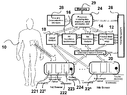
[0027] Figure 1 is a system level block diagram of a physiology analysis
system utilizing
the present invention;
[0028] Figure 2 is an equivalent circuit diagram for ECG measurements;
[0029] Figure 3 illustrates a typical ECG signal showing R-peak;
[0030] Figure 4 is an equivalent circuit for bio-impedance measurements of the
body;
(0031] Figure 5 is an equivalent circuit for local bio-impedance
measurements;.
[0032] Figure 6 is an equivalent circuit for local RF impedance spectroscopy
measurements;
[0033] Figure 7 is a transmissive optical analysis;
[0034] Figure 8 illustrates a backscattered/reflected optical analysis
configuration;
[0035] Figure 9 is a preferred embodiment of a,two sensor module
configuration;
[0036] Figure 10 shows a minimal embodiment in the two sensor module
configuration;
(0037) Figure 11 shows a preferred embodiment in the single sensor module
configuration;
[0038] Figure 12 shows a minimal embodiment in the single sensor module
configuration;
CA 02555807 2006-08-10
WO 2005/077260 PCT/CA2005/000147
(0039] Figure 13 shows the aggregate glucose high level process;
[0040] Figure 14 shows the aggregate blood pressure high level process;
[0041] Figure 15 shows the sensor attachment detection process;
[0042] Figure 16 illustrates an ECG data acquisition process;
S [0043] Figure 17 illustrates a bio-impedance data acquisition process;
[0044] Figure 18 illustrates an RF data acquisition process;
(0045] Figure 19 illustrates an optical data acquisition process;
(0046] Figure 20 illustrates a generic parameter extraction signal processing
process;
(0047] Figure 21 illustrates an aggregate glucose signal processing; and
(0048] Figure 22 illustrates an aggregate blood pressure signal processing
process in
accordance with one embodiment of the invention.
DETAILED DESCR~T~ON OF PREFERRED EMBODIIVVIENTS
(0049] As noted above, in accordance with the principles of the invention, a
final result
for a physiological parameter is obtained from multiple disparate sources of
data.
[0050] Figure 1 discloses a system level block diagram of a preferred
embodiment for
analyzing the physiology of a patient 10. This system combines a physical
noninvasive optical
analysis subsystem with one or more physical noninvasive bioelectric
measurement sub-
systems: a passive block subsystem that passively measures physiology
attributes such as
Electrocardiogram (ECG), temperature and sensor pressure; a bio-impedance
analysis
subsystem I4, an RF-impedance Spectroscopy subsystem; and an optical analysis
subsystem 18.
9
CA 02555807 2006-08-10
WO 2005/077260 PCT/CA2005/000147
[0051] An electrode cross-point switch 20 allows sensor module electrodes 22~
...22" to
be connected to any of the bioelectrical analysis subsystems, giving. maximum
flexibility in
electrode configuration. The electrical cross point switch 20 allows the
electrodes to be
switched to a single subsystem allowing measurements to be made over an
extended period or
to interleave measurements from any combination of several subsystems rapidly.
The cross-
point switch 20 also allows multiple subsystems to be connected to the
electrodes
simultaneously for concurrent measurements. It would also be possible to
design the system
without the switch such that the electrodes are wired into one or more of the
subsystems in a
fixed configuration and with circuitry such as filters to allow for
asynchronous and/or
concurrent subsystem operations.
[0052] Outputs from the bioelectric and optical analysis subsystems are
provided to the
processor subsystem 24, which includes the data acquisition and signal
processing functions.
The data acquisition function takes analog and digital signals from the
optical and bioelectrical
analysis subsystems and convert them into their internal representations for
further analysis.
The physical implementation for the acquisition function could use any number
of analog to
digital converters (ADCs), digital bit-ports or integrated acquisition
peripherals. However, the
preferred embodiment uses an embedded processor with multiple integrated 10
and 12 bit
ADCs since they are readily available and reduce the overall cost of the
device. The sampling
rate for the acquisition function is selected to provide sufficient resolution
of the measured
signals. The sampling rate and duty cycle could be different for the different
sensor types.
[0053] The processor sub-system 24 may include a memory 29 storing a look-up
table
containing calibration data representing different values of the signals for
different values of
the physiological parameter in question.
CA 02555807 2006-08-10
WO 2005/077260 PCT/CA2005/000147
(0054] The processor subsystem 24 also includes a signal processing function,
which
analyzes the data acquired from the optical, RF and bio-impedance analysis
subsystems and
the passive subsystem. The signal processing performs digital signal
conditioning and
statistical analysis functions such as PLS, PCR, etc. with the net result of
turning the captured
data into meaningful physiology attributes and other processed intermediate
results. The
preferred embodiment shows the data acquisition, signal processing and
processor functions
physically contained within the same physiology analysis device. Many
combinations of
components and subsystem configuration are possible depending on the
technology utilized.
Alternatively, they could also be physically separated in a variety of remote
configurations: for
example the sensor modules could be remotely connected through fiber optics
and cables, the
data acquisition system could transmit the raw captured data through wired or
wireless
communications, the signal processing function could transmit the intermediate
or final results
through wired or wireless communications, and the user interface could be
remotely operated
through wired or wireless communications. For example the raw acquired data
could be sent
to an external system such as a PC through a wired, fiber or Bluetooth
wireless connection for
analysis and/or presentation. Thus the external PC would be part of the
physiology system in
such a configuration.
[0055] In the preferred embodiment the processor controls the overall system
and all of
the subsystems either directly or indirectly. The power management subsystem
26 provides
~ power and power conditioning for the entire system.
[0056] The user interface 28 provides interaction with the user. Input is
accepted to
determine what function to execute and to configure the system such as user
information and
calibration parameters. The user interface for a portable device could range
from simple
switches and LEDs to more elaborate touch screen LCD displays and keypads. The
user
11
CA 02555807 2006-08-10
WO 2005/077260 PCT/CA2005/000147
interface for a.remote system can be much more extensive such as a standalone
PC based
application running on a local or remote workstation, or a PDA or cellular
telephone.
[0057] The device can also be accessed remotely, for example, through a
network or via
an attached PC through the Communications Interface, so that configuration,
control, data
collection, analysis and presentation can be done from a separate system
and/or a separate
location. USB, serial, IRDA, wireless are just a few examples of
Communications Interfaces
that could be used for remote access.
[0058] The sensor modules 22' . . .22° are attached to the body or the
body comes in
contact with sensor modules so that physiology information of the body can be
sensed. Each
sensor 22n includes electrodes 221, multiple wavelength optical sensors 222,
electrodes 223,
and passive sensors 224. The physiology sensing.system requires at least one
sensor module
containing a combination of electrodes and optical receiver/detector
components. Optionally
additional sensor modules may be present, each sensor module containing
electrodes and/or
optical components. These sensors are placed in locations sensitive to the
additional
information to be detected. For example, by placing an additional sensor with
a pair of
electrodes on the opposite side of cardiac divide from the first
electrical/optical sensor, ECG,
cardiac output and respiratory function information can be detected. The
sensors can be
conveniently mounted on a single module configured to allow the user to place
a hand on the
module with different fingers and the thumb exposed to different sensors.
[0059] The electrode cross-point~switch 20 is used to interconnect the ECG,
bio-
impedance and RF-impedance spectroscopy analysis blocks to specific electrodes
in the
sensing modules. This switching arrangement allows any combination of two or
more
electrodes on any of these modules to be connected to any of the bioelectric
analysis sub-
12
CA 02555807 2006-08-10
WO 2005/077260 PCT/CA2005/000147
systems so that any combination of two electrode or four electrode
configurations within a
single module or between two or more modules can be configured as needed. It
also allows
electrodes that are not being used at a specific point in time to be left
disconnected from the
analysis circuitry so as to reduce power consumption and eliminate unwanted
interference,
which would require additional compensation circuitry to remove the
interference. The
electrodes can be switched to a single subsystem allowing measurements to be
made over an
extended period (seconds or longer) or to interleave measurements from several
subsystems
rapidly. The electrodes cari also be connected to multiple analysis circuits
simultaneously so
that concurrent measurement can be made if required.
(0060] Figure 17 illustrates how the cross point switch is used to select the
correct
electrodes to perform the Bio-Impedance data acquisition. The process starts
by first selecting
the electrodes on the primary sensor module to acquire data for local bio-
impedance analysis.
After the local bio-impedance analysis time slice is completed the cross point
switch is used
again to switch to the electrode pairs on two separate sensor modules to
acquire data for body
' bio-impedance analysis. Note that the body bio-impedance analysis data
acquisition is only
performed on configurations with two or more sensor modules.
[0061] A method to automatically detect that the sensors are properly attached
improves
the user experience for this type of device and ensures that consistent,
accurate measurements
are made. The determination for proper attachment can be made from a
combination of
sensors in the device: the bio-impedance analysis or RF sensors for electrode
connectivity,
contact pressure sensor, temperature sensor and optical sensor for motion
detection. For this
function the bio-impedance analysis and RF sensors are used to pass an
alternating current
through the different electrode pairs to monitor connectivity. When the
electrodes are properly
attached the current will increase dramatically (to a maximum safe level)
making it an ideal
13
CA 02555807 2006-08-10
WO 2005/077260 PCT/CA2005/000147
trigger for attachment detection. The preferred embodiment uses the bio-
impedance sensors
and the temperature sensor to determine proper attachment. A visual indication
can be given to
the user if the sensors are not properly attached, for example with a text
message to the user
indicating that the sensors must be readjusted. With the sensor modules
properly in place, the
other acquisition and analysis block functions can then start. With proper
mechanical design of
the outei electrodes with respect to all other sensors in the module once the
outer electrodes
are determining to .be properly attached, all other sensors in the module will
also be properly
attached.
[0062) Figure 15 illustrates the steps taken on the preferred embodiment to
detect good
I 0 sensor attachment before the data acquisition phases start. The same
process can be used using
the RF sensors for configurations without bio-impedance sensors. First the
process selects the
bio-impedance electrodes on the main module and applies an AC current. The AC
current is
monitored continuously to detect a sudden rise in current, which is expected
when the sensor
comes in contact with the skin. For configurations with two or more modules,
this process is
repeated for each sensor module. Once good contact has been detected for all
sensor modules
then the skin temperature can be checked to further confirm that good sensor
contact has been
achieved. If any of the sensor attachment checks fail then the entire process
is restarted thus
ensuring that all sensors are well attached at the same time.
[0063) In the passive blockl2, various passive sensors can be added to help
provide
additional information about the target measurement site that can be used by
any signal
processing algorithms. For example, a thermal sensor can measure skin
temperature so as to
compensate for any changes that temperature might have on the other sensor
readings. These
passive sensors can also provide useful data directly related to the parameter
of interest.
Although not shown, other passive sensors such as pressure sensors to account
for sensor
14
CA 02555807 2006-08-10
WO 2005/077260 PCT/CA2005/000147
contact pressure, humidity sensors to account for skin perspiration and/or
environmental
humidity, etc. could also be beneficially added. Further, passive information
received from
electrode pairs in separate modules can be used to pick up ECG signals.
[0064] An example of an ECG equivalent circuit 30 is shown in Figure 2. The
ECG sub-
system 32 is used to pick up passive cardiac voltage potentials between an
electrode on the left
sensor module and an electrode on the right sensor module, for example LEI and
RE1 as
shown. The raw cardiac signal is processed to determine the occurrence of R-
peak as shown in
Figure 3. Most of the QRS complex spectrum is in the 5-30Hz range and the ECG
signal is
very small, typically 4mv or less. 'The primary function of the circuit is to
isolate the QRS
complex, filter out noise, especially 50/60Hz noise and amplify the ECG signal
to a range that
can be properly captured by an analog-to-digital converter (ADC) in the data
acquisition sub-
system.
[0065] The signal is typically sampled at a rate of approximately 100 samples
per second.
The data acquisition sub-system extracts the following data from the ECG sub-
system:
~ R-peak using a peak detection algorithm, as described for example in G.M.
Friesen,
T.C. Jannett, M.A. Jaelallah, S:L. Yates, S.R. Quint, and H.R. Nagle, "A
comparison of
the noise sensitivity of nine QRS detection algorithms", IEEE Trans. Biomed.
Eng.,
vol. 37, pp. 85-98, Jan. 1990.
~ Statistic on timing and interval of R-peaks are analyzed so that false R-
peak detects
and missed R-peaks are adjusted for.
~ Heart rate calculated from the time between R-peaks. The heart rate is
typically
averaged over a 5 second moving window to act as a damper to heart variability
and to
filter out possible invalid and missed R-peak detections.
CA 02555807 2006-08-10
WO 2005/077260 PCT/CA2005/000147
(0066] Figure 16 illustrates how ECG samples are acquired and processed. The
ECG data
acquisition process is designed to operate concurrently with the bio-
impedance, RF and optical
data acquisition processes so that these processes can be run independently or
synchronized
with the ECG R-peak. The electrodes on the preferred embodiment are
permanently
connected to the ECG subsystem therefore it is not necessary for the cross
point switch to
connect the electrodes to the ECG. Configurations without permanent ECG
connections will
require the electrodes to be connected to the ECG subsystem. A single ECG
sample is
acquired and groomed using a digital filter to be used in the R-peak search
algorithm. See
reference [QRS] "A comparison of the noise sensitivity of nine QRS detection
algorithms" for
a description of nine different peak search algorithms. If an R-peak is found
then a time stamp
is taken for use by the bio-impedance, RF and Optical data acquisition
processes for
synchronization. The heart rate is also updated-and displayed on screen.
[0067] Bio-Impedance is defined herein to cover the frequency range from 0 Hz
to 1 MHz
and RF is defined herein to cover the range from 1 MHz and higher. This
distinction has been
made due to the different circuitry required for these ranges and the
different types of
information found in each range.
[0068] The Bio-impedance sub-system is used to inject alternating current in
the sub MHz
range into the body between electrodes on two separate sensor modules as shown
in Figure 4.
Preferably the source supplies less than 1mA (for safety) of sinusoidal
current at several
frequencies in the range of 1 Hz to 100 kHz and less than l OmA in the range
of 100 kHz to
1 MHz. The bio-impedance subsystem measures the complex impedance across the
body
(between electrodes in separate sensor modules - as shown in Figure 4) or
across the local
body part (between electrodes within a single sensor module - as shown in
Figure 5). Different
current levels and periodic waveforms can be used to perform a.similar bio-
impedance
16
CA 02555807 2006-08-10
WO 2005/077260 PCT/CA2005/000147
function. The resultant phase and magnitude information from the Bio-impedance
block is
sampled by the data acquisition system so that it can be used by the signal
processing function
to calculate body composition information such as local and body water
content, local and
body electrolyte content and local and body fat content etc.
[0069] The Bio-impedance circuit can be connected to electrodes simultaneously
with the
ECG sub-system. This allows the signal processing function to use the ECG R-
Peak to
synchronize the Bio-impedance measurements to improve the bio-impedance signal
processing by focussing the processing to a specific interval in the cardiac
period.
(0070) The bio-impedance analysis sub-system measures the complex impedance
across
I 0 the body or across a local tissue area. One method of determining complex
impedance is using
the theory of AC phasors. By injecting a sinusoidal waveform into the body the
magnitude of
the complex impedance can be determined and the phase angle can be determined
using a
phase detector.
~ The current being injected into body (IBoay) is derived by measuring the
voltage (VTx)
across a series source resistor (Rs).
v~
Isow - R
s
~ The complex impedance magnitude of the body (ZBoay) is calculated by
measuring the
current flowing through the body (IBoay) and measuring the voltage drop across
the
body (V,~) (i.e. ohm's law).
__ v~
0 IzBody ( I Body
~ The voltage drop across~the body (V~) is measured through a second set of
electrodes
17
CA 02555807 2006-08-10
WO 2005/077260 PCT/CA2005/000147
(RE2 and LE2). The electrode resistances (RE) do not affect the voltage
measurement
since the high input impedance of the magnitude and phase detectors draws
virtually
no current.
[0071] The phase shift (~~) of the injected signal with respect to the
received signal is
~ measured using a phase detector.
[0072] The real and imaginary parts of the complex impedance can be determined
using
the following formula:
Body - hBody I ~ ~2Y - R + .~~' = I Body I COS(~RX ) + .~ hBody I Sln(I~RY )
[0073] The body impedance is derived from the current and voltage drop across
the body.
A constant current source could be used for the measurement eliminating the
need to measure
the current. However, in this embodiment, a measured current method is used.
This method
requires an additional ADC to measure the voltage drop across a reference
resistor to derive
the injected current. Phase is extracted using a phase detector and is
acquired through an ADC.
[0074] The device acquires all or part of the following data during a fixed
acquisition .
period:
~ Average Impedance (Real): the average real impedance is calculated: However
it may
be sufficient to measure the average magnitude, which avoids having to
calculate the
real impedance from the raw impedance measurement.
~ Average Phase
~ Average Max (dZ/dt): This value can be synchronized with the ECG R-peak to
increase the reliability of detecting dZ/dt peaks vs. other artefacts. The
maximum dZ/dt
typically occurs 200-400 ms through an R-peak to R-peak cycle. This dZ/dt
value is
18
CA 02555807 2006-08-10
WO 2005/077260 PCT/CA2005/000147
averaged over the acquisition period.
~ Average Time from R-peak to Max (dZ/dt) if R-peak synchronization is used.
[0075] Bio-impedance can also be measured locally between electrodes in a
single sensor
module as shown in.Figure 5. The complex impedance information is used to
derive local
water, electrolyte and fat information. The voltage drop across the local
tissue (V~) is
measured through a second set of electrodes (LE2 and LE3). The electrode
resistances (RE) do
not affect the voltage measurement since the high input impedance of the
magnitude and phase
detectors draws virtually no current.
[0076] Figure 17 illustrates how the Bio Impedance data is acquired for use in
the final
parameter signal processing algorithms. The same process is used to acquire
the bio-
impedance data for local (single module) and body (mufti module) measurements
at a number
of frequencies. First the bio-impedance electrode pairs are selected and an AC
current is
injected into the tissue. The injected signal is recovered and the tissue
complex impedance is
I
derived from the raw voltage, current and phase shift measurements (using
ohm's law).
Instantaneous and average complex impedance is recorded. Then the rate of
change of the
complex impedance (dZ/dt) is computed to find the maximum rate of change (max
(d~/dt))
and the time interval from R-peak to max (dZldt) (if R-peak synchronization is
used). These
values are recorded for use in the final data processing algorithms. If R-peak
synchronization
is used then the dZ/dt, max (dZ/dt) and timing measurements calculations are
skipped unless
the sample is taken during the desired time interval from R-peak. The
acquisition process is
repeated for each frequency and set of electrodes. The bio-impedance subsystem
must wait
for the injected signals to stabilize before making measurements, which makes
it di~cult to
switch rapidly to and from the bio-impedance subsystem. For this reason the
bio-impedance
19
CA 02555807 2006-08-10
WO 2005/077260 PCT/CA2005/000147
data acquisition process is given an appropriate time slice to complete all of
its measurements.
[0077] The RF-impedance Spectroscopy block, as shown in 6, is used to inject
RF
frequency alternating current into the body between a pair of electrodes at a
single site in a
single sensor module. The source supplies a sinusoidal current at several
frequencies in the
range of 1 MHz to SGHz and measures the phase and magnitude across the local
body part
between the electrode pair. For safety, the injected current is limited to a
maximum safe Ievel.
Different current and periodic waveforms could be used to perform a similar RF-
impedance
spectroscopy function. The resultant phase and magnitude information from the
RF-
impedance spectroscopy block is sampled by the data acquisition system so that
signal
processing can be performed to determine local composition information such as
water,
electrolyte and glucose content. The sampling of the RF signal can be
referenced with other
strong periodic signals such as R-peak or photo-plethysmograph. This time
referencing is
useful to increase the recovered signal quality and can also be used to more
accurately measure
RF-impedance at the peaks and troughs of the cardiac pulse. These peak and
trough
measurements can then be used to perform RF pulse spectroscopy, a novel
technique of the
present invention to isolate arterial blood RF spectral information.
[0078] RF pulse spectroscopy uses a technique similar to optical pulse
oximetry but uses
the ratio of AC to DC RF impedance at one frequency compared to the RF
impedance ratio at
one or more other frequencies. The benefit of this technique is that the non-
arterial impedance
components such as tissue, venous,blood, fat, etc that are constant in both
measurements can
be cancelled out, and allows isolation of arterial blood component RF effects.
[0079] The RF circuit operates in parallel to the ECG circuit since it can
beneficially use
the ECG R-Peak to synchronize measurements. The phase and impedance are
measured at
CA 02555807 2006-08-10
WO 2005/077260 PCT/CA2005/000147
multiple RF frequencies on one location only. The RF Impedance Spectroscopy
hardware
design differs from the Bio Impedance hardware in that it requires higher
frequencies (greater
than 1 MHz), and it is measured across local body part only (e.g. a finger,
wrist or forearm).
The RF Impedance Analysis Subsystem acquires all or part of the following
data:
~ Instantaneous and Average Impedance at each frequency.
~ Instantaneous and Average Phase shift at each frequency.
~ Arterial pulse peak and trough complex impedance at each frequency. This
measurement can be synchronized to the ECG R-peak to enhance peak
determination
and accuracy.
~ Rate of change of impedance over time (dZ/dt) at one or more frequencies.
~ Maximum rate of change of impedance, Max (dZ/dt), at one or more
frequencies.
~ Instantaneous and Average Time from R-peak to Max (dZ/dt) at one or more
frequencies.
[0080] Figure 18 illustrates how the RF data is acquired for use in the final
parameter
signal processing algorithms. First the RF electrode pairs are selected and an
RF current is
injected into the tissue. The injected signal is recovered and the tissue
complex impedance is
derived from the raw voltage, current and phase shift measurements (using
ohm's law).
Instantaneous and average complex impedance are recorded. Then the rate of
change of the
complex impedance (dZ/dt) is computed to fmd the maximum rate of change (max
(dZ/dt))
and the time interval from R-peak to max (dZ/dt). These values are recorded
for use in the
final data processing algorithms. If R-peak synchronization is used then the
dZ/dt, max
(dZ/dt) and timing measurements calculations are skipped unless the sample is
taken during
21
CA 02555807 2006-08-10
WO 2005/077260 PCT/CA2005/000147
the desired time interval from R-peak. 'The acquisition process is repeated
for each RF
frequency resulting in a discrete complex impedance spectrum. The RF subsystem
must wait
for the injected signals to stabilize before making measurements, which makes
it difficult to
switch rapidly to and from the RF subsystem. For this reason the RF data
acquisition process
is given a time slice to complete all of its measurements. 'The time slice
size depends on the
configuration and the number of frequencies being measured.
[0081] The Optical Analysis block 18 injects light into the body and detects
absorption
and scattering of the light at 1 or more optical wavelengths. The wavelengths
used in the
present embodiment are in the visible-NIR range from 400nm to 2500nm, although
UV, MIR,
FIR and other wavelengths that exhibit good transmission properties through
the skin and have
discernible absorption and/or scattering by chemicals or tissue of interest,
could also be used.
The optical subsystem light source is designed to handle one or more LEDs.
However, laser
diodes, or other light sources that produce sufficient light in the wavelength
bands of interest
could equally well be used. The output intensity and shape of the light source
are set to
I 5 maximize recovered signal for the specific frequency and configuration.
The light source is
positioned so as to illuminate the subject's finger or other body part in
which light absorption
of the blood can be detected. One or more detectors that are sensitive to
light in the
wavelengths required for the specific application are used to collect light in
either a
transmissive and/or reflective/backscattered configuration. Alternate source-
detector
arrangements can be used so long as sufficient power at the necessary
wavelengths for the
specific application can be detected. For example, incandescent or halogen
light bulbs can be
used with narrow band filters at the specific frequencies of interest. For
wavelengths above
about 11 OOnm, some form of shutter or pulsing mechanism may also be required
to provide
for su~cient NIR energy emission during the illumination period but block off
the light for
22
CA 02555807 2006-08-10
WO 2005/077260 PCT/CA2005/000147
the remainder of the period to protect the skin and tissue from thermal
injury.
[0082] The sampling of the optical signals can be referenced with other strong
periodic
signals such as R-peak or photo-plethysmograph signals. Tlus time referencing
is useful to
increase the recovered signal quality and can also be used to more accurately
measure optical
absorption and scatter at the peaks and troughs of the cardiac pulse. These
peak and trough
measurements can then be used to perform optical pulse spectroscopy to isolate
arterial blood
optical spectral information. The resultant optical information from the
Optical Analysis block
is sampled by the data acquisition system so that signal processing can be
performed to
determine local composition information such as water, haemoglobin, oxygen
saturation,
I 0 blood glucose, lactate and others.
[0083] Many Visible - Infra-Red (IR) sensors today are transmissive: they
shine light
from one side of the finger (or earlobe, toe, etc.) and detect the light on
the other side, as
shown in Figure 7. The major disadvantage of transmissive spectroscopy is that
it is
mechanically more difficult to design. The photo detectors need to be built
into the outside
mechanical structure, which means that separate electronic module and cabling
are needed.
Additionally, the range of tissue types and finger sizes etc. that need to be
accommodated
tends to make calibratibn difficult. The big advantage of using transmissive
optics is that it is
possible to do a calibration of the optics before the finger is inserted. When
the LED is turned
on, the received light signal is measured without anything in the light path.
This effectively
calibrates out any aging effects of the LEDs and photo detectors as well as
dust, scratches, etc.
on the lenses.
[0084] Reflective spectroscopy, as shown in Figure 8 is easier to implement
mechanically.
The LED and photo detectors can both be built into the same electronic module
in the main
23
CA 02555807 2006-08-10
WO 2005/077260 PCT/CA2005/000147
device housing. The challenge of reflective spectroscopy is that the optics
are somewhat more
difficult to calibrate after the device is in the field. There are also issues
with isolating the
photo detector from the light source since they are in.such close proximity.
This can be solved
by using some sort of bafBe or by using a lens to ensure that the light goes
directly into the
finger. By tapping off a portion of the emitted light energy for each of the
frequencies, for
example with a 1:100 prism, the transmission energy of each of the frequencies
can be
determined and from this the relative emission energies at each frequency.
These,emission
energies can be used to normalize each of the recovered
reflective/backscattered optical signals
so that the ratios of absorptionlscattering of each frequency can be
determined.
[0085] Figures 8 and 9 illustrate light injected at different frequencies, for
example
660 nm, ~ 10 nm, 970 nm,1054 nm due to their sensitivity to haemoglobin
absorption,
haemoglobin isobestic point, water absorption and glucose absorption
respectively. More or
less than 4 frequencies as well as other frequencies could equally well be
used without
changing the intent of the current system.
[0086] The optical analysis subsystem acquires all or part of the following
data:
1. Average energy at each wavelength without subject in place (Reference
measurement)
2: Average energy (DC) at each wavelength with subject in place
3. Arterial pulse Peak and Trough energy (AC) at each wavelength with subject
in place.
Synchronization with R-Peak can optionally be used to improve the
determination of
these values.
4. Average Max (dI/dt) at one or more frequencies with subject in place. This
can be
synchronized with the ECG R-peak to improve accuracy. It involves measuring
the
maximum dI/dt, which typically occurs 200-300 ms after R-peak. This value is
24
CA 02555807 2006-08-10
WO 2005/077260 PCT/CA2005/000147
averaged over the acquisition period.
5. Average Time from R-peak to Max. (dIldt) at one frequency only with subject
in place.
(0087] Figure 19 illustrates how the Optical data is acquired for use in the
final parameter
signal processing algorithms. 'The first LED and the associated optical
detector are selected.
A short burst of light is produced and the received optical power is acquired
and groomed
from the raw optical detector current. Instantaneous and average optical
received powers are
recorded. Then the rate of change of the optical power (dI/dt) is computed to
find the
maximum rate of change (max(dI/dt)) and the time interval from R-peak to
max(dI/dt). These
values are recorded for use in the final data processing algorithms. If R-peak
synchronization
is used then the dI/dt, max (dI/dt) and timing measurefnent calculations are
skipped unless the
sample is taken during the desired time interval from R-peak. The acquisition
process is
repeated for each optical frequency. The optical data acquisition process is
given a time slice
to complete all of its measurements. The time slice size depends on the
configuration and the
number of frequencies being measured.
(0088] Since many of the sensors are measuring interdependent or identical
attributes, self
consistency between identical attributes can be performed to ensure that the
most accurate
information is determined, and corrections for interfering attributes can be
made. For example,
water concentration can be determined using local and body bio-impedance,
optical analysis
and by using RF-impedance Spectroscopy. However RF water measurements are
shifted by
electrolyte concentrations, which are not easy to isolate in the RF domain,
and optical water
measurements are impacted by lactate and other blood chemical concentration
changes. Since
bio-impedance can isolate electrolyte from water content (1 kHz vs. 50 kHz) to
give accurate
estimates of each, this information can be used by both optical and RF to
correct for water and
CA 02555807 2006-08-10
WO 2005/077260 PCT/CA2005/000147
electrolyte contributions. In a similar fashion both optical and RF can detect
glucose but water
and electrolyte interfere in RF measurements and water and lactate interfere
in Optical. So
using bio-impedance, water and electrolyte corrections, both optical and RF
can improve
determination of glucose concentrations. These adjustments are repeated with
the new refined
measurements until the water, electrolyte, lactate and glucose concentration
information from
each subsystem is as accurate as the system will allow.
[0089] Figure 13 shows a typical sequence of how a physiological parameter is
analyzed
from mufti-sensor information. In this example glucose is measured in the
blood non-
invasively by acquiring data from Bio-impedance, RF and Optical sensors that
is then
l 0 processed and displayed to the user.
[0090] Figure 21 shows how the acquired bio-impedance, RF and optical data are
used in conjunction with population calibration data and user calibration data
to derive the
final Glucose parameter value.
[0091] Figures 14 and 22 show another example for blood pressure measurements.
[0092] A wide range of physiological parameters can be derived using
procedures similar
to the Glucose and Blood pressure described above. The physiological
parameters include, but
are not limited to, lactate, body water, body fat, body electrolytes, local
tissue water, local
tissue fat, local tissue electrolytes, cardiac output, cholesterol, etc.
(0093] Figure 9 shows a preferred two sensor module configuration. The modules
can be
located in a variety of places such'as fingers, wrists or forearms, ideally,
but not restricted to,
where there is plenty of vascular blood in the underlying tissue as well as a
detectable arterial
pulse. Sensor modules must be placed on opposite sides of the cardiac divide
to be able to pick
up cardiac and respiratory information. The left sensor module contains 4 high
conductivity
26
CA 02555807 2006-08-10
WO 2005/077260 PCT/CA2005/000147
electrodes, 2 or more LEDs in the visible-NIR range, detectors) sensitive to
the transmitted
wavelengths and a thermal sensor. Typical wavelengths chosen are those
sensitive to attributes
of interest. For example, 970nm is sensitive to water, 810nm since it is
equally sensitive to
oxygenated and deoxygenated haemoglobin (i.e. haemoglobin isobestic
point),1054nm for ,
sensitivity to glucose, 660nm for higher sensitivity to deoxygenated vs.
oxygenated
haemoglobin and 1660nm for sensitivity to lactate. Other wavelengths, with
sensitivities to
other physiology attributes could also be used. The detectors) are chosen such
that they are
sensitive to those wavelengths and to pick up energy at the desired locations.
For example, a -
single Silicon detector could be used to cover wavelengths from roughly
SOOr~rn-.l 100nm, an
InGaAs detector could be used to cover the range from roughly 900rim-1900nm or
multiple
detectors could be used to pick up both reflective and transmissive energies
and/or cover the
range from SOOnm-1900nm. The right sensor module contains 2 high conductivity
electrodes,
a single LED that emits in the visible-NIR range and a detector that is
sensitive to the single
LED's transmitted wavelength. 'The LED wavelength such as 660nm is chosen to
allow
detection of a strong photo-plethysmograph signal. In such a configuration all
of the analysis
subsystem functions can be performed, allowing blood pressure; cardiac output;
respiratory
function; local and body water, fat and electrolytes; and blood chemistry
attributes to be
determined.
[0094] Figure 10 shows the minimal configuration for a 2 Sensor Module system.
This
configuration accommodates a 4-wire bio-impedance circuit to measure body
composition, a 2
electrode ECG to measure cardiac output and respiratory functions and a simple
optical source
and detector with a single LED. The optical source and detector can be used to
implement a
photo-plethysmograph as well as determine tissue scattering properties and
relative absorption
properties at a pair of wavelengths which can be used to determine oxygen
saturation or
27
CA 02555807 2006-08-10
WO 2005/077260 PCT/CA2005/000147
nneasure other blood chemistry attributes. Additionally blood pressure can be
determined by
analyzing the timing relationship between the ECG and the photo-
plethysmograph.
[0095] Figure 11 shows the preferred configuration for a single sensor module
system.
The sensor module contains four high conductivity electrodes, two or more LEDs
in the
visible-NIR range, detectors) sensitive to the transmitted wavelengths and a
thermal sensor.
The choice of number and wavelengths of LEDs and the number. and frequency of
detectors)
depends on the specific application and sensor location, as described
previously. In such a
configuration optical, RF and local bio-impedance analysis subsystem functions
can be
performed, allowing blood pressure; local water, fat and electrolytes; and
blood chemistry
attributes to be determined.
[0096] Figure 12 shows the minimum configuration for a single sensor module
system.
The sensor module contains 2 high conductivity electrodes, l LEDs in the
visible-NIR range
and a detector sensitive to the transmitted wavelengths. The~choice
wavelengths of LEDs and
detector depend on the specific application and sensor location, as described
previously. In
such a configuration optical, RF and local bio-impedance analysis subsystem
functions can be
performed, allowing blood pressure; local water, fat and electrolytes; and
blood cherriistry
attributes to be determined.
[0097] 'The following Table summarizes the various attributes that each
configuration can
provide and an indication of which technique is best when there is a
difference.
Attribute Minimum Preferred Minimum Preferred
l 1 2 2
Sensor Sensor Sensor Sensor
Module Module Module
Module
28
CA 02555807 2006-08-10
WO 2005/077260 PCT/CA2005/000147
Heart rate -best ~-best
Cardiac ~V
Output
Blood ~-besY9
Pressure
Respiratory
Function
Local ~ ~l-best ~I ~J-best
electrolytes
Local water~l ~l-best ~l ~J-best
Local fat ~I ~I-best ~I ~l-best
Body ~l ~J-best
electrolytes
Body water ~J ~J-best
Body fat -best
Blood glucose ~l ~J-best
Blood ~J ~J-best
Oxygen
Saturation
Blood lactate ~l ~J-best
Other Blood ~1 ~!-best
attributes
In the above table superscript A indicates: ECG sync, BIO-IMPEDANCE valve open
detect
and single or dual PPG PTT. Superscript B indicates 4-wire local composition
corrections
were used.
29