Note: Descriptions are shown in the official language in which they were submitted.
CA 02555984 2006-08-11
WO 2005/079271 PCT/US2005/004237
COMBINED IMMUNOTHERAPY OF FUSION CELLS AND
INTERLEUKIN-12 FOR TREATMENT OF CANCER
RELATED APPLICATIONS
This application claims benefit of United States application no. 10/778,717
filed on
February 12, 2004, which is incorporated herein by reference in its entirety.
1. INTRODUCTION
The present invention relates to methods and treatment protocols for the
immunotherapy of cancer by administering a therapeutically effective dose of
fusion cells
formed by fusion of autologous dendritic cells and autologous non-dendritic
cells in
combination with interleukin-12.
2. BACKGROUND OF THE INVENTION
There is great interest in the development of an effective immunotherapeutic
composition for treating or preventing cancer. Success at such an
immunotherapeutic
approach will require the development of a composition that is both capable of
eliciting a
very strong immune response, and, at the same time, extremely specific for the
target tumor
or infected cell.
2.1 THE IMMUNE RESPONSE
Cells of the immune system arise from pluripotent stem cells through two main
lines
of differentiation, the lymphoid lineage and the myeloid lineage. The lymphoid
lineage
produces lymphocytes, such as T cells, B cells, and natural killer cells,
while the myeloid
lineage produces monocytes, macrophages, and neutrophils and other accessory
cells, such as
dendritic cells, platelets, and mast cells. There are two main types of T
cells of the lymphoid
lineage, cytotoxic T lymphocytes ("CTLs") and helper T cells which mature and
undergo
selection in the thymus, and are distinguished by the presence of one of two
surface markers,
for example, CD8 (CTLs) or CD4 (helper T cells).
Lymphocytes circulate and search for invading foreign pathogens and antigens
that
tend to become trapped in secondary lymphoid organs, such as the spleen and
the lymph
nodes. Antigens are taken up in the periphery by the antigen-presenting cells
(APCs) and
CA 02555984 2006-08-11
WO 2005/079271 PCT/US2005/004237
migrate to secondary organs. Interaction between T cells and APCs triggers
several effector
pathways, including activation of B cells and antibody production as well as
activation of
CD8~ cytotoxic T lymphocytes (CD8+ CTLs) and stimulation of T cell production
of
cytokines.
CTLs then kill target cells that carry the same class I MHC molecule and the
same
antigen that originally induced their activation. CD8+ CTLs are important in
resisting cancer
and pathogens, as well as rejecting allografts (Terstappen et al., 1992, Blood
79:666-677).
Antigens are processed by two distinct routes depending upon whether their
origin is
intracellular or extracellular. Intracellular or endogenous protein antigens
are presented to
CD8+ CTLs by class I major histocompatibility complex (MHC) molecules,
expressed in
most cell types, including tumor cells. On the other hand, extracellular
antigenic
determinants are presented on the cell surface of "specialized" or
"professional" APCs, such
as dendritic cells and macrophages, for example, by class II MHC molecules to
CD4+
"helper" T cells (see generally, W.E. Paul, ed., Fundamental Immunology. New
York: Raven
Press, 1984).
Class I and class II MHC molecules are the most polymorphic proteins knomn. A
further degree of heterogeneity of MHC molecules is generated by the
combination of class I
and class II MHC molecules, known as the MHC haplotype. In humans, HLA-A, HLA-
B
and HLA-C, three distinct genetic loci located on a single chromosome, encode
class I
molecules. Because T cell receptors specifically bind complexes comprising
antigenic
peptides and the polymorphic portion of MHC molecules, T cells respond poorly
when an
MHC molecule of a different genetic type is encountered. This specificity
results in the
phenomenon of MHC-restricted T cell recognition and T cell cytotoxicity.
Lymphocytes circulate in the periphery and become "primed" in the lymphoid
organs
on encountering the appropriate signals (Bretscher and Cohn, 1970, Science
169:1042-1049).
The first signal is received through the T cell receptor after it engages
antigenic peptides
displayed by class I MHC molecules on the surface of APCs. The second signal
is provided
either by a secreted chemical signal or cytokine, such as interleukin-1 (IL-
1), interferon-y,
interleukin-2 (IL-2), interleukin-4 (IL-4), interleukin-7 (IL-7), and
interleukin-l 2 (IL-12),
produced by CD4+ helper T cells or dendritic cells, or by a plasma-membrane-
bound co-
stimulatory molecule, such as B7, which is present on the antigen-presenting
cell membrane
and is recognized by a co-receptor on the cell surface of helper T cells,
called CD28, a
CA 02555984 2006-08-11
WO 2005/079271 PCT/US2005/004237
member of the Ig superfamily. Interferon-y and IL-12 are associated with the
helper T cell
subtype known as TH1, which promote the development of CDB~ T cells, and IL-4
is
associated with the T helper cell subtype known as TH2, which promote the
development and
activation of B cells to produce antibodies.
In addition to antigen-specific interactions during antigen presentation,
antigen non-
specific adhesive mechanisms also operate. These stabilize the binding of T
lymphocytes to
APC. Receptor molecules on APC, such as ICAM-1/CD54, LFA-3/CD58, and B7, bind
corresponding co-receptors on T cells.
Thus, helper T cells receiving both signals are activated to proliferate and
to secrete a
variety of interleukins. CTLs receiving both signals are activated to kill
target cells.
However, T cells receiving the first signal in the absence of co-stimulation
become anergized,
leading to tolerance (Lamb et al., 1983, J. Exp. Med. 157:1434-1447; Mueller
et al., 1989,
Annu. Rev. Immunol. 7:445-480; Schwartz, 1992, Cell 71:1065-1068; Mueller and
Jenkins,
1995, Curr. Opin. Immunol. 7:375-381).
2.2 IMMUNOTHERAPY AGAINST CANCER
The cytotoxic T cell response is the most important host response for the
control of
growth of antigenic tumor cells (Anichimi et al., 1987, Immunol. Today 8:385-
389). Studies
with experimental animal tumors as well as spontaneous human tumors have
demonstrated
that many tumors express antigens that can induce an immune response. Some
antigens are
unique to the tumor, and some are found on both tumor and normal cells.
Several factors
influence the immunogenicity of the tumor, including, for example, the
specific type of
carcinogen involved, and immunocompetence of the host and the latency period
(Old et al.,
1962, Ann. N.Y. Acad. Sci. 101:80-106; Bartlett, 1972, J. Natl. Cancer. Inst.
49:493-504). It
has been demonstrated that T cell-mediated immunity is of critical importance
for rejection of
virally and chemically induced tumors (I~lein et al., 1960, Cancer Res.
20:1561-1572;
Tevethia et al., 1974, J. Immunol. 13:1417-1423).
Adoptive immunotherapy for tumors refers to the therapeutic approach wherein
immune cells with antitumor activity are administered to a tumor-bearing host,
with the
objective that the cells cause the regression of an established tumor, either
directly or
indirectly. Immunization of hosts bearing established tumors with tumor cells
or tumor
antigens, as well a spontaneous tumors, has often been ineffective since the
tumor may have
CA 02555984 2006-08-11
WO 2005/079271 PCT/US2005/004237
already elicited an immunosuppressive response (Greenberg, 1987, Chapter 14,
in Basic and
Clinical Immunology, 6th ed., ed. by Stites, Stobo and Wells, Appleton and
Lange, pp. 186-
196; Bruggen, 1993). Thus, prior to immunotherapy, it had been necessary to
reduce the
tumor mass and deplete all the T cells in the tumor-bearing host (Greenberg et
al., 1983, page
301-335, in "Basic and Clinical Tumor Immunology", ed. Herbermann RR, Martinus
Nijhoff).
Animal models have been developed in which hosts bearing advanced tumors can
be
treated by the transfer of tumor-specific syngeneic T cells (Mule et al.,
1984, Science
225:1487-1489). Investigators at the National Gancer Institute (NCI) have used
autologous
reinfusion of peripheral blood lymphocytes or tumor-infiltrating lymphocytes
(TIL), T cell
cultures from biopsies of subcutaneous lymph nodules, to treat several human
cancers
(Rosenberg, S.A., U.S. Patent No. 4,690,914, issued September 1, 1987;
Rosenberg et al.,
1988, N. Engl. J. Med., 319:1676-1680). For example, TIL expanded ih
vitf°o in the presence
of IL-2 have been adoptively transferred to cancer patients, resulting in
tumor regression in
select patients with metastatic melanoma. Melanoma TIL grown in IL-2 have been
identified
as CD3+ activated T lymphocytes, which are predominantly CD8+ cells with
unique i~c vitro
anti-tumor properties. Many long-term melanoma TIL cultures lyse autologous
tumors in a
specific class I MHC- and T cell antigen receptor-dependent manner (Topalian
et al., 1989, J.
Immunol. 142:3714).
Application of these methods for treatment of human cancers would entail
isolating a
specific set of tumor-reactive lymphocytes present in a patient, expanding
these cells to large
numbers ifz vitro, and then putting these cells back into the host by multiple
infusions. Since
T cells expanded in the presence of IL-2 are dependent upon IL-2 for survival,
infusion of
IL-2 after cell transfer prolongs the survival and augments the therapeutic
efficacy of cultured
T cells (Rosenberg et al., 1987, N. Engl. J. Med. 316:889-897). However, the
toxicity of the
high-dose IL-2 and activated lymphocyte treatment has been considerable,
including high
fevers, hypotension, damage to the endothelial wall due to capillary leak
syndrome, and
various adverse cardiac events such as arrhythmia and myocardial infarction
(Rosenberg et
al., 1988, N. Engl. J. Med. 319:1676-1680). Furthermore, the demanding
technical expertise
required to generate TILs, the quantity of material needed, and the severe
adverse side effects
limit the use of these techniques to specialized treatment centers.
CA 02555984 2006-08-11
WO 2005/079271 PCT/US2005/004237
CTLs specific for class I MHC-peptide complexes could be used in treatment of
cancer and viral infections, and ways have been sought to generate them in
vitro without the
requirement for priming ih vivo. These include the use of dendritic cells
pulsed with
appropriate antigens (Inaba et al., 1987, J. Exp. Med. 166:182-194; Macatonia
et al., 1989, J.
Exp. Med. 169:1255-1264; De Bruijn et al., 1992, Eur. J. Immunol. 22:3013-
3020). RMA-S
cells (mutant cells expressing high numbers of'empty' cell surface class I MHC
molecules)
loaded with peptide (De Bruijn et al., 1991, Eur. J. Immunol. 21:2963-2970; De
Bruijn et al.,
1992, supra; Houbiers et al., 1993, Eur. J. Immunol. 26:2072-2077) and
macrophage
phagocytosed-peptide loaded beads (De Bruijn et al., 1995, Eur. J. Immunol.
25, 1274-1285).
2.3 DENDRITIC CELLS AND INDUCTION OF CANCER IMMUNITY
Dendritic cells are immunocytes classified as specialized antigen presenting
cells.
They are distributed throughout the body, especially subcutaneously. When
bacteria, viruses,
or foreign bodies, dendritic cells convey the information about the
antigenicity of the
bacterium, virus, or foreign body to lymphocytes and instruct lymphocytes to
recognize the
antigenicity and to react to it. Thus, dendritic cells play an important role
at the earliest stage
in causing the body to react immunologically. Cancer cells also have their own
specific
antigenicity, which can be recognized as a foreign body to the organism such
as bacteria and
viruses. However, cancer cells which arise and proliferate in the patient's
body produce
substances which inhibit such action of dendritic cells. Cancer cells are so
structured as not
to be killed by immunity.
Fusion of B cells or dendritic cells with tumor cells has been previously
demonstrated
to elicit anti-tumor immune responses in animal models (Guo et al., 1994,
Science, 263:518-
520; Stuhler and Walden, 1994, Cancer Immunol. Immuntother. 1994, 39:342-345;
Gong et
al., 1997, Nat. Med. 3:558-561; Celluzzi, 1998, J. Immunol. 160:3081-3085;
Gong, PCT
publication WO 98/46785, dated October 23, 1998). In particular, immunization
with
hybrids of tumor cells and antigen presenting cells has been shown to result
in protective
immunity in various rodent models. Fused cells have functions of two kinds of
cells: the
function of cancer cells to produce cancer antigen and the function of
dendritic cells to elicit
an immune response.
However, the current treatments, while stimulating protective immunity, may
not
effectively treat a patient who already has an established disease. In other
words,
administration of fusion cells to a subject with cancer does not always
stimulate an immune
CA 02555984 2006-08-11
WO 2005/079271 PCT/US2005/004237
response sufficient to eliminate the disease. Thus, a need exists for a
therapeutic composition
which can be used to treat, e.g., cause the regression of an existing disease,
e.g., cancer or
infectious disease, in a patient.
Citation or discussion of a reference herein shall not be construed as an
admission that
such is prior art to the present invention.
3. SUMMARY OF THE INVENTION
The present invention provides methods and compositions for eliciting tumor-
specific
immunity in a subject by administering fusion cells comprising dendritic cells
and tumor
cells, together with recombinant human interleukin-12 (rhIL-12).
The present invention relates to methods and protocols for treating cancer
using
fusion cells formed by fusion of autologous dendritic cells and autologous non-
dendritic cells
administered in combination with a molecule which stimulates a CTL and/or
humoral
immune response. The invention is based, in part, on the discovery and
demonstration that
fusion cells of autologous dendritic cells (DCs) and autologous non-dendritic
cells, e.g.,
tumor cells, when administered in combination with a molecule which stimulates
a CTL
and/or humoral immune response, results in a potentiated immune response
against cancer.
Such fusion cells combine the vigorous immunostimulatory effect of DCs with
the specific
antigenicity of tumor cells, thereby eliciting a specific and vigorous immune
response. When
autologous cells are used to prepare fusion cells, co-administration of the
immune activator
IL-12, enhances stimulation of the CTL and/or a humoral response.
The present invention further provides therapeutic methods by which dendritic
cells
are removed from a patient, treated with a cancer antigen ex vivo, and then
returned into
circulation of the patient together with recombinant human IL-12 (rhIL-12).
The present invention provides methods for administering fusion cells in
combination
with recombinant human interleukin-12. In particular, the invention provides
specific
regimens and dosages for administration of fusion cells and recombinant human
interleukin-
12. The present invention further provides specific methods for the generation
of the fusion
cells, and the treatment of the fusion cells before administering the fusion
cells to the subject.
4. BRIEF DESCRIPTION OF THE
FIGURES
CA 02555984 2006-08-11
WO 2005/079271 PCT/US2005/004237
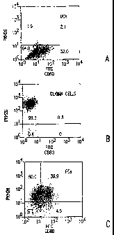
Figures lA-C. Fluorescence activated cell sorter (FACS) analysis of FCs. (A)
DCs
were stained by FITC-labeled anti-CD 80 antibody. A total of 34% of DCs were
stained with
anti-CD80 monoclonal antibody. (B) PKH26 was incorporated into glioma cells.
More than
95% of glioma cells were positive for PKH26. (C) After incorporation of PKH26
into glioma
cells, DCs and glioma cells were fused. DCs were stained with FITC-labeled
anti-CD80
monoclonal antibody. A total of 39.9% of cells were positive for both PKH26
and CD80,
suggesting that most DCs were fused with glioma cells.
Figures 2A-B. Antitumor effects of immunization with FCs. (A) FCs (0), DCs_ (
~),
or irradiated parental cells as a control (~) were injected into syngeneic
mice subcutaneously
on days 0 and 7 (n=11 in each group). On day 14,1 x 10⁶ parental cells were
subcutaneously
inoculated into the flank. The inoculated tumor cells caused large tumors
within two weeks
in all mice injected with irradiated parental cells. In contrast, none of the
mice immunized
with FCs died within six weeks. Whereas six of 11 mice immunized with DCs
developed a
palpable tumor that subsequently grew, none of 11 mice immunized with FCs
developed a
palpable tmnor. (B) After immunization with FCs on days 0 and 7, 1 x 10⁴ tumor
cells were
stereotactically inoculated into the right frontal lobe of the brain (day 14).
Half of the mice
immunized with FCs survived longer than 70 days (~; n =20 in each group; p
<0.001) (Fig.
2-B). All control mice died within 6 weeks (~).
Figure 3. Survival of mice following treatment with FCs and rIL-12. Parental
cells
(1x10⁴) were stereotactically inoculated into the right frontal lobe (day 0).
On days 5 and 12,
3 x 10⁵ FCs were subcutaneously inoculated. Several mice were given an
intraperitoneal
(i.p.) injection of 0.5 pg/100 ml of rmlL-12, or 100 ml of saline, every other
day for two
weeks (3.5 pg/mouse total) starting on Day 5 and observed for 70 days. While
vaccination
with FCs alone did not prolong the survival of tumor-bearing mice (~'; p >
0.05), vaccination
with both FCs and rIL-12 prolonged the survival compared with the control (0;
p = 0.01).
Five of ten mice treated with FCs and rIL-1 2 survived over seventy days.
Figure 4. Cytotoxicity of spleen cells from tumor-bearing mice. SPCs were
separated
from untreated mice (~), mice injected with rIL-12 alone (~), mice injected
DCs twice (days
0 and 7; ~), mice immunized with FCs once (day 0; O) or twice (days 0 and 7;
~) and mice
immunized with rIL-12 and FCs twice (days 0 and 7; o) on day 28. CTL activity
on tumor
cells from immunized mice, especially mice injected with rIL-12 and immunized
with FCs
twice, was considerably increased compared with the control and others.
Antitumor activity
CA 02555984 2006-08-11
WO 2005/079271 PCT/US2005/004237
on Yac-1 cell from treated mice increased but not considerably compared with
the control
(data not shown).
Figure 5. Regression of established subcutaneous tumors following vaccination
with
FCs and depletion of T-cell subsets. Lymphocyte subsets were depleted by
administering
anti-CD4 (D), anti-CD8 (~'), anti-asialo GMl (O), or control rat 1gG (~) into
mice given
injections of glioma cells and FCs. On days 0 and 7, FCs were subcutaneously
inoculated
into the flank. Subsequently parental cells were inoculated into the opposite
flank on day 14.
The mAbs were injected i.p. on days 7, 10, 14, and 17. The antitumor effect
was reduced in
mice depleted of CD8+ T cells ('.~) (n =4 in each group). The protection
conferred by FCs
was not abolished by CD4+ T and NIA cell depletion. Control mice were not
vaccinated with
FCs (~). Data represent means + SD.
Figures 6A-D. Immunofluorescence analysis of the developed brain tumors. A few
CD4+ and CD8+ T cells were present in the tumors of non-vaccinated mice
(Figures 6A, B).
In contrast, many CD4+ and CD8+ T cells were seen in the tumors of vaccinated
mice
(Figures 6C, D). The 'numbers of infiltrating CD4+ and CD8+ T cells were
almost the same.
SR-B 10.A cells were positive for GFAP.
Figure 7. Fused cells stained with both FITC (green) and PKH-26 (red) among
the
PEG-treated cells
Figure 8. FACS analysis, cells stained with both PKH-2GL and PKH-26, which
were
considered to be fusions of DCs and BNL cells, are shown in upper area of cell
scattergram
with high forward scatter and high side scatter. The cell fraction of high and
moderate
forward scatter and low side scatter contained many non-fused BNL cells, which
those of low
forward scatter and low side scatter contained non-fused DCs and non-fused BNL
cells.
About 30% of the nonadherent cells were fusions as judged from the width of
area of double
positive cells occupying in the whole scattergram.
Figure 9. FACS analysis of the cell fractions positive for both PKH-2GL and
PKH-
26 gated on scattergram and examined for antigen expression. I-Aa/I-Ed (MCH
class II),
CD80, CD86 and CD54 molecules, which are found on DCs, were expressed by the
fusions
Figure 10. Scanning Electron Microscopy of BNL cells expressing short
processes on
a plain cell surface, whereas DCs have many long dendritic processes. The
nonadherent
fusion cells are large and ovoid with short dendritic processes.
s
CA 02555984 2006-08-11
WO 2005/079271 PCT/US2005/004237
Figure 11. Vaccination of mice with DCBNL fusions resulted in the rejection of
a
challenge with BNL cells inoculated in BALBIc mice. By contrast, injection of
only DCs or
only irradiated BNL cells failed to prevent the development and growth of
tumors.
Figure 12. Chromium-51 release assay of CTL. The effect of treatment with
DCBNL fusion cells alone against BNL tumor was not significant. However,
injection of
DCBNL fusions followed by administration of IL-12 elicited a significant
antitumor effect.
Figure 13. Significant cytolytic activity against BNL cells was observed using
splenocytes derived from mice treated with DCBNL fusions. The solid bars are
the BNL-
cells and the hatched bars are the C26-cells.
Figure 14. Splenocytes from mice treated with DCBNL fusions in combination
with
IL-12 showed greater cytolytic activity against BNL cells than those treated
with DCBNL
fusions alone.
Figure 15. Lytic activity of the splenocytes treated with antibody against CD4
was
significantly reduced, while those treated with antibody against CD8 exhibited
almost the
same lytic activity as those treated with an isotype identical antibody, rat
IgG2a.
Figure 16. Vaccination schedule. FGs were injected intradermally close to a
cervical
lymph node on day 1. rhIL-12 was injected subcutaneously at the same site on
days 3 and 7.
This cycle was repeated every 2 weeks for 6 weeks (upper). In the absence of
progressive
disease or grade 3 or 4 major organ toxicity, patients could receive a second
6-week course
beginning 2 to 5 weeks after the last dose of rhIL-12 during course 1 (lower).
Figure 17. Analysis of fusion efficiency using FACScan. (A) Negative control.
(B)
PKH 26 was incorporated into glioma cells. 93.0% of glioma cells were positive
for PKH 26.
(C) PKH 2 was incorporated into DCs. 99.6% of DCs were positive for PKH 26.
(D) Stained
glioma cells and DCs were fused with PEG. Double positive cells (66.2%) were
determined
to be fusion cells. The numbers show the percentage of cells. Vertical axis:
PKH 26,
horizontal axis: PKH 2.
Figure 18. MRI of case 1 shows that the tumor recurred 2 months after the
first
operation. Inoculation of FCs did not inhibit the growth of the tumor. After
combination
CA 02555984 2006-08-11
WO 2005/079271 PCT/US2005/004237
therapy using FCs and rhIL-12, the high intensity area around the tumor on the
T2-weighted
image and the size of tumor on the T1-weighted image decreased remarkably. T1-
(A) and
T2-weighted (B) images of recurrent tmnor. T1- (C) and T2-weighted (D) images
after
immunization with FCs and rhIL-12.
Figure 19. MRI of case 3 shows the reduction in the high intensity area around
the
tumors on the T2-weighted image. (A) T2-weighted images before immunization.
(B) T2-
weighted images after immunization with FCs and rhIL-12.
Figure 20. Pathological findings for tumor specimens. Many larger tumor cells
containing multiple nuclei and wide cytoplasm were observed in recurrent tumor
specimens
compared with primary tumors. A robust CD8+, but not CD4+, T lymphocyte
infiltration was
observed in areas of the tumor. HE staining of primary and recurrent tumors in
cases 1 (A, B)
and 6 (C, D). Immunohistochemical staining of recurrent tumor specimens with
anti-CD4 and
anti-CD8 monoclonal antibodies in cases 1 (E, F) and 6 (G., H).
Figure 21. Cytolytic activity of PBLs against autologous glioma cells. PBLs
were
separated from blood taken before (black bar) and 8 to 10 weeks after first
immunization
(white bar). In 2 cases (cases 1 and 2), cytolytic activity against autologous
tumor cells
increased after treatment, while in other cases, cytolytic activity was almost
non-existent after
treatment. In case 6, the cytolytic activity after the treatment was lower
than that before the
treatment. The effectoraarget ratio was 80:1.
Figure 22. Cytokine flow cytometry for detection of IFN-y-expressing CD8+ T
lymphocytes in the peripheral circulation of patients before and after the
treatment.
Representative cases are shown (cases 9 and 15). In case 15, the parcentage of
double
positive cells increased after the treatment.
5. DETAILED DESCRIPTION OF THE INVENTION
to
CA 02555984 2006-08-11
WO 2005/079271 PCT/US2005/004237
The invention provides methods and compositions for the treatment of cancer.
In a
preferred embodiment, the methods of the invention provide the administration
of fusion cells
in combination with interleukin-12 (IL-12), e.g., recombinant human
interleukin-12 (rhIL-
12). The fusion cells of the invention are produced by fusion of autologous
dendritic cells
with autologous non-dendritic cells. Subsequently, the fused cells are
administered to a
subject in need thereof, in combination with a therapeutically effective dose
of a molecule
which stimulates a cytotoxic T-lymphocyte (CTL) response. In a preferred
embodiment, the
invention relates to methods and compositions for treating cancer comprising a
therapeutically effective dose of fusion cells in combination with IL-12.
Using the methods described herein, autologous dendritic cells can be fused to
a non-
dendritic cell containing an antigen of interest, such as a cancer antigen.
The resulting
hybrids of dendritic cells and non-dendritic cells can be used as a potent
composition against
a disease condition involving an antigen, such as a cancer. This approach is
particularly
advantageous when a specific antigen is not readily identifiable, as in the
case of many
cancers. For treatment of human cancer, for example, non-dendritic cells can
be obtained
directly from the tumor of a patient. Fusion cell compositions prepared in
this way are highly
specific for the individual tumor being treated.
Described in the sections below are compositions and methods relating to such
immunotherapeutic compositions. In particular, Sections 5.1, 5.2, and 5.3
describe the non-
dendritic, dendritic, and the fusion cells, respectively, that are used with
in the invention, and
methods for their isolation, preparation, and/or generation. Target cancers
that can be treated
or prevented using such compositions are described below in Section 5.6.
Sections 5.8, 5.9,
and 5.10 describes the methods and use of these fusion cells as therapeutic
compositions
against cancer.
5.1 NON-DENDRITIC CELLS
A non-dendritic cell of the present invention can be any cell bearing an
antigen of
interest for use in a fusion cell-cytokine composition. Such non-dendritic
cells may be
isolated from a variety of desired subjects, such as a tumor of a cancer
subject. The non-
dendritic cells may also be from an established cell line or a primary cell
culture. The
methods for isolation and preparation of the non-dendritic cells are described
in detail
hereinbelow.
11
CA 02555984 2006-08-11
WO 2005/079271 PCT/US2005/004237
The source of the non-dendritic cells may be selected, depending on the nature
of the
disease with which the antigen is associated. Preferably, the non-dendritic
cells are
autologous to the subject being treated, i.e., the cells used are obtained
from cells of the
ultimate target tumor in vivo (e.g., tumor cells of the patient being
treated), however, any
non-dendritic cell can be used as long as at least one antigen present on the
cell is an antigen
specific to a cell obtained from the target tumor, and as long as the non-
dendritic cell has the
same class I MHC haplotype as the patient being treated. Thus, while whole
cancer cells or
other non-dendritic cells may be used in the present methods, it is not
necessary to isolate
them, or characterize or even know the identities of their antigens prior to
performing the
present methods.
For treatment or prevention of cancer, the non-dendritic cell is a cancer
cell. In this
embodiment, the invention provides fusion cells that express antigens
expressed by cancer
cells, e.g., tumor-specific antigens and tumor associated antigens, and are
capable of eliciting
an immune response against such cancer cells. In one embodiment of the
invention, any
tissues, or cells isolated from a cancer, including cancer that has
metastasized to multiple
sites, can be used for the preparation of non-dendritic cells. For example,
leukemic cells
circulating in blood, lymph or other body fluids can also be used, solid tumor
tissue (e.g.,
primary tissue from a biopsy) can be used. Examples of cancers that are
amenable to the
methods of the invention are listed in Section 5.6 infra.
In a preferred embodiment, the tumor cells are not freshly isolated, but are
instead
cultured to select for tumor cells to be fused with dendritic cells and
prevent or limit
contamination of cells to be fused with healthy, non-cancerous or uninfected
cells.
In a preferred embodiment, the non-dendritic cells of the invention may be
isolated
from a tumor that is surgically removed from mammal to be the recipient of the
hybrid cell
compositions. Prior to use, solid cancer tissue or aggregated cancer cells
should be
dispersed, preferably mechanically, into a single cell suspension by standard
techniques.
Enzymes, such as but not limited to, collagenase and DNase may also be used to
disperse
cancer cells. In yet another preferred embodiment, the non-dendritic cells of
the invention
are obtained from primary cell cultures, i.e., cultures of original cells
obtained from the body.
Typically, approximately 1x10⁶ to 1x10⁹ non-dendritic cells are used for
formation of fusion
cells.
12
CA 02555984 2006-08-11
WO 2005/079271 PCT/US2005/004237
In one embodiment, approximately 1 x 10⁶ to 1 x 10⁹ non-dendritic cells are
used for
formation of fusion cells. In another embodiment, 5 x 10~ to 2 x 10⁸ cells are
used. In yet
another embodiment, 5 x 10~ non-dendritic cells are used.
Cell lines derived from cancer or infected cells or tissues can also be used
as non-
dendritic cells, provided that the cells of the cell line have the same
antigenic determinants)
as the antigen of interest on the non-dendritic cells. Cancer or infected
tissues, cells, or cell
lines of human origin are preferred.
In an alternative embodiment, in order to prepare suitable non-dendritic cells
that are
cancer cells, noncancerous cells, preferably of the same cell type as the
cancer desired to be
inhibited can be isolated from the recipient or, less preferably, other
individual who shares at
least one MHC allele with the intended recipient, and treated with agents that
cause the
particular or a similar cancer or a transformed state; such agents may include
but not limited
to, radiation, chemical carcinogens, and viruses. Standard techniques can be
used to treat the
cells and propagate the cancer or transformed cells so produced.
Alternatively, if the gene encoding a tumor-specific antigen, tumor-associated
antigen
or antigen of the pathogen is available, normal cells of the appropriate cell
type from the
intended recipient. Optionally, more than one such antigen may be expressed in
the
recipient's cell in this fashion, as will be appreciated by those skilled in
the art, any
techniques known, such as those described in Ausubel et al. (eds., 199,
Current Protocols in
Molecular Biology, Greene Publishing Associates and Wiley Interscience, New
York), may
be used to perform the transformation or transfection and subsequent
recombinant expression
of the antigen gene in recipient's cells. These non-dendritic cells bearing
one or more MHC
molecules in common with the recipient are suitable for use in the methods for
formation of
fusion cells of the invention.
The non-dendritic cells used for the generation of fusion cells and the target
tumor or
pathogen infected cell must have at least one common MHC allele in order to
elicit an
immune response in the mammal. Most preferred is where the non-dendritic cells
are derived
from the intended recipient (i.e., are autologous). Less preferred, the non-
dendritic cells are
nonautologous, but share at least one MHC allele with the cancer cells of the
recipient. If the
non-dendritic cells are obtained from the same or syngeneic individual, such
cells will all
have the same class I MHC haplotype. If they are not all obtained from the
same subject, the
MHC haplotype can be determined by standard HLA typing techniques well known
in the art,
13
CA 02555984 2006-08-11
WO 2005/079271 PCT/US2005/004237
such as serological tests and DNA analysis of the MHC loci. An MHC haplotype
determination does not need to be undertaken prior to carrying out the
procedure for
generation of the fusion cells of the invention.
Non-dendritic cells, such as cells containing an antigen having the
antigenicity of a
cancer cell, can be identified and isolated by any method known in the art.
For example,
cancer or infected cells can be identified by morphology, enzyme assays,
proliferation assays,
or the presence of cancer-causing viruses. If the characteristics of the
antigen of interest are
known, non-dendritic cells can also be identified or isolated by any
biochemical or
immunological methods known in the art. For example, cancer cells or infected
cells can be
isolated by surgery, endoscopy, other biopsy techniques, affinity
chromatography, and
fluorescence activated cell sorting (e.g., with fluorescently tagged antibody
against an antigen
expressed by the cells).
There is no requirement that a clonal or homogeneous or purified population of
non-
dendritic cells be used. A mixture of cells can be used provided that a
substantial number of
cells in the mixture contain the antigen or antigens present on the tumor
cells being targeted.
In a specific embodiment, the non-dendritic cells and/or dendritic cells are
purified.
In a specific embodiment, cancer tissue from the subject to be treated is
collected
during operation or by biopsy. The collected tissue is maintained in such a
way as to keep
the cancer cells alive. Cancer cells are preferably collected just prior
preparation of the
fusion cells. Most preferably, they are collected from ascites and pleural
fluid just prior to
preparation of fusion cells. When it is not possible to obtain sufficient
quantities of
malignant tumor cells in this manner, collection of malignant tumor cells from
abdominal or
thoracic liquid, or from a needle biopsy, may be possible.
5.2 DENDRITIC CELLS
Dendritic cells can be isolated or generated from blood or bone marrow, or
secondary
lymphoid organs of the subject, such as but not limited to spleen, lymph
nodes, tonsils,
Peyer's patch of the intestine, and bone marrow, by any of the methods known
in the art.
Preferably, DCs used in the methods of the invention are (or terminally
differentiated)
dendritic cells. The source of dendritic cells is preferably human blood
monocytes.
Immune cells obtained from such sources typically comprise predominantly
recirculating lymphocytes and macrophages at various stages of differentiation
and
14
CA 02555984 2006-08-11
WO 2005/079271 PCT/US2005/004237
maturation. Dendritic cell preparations can be enriched by standard techniques
(see e.g.,
Current Protocols in Immunology, 7.32.1-7.32.16, John Wiley and Sons, Inc.,
1997). In one
embodiment, for example, DCs may be enriched by depletion of T cells and
adherent cells,
followed by density gradient centrifugation. DCs may optionally be further
purified by
sorting of fuorescence-labeled cells, or by using anti-CD83 MAb magnetic
beads.
Alternatively, a high yield of a relatively homogenous population of DCs can
be
obtained by treating DC progenitors present in blood samples or bone marrow
with cytokines,
such as granulocyte-macrophage colony stimulating factor (GM-CSF) and
interleukin 4 (IL-
4). Under such conditions, monocytes differentiate into dendritic cells
without cell
proliferation. Further treatment with agents such as TNFa stimulates terminal
differentiation
of DGs.
By way of example but not limitation, dendritic cells can be obtained from
blood
monocytes as follows: peripheral blood monocytes are obtained by standard
methods (see,
e.g., Sallusto et al., 1994, J. Exp. Med. 179:1109-1118). Leukocytes from
healthy blood
donors are collected by leukapheresis pack or huffy coat preparation using
Ficoll-Paque
density gradient centrifugation and plastic adherence. If mature DCs are
desired, the
following protocol may be used to culture DCs. Cells are allowed to adhere to
plastic dishes
for 4 hours at 37°C. Nonadherent cells are removed and adherent
monocytes are cultured for
7 days in culture media containing 0.1 ~,g/ml granulocyte-monocyte colony
stimulating factor
(GM-CSF) and O.OS~,g/ml interleukin-4 (IL-4). In order to prepare mature
dendritic cells,
tumor necrosis factor-a is added on day 5, and cells are collected on day 7.
In a specific embodiment, the following protocol is used. First, bone marrow
is
isolated and red cells lysed with ammonium chloride (Sigma, St. Louis, MO).
Lymphocytes,
granulocytes and DCs are depleted from the bone marrow cells and the remaining
cells are
plated in 24-well culture plates (1 x 10⁶ cells/well) in RPMI 1640 medium
supplemented with
5% heat-inactivated FBS, 50 ~.M 2-mercaptoethanol, 2 mM glutamate, 100 U/ml
penicillin,
100 pg/ml streptomycin, 10 ng/ml recombinant marine granulocyte-macrophage
colony
stimulating factor (GM-CSF; Becton Dickinson, San Jose, CA) and 30 U/ml
recombinant
mouse interleukin-4 (IL-4; Becton Dickinson). Second, on day 5 of culture,
nonadherent and
loosely adherent cells are collected and replated on 100-mm petri dishes (1 x
106 cells/ml; 10
ml/dish). Next, GM-CSF and IL-4 in RPMI medium are added to the cells and 1 x
106 DCs
CA 02555984 2006-08-11
WO 2005/079271 PCT/US2005/004237
are mixed with 3 x 10⁶ irradiated (50 Gy, Hitachi MBR-15208, dose rate: 1.1
Gy/min.) SR-
B 1 O.A cells. After 48 h, cells are collected for fusion with tumor cells.
Dendritic cells obtained in this way characteristically express the cell
surface marker
CD83. In addition, such cells characteristically express high levels of MHC
class II
molecules, as well as cell surface markers CDla, CD40, CD86, CD54, and CD80,
but lose
expression of CD 14. Other cell surface markers characteristically include the
T cell markers
CD2 and CDS, the B cell marker CD7 and the myeloid cell markers CD13, CD32
(FcyR II),
CD33, CD36, and CD63, as well as a large number of leukocyte-associated
antigens
Optionally, standard techniques, such as morphological observation and
immunochemical staining, can be used to verify the presence of dendritic
cells. For example,
the purity of dendritic cells can,be assessed by flow cytometry using
fluorochrome-labeled
antibodies directed against one or more of the characteristic cell surface
markers noted above,
e.g., CD83, HLA-ABC, HLA-DR, CDla, CD40, and/or CD54. This technique can also
be
used to distinguish between immature and mature DCs, using fluorochrome-
labeled
antibodies directed against CD14, which is present in immature, but not
mature, DCs.
In a preferred embodiment, venous blood is collected from the brachial vein by
any
method well-known to the skilled artisan. In a specific embodiment, 60 ml of
blood is
collected from the subject to be treated. White blood cells are separated from
the collected
blood, and only white blood cells with high adherent capacity are collected
(see, e.g., Kikuchi
et al., 2001, Cancer Immunol Immunother 50:337-344). An exemplary protocol for
the
cultivation of white blood cells with high adherent capacity is as follows.
Briefly, peripheral
blood mononuclear cells are separated from peripheral blood using Ficoll-
Hypaque density
centrifugation. Peripheral blood mononuclear cells are resuspended in RPMI-
1640 (Sigma)
and allowed to adhere to 24-well cluster plates. The nonadherent cells are
removed after 2
hours at 37°C, and the adherent cells are subsequently cultured for 7
days in X-VIVO-15
medium (BioWhittaker, Walkersville, MD) supplemented with 1% heat-inactivated
autologous serum, 10 ng/ml recombinant human granulocyte-macrophage colony
stimulating
factor (GM-CSF; Becton Dickinson, San Jose, CA), 30 U/ml recombinant human
interleukin-
4 (IL-4; Becton Dickinson), and 20 ng/ml tumor necrosis factor-a (TNF-a;
Becton
Dickinson). The cultures are fed every third day and are split when necessary.
Thereafter,
the semi-adherent and non-adherent cells are harvested by vigorous pipetting
and used as
dendritic cells for fusion. In certain embodiments, 50 mM 2-mercaptoethanol, 2
mM
16
CA 02555984 2006-08-11
WO 2005/079271 PCT/US2005/004237
glutamate, 100 U/ml penicillin, and 100 mg/ml streptomycin are also present in
the culture
medium. GM-CSF and IL-4 cause white blood cells and lymphocytes to proliferate
or to
exhibit various functions. While culturing, serum of the appropriate subject
is added to a
concentration of 10% in the culture solution, avoiding any contact with
heterogenous antigen.
In certain embodiments, the adherent cells are cultured in medium supplemented
with
at least 10 ng/ml, 20 ng/ml, 30 ng/ml, 40 ng/ml, 50 ng/ml, 60 ng/ml, 70 ng/ml,
80 ng/ml, 90
ng/ml or 100 ng/ml GM-CSF. In certain embodiments, the adherent cells are
cultured in
medium supplemented with at most 10 nglml, 20 ng/ml, 30 ng/ml, 40 ng/ml, 50
ng/ml, 60
ng/ml, 70 ng/ml, 80 ng/ml, 90 ng/ml or 100 ng/ml GM-CSF. In certain
embodiments, the
adherent cells are cultured in medium supplemented with between 10 ng/ml and
100 ng/ml,
20 ng/ml and 80 ng/ml, 30 ng/ml and 70 ng/ml, or 40 ng/ml and 60 ng/ml GM-C
SF.
In certain embodiments, the adherent cells are cultured in medium supplemented
with
at least 10 ng/ml, 20 ng/ml, 30 ng/ml, 40 ng/ml, 50 ng/ml, 60 ng/ml, 70 ng/ml,
80 ng/ml, 90
ng/ml or 100 ng/ml TNF-a. In certain embodiments, the adherent cells are
cultured in
medium supplemented with at most 10 ng/ml, 20 ng/ml, 30 ng/ml, 40 ng/ml, 50
ng/ml, 60
ng/ml, 70 ng/ml, 80 ng/ml, 90 ng/ml or 100 ng/ml TNF-a. In certain
embodiments, the
adherent cells are cultured in medium supplemented with between 10 ng/ml and
100 ng/ml,
20 ng/ml and 80 ng/ml, 30 ng/ml and 70 ng/ml, or 40 ng/ml and 60 ng/ml TNF'-a.
In certain embodiments, the adherent cells are cultured in medium supplemented
with
at least 10 U/ml, 20 U/ml, 30 ng/ml, 40 U/ml, 50 Ulml, 60 U/ml, 70 U/ml, 80
U/ml, 90 U/ml
or 100 U/ml IL-4. In certain embodiments, the adherent cells are cultured in
medium
supplemented with at most 10 U/ml, 20 U/ml, 30 U/ml, 40 U/ml, 50 U/ml, 60
U/ml, 70 U/ml,
80 U/ml, 90 U/ml or 100 U/ml IL-4. In certain embodiments, the adherent cells
are cultured
in medium supplemented with between 10 U/ml and 100 U/ml, 20 Ulml and 80 U/ml,
30
U/ml and 70 U/ml, or 40 U/ml and 60 U/ml IL-4.
5.3 GENERATION OF FUSION CELLS
Non-dendritic cells can be fused to autologous DCs as followed. Cells can be
washed
prior to fusion under sterile conditions. Fusion can be accomplished by any
cell fusion
technique in the art provided that the fusion technique results in a mixture
of fused cells
suitable for injection into a mammal for treatment of cancer. In a specific
example,
electrofusion is used. Electrofusion techniques are well known in the art
(Stuhler and
17
CA 02555984 2006-08-11
WO 2005/079271 PCT/US2005/004237
Walden, 1994, Cancer Immunol. Immunother. 39: 342-345; see Chang et al.
(eds.), Guide to
Electroporation and Electrofusion. Academic Press, San Diego, 1992).
In a specific embodiment, the following protocol is used. In the first step,
approximately 5 x 10~ tumor cells and 5 x 10~ dendritic cells (DCs) are
suspended in 0.3 M
glucose and transferred into an electrofusion cuvette. The sample is
dielectrophoretically
aligned to form cell-cell conjugates by pulsing the cell sample at 100 V/cm
for 5-10 secs.
Optionally, alignment may be optimized by applying a drop of dielectrical wax
onto one
aspect of the electroporation cuvette to 'inhomogenize' the electric field,
thus directing the
cells to the area of the highest field strength. In a second step, a fusion
pulse is applied.
Various parameters may be used for the electrofusion. For example, in one
embodiment, the
fusion pulse may be from a single to a triple pulse. In another embodiment,
electrofusion is
accomplished using from 500 to 1500 V/cm, preferably, 1,200V/cm at about 25
wF.
In a preferred embodiment, matured dendritic cells are fused with cancer cells
by use
of polyethyleneglycol. Briefly, the dendritic cells are mixed with lethally
irradiated cancer
cells (300 Gy, Hitachi MBR-15208, dose rate 1.1 Gy/min). In certain
embodiments, the
cancer cells are irradiated with 10 Gy, 25 Gy, 50 Gy, 100 Gy, 200 Gy, 300 Gy,
400 Gy, 500
~Gy, 750 Gy, or 1,000 Gy. In certain embodiments, the cancer cells are
irradiated with 50 to
500 Gy. The ratio of dendritic cells and cancer cells can range from 3:1 to
10:1 depending on
the numbers of acquired dendritic cells and cancer cells. Subsequently, fusion
is initiated by
adding 500 ~,1 of a 50% solution of polyethylene glycol (PEG; Sigma) dropwise
for 60
seconds. The fusion is stopped by stepwise addition of serum-free RPMI medium.
After
washing 3 times with phosphate-buffered saline (PBS; Cosmo Bio), fusion cells
are plated in
100-mm petri dishes in the presence of GM-CSF, IL-4, and TNF-a in RPMI medium
for 24
hours. In a specific embodiment, fusion cells are plated in the presence of 10
ng/ml GM-
CSF, 30 U/ml IL-4, and 20 ng/ml TNF-a in RPMI medium for 24 hours After
overnight
culture, the fused cells are suspended in about 1 mL of physiological saline,
and injected
subcutaneously to the subject. In a preferred embodiment the suspension of
fusion cells is
injected in the groin area as this area is rich in lymph nodes.
In another specific embodiment, the following protocol is used. First,
dendritic cells
are prepared, as described in Section 5.2, above. On day 5 of dendritic cell
culture,
nonadherent and loosely adherent cells are collected and replated on 100-mm
petri dishes (1 x
106 cells/ml; 10 ml/dish). Next, GM-CSF and IL-4 in RPMI medium are added to
the cells
is
CA 02555984 2006-08-11
WO 2005/079271 PCT/US2005/004237
and 1 x 10⁶ DCs are mixed with 3 x l06 irradiated (50 Gy, Hitachi MBR-15208,
dose rate:
1.1 Gy/min.) SR-B l O.A cells. After 48 h, fusion is started by adding
dropwise for 60 sec,
500 ~1 of a 50% solution of polyethylene glycol (PEG; Sigma). In a specific
embodiment,
the final concentration of PEG is 2.5%. In certain embodiments of the
invention, the final
concentration of PEG is 0.5%, 1%, 1.5%, 2.5%, 5%, 10%, 15%, 20% or 25%. In
certain
embodiments, the final concentration of PEG is 0.5% to 25%, 1% to 20%, or 5%
to 15%.
The fusion is stopped by stepwise addition of serum-free RPMI medium. FCs are
plated in
100-mm petri dishes in the presence of GM-CSF and IL-4 in RPMI medium for 48
h.
In another embodiment, the dendritic cell and the non-dendritic cell are fused
as
described above. Subsequently, the fused cells are transfected with genetic
material which
encodes a molecule which stimulates a CTL and/or humoral immune response. In a
preferred
embodiment, the genetic material is mRNA which encodes IL-12. Preferred
methods of
transfection include electroporation or cationic polymers.
In certain embodiments, the cancer cells are fused with the dendritic cells at
a ratio of
1 cancer cell per dendritic cell (DC), 2 cancer cells per DC, 3 cancer cells
per DC, 4 cancer
cells per DC, 5 cancer cells per DC, 6 cancer cells per DC, 7 cancer cells per
DC, 8 cancer
cells per DC, 9 cancer cells per DC, or 10 cancer cells per DC.
The extent of fusion cell formation within a population of antigenic and
dendritic cells
can be determined by a number of diagnostic techniques known in the art. In
one
embodiment, for example, hybrids are characterized by emission of both colors
after labeling
of DCs and tumor cells with red and green intracellular fluorescent dyes,
respectively.
Samples of DCs without tumor cells, and tumor cells without DCs can be used as
negative
controls, as well as tumor + DC mixture without electrofusion.
In one embodiment, the fusion cells prepared by this method comprise
approximately
and 20% of the total cell population. In yet another embodiment, the fusion
cells
prepared by this method comprise approximately 5 to 50% of the total cell
population.
In certain embodiments, the fusion cells are cultured in medium supplemented
with at
least 10 ng/ml, 20 ng/ml, 30 ng/ml, 40 ng/ml, 50 ng/ml, 60 ng/ml, 70 ng/ml, 80
ng/ml, 90
ng/ml or 100 ng/ml GM-CSF. In certain embodiments, the adherent cells are
cultured in
medium supplemented with at most 10 ng/ml, 20 ng/ml, 30 ng/ml, 40 ng/ml, 50
ng/ml, 60
ng/ml, 70 ng/ml, 80 ng/ml, 90 ng/ml or 100 ng/ml GM-CSF. In certain
embodiments, the
19
CA 02555984 2006-08-11
WO 2005/079271 PCT/US2005/004237
adherent cells are cultured in medium supplemented with between 10 ng/ml and
100 ng/ml,
20 ng/ml and 80 ng/ml, 30 ng/ml and 70 ng/ml, or 40 ng/ml and 60 ng/ml GM-CSF.
In certain embodiments, the fusion cells are cultured in medium supplemented
with at
least 10 ng/ml, 20 ng/ml, 30 ng/ml, 40 ng/ml, 50 ng/ml, 60 ng/ml, 70 ng/ml, 80
ng/ml, 90
nghnl or 100 ng/ml TNF-a. In certain embodiments, the adherent cells are
cultured in
medium supplemented with at most 10 ng/ml, 20 ng/ml, 30 ng/ml, 40 ng/ml, 50
ng/ml, 60
ng/ml, 70 ng/ml, 80 ng/ml, 90 ng/ml or 100 ng/ml TNF-a. In certain
embodiments, the
adherent cells are cultured in medium supplemented with between 10 ng/ml and
100 ng/ml,
20 ng/ml and 80 ng/ml, 30 ng/ml and 70 ng/ml, or 40 ng/ml and 60 ng/ml TNF-a.
In certain embodiments, the fusion cells are cultured in medium supplemented
with at
least 10 U/ml, 20 Uhnl, 30 ng/ml, 40 U/ml, 50 U/ml, 60 U/ml, 70 U/ml, 80 U/ml,
90 U/ml or
100 U/ml IL-4. In certain embodiments, the adherent cells are cultured in
medium
supplemented with at most 10 U/ml, 20 U/ml, 30 U/ml, 40 U/ml, 50 U/ml, 60
U/ml, 70 U/ml,
80 U/ml, 90 U/ml or 100 U/ml IL-4. In certain embodiments, the adherent cells
are cultured
in medium supplemented with between 10 U/ml and 100 U/ml, 20 U/ml and 80 U/ml,
30
Ulml and 70 U/ml, or 40 U/ml and 60 U/ml IL-4.
To prevent contamination of the fusion cells with bacteria or viruses, cell
culturing
and fusion of cells may be conducted in a room for exclusive use for mammalian
cell
culturing. These cells are monitored to confirm they are not infected with
bacteria or
contaminated with the toxin of bacteria.
In instances where dendritic cells are fused with cancer cells by use of
polyethylene
glycol or another method, some, but not all of the cancer cells may be fused.
When such
non-fused cancer cells are injected into the subject's body, unfavorable
effects may occur.
Therefore, in certain embodiments, the cancer cells are irradiated before
administration. By
use of these safety measures, there is little possibility that cancer cells
proliferate actively
even if the fused cells are contaminated with a trace amount of cancer cells.
In a preferred
embodiment, the cancer cells are irradiated before fusion.
In certain embodiments, the cancer cells are obtained from a subject at least
10 min,
30 min, 60 min, 2 hours, 5 hours, 10 hours, or 24 hours before fusing the
cancer cells with
dendritic cells. In certain embodiments, the cancer cells are obtained from a
subject at most
min, 30 min, 60 min, 2 hours, 5 hours, 10 hours, or 24 hours before fusing the
cancer cells
CA 02555984 2006-08-11
WO 2005/079271 PCT/US2005/004237
with dendritic cells. In certain embodiments, the cancer cells are obtained
from a subject and
subsequently a cell line is established before fusing the cancer cells with
dendritic cells. In
certain embodiments, the dendritic cells are obtained from a subject at least
10 min, 30 min,
60 min, 2 hours, 5 hours, 10 hours, or 24 hours before fusing the cancer cells
with dendritic
cells. In certain embodiments, the dendritic cells are obtained from a subject
at most 10 min,
30 min, 60 min, 2 hours, 5 hours, 10 hours, or 24 hours before fusing the
cancer cells with
dendritic cells.
5.3.1 RECOMBINANT CELLS
In an alternative embodiment, rather than fusing a dendritic cell to a cancer
cell, the
non-dendritic cells are transfected with a gene encoding a known antigen of a
cancer. The
non-dendritic cells are then selected for those expressing the recombinant
antigen and
administered to the subj ect in need thereof in combination with a cytokine or
molecule which
stimulates or induces a CTL and/or humoral immune response.
Recombinant expression of a gene by gene transfer, or gene therapy, refers to
the
administration of a nucleic acid to a subject. The nucleic acid, either
directly or indirectly via
its encoded protein, mediates a therapeutic effect in the subject. The present
invention
provides methods of gene therapy wherein genetic material, e.g., DNA or mRNA,
encoding a
protein of therapeutic value (preferably to humans) is introduced into the
fused cells
according to the methods of the invention, such that the nucleic acid is
expressible by the
fused cells, followed by administration of the recombinant fused cells to a
subject.
The recombinant fused cells of the present invention can be used in any of the
methods for gene therapy available in the art. Thus, the nucleic acid
introduced into the cells
may encode any desired protein, e.g., an antigenic protein or portion thereof
or a protein that
stimulates a CTL and/or humoral immune response. The descriptions below are
meant to be
illustrative of such methods. It will be readily understood by those of skill
in the art that the
methods illustrated represent only a sample of all available methods of gene
therapy.
For general reviews of the methods of gene therapy, see Lundstrom, 1999, J.
Recept.
Signal Transduct. Res. 19:673-686; Robbins and Ghivizzani, 1998, Pharmacol.
Ther.80:35-47; Pelegrin et al., 1998, Hum. Gene Ther. 9:2165-2175; Harvey and
Caskey,
1998, Curr. Opin. Chem. Biol. 2:512-518; Guntaka and Swamynathan, 1998, Indian
J. Exp.
Biol. 36:539-535; Desnick and Schuchman, 1998, Acta Paediatr. Jpn. 40:191-203;
Vos, 1998,
21
CA 02555984 2006-08-11
WO 2005/079271 PCT/US2005/004237
Curr. Opin. Genet. Dev. 8:351-359; Tarahovsky and Ivanitsky, 1998,
Biochemistry (Mosc)
63:607-618; Morishita et al., 1998, Circ. Res. 2:1023-1028; Vile et al., 1998,
Mol. Med.
Today 4:84-92; Branch and Klotman,1998, Exp. Nephrol. 6:78-83; Ascenzioni et
al., 1997,
Cancer Lett. 118:135-142; Chan and Glazer, 1997, J. Mol. Med. 75:267-282.
Methods
commonly known in the art of recombinant DNA technology which can be used are
described in Ausubel et al. (eds.), 1993, Current Protocols in Molecular
Biology, John Wiley
& Sons, NY; and Kriegler, 1990, Gene Transfer and Expression, A Laboratory
Manual,
Stockton Press, NY.
In an embodiment in which recombinant cells are used in gene therapy, a gene
whose
expression is desired in a subject is introduced into the fused cells such
that it is expressible
by the cells and the recombinant cells are then administered i~r vivo for
therapeutic effect.
Recombinant fused cells can be used in any appropriate method of gene therapy,
as
would be recognized by those in the art upon considering this disclosure. The
resulting
action of recombinant manipulated cells administered to a subject can, for
example, lead to
the activation or inhibition of a pre-selected gene, such as activation of IL-
12, in the patient,
thus leading to improvement of the diseased condition afflicting the patient.
The desired gene is transferred, via transfection, into fused by such methods
as
electroporation, lipofection, calcimn phosphate mediated transfection, or
viral infection.
Usually, the method of transfer includes the transfer of a vector containing a
selectable
marker. The cells are then placed under selection to isolate those cells that
have taken up and
are expressing the vector, containing the selectable marker and also the
transferred gene.
Those cells are then delivered to a patient.
In this embodiment, the desired gene is introduced into fused, cells prior to
administration in vivo of the resulting recombinant cell. Such introduction
can be carried out
by any method known in the art, including but not limited to transfection,
electroporation,
microinjection, infection with a viral or bacteriophage vector containing the
gene sequences,
cell fusion, chromosome-mediated gene transfer, microcell-mediated gene
transfer,
spheroplast fusion, etc. Numerous techniques are known in the art for the
introduction of
foreign genes into cells (see e.g., Loeffler and Behr, 1993, Meth. Enzymol.
217:599-618;
Cohen et al., 1993, Meth. Enzymol. 217:618-644; Cline, 1985, Pharmac. Ther.
29:69-92) and
may be'used in accordance with the present invention, provided that the
necessary
developmental and physiological functions of the recipient cells are not
disrupted. The
22
CA 02555984 2006-08-11
WO 2005/079271 PCT/US2005/004237
teclinique should provide for the stable transfer of the gene to the cell, so
that the gene is
expressible by the cell and preferably heritable and expressible by its cell
progeny.
One common method of practicing gene therapy is by making use of retroviral
vectors
(see Miller et al., 1993, Meth. Enzymol. 217:581-599). A retroviral vector is
a retrovirus that
has been modified to incorporate a preselected gene in order to effect the
expression of that
gene. It has been found that many of the naturally occurring DNA sequences of
retroviruses
are dispensable in retroviral vectors. Only a small subset of the naturally
occurring DNA
sequences of retroviruses is necessary. In general, a retroviral vector must
contain all of the
cis-acting sequences necessary for the packaging and integration of the viral
genome. These
cis-acting sequences are:
a) a long terminal repeat (LTR), or portions thereof, at each end of the
vector;
b) primer binding sites for negative and positive strand DNA synthesis; and
c) a packaging signal, necessary for the incorporation of genomic RNA into
virions.
The gene to be used in gene therapy is cloned into the vector, which
facilitates
delivery of the gene into an cell by infection or delivery of the vector into
the cell.
More detail about retroviral vectors can be found in Boesen et al., 1994,
Biotherapy
6:291-302, which describes the use of a retroviral vector to deliver the mdrl
gene to
hematopoietic stem cells in order to make the stem cells more resistant to
chemotherapy.
Other references illustrating the use of retroviral vectors in gene therapy
are: Cloves et al.,
1994, J. Clin. Invest. 93:644-651; Kiem et al., 1994, Blood 83:1467-1473;
Salmons and
Gunzberg, 1993, Human Gene Therapy 4:129-141; and Grossman and Wilson, 1993,
Curr.
Opin. in Genetics and Devel. 3:110-114.
Adenoviruses can be used to deliver genes to non-dendritic cells derived from
the
liver, the central nervous system, endothelium, and muscle. Adenoviruses have
the
advantage of being capable of infecting non-dividing cells. Kozarsky and
Wilson, 1993,
Current Opinion in Genetics and Development 3:499-503 present a review of
adenovirus-based gene therapy. Other instances of the use of adenoviruses in
gene therapy
can be found in Rosenfeld et al., 1991, Science 252:431-434; Rosenfeld et al.,
1992, Cell
68:143-155; and Mastrangeli et al., 1993, J. Clin. Invest. 91:225-234.
It has been proposed that adeno-associated virus (AAV) be used in gene therapy
(Walsh et al., 1993, Proc. Soc. Exp. Biol. Med. 204:289-300). It has also been
proposed that
23
CA 02555984 2006-08-11
WO 2005/079271 PCT/US2005/004237
alphaviruses be used in gene therapy (Lundstrom, 1999, J. Recept. Signal
Transduct. Res.
19:673-686).
Other methods of gene delivery in gene therapy include mammalian artificial
chromosomes (Vos, 1998, Curr. Op. Genet. Dev. 8:351-359); liposomes
(Tarahovsky and
Ivanitsky, 1998, Biochemistry (Mosc) 63:607-618); ribozymes (Branch and
Klotman, 1998,
Exp. Nephrol. 6:78-83); and triplex DNA (Chan and Glazer, 1997, J. Mol. Med.
75:267-282).
A desired gene can be introduced intracellularly and incorporated within host
cell
DNA for expression, by homologous recombination (Koller and Smithies, 1989,
Proc. Natl.
Acad. Sci. USA 86:8932-8935; Zijlstra et al., 1989, Nature 342:435-438).
In a specific embodiment, the desired gene recombinantly expressed in the cell
to be
introduced for purposes of gene therapy comprises an inducible promoter
operably linked to
the coding region, such that expression of the recombinant gene is
controllable by controlling
the presence or absence of the appropriate inducer of transcription.
In a preferred embodiment, the desired gene recombinantly expressed in the
cells,
whether its function is to elicit a cell fate change according to the methods
of the invention, is
flanked by Cre sites. When the gene function is no longer required, the cells
comprising the
recombinant gene are subjected to Lox protein, for example be means of
supplying a nucleic
acid containing the Lox coding sequences functionally coupled to an inducible
or tissue
specific promoter, or by supplying Lox protein functionally coupled to a
nuclear
internalization signal. Lox recombinase functions to recombine the Cre
sequences (Hamilton
et al., 1984, J. Mol. Biol. 178:481-486), excising the intervening sequences
in the process,
which according to this embodiment contain a nucleic acid of a desired gene.
The method
has been used successfully to manipulate recombinant gene expression
(Fukushige et al.,
1992, Proc. Natl. Acad. Sci. USA 89:7905-7909). Alternatively, the FLP/FRT
recombination
system can be used to control the presence and expression of genes through
site-specific
recombination (Brand and Perrimon, 1993, Development 118:401-415).
In a preferred aspect of the invention, gene therapy using nucleic acids
encoding
hepatitis B or hepatitis C major antigens are directed to the treatment of
viral hepatitis.
5.4 IMMUNE CELL ACTIVATING MOLECULES
The present invention provides a method which comprises administering first, a
fusion cell derived from the fusion of a dendritic and non-dendritic cell, and
second, a
24
CA 02555984 2006-08-11
WO 2005/079271 PCT/US2005/004237
cytokine or other molecule which can stimulate or induce a cytotoxic T cell
(CTL) response,
such as interleukin-12 (IL-12).
IL-12 plays a major role in regulating the migration and proper selection of
effector
cells in an immune response. The IL-12 gene product polarizes the immtme
response toward
the THl subset of T helper cells and strongly stimulates CTL activity. In a
preferred
embodiment, the CTL stimulating molecule is IL-12. As elevated doses of IL-12
exhibits
toxicity when administered systemically, IL-12 is preferably administered
locally. Additional
modes of administration are described below in Section 5.7.1.
Expression of IL-12 receptor b2 (IL-12R-b2) is necessary for maintaining IL-12
responsiveness and controlling TH1 lineage commitment. Furthermore, IL-12
signaling
results in STAT4 activation, i.e., measured by an increase of phosphorylation
of STAT4, and
interferon-g (IFN-g) production. Thus, in one embodiment, the present
invention
contemplates the use of a molecule, which is not IL-12, which can activate
STAT4, for
example a small molecule activator of STAT4 identified by the use of
combinatorial
chemistry.
In an alternative embodiment, the immune stimulating molecule is IL-18. In yet
another embodiment, the immune stimulating molecule is IL-15. In yet another
embodiment,
the immune stimulating molecule is interferon-y.
In another embodiment, the subject to be treated is given any combination of
molecules or cytokines described herein which stimulate or induce a CTL and/or
humoral
immune response.
In a less preferred embodiment, to increase the cytotoxic T-cell pool, i.e.,
the TH1 cell
subpopulation, anti-IL-4 antibodies can be added to inhibit the polarization
of T-helper cells
into THZ cells, thereby creating selective pressure toward the TH1 subset of T-
helper cells.
Further, anti-IL-4 antibodies can be administered concurrent with the
administration of IL-12,
to induce the TH cells to differentiate into THI cells. After differentiation,
cells can be
washed, resuspended in, for example, buffered saline, and reintroduced into a
subject via,
preferably, intravenous administration.
The present invention also pertains to variants of the above-described
interleukins.
Such variants have an altered amino acid sequence which can function as
agonists (mimetics)
to promote a CTL and/or humoral immune response response. Variants can be
generated by
CA 02555984 2006-08-11
WO 2005/079271 PCT/US2005/004237
mutagenesis, e.g., discrete point mutation or truncation. An agonist can
retain substantially
the same, or a subset, of the biological activities of the naturally occurring
form of the
protein. An antagonist of a protein can inhibit one or more of the activities
of the naturally
occurring form of the protein by, for example, competitively binding to a
downstream or
upstream member of a cellular signaling cascade which includes the protein of
interest. Thus,
specific biological effects can be elicited by treatment with a variant of
limited function.
Treatment of a subject with a variant having a subset of the biological
activities of the
naturally occurring form of the protein can have fewer side effects in a
subject relative to
treatment with the naturally occurring form of the protein.
Variants of a molecule capable of stimulating a CTL and/or humoral immure
response can be identified by screening combinatorial libraries of mutants,
e.g., truncation
mutants, for agonist activity. In one embodiment, a variegated library of
variants is generated
by combinatorial mutagenesis at the nucleic acid level and is encoded by a
variegated gene
library. A variegated library of variants can be produced by, for example,
enzymatically
ligating a mixture of synthetic oligonucleotides into gene sequences such that
a degenerate
set of potential protein sequences is expressible as individual polypeptides,
or alternatively,
as a set of larger fusion proteins (e.g., for phage display). There are a
variety of methods
which can be used to produce libraries of potential variants of IL-12 from a
degenerate
oligonucleotide sequence. Methods for synthesizing degenerate oligonucleotides
are known
in the art (see, e.g., Narang, 1983, Tetrahedron 39:3; Itakura et al., 1984,
Annu. Rev.
Biochem., 53:323; Itakura et al., 1984, Science, 198:1056; Ike et al., 1983,
Nucleic Acid
Res., 11:477).
In addition, libraries of fragments of the coding sequence of an interleukin
capable of
promoting a CTL and/or humoral immune response can be used to generate a
variegated
population of polypeptides for screening and subsequent selection of variants.
For example,
a library of coding sequence fragments can be generated by treating a double
stranded PCR
fragment of the coding sequence of interest with a nuclease under conditions
wherein nicking
occurs only about once per molecule, denaturing the double stranded DNA,
renaturing the
DNA to form double stranded DNA which can include sense/antisense pairs from
different
nicked products, removing single stranded portions from reformed duplexes by
treatment
with S 1 nuclease, and ligating the resulting fragment library into an
expression vector. By
26
CA 02555984 2006-08-11
WO 2005/079271 PCT/US2005/004237
this method, an expression library can be derived which encodes N-terminal and
internal
fragments of various sizes of the protein of interest.
Several techniques are known in the art for screening gene products of
combinatorial
libraries made by point mutations or truncation, and for screening cDNA
libraries for gene
products having a selected property. The most widely used techniques, which
are amenable
to high through-put analysis, for screening large gene libraries typically
include cloning the
gene library into replicable expression vectors, transforming appropriate
cells with the
resulting library of vectors, and expressing the combinatorial genes under
conditions in which
detection of a desired activity facilitates isolation of the vector encoding
the gene whose
product was detected. Recursive ensemble mutagenesis (REM), a technique which
enhances
the frequency of functional mutants in the libraries, can be used in
combination with the
screening assays to identify variants of an interleukin capable of promoting a
CTL and/or
humoral immune response (Arkin and Yourvan, 1992, Proc. Natl. Acad. Sci. USA,
89:7811-7815; Delgrave et al., 1993, Protein Engineering, 6(3):327-331).
In a specific embodiment of the invention fusion cells are administered in
combination with recombinant human interleukin-12. Recombinant human
interleukin-12,
previously called cytotoxic lymphocyte maturation factor (CLMF) or NK cell
stimulatory
factor (NKSF), is generated according to the methods provided in the following
publications,
which are incorporated by reference in their entireties herein: Stern et al.,
1990, Proc Natl
Acad Sci 87:6808-6812; Gubler et al., 1991, Proc Natl Acad Sci 88:4143-4147;
and Wolf et
al., 1991, J Immunol 146:3074-3081.
In a preferred embodiment of the invention, rhIL-12 is administered to the
subject
before the combined immunotherapy to determine whether the subject is
hypersensitive to
hIL-12. In a specific embodiment, hypersensitivity to hIL-12 is tested by
injecting rhIL-12
subcutaneously. In specific embodiments, a prick-test is used to test whether
the subject is
hypersensitive to hIL-12. In specific embodiments, drops of solutions of
different
concentrations of hIL-12, le.g., 1 femtomole, 10 femtomole, 100 femtomole, 1
picomole, 10
picomole, 100 picomole, 1 nanomole, 10 nanomole, 100 nanomole, 1 micromole, 10
micromole, 100 micromole, 1 millimole, 10 millimole, or 100 millimole are
applied to the
skin of the subject's arm. The skin is pricked with a needle at the positions
of the drops, and
the reaction of the skin is observed over a time period of 5 minutes to 60
minutes. Redness of
the skin and skin rash are indicators of a hypersensitive reaction. The
severity of possible
27
CA 02555984 2006-08-11
WO 2005/079271 PCT/US2005/004237
adverse reactions to and therapeutic efficacy of rhIL-12 in a subject are
evaluated by
conducting the following tests: hematological tests, urinalysis and fecal
test, and imaging
examinations including CT scan. The efficacy of rhIL-12 can be estimated by
measuring
tumor size in the subject. From animal experiments conducted heretofore, it
has been shown
that administration of interleukin-12 may be followed by reduction in size or
disappearance
of tumor implanted experimentally.
In certain embodiments, about 10 ng, 20 ng, 30 ng, 40 ng, 50 ng, 60 ng, 70 ng,
80 ng,
90 ng or 100 ng of interleukin-12 per kg of body weight are administered per
administration.
In certain embodiments, between 10 ng and 25ng, 25ng and 50 ng, 50 ng and
75ng, 75ng and
100 ng, 10 ng and 100 ng, or 25ng and 75 ng of interleukin-12 per kg of body
weight are
administered per administration. In a preferred embodiment, 30 ng of
interleukin-12 per kg
of body weight are administered per administration.
5.5 ASSAYS FOR MEASURING AN IMMUNE RESPONSE
The fusion cell-cytokine compositions can be assayed for immunogenicity using
any
method known in the art. By way of example but not limitation, one of the
following
procedures can be used.
A humoral immune response can be measured using standard detection assays
including but not limited to an ELISA, to determine the relative amount of
antibodies which
recognize the target antigen in the sera of a treated subject, relative to the
amount of
antibodies in untreated subjects. A CTL response can be measured using
standard
immunoassays including chromium release assays as described herein. More
particularly, a
CTL response is determined by the measurable difference in CTL activity upon
administration a stimulator, relative to CTL activity in the absence of a
stimulator.
5.5.1 MLTC ASSAY
The fusion cell-cytokine compositions may be tested for immunogenicity using a
MLTC assay. For example, 1x10 fusion cells are y-irradiated, and mixed with T
lymphocytes. At various intervals the T lymphocytes are tested for
cytotoxicity in a 4 hour
siCr-release assay (see Palladino et al., 1987, Cancer Res. 47:5074-5079). In
this assay, the
mixed lymphocyte culture is added to a target cell suspension to give
different effectoraarget
(E:T) ratios (usually 1:1 to 40:1). The target cells are prelabelled by
incubating 1x10⁶ target
cells in culture medium containing 500 ~,Ci slCr/ml for one hour at
37°C. The cells are
2s
CA 02555984 2006-08-11
WO 2005/079271 PCT/US2005/004237
washed three times following labeling. Each assay point (E:T ratio) is
performed in triplicate
and the appropriate controls incorporated to measure spontaneous SICr release
(no
lymphocytes added to assay) and 100% release (cells lysed with detergent).
After incubating
the cell mixtures for 4 hours, the cells are pelletted by centrifugation at
200g for 5 minutes.
The amount of SICr released into the supernatant is measured by a gamma
counter. The
percent cytotoxicity is measured as cpm in the test sample minus spontaneously
released cpm
divided by the total detergent released cpm minus spontaneously released cpm.
In order to block the MHC class I cascade a concentrated hybridoma supernatant
derived from K-44 hybridoma cells (an anti-MHC class I hybridoma) is added to
the test
samples to a final concentration of 12.5%.
5.5.2 ANTIBODY RESPONSE ASSAY
In one embodiment of the invention, the immunogenicity of fusion cells is
determined
by measuring antibodies produced in response to the vaccination, by an
antibody response
assay, such as an enzyme-linked immunosorbent assay (ELISA) assay. Methods for
such
assays are well known iri the art (see, e.g., Section 2.1 of Current Protocols
in Immunology,
Coligan et al. (eds.), John Wiley and Sons, Inc. 1997). In one mode of the
embodiment,
microtitre plates (96-well Immuno Plate II, Nunc) are coated with 50 ~.1/well
of a 0.75 ~.g/ml
solution of a purified cancer cell or infected used in the composition in PBS
at 4°C for 16
hours and at 20°C for 1 hour. The wells are emptied and blocked with
200 ~,1 PBS-T-BSA
(PBS containing 0.05% (v/v) TWEEN 20 and 1% (w/v) bovine serum albumin) per
well at
20°C for 1 hour, then washed 3 times with PBS-T. Fifty ~.l/well of
plasma or CSF from a
vaccinated animal (such as a model mouse or a human patient) is applied at
20°C for 1 hour,
and the plates are washed 3 times with PBS-T. The antigen antibody activity is
then
measured calorimetrically after incubating at 20°C for 1 hour with
50~.1/well of sheep
anti-mouse or anti-human immunoglobulin, as appropriate, conjugated with
horseradish
peroxidase diluted 1:1,500 in PBS-T-BSA and (after 3 further PBS-T washes as
above) with
50 ~,1 of an o-phenylene diamine (OPD)-H202 substrate solution. The reaction
is stopped
with 150 ~,1 of 2M HZS04 after 5 minutes and absorbance is determined in a
photometer at
492 nm (ref. 620 nm), using standard techniques.
5.5.3 CYTOHINE DETECTION ASSAYS
29
CA 02555984 2006-08-11
WO 2005/079271 PCT/US2005/004237
The CD4+ T cell proliferative response to the fusion cell-cytokine composition
may
be measured by detection and quantitation of the levels of specific cytokines.
In one
embodiment, for example, intracellular cytokines may be measured using an IFN-
y detection
assay to test for immunogenicity of the fusion cell-cytokine composition. In
an example of
this method, peripheral blood mononuclear cells from a subject treated with
the fusion cell-
cytokine composition are stimulated with peptide antigens such as mucin
peptide antigens or
Her2/neu derived epitopes. Cells are then stained with T cell-specific labeled
antibodies
detectable by flow cytometry, for example FITC-conjugated anti-CD8 and PerCP-
labeled
anti-CD4 antibodies. After washing, cells are fixed, permeabilized, and
reacted with dye-
labeled antibodies reactive with human IFN-'y (PE- anti-IFN-'y). Samples are
analyzed by
flow cytometry using standard techniques.
Alternatively, a filter immunoassay, the enzyme-linked immunospot assay
(ELISPOT) assay, may be used to detect specifc cytokines surrounding a T cell.
In one
embodiment, for example, a nitrocellulose-backed microtiter plate is coated
with a purified
cytokine-specific primary antibody, i.e., anti-IFN-y, and the plate is blocked
to avoid
background due to nonspecific binding of other proteins. A sample of
mononuclear blood
cells, containing cytokine-secreting cells, obtained from a subject vaccinated
with a fusion
cell-cytokine composition, is diluted onto the wells of the microtitre plate.
A labeled, e.g.,
biotin-labeled, secondary anti-cytokine antibody is added. The antibody
cytokine complex
can then be detected, i. e. by enzyme-conjugated streptavidin - cytokine-
secreting cells will
appear as "spots" by visual, microscopic, or electronic detection methods.
5.5.4 TETRAMER STAINING ASSAY
In another embodiment, the "tetramer staining" assay (Altman et al., 1996,
Science
274: 94-96) may be used to identify antigen-specific T-cells. For example, in
one
embodiment, an MHC molecule containing a specific peptide antigen, such as a
tumor-
specific antigen, is multimerized to make soluble peptide tetramers and
labeled, for example,
by complexing to streptavidin. The MHC complex is then mixed with a population
of T cells
obtained from a subject treated with a fusion cell composition. Biotin is then
used to stain T
cells which express the antigen of interest, i.e., the tumor-specific antigen.
Cytotoxic T-cells are immune cells which are CD8 positive and have been
activated
by antigen presenting cells (APCs), which have processed and are displaying an
antigen of a
target cell. The antigen presentation, in conjunction with activation of co-
stimulatory
CA 02555984 2006-08-11
WO 2005/079271 PCT/US2005/004237
molecules such as B-7/CTLA-4 and CD40 leads to priming of the T-cell to target
and destroy
cells expressing the antigen.
Cytotoxic T-cells are generally characterized as expressing CD8 in addition to
secreting TNF-(3, perform and IL-2. A cytotoxic T cell response can be
measured in various
assays, including but not limited to increased target cell lysis in SICr
release assays using T-
cells from treated subjects, in comparison to T-cells from untreated subjects,
as shown in the
examples herein, as well as measuring an increase in the levels of IFN-g and
IL-2 in treated
subjects relative to untreated subjects.
5.6 TARGET CANCERS
The cancers and oncogenic diseases that can be treated or prevented using the
fusion
cells of the invention of the present invention include, but are not limited
to: human sarcomas
and carcinomas, e.g., , renal cell carcinoma, fibrosarcoma, myxosarcoma,
liposarcoma,
chondrosarcoma, osteogenic sarcoma, chordoma, angiosarcoma, endotheliosarcoma,
lymphangiosarcoma, lymphangioendotheliosarcoma, synovioma, mesothelioma,
Ewing's
tumor, leiomyosarcoma, rhabdomyosarcoma, colon carcinoma, pancreatic cancer,
breast
cancer, ovarian cancer, prostate cancer, squamous cell carcinoma, basal cell
carcinoma,
adenocarcinoma, sweat gland carcinoma, sebaceous gland carcinoma, papillary
carcinoma,
papillary adenocarcinomas, cystadenocarcinoma, medullary carcinoma,
bronchogenic
carcinoma, hepatoma, bile duct carcinoma, choriocarcinoma, seminoma, embryonal
carcinoma, Wilms' tumor, cervical cancer, testicular tumor, lung carcinoma,
small cell lung
carcinoma, bladder carcinoma, epithelial carcinoma, glioma, astrocytoma,
medulloblastoma,
craniopharyngioma, ependymoma, pinealoma, hemangioblastoma, acoustic neuroma,
oligodendroglioma, meningioma, melanoma, neuroblastoma, retinoblastoma;
leukemias, e.g.,
acute lymphocytic leukemia and acute myelocytic leukemia (myeloblastic,
promyelocytic,
myelomonocytic, monocytic and erythroleukemia); chronic leukemia (chronic
myelocytic
(granulocytic) leukemia and chronic lymphocytic leukemia); and polycythemia
vera,
lymphoma (Hodgkin's disease and non-Hodgkin's disease), multiple myeloma,
Waldenstrom's macroglobulinemia, and heavy chain disease.
31
CA 02555984 2006-08-11
WO 2005/079271 PCT/US2005/004237
5,7 PATIENT SELECTION AND POSSIBLE SIDE EFFECTS AND THEIR
TREATMENT
A variety of therapies have been developed for the treatment of cancer. The
optimal
treatment for individual patients should be chosen by taking into
consideration the type and
stage of cancers in individual patients. The basic selection criteria for
patients to be enrolled
in a clinical study involving combined immunotherapy are as follows:
In accordance with the present invention, the following inclusion criteria
should be
considered in order to select patients:
Patients who received existing established anticancer therapy with no
satisfying or reliable effects, and whose lesions are being progressing, i.e.,
patients who received surgical operation, chemotherapy, radiotherapy and
other established anticancer therapy with no satisfying or reliable effect and
in
whom the cancer is progressing or expected to progress in the future;
2. Patients for whom, for some reason, the existing anticancer therapies
cannot
be applied;
3. Patients for whom any of the existing anticancer therapy is not indicated
due
to the stage of advance of the lesion, but some efficacy or recovery in
general
condition to some extent or more can be expected from the treatment;
4. Patients in whom no severe immunodeficiency is present regardless of cause;
and
5. Patients from whom a small amount of cancer tissues or cells can be
collected
safely and assuredly and it is feasible to draw 60 mL of blood sample at one
time to collect dendritic cells.
In a preferred embodiment of the invention, the subject is continuously
monitored for
the occurrence of possible side effects of rhIL-12. The skilled practitioner
will be aware of
such potential side effects experienced by patients who have received single
or multiple doses
of rhIL-12, the most common of which effects fever, headache, nausea, chills,
weakness and
swelling, redness, irritation, itching and/or pain at injection site (for
injections under the
skin). Also, patients have experienced a temporary rise in blood sugar levels
and liver
enzymes and a temporary decrease in numbers of white blood cells.
32
CA 02555984 2006-08-11
WO 2005/079271 PCT/US2005/004237
Other less common side effects following doses of rhIL-12 include muscle and
joint
aches, sleeplessness, dizziness, stomach pain, diarrhea, vomiting, loss of
appetite, sore throat,
increased cough, runny nose, sweating, pain, general discomfort, constipation,
mouth sores,
and decrease in platelets (cells that help the blood clot) which may result in
easy bruising or
bleeding because of a decreased ability of the subject's blood to clot.
On rare occasions, after receiving multiple high doses of rhIL-12, patients
have
experienced anxiety, confusion, depression, blood in stool and vomit, failed
kidney function,
bunting or tingling of the skin, shortness of breath, upset stomach, more acid
in blood than
normal, low blood pressure and high blood pressure. Deaths have occurred in
cancer patients
receiving high doses of rhIL-12 given multiple times intravenously (into the
vein).
Animal reproductive studies have been performed with rhIL-12, and death of the
fetus, abortion, and reduced fetal weights have occurred. These effects are
consistent with
those that occur with other compounds of this nature. No studies examining the
effects of
rhIL-12 on fertility have been performed to date. It is not known whether rhIL-
12 is excreted
in human milk. Since many drugs are excreted in milk, women who are nursing
should not
receive rhIL-12.
Mutagenicity studies have shown no effects of rhIL-12. No studies examining
the
cancer-causing potential of rhIL-12 have been performed.
Immediate severe reactions such as allergic reactions, shortness of breath,
wheezing,
and hives have not been observed in animal or human studies of rhIL-12.
However, such
reactions are possible after receiving any protein drug.
The subject receiving combined immunotherapy is instructed to tell the
physician
about any new health problems that develop. In a most preferred embodiment,
the subject is
monitored closely for these side effects. If symptoms develop, the skilled
practitioner will
reduce or withdraw therapy or initiate appropriate treatment. Other unexpected
side effects
that have not yet been previously observed may also occur.
The use of rhIL-12 poses possible risks to a fetus. If the subject is a woman
of child
bearing potential, the subject is required to have a pregnancy test (blood)
done during the
screening period. If the subject becomes pregnant during treatment with rhIL-
12, the subject
must tell the study physician. Because it is not known whether rhIL-12 is
excreted in human
milk, the subject must not nurse an infant or child while receiving rhIL-12.
33
CA 02555984 2006-08-11
WO 2005/079271 PCT/US2005/004237
The immunotherapeutic methods encompassed by the present invention may pose
additional side effects. For example, lymphocytes release cytokines as part of
the tumor-
specific immune response, which may result in such symptoms such as fever,
chill,
discomfort, and hot feeling of the tumor site. These symptoms may be
interpreted as
inflammatory reactions in cancer tissues. When remarkable fever appears,
treatment with
antipyretics may be provided, as would be appreciated by the skilled
practitioner.
Antipyretics are substances capable of relieving or reducing fever and anti-
inflammatory
agents are substances capable of counteracting or suppressing inflammation.
Examples of
such agents include aspirin (salicylic acid), indomethacin, sodium
indomethacin trihydrate,
salicylamide, naproxen, colchicine, fenoprofen, sulindac, diflunisal,
diclofenac, indoprofen
and sodium salicylamide.
Fusion cells comprising dendritic and cancer cells are artificial cells and
foreign to the
patient being treated, they are not expected to survive in the patient after
antigen is presented
to the lymphocytes. In animal experiments, fused cells do not survive, and
have not been
observed to generate tumors. However, fusion cells for use in human
immunotherapy should
be irradiated to remove proliferating capacity before administering to the
patient to prevent
proliferation. Efficacy of fusion cells to induce cancer immunity in vivo is
little affected by
their irradiation.
The antigenicity of cancer cells may be largely overlapping with the
antigenicity of
normal cells of the subject receiving combined immunotherapy. Therefore, when
cancer cells
are killed immunologically, normal cells of the organs in which the cancer
developed may
also be injured by the same immunological effect, known as induction of
autoimmune
phenomenon. When dendritic cell immunotherapy and rhIL-12 therapy is combined,
the
immuno-reaction is expected to be enhanced. If such a phenomenon occurs in a
subject
receiving combined immunotherapy, it is possible that not only the cancerous
tissues, but also
normal tissues, are injured. Currently, there is no evidence that an
autoimmune phenomenon
powerful enough to damage normal tissues actually occurs in patients treated
undergoing the
immunotherapeutic methods described herein. However, results of animal
experiments
indicate that this point should be taken into consideration. If administration
of
immunosuppressants such as steroids is considered to be necessary based on the
judgement of
the skilled practitioner in view of the therapeutic efficacy for cancer and
severity of damages
to normal tissues, immunosuppressive therapy may be provided.
Immunosuppressive agents
34
CA 02555984 2006-08-11
WO 2005/079271 PCT/US2005/004237
are, inter alia, glucocorticoids (methylprednisolone), myelin basic protein
(e.g., 7-capaxone),
anti-Fc receptor monoclonal antibodies, hydroorotate dehydrogenase inhibitor,
anti-IL2
monoclonal antibodies (e.g., CHI-621 and daclixiinab), buspirone,
castanospermine, CD-59
(complement factor inhibitor), 5-lipoxygenase inhibitor (e.g., CMI-392),
phosphatidic acid
synthesis antagonists, ebselen, edelfosine, enlimomab, galaptin, platelet
activating factor
antagonists, selectin antagonists (e.g., ICAM4), interleukin-10 agonist,
macrocylic lactone,
methoxatone, mizoribine, OX-19, peptigen agents, PG-27, protein kinase C
inhibitors,
phosphodiesterase IV inhibitor, single chain antigen binding proteins,
complement factor
inhibitor, sialophorin, sirolimus, spirocyclic lactams, 5-hydroxytryptamine
antagonist, anti-
TCR monoclonal antibodies, CDS gelonin and TOIL-8801.
5.8 PHARMACEUTICAL PREPARATIONS AND METHODS OF
ADMINISTRATION
The composition formulations of the invention comprise an effective immunizing
amount of the fusion cells which are to be administered with a molecule
capable of
stimulating a CTL and/or humoral immune response, e.g., cytokines.
Suitable preparations of fusion cell-cytokine compositions include
injectables,
preferably as a liquid solution.
Many methods may be used to introduce the composition formulations of the
invention; these include but are not limited to subcutaneous injection,
intralymphatically,
intradermal, intramuscular, intravenous, and via scarification (scratching
through the top
layers of skin, e.g., using a bifurcated needle). In a specific embodiment,
fusion cell-cytokine
compositions are injected intradermally.
In addition, if desired, the composition preparation may also include minor
amounts
of auxiliary substances such as wetting or emulsifying agents, pH buffering
agents, and/or
compounds which enhance the effectiveness of the composition. The
effectiveness of an
auxiliary substances may be determined by measuring the induction of
antibodies directed
against a fusion cell.
The mammal to which the composition is administered is preferably a human, but
can
also be a non-human animal including but not limited to cows, horses, sheep,
pigs, fowl (e.g.,
chickens), goats, cats, dogs, hamsters, mice and rats.
CA 02555984 2006-08-11
WO 2005/079271 PCT/US2005/004237
The severity of possible adverse reactions to the combined immunotherapy and
its
therapeutic efficacy in a subject may be evaluated by the following tests:
hematological tests,
urinalysis and fecal test, and imaging examinations including CT scan. The
efficacy of rhIL-
12 can be estimated by measuring changes in tumor size in the subject. These
tests can be
done, as will be appreciated by the skilled artisan, at any time and in any
combination during
the course of the treatment.
Further, before administration of fusion cells to the subject, the fusion
cells are
washed to reduce contaminations with cytokines, such as GM-CSF, IL-4, and TNF-
alpha. In
a specific embodiment of the invention, contamination with cytokines does not
amount to
more than 10-9 gram cytokine per 106 fusion cells. In a preferred embodiment,
the fusion
cells to be administered to a subject are suspended in about 1 mL of
physiological saline, and
injected subcutaneously to the subject. As the injection site, the groin area
is chosen as it is
rich in lymph nodes.
5.9 ADMINISTRATION SCHEDULE
In certain embodiments, fusion cells can be administered several times in
cycles as
described below. In various embodiments of the invention from about 104 to
about 1 Og
fusion cells are used per administration. In certain embodiments, the number
of fusion cells
per administration (see below) is from about 104 to about 105 fusion cells,
from about 5x10⁴
to about 5x10⁵ fusion cells, from about 105 to about 106 fusion cells, from
about 5x10⁵ to
about 5x10⁶ fusion cells, from about 106 to about 10~ fusion cells, from about
5x10⁶ to about
SxlO~ fusion cells, from about 10'to about 10$ fusion or from about 108 to
about 109 fusion
cells.
In various embodiments of the invention from about 104 to about 109 fusion
cells are
administered per cycle In certain embodiments, the number of fusion cells per
cycle (see
below) is from about 104 to about 105 fusion cells, from about 5x10⁴ to about
Sx105 fusion
cells from about 105 to about 106 fusion cells, from about 5x10⁵ to about
5x10⁶ fusion cells,
from about 106 to about 10' fusion cells, from about 5x10⁶ to about SxlO~
fusion cells, from
about 10~ to about 108 fusion cells, or from about 108 to about 109 fusion
cells.
In various embodiments of the invention a total of about 104 to about
101°' , or more
fusion cells are administered per treatment regimen. In certain embodiments,
the total
number of fusion cells administered (i.e., per treatment) is from about 105 to
about 106 fusion
36
CA 02555984 2006-08-11
WO 2005/079271 PCT/US2005/004237
cells, from about 5x10⁵ to about 5x10⁶ fusion cells, from about 106 to about
10' fusion cells,
from about Sx106 to about SxlO~ fusion cells, from about 10~ to about 108
fusion cells, or
from about SxlO~ to about 5x10⁸ fusion cells, from about 108 to about 109
fusion cells, or
from about 109 to about 101° fusion cells. In a specific embodiment,
the total number of
fusion cells achninistered per treatment is from about 3x10⁶ to about 3x10'
fusion cells.
In certain embodiments, the administration of fusion cells and rhIL-12 is
performed in
cycles. Each cycle can be composed of one or more administrations) of fusion
cells and one
or more administrations) of rhIL-12. The treatment of a patient can be
composed of one or
more cycles. In certain embodiments the number of cycles per treatment is 1,
2, 3, 4, 5, 6, 7,
8, 9, or 10. In certain embodiments, administration is performed in courses.
Each course is
composed of 2 or more cycles. The treatment can be composed of two or more
courses. The
courses can be interrupted by a break without administration of fusion cells
or rhIL-12. In
certain embodiments, the break can last at least 1 week, 2 weeks, 3 weeks, 4
weeks, 5 weeks,
7 weeks, 10 weeks, 15 weeks or at least 20 weeks. In certain embodiments, the
break can last
at most 1 week, 2 weeks, 3 weeks, 4 weeks, 5 weeks, 7 weeks, 10 weeks, 15
weeks or at most
20 weeks.
In a specific embodiment, each cycle consists of two weeks. In the first week
of a
cycle, 1-5 x 10⁵ fusion cells are administered first, followed by the
administration of 30 ng/kg
rhIL-12, followed by another administration of 30 ng/kg rhIL-12. In a specific
embodiment
rhIL-12 is administered on days 3 and 7 of the week 1 of the cycle. In the
second week of the
cycle, neither fusion cells nor rhIL-12 is administered. In a preferred
embodiment, the
treatment of a subject consists of three cycles. In another preferred
embodiment, the
treatment of a subject consists of four or five cycles. In a specific
embodiment the cycle is
repeated six times. The number of cycles depends on the condition of the
patient, and can be
determined by the skilled artisan according to the individual circumstances.
In certain embodiments, fusion cells can be administered several times in
cycles as
described below. In certain embodiments, the number of fusion cells per
administration (see
below) is between 104 and 105 fusion cells, between 5x10⁴ and 5x10⁵ fusion
cells between
105 and 106 fusion cells, between 5x10⁵ and 5x10⁶ fusion cells, between 106
and 10' fusion
cells, between 5x10⁶ and SxlO~ fusion cells, or between 10' and 108 fusion
cells. In certain
embodiments, the number of fusion cells per cycle (see below) is between 104
and 105 fusion
cells, between 5x10⁴ and 5x10⁵ fusion cells between 105 and 106 fusion cells,
between 5x10⁵
37
CA 02555984 2006-08-11
WO 2005/079271 PCT/US2005/004237
and 5x10⁶ fusion cells, between 106 and 10~ fusion cells, between Sx106 and
SxlO~ fusion
cells, or between 10~ and 108 fusion cells. In certain embodiments, the total
number of fusion
cells administered (i.e., per treatment) is between 105 and 106 fusion cells,
between 5x10⁵ and
5x10⁶ fusion cells, between 106 and 10~ fusion cells, between 5x10⁶ and 5x10'
fusion cells,
between 10~ and 108 fusion cells, or between SxlO~ and Sx108 fusion cells. In
a specific
embodiment, the total number of fusion cells administered per treatment is
between 3x10⁶
and 3x10' fusion cells.
In certain embodiments, the administration of fusion cells and rhIL-12 is
performed in
cycles. Each cycle can be composed of one or more administrations) of fusion
cells and one
or more administrations) of rhIL-12. The treatment of a patient can be
composed of one or
more cycles. In certain embodiments the number of cycles per treatment is l,
2, 3, 4, 5, 6, 7,
8, 9, or 10. In certain embodiments, administration is performed in courses.
Each course is
composed of 2 or more cycles. The treatment can be composed of two or more
courses. The
courses can be interrupted by a break without administration of fusion cells
or rhIL-12. In
certain embodiments, the break can last at least 1 week, 2 weeks, 3 weeks, 4
weeks, 5 weeks,
7 weeks, 10 weeks, 15 weeks or at least 20 weeks. In certain embodiments, the
break can last
at most 1 week, 2 weeks, 3 weeks, 4 weeks, 5 weeks, 7 weeks, 10 weeks, 15
weeks or at most
20 weeks.
In a specific embodiment, fusion cells and rhIL-12 are administered in cycles.
Each
cycle consists of two weeks. In the first week of a cycle, 1-5 x 10⁵ fusion
cells are
administered first, followed by the administration of 30 ng/kg rhIL-12,
followed by another
administration of 30 ng/kg rhIL-12. In a specific embodiment rhIL-12 is
administered on
days 3 and 7 of the week 1 of the cycle. In the second week of the cycle,
neither fusion cells
nor rhIL-12 is administered. In a preferred embodiment, the treatment of a
subject consists of
three cycles. In another preferred embodiment, the treatment of a subject
consists of four or
five cycles. In a specific embodiment the cycle is repeated six times. The
number of cycles
depends on the condition of the patient, and can be determined by the skilled
artisan
according to the individual circumstances.
Throughout the course of the combined immunotherapy the subject is monitored
closely. Several test are conducted to evaluate the state of the subject
receiving the treatment.
Such tests include, but are not limited to, hematological tests, urinalysis
and fecal test, and
imaging examinations including CT scan. Further, the efficacy of the treatment
can be tested
38
CA 02555984 2006-08-11
WO 2005/079271 PCT/US2005/004237
by monitoring the size of the tumor of the subject receiving the combined
immunotherapy
treatment.
Throughout the course of the combined immunotherapy the subject receiving the
treatment is monitored for possible side effects. Side effects caused be
administering rhIL-12
have been discussed in Section 5.7.
In a specific embodiment, fusion cells are kept in cell culture for up to 10
days prior
to administration to the patient in need thereof. In a specific embodiment,
fusion cells are
kept in X-VIVO-15 medium (BioWhittaker, Walkersville, MD) supplemented with 10
ng/ml
GM-CSF (Becton Dickinson), 30 U/ml IL-4 (Becton Dickinson), and 20 ng/ml TNF-a
(Becton Dickinson).
In another specific embodiment, fusion cells are kept in RPMI medium (Sigma,
St.
Louis, MO) supplemented with 10 ng/ml GM-CSF (Becton Dickinson), 30 U/ml IL-4
,
(Becton Dickinson), and 20 ng/ml TNF-a (Becton Dickinson). In this embodiment,
fusion
cells do not have to be generated before each injection but can be obtained
from the culture.
5.10 EFFECTIVE DOSE
The compositions can be administered to a subject at therapeutically effective
doses to
treat or prevent cancer. A therapeutically effective amount refers to that
amount of the fusion
cells sufficient to ameliorate the symptoms of such a disease or disorder,
such as, e.g., cause
or commence regression of a tumor. Effective doses (immunizing amounts) of the
compositions of the invention may also be extrapolated from dose-response
curves derived
from animal model test systems. The precise dose of fusion cells to be
employed in the
pharnaceutical formulation will also depend on the particular type of disorder
being treated.
For example, if a tumor is being treated, the aggressiveness of the tumor is
an important
consideration when considering dosage. Other important considerations are the
route of
administration, and the nature of the patient. Thus the precise dosage should
be decided
according to the judgment of the practitioner and each patient's
circumstances, e.g., the
immune status of the patient, according to standard clinical techniques.
In a specific embodiment, for example, to treat a human tumor, a fusion cell-
cytokine
composition formed by cells of the tumor fused to autologous DCs at a site
away from the
tumor, and preferably near the lymph tissue. The administration of the
composition may be
39
CA 02555984 2006-08-11
WO 2005/079271 PCT/US2005/004237
repeated after an appropriate interval, e.g., every 3-6 months, using
approximately 1 x 10⁸
cells per administration.
The present invention thus provides a method of immunizing a mammal, or
treating or
preventing cancer in a mammal, comprising administering to the mammal a
therapeutically
effective amount of a fusion cell-cytokine composition of the present
invention.
In a preferred embodiment of the invention, rhIL-12 is administered at a dose
between
about 10 ng/kg to about 100 ng/kg body weight of the subject to whom the
substance is to be
administered. In another preferred embodiment, the dose of administration of
rhIL-12 is 30
ng/kg body weight.
In a preferred embodiment, 1 - 5 x 10⁵ fusion cells are administered per
injection.
Before injection of the fusion cell, the safety of the fusion cells is
monitored and confirmed.
In a specific embodiment, the pH of the medium in which the fusion cells are
cultured is
measured. The pH should be between 7.0 and 7.4. In specific embodiments, the
morphology
of the fusion cells is analyzed by microscopy.
6. EXAMPLES
6.1 VACCINATION WITH DENDRITIC CELLS AND GLIOMA CELLS
AGAINST BRAIN TUMORS
In the present example, the therapeutic use of dendritic cells fused to glioma
cells
against tumors in the brain, an immunologically privileged site, was
investigated. Prior
immunization with fusion cells (FCs) resulted in prevention of tumor formation
upon
challenge with glioma cells in the flank or in the brain. Efficacy was reduced
when studies
were performed in mice depleted of CD8+ cells. In a treatment model, FCs were
injected
subcutaneously after tumor development in the brain. Administration of FCs
alone had
limited effects on survival of brain tumor-bearing mice. Importantly, however,
administration of FCs and recombinant IL-12 (rIL-12) remarkably prolonged
survival of mice
with brain tumors. CTL activity against glioma cells from immunized mice was
also
stimulated by co-administration of FCs and rIL-12 compared with that obtained
with FCs or
rIL-12 alone. These data support the therapeutic efficacy of combining fusion
cell-based
vaccine therapy and rIL-12.
6.1.1 MATERIALS AND METHODS
Cell lines agents and animals
CA 02555984 2006-08-11
WO 2005/079271 PCT/US2005/004237
The mouse glioma cell line, SR-B 1 O.A, was maintained as monolayer cultures
in
DMEM (Cosmo Bio, Tokyo, Japan) supplemented with 100 Uhnl penicillin, 0.1
mg/ml
streptomycin, and 10% heat-inactivated fetal bovine serum (FBS; GIBCO,
Gaithersburg,
MD). Yac-1 cells, obtained from RIKEN CELL BANK (Tsukuba, Japan), were
maintained
in RPMII640 (Cosmo Bio) with 10% FBS.
Recombinant mouse IL-12 (nnIL-12) was kindly provided by Genetics Institute,
Cambridge, MA.
Female BlO.A mice, purchased from Sankyo Laboratory Inc. (Shizuoka, Japan),
were
maintained in a specific pathogen-free room at 253°C. Mice were used at
8 weeks of age.
Fusions of dendritic and tumor cells
Bone marrow was flushed from long bones of B 1 O.A mice, and red cells were
lysed
with ammonium chloride (Sigma, St. Louis, MO). Lymphocytes, granulocytes and
DCs
were depleted from the bone marrow cells and the cells were plated in 24-well
culture plates
(1 x 10⁶ cells/well) in RPMI 1640 medium supplemented with 5% heat-inactivated
FBS, 50
~,M 2-mercaptoethanol, 2 mM glutamate, 100 U/ml penicillin, 100 pg/ml
streptomycin, 10
ng/ml recombinant murine granulocyte-macrophage colony stimulating factor (GM-
CSF;
Becton Dickinson, San Jose, CA) and 30 U/ml recombinant mouse interleukin-4
(IL-4;
Becton Dickinson). On day 5 of culture, nonadherent and loosely adherent cells
were
collected and replated on 100-mm petri dishes (1 x 10⁶ cells/mi; 10 ml/dish).
GM-CSF and
IL-4 in RPMI medium were added to the cells and 1 x 10⁶ DCs were mixed with 3
x 106
irradiated (50 Gy, Hitachi MBR-15208, dose rate: 1.1 Gy/min.) SR-B 10.A cells.
After 48 h,
fusion was started by adding dropwise for 60 sec, 500 ~.l of a 50% solution of
polyethylene
glycol (PEG; Sigma). The fusion was stopped by stepwise addition of serum-free
RPMI
medium. FCs were plated in 100-mm petri dishes in the presence of GM-CSF and
IL-4 in
RPMI medium for 48 h.
Flow c ometry
Tumor cells (3 x10⁶) were harvested and washed twice with phosphate-buffered
saline
(PBS; Cosmo Bio). PKH26 (2 ~,l;Sigma) was added to the tumor cells and the
mixture was
kept at room temperature for 5 mm. Then, 500 ~,1 FBS was added to stop the
reaction. Cells
were washed twice using PBS and resuspended in 500 ~,1 of PBS. Single cell
suspensions of
DCs and FCs were prepared, washed, resuspended in buffer (1% BSA, 0.1% Sodium
azide in
41
CA 02555984 2006-08-11
WO 2005/079271 PCT/US2005/004237
PBS) and stained with an FITC-labeled anti-mouse CD80 monoclonal antibody
(Pharmingen,
San Diego, CA) for 30 mm at 4°C. Stained cells were analyzed using a
FACScan flow
cytometer (Becton Dickinson, San Jose, CA).
Animal models
FCs were washed twice with PBS, then suspended in PBS at a density of 1 x 10⁶
ml.
FCs (3 x10⁵) were subcutaneously (s.c.) inoculated into the flank of B10.A
mice on days 0
and 7. Subsequently, tumor cells (1x10⁶) were inoculated s.c. into the
opposite flank on day
14. In the brain tumor model, 1 x 10⁴ SR-B 10.A. Tumor cells were
stereotactically
inoculated into the right frontal lobes of the brains of syngeneic mice on day
14 after
immunization with FCs.
In the treatment model, 1 x 10⁴ tumor cells were stereotactically inoculated
into the
brains (day 0) followed by s.c. injection of FCs (3 x10⁵) on days 5 and 12. In
certain
experiments, nnIL-12 was injected intraperitoneally (i.p.). Autopsy was
performed on
deceased mice.
Assa~~tolytic activity
The cytolytic activity of activated spleen cells (SPC) was tested in vitro in
a SICr
release assay. Single cell suspensions of SPC from individual mice were washed
and
resuspended in 10% FCS-RPMI at a density of 1 x 10~/ml in six-well plates
(Falcon Labware,
Lincoln Park, NJ) (Day 0). After removing adherent cells, 10 U/ml of
recombinant human
IL-2 was added to the cultures every other day. Four days after culture
initiation, cells were
harvested and cytotoxic T cells (CTL) activity was determined. Target cells
were labeled by
incubation with SICr for 90 mm at 37°C, then co-cultured with effector
lymphocytes for 4
hours. The effectoraarget ratio ranged from 10:1 to 80:1. All determinations
were made in
triplicate and percentage lysis was calculated using the formula:
(experimental cpm -
spontaneous cpm / maximum cpm - spontaneous cpm) x 100%.
Antibody ablation studies
In vivo ablation of T-cell subsets was accomplished as previously described
(Kikuchi
et al., 1999, Int J Cancer, 80:425-430). Briefly, 3 x10⁵ FCs were inoculated
subcutaneously
into the flank of B10.A mice on days 0 and 7. Subsequently, tumor cells (1
x106) were
inoculated into the opposite flank on day 14. The rat monoclonal antibodies
anti-mCD4
(ATCC hybridoma GK1.5), anti-mCD8 (ATCC hybridoma 56.6.73), anti-asialo GMI
(Wako
42
CA 02555984 2006-08-11
WO 2005/079271 PCT/US2005/004237
Pure Chemicals, Tokyo, Japan) or normal rat 1gG was injected i.p. (0.5
mg/injection/mouse)
on days 7, 10, 14 and 17.
Immunofluorescence staining
Tumor cells (1 x 10⁴) were stereotactically inoculated into the brains (day 0)
followed
by subcutaneous (s.c.) injection of FCs (3 x10⁵) or irradiated glioma cells (3
x 105) on day 3
as a control. After sacrificing the mouse on day 17, we fixed the brain in
fixation buffer (1%
paraformaldehyde a,nd 0.1 % glutaraldehyde in PBS) for 10 mm. Sections (6 ~,m
thickness)
were incubated overnight at 4°C with the first antibody, anti-glial
fibrillary acidic protein
(anti-GFAP; Zymed Laboratories, San Francisco, CA). The primary antibody was
detected
by FITC-conjugated goat anti-rabbit 1gG (Jackson ImmunoResearch Laboratories,
West
Grove, PA) in a 2 h incubation at room temperature. Subsequently, sections
were incubated
overnight at 4°C with anti-CD4-PE (Pharmingen) or anti-CD8-PE
(Pharmingen) antibody.
Data anal skis
Calculated tumor sizes were compared using a two-sample t test. Survival was
evaluated by generation of Kaplan-Meier cumulative hazard plots and Wilcoxon
analysis.
Differences were considered significant at p < 0.05.
6.1.2 RESULTS
DCs and glioma cells were fused after incorporation of PKH26 into glioma
cells.
DCs were stained by FITC-labeled anti-CD80 monoclonal antibody. Figure 1A
shows that
34% of DCs were stained by anti-CD80 monoclonal antibody. More than 95% of
glioma
cells were positive for PKH26 (Figure 1B). The percentage of double positive
cells (39.9%;
Figure 1C) was nearly identical to the percent of CD80-positive DCs and 10% of
FCs were
PKH26-negative, suggesting that most DCs were fused with glioma cells.
The antitumor effects of prior immunization with FCs on subcutaneous gliomas
was
examined. FCs, DCs, or irradiated parental cells as a control (1x10⁶) were
injected s.c. into
syngeneic mice on days 0 and 7 (n=11 in each group). On day 14, 1 x 10⁶
parental cells were
inoculated s.c. into the opposite flank. Within two weeks, the inoculated
tumor cells caused
large tumors in all mice injected with irradiated parental cells. All of the
mice died within six
weeks. In contrast, none of the mice immunized with FCs died within six weeks.
Whereas
six of 11 mice immunized with DCs developed tumors, none of 11 mice immunized
with FCs
developed a palpable tumor (Figure 2A).
43
CA 02555984 2006-08-11
WO 2005/079271 PCT/US2005/004237
We also investigated the antitumor effects of prior immunization with FCs on
gliomas
in the brain. After immunization with FCs on days 0 and 7, 1 x 10⁴ tumor cells
were
stereotactically inoculated into the right frontal lobe of the brain (day 14).
These mice were
observed for 70 days. Half of the mice immunized with FCs survived longer than
70 days (n
= 20 in each group; p < 0.01) (Figure 2B). All control mice died within 6
weeks. Autopsy
was performed on all mice. Large tumors had developed in the dead mice, but
not in the
surviving mice. These findings indicate that immunization with FCs prevents
the
development of glioma cell tumor in the flank and in the brain.
As an experimental treatment model, FCs were injected after brain tumor
development. Tumor cells (1 x 10⁴) were stereotactically inoculated into the
right frontal
lobes of the brains of syngeneic mice (day 0). On days 5 and 12, 3 x 10⁵ FCs
were inoculated
s.c.. Although, vaccination with FCs prolonged the survival of tumor-bearing
mice (n = 15
each; Figure 3), the difference was not significant (p > 0.05). Inoculation of
DCs alone had
no effect on survival (data not shown). We then analyzed antitumor effects of
combined FCs
and rmIL-12 therapy. Tumor cells (1 x 10⁴) were stereotactically inoculated
into the brains of
syngeneic mice (day 0). On days 5 and 12, 3 x 10⁵ FCs were inoculated s.c..
All mice were
given an i.p. injection of 0.5 ~,g/100 ~.1 rmIL-12 or 100 ~.1 saline every
other day for two
weeks (3.5 ~.g/mouse total) starting on day 5. Vaccination with both FCs and
rIL-1 2
prolonged survival in comparison with the control (p = 0.01; Figure 3). Five
of ten mice
treated with FCs and rIL-12 survived over 70 days. The difference in survival
rates between
the controls and mice treated with rmIL-12 alone or both DCs and nnIL-12 was
not
statistically significant (data not shown). These results demonstrate that
rmIL-12 potentiates
the antitumor effects of the FC composition.
CTL activity was analyzed by a SICr release assay. After immunization with FCs
(on
day 0 and/or 7) and/or rIL-12 (every other day for 10 days starting on day 7;
2.5 pg/mouse
total), splenocytes (SPCs) were separated from untreated mice and the mice
immunized with
FCs once or twice. Figure 4 shows that CTL activity on tumor cells from
immunized mice,
especially mice injected with rIL-12 and immunized with FCs twice, was
considerably
increased compared with the control and others and that antitumor activity on
Yac-1 cells
from treated mice did not significantly increase (data not shown). These
results suggest that
vaccination with FCs induced antitumor activity and that the cytolytic
activity of SPCs from
treated mice was tumor-specific.
44
CA 02555984 2006-08-11
WO 2005/079271 PCT/US2005/004237
In addition, lymphocyte subsets were depleted by administering anti-CD4, anti-
CDB,
anti-asialo GMI, or control rat 1gG into mice given injections of glioma cells
and FCs. On
days 0 and 7, FCs were subcutaneously inoculated into the flank. Subsequently,
on day 14
parental cells were inoculated into the opposite flank. The mAbs were injected
i.p. on days 7,
10, 14, and 17. The antitumor effect was reduced in mice depleted of CD8~ T
cells (n = 4 in
each group; Figure 5). The protection conferred by FCs was not abolished by
CD4+ T or NK
cell depletion. These results demonstrate that CD8+ T cells are required for
the antitulnor
effect of FCs in this model.
In the experimental treatment model, we analyzed whether CD4+ and/or CD8+ T
cells
were infiltrating into the brain tumor. Immunofluorescence analysis of the
brain tumors
showed that a few CD4+ and CD8+ T cells were present in the tumors of non-
vaccinated mice
(Figure 6A, B). In contrast, numerous CD4+ and CD8+ T cells were detectable in
the tumors
of vaccinated mice (Figure 6C, D). As reported previously, SR-B 10.A cells
were positive for
GFAP (10).
6.1.3 DISCUSSION
Genetically engineered glioma cells can be used as APCs for vaccination
against
gliomas, but the antitumor effect 15 llOt sufficient to eradicate established
brain tumors in the
mouse model (Aoki et al., 1992, Proc Natl Acad Sci U S A, 89:3850-4);
Wakimoto, H. et al.,
1996, Cancer Res, 56:1828-33). Therefore, a DC-based composition is a
potential approach
that could be used for the treatment of brain tumors. DCs lose the ability to
take up antigens.
Therefore, use of DCs requires efficient methods to incorporate TAAs into DCs.
So far,
several methods using DCs for the induction of antitumor immunity have been
investigated:
DCs pulsed with proteins or peptides extracted from tumor cells (Zitvogel et
al., 1996; Nair et
al., 1997, Int J Cancer, 70:706-15; Tjandrawan et al., 1998, J hnmunother,
21:149-57), QCs
transfected with genes encoding TAAs (Tuting et al., 1998, J Immunol, 160:1139-
47), DCs
cultured with tumor cells (Celluzi and Falo, 1998) and DCs fused with tumor
cells (Gong et
al., 1997, Nat Med, 3:558-61; Gong et al., 1998, Proc Natl Acad Sci U S A,
95:6279-83;
Lespagnard et al., 1998, Int J Cancer, 76:250-8; Wang et al., 1998, J Immunol,
161:5516-24).
Since, 1) FCs can be used to induce antitumor immunity against unknown TAAs,
2) the
common TAAs of gliomas have not been identified and 3) antitumor effects of
FCs provide a
more thorough cure than mixture of DCs and tumor cells, FCs may have an
advantage as a
potential therapeutic approach for malignant gliomas.
CA 02555984 2006-08-11
WO 2005/079271 PCT/US2005/004237
Although the effects of FCs on tumor cells in a mouse subcutaneous tumor model
were previously reported (Gong et al., 1997, Nat Med, 3:55-61), the effects in
the brain
remained unclear. In our brain tumor model, systemic vaccination with FCs
rendered tumor
cells susceptible to rejection, which resulted in the establishment of
systemic immunity and
prolonged survival. The central nervous system (CNS) is generally considered
an
immunologically privileged site due to the lack of lymphatic drainage and the
nature of the
blood brain barrier in which tight junctions between cerebral vascular
endothelial cells form a
physical barrier to the passage of cells and antibodies (Cserr, H.F. and
I~nopf, P.M., 1992,
Immunol Today, 13:507-12). However, the present study shows that systemic
vaccination
with FCs can be used to treat established brain tumors. Therefore, the brain
may not be
completely immuno-privileged or, alternatively, barriers to the immune system
can be
surmounted for certain tumors, resulting in crosstalk between systemic and
focal immunity.
In the present study, vaccination with FCs alone prolonged survival of mice
with
brain tumors. We therefore reasoned that the immunization treatment schedule
and method
might be improved by injecting FCs with stimulatory cytokines. Indeed,
administration of
rmIL-I 2 enhanced the antitumor effect of FCs against mouse gliomas. IL-12,
originally
called natural killer cell stimulatory factor or cytotoxic lymphocyte
maturation factor,
enhances the lytic activity of NK/lymphokine-activated killer (LAIC) cells,
facilitates specific
cytotoxic T lymphocyte (CTL) responses, acts as a growth factor for activated
T and NIA
cells, induces production of IFN-y from T and NK cells, and acts as an
angiogenesis inhibitor
(Brenda, M.J., 1994, J. Leukoc Biol, 55:20-8). Although IL-12 has the
potential to be used
as an immunomodulator in the therapy of malignancies and has been shown to
significantly
retard the growth of certain murine tumors (Gately et al., 1994, Int Immunol,
6:157-67);
Nastala et al., 1994, J Immunol, 153:1697-706), systemic administration of
rmIL-12 did not
prolong the survival of mice with brain tumors (Kikuchi et al., 1999, Int J
Cancer, 80:425-
430), indicating that the antitumor effect of combined FCs and rmIL-12 therapy
may be
synergistic. There were few lymphocytes present in the brain tumors from
control mice.
Importantly, however, immunization with FCs substantially increased lymphocyte
infiltration. In addition, at the tumor site, the concentration of tumor-
derived immuno-
suppressive factors (e.g. TGF-(3, IL-10, prostaglandin E2) may be high,
indicating that more
potent CTL may be needed to cure brain tumors.
46
CA 02555984 2006-08-11
WO 2005/079271 PCT/US2005/004237
DCs can sensitize CD4+ T cells to specific antigens in a MHC-restricted
manner.
CD4+ T cells are critical in priming both cytotoxic T lymphocytes and antigen
non-specific
effector immune responses, implying that both CD4+ and CD8+ T cells are
equally important
in antitumor immunity. As reported previously, antitumor effects of cells
fused with DCs and
MC38 were mediated via both CD4+ and CD8+ T cells (Gong et al., 1997, Nat Med,
3:558-
61). However, our results demonstrated that CD8+ T cells were required for the
antitumor
effect of FCs and that the role of CD4+ T cells less obvious. Okada et al.
(1998, Int J Cancer,
78:196-201) reported that only CD8+ T cells were required for antitumor
effects of peptide-
pulsed DCs in a brain tumor model (Okada et al., 1998, Int J Cancer, 78: 196-
201).
Therefore, the cell type mediating the anti-tumor effects of DCs may not be
universal, but
rather dependent upon the experimental model. Histopathological findings
showed that both
CD4+ and CD8+ T cells were present in the brain tumors. It may be speculated
that CTLs
were already primed before starting the vaccination with FCs. That is, CD4+ T
cells have
already finished priming CTLs before immunization with FCs and pre-CTLs
(primed CTLs)
were stimulated by FCs, resulting in induction of activated CTLs and
acquisition of antitumor
activity.
In conclusion, our data suggest that vaccination with FCs and rIL-12 can be
used to
treat malignant gliomas in a mouse model. In the present study, we fused DCs
with an
established tumor cell line. However, for clinical application, DCs should be
fused with
removed tumor materials or primary cultured cells. Future research will focus
on
characterizing the antitumor activities of cells fused with DCs and primary
cultured human
glioma cells.
6.2 TREATMENT WITH TUMOR CELL-DENDRITIC CELL HYBRIDS
IN COMBINATION WITH INTERLEUHIN-12
Hepatocellular carcinoma (HCC) is one of the most common cancers in the world,
especially in Asian and African countries. While this disease is rare
elsewhere (a), recent
reports have indicated that HCC is now increasing in Western countries (El-
Selag et al.,
1999, N. Engl. J. Med., 340:745-750). Epidemiological and prospective studies
have
demonstrated a strong etiological association between hepatitis B virus (HBV)
and/or
hepatitis C virus (HCV) infection and HCC (Ikeda et al., 1993, Hepatology,
18:47-5; Obata et
al., 1980, Int. J. Cancer, 25:741-747; Saito et al., 1990, Proc. Natl. Acad.
Sci. USA, 87:6547-
6549). In Japan, about 76% of HCC patients had chronic HCV infection and 78%
of them
47
CA 02555984 2006-08-11
WO 2005/079271 PCT/US2005/004237
had liver cirrhosis (Liver Cancer Study Group of Japan, 1998). The reduction
in functional
reserve due to the coexisting liver cirrhosis has limited surgical resection
of the tumor.
Consequently, treatment has involved cancer chemotherapy, transcatheter
arterial
embolization, transcatheter arterial chemotherapy, percutaneous ethanol inj
ection and
percutaneous microwave coagulation therapy. However, the recurrence rate after
these
therapies is high (Liver Cancer Study Group of Japan, 1998; Tarao et al.,
Cancer, 79:688-
694), probably because of the insufficient therapeutic effect and multicentric
development of
HCC in a cirrhotic liver.
In the present study, we show that the growth of HCC tumors is prevented by
vaccination of DCs fused to HCC cells prior to inoculation of HCC cells. In
addition,
treatment of established HCC tumors with DC/HCC requires co-administration
with IL-12.
Importantly, IL- 12 can also enhance the effectiveness of fusion cell-based
immunotherapy.
6.2.1 MATERIALS AND METHODS
Mice tumor cell lines cytokines and antibodies
Female BALB/c mice, 8 to 10 weeks old, were purchased from Nippon SLO
(Sbizuoka, Japan). A marine HCC cell line, BNL, was kindly provided by Dr. S.
Kuriyama
(tiara Medical University, Nan., Japan). C26, a colon carcinoma cell line of
BALB/c mouse,
was provided from Tyugai Pharmaceutical Company, Tokyo. Marine recombinant IL-
12
(mrIL-12) was kindly provided by Genetics Institute, Cambridge, MA. Human
recombinant
IL-2 (hrIL-2) was kindly provided by Sbionogi Pharmaceutical Company, Tokyo.
Rat
monoclonal antibodies against marine CD4, CDB, H-2Kd and I-Ad/I-Ed were
purchased from
Pharmingen, San Diego.
Preparation of DCs
DCs were prepared with the method described by Inaba et al (Inaba et al.,
1992, J.
Exp. Med., 176:1693-1702) with modifications. Briefly, bone marrow cells were
obtained
from the femur and tibiae of female BALB/c mice (8 to 10 weeks old). Red blood
cells were
lysed by treatment With 0.83% ammonium chloride solution. The cells were
incubated for 1
hour at 3700 on a plate coated with human y-globulin (Cappel, Aurora, OH)
(Yamaguchi et
al., 1997, Stem Cell, 15:144-153). Nonadherent cells were harvested and
cultured on 24-well
plates (105 cells/ml/well) in medium containing 10 ng/ml marine recombinant
granulocyte/macrophage) colony-stimulating factor (GM-CSP) (Becton-Dickinson,
Bedford,
48
CA 02555984 2006-08-11
WO 2005/079271 PCT/US2005/004237
MA) and 60 U/mm of recombinant marine IL-4 (Becton-Dickinson). After 5 days of
culture,
nonadherent or loosely attached calls were collected by gentle pipetting and
transferred to a
100-nun Petri dish. Floating cells, which included many DCs, were collected
after overnight
culture. The cells obtained in this manner exhibited dendritic features and
cell surface
expression of MHC class 1, class II CD80, CD86, CD54 but not CD4, CD8 and
CD4SR.
Fusion of DCs and BNL cells
Fusion of DCs and BNL cells were performed according to Gong et al. (Gong et
al.,
1997, Nat. Med., 3:558-561) with modifications. Briefly, BNL cells were
irradiated in the 35
Gy, mixed with DCs at a ratio of 1:3 (BNL:DC) and then centrifuged. Cell
pellets were
treated with 50% polyethylene glycol (PEG 1460, Sigma Chemical Co., St. Louis,
MO) for 1
minute at 37°C, after which the PEG solution was diluted with warm RPMI
1640 medium.
The PEG treated cells were cultured overnight at 37°C in medium
containing GM-CSF and
IL-4.
FACS analvsis of the cells
To determine the efficiency of cell fusion, BNL cells were stained with PITH-
26 (red
fluorescence) and DCs were stained with PKH-2GL (green fluorescence). The
cells stained
with the fluorescent dyes were treated with PEG and cultured overnight as
described above.
The fusions were also stained with phycoerythin (PE) or fluorescein
isothiocyanate (FITC)
conjugated with monoclonal antibodies against I-Aa/I-Ed, CD80, CD86 and CD54
(Pharmingen, San Diego). Fluorescence profiles were generated with a
FACSCalibur flow
cytometer (Becton-Dickinson, San Jose, CA). Histograms and density plots were
generated
with the Cell Quest software package (Becton Dickinson, San Jose, CA).
Scanning electron microscopy
Cells were fixed with 1.2% glutaraldehyde in 0.1 M phosphate buffer (pH 7.4).
Fixed
cells were attached to slides previously coated with 0.1% poly-L-lysine,
dehydrated in
ascending concentrations of ethanol, treated with isoamyl acetate and critical-
point dried with
liquid C02. Specimens were coated with vacuum-evaporated, iron-sputtered gold
and
observed with a JSM-35 scanning electron microscope (Japan Electric Optical
Laboratory,
Tokyo, Japan) at an accelerating voltage of 10 kV.
Injection of the fusions to mice and administration of IL-12
49
CA 02555984 2006-08-11
WO 2005/079271 PCT/US2005/004237
In tumor prevention studies, DCBNL fusions were suspended in phosphate-
buffered
saline (PBS) and injected into the tail vein of mice (4 x 10⁵ cells/mouse),
twice, at an interval
of 2 weeks. One week after the second immunization, tumor challenge was
performed by
subcutaneous injection of 106 BNL cells. The mice were monitored each week for
the
development of tumor by measurement of tumor size (>_ 3mm scored as positive).
The
control mice received phosphate-buffered saline (PBS), irradiated BNL cells
(105/mouse),
DCs (3 x 10⁵/mouse) or mixture of irradiated BNL cells and DCs (4 x 10⁵/mouse,
DC:BNL
ratio 3:1) instead of the DCBNL fusions, and were examined for development of
the tumor
as those which received the fusions. Each group consisted of 10 mice.
In treatment studies, the mice were divided into four groups. Ten mice in each
group
had BNL cells inoculated subcutaneously. In group A, DCBNL fusions were
injected
subcutaneously on days 3 and 10 after inoculation of BNL cells. IL-12,
dissolved in PBS
containing 0.3% bovine serum albumin, was injected intraperitoneally on 2, 4
and 6 days
after the first inoculation of the fusions and 3 and 5 days after the second
inoculation. The
mice in group B were treated in the same way as those in group A except that
they did not
receive IL-12. The mice in group C were treated in the same way as those in
group A except
that they did not receive the fusions. The mice in group D were treated in the
same way as
those in group A except that they received neither IL-12, nor the fusions.
Assay of l~tic activit~of splenoc es against BNL cells
Splenocytes were obtained by gentle disruption of the spleen on a steel mesh
and
depletion of red blood cells by hypotonic treatment. Splenocytes from the mice
were
cultured in RPMI-1640 medium supplemented with 10% heat inactivated fetal calf
serum
(FCS) containing 50 U/ml of human recombinant IL-2 for 4 days. BNL cells (10~
cells/well)
were labeled with SICr and incubated in RPMI-1640 medium supplemented with 10%
heat
inactivated FCS with splenocytes (effector cells) at the indicated effector
target ratios in a
volume of 200 u1 in triplicate in a 96 multiwell plate for 4 hours at
37°C. After incubation,
100 ~.l of supernatant was collected and the percent specific SICr release was
calculated with
the following formula: percent SICr release = 100 x (cpm experimental - cpm
spontaneous
release)\(cpm maximum release - cpm spontaneous release), where maximum
release was
that obtained from target cells incubated with 0.33N HCl and spontaneous
release was that
obtained from target cells incubated without the effector cells. For assessing
inhibition of
so
CA 02555984 2006-08-11
WO 2005/079271 PCT/US2005/004237
lytic activity by rat monoclonal antibodies against murine CD4, CDB, H-2Kd, I-
Ad/I-Ed, 50
ug/ml of each antibody was added to the culture during the 4 hour incubation.
Immunohistochemical studies
Immunofluorescent staining was performed by direct immunofluorescence. Frozen
sections of tumor tissue were made and fixed with acetone for 10 minutes at
room
temperature. After washing with PBS, the sections were incubated in 10% normal
goat
serum in PBS for 20 minutes at room temperature, and then with the PE or FITC-
labeled
antibody in 10% normal goat serum in PBS for 2-3 hours at room temperature in
a dark box.
Sections were washed with PBS, mounted and observed under a fluorescent
microscope.
6.2.2 RESULTS
Characteristics of fusions of DCs and BNL cells
DCs and BNL cells were combined, treated with PEG and incubated overnight.
Nonadherent and adherent cells obtained from PEG-treated cells exhibited
dendritic features
and epithelial characteristics, respectively, under a phase contrast
microscope. Nonadherent
cells expressed DC markers, I-Ad (MHC class II) and CD1 lc, by FACS analysis
(data not
shown). The finding that the adherent cells are negative for I-Ad and CD1 lc
expression
indicated that BNL cells were in the adherent cell fraction.
Prior to PEG treatment, DCs were treated with an FITC conjugated antibody
against
CDllc and BNL cells were stained with PKH-26. The cells were fused by PEG
treatment
and observed under a fluorescence microscope. Cells stained with both FITC
(green) and
PKH-26 (red) were observe among the PEG-treated cells (Figure 7). For
determination of the
fusion efficacy, DCs and BNL cells were stained with fluorescent dyes, PKH-2GL
and PKH-
26, respectively, and then treated with PEG. By FAGS analysis, cells stained
with both PKH-
2GL and PKH-26, which were considered to be fusions of DCs and BNL cells, are
shown in
upper area of cell scattergram with high forward scatter and high side scatter
(Figure 8). The
cell fraction of high and moderate forward scatter and low side scatter
contained many non-
fused BNL cells, which those of low forward scatter and low side scatter
contained non-fused
DCs and non-fused BNL cells (Figure 8). About 30% of the nonadherent cells
were fusions
as judged from the width of area of double positive cells occupying in the
whole scattergram.
Phenotypes of the fusions were analyzed by FACS. The cell fraction positive
for both
PKH-2GL and PKH-26 were gated on scattergram and examined for antigen
expression. I-
s1
CA 02555984 2006-08-11
WO 2005/079271 PCT/US2005/004237
Ad/I-Ed (MCH class II), CD80, CD86 and CD54 molecules, which are found on DCs,
were
expressed by the fusions (Figure 9).
In addition, scanning electron microscopy showed that BNL cells express short
processes on a plain cell surface, whereas DCs had many long dendritic
processes. The
nonadherent fusion cells were large and ovoid with short dendritic processes
(Figure 10).
Effect of vaccination with DCBNL fusions on prevention of tumor development.
Vaccination with DCBNL fusions resulted in the rejection of a challenge with
BNL
cells inoculated in BALB/c mice. By contrast, injection of only DCs or only
irradiated BNL
cells failed to prevent the development and growth of tumors (Figure 11).
Injection of
mixture of DCs and BNL cells, in numbers corresponding to those used to
produce the
fusions, transiently inhibited tumor growth, but after 4 weeks, tumors grew at
rates
comparable to controls. The finding that C26 colon carcinoma cells were not
rejected by
prior injection of DCBNL fusions indicated that the immunity induced by DCBNL
fusions
was specific for BNL cells (data not shown).
Effects of vaccination with DCBNL fusions on treatment of pre-established BNL
tumors.
BNL cells (106/mouse) were inoculated 3 days before treatment with DCBNL
fusions. The effect of treatment with DCBNL fusion cells alone against BNL
tumor was not
significant (Figure 12). In addition, systemic administration of IL-12 (200
ng/mouse,
intraperitoneal) alone had no significant therapeutic effect against growth of
BNL cells;
tumors were observed in all mice within 7 weeks after inoculation. However,
injection of
DCBNL fusions followed by administration of IL-12 elicited a significant
antitumor effect.
Four of the seven mice showed no BNL tumor development. Thus, tumor incidence
7 weeks
after BNL cell inoculation was 43% (3/7). Neither increasing nor decreasing
the dose of IL-
12 in this protocol improved the antitumor effect.
Lytic activit~of ~lenocytes against BNL cells in mice treated with DCBNL
fusions and IL-
12.
Significant cytolytic activity against BNL cells was observed using
splenocytes
derived from mice treated with DCBNL fusions (Figure 13). Splenocytes from
mice treated
with both DCBNL fusions and IL-12 showed stronger cytolytic activity against
BNL cells
than splenocytes from mice treated with DCBNL fusions only. By contrast, there
was no
evidence of cytolytic activity against C26 colon carcinoma cells (Figure 14).
s2
CA 02555984 2006-08-11
WO 2005/079271 PCT/US2005/004237
Identification of effector cells induced by vaccination with the fusions
Splenocytes from mice immunized with DCBNL fusions were examined for lytic
activity against BNL cells in the presence of antibodies against CD4, CDB, H-
2Kd and I-Ad/I-
Ea. Lytic activity of the splenocytes treated with antibody against CD4 was
significantly
reduced, while those treated with antibody against CD8 exhibited almost the
same lytic
activity as those treated with an isotype identical antibody, rat IgGZa
(Figure 15A). Lytic
activity of the splenocytes from the fusion-treated mice was significantly
inhibited when
BNL cells were treated with antibody against I-Ad/I-Ed, but not H-2Kd. These
results suggest
that effector cells induced by immunization with DCBNL fusions are CD4+ CTLs
and the
cytotoxicity is MHC class II-dependent.
Immunohistochemical studies on BNL tumors ~rowin in the fusion-treated mice.
BNL tumors which grew in spite of the prior injection of DCBNL fusions were
examined by immunohistochemistry, for infiltration of CD4+ cells and
expression of I-Ad/I-
Ed and for ICAM-1. In this study, DCBNL fusions were injected subcutaneously,
twice, at a
two week interval. BNL cells, 109 /mouse, were inoculated subcutaneously 7
days after the
second injection of the fusions.
When small tumors emerged, some mice were treated with 200 ng of IL-12 three
times a week. The tumor was resected one day after the third administration of
IL-12. CD4+
cells were detectable in the tumors that formed in the fusion-treated mice
which had received
IL-12. By contrast, few CD4~ cells were seen in tumors formed in mice treated
with the
fusions alone. I-Ad/I-Ed molecules were expressed more abundantly in BNL
tumors formed
in mice which had received administration of IL-12.
CD54 (Intercellular adhesion molecule 1; ICAM-1) was also expressed at higher
levels on BNL tumor cells in mice treated with IL-12. These results suggest
that main
effector cells reactive with BNL cells induced by immunization with DCBNL
fusions were
CD4+ CTLs. Moreover, treatment with IL-12 induces tumor cell susceptibility to
CD4+
CTLs by enhanced expression of MHC class II and ICAM-1 molecules.
6.2.3 DISCUSSION
DCs are potent antigen-presenting cells that can present tumor antigens to
naive T
cells and prime them against these antigens (Grabbe et al., 1995, Immunolo.
Today, 16:117-
121; Shurin, M. R., 1996, Cancer Immunol., 43:158-164). A current focus of
cancer
53
CA 02555984 2006-08-11
WO 2005/079271 PCT/US2005/004237
immunotherapy is the utilization of DCs as an immunotherapeutic agent. Because
DCs can
process and present exogenous antigens to not only CD4+ T cells, but also CD8+
T cells,
antitumor immunity induced by loading DCs with tmnor lysate or antigenic
peptides carried
in the context of MHC molecules on the tumor cell surface may be a promising
antitumor
strategy (Paglia et al., 1996, J. Exp. Med., 183:317-322; Mayordomo et al.,
1995, Nat. Med.,
1:1297-1302; Celluzzi et al., 1996, J. Exp. Med., 183:283-287, Zivogel et al.,
1996, J. Exp.
Med., 183:87-97; Nestle et al., 1998, Nat. Med., 4:328-332; Porgador et al.,
1995, J. Exp.
Med., 182:255-260).
It has been reported that DCs fused with tumor cells induce antitumor immunity
(Gong et al., 1997, Nat. Med. 3:558-561). In this setting, fusion cells
present antigenic
epitopes of tumor antigens to naive T cells and prime them against these
antigens, because
fusion cells simultaneously carry antigenic epitopes of the tumor cell and
retain expression of
MHC class I and class II molecules, co-stimulatory molecules (CD80, CD86) and
intercellular adhesion molecule-1 (ICAM-1).
By fusing autologous DCs and tumor cells, obstacles to the induction of
antitumor
immunity such as MHC restriction, unique mutations of tumor antigens (Robbins
et al., 1996,
J. Exp. Med., 183:1185-1192; Brandle et al., 1996, J. Exp. Med., 183:2501-
2508), and the
multiplicity of tumor-specific epitopes may be overcome. Furthermore, problems
of peptide-
pulsed DCs, such as the low affinity of pulsed antigenic peptides to MHC
molecules
(Banchereau et al., 1998, Nature, 392:245-252) and the short life span of
peptide-pulsed
MHC class I molecules (Cella et al., 1997, Nature, 388:782-792) are not issues
in fusion-
based immunization. In addition, the number of BNL cells required for cell
fusion is one half
to one third that of DCs. A small number of requisite tumor cells is an
advantage for the
clinical application of fusion-based immunotherapy. Tumor cells that can be
obtained at
tumor biopsy might suffice as a source of fusion partners for DCs.
For the clinical application of DC/cancel cell fusions, assessment of the
fusion
efficacy of DCs and tumor cells by treatment with PEG and exclusion of cancer
cells are
important. Nonadherent cells showed DC markers, I-Ad and CD 11 c, whereas
adherent cells
did not, indicating that the nonadherent cell fraction contained fusion cells
and DCs, and that
most adherent cells were BNL cells which were not fused with DCs. In the
nonadherent cell
fraction, phase-contrast microscopy and scanning electron microscopy showed
multi-
dendritic cells larger than DCs. Two-color FACS analysis showed that
approximately 30%
54
CA 02555984 2006-08-11
WO 2005/079271 PCT/US2005/004237
of the PEG-treated nonadherent cells were positive for both PKH-2GL and PKH-
26. Cells
positive for both fluorescent dyes expressed MHC class II, CD80, CD86 and CD54
molecules which are required for antigen presentation. It is conceivable,
therefore, that the
fusions can present BNL tumor antigens) to naive T cells by means of DC
capability.
Immunization of BALB/c mice with DCBNL was associated with protection against
challenge with BNL cells. Moreover, splenocytes from the immunized mice showed
significant lytic activity against BNL cells. By contrast, the finding that
the splenocytes do
not exhibit lytic activity against C26 marine colon carcinoma cells indicates
that the
antitumor immunity is specific for BNL cells. Mice immunized with a mixture of
DCs and
BNL cells, which were not treated with PEG, exhibited less protection against
BNL cell
challenge than did the DCBNL fusion cells. Celluzzi, C.M. and Falo, L.J.
(1998, J.
Immunol, 160, 3081-5) found no difference of antitumor immunity between DCB16
melanoma cell fusions and a mixture of DCs and B 16 melanoma cells. This
discrepancy
might be due to differences in antigenicity between BNL HCC cells and B 16
melanoma cells.
IL-12 is a heterodimeric (p35/p40) cytokine originally termed cytotoxic
lymphocyte
maturation factor (CLMF) (Stern et al., 1990, Proc. Natl. Acad. Sci. USA,
87:6808-6812) or
natural killer cell stimulating factor (NKSF) (Kobayashi et al., 1989, J. Exp.
Med., 170:827-
845). IL-12 plays a key role in differentiation of naive precursors to THl
cells to induce
antitumor immunity (Tahara et al., 1995, Gene Ther., 2:96-106; Dustin et al.,
1986, J.
Immunol., 137:245-254; Schmitt et al., 1994, Eur. J. Immunol., 24:793-798).
Dendritic cells
that produce high levels of IL-12 drive naive helper T cells to differentiate
to TH1 (Macatonia
et al., 1995, J. Immunol., 154:5071-5079). Splenocytes from mice treated with
DCBNL
fusions in combination with IL-12 showed greater cytolytic activity against
BNL cells than
those treated with DCBNL fusions alone (Figure 14). Helper T lymphocytes
stimulated by a
specific antigen and co-stimulated through CD80 and CD86 express IL-12
receptor (Igarashi
et al., 1998, J. Immunol., 160:1638-1646). Immunization with DCs pulsed with
tumor
peptide and systemic administration of IL-12 elicit effective antitumor
immunity (Zitvogel et
al., 1996, Anal. New York Acad. Sci., 795:284-293). IFN-y induced by IL-12
enhances the
function of proteosomes and efficacy of antigen presentation by DCs (Griffin
et al., 1998, J.
Exp. Med., 187:97-104) and possibly by the fusion cells. In the present
studies, systemic
administration of IL-12 alone had no effect against pre-established BNL
tumors. Nonspecific
activation of CTLs or NK cells by treatment with IL-12 is apparently not
sufficient to induce
tumoricidal activity. The present studies also demonstrate that induction of
specific CTLs by
CA 02555984 2006-08-11
WO 2005/079271 PCT/US2005/004237
immunization with DC/tumor cell fusions and activation of the induced CTLs by
IL-12
produce effective and tumor-specific antitumor immunity. It is also
conceivable that DC-
tumor cell fusions can not produce sufficient IL-12 to induce Thl condition.
IL-12 produced
and released from DCs presenting a specific antigen to naive T helper cells
activates Thl
cells (Macatonia et al., 1995, J. Immunol., 154:5071-5079). If the ability of
DC to produce
IL-12 is attenuated by cell fusion, systemic administration of IL-12 to the
fusion-immunized
host may contribute to the development of Thl cells and generation of specific
CTLs.
Another possibility is that antigen presentation by the fusions induces a Th2
response and
secretion of IL-10, an inhibitor of IL-12 production (Hino et al., 1996, Eur.
J. Immunol.,
26:623-628). Systemic administration of IL-12 could also inhibit Th2 response
and generate
tumoricidal CTLs.
Cytolytic activity of splenocytes from mice treated with the fusions was
inhibited by
treatment of the splenocytes with antibody against CD4 and treatment of the
target cells with
antibody against I-Ad/I-Ed. These findings suggest that BNL-specific effector
cells are CD4+
CTLs and cytotoxicity is dependant on MHC class II (Shinohara N.,1987,
Cellular Immunol.,
107:395-407; Ozdemirli et al., 1992, J. Immunol., 149:1889-1885; Yasukawa et
al., 1993,
Blood, 81:1527-1534). DCs present specific tumor antigen to CD8+ CTLs and
tumoricidal
activity is MHC class I dependent (Porgador et al., 1995, J. Exp. Med.,
182:255-260).
Although CD4+ CTLs are uncommon, CD4+ CTLs work in almost the same manner as
CD8+
CTLs (Yasukawa et al., 1993, Blood, 81:1527-1534). In this study, cytolytic
activity was not
inhibited by treatment of effector cells with antibodies against CD8 nor
treatment of the
target cells with antibody against MHC class I. Expression of MHC class II (I-
Aa/I-Ed)
molecules on BNL tumor in vivo was greatly enhanced when BNL bearing mice were
treated
with IL-12. This response may be due to the induction of interferon-y, tumor
necrosis factor
(TNF) or interleukin-1 (Gately et al., 1994, Int. Immunol., 6:157-167; Nastala
et al., 1994, J.
Immunol., 153:1697-1706). Enhanced expression of MHC class II molecules
increases
exposure of antigenic peptides from BNL tumor antigens to CD4+ CTLs.
Furthermore,
expression of ICAM-1 by BNL tumor tissue was more enhanced by treatment of the
tumor-
bearing mice with IL-12. This effect could also be due to the effect of IFN-~y
or IL-1 directly
or indirectly induced by IL-12 (Dustin et al., 1986, J. Immunol., 137:245-
254). These results
suggest that CTLs are able to attach to endothelial cells of the tumor and
migrate into the
tumor tissue more efficiently by IL-12 treatment, leading to enhanced
antitmnor activity
against established lesions.
56
CA 02555984 2006-08-11
WO 2005/079271 PCT/US2005/004237
The development and frequent recurrence of multicentric HCC are serious
problems
in patients with virus-induced cirrhosis. Therefore, methods of preventing the
development
of HCC are needed. Small HCCs can be detected with ultrasonography and
curatively treated
with percutaneous ethanol injection therapy or surgical resection. To prevent
the
development of new HCCs and treat remaining micrometastases, tumor cells
obtained at
biopsy or resection can be fused with DCs. Thus, as demonstrated in this
example,
immunization with fusions of autologous DCs and tumor cells combined. with IL-
12
administration is a promising method for the treatment of HCC.
6.3. VACCINATION OF GLIOMA PATIENTS WITH FUSIONS OF
DENDRITIC AND GLIOMA CELLS AND RECOMBINANT HUMAN
INTERLEUKIN 12
SUMMARY
Despite aggressive treatment the median survival time of patients with high-
grade
malignant astrocytoma is about 1 year. In the present study, the safety and
clinical response
to immunotherapy using fusions of dendritic and glioma cells combined with
recombinant
human interleukin 12 (rhIL-12) for the treatment of malignant glioma was
investigated.
Fifteen patients with malignant glioma participated in this study. Dendritic
cells were
generated from peripheral blood. Cultured autologous glioma cells were
established from
surgical specimens in each case. Fusion cells were prepared from dendritic and
glioma cells
using polyethylene glycol. All patients received fusion cells (FCs)
intradermally on day 1.
rhIL-12 was injected subcutaneously at the same site on days 3 and 7. Response
to the
treatment was evaluated by clinical observations and radiological findings. No
serious
adverse effects were observed. In 4 patients, magnetic resonance imaging
demonstrated a
greater than 50% reduction in tumor size. One patient had a mixed response.
Clinical
responses were associated with induction of cytolytic T cells against
autologous tumor. These
results demonstrate that FCs and rhIL-12 safely induces immune responses and
clinically
significant antitumor effects in patients with malignant glioma.
INTRODUCTION
Malignant astrocytoma is the most common primary brain tumor in adults. The
median survival time of patients with high-grade malignant astrocytoma is
about 1 year,
s7
CA 02555984 2006-08-11
WO 2005/079271 PCT/US2005/004237
despite aggressive treatment with surgical resection, radiotherapy, and
cytotoxic
chemotherapyl. Novel therapeutic approaches are therefore needed to prolong
survival.
Immunotherapy is one such novel approach that has been investigated for
different types of
tumors, including brain tumors.
Dendritic cells (DCs) are professional antigen presenting cells (APCs) that
have a
unique potency for activating T cells. DCs express high levels of major
histocompatibility
complex (MHC), adhesion and costimulatory molecules2. Efficient isolation and
preparation
of both human and marine DCs is now possible 3 4. Several methods that use DCs
for the
induction of antitumor immunity have been investigated including DCs pulsed
with proteins
or peptides extracted from tumor cells 5 6 ~, DCs transfected with genes
encoding tumor
associated antigens (TAAs) 8, DCs cultured with tumor cells 9, and DCs fused
with tumor
cells to I1 i2 i3, Several of these approaches require a known TAA. However,
since 1) fusion
cells (FCs) can induce antitumor immunity against unknown TAAs and 2) the TAAs
of
gliomas have not yet been identified, use of FCs may offer a useful
therapeutic approach for
malignant gliomas. In this regard, vaccination with FCs has been shown to
prolong the
survival of mice with brain tumors 11.
As reported previously, the results of a Phase I clinical trial of FCs
prepared with
DCs and cultured autologous glioma cells indicated that this treatment safely
induces immune
responses 14. However, statistically significance of the treatment associated
response rate had
not been demonstrated. A study of a mouse brain tumor model demonstrated that
systemic
administration of recombinant interleukin 12 (rIL-12) enhances the antitumor
effects of FCs
11. IL-12, originally known as natural killer cell stimulatory factor or
cytotoxic lymphocyte
maturation factor, enhances the lytic activity of natural killer
(NK)/lymphokine-activated
killer (LAK) cells, facilitates specific cytotoxic T lymphocyte (CTL)
responses, acts as a
growth factor for activated T and NK cells, induces production of IFN-y from T
and NK
cells, and acts as an angiogenesis inhibitor 15. The present study describes
the results of 15
patients with recurrent malignant glioma who were vaccinated with rhIL-12 and
dendritic
cells fused with autologous glioma cells. The safety, feasibility, and
immunological and
clinical responses of this approach are discussed.
5s
CA 02555984 2006-08-11
WO 2005/079271 PCT/US2005/004237
PATIENTS AND METHODS
Patient Selection
For the clinical trial, patients were selected using the following inclusion
criteria: 1)
histologically proven glioblastoma, anaplastic astrocytoma or other malignant
gliomas
according to the World Health Organization criteria; 2) Karnofsky performance
status >_70%;
3) age >_19; 4) progression of their tumor despite radiotherapy and/or
chemotherapy; 5) no
antineoplastic chemotherapy or radiotherapy during the previous 4 weeks; 6)
residual tumors
detectable by magnetic resonance imaging (MRI) or computed tomography (CT);
and 7)
available cultured autologous tumor cells. All of the patients gave a written
informed consent
and the study was approved by the Ethical Committee of Jikei University.
Treatment was
carried out in the Department of Neurosurgery, Jikei University. Patient
recruitment started in
July 2001. Fifteen patients, ranging in age from 29 to 64 years (mean, 45
years), were
enrolled and their characteristics are summarized in Table 1. Steroids were
not administered
during the immunotherapy. The median Karnofsky performance scale was 90%,
ranging
from 70 to 100%.
59
CA 02555984 2006-08-11
WO 2005/079271 PCT/US2005/004237
Table 1 Patient characteristics
Case Age/Sex Pathological Previous Karnofsky
(years) Diagnosis Therapy Score (%)
1 40/M AA S, C, R 100
2 64/F AOA S, C, R 70
3 29/M AA S, R 100
4 60/M GBM S, C, R 90
40/F AOA S, 100
6 55/F AA S, C, R 80
7 32/M GBM S, C, R 70
8 50/M AA S, C, R 70
9 45/M AA S, C, R 100
46/M GBM S, C, R 100
11 42/F GBM S, C, R 100
12 55/M GBM S, C, R 90
13 56/F GBM S, C, R 90
14 32/M AA S, R 100
49/M AA S, C, R 100
GBM: glioblastoma multiforme, AA: anaplastic astrocytoma, AOA: anaplastic
oligoastrocytoma, S: surgery, C: chemotherapy, R: radiotherapy, ND: not done
Generation of Dendritic Cells from Peripheral Blood
Dendritic cells were separated from peripheral blood as described previously
14.
Briefly, peripheral blood mononuclear cells (PBMCs) were separated from
peripheral blood
(50 ml) using Ficoll-Hypaque density centrifugation. PBMCs were resuspended in
RPMI1640 medium (Sigma, St. Louis, MO) and allowed to adhere to 24-well
cluster plates.
The nonadherent cells were removed after 2 h at 37°C, and the adherent
cells were
subsequently cultured for 9 days in X-VIVO15 medium (BioWhittaker,
Walkersville, MD)
supplemented with 1 % heat-inactivated autologous serum, 10 ng/ml recombinant
human
granulocyte-macrophage colony stimulating factor (GM-CSF; Becton Dickinson,
San Jose,
CA), 10 U/ml recombinant human interleukin-4 (IL-4; Becton Dickinson), and 10
ng/ml
CA 02555984 2006-08-11
WO 2005/079271 PCT/US2005/004237
Tumor Necrosis Factor-a (TNF-a; Becton Dickinson). The cultures were fed every
third day
and were split when necessary. Thereafter, the semi-adherent and nonadherent
cells were
harvested by vigorous pipetting and used as DCs for fusion.
Generation of Cultured Glioma Cells from Surgical Specimens
Single cell suspensions of tumor cells were obtained by enzymatic digestion as
described previously 14. Briefly, each resected tumor was collected from
surgery and handled
under sterile conditions. Necrotic tissue, fatty tissue, clotted blood, and
apparently normal
tissue were removed and the remaining specimen was minced into small pieces
using surgical
blades. The chopped tissue was dissociated by mechanical stirring for 30 min
at room
temperature in a flask containing dispase (103 U/ml; Goudou Inc., Tokyo,
Japan). The
resulting mixture was resuspended at 1 x 10⁵ cells/ml in Dulbecco's MEM (Cosmo
Bio)
containing 10% fetal calf serum (FCS, GIBCO, Gaithersburg, MD). The cells were
cultured
at 37°C in 5% COz.
Preparation of Fusion Cells
DCs were fused with glioma cells as described previously 14. Briefly, DCs were
mixed with lethally irradiated (300 Gy, Hitachi MBR-15208, dose rate: 1.1
Gy/min.)
autologous glioma cells. The ratio of DCs and glioma cells ranged from 3:1 to
10:1
depending on the numbers of acquired DCs and glioma cells. Fusion was started
by adding
500 ~,1 of a 50% solution of polyethylene glycol (PEG; Sigma) dropwise for 60
s. The fusion
was stopped by stepwise addition of serum-free RPMI medium. After washing 3
times with
phosphate-buffered saline (PBS; Cosmo Bio), FCs were plated onto 100-mm Petri
dishes in
the presence of GM-CSF, IL-4, and TNF-a in RPMI medium for 24 h.
To determine fusion efficiency, DCs and glioma cells were stained with PKH-2
(Sigma) and PKH-26 (Sigma), respectively, and then fused as described above.
Fusion cells
were resuspended in a buffer (1% BSA, 0.1% Sodium azide in PBS) and analyzed
using a
FACScan flow cytometer (Becton Dickinson, San Jose, CA). Double positive cells
were
determined to be fusion cells. Fusion efficiency was calculated as follows:
Fusion efficiency
= double positive cells/total cells x 100 (%).
61
CA 02555984 2006-08-11
WO 2005/079271 PCT/US2005/004237
Study Design and Vaccination Schedule
The primary endpoints for the present study were to assess feasibility and
toxicity of
vaccination with FCs and rhIL-12. The secondary endpoints were to assess
immune,
radiological, and clinical responses induced by the vaccination procedure. The
study protocol
was approved by the ethical committee of Jikei University. All patients
provided informed
consent before treatment. All patients received the FCs on day 1. FCs, ranging
from a total of
3.6 to 32.3 x 10⁶ cells were injected. FCs were suspended in 0.3 ml normal
saline and then
injected intradermally close to a cervical lymph node. rhIL-12 (30 ng/kg,
provided by Wyeth
Research, Cambridge, MA) was injected subcutaneously at the same site on days
3 and 7. .
This treatment was repeated every 2 weeks for 6 weeks. In the absence of
progressive disease
or grade 3 or 4 major organ toxicity, patients could receive a second 6-week
course beginning
2 to 5 weeks after the last dose of rhIL-12 in course 1 (Fig. 16). Patients
were monitored for
immediate and delayed toxicities and the injection sites were examined at 48
h. All toxicity
was graded using the National Cancer Institute Common Toxicity Criteria. The
response to
the treatment was evaluated by clinical observations and radiological
findings. MRI or CT
was performed to evaluate intracranial lesions before treatment, and 6 and 10
weeks after the
first immunization. Patients subsequently underwent MRI or CT every 2 months.
Tumor size
was estimated as the volume of the region of abnormal enhancement observed on
MRI or CT.
Response was classified as one of the following 4 categories: 1) complete
response (CR),
defined as disappearance of the entire tumor; 2) partial response (PR),
defined as a reduction
of 50% or more in tumor size; 3) no change (NC), defined as either a decrease
of less than
50% or an increase of less than 25% in tumor size; and 4) progressive disease
(PD), defined
as an increase of 25% or more in tumor size.
Cell Surface Analysis
PBMCs were resuspended in 1% bovine serum albumin (BSA), 0.1% sodium azide in
phosphate buffer saline (PBS) and stained with anti-human CD3, CD4, CDB, CD16,
CD19,
and CD56 monoclonal antibodies (Pharmingen, San Diego, CA) for 30 min at 4
°C. Stained
cells were washed with PBS and analyzed using a FACScan flow cytometer.
62
CA 02555984 2006-08-11
WO 2005/079271 PCT/US2005/004237
siCr release assay
The cytolytic activity of peripheral blood lymphocytes (PBLs) was tested in
vitro in a
standard 5lCr release assay. Single cell suspensions of PBLs were washed and
resuspended
in 10% FCS-RPMI at a density of 1 x 10~/ml in 6-well plates (day 0).
Recombinant human
IL-2 (10 U/ml), provided by Shionogi, Osaka, Japan, was added to the cultures
every other
day. Four days after culture initiation, cells were harvested and CTL activity
was
determined. Target cells were labeled by incubation with SICr for 90 min at 37
°C, then co-
cultured with effector lymphocytes for 4 h. The effectoraarget ratio was 80:1,
due to the
limited number of lymphocytes. All determinations were made in triplicate and
percentage
lysis was calculated using the formula: (experimental cpm - spontaneous cpm /
maximum
cpm - spontaneous cpm) x 100%.
Intracellular staining for interferon-y (IFN-y)
Single cell suspensions of PBLs were washed and resuspended in 10% FCS-RPMI at
a density of 1 x 10~/ml in 6-well plates (day 0). Recombinant human IL-2 (10
U/ml) was
added to the cultures every other day. Four days after culture initiation,
cells were harvested
and CTL activity was determined. PBLs were stained with both labeled anti-
human CD8 and
anti-human IFN-y antibodies (Pharmingen) using FIX AND PERM CELL
PERMEABILIZATION REAGENTS (CALTAG Lab., Burlingame, CA) according to the
manufacturer's instructions. Stained cells were washed with PBS and analyzed
using a
FACScan flow cytometer.
Light microscopic and immunohistochemical analysis
In 2 cases (cases 1 and 6), tumors were resected after vaccination for the
purpose of
internal decompression. In addition to routine light microscopic assessment of
formalin-
fixed, paraffin-embedded sections stained using hematoxylin and eosin (HE),
immunopathological examinations were also performed. Serial sections of the
paraffin blocks
63
CA 02555984 2006-08-11
WO 2005/079271 PCT/US2005/004237
were immunostained using an avidin-biotin immunoperoxidase technique. Tumor
infiltrating
lymphocytes were detected using anti-CD4 and anti-CD8 antibodies (Becton
Dickinson).
RESULTS
Vaccine Preparation and Characterization
To assess fusion efficiency (FE), DCs and glioma cells were stained with PKH 2
and
PKH 26, respectively, and fused with PEG. Double positive cells were
determined to be
fusion cells. A representative case is shown in Figure 17. The percentage of
double positive
cells (FCs) was 66.2 %, while the PKH2 positive cells (infused tumor cells)
were less than 1
%, suggesting that cells injected into patients consisted predominantly of FCs
and unfused
DCs. Double positive cells were not detected after the fusion without PEG
(data not shown).
Vaccine Administration
Five patients received at least 2 courses of intradermal vaccination with FCs
and rhIL-
12. Three courses of vaccination were given to case l, 5 and 9. The total
number of
inoculated FCs was 13.7 x 10⁶ cells (mean), ranging from 3.6 x 10⁶ to 3.2 x
10' (Table 2).
The total dose of rhIL-12 was 15.7 ~,g (mean), ranging from 6.0 to 37.8 ~g
(Table 2).
Toxicity of Vaccination
Vaccination with FCs and rhIL-12 was well tolerated in all patients. No
serious
adverse effects, clinical signs of autoimmune reaction, or substantial changes
in the results of
routine blood tests including absolute lymphocyte count were observed.
Transient grade 1
fever occurred in 4 patients (cases 1, 2, 9 and 11). In case 7, general
convulsion occurred
once during the second course of the treatment. It remains unclear whether
there was any
causal relationship between the convulsion and immunotherapy. In 13 cases,
erythema and
induration were observed at the injection site after the second and/or the
third immunization
with FCs during the first course, suggesting a delayed-type hypersensitivity
reaction. During
the second course, all patients developed injection site erythema and
induration. Although
64
CA 02555984 2006-08-11
WO 2005/079271 PCT/US2005/004237
transient liver dysfunction and leucocytopenia occurred in 6 and 7 cases,
respectively, in
none of the patients was the treatment abandoned due to adverse effects.
Clinical Responses
Clinical response data are listed in Table 2. Four cases experienced
deterioration in
symptoms. In cases 4, 10 and 12, the patients' level of consciousness worsened
at the end of
first course of vaccination. In case 6, hemiparesis worsened during the study.
In both cases,
therapy was discontinued because of the need to administer steroids. In the
remaining 11
patients, clinical symptoms were not observed before treatment and did not
worsen during
therapy. Radiological findings showed that 4 patients had partial responses
(PR; cases 1, 2, 9
and 15). One patient had a mixed response (MR; case 3) and two patients
exhibited stable
disease (cases 5 and 7).
CA 02555984 2006-08-11
WO PCT/US2005/004237
2005/079271
W
w
ai
0
0
_ _ _ _ _
N M O ~ ~ N N
o
~ ~ n O O o o cn
'ra ~,.ra ~ '~.ra a '.ra a ~r~ ~i
~ ~ ~ ~ ~ ~ ~ ~ ~ ~
~ P ~ G ~ ~ P P ~ P P P P
-~ .~ -~-~ .r-~.~ -~
N
~,
U
.?;
~
O
~
. ~ aF
~
. ~Ol~N ~ N ~ d'm D ~ ~ ~ ~ ~ M
O
~
U .
P~ .s",
. ,..,
z~
r~w'~ ~ U ~ U G r~ f~~l~l~ ~ c~
~~ ~,~,~ ~ z ~ z ~ ~ ~ ~ ~ ~, ~,~,
a w
~
0
ox .
w
0
~ ~ ~ ~ c~ ~ ~ ~ ~ ~ ~ a ~ ~ ~
U N U
O
:.~
N U a~ ~
a~
0 0 0
o0_,vD~nO O O l~'p to~p O ~D~O
N ~ , (VM oo ~ ~-iN N N ~ ~:~ N N ~ ~O
.--W ~ ~
i M .-i,--a,-r N .-a~
~H
~
1
U
~O
N
M O d;d'~O ~ ~,d.oo M M N ~ d1O .
+d w N N ~oN ~ M ool~N ,.-~p ~ ~ wj.-i
M -~~ .-r..-~ .-r,-i~ N -~ ,aE
H p
er
p
., .
N
O v~
~ N -~N -aN ~-,N .-~~ ,-~~ ~ ,-i~ ' d
M , . .
U '-
o H
0
.-aN M d'V~ ~Ol~COO1 O ~ N M d'~1
U ,--.~ ,-....,-~.-~,~
~ a~
CA 02555984 2006-08-11
WO 2005/079271 PCT/US2005/004237
In case 1, the tumor recurred 2 months after the initial operation, despite
postoperative chemotherapy and radiotherapy (Fig. 18 A, B). FCs without rhIL-
12 were
administered, but there was no effect on tumor growth. Therefore, combination
therapy with
FCs and rhIL-12 was initiated in July 2001. The size of tumor on the T1-
weighted image
decreased 70.2% by 4 months after the first immunization (Fig. 18 C). The high
intensity
area around the tumor on the T2-weighted image decreased 4 weeks after the
first
immunization (Fig. 18 D). Recurrence of the tumor required surgical removal 6
months
after initial immunization (Jan 2002). Following culture of this specimen, one
course of the
0 vaccination with FCs prepared with DCs and newly established glioma cells
was
administered with rhIL-12. However, therapy was discontinued because of
deterioration in
symptoms and progression of tumor size. In patient 3, the high intensity area
around the
tumors on T2-weighted imaging decreased 6 weeks after first immunization (Fig.
19),
although a reduction on T1-weighted imaging was not apparent (data not shown).
This case
5 was therefore categorized as MR.
Pathological Responses
In cases 1 and 6, operations to remove growing tumors were performed after
immunization. In both cases, many larger tumor cells containing multiple
nuclei and
>.0 extended cytoplasm were observed in the recurrent tumor specimens (Fig. 20
B, D) as
compared to that in the primary tumors (Fig. 20 A, C). These patients also
exhibited a
robust infiltration of CD8+ T lymphocytes in areas of the tumor (Fig. 20 F,
H), which was
not apparent on tumor specimens obtained before vaccination (data not shown).
By contrast,
infiltration of CD4+ T-cells was not apparent (Fig. 20 E, G).
~5
Immunological Responses
The surface phenotype of PBLs was investigated using FACScan before and after
immunotherapy in 7 cases. The expression of CD3, 4, 8, 16, 19, and 56 was
analyzed. The
percentage of each surface phenotype before and after therapy (data not shown)
did not
30 change significantly. Subsequently, it was analyzed whether the
immunotherapy affected
the response of PBLs against autologous glioma cells. The cytolytic activity
of PBLs was
tested in vitro using a standard SICr release assay in cases 1 to 8. PBLs were
separated from
blood taken before and 8 to 10 weeks after first immunization. In 2 cases
(cases 1 and 2),
67
CA 02555984 2006-08-11
WO 2005/079271 PCT/US2005/004237
cytolytic activity against autologous tumor cells increased after treatment,
while in other
cases, cytolytic activity was almost non-existent after treatment (Fig. 21).
In case 6, the
cytolytic activity after the treatment was lower than that before the
treatment. In cases 9 to
15, the cytolytic activity of PBLs was tested in vitro using intracellular
staining for IFN-y.
In case 15, the parcentage of double positive cells increased after the
treatment, while in
other cases, the parcentage of double positive cells was almost zero both
before and after
the treatment (Fig. 22).
DISCUSSION
0 Genetically engineered glioma cells can be used as APCs for vaccination
against
gliomas, but the antitumor effect is insufficient to eradicate established
brain tumors in the
mouse model 16 1~. However, an intradermal injection of fusions prepared with
DGs and
glioma cells prolongs the survival of mice with brain tumors 11. In the
present study, a
clinical trial of immunotherapy for gliomas using FCs was performed. As
reported
previously, the results of a Phase I clinical trial of FCs from DCs and
cultured autologous
glioma cells indicated that this treatment safely induces antitumor immune
responses 14.
However, the statistically significance of the treatment associated response
rate had not
been reported. A study in a mouse brain tumor model demonstrated that systemic
admiustration of rIL-12 enhances the antitumor effects of FCsll.
'0 Treatment efficacy for this method was 30% (CR+PR/total cases),
demonstrating
that the anti-tumor effects of FCs and rhIL-12 are more potent than that of
FCs alone.
These data are compatible with the results from the experiments in a mouse
brain tumor
model in which administration of FCs and rIL-12 markably prolonged the
survival of mice
with brain tumors compared with FCs or rIL-12 alone 11. In the mouse brain
tumor model,
?5 many CD4+ and CD8+ T cells were detected in the tumors of vaccinated mice.
In the
present results, pathological findings of a recurrent tumor resected after the
immunization
showed infiltration by CD8+, and not CD4+, T lymphocytes. In SICr release
assays, anti-
tumor CTL activity was increased after vaccination in 2 cases with PR (cases 1
and 2).
These data demonstrate that anti-tumor effects of FCs and rhIL-12 are mainly
induced by
30 CD8+ cytotoxic T lymphocytes. Conversely, in cases 4 and 6, CTL activity
against
autologous glioma cells decreased after treatment. In both cases, therapy was
discontinued
because of deterioration in symptoms and progression of the tumor size.
Potential reasons
for the decrease in immunological response are 1) tumor progression that
suppresses
immunological reactivity, and/or 2) tolerance against the tumor induced by the
vaccination.
68
CA 02555984 2006-08-11
WO 2005/079271 PCT/US2005/004237
Interestingly, in 2 cases (cases 1 and 3), the high intensity area around the
tumor on
the T2-weighted image decreased 4 weeks after the first immunization and, in
case 1, this
finding was followed by a reduction in the tumor on the Tl-weighted image.
That in 1 of 8
cases treated with FCs alone, the high intensity area decreased around the
tumor on the T2-
weighted image had been reported previously 14. High intensity areas on T2-
weighted
images are caused by glioma cells migrating into the tumor periphery. Thus,
induction of
anti-tumor immunity may result in death or inhibition of the activity of
migrating tumor
cells in the periphery. Likewise, vascular permeability may be affected and
thereby
contribute to a reduction in the tumor volume.
rhIL-12 has been investigated in several clinical trials in patients with
malignant
tumors 18. Common toxicities included fever, chills, pulmonary toxicity,
depression, and
gastrointestinal bleeding. Laboratory changes including anemia, leukopenia,
and liver
dysfunction. The maximum tolerated rhIL-12 dose was previously reported as 500
to 1000
ng/kg, whereas, in the present study, the rhIL-12 dose was 30 ng/kg. Low dose
rhIL-12 was
administered because the FCs and rhIL-12 in combination may have
synergistically induced
adverse effects. No serious adverse effects, such as autoimmune responses,
were observed.
The advantages of the treatment outlined in the present study include: 1) FCs
can be
used to induce antitumor immunity against unknown TAAs, and 2) there is no
evidence for
induction of autoimmune responses. One of the disadvantages is that cultured
glioma cells
0 are needed. Kugler et al. reported the fusion of DCs with fresh renal cancer
cells 12, whereas
we fused DCs with cultured glioma cells. Our method avoids fusion with normal
cells.
However, in the present study, glioma cells established from specimens taken
during the
initial operation were used as a fusion partner. TAAs of recurrent tumors may
not be the
same as those of cultured tumor cells, resulting in an "escape phenomenon" in
which CTLs
;5 induced by FCs kill only tumor cells expressing the same TAAs as those of
the cultured
tumor cells. Therefore, the escape phenomenon may have been responsible for
disease
progression in patients on our trial.
The results of the present clinical trial of rhIL-12 and FCs containing DCs
and
cultured autologous glioma cells demonstrates that this treatment can safely
induce immune
i0 responses and that a high treatment-associated response rate is achieved. A
combination of
FCs and high dose rhIL-12 (60-100 ng/kg) may result in better outcomes.
Therefore, as no
serious adverse effects have observed to date, a dose escalation study is
planned.
REFERENCES
69
CA 02555984 2006-08-11
WO 2005/079271 PCT/US2005/004237
1. Brandes A, Soesan M, Fiorentino MV. Medical treatment of high grade
malignant
gliomas in adults: an overview. Anticancer Res 1991;11(2):719-27.
2. Steinman RM. The dendritic cell system and its role in immunogenicity. Annu
Rev Immunol 1991;9:271-96.
3. Nestle FO, Alijagic S, Gilliet M, Sun Y, Grabbe S, Dummer R, et al.
Vaccination
of melanoma patients with peptide- or tumor lysate-pulsed dendritic cells. Nat
Med
1998;4(3):328-32.
4. Reeves ME, Royal RE, Lam JS, Rosenberg SA, Hwu P. Retroviral transduction
0 of human dendritic cells with a tumor- associated antigen gene. Cancer Res
1996;56(24):5672-7..
5. Nair SK, Snyder D, Rouse BT, Gilboa E. Regression of tumors in mice
vaccinated with professional antigen- presenting cells pulsed with tumor
extracts. Int J
Cancer 1997;70(6):706-15.
5 6. Tjandrawan T, Martin DM, Maeurer MJ, Castelli C, Lotze MT, Storkus WJ.
Autologous human dendriphages pulsed with synthetic or natural tumor peptides
elicit
tumor-specific CTLs in vitro. Jlmmunothe~ 1998;21(2):149-57.
7. Zitvogel L, Mayordomo JI, Tjandrawan T, DeLeo AB, Clarke MR, Lotze MT, et
al. Therapy of murine tumors with tumor peptide-pulsed dendritic cells:
dependence on T
!0 cells, B7 costimulation, and T helper cell 1-associated cytokines. JExp Med
1996;183(1):87-97.
8. Tuting T, Wilson CC, Martin DM, Kasamon YL, Rowles J, Ma DI, et al.
Autologous human monocyte-derived dendritic cells genetically modified to
express
melanoma antigens elicit primary cytotoxic T cell responses in vitro:
enhancement by
?5 cotransfection of genes encoding the Thl- biasing cytokines IL-12 and IFN-
alpha. J
Imnaunol 1998;160(3):1139-47.
9. Celluzzi CM, Falo LD. Physical interaction between dendritic cells and
tumor
cells results in an immunogen that induces protective and therapeutic tumor
rejection. J
Immunol 1998;160(7):3081-5.
CA 02555984 2006-08-11
WO 2005/079271 PCT/US2005/004237
10. Gong J, Chen D, Kashiwaba M, Kufe D. Induction of antitumor activity by
immunization with fusions of dendritic and carcinoma cells. Nat Med
1997;3(5):558-61..
11. Akasaki Y, Kikuchi T, Homma S, Abe T, Kufe D, Ohno T. Antitumor effect of
immunizations with fusions of dendritic and glioma cells in a mouse brain
tumor model. J
Immu~other~ 2001;24:106-13.
12. Kugler A, Stuhler G, Walden P, Zoller G, Zobywalski A, Brossart P, et al.
Regression of human metastatic renal cell carcinoma after vaccination with
tumor cell-
dendritic cell hybrids. Nat Med 2000;6(3):332-6.
13. Wang J, Saffold S, Cao X, Krauss J, Chen W. Eliciting T cell immunity
against
0 poorly immunogenic tumors by immunization with dendritic cell-tumor fusion
vaccines. J
Imnaunol 1998;161(10):5516-24.
14. Kikuchi T, Akasaki Y, Irie M, Homma S, Abe T, Ohno T. Results of a phase I
clinical trial of vaccination of glioma patients with fusions of dendritic and
glioma cells.
Cancel- Immu~ol Immuvtother 2001;50(7):337-44.
LS 15. Brunda M. Interleukin-12. JLeukoc Biol 1994;55:280-88.
16. Aoki T, Tashiro K, Miyatake S, Kinashi T, Nakano T, Oda Y, et al.
Expression
of marine interleukin 7 in a marine glioma cell line results in reduced
tumorigenicity in
vivo. Proc Natl Acad Sci USA 1992;89(9):3850-4.
17. Wakimoto H, Abe J, Tsunoda R, Aoyagi M, Hirakawa K, Hamada H. Intensified
)0 antitumor immunity by a cancer vaccine that produces granulocyte-macrophage
colony-
stimulating factor plus interleukin 4. Cancer Res 1996;56(8):1828-33.
18. Leonard JP, Sherman ML, Fisher GL, Buchanan LJ, Larsen G, Atkins MB, et
al.
Effects of single-dose interleukin-12 exposure on interleukin-12- associated
toxicity and
interferon-gamma production. Blood 1997;90(7):2541-8.
The invention is not to be limited in scope by the specific embodiments
described
which are intended as single illustrations of individual aspects of the
invention, and
functionally equivalent methods and components are within the scope of the
invention.
Indeed various modifications of the invention, in addition to those shown and
described
herein will become apparent to those skilled in the art from the foregoing
description and
71
CA 02555984 2006-08-11
WO 2005/079271 PCT/US2005/004237
accompanying drawings. Such modifications are intended to fall within the
scope of the
appended claims.
All references cited herein are incorporated by reference herein in their
entireties for
all purposes.
72