Note: Descriptions are shown in the official language in which they were submitted.
CA 02557800 2006-08-29
WO 2005/093049 PCT/US2005/009559
CELLULAR AND VIRAL INACTIVATION
This application claims benefit of the filing date of United States
Provisional Application Serial No. 60/555,268, filed March 22, 2004, the
contents of which are incorporated herein in their entirety.
Government Funding
The invention described herein was developed with support from the
National Cancer Institute. The United States Government has certain rights in
the invention.
Field of the Invention
The invention is related to a method for universal inactivation of viruses,
parasites and tumor cells. These inactivated agents can be used as vaccines
against the diseases caused by such viruses, parasites and tumor cells. The
inventive inactivation method preserves the integrity of structural and
conformational features of the agent. Hence, the immunogenicity of the agent
as
a whole is maintained and can be safely used for vaccination without the
threat
of infection.
Background of the Invention
Vaccination against pathogens has been one of the major
accomplishments of medicine over the past century. While effective vaccines
have been developed for a large number of diseases, development of safe and
effective vaccines for a number of other diseases remains problematic. The use
of inactivated or killed microbial agents as a vaccine, although generally
safe,
will not always be effective if the immunogenic characteristics of the agent
are
altered. Indeed, the preferential degradation of certain antigens on the
inactivated microorganisms might produce a weak or poorly targeted immune
response that permits a pathological response when the host is later
challenged
with the live microorganism. On the other hand, while the preparation of live
attenuated microbial agents as vaccines will often provide improved
immunologic reactivity, use of such live attenuated microbial agents has an
CA 02557800 2006-08-29
WO 2005/093049 PCT/US2005/009559
increased risk that the vaccine itself will be infectious. Such live
attenuated
vaccines can be infectious, for example, as a result of reversion, or the
organism
may be able to propagate and provide a reservoir for future infection.
Thus, one must generally choose between improved effectiveness and
S greater degree of safety when selecting between the viral inactivation and
viral
attenuation techniques for vaccine preparation. The choice is particularly
difficult when the virus is resistant to inactivation and requires highly
rigorous
inactivation conditions that are likely to degrade the antigenic
characteristics.
It is therefore desirable to provide improved methods for inactivating
agents such as viruses, bacteria, cancer cells and other cell types, where the
methods are capable of inactivating these agents without causing substantial
degradation of the antigenic structure of the agents. In particular, the
inactivated
agents should be useful as vaccines and free from adverse side effects at the
time
of administration as well as upon subsequent challenge with the live agent.
Summary of the Invention
The invention provides methods for inactivating an infective agent or
cancer cell that involve exposing the agent or cell to a hydrophobic
photoactivatable compound, for example, 1,5-iodonaphthylazide (INA). These
photoactivatable compounds are non-toxic, hydrophobic compounds that
penetrate into the innermost regions of biological membrane bilayers and
selectively accumulate in such inner membrane regions. Upon irradiation with
light, a reactive derivative of the compound is generated that binds to
membrane
proteins deep in the lipid bilayer. This process specifically inactivates
integral
membrane proteins embedded in the membrane while maintaining the structural
integrity and activity of the proteins that protrude from the extracellular
surface
of the membrane. Such inactivation is so successful that the inactivated
infective agent, cancer cell or other agent of interest, can be used as a
vaccine.
Description of the Figures
Figure 1 illustrates that the integrity of SIV proteins was substantially
unaffected by INA treatment. The integrity of the virus after the INA
treatment
was evaluated by recovery of the virus in the pellet using standard procedures
for
2
CA 02557800 2006-08-29
WO 2005/093049 PCT/US2005/009559
centrifugation of virus and by identifying the major viral proteins in the
pellet by
SDS-PAGE. Similar results were obtained with INA treated HIV (not shown).
Figure 2 shows that all detected viral proteins in INA-treated viruses
were modified to some extent by INA as measured by their migration patterns on
a reverse phase HPLC column. Hence, while the molecular masses of INA-
treated viral proteins as observed by SDS-PAGE in Figure 1 were not changed,
some chemical modifications could be observed with HPLC.
Figure 3 shows~that viral proteins from INA treated virus were still
recognized by monoclonal antibodies as revealed by western blot analysis under
reducing (R) and non-reducing (NR) conditions.
Figure 4 shows that treatment of SIV with 200 ~M INA, which
completely inactivated the SIV (see Table 1), decreased CD4-independent
binding of SIV to target cells by only 30%. Binding was measured by incubation
of the virus with cells at room temperature. The cells were washed to remove
unbound virus and the amount of gp32 that remained attached to the cells was
measured by western blot analysis. CD4 dependent binding was not determined.
Figure 5 illustrates that INA treatment blocks fusion of SIV with the
target cell at the plasma membrane level, as measured by a photosensitized
labeling method developed by the inventors. See Raviv et al. (2002) Virology,
293, 243-251.
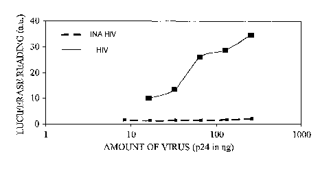
Figure 6 illustrates the effect of INA treatment on HIV infectivity as
measured by a luciferase reporter gene assay. As illustrated, INA-treated HIV
exhibit essentially no transcription from viral promoters within the HIV LTR.
These results further confirm that the INA-treated viruses used to generate
the
results in Figure 1 were indeed inactivated.
Figure 7 illustrates that INA-treatment of HIV causes substantially no
change in the epitopes recognized by three anti-HIV neutralizing antibody
preparations. The antibody preparations tested were the 2612, B 12 and 4E 10
antibody preparations. As shown, the amount of virus bound by the three
antibody preparations did not change when HIV was treated with INA (dashed
lines) as compared to untreated HIV (solid lines).
FIG. 8 shows that INA treatment of Ebola viral particles effectively
eliminates viral growth in mammalian cells (Vero-E6 cells). Ebola viral
CA 02557800 2006-08-29
WO 2005/093049 PCT/US2005/009559
particles were incubated with INA or DMSO (Control), exposed to ultraviolet
light and then cultured with Vero-E6 cells. At selected time points (shown on
the x-axis), aliquots of the virus/cell mixture were removed and the number of
viruses (plaque-forming units, pfu) was determined. As shown, control-treated
Ebola virus grew well on Vero-E6 cells but INA-treated Ebola virus failed to
grow.
Detailed Description of the Invention
According to the invention, treatment of tumor cells with a
photoactivatable hydrophobic compound of the invention bloclcs cell division
and colony formation with substantially no detectable damage to the structural
integrity of the cells. Moreover, when live HIV, SIV and Ebola viral particles
are treated with appropriate concentration of such photoactivatable
hydrophobic
compounds, substantially no infectivity is observed. Minor, generally
insubstantial changes in the structural integrity of virus particles were
observed.
These modified viral particles reacted with monoclonal antibodies directed
against selected viral proteins and the inactivated viruses bound to their
target
cells. However, viral fusion with the membrane was impaired by use of the
present inventive methods.
Hence, the invention provides new methods for inactivating viruses,
bacteria, parasites and tumor cells. These inactivated agents can be used in
compositions to stimulate an immune response against active viruses, bacteria,
parasites and tumor cells. In another embodiment, the invention provides
vaccines to prevent the diseases caused by such viruses, bacteria, parasites
and
tumor cells.
Photoactivatable Hydrophobic Compounds
Accordingly, as provided herein, a photoactivatable hydrophobic
compound of the following formula (I) Call be used to inactivate vinises,
parasites and tumor cells.
X-Ar-Y I
4
CA 02557800 2006-08-29
WO 2005/093049 PCT/US2005/009559
wherein:
Ar is a hydrophobic moiety; and
X and Y are each independently hydrogen or a reactive group, provided
that at least one of X or Y is a reactive group.
The Ar hydrophobic moiety can be any moiety that preferentially
partitions out of an aqueous environment and into a cellular or viral
membrane.
Examples of Ar hydrophobic moieties include linear, branched, cyclic and
acyclic hydrocarbons and combinations thereof. The cyclic groups employed
can be non-aromatic or aromatic ring moieties. For example, the Ar
hydrophobic moiety can be a fatty acid, alkyl, adamantane, phenyl, naphthyl,
anthracene, pyrene, phenanthracene or similar moiety.
The X and Y reactive groups are functional groups that are chemically
reactive (or that can be made or activated to be chemically reactive) with
functional groups typically found in biological materials, or with functional
groups that can be readily converted to chemically reactive groups using
methods well known in the art. In one embodiment of the invention, the X
and/or Y reactive groups are separately azido (-N3), halo (Cl, Br or I), halo
lower
alkyl (e.g. CF3), diazirene, azidocarbonyloxy (-O-CO-N3), haloacetamide (-NH-
(C=O)-CH2-Z), where Z is Cl, Br or I. Alternatively, the reactive groups are
separately amine, maleimide, isocyanato (-N=C=O), isothiocyanato (-N=C=S),
acyl halide, succinimidyl ester, or sulfosuccinimidyl ester. In another
embodiment, the reactive groups are carboxylic acid (COOH), or derivatives of
a
carboxylic acid. An appropriate derivative of a carboxylic acid includes an
alkali
or alkaline earth metal salt of carboxylic acid. Alternatively, the reactive
groups
are reactive derivatives of a carboxylic acid (-COOR), where the reactive
group
R is one that activates the carbonyl group of-COOR toward nucleophilic
displacement. In particular, R is any group that activates the carbonyl
towards
nucleophilic displacement without being incorporated into the final
displacement
product. Examples of COOR groups include esters of phenol or naphtol that are
further substituted by at least one strong electron withdrawing group, or
carboxylic acid activated by carbodiimide, or constitute aryl chloride, azido,
succinimidyl or sulfosuccinimidyl ester. Additional charged groups include,
CA 02557800 2006-08-29
WO 2005/093049 PCT/US2005/009559
among others, sulfonyl halides, sulfonyl azides, alcohols, thiols,
semicarbazides,
hydrazines or hydroxylamines.
Examples of photoactivatable hydrophobic compounds that can be used
in the invention include the following compounds:
N3 N3 O.,N
N
azidobenzene 1-azidonaphthalene 4-azido-2-nitro-1-(phenylthio)benzene
N
N3 N~
I
1-azido-4-iodobenzene 1-azido-5-iodonaphthalene 3-phenyl-3H diazirene
N N.
N N
3-phenyl-3-(trifluoromethyl)- 3-(3-iodophenyl)-3-(trifluoromethyl)
3H diazirene -3H diazirene
6
CA 02557800 2006-08-29
WO 2005/093049 PCT/US2005/009559
N3
N
1-azidopyrene adamantanediazirene
CH3 i o-COOH
NOZ CHZ-O-(CHZ),; COOH
n=8-15
N
12-(4-azido-2-nitrophenoxy)- w-(m-diazirinophenoxy)fatty acids
stearic acid
CH3-(CH2)5-CH-(CH2)lo-COOH CH3-(CH2)5_CH-(CH2)IO-COOH
O
N3
O
N3
12[(azidocarbonyl)oxy]stearic 12-azidostearic acid
acid
N
N 1 o-COOH
11-(3-azidophenoxy)undecanoic acid w-(m-diazirinophenoxy)undecanoic acid
7
CA 02557800 2006-08-29
WO 2005/093049 PCT/US2005/009559
In one embodiment, 1,5-iodonaphthyl azide (INA) is employed as a
photoactivatable hydrophobic compound. INA is a non toxic hydrophobic
compound. The structure for 1,5-iodonaphthyl azide (INA) is provided below.
See also, Bercovici and Gitler 1978, Biochemistry, 17: 1484-89.
Upon exposure to cells, photoactivatable hydrophobic compounds of the
invention will penetrate into the innermost regions of biological membrane
bilayers and will accumulate selectively in these regions. Photoactivatable
hydrophobic compounds of the invention are also light sensitive. Upon
irradiation with ultraviolet light (e.g., 320 to 400 mn) a reactive derivative
is
generated that binds to membrane proteins deep in the lipid bilayer. This
process specifically inactivates integral membrane proteins embedded in the
membrane while maintaining the integrity and activity of the proteins that
protrude from the extracellular surface of the membrane.
In another embodiment, the photoactivatable hydrophobic compounds of
the invention can be used for inactivation of viruses, bacteria, parasites and
tumor cells using visible light. However, when visible light is used a
photosensitizes chromophore is needed. This photosensitizes chromophore has
an absorption maximum in the visible light range and can photosensitize the
photoactivatable hydrophobic compounds of the invention. In general, the
photosensitizes chromophores have absorption maxima in the range of about 450
to about 525 nm or about 600 to about 700 nm. The photosensitizes
chromophore can be a porphyrin, chlorin, bacteriochlorin, purpurin,
phthalocyanine, naphthalocyanine, merocyanines, carbocyanine, texaphyrin,
non-tetrapyrrole, or other photosensitizes known to one of skill in the art.
Specific examples of photosensitizes chromophores include fluorescein, eosin,
CA 02557800 2006-08-29
WO 2005/093049 PCT/US2005/009559
bodipy, nitro-benzo-diazol (NBD), erythrosine, acridine orange, doxorubicin,
rhodamine 123, picoerythrin and the like.
Treatment with Photoactivatable Hydrophobic Compounds
S As provided herein, viruses, bacteria, parasites and tumor cells can be
inactivated by exposure to photoactivatable hydrophobic compounds. In some
embodiments the photoactivatable hydrophobic compound is 1,5-iodonaphthyl
azide (INA) or a related compound. After contacting the photoactivatable
hydrophobic compound with the virus, parasite or tumor cell to form a mixture
thereof, the mixture is exposed to light. If the virus, parasite or tumor cell
is
contacted with just the photoactivatable hydrophobic compound, ultraviolet
light
is used. If the virus, parasite or tumor cell is contacted with both the
photoactivatable hydrophobic compound and a photosensitizer chromophore that
absorbs visible light, then visible light can be used instead. Exposure to
ultraviolet light directly photoactivates the photoactivatable hydrophobic
compound within viral and cellular membranes. Exposure to visible light first
photoactivates the photosensitizer chromophore, which then activates or
photosensitizes the photoactivatable hydrophobic compound within viral or
cellular membranes. In either case, a reactive derivative of the
photoactivatable
hydrophobic compound is generated that binds to membrane proteins deep
within the lipid bilayer. This process causes specific inactivation of
integral
membrane proteins embedded in the membrane, while maintaining the integrity
and activity of proteins that protrude outside of the membrane.
Prior to exposure to a photoactivatable hydrophobic compound, the
viruses, parasites or tmnor cells can be washed to remove media, waste and
other
materials that might reduce partitioning of the photoactivatable hydrophobic
compound into viral or cellular membranes. For example, the viruses, parasites
or tumor cells can be washed in serum-free media, phosphate-buffered saline or
other solutions selected by one of slcill in the art.
The amount of photoactivatable hydrophobic compound used to
inactivate a virus, bacteria, parasite or tumor cell can vary and may depend
upon
the type of virus, bacteria, parasite or tumor cell as well as the conditions
under
which the photoactivatable hydrophobic compound is reacted with the virus,
9
CA 02557800 2006-08-29
WO 2005/093049 PCT/US2005/009559
bacteua, parasite or tumor cell. For example, if competing hydrophobic
molecules are present in the media, then larger amounts of the
photoactivatable
hydrophobic compound may be needed.
In some embodiments, the concentration of photoactivatable hydrophobic
compound employed in a mixture with a virus, parasite or tumor can vary from
about 0.1 micromolar to about 1 millimolar, or from about 1 micromolar to
about 700 micromolar, or from about 10 micromolar to about 500 micromolar,
or from about 20 micromolar to about 400 micromolar, or from about 50
micromolar to about 300 micromolar, or from about 100 micromolar to about
250 micromolar.
When expressed as a r atio of the amount of photoactivatable hydrophobic
compound employed per amount of viral, parasite or tumor protein, this ratio
can
vary from about O.lmicrograms photoactivatable hydrophobic compound per
milligram of viral, parasite or tumor protein to about 500 micrograms
photoactivatable hydrophobic compound per milligram of viral, parasite or
tumor protein. In other embodiments, the amount of photoactivatable
hydrophobic compound used can vary from about 0.5 to about 200, or about 1 to
about 150, or about 2 to about 125, or about 3 to about 100 micrograms
photoactieatable hydrophobic compound per milligram of viral, parasite or
tumor protein.
The amount of photosensitizes chromophore used to activate the
photoactivatable hydrophobic compound can also vary and depends to some
extent on the photosensitizes chromophore used, the photoactivatable
hydrophobic compound employed and the type of virus, bacteria, parasite or
tumor cell. For example, about 0.01 mg/ml to about 50 mg/ml photosensitizes
chromophore can be used, or about 0.1 mg/ml to about 5 mg/ml photosensitizes
chromophore can be used, or about 0.3 mg/ml to about 1 mg/ml photosensitizes
chromophore can be used.
After forming a mixture of the virus, bacteria, parasite or tumor cell with
a photoactivatable hydrophobic compound, the mixture is exposed to light for a
time and under conditions sufficient for generating a reactive hydrophobic
derivative that can bind to membrane proteins within the lipid bilayer. The
ultraviolet light employed when only the photoactivatable hydrophobic
CA 02557800 2006-08-29
WO 2005/093049 PCT/US2005/009559
compound is present has a wavelength that is generally above that absorbed by
proteins and nucleic acids. Such a wavelength of ultraviolet light does not
cause
substantial damage to such proteins and nucleic acids. Thus, for example, the
wavelength can be about 320 nm to about 400 mn. W some embodiments, the
wavelength is about 330 nm to about 380 nm. In other embodiments, the
wavelength is about 340 nm to about 360 nm.
Visible light of an appropriate wavelength can be used when a
photosensitizer chromophore is employed that is incubated with or is localized
in
the vicinity of the hydrophobic photoactivatable compound. In general, the
photosensitizer chromophores have absorption maxima in the range of about 450
to about 525 nm or about 600 to about 700 nm.
Light for photoactivation of the photosensitizer chromophore or the
hydrophobic derivative can be from various light sources. For example,
suitable
light sources include broadband conventional light sources, broad arrays of
LEDs, laser beams, defocused laser beams, optical fiber devices and
transillumination. The light can be filtered to eliminate certain types or
wavelengths of light. Hence, the light can be filtered to provide ultraviolet
light
(e.g., 320 to 400 nm), or visible light of selected wavelengths (e.g., 450 to
525
nm or 600 to 700 nm). The light can also be filtered to reduce heat
production,
for example, by passing the light through water.
Different light sources of different powers can be used: An incaaidescent
light source like tungsten or halogen lamps will have a power range from 100-
200 Watt. Mercury or Xenon light sources have a power range between 100-
1000 Watt. A laser source will have the power range of 1-10 Watts. When
visible light is used in the presence of a photosensitizer chromophore, the
tungsten, halogen, Mercury and Xenon light sources should be equipped with
optical filters or a monochromator that will filter out all wavelengths below
400
nm. When a laser is used, the appropriate wavelength line of 400 nm or higher
should be used depending on the photosensitizer chromophore employed.
Regardless of the light source the intensities of light on the target sample
should
be in the range of 1-50 milliwatt/cm~lmin depending on the nature of the
sample
and the area irradiated.
11
CA 02557800 2006-08-29
WO 2005/093049 PCT/US2005/009559
Light exposure times can vary. For example, one of skill in the art may
choose to expose a mixture of a photosensitizer chromophore and/or a
photoactivatable hydrophobic compound with a virus, bacteria, parasite or
tumor
cell to a light source for about 1 second to about 20 minutes, or about 3
seconds
to about 15 minutes, or about 5 seconds to about 10 minutes, or about 7
seconds
to about 7 minutes, or about 30 seconds to about 5 minutes. A series of short
(e.g., about 1 to about 60 seconds) or longer (e.g., about 20 to about 60
seconds)
light exposures can also be employed. When a laser is used, substantially
shorter
exposure times are typically used, for example, about 0.1 second to about 5
seconds, or about 0.5 seconds to about 3 seconds.
As is appreciated by one of skill in the art, the exposure time can vary
depending on the wattage of the light employed. Either cultures or plates of
viruses, bacteria, parasites or tumor cells can be treated with a selected
photoactivatable hydrophobic compound and/or a photosensitizer chromophore
and then exposed to light. The exposure time and wattage of the light employed
may be different if a culture or plate of viruseslcells is employed. For
example,
less exposure may be needed for plated viruses/cells than for viruses/cells
cultured in suspension because the depth of the culture may influence the
degree
to which the light penetrates the culture. Hence, some variation and deviation
from the ranges provided herein is possible without deviating from the scope
of
the invention.
As described in more detail herein, INA has been shown by the inventors
to penetrate into the inner most segments of membrane bilayers and accumulate
selectively in this domain. As shown herein, upon irradiation of the organism
or
cell with ultraviolet light (e.g., 320-400 nm), INA is photoactivated in the
membrane to generate a reactive derivative that binds to membrane proteins
deep
within the lipid bilayer. This process causes specific inactivation of
integral
membrane proteins embedded in the membrane, while maintaining the integrity
and activity of proteins that protrude outside the membrane (Raviv et al, 1984
Biochemistry, 23, 503-508).
12
CA 02557800 2006-08-29
WO 2005/093049 PCT/US2005/009559
Methods of Using the Inactivated Microbes, Parasites and Tumor Cells
The invention provides a method that can universally inactivate viruses,
bacteria, parasites and tumor cells in a way that they can be safely used as
immunological compositions or vaccines to inhibit the disease they cause. The
inactivation kills the organism or cell in a specific manner that maintains
its
structure and conformation. Hence, the structure of the inactivated virus/cell
is
similar to that of the live virus/cell. W this way, the immunogenicity of the
organism or cell as a whole is maintained and can be safely used to stimulate
the
immune system of a subject animal or patient. Similarly, the inactivated
viruses,
bacteria, cancer cells or parasites of the invention can be used for
vaccination
without causing disease or other negative side effects.
A study conducted by the inventors showed that INA treatment of tumor
cells blocked their ability to divide and form colonies, with no detectable
damage to the structural integrity of the cells.
Studies by the inventors show that INA can also be used to inactivate live
HIV, SIV and Ebola viruses. In particular, INA treatment produced inactive
viruses with no detectable infectivity (Table 1 and Figure 6) and with no
significant change to their structural integrity (Figures 1, 3 and 4). Minor
modifications to viral proteins were detected (Figure 2). However, these
modifications did not affect the ability of these proteins to react with
antibodies
that are known to bind to SIV or HIV (Figures 3 and 7). Likewise, the inactive
virus was not significantly impaired in its ability to bind to target cells,
with the
highest concentration of INA (0.2 mM) only reducing the binding by 30%
(Figure 4). However, the INA treatment impaired the ability of the virus to
fuse
with the target cell at the plasma membrane level (Figure 5) and to express
virally encoded functions (Figure 6). Viral growth in cells that normally
would
become infected was essentially eliminated.
Hence, the INA treatment procedures of the invention generate inactive
viruses that can be used in a manner similar to aldrithiol inactivated HIV
(developed by the AIDS vaccine program SAIC). Alternatively, the INA-
inactivation procedures of the invention can be used in conjunction with
aldrithiol inactivation procedures to generate inactive HIV that comply with
the
13
CA 02557800 2006-08-29
WO 2005/093049 PCT/US2005/009559
requirements of the FDA. Thus, two mechanistically independent methods of
inactivation can be used to provide a prophylactic AIDS or HIV vaccine.
The present invention is therefore directed to methods of treating or
preventing or otherwise ameliorating microbial or parasitic infections in a
marmnal, as well as other animals, such as farm animals and birds. In another
embodiment, the invention provides to methods of treating or preventing or
otherwise ameliorating cancer in a mammal, as well as other animals, such as
farm animals and birds. These methods include administering to the animal an
effective amount, for example, a therapeutically effective amount of an
inactivated agent of the present invention, wherein the agent may cause an
infection or cancer when not inactivated as described herein.
Prevention or treatment of microbial infections, parasitic infections or
cancer is intended to include the alleviation of or diminishment of at least
one
symptom typically associated with the infection or cancer. Prevention or
treatment also includes alleviation or diminishment of more than one synptom.
Ideally, treatment with the inactivated agents of the invention generates
irrummity in the animal towards the agent while prevention by the inactivated
agents of the invention substantially eliminates the symptoms associated with
the
infection or cancer.
Microbial infections that can be treated by the present inactivated agents
include infections by any target microbial organisms that can infect a mammal
or
other animal. Such target microbial organisms include essentially any virus,
bacterium, fungus, single cell organism or parasite that can infect an animal,
including mammals. For example, target microbial organisms include viruses,
bacteria, fungi, yeast strains and other single cell organisms. In another
embodiment, the inactivated agents of the invention can give rise to immunity
against both gram-negative and gram-positive bacteria.
Treatment of, or prevention of, viral, bacterial, fungal, microbial or
parasitic infections is intended to include the alleviation of or diminishment
of at
least one symptom typically associated with the infection. The treatment also
includes alleviation or diminishment of more than one symptom. The treatment
may cure the infection, e.g., it may substantially prevent the infection
and/or
eliminate the symptoms associated with the infection.
14
CA 02557800 2006-08-29
WO 2005/093049 PCT/US2005/009559
Exemplary viral infections that can be treated by the present inactivated
agents include infections by any virus that can infect animals (including but
not
limited to mammals), including enveloped and non-enveloped viuuses, DNA and
RNA viruses, viroids, and prions. Hence, for example, infections or unwanted
levels of the following viruses and viral types can be treated, prevented or
addressed by the present inactivated agents: human immunodeficiency virus
(HIV), simian immunodeficiency virus (SIV), hemorrhagic fever viruses,
hepatitis A virus, hepatitis B virus, hepatitis C virus, poxviruses, herpes
viruses,
adenoviruses, papovaviruses, parvoviruses, reoviruses, orbiviruses,
picornaviruses, rotaviruses, alphaviruses, rubiviruses, influenza virus type A
and
B, flaviviruses, coronaviruses, paramyxoviruses, morbilliviruses,
pneumoviruses, rhabdoviruses, lyssaviruses, orthmyxoviruses, bunyaviruses,
phleboviruses, nairoviruses, hepadnaviruses, arenaviruses, retroviruses,
enteroviruses, rhinoviruses and the filovirus.
Infections or unwanted levels of the following target viruses and viral
types that are believed to have potential as biological weapons can be
treated,
prevented or addressed by the present inactivated agents: hemorrhagic fever
viruses (HFVs), Chilcungunya virus, Japanese encephalitis virus, Monkey pox
virus, variola viuus, Congo-Crimean hemorrhagic fever virus, Junin virus,
Omslc
hemorrhagic fever virus, Venezuelan equine encephalitis virus, Dengue fever
virus, Lassa fever virus, Rift valley fever virus, Western equine encephalitis
virus, Eastern equine encephalitis virus, Lymphocytic choriomeningitis virus,
Russian Spring-Summer encephalitis virus, White pox, Ebola virus, Machupo
virus, Smallpox virus, Yellow fever virus, Hantaan virus, Marburg virus, and
Ticlc-borne encephalitis virus.
Similarly, infections or unwanted levels of the following examples of
target microbial organisms can be treated, prevented or addressed by the
present
inactivated agents: Aeromoraas spp. (including, for example,
Aey°onaonas
hydr~ophila, Aeromoraas caviae and Aeronaonas sobs°ia), Bacillus spp.
(including,
for example, Bacillus cereus, Bacillus afZtlaracis and Bacillus
thuringierasis),
Bacte~oides spp. (including, for example, B. fragilis, B. tlaetaiotaomicron,
B.
vulgatus, B. ovatus, B. distasonis, B. unifonmis, B. stercoris, B. eggerthii,
B.
merdae, and B. caccae), Canapylobacter spp. (including, for example,
CA 02557800 2006-08-29
WO 2005/093049 PCT/US2005/009559
CampylobacteY jejuni, Canapylobacte>~ laridis, and Campylobactez°
hyointestinalis), ClostYidium spp. (such as the pathogenic clostridia
including all
types of Clostridium botulinum (including those in Groups I, II, III and IV,
and
including those that produce botulism A, B, C, D, E, F and G), all types of
Clostf~idimta tetazti, all types of Clostridium difficile, and all types of
Closty~idiustt
perfi°ingens), Ebola spp. (e.g. EBOV Zaire), Eraterobacter spp.
(including, for
example, EnteYObactez° aet°ogenes (also sometimes referred to as
Klebsiella
naobilis), Ente~obactet~ agglonaet~ans (also sometimes referred to as Pantoea
agglomez°ans), Ente~obacter amnigenus, Entenobactef° asbu>"iae,
Erttez°obacten
cancez°ogenus (also sometimes referred to as Enterobactez°
tayloz°ae and/or
Erwinia catZCe>"ogena), Enterobactet° cloacae, EnteYObactet~
cowanii,
EnteYObactet° dissolvens (also sometimes referred to as Ez"winia
dissolvens),
Ente~obacte>" geYgoviae, Entet~obacter lZOrmaechei, EnterobacteY
ifttet~medium,
Enterobactez° intermedius (also sometimes referred to as
Entet~obactet~
intenmedium), Enterobactet~ l~obei, Enterobactez° nitnipressut~alis
(also
sometimes referred to as Et°winia nimip>~essuz°alis),
Ente>~obacte>~ salrazakii, and
Ente>~obacter taylo~ae (also sometimes referred to as Entez-
obactez°
catace~°ogenus)), Eytterococcus spp. (including, for example,
Vancomycin
Resistant Enterococcus (VRE), Enterococcus faecalis, Enterococcus faecium,
Enterococcus durarts, Enterococcus gallinaz~una, and
Entez°ococctts
casseliflavus), Esche~ichia spp. (including the enterotoxigenic (ETEC)
strains,
the enteropathogenic (EPEC) strains, the enterohemorrhagic (EHEC) strain
designated E. coli 0157:H7, and the enteroinvasive (EIEC) strains),
Gastrospir°illun2 spp. (including, for example,
Gastrospit°illunZ hominis (also
sometimes now referred to as Helicobactez" heilman ztii), Helicobacter spp.
(including, for example, Helicobactez° pylori and Helicobacten
hepaticus),
Klebsiella spp. (including, for example, Klebsiella pneumoniae, Klebsiella
ozaenae, Klebsiella >"hirtoscleromatis, Klebsiella oxytoca, Klebsiella
planticola,
Klebsiella tet~f~igena, and Klebsiella o~ftithinolytica), Salmonella spp.
(including,
for example, S. typhi and S. pat~atyphi A, B, and C, S. entez~itidis, and S.
dublin),
Shigella spp. (including, for example, Shigella sonnei, Shigella boydii,
Shigella
flexneri, and Slaigella dysentet~iae), Staphylococcus spp. (including, for
example,
Staphylococcus aureus, methicillin-resistant Staphylococcus aureus (MRSA),
16
CA 02557800 2006-08-29
WO 2005/093049 PCT/US2005/009559
Staphylococcus saprophyticus and Staphylococcus epiderntis),
Stj°eptococcus
ssp. (including Groups A (one species with 40 antigenic types, Streptococcus
pyogenes), B, C, D (five species (Streptococcus faecalis, Streptococcus
faeciunt,
Streptococcus durans, Streptococcus aviunt, and Streptococcus bovis)), F, and
G,
including Streptococcus pneumoniae), Pseudontortas spp. (including, for
example, Pseudomonas aerugirtosa, Pseudotnonas tnaltophilia, Pseudotnonas
fluorescens, Pseudontonas putida, Pseudotnonas cepacia, Pseudontortas
stutzeri,
Pseudotnonas mallei, Pseudomonas pseudontallei and Pseudotnottas
putrefaciens), Yibrio spp. (including, for example, Tribrio cholera Serogroup
O1
and T~ibrio cholera Serogroup Non-O1, Vibrio parah.aentolyticus, llibrio
alginolyticus, l~ibrio furnissii, hibrio carclZariae, Tlibrio hollisae,
T~ibrio
cincinnatiensis, Tribrio ntetschnikovii, Vibrio damsela, Vibrio ntimicus,
T~ibrio
vulnificus, and Yibrio fluvialis), Yetsinia spp. (including, for example,
Yei°sinia
pestis, Yeisittia enterocolitica and Yersinia pseudotuberculosis), Neisseria,
Proteus, Citrobacter, Aerobacter, Providencia, Set°ratia, Brucella,
Fraytcisella
tularensis (also sometimes referred to as Pasteurella tulat°ertsis,
Bacillus
tularensis, Brucella tularensis, tularemia, rabbit fever, deerfly fever,
Ohara's
disease, and/or Francis disease), and the like. Thus, for example, various
bacterial infections or unwanted levels of bacteria that can be treated,
prevented
or addressed by the present inactivated agents include but are not limited to
those
associated with anthrax (Bacillus atZthracis), staph infections
(Stap7Zylococcus
aus°eus), typhus (Salmotzella typhi), food poisoning (Esc7zerichia
coli, such as
0157:H7), bascillary dysentery (Shigella dysenteric), pneumonia (Psuedomonas
aerugenosa and/or Pseudontonas cepacia), cholera (Yibrio cholerae), ulcers
(Flelicobacter pylori), Bacillus cereus, Salmonella, Clostridium perfi-ingens,
Catnpylobacter, Listeria mortocytogenes, Vibrio parahaemolyticus, botulism
(Clostridiutn botulinum), smallpox (variola tnajor), listeriosis (Listeria
monocytogenes), tularemia (Francisella tularensis), plague (Yersinia pesos;
also
sometimes referred to as bubonic plague, pneumonic plague, and/or blaclc
death)
and others. E. coli serotype 0157:H7 has been implicated in the pathogenesis
of
diarrhea, hemorrhagic colitis, hemolytic uremic syndrome (HUS) and thrombotic
thrombocytopenic purpura (TTP). As indicated herein, the inactivated agents of
the invention are also active against drug-resistant and multiply-drug
resistant
17
CA 02557800 2006-08-29
WO 2005/093049 PCT/US2005/009559
strains of bacteria, for example, multiply-resistant strains of Staphylococcus
aureus and vancomycin-resistant strains of Enterococcus faeciuna and
Ente~ococcus faecalis.
Fungal infections that can be treated or prevented by the present
inactivated agents include infections by fungi that infect a mammal, including
Histoplasma capsulatuna, Coccidioides immitis, Cryptococcus neofof~fyaans,
Candida ssp. including Candida albicans, Aspefgilli ssp. including Aspefgillus
fumigatus, Spof°otlZrix, Triclzophyton ssp., Fusaf~ium ssp.,
Tf°icosporon ssp.,
Pneumocystis caf°inii, and Ty~ichophyton mentagnophytes. Hence, for
example,
infections or unwanted levels of target fungi can be treated, prevented or
addressed by the present inactivated agents. Such fungi also include fungal
pathogens that may have potential for use biological weapons, including
Coccidioides inamitis and Histoplasma capsulatum..
Anti-microbial activity can be evaluated against these varieties of
microbes (viruses, bacteria, fungi and parasites) using methods available to
one
of skill in the art. In one embodiment, anti-microbial activity is the amount
of
the inactivated agent that stimulates an immune response against the microbe.
In
another embodiment, anti-microbial activity is the amount of the inactivated
agent that effectively immunizes a mammal against the microbe.
Treatment of, or treating, cancer is intended to include the alleviation of
or diminishment of at least one symptom typically associated with the disease.
The treatment also includes alleviation or diminishment of more than one
symptom. The treatment may cure the cancer, e.g., it may reduce the number of
cancer cells and/or arrest the growth of the cancerous tumor.
Cancers that can be treated by the present inactivated agents include solid
mammalian tumors as well as hematological malignancies. Solid mammalian
tumors include cancers of the head and neclc, lung, mesothelioma, mediastinum,
esophagus, stomach, pancreas, hepatobiliary system, small intestine, colon,
colorectal, rectum, anus, kidney, urethra, bladder, prostate, urethra, penis,
testis,
gynecological organs, ovaries, breast, endocrine system, skin central nervous
system; sarcomas of the soft tissue and bone; and melanoma of cutaneous and
intraocular origin. Hematological malignancies include childhood leukemia and
lymphomas, Hodglcin's disease, lymphomas of lymphocytic and cutaneous
18
CA 02557800 2006-08-29
WO 2005/093049 PCT/US2005/009559
origin, acute and chronic leukemia, plasma cell neoplasm and cancers
associated
with AIDS. In addition, a cancer at any stage of progression can be treated,
such
as primary, metastatic, and recurrent cancers. Information regarding numerous
types of cancer can be found, e.g., from the American Cancer Society
(www.cancer.org), or from, e.g., Wilson et al. (1991) Harrison's Principles of
Internal Medicine, l2<sup>th</sup> Edition, McGraw-Hill, Inc. Both human and
veterinary uses are contemplated.
Anti-cancer activity can be evaluated against varieties of cancers using
methods available to one of skill in the art. Anti-cancer activity, for
example, is
determined by identifying the LDlon or EDSO of an inactivated tumor or cancer
cell of the present invention that prevents the growth of a cancer. In one
embodiment, anti-cancer activity is the amount of the inactivated agent that
effectively immunizes a mammal against that cancer type.
According to the present invention, the inactivated agents provided
herein do not have substantial or undesired toxicity or infectivity within the
mammalian organism to be treated.
Administration of the Inactivated Agents
The inactivated agents of the invention are administered so as to achieve
a reduction in at least one synptom associated with an infection, cancer,
tumor
or other disease, or a decrease in the amount of antibody associated with the
infection, cancer, tumor or other disease.
To achieve the desired effect(s), the inactivated agent, or a combination
of inactivated agents, may be administered as single or divided dosages, for
example, of at least about 0.01 mg/kg to about 500 to 750 mg/lcg, of at least
about 0.01 mg/kg to about 300 to 500 mg/kg, at least about 0.1 mg/kg to about
100 to 300 mg/kg or at least about 1 mg/kg to about 50 to 100 mg/lcg of body
weight, although other dosages may provide beneficial results. The amount
administered will vary depending on various factors including, but not limited
to, the inactivated agent chosen, the disease, the weight, the physical
condition,
the health, the age of the mammal, or whether prevention or treatment is to be
achieved. Such factors can be readily determined by the clinician employing
animal models or other test systems that are available in the art.
19
CA 02557800 2006-08-29
WO 2005/093049 PCT/US2005/009559
Administration of the therapeutic agents in accordance with the present
invention may be in a single dose, in multiple doses, in a continuous or
intermittent manner, depending, for example, upon the recipient's
physiological
condition, whether the purpose of the administration is therapeutic or
prophylactic, and other factors known to skilled practitioners. The
administration of the inactivated agents of the invention is generally
intermittent
over a preselected period of time, for example, in a series of spaced doses.
Both
local and systemic administration is contemplated.
To prepare the composition, inactivated agents are prepared according to
the methods described herein, and purified as necessary or desired. In some
embodiments the inactivated agents can be lyophilized and/or stabilized. The
inactivated agent can then be adjusted to the appropriate concentration, and
optionally combined with other agents.
The absolute weight of a given inactivated agent included in a unit dose
can vary widely. For example, about 0.01 to about 2 g, or about 0.1 to about
500
mg, of at least one inactivated agent of the invention, or a plurality of
inactivated
agents, can be administered. Alternatively, the unit dosage can vary from
about
0.01 g to about 5 g, from about 0.01 g to about 3.5 g, from about 0.01 g to
about
2.5 g, from about 0.1 g to about 1 g, from about 0.1 g to about 0.8 g, from
about
0.1 g to about 0.4 g, or from about 0.1 g to about 0.2 g.
One or more suitable unit dosage forms comprising the therapeutic
inactivated agents of the invention can be administered by a variety of routes
including oral, parenteral (including subcutaneous, intravenous, intramuscular
and intraperitoneal), rectal, dermal, transdermal, intrathoracic,
intrapulmonary
and intranasal (respiratory) routes. The therapeutic inactivated agents may
also
be formulated for sustained release (for example, using microencapsulation,
see
WO 94/ 07529, and U.S. Patent No.4,962,091). The formulations may, where
appropriate, be conveniently presented in discrete unit dosage forms and may
be
prepared by any of the methods well known to the pharmaceutical arts. Such
methods may include the step of mixing the therapeutic agent with liquid
carriers, solid matrices, semi-solid carriers, finely divided solid carriers
or
combinations thereof, and then, if necessary, introducing or shaping the
product
into the desired delivery system.
CA 02557800 2006-08-29
WO 2005/093049 PCT/US2005/009559
When the therapeutic inactivated agents of the invention are prepared for
oral administration, they are generally combined with a pharmaceutically
acceptable carrier, diluent or excipient to form a pharmaceutical formulation,
or
unit dosage form. For oral administration, the inactivated agents may be
present
as a powder, a granular formulation, a solution, a suspension, an emulsion or
in a
natural or synthetic polymer or resin for ingestion of the agents from a
chewing
gum. The inactivated agents may also be presented as a bolus, electuary or
paste. Orally administered therapeutic inactivated agents of the invention can
also be formulated for sustained release, e.g., the inactivated agents can be
coated, micro-encapsulated, or otherwise placed within a sustained delivery
device. The total active ingredients in such formulations comprise from 0.1 to
99.9% by weight of the formulation.
By "pharmaceutically acceptable" it is meant a carrier, diluent, excipient,
and/or salt that is compatible with the other ingredients of the formulation,
and
not deleterious to the recipient thereof.
Pharmaceutical formulations containing the therapeutic inactivated
agents of the invention can be prepared by procedures known in the art using
well-known and readily available ingredients. For example, the inactivated
agent can be formulated with common excipients, diluents, or carriers, and
formed into tablets, capsules, solutions, suspensions, powders, aerosols and
the
like. Examples of excipients, diluents, and carriers that are suitable for
such
formulations include buffers, as well as fillers and extenders such as starch,
cellulose, sugars, mamlitol, and silicic derivatives. Binding agents can also
be
included such as carboxymethyl cellulose, hydroxymethylcellulose,
hydroxypropyl methylcellulose and other cellulose derivatives, alginates,
gelatin,
and polyvinyl-pyrrolidone. Moisturizing agents can be included such as
glycerol, disintegrating agents such as calcium carbonate and sodium
bicarbonate. Agents for retarding dissolution can also be included such as
paraffin. Resorption accelerators such as quaternary armnonium compounds can
also be included. Surface active agents such as cetyl alcohol and glycerol
monostearate can be included. Adsorptive carriers such as lcaolin and
bentonite
can be added. Lubricants such as talc, calcium and magnesium stearate, and
solid polyethyl glycols can also be included. Preservatives may also be added.
21
CA 02557800 2006-08-29
WO 2005/093049 PCT/US2005/009559
The compositions of the invention can also contain thickening agents such as
cellulose and/or cellulose derivatives. They may also contain gums such as
xanthan, guar or carbo gum or gum arabic, or alternatively polyethylene
glycols,
bentones and montmorillonites, and the like.
For example, tablets or caplets containing the inactivated agents of the
invention can include buffering agents such as calcium carbonate, magnesium
oxide and magnesium carbonate. Caplets and tablets can also include inactive
ingredients such as cellulose, pre-gelatinized starch, silicon dioxide,
hydroxy
propyl methyl cellulose, magnesium stearate, microcrystalline cellulose,
starch,
talc, titanium dioxide, benzoic acid, citric acid, corn starch, mineral oil,
polypropylene glycol, sodium phosphate, zinc stearate, and the like. Hard or
soft
gelatin capsules containing at least one inactivated agent of the invention
can
contain inactive ingredients such as gelatin, microcrystalline cellulose,
sodium
lauryl sulfate, starch, talc, and titanium dioxide, and the like, as well as
liquid
vehicles such as polyethylene glycols (PEGs) and vegetable oil. Moreover,
enteric-coated caplets or tablets containing one or more inactivated agents of
the
invention are designed to resist disintegration in the stomach and dissolve in
the
more neutral to allcaline environment of the duodenum.
The inactivated agents of the invention can also be formulated as elixirs
or solutions for convenient oral administration or as solutions appropriate
for
parenteral administration, for instance by intramuscular, subcutaneous,
intraperitoneal or intravenous routes. The pharmaceutical formulations of the
therapeutic inactivated agents of the invention can also take the form of an
aqueous or anhydrous solution or dispersion, or alternatively the form of an
emulsion or suspension or salve.
Thus, the therapeutic inactivated agents may be formulated for parenteral
administration (e.g., by injection, for example, bolus injection or continuous
infusion) and may be presented in unit dose form in ampoules, pre-filled
syringes, small volume infusion containers or in mufti-dose containers. As
noted
above, preservatives can be added to help maintain the shelve life of the
dosage
form. The inactivated agents and other ingredients may form suspensions,
solutions, or emulsions in oily or aqueous vehicles, and may contain
fonnulatory
agents such as suspending, stabilizing and/or dispersing agents.
Alternatively,
22
CA 02557800 2006-08-29
WO 2005/093049 PCT/US2005/009559
the inactivated agents and other ingredients may be in powder form, obtained
by
aseptic isolation of sterile solid or by lyophilization from solution, for
constitution with a suitable vehicle, e.g., sterile, pyrogen-free water,
before use.
These formulations can contain pharmaceutically acceptable carriers,
vehicles and adjuvants that are well known in the art. It is possible, for
example,
to prepare solutions using one or more organic solvents) that islare
acceptable
from the physiological standpoint, chosen, in addition to water, from solvents
such as acetone, ethanol, isopropyl alcohol, glycol ethers such as the
products
sold under the name "Dowanol," polyglycols and polyethylene glycols, C1-C4
alkyl esters of short-chain acids, ethyl or isopropyl lactate, fatty acid
triglycerides such as the products marketed under the name "Miglyol,"
isopropyl
myristate, animal, mineral and vegetable oils and polysiloxanes.
It is possible to add, if desired, an adjuvant chosen from antioxidants,
surfactants, other preservatives, film-forming, keratolytic or comedolytic
agents,
perfumes, flavorings and colorings. Antioxidants such as t-butylhydroquinone,
butylated hydroxyanisole, butylated hydroxytoluene and a tocopherol and its
derivatives can be added.
Also contemplated are combination products that include one or more
inactivated agents of the present invention and one or more other anti-
microbial
agents. For example, a variety of antibiotics can be included in the
pharmaceutical compositions of the invention, such as aminoglycosides (e.g.,
streptomycin, gentamicin, sisomicin, tobramycin and amicacin), ansamycins
(e.g. rifamycin), antimycotics (e.g. polyenes and benzofuran derivatives), (3-
lactams (e.g. penicillins and cephalosporins), chloramphenical (including
thiamphenol and azidamphenicol), linosamides (lincomycin, clindamycin),
macrolides (erythromycin, oleandomycin, spiramycin), polymyxins, bacitracins,
tyrothycin, capreomycin, vancomycin, tetracyclines (including oxytetracycline,
minocycline, doxycycline), phosphomycin and fusidic acid.
Additionally, the inactivated agents are well suited to formulation as
sustained release dosage forms and the like. The formulations can be so
constituted that they release the inactivated agent, for example, in a
particular
part of the intestinal or respiratory tract, possibly over a period of time.
Coatings, envelopes, and protective matrices may be made, for example, from
23
CA 02557800 2006-08-29
WO 2005/093049 PCT/US2005/009559
polymeric substances, such as polylactide-glycolates, liposomes,
microemulsions, microparticles, nanoparticles, or waxes. These coatings,
envelopes, and protective matrices are useful to coat indwelling devices,
e.g.,
stems, catheters, peritoneal dialysis tubing, draining devices and the like.
For topical administration, the inactivated agents may be formulated as is
known in the art for direct application to a target area. Forms chiefly
conditioned for topical application talce the form, for example, of creams,
milks,
gels, dispersion or microemulsions, lotions thickened to a greater or lesser
extent, impregnated pads, ointments or sticks, aerosol fomnulations (e.g.,
sprays
or foams), soaps, detergents, lotions or cakes of soap. Other conventional
forms
for this purpose include wound dressings, coated bandages or other polymer
coverings, ointments, creams, lotions, pastes, jellies, sprays, and aerosols.
Thus,
the therapeutic inactivated agents of the invention can be delivered via
patches
or bandages for dermal administration. Ahternatively, the inactivated agent
can
be formulated to be part of an adhesive polymer, such as polyacrylate or
acryhate/vinyl acetate copolymer. For long-term applications it might be
desirable to use microporous and/or breathable backing laminates, so hydration
or maceration of the skin can be minimized. The backing layer can be any
appropriate thickness that will provide the desired protective and support
functions. A suitable thickness will generally be from about 10 to about 200
microns.
Ointments and creams may, for example, be formulated with an aqueous
or oily base with the addition of suitable thichcening and/or gelling agents.
Lotions may be formulated with an aqueous or oily base and will in general
also
contain one or more emulsifying agents, stabilizing agents, dispersing agents,
suspending agents, thickening agents, or coloring agents. The inactivated
agents
can also be delivered via iontophoresis, e.g., as disclosed in U.S. Patent
Nos.
4,140,122; 4,383,529; or 4,051,842. The percent by weight of a therapeutic
agent of the invention present in a topical formulation will depend on various
factors, but generally will be from 0.01 % to 95% of the total weight of the
formulation, and typically 0.1-85% by weight.
Drops, such as eye drops or nose drops, may be formulated with one or
more of the inactivated agents in an aqueous or non-aqueous base also
24
CA 02557800 2006-08-29
WO 2005/093049 PCT/US2005/009559
comprising one or more dispersing agents, solubilizing agents or suspending
agents. Liquid sprays are conveniently delivered from pressurized pacl~s.
Drops
can be delivered via a simple eye dropper-capped bottle, or via a plastic
bottle
adapted to deliver liquid contents dropwise, via a specially shaped closure.
The therapeutic inactivated agent may further be formulated for topical
achninistration in the mouth or throat. For example, the active ingredients
may
be formulated as a lozenge further comprising a flavored base, for example,
sucrose and acacia or tragacanth; pastilles comprising the composition in an
inert
base such as gelatin and glycerin or sucrose and acacia; and mouthwashes
comprising the composition of the present invention in a suitable liquid
carrier.
The pharmaceutical formulations of the present invention may include, as
optional ingredients, pharmaceutically acceptable carriers, diluents,
solubilizing
or emulsifying agents, and salts of the type that are available in the art.
Examples of such substances include normal saline solutions such as
physiologically buffered saline solutions and water. Specific non-limiting
examples of the carriers andJor diluents that are useful in the pharmaceutical
formulations of the present invention include water and physiologically
acceptable buffered saline solutions such as phosphate buffered saline
solutions
pH 7.0-8Ø
The inactivated agents of the invention can also be administered to the
respiratory tract. Thus, the present invention also provides aerosol
pharmaceutical formulations and dosage forms for use in the methods of the
invention. h1 general, such dosage forms comprise an amount of at least one of
the agents of the invention effective to treat or prevent the clinical
symptoms of
a specific infection, cancer, tumor or disease. Any statistically significant
attenuation of one or more symptoms of an infection, cancer, tumor or disease
that has been treated pursuant to the methods of the present invention is
considered to be a treatment or prevention of such infection, cancer, tumor or
disease within the scope of the invention.
Alternatively, for administration by inhalation or insufflation, the
composition may take the form of a dry powder, for example, a powder mix of
the therapeutic agent and a suitable powder base such as lactose or starch.
The
powder composition may be presented in unit dosage forn in, for example,
CA 02557800 2006-08-29
WO 2005/093049 PCT/US2005/009559
capsules or cartridges, or, e.g., gelatin or blister packs from which the
powder
may be administered with the aid of an inhalator, insufflator, or a metered-
dose
inhaler (see, for example, the pressurized metered dose inhaler (MDI) and the
dry powder inhaler disclosed in Newman, S. P. in AEROSOLS AND THE LUNG,
Clarke, S. W. and Davia, D. eds., pp. 197-224, Butterworths, London, England,
194).
Therapeutic inactivated agents of the present invention can also be
administered in an aqueous solution when administered in an aerosol or inhaled
form. Thus, other aerosol pharmaceutical formulations may comprise, for
example, a physiologically acceptable buffered saline solution containing
between about 0.1 mg/ml and about 100 mg/ml of one or more of the inactivated
agents of the present invention specific for the indication or disease to be
treated
or prevented. Dry aerosol in the form of finely divided solid inactivated
agent
that are not dissolved or suspended in a liquid are also useful in the
practice of
the present invention. Inactivated agents of the present invention may be
formulated as dusting powders and comprise finely divided particles having an
average particle size of between about 1 and 5 ~.m, alternatively between 2
and 3
,um. Finely divided particles may be prepared by pulverization and screen
filtration using techniques well known in the art. The particles may be
administered by inhaling a predetermined quantity of the finely divided
material,
which can be in the form of a powder. It will be appreciated that the unit
content
of active ingredient or ingredients contained in an individual aerosol dose of
each dosage form need not in itself constitute an effective amount for
treating or
preventing the particular infection, indication or disease since the necessary
effective amount can be reached by administration of a plurality of dosage
units.
Moreover, the effective amount may be achieved using less than the dose in the
dosage form, either individually, or in a series of administrations.
For administration to the upper (nasal) or lower respiratory tract by
inhalation, the therapeutic inactivated agents of the invention are
conveniently
delivered from a nebulizer or a pressurized pack or other convenient means of
delivering an aerosol spray. Pressurized packs may comprise a suitable
propellant such as dichlorodifluoromethane, trichlorofluoromethane,
dichlorotetrafluoroethane, carbon dioxide or other suitable gas. In the case
of a
26
CA 02557800 2006-08-29
WO 2005/093049 PCT/US2005/009559
pressurized aerosol, the dosage unit may be determined by providing a valve to
deliver a metered amount. Nebulizers include, but are not limited to, those
described in U.S. Patent Nos. 4,624,251; 3,703,173; 3,561,444; and 4,635,627.
Aerosol delivery systems of the type disclosed herein are available from
numerous commercial sources including Fisons Corporation (Bedford, Mass.),
Schering Corp. (Kenilworth, NJ) and American Pharmoseal Co., (Valencia, CA).
For intra-nasal administration, the therapeutic agent may also be administered
via nose drops, a liquid spray, such as via a plastic bottle atomizer or
metered-
dose inhaler. Typical of atomizers are the Mistometer (Wintrop) and the
Medihaler (Rilcer).
Furthermore, the active ingredients may also be used in combination with
other therapeutic agents, for example, pain relievers, anti-inflammatory
agents,
antihistamines, bronchodilators and the like, whether for the conditions
described or some other condition.
The present invention further pertains to a packaged pharmaceutical
composition for controlling microbial infections or cancer such as a kit or
other
container. The kit or container holds a therapeutically effective amount of a
pharmaceutical composition for controlling microbial infections, or cancer or
tumor growth and instructions for using the pharmaceutical composition for
control of the microbial infection or for control of the cancer or tumor. The
pharmaceutical composition includes at least one inactivated agent of the
present
invention, in a therapeutically effective amount such that microbial
infection,
cancer or tumor is controlled.
The invention is further illustrated by the following non-limiting
Examples.
EXAMPLE 1: Illustrative Materials and Methods
This Example provides many of the reagents and procedures employed
for several experiments described herein.
Mateauals
Antibodies and their sources were as follows: anti-HLA-DR IgG L243
(mAb from Elena Chertova), anti-HLA-DR IgG DA6-147 (mAb from Paul
27
CA 02557800 2006-08-29
WO 2005/093049 PCT/US2005/009559
Roche), and anti-Gp32 IgG (rabbit polyclonal Ab from Raoul Benveniste).
yzsI~INA (300 mCi/rnlnol) was purchased from Lofstrand Laboratories
(Gaithersburg, MD). All other biochemical reagents used were of the highest
purity available and were obtained from regular commercial sources.
Viruses
HIV-1 MN/H9 clone 4 was propagated in H9 cells, as described
previously (Ott at al. 1995). SIVmne was obtained from supernatants of the
cloned E11 S cell lines derived from a culture of HuT-78 cells infected with
SIVmne (Benveniste at al. 1990). Concentrated virus preparations were
produced by sucrose gradient banding in a continuous-flow centrifuge (Bess at
al. 1997). Inactivation of SIV by treatment with aldrithiol-2 was performed as
described (Rossio at al. 1998).
Cell cultures
Ghost-345 cells (derived from human osteosarcoma cells) that stably
express CD4, as well as CXCR4 and CCRS, and NIH3T3 CD4/X4 were
obtained from Dan Littman and Vineet KewalRamani. TF228 cells derived from
the BJAB human B cell line and that stably express the HIV-lLa,i envelope
glycoprotein (Jonak at al. 1993) were from Zdenka L. Jonak (Smith-Kline &
Beecham, King of Prussia, PA). SupTl (human CD4-expressing T-
Lymphoblastic cell line) and TF228 were grown in RPMI supplemented with
10% fetal bovine serum (FBS) (Life Technologies, Inc., Roclcville). NIH3T3
CD4 cells were grown in Dulbecco's modified Eagle's medium + 10% FBS
(D10). NIH3T3 CD4/X4 cells were grown in D10 + 3 mg/ml puromycin. Ghost
345 cells were grown in D10 + 500 mg/ml 6418 + 100 mg/ml hygromycin + 1
mg/ml puromycin. All the cells were grown in the presence of penicillin and
streptomycin.
'Treutmeant with INA
Viruses or cells were suspended in Phosphate Buffered saline (PBS) at a
concentration of 0.5-1.0 mg/ ml. A stock solution of 30 mM INA in DMSO was
prepared. INA was added to the cell or viral suspension under dim light to a
final
28
CA 02557800 2006-08-29
WO 2005/093049 PCT/US2005/009559
concentration of 1-200 ,uM. The TNA was added so that the total DMSO will not
exceed 1% of the total sample volume. Addition of INA was done in
installments of 3-4 aliquots while mixing vigorously after each aliquot. The
sample was incubated at room temperature for 30 minutes and washed once in
PBS.
The suspension was then irradiated with an ozone free 100 W mercury
arc lamp and through a water filter to eliminate heat and a 320nm cut-off
filter.
Time of irradiation vary with the size of the sample. For a 1 ml sample and a
cross-area of 1 cmz the irradiation time was 2 minutes. For a 20 ml sample and
a
cross area of 10 cmz the irradiation time was 5 minutes.
Labeling of the target cells
The fluorescent lipid Di0 (Molecular Probes, Eugene, OR) was diluted
in 50% Diluent C (Sigma-Aldrich, St. Louis, MO) and 50% senun-free RPMI
(RPMI) to a final concentration of 50 mM. After two washes in RPMI the cells
were incubated in the Di0 solution for 30 min at room temperature. They were
then washed once with clear RPMI and further incubated 30 min in medium at
room temperature. They were then washed three times with PBS, in which they
were finally resuspended. At this point [lzsl]INA (1 Ci/mmol) was added in the
amount of 10 mCi for each experimental group. Upon 20 min incubation in the
darl~, the cells were washed with PBS and subsequently used for the
photolabeling experiment.
Measurement of fusion by photo-sensitized labeling
The HLA-DRS virions are incubated with the HLA-DR- target cells
labeled with the fluorescent lipid analog 3,3'-dioctadecyloxacarbocyanine
(Di0)
and [lzsl]INA for binding at room temperature. Plasma membranes of target
cells bearing CD4 and coreceptors are labeled with the fluorescent lipid
analog 3
dioctadecyloxacarbocyanine (Di0). [lzsl]INA spontaneously partitions from the
medium into viral and other target membranes. In the bound state only integral
membrane proteins of the Di0-labeled target membranes react with [lzsI~INA
following photoactivation by visible light. Upon incubation of virus-cell
complexes at 37 °C, Di0 becomes part of the viral membrane as a result
of
29
CA 02557800 2006-08-29
WO 2005/093049 PCT/US2005/009559
fusion and therefore photoactivation using visible light results in covalent
attachment of [lzsl]INA to viral membrane-resident proteins. At different
times
following incubation at 37 °C, samples are irradiated with visible
light, the cells
are lysed, and the HIV or SIV Env, as well other viral envelope-resident
proteins
such as HLA-DR, is isolated from other radioactively labeled proteins by
immunoprecipitation. The extent of radioactivity incorporated into these
proteins
is then a quantitative measure of viral fusion at the plasma membrane level.
In the case of HIV-1, 1 ml virus (0. 79 mg/ml capsid) was added to 3 x
108 SupTl cells in 3 ml. hi case of SIVmne, 0.2 ml of virus (0.084 mg/ml
capsid) was added to 3 ml medium overlaid on attached Ghost-345 cells. The
unbound virions were then removed and the samples subjected to fusion at the
desired temperature. At defined times cells were irradiated with an argon
laser
(Lexel Laser, Inc., Freemont, CA) in the multiline mode of 488/514 nm.
Suspension cells were irradiated horizontally for two consecutive 10-s periods
with a beam of 400 mW that was passed through a UV cut-off filter and focused
on an area of 1 cm2 (133 mW/cm2/min). Plated cells were irradiated for 60 s
vertically using a 5-W beam focused on an area of 144 cm2 (11 mW/cm2/min).
The cells were then collected and lysed (2% Triton X-100 in Tris-
buffered saline (TBS; 50 mM Tris, 138 mM NaCI, 2.7 mM KCI, pH 8)
containing protease inhibitors) for 2 h at 4 °C. The insoluble material
was spun
down at 15,000 rpm for 15 min in an Eppendorf microcentrifuge. The
supernatant was then diluted twice in TBS and total protein was measured using
the BCA protein determination reagent (Pierce, Rockford, IL). Samples were
subjected to immunoprecipitation using L243 (for HLA-DR) or anti-SIV gp32
for the SIV Env. Upon overnight incubation with the respective antibody,
protein G-agarose was added for 2 h and washed five times with TBS containing
1 % Triton X- 100. Proteins were separated by 14% SDS-PAGE and transferred
to nitrocellulose membranes. Blots were incubated for 1 h in PBST (phosphate-
buffered saline, 0.2% Tween 20) containing 5% powdered skim milk.
Membranes were incubated for 2 h with the primary antibody in a 3% BSA
solution containing 0.2% Tween 20 and for 1 h 30 min with a peroxidase-
conjugated secondary antibody in PBST. hnmunoreactivity was detected by
using an ECL kit (Amersham, Piscataway, N~ and an imaging system with high
CA 02557800 2006-08-29
WO 2005/093049 PCT/US2005/009559
dynamic range (Bio-Rad GS 505 Molecular Imager System, Hercules, CA). The
blots were then exposed to Phosphorimager screens; bands were quantified using
a Storm system (Molecular Dynamics Sunnyvale, CA) and the Image Quant
software (Molecular Dynamics).
HIV-1 envelope glycoprotein-mediated cell-cell fusion
For the photo-sensitized labeling experiments HLA-DR+ TF22S.1.16
effector cells and Di0-labeled HLA-DR target cells were loaded with [lzsl]1NA
and incubated for various times at 37 °C. The plates were irradiated
for 60 s
with a 5-W laser beam over an area of 144 cmz (11 mW/cmz/min) and
incorporation of [lzsl]1NA into HLA-DR was measured as described above. For
the dye redistribution experiments target cells were labeled with the
cytoplasmic
dye 5- and 6-([(4-chloromethyl)benzoyl]amino) tetramethylrhodamine
(CMTMR) at a concentration of 1.5 mM for 1 h at 37 °C. Envelope-
expressing
cells were labeled with calcein AM at a concentration of 1 mM for 1 h at 37
°C.
Calcein-labeled effector cells were co-cultured with CMTMR-labeled target
cells for 2 h at 37 °C, and dye redistribution was monitored
microscopically as
described previously (Munoz-Barroso et al. 1990. The extent of fusion was
calculated as:
percent fusion = 100 x number of bound cells positive for both dyes
number of bound cells positive for CMTMR
EXAMPLE 2: INA-Treated SIV
Cannot Fuse with Mammalian Cells
This Example describes the results of experiments showing that INA
treatment inactivates viruses but leaves them substantially intact. However,
such
treatment inhibits viral fusion with host cells and prevents viral infection.
Figure 1 shows a Coomassie-stained SDS-PAGE gel illustrating that
treatment of SIV virions with INA causes insubstantial changes in the
molecular
weights of viral proteins. As shown, exposure to INA at concentrations ranging
from 2 ~M to 200 ~.M caused substantially no change in the separation pattern
of
SIV proteins as compared to untreated virions (DMSO) and virions that were
treated with either THE (0.1 M Tris HCl, 0.1 M NaCI, 1 mM EDTA) or 200 ~M
31
CA 02557800 2006-08-29
WO 2005/093049 PCT/US2005/009559
INA but not exposed to light. Similar results were obtained when these
experiments were repeated with HIV. These results indicate that INA treatment
maintains the integrity of the majority of viral proteins.
However, as shown by reverse phase HPLC analysis of viral proteins
under reducing conditions (Figure 2), many viral proteins were modified to
some
extent by INA. As a result, the migration patterns of these viral proteins on
the
HPLC column were altered. But even though there are some changes in viral
proteins after treatment with INA, several maj or viral proteins were still
recognized by monoclonal antibodies directed against those proteins (Figure
3).
Hence, for example, the GP120, P28 and GP32 proteins from INA-treated
virions were recognized by monoclonal antibodies directed against the
respective
untreated proteins.
When 200 ~,M INA was used to treat SIV, CD4 independent binding of
SIV decreased only by 30% (Figure 4). Binding was measured by incubation of
the virus with cells at room temperature. The cells were washed to remove
unbound virus and the amount of gp32 that remained attached to the cells was
measured by western blot analysis. CD4 dependent binding was not determined.
These results show that SIV can bind to host cells even though the SIV has
been
treated with INA. These results further illustrate that INA treatment has
little
effect on the structural integrity and activity of the majority of viral
proteins.
However, even though INA-treated virions can bind to host cells, they
exhibit reduced fusion with those host cells. As shown by Figure 5, INA
treatment blocked fusion of SIV with the target cell at the plasma membrane
level, as measured by a photosensitized labeling method developed by the
inventors (see Example 1). Hence, the types of minor structural changes caused
by INA treatment appear to be sufficient to undermine the functioning of the
viruses.
More significantly, the infectivity of SIV was 100% blocked by treatment
with appropriate levels of INA. Table 1 illustrates that INA treatment
completely bloclcs infection of SIV as measured by the expression of the viral
protein P-28 at different times after the introduction of the virus. In
particular, at
200 ,uM INA infectivity was blocked by 100%.
32
CA 02557800 2006-08-29
WO 2005/093049 PCT/US2005/009559
Table 1: INA Blocks SIV Infectivity
SIV P28 (PG/ML)
SAMPLE DAY 3 DAY 7 DAY 11
NO Treatment 5,490 156,987 179,324
DMSO Treatment <955 71,363 94,730
200 uM INA <955 <955 <955
20 uM INA <955 1,939 32,670
2 uM INA <955 94,084 126,480
200 uM INA 4,978 124,939 200,413
(NO LIGHT)
NEG CTRL <955 <955 <955
These data indicate that INA treatment gives rise to viral particles that
have minor but significant structural changes. The structural changes do not
affect the ability of the viral particles to be recognized by antibodies
(Figures 3
and 7) or bind with host cells (Figure 4). However, INA treatment does inhibit
viral fusion with host cells (Figure 5). Even more importantly, INA treatment
substantially eliminates viral infectivity (Table 1). Hence,1NA is a useful
reagent for inactivating infectious agents, for example, so that those
inactivated
infectious agents may be used as vaccines.
EXAMPLE 3: INA-Treated HIV
Are Transcriptionally Inactive in Mammalian Cells
This Example describes the results of experiments showing that INA
treatment inactivates human immunodeficiency viral transcription, thereby
illustrating by another procedure that INA treatment inactivates HIV.
Infectivity assay was carried out using the luciferase reporter gene assay,
essentially as described in Spenlehauer, C., Gordon, C.,Trlcola, A. and Moore,
J.
(2001) Virology 280, 292-300; and Wei, X., Decker, J., Liu, Z., Zhang, Z.,
33
CA 02557800 2006-08-29
WO 2005/093049 PCT/US2005/009559
Arani, R.,Kilby, M.,Saag, M., Wu, X.,Shaw, G.,and Kappes, J. (2002)
Antimicrobial Agents and Chemotherapy, 46, 1896-1905.
Briefly, JC53BL cells were used that express the luciferase enzyme under
the transcriptional control of HIV long terminal repeat (LTR). Upon HIV
infection the TAT protein from the virus binds to the LTR to induce the
expression of Luciferase. The level of Luciferase expression can be assessed
by
incubation of the sample with a luciferase substrate which triggers a
chemiluminescent signal that can be easily quantified by a lmninometer.
As shown in Figure 6, substantially no luciferase expression is detected
after JC53BL cells were exposed to 1NA-treated HIV. However, HIV viruses
that were not exposed to INA readily induced expression of luciferase.
These results further demonstrate the effectiveness of INA for
inactivating HIV. No effective vaccines are currently available for preventing
HIV infection. However, the results provided herein indicate that the present
compositions involving INA-inactivated HIV may be useful as vaccines.
EXAMPLE 4: INA-Treated HIV
Bind to Neutralizing Anti-HIV Antibodies
This Example describes the results of experiments showing that INA
treatment does not destroy the antigenicity of HIV. W stead, INA-treated HIV
readily binds to available anti-HIV neutralizing antibodies.
The antibodies employed were the 2612 and B 12 antibodies that target
Gp120 and the 4E10 antibody that targets gp4l. Each of these antibody
preparations is broadly neutralizing of HIV infectivity.
Antibody binding to HIV virions was measured by an immunocapture
procedure essentially as described in Nyambi, P., Burda, S., Bastani, L., and
Williams, C. (2001) Journal of Inununological Methods, 253, 253-262.
Briefly, 10 microgram of each antibody was coated onto 96 well ELISA plates
and non-specific binding was blocked with BSA. HIV was then added and
incubated for binding for one hour at 37°C using different amounts of
virus as
indicated in Figure 7. A control assay was performed in which no antibody was
used. After washing, the samples were lysed and analyzed for the presence of
34
CA 02557800 2006-08-29
WO 2005/093049 PCT/US2005/009559
virus by measuring the viral protein, p24, using an ELISA assay. Each
experimental point was carried out in triplicate.
The results are provided in Figure 7. As shown, Figure 7 illustrates that
INA-treated HIV interacts substantially the same as the non-treated virus with
all
three antibody preparations. These antibodies were originally derived from
human AIDS patients that developed these antibodies spontaneously. Cells
producing these antibody preparations were cloned to generate anti-HIV
monoclonal antibody preparations. Each of these human monoclonal antibody
preparations specifically recognizes structural epitopes on HIV envelope
proteins. The 2612 and B12 antibodies recognize epitopes on the gp120 protein
and the 4E10 antibodies recognize an epitope on the gp41 fusion protein. These
three antibody clones are broadly neutralizing, i.e. they blocl~ infection by
many
types of HIV in cell culture assays. Hence, these antibodies probe epitopes on
HIV that have the potential of inducing antibodies in humans that will blocl~
viral infections.
As illustrated herein, each of these antibodies recognizes and binds to
INA-inactivated HIV, demonstrating that the epitopes recognized by the
antibodies are substantially unaffected by INA treatment.
EXAMPLE 5: INA-Treated Ebola Viruses
Fail to Grow in Mammalian Cells
This Example illustrates that INA inhibits growth of Ebola virus cultured
with mammalian cells.
The EBOV Zaire strain of Ebola virus was used for these studies.
Confluent Vero E6 cells were used to monitor the viral replication. 4x10⁴
virus
particles (PFUs) were treated with O.lmM INA or 0.33% DMSO (control) for 30
min at 4°C in the darl~. After adding 20 mM Glutathione (reduced form,
pH
7.5), the viral suspensions were exposed to UV light for 10 minutes. The viral
suspensions were then added to cells and incubated for 50 minutes at
37°C to
allow attachment. Subsequently, excess virus was washed and medium added.
At the time points indicated in FIG. 8, a fraction of the supernatant was
removed
and lysed in triazole. Viral RNA was prepared and the particle number was
CA 02557800 2006-08-29
WO 2005/093049 PCT/US2005/009559
assessed by real time PCR. As shown in FIG. 8, INA-treated viral particles
failed
to grow in Vero-E6 cells.
These data indicate that INA may be an effective inactivation agent for
use in preparing immune system-stimulating compositions of hemorrhagic fever
viruses such as Ebola virus.
References
Arthur et al. (1998). Chemical inactivation of retroviral infectivity by
targeting
nucleocapsid protein zinc fingers: A candidate SIV vaccine. AIDS Res.
Hum. Retroviruses 14(Suppl. 3), 5311-5319.
Arthur et al. (1992). Cellular proteins bound to immunodeficiency viruses:
Implications for pathogenesis and vaccines. Science 258, 1935- 1938.
Benveniste et al. (1990). Characterization of clones of HIV-1 infected HuT 78
cells defective in gag gene processing and of SIV clones producing large
amounts of envelope glycoprotein. J. Med. Prima 19, 351-366.
Bercovici, T, and Gitler, C. (1978). ~l2sl]Iodonaphthyl azide, a reagent to
determine the penetration of proteins into the lipid bilayer of biological
membranes. Biochemistry 17: 1484-89.
Berger, E. A, Murphy, P. M., and Farber, J M. (1999). Chemolcine receptors as
HIV- 1 coreceptors: Roles in viral entry, tropism, and disease. Annu.
Rev. Immunol., 657-700.
Bess et al. (1997). Microvesicles are a source of contaminating cellular
proteins
found in purified HIV- 1 preparations. Virology 230, 134- 144.
Chan, D. C., Chutlcowski, C. T, and Kim, P. S. (1998). Evidence that a
prominent cavity in the coiled coil of HIV type 1 gp41 is an attractive
drug target. Proc. Natl. Acad. Sci. USA 95: 15613-17.
Chan, D. C., Fass, D., Berger, J M., and Kim, P. S. (1997). Core structure of
gp41 from the HIV envelope glycoprotein. Cell 89: 263-273.
Chan, D. C., and Kim, P. S. (1998). HIV entry and its inhibition. Cell 93: 681-
684.
Chen, C. H., Matthews, T. J., McDanal, C. B., Bolognesi, D. P., and Greenberg,
M. L. (1995). A molecular clasp in the human immunodeficiency virus
36
CA 02557800 2006-08-29
WO 2005/093049 PCT/US2005/009559
(HIV) type 1 TM protein determines the anti-HIV activity of gp41
derivatives: Implication for viral fusion. J. Viral. 69: 3771-3777.
Chen, Z., Gettie, A, Ho, D. D., and Marx, P. A (1998). Primary SIVsm isolates
use the CCRS coreceptor from sooty mangabeys naturally infected in
west Africa: A comparison of coreceptor usage of primary SIVsm, HIV-
2, and SIVmac. Virology 246, 113-124.
Dimitrov, D. S. (2000). Cell biology of virus entry. Cell 101, 697-702.
Dimitrov, D. S., Willey, R., Martin, M., and Blumenthal, R. (1992). Kinetics
of
HIV-1 interactions with sCD4 and CD4 + cells: Implications for
inhibition of virus infection and initial steps of virus entry into cells.
Virology 187, 398-406.
Doms, R. W (2000). Beyond receptor expression: The influence of receptor
conformation, density, and affinity in HIV- 1 infection. Virology 276,
229-237.
Duzgunes, N., Larsen, C. E., Konopka, K., Alford, D. R., Young, L. J., McGraw,
T. P., Davis, B. R., Nir, S., and Jennings, M. (1991). Fusion of HIV-1
and SIVmac with liposomes and modulation of HIV-1 infectivity. Adv.
Exp. Med. Biol. 300, 167-189.
Frey, S., Marsh, M., Gunther, S., Pelchen-Matthews, A, Stephens, P., Ortlepp,
S., and Stegmann, T (1995). Temperature dependence of cell-cell fusion
induced by the envelope glycoprotein of human immunodeficiency virus
type 1. J. Virol., 1462-72.
Furuta, R. A., Wild, C. T, Weng, Y, and Weiss, C. D. (1998). Capture of an
early fusion-active conformation of HIV-1 gp41. Nat. Struct. Biol. 5:
276-279.
Gallo, S. A., Puri, A, and Blumenthal, R. (2001). HIV-1 gp41 six-helix bundle
formation occurs rapidly after the engagement of gp120 by CXCR4 in
the HIV-1 Env- mediated fusion process. Biochemistry 40:12231- 12236.
Hoelcstra, D., de Boer, T, Klappe, K., and Wilschut, J (1984). Fluorescence
method for measuring the kinetics of fusion between biological
membranes. Biochemistry 23, 5675-5681.
Hug, P., Lin, H. M., Korte, T, Xiao, X., Dimitrov, D. S., Wang, J M., Puri,
and
Blumenthal, R. (2000). Glycosphingolipids promote entry of a broad
37
CA 02557800 2006-08-29
WO 2005/093049 PCT/US2005/009559
range of human immunodeficiency virus type 1 isolates into cell lines
expressing CD4, CXCR4, and/or CCRS. J. Virol., 74: 6377-6385.
Jernigan, K. M, Blumenthal, R., and Puri, A (2000). Varying effects of
temperature, Ca(2 +) and cytochalasin on fusion activity mediated by
human immunodeficiency virus type 1 and type 2 glycoproteins. FEBS
Lett. 474, 246-251.
Jiang, S., Lin, K., Strick, N., and Neurath, A R. (1993). Inhibition of HIV-1
infection by a fusion domain binding peptide from the HIV- 1 envelope
glycoprotein GP41. Biochem. Biophys. Res. Commun. 195, 533-538.
Jonak, Z. L., Clark, R. K., Matour, D., Trulli, S., Craig, R., Henri, E., Lee,
E. V.,
Greig, R., and Debouclc, C. (1993). A human lymphoid recombinant cell
line with functional human immunodeficiency virus type envelope. AIDS
Res. Hiun. Retroviruses, 9: 23-32.
Kowalski, M., Potz, J, Basiripour, L., Dorfinan, T, Goh, W C., Terwilliger,
Dayton, A, Rosen, C., Haseltine, W, and Sodroski, J (1987). Functional
regions of the envelope glycoprotein of human immunodeficiency virus
type 1. Science 237, 1351-1355.
Krumbiegel, M., Herrmann, A, and Blumenthal, R. (1994). Kinetics of the low-
pH induced conformational changes and fusogenic activity of influenza
hemagglutinin. Biophys. J. 67, 2355-2360.
LaCasse, R. A, Follis, K. E., Trahey, M., Scarborough, J. D., Littman, D. R.,
and
Nunberg, J H. (1999). Fusion-competent vaccines: Broad neutralization
of primary isolates of HIV. Science 283, 357-362.
Liao, Z., Roos, J. W., and Hildreth, J. E. (2000). Increased infectivity of
HIV
type 1 particles bound to cell surface and solid-phase ICAM- 1 and
VCAM-1 through acquired adhesion molecules LFA- 1 and VLA-4.
AIDS Res. Hum. Retroviruses 16, 355-366.
Lifson, J D., Feinberg, M. B., Reyes, G. R., Rabins, L., Banapour, B.
Chalcrabarti, S., Moss, B., Wong-Staal, F., Steimer, K. S., and Engleman,
E. G. (1986). Induction of CD4-dependent cell fusion by the HTLV
III/LAV envelope glycoprotein. Nature 323, 725-728.
Melikyan, G. B., Markosyan, R. M., Hemmati, H., Delmedico, M. K., Lambert,
D. M., and Cohen, F. S. (2000). Evidence that the transition of HIV-1
38
CA 02557800 2006-08-29
WO 2005/093049 PCT/US2005/009559
gp41 into a six- helix bundle, not the bundle configuration, induces
membrane fusion. J. Cell Biol. 151: 413-423.
Merezhinslcaya, N., Kuijpers, G. A, and Raviv, Y. (1998). Reversible
penetration
of alpha-glutathione S-transferase into biological membranes revealed by
photosensitized labeling in situ. Biochem. J. 335, 597-604.
Munoz- Barroso, L, Durell, S., Sakaguchi, K., Appella, E., and Blumenthal, R.
(1998). Dilation of the human immunodeficiency virus-1 envelope
glycoprotein fusion pore revealed by the inhibitory action of a synthetic
peptide from gp4l. J. Cell Biol. 140, 315-323.
Ott, D. E., Nigida, S. M., Jr., Henderson, L. E, and Arthur, L. O. (1995). The
majority of cells are superinfected in a cloned cell line that produces high
levels of human immunodeficiency virus type 1 strain MN. J. Virol. 69,
2443-2450.
Palc, C. C., Krumbiegel, M., Blumenthal, R., and Raviv, Y. (1994). Detection
of
influenza hemagglutinin interaction with biological membranes by
photosensitized activation of [IZSI]Iodonaphthylazide. J. Biol. Chem.
269, 14614-14619.
Pak, C. C., Puri, A., and Blumenthal, R. (1997). Conformational changes and
fusion activity of vesicular stomatitis virus glycoprotein:
[lzSI]Iodonaphthylazide photo labeling studies in biological membranes.
Biochemistry 36, 8890-8896.
Raviv, Y., Bercovici, T, and Salomon, Y. (1984) Biochemistry 23: 503-508.
Raviv, Y., Bercovici, T., Gitler, C., and Salomon, Y. (1989). Detection of
nearest neighbors to specific fluorescently tagged ligands in rod outer
segment and lymphocyte plasma membranes by photosensitization of 5-
iodonaphthyl 1-azide. Biochemistry 28, 1313-1319.
Raviv, Y., Pollard, H. B., Bruggemann, E. P., Pastan, L, and Gottesman, M. M.
(1990). Photosensitized labeling of a functional multidrug transporter in
living drug- resistant tumor cells. J. Biol. Chem. 265, 3975-3980.
Raviv, Y., Puri, A., and Blumenthal, R. (2000). P-glycoprotein-overexpressing
multidrug-resistant cells are resistant to infection by enveloped viruses
that enter via the plasma membrane. FASEB J. 14: 511-515.
39
CA 02557800 2006-08-29
WO 2005/093049 PCT/US2005/009559
Raviv, Y., Salomon, Y., Gitler, C., and Bercovici, T. (1987). Selective
labeling
of proteins in biological systems by photosensitization of
iodonaphthalene-1-azide. Proc. Natl. Acad. Sci. USA 84, 6103-6107.
Raviv, Y., Viard, M., Bess Jr., J. and Blumenthal, R. (2002) Virology 293: 243-
351.
Rossio, J. L., Esser, M. T, Suryanarayana, K., Schneider, D. K., Bess, J. W.,
Jr.,
Vasquez, G. M., Wiltrout, T. A, Chertova, E., Grimes, M. K., Sattentau,
Q., Arthur, L. O., Henderson, L. E., and Lifson, J D. (1998). Inactivation
of human immunodeficiency virus type 1 infectivity with preservation of
conformational and functional integrity of virion surface proteins. J.
Virol. 72: 7992-8001.
Ugolini, S., Mondor, L, and Sattentau, Q. J. (1999). HIV-1 attachment: Another
look. Trends Microbiol. 7: 144-149.
Volsky, D. J. (1990). Fusion of human immunodeficiency virus type 1 (HIV-1)
with human cells as measured by membrane fluorescence dequenching
(DQ) method: Roles of HIV- cell fusion in AIDS pathogenesis. In
"Horizons in Membrane Biotechnology," pp. 179- 198, Wiley- Liss, New
Yorlc.
Weissenhorn, W., Dessen, A., Harrison, S. C., Skehel, J. J, and Wiley, D. C.
(1997). Atomic structure of the ectodomain from HIV- 1 gp4l. Nature
387, 426-428.
Wild, C., Greenwell, T, and Matthews, T (1993). A synthetic peptide from HIV-
1 gp41 is a potent inhibitor of virus-mediated cell-cell fusion. AIDS Res.
Hum. Retroviruses 9, 1051-1053.
All patents and publications referenced or mentioned herein are
indicative of the levels of slcill of those skilled in the art to which the
invention
pertains, and each such referenced patent or publication is hereby
incorporated
by reference to the same extent as if it had been incorporated by reference in
its
entirety individually or set forth herein in its entirety. Applicants reserve
the
right to physically incorporate into this specification any and all materials
and
information from any such cited patents or publications.
CA 02557800 2006-08-29
WO 2005/093049 PCT/US2005/009559
The specific methods and compositions described herein are
representative of preferred embodiments and are exemplary and not intended as
limitations on the scope of the invention. Other objects, aspects, and
embodiments will occur to those skilled in the art upon consideration of this
specification, and are encompassed within the spirit of the invention as
defined
by the scope of the claims. It will be readily apparent to one skilled in the
art
that varying substitutions and modifications may be made to the invention
disclosed herein without departing from the scope and spirit of the invention.
The invention illustratively described herein suitably may be practiced in the
absence of any element or elements, or limitation or limitations, which is not
specifically disclosed herein as essential. The methods and processes
illustratively described herein suitably may be practiced in differing orders
of
steps, and that they are not necessarily restricted to the orders of steps
indicated
herein or in the claims. As used herein and in the appended claims, the
singular
forms "a," "an," and "the" include plural reference unless the context clearly
dictates otherwise. Thus, for example, a reference to "a host cell" includes a
plurality (for example, a culture or population) of such host cells, and so
forth.
Under no circumstances may the patent be interpreted to be limited to the
specific examples or embodiments or methods specifically disclosed herein.
Under no circumstances may the patent be interpreted to be limited by any
statement made by any Examiner or any other official or employee of the Patent
and Trademark Office unless such statement is specifically and without
qualification or reservation expressly adopted in a responsive writing by
Applicants.
The terms and expressions that have been employed are used as terms of
description and not of limitation, and there is no intent in the use of such
terms
and expressions to exclude any equivalent of the features shown and described
or portions thereof, but it is recognized that various modifications are
possible
within the scope of the invention as claimed. Thus, it will be understood that
although the present invention has been specifically disclosed by preferred
embodiments and optional features, modification and variation of the concepts
herein disclosed may be resorted to by those spilled in the art, and that such
41
CA 02557800 2006-08-29
WO 2005/093049 PCT/US2005/009559
modifications and variations are considered to be within the scope of this
invention as defined by the appended claims.
The invention has been described broadly and generically herein. Each
of the narrower species and subgeneric groupings falling within the generic
disclosure also form part of the invention. This includes the generic
description
of the invention with a proviso or negative limitation removing any subject
matter from the genus, regardless of whether or not the excised material is
specifically recited herein.
Other embodiments are within the following claims. In addition, where
features or aspects of the invention are described in terms of Markush groups,
those skilled in the art will recognize that the invention is also thereby
described
in terms of any individual member or subgroup of members of the Markush
group.
42