Note: Descriptions are shown in the official language in which they were submitted.
CA 02558610 2006-09-O1
WO 2005/072488 PCT/US2005/000767
BRUSH ELECTRODE AND METHOD FOR ABLATION
CROSS-REFERENCE TO RELATED APPLICATIONS
[0001] This application claims priority to U.S. provisional application no.
60/537,092,
filed 16 January 2004, and to U.S. application no. 10/808,919, filed 24 March
2004, which
are hereby incorporated by reference in their entirety as though fully set
forth herein.
BACKGROUND OF THE INVENTION
a. Field of the Invention
[0002] The instant invention is directed toward a brush electrode and a method
for
using the brush electrode for tissue ablation. In particular, the brush
electrode of the
present invention comprises a plurality of flexible filaments or bristles for
applying
ablative energy (e.g., RF energy) to target tissue during the formation of
spot or continuous
linear lesions.
b. Background Art
[0003] It is well known that benefits may be gained by forming lesions in
tissue if the
depth and location of the lesions being formed can be controlled. In
particular, it can be
desirable to elevate tissue temperature to around 50°C until lesions
are formed via
coagulation necrosis, which changes the electrical properties of the tissue.
For example,
when sufficiently deep lesions are formed at specific locations in cardiac
tissue via
coagulation necrosis, undesirable atrial fibrillations may be lessened or
eliminated.
"Sufficiently deep" lesions means transmural lesions in some cardiac
applications.
[0004] Several difficulties may be encountered, however, when attempting to
form
adequately-deep lesions at specific locations using some existing ablation
electrodes. For
example, when forming lesions with RF energy, high temperature gradients are
often
encountered in the vicinity of the electrode. At the edges of some existing
electrodes are
regions of very high current density, leading to large temperature gradients
and hot spots.
These "edge effects" may result in the formation of undesirable coagulum and
charring of
the surface tissue. For example, undesirable coagulum may begin to form when
blood
reaches around 80°C for an appreciable length of time, and undesirable
tissue charring and
desiccation may be seen when tissue reaches around 100°C for an
appreciable length of
time. There two types of undesirable coagulum: coagulum that adheres to and
damages
the medical device; and coagulum blood clots or curds that may enter a
patient's
CA 02558610 2006-09-O1
WO 2005/072488 PCT/US2005/000767
bloodstream, possibly resulting in other health problems for the patient.
Charring of the
surface tissue may also have deleterious effects on a patient.
[0005] As the temperature of the electrode is increased, the contact time
required to
form an adequately-deep lesion decreases, but the likelihood of charring
surface tissue and
forming undesirable coagulum increases. As the temperature of the electrode is
decreased,
the contact time required to form an adequately-deep lesion increases, but the
likelihood of
charring surface tissue and forming undesirable coagulum decreases. It is,
therefore, a
balancing act trying to ensure that tissue temperatures are adequately high
for long enough
to create deep lesions, while still preventing or minimizing coagulum
formation and/or
charring of the surface tissue. Active temperature control may help, but the
placement of
thermocouples, for example, is tricky and setting the RF generator for a
certain temperature
becomes an empirical exercise as actual tissue temperatures are generally
different from
those recorded next to the electrode due to factors such as convection and
catheter design.
[0006] Another difficulty encountered with existing ablation electrodes is how
to
ensure adequate tissue contact. Current techniques for creating continuous
linear lesions in
endocardial applications include, for example, dragging a conventional
catheter on the
tissue, using an array electrode, or using pre-formed electrodes. All of these
devices
comprise rigid electrodes that do not always conform to the tissue surface,
especially when
sharp gradients and undulations are present, such as at the ostium of the
pulmonary vein in
the left atrium and the istlunus of the right atrium. Consequently, continuous
linear lesions
are difficult to achieve. When forming lesions in a heart, the beating of the
heart further
complicates matters, making it difficult to keep adequate contact between the
electrode and
the tissue for a sufficient length of time to form a desired lesion. With a
rigid electrode, it
can be quite difficult to maintain sufficient contact pressure until an
adequate lesion has
been formed. This problem is exacerbated on contoured or trabeculated
surfaces. If the
contact between the electrode and the tissue cannot be properly maintained, a
quality lesion
is unlikely to be formed.
[0007] Catheters based upon a virtual electrode may address some of the
difficulties,
but these catheters often require high flow rates of conductive fluid (e.g.,
typically around
70 milliliters per minute) to maintain effective cooling for high-power RF
applications.
The introduction of a large amount of conductive fluid into a patient's
bloodstream may
have detrimental effects on the patient.
2
CA 02558610 2006-09-O1
WO 2005/072488 . PCT/US2005/000767
[0008] Thus, there remains a need for an ablation catheter that address these
issues
with the existing designs and that permits the formation of uniform,
transmural spot and
continuous linear lesions on smooth or contoured surfaces.
BRIEF SUMMARY OF THE INVENTION
[0009] It is desirable to be able to form adequately-deep spot or continuous
linear
lesions in tissue while reducing the fomnation of undesirable coagulum and
charring of the
surface tissue, while applying a reasonable amount of RF energy, while
mitigating
electrode-tissue contact problems, and/or while reducing the amount of
conductive fluid
(e.g., isotonic saline) possibly entering a patient's bloodstream during the
procedure. The
present invention is an improved ablation electrode.
[0010] In one form, the present invention comprises a wet-brush electrode that
facilitates electrode-tissue contact in target tissue having contoured
surfaces. The wet-
brush electrode comprises a plurality of flexible filaments adapted to
transfer ablative
energy to target tissue, the flexible filaments having longitudinal axes and
defining
interstitial spaces among the plurality of filaments, wherein the interstitial
spaces are
adapted to direct conductive fluid predominantly parallel to the filament
longitudinal axes.
This wet-brush electrode also comprises a primary conductor operatively
connected to, and
adapted to transfer ablative energy to, the plurality of flexible filaments;
and a
fluid-delivery means adapted to deliver conductive fluid to the interstitial
spaces.
[0011] In another form, the present invention comprises a catheter for tissue
ablation.
The catheter comprises an outer sheath having a distal end and a brush
electrode, the brush
electrode comprising (a) a plurality of flexible filaments adapted to transfer
ablative energy
to target tissue during lesion formation, wherein the flexible filaments
extend
from the distal end of the outer sheath; and (b) a primary conductor in
electrical contact
with the plurality of filaments. Although the brush electrode may be merely
frictionally
engaged with the distal end of the outer sheath, the catheter may also
comprises an
attachment means for physically securing the brush electrode to the distal end
of the outer
sheath. The filaments may be conductive filaments and/or nonconductive
filaments, and
the filaments may have nonuniform cross-sectional configurations (e.g., the
may be
tapered). Further, nonconductive tips may be present at the distal ends of at
least some of
the flexible filaments.
3
CA 02558610 2006-09-O1
WO 2005/072488 PCT/US2005/000767
[0012] In yet another form, the present invention comprises a catheter for
ablating
tissue inside a human body. The catheter comprises an outer sheath having a
distal end; a
conforming electrode adapted to apply ablative energy to target tissue, the
conforming
electrode comprises an embedded portion and an exposed portion, wherein the
exposed
portion has a distal end, wherein a working surface is present at the distal
end of the
exposed poution, and wherein the exposed portion extends from the distal end
of the outer
sheath; and a primary conductor in direct electrical contact with the
conforming electrode
and adapted to carry ablative energy from an energy source to the conforming
electrode.
The conforming electrode may comprise a dry or wetted brush electrode having a
plurality
of flexible filaments. The flexible filaments may be trimmed to give a desired
tip
configuration or a desired standoff distance between the tissue and the
conductive
filaments in the brush electrode. Also, the filaments may be grouped into
clusters.
[0013] In still another form, the present invention comprises a catheter for
tissue
ablation, wherein the catheter includes a shielded-tip brush electrode. In
particular, the
catheter comprises an outer sheath having a distal end; a shielded-tip brush
electrode at the
distal end of the outer sheath, the shielded-tip brush electrode comprising
(a) a bundle of
filaments adapted to transfer ablative energy to target tissue during the
formation of a
lesion, wherein the bundle of filaments extend from the distal end of the
outer sheath, and
wherein the bundle of filaments has an outer surface; and (b) a primary
conductor having
an uninsulated portion, wherein the uninsulated portion is in electrical
contact with the
plurality of filaments. Attachment means may be present to secure the shielded-
tip brush
electrode to the distal end of the outer sheath.
[0014] In another form, the present invention comprises a catheter having an
outer
sheath with a distal end; an inner sheath with a distal end; an annular
channel defined
between the outer sheath and the inner sheath, wherein the annular channel is
adapted to
carry fluid; a mechanical interface supported at least in part by the distal
end of the inner
sheath; a flexible electrode adapted to apply ablative energy to target
tissue, wherein the
flexible electrode is supported by the mechanical interface, wherein the
flexible electrode
comprises an embedded portion and an exposed portion, and wherein the exposed
portion
extends from the distal end of the outer sheath and comprises a working
surface; a primary
conductor adapted to carry ablative energy from an energy source to the
flexible electrode,
wherein the primary conductor comprises an uninsulated portion in electrical
contact with
4
CA 02558610 2006-09-O1
WO 2005/072488 PCT/US2005/000767
the flexible electrode; and a flexible boot at the distal end of the outer
sheath, the
flexible boot defining an annular fluid jacket around a booted portion of the
flexible
electrode, wherein the booted portion comprises at least a portion of the
exposed portion of
the flexible electrode, and wherein the annular fluid jacket is adapted to
carry fluid that is
in fluid communication with the annular channel.
[0015] The present invention also comprises a method of ablating tissue inside
a
human body using a flexible brush electrode affixed at a distal end of an
outer sheath of a
catheter. The method comprising the steps of placing an exposed portion of the
brush
electrode adjacent to tissue to be treated; applying ablative energy to the
exposed portion of
the brush electrode; and forming a lesion in the tissue via coagulation
necrosis.
[0016] In each of the brush electrode embodiments described above, the
filaments
comprising the brush have interstitial gaps between them. The interstitial
gaps are adapted
to direct fluid, when pxesent, toward the tissue being treated.
[0017] In each of the brush electrodes described above, a secondary lead may
also be
present and may have a device (e.g., a thermocouple, a pressure sensor, and an
ultrasound
sensor) operatively connected with it.
[0018] The foregoing and other aspects, features, details, utilities, and
advantages of
the present invention will be apparent from reading the following description
and claims,
and from reviewing the accompanying drawings.
BRIEF DESCRIPTION OF THE DRAWINGS
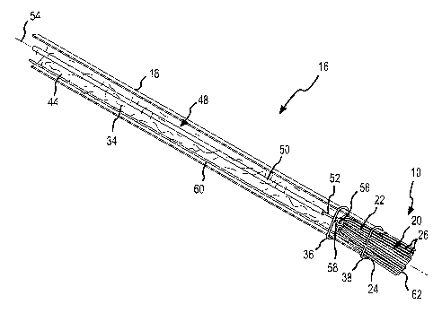
[0019] Fig. 1 is an isometric view of one embodiment of a catheter having a
brush
electrode according to the present invention, and depicts the filaments
comprising the
brush electrode extending from a distal end of an outer sheath.
[0020] Fig. 2 is an enlarged view of the circled region of Fig. 1.
[0021] Fig. 3 is similar to Fig. 2, but depicts an alternative embodiment
where the
brush electrode is secured at the distal end of the outer sheath by at least
one suture that is
covered by a section of shrink tube.
j0022] Fig. 4 is similar to Fig. 3, but a portion of the shrink tube has been
broken
away to reveal two sutures through the outer sheath.
[0023] Fig. 5 is an isometric, cross-sectional view of the catheter depicted
in Figs. 3
and 4, taken along line 5-5 of Fig. 3, revealing a primary conductor making
electrical
CA 02558610 2006-09-O1
WO 2005/072488 PCT/US2005/000767
contact with the filaments comprising the brush electrode, and depicting a
secondary lead
(e.g., for a thermocouple) extending adjacent to the primary conductor and
becoming
embedded within the brush filaments.
[0024] Figs. 6 and 7 depict steps that may be used to form the brush electrode
depicted
in, for example, Fig. 5.
[0025] Fig. 8 is similar to Fig. 5, but is a cross-sectional view of an
alternative
embodiment of the brush electrode, wherein conductive filaments are
interspersed among
relatively longer nonconductive filaments.
[0026] Fig. 9 is a cross-sectional view taken along line 9-9 of Fig. 8.
[0027] Fig. 10 is an enlaxged view of the circled region of Fig. 8.
[0028] Figs. 11-14 depict alternative shapes for the filaments comprising the
tip of the
brush electrode.
[0029] Fig. 15 depicts an alternative embodiment of the filaments comprising
the
brush electrode, wherein the individual filaments gradually taper toward their
distal ends.
(0030] Fig. 16 depicts an alternative embodiment of the filaments comprising
the
brush electrode, wherein the individual filaments have nonconductive tips at
their distal
ends creating a stand-off distance.
(0031] Fig. 17 is a fragmentary, isometric view of an embodiment of the outer
sheath
having a concentric ring of sub-channels around a main or central channel
through which
the brush filaments extend.
[0032] Fig. 18 is a fragmentary, isometric view of an embodiment wherein the
sheath
surrounding the filaments of the brush electrode is porous adjacent to the
exposed portion
a
of the brush electrode.
[0033] Fig. 19 is a fragmentary, isometric view of an embodiment wherein the
sheath
surrounding the filaments of the brush electrode is a threaded sheath, having
a spiral or
helical ridge on its outer surface, adjacent to the exposed portion of the
brush electrode.
[0034] Fig. 20 is a fragmentary view of a section of the threaded sheath
depicted in
Fig. 19, surrounded by a covering shown in phantom and cross-section to create
a helical
flow channel between the threaded sheath and the covering.
[0035] Fig. 21 is a fragmentary, isometric view of an embodiment wherein the
sheath
surrounding the filaments of the brush electrode is a grooved sheath, having a
plurality of
6
CA 02558610 2006-09-O1
WO 2005/072488 PCT/US2005/000767
longitudinally-extending grooves or cuts on its outer surface, adjacent to the
exposed
portion of the brush electrode.
[0036] Fig. 22 is a fragmentary view of a section of the grooved sheath
depicted in
Fig. 21, surrounded by a covering (shown cross-section) to create a plurality
of
longitudinally-extending flow channels between the grooved sheath and the
covering.
[0037] Fig. 23 is a cross-sectional view taken along line 23-23 of Fig. 21,
with the
covering shown in phantom and with the longitudinally-extending flow channels
clearly
visible.
[0038] Fig. 24 is similar to Fig. 5, but depicts an isometric, cross-sectional
view of a
catheter wherein the primary conductor makes electrical contact with the
filaments via an
energy transfer coil or spring surrounding at least the embedded portion of
the brush
electrode.
[0039] Fig. 25 is similar to Figs. 5 and 24, but depicts an isometric, cross-
sectional
view of a catheter wherein the primary conductor makes electrical contact with
the
filaments via an energy transfer mesh or fabric surrounding at least the
embedded portion
of the brush electrode.
[0040] Fig. 26 is a cross-sectional view of a first embodiment of a shielded-
tip brush
electrode, wherein an uninsulated portion of the primary conductor is looped
around the
outer surface of the brush electrode.
[0041] Fig. 27 is similar to Fig. 26, but depicts a second embodiment of a
shielded-tip
brush electrode.
[0042] Figs. 28-35 depict different cross-sectional configurations for brush
electrodes
according to the present invention.
[0043] Fig. 36 is a cross-sectional view of a brush electrode wherein some of
the filaments comprise hollow or porous members.
[0044] Fig. 37 is a cross-sectional view of a brush electrode having devices
(e.g., a
thermocouple or other temperature sensor, a pressure sensor, or an ultrasound
sensor)
embedded among the conductive and nonconductive filaments.
[0045] Fig. 38 is an isometric view of a catheter having a brush electrode
according to
the present invention forming a spot or point lesion on a section of tissue.
[0046] Fig. 39 is an isometric view of a catheter having a brush electrode
according to
the present invention forming a linear' or drag lesion on a section of tissue.
7
CA 02558610 2006-09-O1
WO 2005/072488 PCT/US2005/000767
[0047] Figs. 40-42 depict a brush electrode according to the present invention
forming
different-sized lesions based in part upon the amount of splay of the brush
electrode.
DETAILED DESCRIPTION OF THE INVENTION
[0048] Several embodiments of a brush electrode 10 according to the present
invention are depicted in the figures. As described further below, the brush
electrode of
the present invention provides a number of advantages, including, for example,
the ability
to form deep lesions in tissue while reducing the formation of undesirable
coagulum and
charring of the surface tissue, while applying a reasonable amount of RF
energy, while
mitigating electrode-tissue contact problems, and/or while reducing the amount
of
conductive fluid (e.g., saline) possibly entering a patient's bloodstream
during the
procedure. The present invention facilitates the formation of a deep lesion in
a shorter
period of time than required by other ablation devices, and it provides the
ability to create
lesions in highly perfused tissue or in fluid-rich environments. The brush
electrode 10
facilitates enhanced tissue contact in difficult environments (e.g., during
ablation of a
contoured or trabeculated surface inside a beating heart), whether creating a
spot lesion 12
(e.g., Fig. 38) or a continuous linear lesion 14 (e.g., Fig. 39), by readily
conforming to
surface contours.
[0049] Fig. 1 is an isometric view of one embodiment of a catheter 16 having a
brush
electrode 10 according to the present invention. As depicted in this figure,
the catheter
comprises a catheter shaft with an outer sheath 18. In the embodiment depicted
in Fig. 1,
the outer sheath is formed from sections of different material (e.g.,.in the
embodiment
depicted Fig. 1, five different sections comprise the outer sheath). These
sections of
different material enable the catheter 16 to have, for example, different
mechanical
properties (e.g., flexibility) at different locations along the catheter
shaft. The outer sheath
18 may or may not comprise these sections of different material depending upon
the
intended application for the catheter. Although the outer sheath 18 depicted
in Fig. 1 has a
circular cross section, the cross-section of the outer sheath may be other
than circular.
[0050] As also shown in Fig. 1, the brush electrode 10, which comprises an
exposed
portion 20 and an embedded portion 22 (see, e.g., Fig. 5), is present at a
distal end 24 of the
outer sheath 18. In particular, at the distal end of the outer sheath, the
exposed portion 20
of the brush electrode 10, comprising a plurality of filaments 26, may be seen
(see, e.g.,
8
CA 02558610 2006-09-O1
WO 2005/072488 PCT/US2005/000767
Fig. 2). The exposed portion of the brush electrode may project a few
millimeters from the
distal end of the outer sheath. The distance that the exposed portion of the
brush electrode
extends from the distal end of the outer sheath varies depending upon a number
of factors
including the composition of the filaments comprising the brush and the
particular area to
be treated with the brush electrode 10. The distal end 24 of the outer sheath
18 may
include a conductive or nonconductive base 28. As explained further below, the
flexible
brush electrode provides enhanced tissue contact, particularly for use on
contoured ar
trabeculated surfaces.
[0051] Fig. 2 is an enlarge view of the circled region of Fig. 1. As clearly
shown in
Fig. 2, the brush electrode 10 according to this embodiment has a relatively
flat working
surface 30 at the distal end 32 of the brush electrode 10. In other words, in
this depicted
embodiment, all of the filaments 26 comprising the brush electrode 10 extend
approximately the same distance from the distal end 24 of the outer sheath 18.
Thus, the
brush tip provides a relatively flat working surface 30 comprising the
longitudinal ends of
the filaments. The outer sheath of the catheter provides mechanical support
for the
filaments and may also provide electrical shielding. As explained further
below, the brush
electrode comprises a bundle of bristles or filaments that each may he
constructed from a
variety of different materials, including nonconductive materials, semi-
conductive
materials, and conductive materials. For example, the filaments may be formed
from metal
fibers, metal plated fibers, carbon-compound fibers, and other materials. Very
thin, carbon
fibers may be used, or relatively thicker but less conductive Thunderon~
acrylic fibers
may be used for the brush electrode filaments. Thunderon~ is manufactured by
Nihon
Sanmo Dyeing Company Ltd. of Kyoto, Japan. Nylon fibers coated with conductive
material may also be used. Filaments 26 constructed from metal plated fibers,
like coated
nylon fibers, may comprise flattened areas around their outer surfaces,
resulting in the
filaments having noncircular cross-sectional shapes. The brush filaments may
be insulated
from each other, or they may be in electrical contact with each other. As
explained further
below, conductive or nonconductive fluids 34 may flow within the filaments
themselves
(see, e.g., Fig. 36) or along the outer surface of the filaments (see, e.g.,
Fig 5).
[0052] Once the distance that the filaments extend from the distal end 24 of
the other.
sheath 18 is set to a desired length, the bundle of filaments comprising the
brush electrode
may be fixed to the outer sheath 18. Figs. 3-5 depict one technique for fixing
or
9
CA 02558610 2006-09-O1
WO 2005/072488 PCT/US2005/000767
anchoring the brush electrode 10 relative to the outer sheath using sutures.
In Fig. 3, a
rea~.-ward suture 36 and a forward suture 38 are shown in phantom under a
section of shrink
tube 40 surrounding the outer surface of the outer sheath 18. The shrink tube
protects the
sutures and makes it easier to insert the catheter by mitigating possible
snags that may
occur due to the presence of the sutures. Fig. 4 is similar to Fig. 3, but
depicts a portion of
the shrink tube 40 broken away to reveal a portion of the two sutures 36, 38.
The suture
knots 42 are clearly visible in Fig. 4.
[0053] Fig. 5 is an isometric, cross-sectional view of the catheter 16
depicted in Figs.
3 and 4, taken along line 5-5 of Fig. 3. In Fig. 5, it is apparent that the
rearward suture 36
may be used to set the depth that the brush electrode 10 may be inserted into
the distal end
24 of the outer sheath 18. In this figure, the forward suture 38 pierces the
filaments 26
comprising the embedded portion 22 of the brush electrode 10 and thereby help
prevent
movement of the brush electrode relative to the outer sheath of the catheter.
In the
embodiment depicted in Fig. 5, conductive fluid 34 is shown flowing through a
lumen 44
of the outer sheath (depicted as a single, embedded channel) from a fluid
source (not
shown) to the brush electrode 10. When the conductive fluid 34 flows through
the brush
electrode, it creates a wet-brush electrode in which impinging jets of fluid
traveling
interstitially impact the tissue 46 (see, e.g., Figs. 38 and 39) at the tissue-
electrode
interface, which makes it easier to control temperature rises at the
interface. Wet-brush
electrodes are discussed further below. In an alternative embodiment, the
lumen 44
depicted in Fig. 5 may comprise a plurality of separate lumen.
[0054] Fig. 5 also clearly depicts a primary conductor 48 having an insulated
portion
50 and an uninsulated portion 52. The primary conductor carries ablative
energy (e.g., RF
current) from an energy source (not shown) to the brush electrode 10. As
depicted in Fig.
5, the primary conductor 48 extends within the fluid-carrying lumen 44 of the
catheter,
along a longitudinal axis 54 of the catheter 16. The primary conductor may
comprise, for
example, insulated copper wire with an uninsulated portion in electrical
contact with the
brush electrode. In this embodiment, the uninsulated portion 52 of the pximary
conductor
is looped or noosed around the filaments comprising the brush electrode at a
connection
point 56 (Fig. 7). At the loop or noose 58, ablative energy is transferred
from the primary
conductor to the conductive filaments comprising part of the brush electrode
10. In this
embodiment, the uninsulated portion 52 of the primary conductor 48 is
connected to the
CA 02558610 2006-09-O1
WO 2005/072488 PCT/US2005/000767
embedded portion 22 of the brush electrode 10 so that the connection between
the primary
conductor and the brush electrode is protected within the outer sheath 18 of
the catheter 16.
[0055] Also clearly visible in Fig. 5 is an embedded or secondary lead 60,
which
extends substantially parallel to the primary conductor 48. A distal end 62 of
the
secondary lead 60 becomes embedded with the filaments 26 comprising the brush
electrode 10. As discussed further below in connection with, for example, Fig.
37, the
secondary lead 60, when present, may be operatively connected to some type of
sensor
embedded in the brush electrode (e.g., a thermal sensor 64, an ultrasound
sensor 66, or a
pressure sensor 68). The brush electrode depicted in Fig. 5 acts as a surface-
cooled
electrode 10.
[0056] Figs. 6 and 7 depict possible steps for forming the brush electrode 10
depicted
in Figs. I -5. In Fig. 6, a bundle 70 of conductive filaments 72 and
nonconductive
filaments 74 is being formed by using the uninsulated portion 52 of the
primary conductor
48 to bind or tie together the filaments. In Fig. 6, the uninsulated portion
has been noosed
around the bundle of filaments 70, but has not been tightened or snugged
against the
bundle. In Fig. 7, the uninsulated portion 52 of the primary conductor has
been snuggly
noosed around the connection point 56 at approximately the mid-section of the
bundle of
filaments that will ultimately form the brush electrode 10. The conductive
filaments 72
and the nonconductive filaments 74 are then bent around the connection point
56 in the
direction of the arrows 76, 78 depicted in Fig. 7. Once the filaments are
folded upon
themselves about the connection point 56, they are inserted into the distal
end 24 of the
outer sheath 18 and positioned relative to the distal end 24 of the outer
sheath 18 so that the
desired amount of the filaments extends from the distal end of the sheath and
comprises the
exposed portion 20 of the brush electrode 10. The ends of the filaments may
then be
trimmed, if desired, to create a desired shape for the working surface 30 at
the distal end 32
of the brush electrode 10 (see, e.g., Figs. 11-14).
[0057j Figs. 8, 9, and I 0 depict an alternative embodiment of the brush
electrode.
This standoff brush electrode 10' includes an exposed portion 20' with a
working surface
30' wherein the longitudinal ends of the conductive filaments 72 are not flush
with the
longitudinal ends of the nonconductive filaments 74. As shown to better
advantage in
Fig. 10, which is an enlarged view of the circled region of Fig. 8, in this
alternative
embodiment of the brush electrode, the conductive filaments 72 are
interspersed among
11
CA 02558610 2006-09-O1
WO 2005/072488 PCT/US2005/000767
relatively longer nonconductive filaments 74. The relatively longer
nonconductive
filaments prevent the conductive filaments from directly touching the tissue
46 (see, e.g.,
Fig. 40) when the working surface 30' of the brush electrode is placed normal
to the tissue
being treated. With this brush configuration and substantially perpendicular
orientation of
the brush worlcing surface 30' relative to the tissue being treated, the brush
electrode acts as
a virtual electrode. If the perpendicular orientation can be maintained, there
is no direct
contact between the conductive filaments and the tissue, and the conductive
fluid 34 (see
Fig. 5) flowing through the lumen 44 of the outer sheath 18 makes the
electrical contact at
the brush-tissue interface. Although Figs. 8 and 10 depict each of the
conductive filaments
72 as being shorter than each of the nonconductive filaments 74, the
electrical
characteristics of the brush electrode may be adjusted by having some
conductive filaments
that extend to the working surface at the tip of the brush electrode, if
desired.
[005] Fig. 9 is a cross-sectional view taken along line 9-9 of Fig. 8 and
clearly
depicts the bundled filaments 70 at the connection point 56 between the
filaments and the
uninsulated portion 52 of the primary conductor. The secondary lead 60 is also
visible in
Fig. 9. In this embodiment, it is possible to adjust the fluid and electrical
contact at the
brush-tissue interface through appropriate selection of the conductive and
nonconductive
filaments. Since this configuration of the brush electrode performs most
effectively when
placed normal or perpendicular to the tissue, a relatively short exposed
portion 20' for the
brush electrode 10' may be desirable with relatively stiff filaments (e.g.,
Thunderon~
filaments). '
[0059] Figs. 11-I4 depict alternative shapes for the filaments 26 comprising
the tip of
the brush electrode. The various tip configurations may provide advantages for
special
applications of brush electrodes. Fig. 11 depicts a blade-shaped distal tip 80
creating a line
of contact with the longest filaments of the brush electrode. As depicted in
Fig. 1 l, the line
of contact at the most distal end of the brush electrode extends
perpendicularly into the
page. In Fig. I2, the working surface of the electrode tip has a concave
portion or channel
82. The concave-tip embodiment depicted in Fig. 12 is beneficial for wrap-
around
applications and provides advantages when ablating curved surfaces like the
outer surface
of a blood vessel. Fig. 13 depicts a convex, trough-shaped tip 84. This
particular
configuration is beneficial, for example, when reaching into troughs or
depressions on a
contoured surface. The distal tip could also be domed or hemispherical rather
than having
12
CA 02558610 2006-09-O1
WO 2005/072488 PCT/US2005/000767
the trough-shaped contact surface shown in Fig. 13. In Fig. 14, the brush
electrode has a
wedge-shaped tip 86. The wedge-shaped tip facilitates angular placement and
increases
the area of the working surface 30". The distal tip could also be conical (not
shown),
coming nearly to a point at the most distal end of the brush electrode, with
its longest
filaments proximal to the longitudinal axis 54 of the catheter 16 (see Fig.
5). This latter
configuration may be advantageous for point applications of ablative energy.
The brush
electrodes are depicted in many of the drawings with circular cross sections,
but may have
different cross-sectional configurations.
[0060] Fig. 15 depicts an example of a brush electrode 10" having continuously
varying conductivity along the longitudinal axes of the filaments. In
particular, the brush
electrode comprises tapered filaments 26'. In this alternative embodiment, at
least a
portion of the individual filaments 26' comprising the brush electrode 10"
gradually taper
toward their distal or free ends 88. In other words, at the distal end 24 of
the outer sheath
18, the filaments 26' have larger cross-sectional areas than they have at
their distal ends 88,
adjacent to the working surface 30"' of the brush electrode 10". The filaments
26' are thus
more conductive adjacent to the distal end of the outer sheath and less
conductive at the
distal ends of the filaments. Since the filaments are more conductive adjacent
to the distal
end of the outer sheath, this minimizes current flow to the less conductive
fluid wetting the
brush from the lumen of the outer sheath. When Less of the ablative energy
flows into the
conductive fluid adjacent to the distal end of the outer sheath, this
minimizes the energy
transfer into the conductive fluid and the concomitant heating of the
conductive fluid
before it contacts the surface of the tissue. At the distal ends 88 of the
filaments 26'
depicted in Fig. 15, the conductivity of the filaments may be matched to the
conductivity of
the fluid to create a relatively uniform electric field at the brush-tissue
interface.
[0061] The taper depicted in Fig. 15 could be an inverse taper, which may be
advantageous for certain applications. It should be noted that, in order to
vary the
conductivity along the length of the filaments, the filaments may also be
coated or plated
with materials having different or varying electrical conductivity. For
example, the
filaments, whether tapering or not, could be coated with conductive material.
The
conductive material coating the filaments in the region most closely adjacent
to the distal
end 24 of the outer sheath 18 may be more conductive than the coating on the
portion of
the filaments most closely adjacent to the distal end of the filaments
themselves. Thus, the
13
CA 02558610 2006-09-O1
WO 2005/072488 PCT/US2005/000767
conductivity of the filaments would be greater near the distal end of the
outer sheath than
near the distal ends of the filaments, even though the cross-sectional areas
of the filaments
may not be changing substantially as one moves longitudinally along the
filaments toward
their distal ends. Although not specifically shown in the figures, the
conductivity of all of
the disclosed filaments may also vary radially rather than, or in addition to,
varying
longitudinally. In other words, the conductivity of the filaments may vary as
one moves
from the center of the filaments to the surface of the filaments.
[0062] Fig. 16 depicts a brush electrode 10"' in which the conductivity of the
filaments varies discontinuously. In particular, Fig. 16 depicts filaments 26"
that are
conductive except at their distal ends. The distal end of each filament
includes a
nonconductive tip 90. These nonconductive tips provide a stand-off distance
when the
working surface of the brush electrode is placed substantially perpendicular
to the tissue
being treated since the conductive portions of the filaments do not actually
touch the tissue
in this embodiment. Similar to what occurs in the embodiment depicted in Figs.
8-10, the
conductive fluid would pass through the ltunen of the catheter and wet the
brush. The
conductive fluid would carry the current over the stand-off distance and to
the tissue,
thereby acting as a virtual electrode. It should be noted that, although the
embodiment
depicted in Fig. 16 shows each of the conductive filaments 26" having a
nonconductive tip
90, some of the conductive filaments 26" may extend all the way to the working
surface
30"" of the brush electrode and thus would, in fact, contact the tissue during
use of the
brush electrode.
[0063] Fig. 17 depicts an embodiment of the outer sheath 18' having a
concentric ring
of sub-channels 92 around a main or central channel 94 through which the brush
filaments
26 extend. The circumferential ring of sub-channels around the brush-carrying
central
channel may be used to carry conductive or nonconductive fluid, including
therapeutic
fluid or medicine. The embedded sub-channels depicted in this figure could
define spiral
or helical paths toward the distal end 24' of the outer sheath, similar to the
paths or
channels 104 described below in connection with Fig. 19 and Fig. 20.
[0064) Fig. 18 depicts an embodiment wherein the sheath 18" surrounding the
filaments of the brush electrode 10 is porous adjacent to the exposed portion
20 of the
brush electrode. An outer covering (not shown) may be placed around the outer
cylindrical
14
CA 02558610 2006-09-O1
WO 2005/072488 PCT/US2005/000767
surface of the porous sheath, possibly leaving an angular ring of material 96
exposed at the
distal end 24" of the sheath 18" adjacent to the brush electrode 10.
[0065] Fig. 19 is a fragmentary, isometric view of an embodiment wherein a
threaded
sheath 98 surrounds the filaments of the brush electrode 10. The threaded
sheath 98 has a
spiral or helical ridge 100 on its outer surface. As shown to good advantage
in Fig. 20,
when the threaded sheath is inserted into a covering 102 (shown in phantom and
cross-section), a helical flow channel 104 is created between the threaded
sheath 98 and the
covering 102. Conductive fluid, nonconductive fluid, or medication may be
delivered to
the tissue adjacent to the brush electrode via this flow channel.
[0066] Fig. 21 is a fragmentary, isometric view of another embodiment, wherein
the
sheath surrounding the filaments of the brush electrode is a grooved sheath
106. The
grooved sheath has a plurality of longitudinally-extending grooves or cuts 108
formed on
its outer surface, adjacent to the exposed portion of the brush electrode 10.
As shown to
best advantage in Fig. 23, when the grooved sheath 106 is inserted into a
covering 102'
(shown in phantom and cross-section), a plurality of longitudinally-extending
flow
channels 110 axe created between the grooved sheath 106 and the covering 102'.
Again,
conductive fluid, nonconductive fluid, or medication may be delivered to the
tissue
adjacent to the brush electrode via these flow channels. Fig. 22 is a
fragmentary view of a
section of the grooved sheath 106 depicted in Fig. 21, surrounded by a
covering 102'
(shown in cross-section) to create the plurality of longitudinally-extending
flow channels
110 between the grooved sheath and the covering.
[0067] Figs. 24 and 25 depict alternative mechanical interfaces between the
filaments
26 of the brush electrode 10 and the primary conductor 48. Fig. 24 is similar
to Fig. 5, but
depicts an isometric, cross-sectional view of a catheter 16' wherein the
exposed portion 52
of the primary conductor 48 makes electrical contact with the brush filaments
26 via an
energy transfer coil or spring 112 surrounding at least the concealed or
embedded portion
22 of the brush electrode 10. In this embodiment, the ablative energy is
transferred to the
brush electrode 10 over a large surface area (i.e., over the entire inner
surface area of the
coil 112). Thus, less damage to the filaments may occur in this embodiment
than may
occur in the embodiment depicted in Fig. 5, wherein all of the ablative energy
is transferred
from the uninsulated portion 52 of the primary conductor to the brush
electrode at the
single connection point 56. As depicted in Fig. 24, a loop of wire 114 may be
present to
CA 02558610 2006-09-O1
WO 2005/072488 PCT/US2005/000767
help collect and stabilize the filaments 26 during assembly of the catheter
16'. This loop of
wire 114 may be anchored to, for example, the inner surface 116 of the outer
sheath 18. As
previously described, a secondary lead 60 may also be present in the lumen 44
of the outer
sheath 18.
[006] Fig. 25 is similar to Figs. 5 and 24, but depicts an isometric, cross-
sectional
view of a catheter 16" wherein the primary conductor 48 makes electrical
contact with the
filaments of the brush electrode 10 via an energy transfer mesh or fabric 118
surrounding
at least the concealed or embedded portion 22 of the brush electrode 10. This
embodiment
has the same advantages that were just described for the embodiment depicted
in Fig. 24.
In another embodiment, the primary conductor 48 makes electrical contact with
the
filaments of the brush electrode 10 via an energy transfer wrap (not shown),
which is
similar to the mesh or fabric 118, but comprises a solid or porous sheet of
conductive
material.
[0069] Fig. 26 is a cross-sectional view of a first embodiment of a shielded-
tip brush
electrode 120. In this embodiment, the uninsulated portion 52 of the primary
conductor 48
is looped around the outer surface of the brush electrode after passing
through a
mechanical interface 122 supporting the filaments 26 of the brush electrode
adj acent to the
distal end 124 of an inner sheath 126. Since fluid may or may not travel
through the lumen
128 of the inner sheath 126, the mechanical interface 122 may or may not be
porous. In
the embodiment depicted in Fig. 26, there is an outer sheath 130 surrounding
the inner
sheath 126. The inner sheath houses the primary conductor 48 and supports the
mechanical interface 122 for the filaments 26 of the brush electrode 120. The
primary
conductor again includes an uninsulated portion 52 that transfers ablative
energy 150 (e.g.,
RF energy) to the conductive filaments in the brush electrode 120. As
mentioned, in this
embodiment the uninsulated portion 52 of the primary conductor forms loops or
coils 132
around the circumference of the brush. These loops or coils increase the
surface area
through which the ablative energy is transferred, thereby providing for more
effective, and
potentially less destructive, energy transfer to the brush electrode 120.
[0070) As shown in Fig. 26, the outer sheath, which may be a typical braided
sheath,
is placed around the inner sheath 126, but is radially and longitudinally
offset from the
inner sheath. The radial offset creates an annular gap or channel 134 between
the inner
sheath 126 and the outer sheath 130 through which conductive fluid may, for
example, be
16
CA 02558610 2006-09-O1
WO 2005/072488 PCT/US2005/000767
introduced to the sides of the brush electrode filaments. The conductive
fluid, if present,
would flow through the annular channel 134 in the direction of the arrows 136
shown at
the top of Fig. 26. The longitudinal offset between the inner sheath 126 and
the outer
sheath 130 ensures that the channel 134 for the conductive fluid extends past
the distal end
I24 of the inner sheath 126 to the sides of the brush electrode filaments. In
this
embodiment, the conductive fluid would flow through the annular channel
between the
inner sheath and the outer sheath, past the coils 132 of uninsulated
conductive wire, into an
annular fluid jacket 138 surrounding a region of the brush electrode adjacent
to the distal
ends of the inner and outer sheaths, and then into the sides of the brush
electrode itself and
through the interstitial gaps between the filaments comprising the brush
electrode. The
ablative energy (e.g., the RF energy 150) is thus carried by the conductive
fluid into the
core of the brush electrode and toward its working surface 140. In this
embodiment, a
flexible polymer nipple or boot 142, defining an outer wall of the annular
fluid jacket 138,
also supports the filaments in a ring 144 of direct contact extending around
the perimeter of
the filament bundle. The flexible boot or nipple may be porous. Finally, a
smooth outer
wall 146 to facilitate easier insertion and manipulation of the catheter in a
patient may
cover the outer sheath 130 and abut a corresponding edge 148 of the flexible
polymer
nipple or boot 142. Alternatively, the outer wall material may actually form
the nipple or
boot in addition to forming a perimetric covering around the outer sheath.' An
annular
layer of porous material or mesh fabric (not shown) may be placed in the
annular fluid
j acket 13 8 to keep the brush wetted and to help prevent splaying (see Figs.
40-42) of the
brush electrode.
[0071] Fig. 27 is similar to Fig. 26, but depicts a second embodiment of a
shielded tip
brush electrode 120'. The only differences between the embodiment depicted in
Fig. 26
and the embodiment depicted in Fig. 27 are the size of the fluid j acket 13 8'
and the
configuration of the flexible polymer nipple or boot 142' that supports the
brush filaments.
In the embodiment depicted in Fig. 27, an alternative flexible polymer nipple
or boot 142'
defines a smaller fluid jacket 138' and supports the filaments in a band of
direct contact
extending around the perimeter of the filament bundle. The band of direct
contact 152
supports the filaments over a larger section of the outer surface of the brush
electrode than
does the ring of direct contact 144 depicted in Fig. 26. By adjusting the
configuration of
the flexible polymer nipple or boot in this manner, the amount of conductive
fluid flowing
17
CA 02558610 2006-09-O1
WO 2005/072488 PCT/US2005/000767
into the brush electrode and the overall flexibility of the brush electrode
can be
manipulated.
[0072] It should be noted that, although the filaments depicted in Figs. 26
and 27 are
shown as extending just into the distal end 124 of the inner sheath 126, the
filaments may
extend further into the inner sheath and may even extend all the way to the
proximal end
(not shown) of the catheter.
[0073] Figs. 28-35 depict different cross-sectional configurations for brush
electrodes
according to the present invention. Interstitial spaces 156 are clearly
visible in each of
these figures. In Figs. 28-31, the brush electrode 10 has a conductive core
154. In these
four figures, the conductive filaments 72 are shown with cross hatching, and
the
nonconductive filaments 74 are shown without cross hatching. Thus, the brush
electrode
depicted in Fig. 28 is fully conductive and does not comprise any
nonconductive filaments.
In each of the embodiments depicted in Figs. 29-31, a conductive core 154 is
shielded by a
barrier of nonconductive filaments 74. In particular, Fig. 29 depicts a core
of relatively
large conductive filaments surrounded by two rings of nonconductive filaments
of
approximately the same size. In Fig. 30, a core 154 of relatively small
conductive
filaments 72 is surrounded by two rings of relatively large nonconductive
filaments 74. In
Fig. 31, a conductive core 154 of relatively large conductive filaments 72 is
surrounded by
two rings of relatively small nonconductive filaments 74.
[0074] Figs. 32 and 33 depict cross-sectional configurations for brush
electrodes that
have conductive perimeters 158. Thus, in the embodiments depicted in Figs. 32
and 33, a
nonconductive core 160 of nonconductive filaments 74 is surrounded by
conductive
filaments 72. Fig. 32 depicts a core of relatively small nonconductive
filaments
surrounded by two rings of relatively large conductive filaments. In Fig. 33,
a core of
relatively large nonconductive filaments is surrounded by two rings of
relatively small
conductive filaments.
[0075] In Fig. 34, conductive clusters 162 of relatively small filaments are
interspersed among relatively large nonconductive filaments 74. The
interspersed
conductive clusters may be interspersed in a specific pattern, pseudo
randomly, or
randomly among the nonconductive filaments in order to achieve a desired
electric field
from the resulting brush electrode. In Fig. 35, nonconductive clusters 164 of
relatively
small filaments are interspersed among relatively large conductive filaments
72.
18
CA 02558610 2006-09-O1
WO 2005/072488 PCT/US2005/000767
[0076] Fig. 36 is a cross-sectional view of a brush electrode wherein some of
the
filaments are hollow or porous 166. Such hollow or porous filaments 166 may be
used as
conduits for conductive fluid, they may be used to supply therapeutic
chemicals, and/or
they may provide suction ports at the brush-tissue interface to control field
smearing on the
tissue surface. If the filaments are porous, they may retain a small amount of
fluid in pores
that are oriented at various angles to the longitudinal axis of the filaments.
During an
ablation procedure, some of the ablative energy may dehydrate the porous
filaments before
affecting the surrounding blood, particularly when the conductivity of the
tissue lessens as
the ablation progresses. Thus, if excess ablative energy is present during an
ablation
procedure, that energy may harmlessly dehydrate the porous filaments rather
than
negatively affecting the tissue being ablated or the blood in the area of that
tissue. In one
embodiment (not shown), some of these hollow filaments 166 do not extend to
the distal
end 32 (labeled on, for example, Fig. 2) of the brush electrode. For example,
some of the
hollow filaments 166 may only extend part way into the exposed portion 20
(labeled on,
for example, Fig. 3) of the brush electrode. These shortened hollow filaments
may deliver
conductive fluid or therapeutic chemicals, for example, to an interior region
of the bundle
of brush filaments. In the embodiment depicted in Fig. 36, the other filaments
26 may be
conductive or nonconductive filaments.
[0077] Fig. 37 is a cross sectional view of a brush electrode having devices
64, 66, 68
embedded among the conductive and nonconductive filaments 26. The devices may
include, for example, pressure sensors 68 to measure contact pressure between
the brush
electrode and the tissue, thermal sensors 64 (e.g., a thermocouple) at the tip
of the brush
electrode to sense the brush-tissue interface temperature, or fiber optic or
ultrasound
sensors 66 for in situ lesion identification and characterization. The devices
may be
operatively connected to equipment (not shown) at the proximal end of the
catheter by
secondary leads like the secondary lead 60 depicted in, for example, Figs. 5
and 8-16.
[0078] Fig. 38 is a fragmentary, isometric view of a catheter 16 having a
brush
electrode 10 according to the present invention forming a spot or point lesion
12 on a
section of tissue 46. As shown in this figure, the brush electrode is placed
against the
tissue with its filaments in contact with or in close proximity to the tissue.
The conductive
filaments are connected to, for example, an RF source (not shown) and serve as
the active
electrode. When present, conductive fluid from a fluid source (not shown)
flows through
19
CA 02558610 2006-09-O1
WO 2005/072488 PCT/US2005/000767
the lumen 44 (e.g., Fig. 5) of the catheter and through the brush filaments to
the working
surface at the brush tip, thereby creating a wet-brush electrode. Rather than
being localized
on the tissue to create a spot or point lesion 12 as showxn in Fig. 38, the
brush electrode 10
may be dragged along the surface of the tissue 46 to create a continuous
linear lesion 14, as
shown in Fig. 39. Fig. 39 is a fragmentary, isometric view of a catheter 16
having a brush
electrode according to the present invention forming a linear or drag lesion
on a section of
tissue.
[0079] Figs. 40-42 depict a brush electrode 10 according to the present
invention
forming different size spot lesions 12 based in part upon the amount of splay
of the brush
electrode. In Fig. 40, relatively light contact pressure is being used to
press the brush
electrode 10 against the tissue 46 while forming a lesion 12. This application
of light
pressure results in minimal splaying of the filaments comprising the brush
electrode, and
thus a relatively small lesion is formed. In Fig. 41, more pressure is being
used to press the
brush electrode 10 into contact with the tissue 46, resulting in relatively
more splaying of
the brush electrode. As long as the efficiency of the brush electrode is not
degraded too
greatly by the splaying, a relatively larger lesion 12 may thus be formed by
applying
additional pressure to press the brush electrode toward the tissue. Finally,
in Fig. 42, even
more contact pressure is being applied to the brush electrode 10 than is being
applied in
Figs. 40 and 41, resulting in even more splaying of the brush electrode and
the formation
of a relatively larger lesion 12 on the tissue 46 than is being formed in
Figs. 40 and 41.
[0080] The brush electrode according to the present invention delivers
ablative energy
to the tissue via the conductive filaments alone, via the conductive fluid
alone, or via both
the conductive filaments and the conductive fluid. In the latter two
configurations, the
brush electrode is referred to as a wet-brush electrode. Since it is possible
for the
conductive fluid to escape from the exposed portion of the wet-brush electrode
prior to
reaching the working surface at the distal tip of the wet-brush electrode,
there is same
ablative energy leakage to the surrounding blood. The leakage of ablative
energy to the
surrounding blood is in part due to direct contact between the blood and the
conductive
filaments and in part due to the conductive fluid escaping between the
filaments to the
surrounding blood, particularly when substantial splaying of the filaments
occurs (see, e.g.,
Fig. 42).
CA 02558610 2006-09-O1
WO 2005/072488 PCT/US2005/000767
[0081] The design parameters for the brush electrode include both filament and
brush
parameters. The filament parameters include, for example, the material and
structural
properties of the individual filaments (e.g., what materials) each individual
filament is
constructed from, whether the filaments are hollow or solid, whether the
filaments are
porous, and how flexible or stiff the filaments are), the shape and cross-
sectional areas of
the individual filaments, and the electrical conductivity of the individual
filaments. The
electrical conductivity of the individual filaments may be constant along the
length of the
filaments or may vary along the length of the filaments. Also, if the
conductivity of a
filament varies along its length, it may vary continuously or discontinuously.
The filament
design parameters may be different for each filament.
[0082] The design parameters for the brush electrode include, for example, the
overall
shape and cross-sectional area of the brush (i.e., the overall shape and size
of the filament
bundle forming the brush electrode), the tip length of the brush itself (i.e.,
the length of the
portions of the filaments that extend the farthest from the distal end of the
outer sheath),
the shape of the brush tip, the length of the individual filaments relative to
each other, the
packing density of the filaments comprising the brush, and the overall
electrical resistance
of the brush. When both nonconductive and conductive filaments are present,
the
conductive filaments may be distributed evenly, randomly, or pseudo-randomly
among the
nonconductive filaments comprising the brush electrode.
[0083] By controlling, among other things, the cross-sectional shapes of the
filaments,
the cross-sectional areas of the filaments, the flexibility or stiffness of
the filaments, the
packing density of the filaments, the ratio of the nonconductive filaments to
the conductive
filaments, and the placement of the nonconductive and conductive filaments
relative to
each other, it is possible to obtain brush electrodes having desired
electrical and thermal
characteristics, which ultimately determine the types of lesions that may be
obtained when
using the brush electrodes for ablation. As mentioned above, it is even
possible to vary the
mechanical and electrical properties of each individual filament, if
necessary, to achieve
desired results.
[0084] The shapes and cross-sectional areas of the individual filaments and
the
packing density of the brush electrode affect the interstitial spaces between
the filaments.
The interstitial spaces between the filaments determine the flow path of the
conductive or
nonconductive fluid when the brush electrode is being used as a wet-brush
electrode. The
21
CA 02558610 2006-09-O1
WO 2005/072488 PCT/US2005/000767
flow path of the conductive or nonconductive fluid determines to a great
extent the
electrical and thermal characteristics of the wet-brush electrode. The use of
a large number
of individual filaments defining interstitial spaces among the filaments
results in efficient
and effective cooling of the brush electrode and of the tissue surface. The
effective cooling
of the brush electrode achieved by the present invention reduces the formation
of coagulum
on the electrode, and the effective cooling of the tissue surface achieved by
the present
invention allows for the application of high-power ablation energy for long
durations,
ultimately resulting in the formation of better lesions.
[0085] During use of a brush electrode, the following operating parameters may
be
taken into account: the incidence angle between the brush electrode and the
tissue, the
stand-off distance between the brush electrode and the tissue, the power being
applied, the
rate of fluid flow when present, and the duration of contact between the
electrode and the
tissue.
[0086] In one set of tests, Thunderon~ filaments were used favorably in a wet-
brush
electrode having a circular cross section with an overall diameter of 6-8
French, a tip
length of 2-3 millimeters, and electrical resistance of 100-150 ohms. In this
embodiment,
the size of the Thunderon~ filaments was 40 decitex. When using this brush
electrode
with zero stand-off distance, 30 watts of power, saline flowing at 12
milliliters per minute,
and contact between the wet-brush electrode and the tissue occurring for 60
seconds, 5-to-6
millimeter deep lesions were formed with an incidence angle of 90°
between the wet-brush
electrode and the tissue. Four millimeter deep lesions were formed when the
incidence
angle between the wet-brush electrode and the tissue was 0°. When a
stand-off distance of
1 millimeter was used during tests with similar operating parameters, a
slightly less deep
(on the order of 3 millimeters deep) lesion was formed.
[0087] In another set of tests, lesions 3-13 millimeters deep were created
using 20-50
watts of power and flow rates of 3-18 milliliters per minute with wet-brush
electrodes
made from commercially available carbon fibers (e.g., carbon fibers available
through
Cytec Carbon Fibers LLC of South Carolina, United States of America. Isotonic
saline
infusion was used in these tests. Isotonic saline is generally about twice as
conductive as
the smTOUnding blood. In other tests, linear lesions 20-42 millimeters long
and 3-8
millimeters deep were created by applying 20-50 watts of power for 60 seconds
in the
22
CA 02558610 2006-09-O1
WO 2005/072488 PCT/US2005/000767
presence of flow rates of 3-18 milliliters per minute using wet-brush
electrodes produced
with conductive filaments made from ThunderonC~.
[0088] As already mentioned, when conductive fluid is used, the brush
electrode
becomes a wet-brush electrode. In a wet-brush electrode, the conductive fluid
serves both
thermodynamic functions and electrical functions. Thermodynamically, the
conductive
fluid cools both the electrode and the tissue surface. As previously
mentioned, effective
cooling of the electrode inhibits or prevents coagulum formation on the
electrode; and
effective cooling of the tissue surface permits longer application of
relatively high ablative
energy, resulting in the formation of the deeper lesions. Electrically, the
conductive fluid
serves as a virtual electrode. The conductive fluid also insulates the
conductive brush
filaments from the surrounding blood, which helps prevent the formation of
coagulum.
The conductive fluid also creates a conductivity gradient resulting from a
concentration
gradient. The conductive fluid flowing through the brush interstitium has a
field
homogenizing effect. The conductive fluid flowing through the working surface
at the
distal tip of the wet-brush electrode thus helps to mitigate hot spots
resulting from edge
effects. Further, since the number of edges present in a brush electrode
greatly exceeds the
number of edges present in many existing electrodes, the energy build up at
each flament
edge in a brush electrode is less than it would be for existing electrodes,
assuming the same
power setting. This results in less severe edge effects when using the brush
electrode of
the present invention. The conductive fluid, when used, further smoothes or
reduces the
undesirable edge effects.
[0089] In the wet-brush electrode, the f laments serve both mechanical and
electrical
fLlnCtlo115. Mechanically, the filaments create a flexible electrode that
provides improved
tissue contact. The filaments also create interstitial spaces, which not only
provide
effective fluid cha~meling, but also prevents the "virtual electrode" from
being washed
away by the surrounding blood, and helps to smooth the concentration gradient
of the
conductive fluid. Electrically, the filaments serve as a conductive electrode.
[0090] Again, it should be noted that although the filaments axe depicted in
nearly all
of the figures as having circular cross-sections for visual simplicity, the
individual
filaments may intentionally or unintentionally have a wide variety of cross-
sectional
configurations and areas, and need not be circulax. Manufacturing
irregularities may result
in various cross-sectional configurations, or filaments having a vaxiety of
different cross-
23
CA 02558610 2006-09-O1
WO 2005/072488 PCT/US2005/000767
sectional configurations may be intentionally selected to achieve a desired
electric field at
the brush-tissue interface. The filaments also may not be perfectly aligned
longitudinally.
Further, the filaments may comprise a yarn of braided or twisted groups of
fibers, or the
filaments may comprise a roving pattern of untwisted, longitudinally-
extending,
substantially-parallel, conductive and nonconductive fibers.
[0091] Although several embodiments of this invention have been described
above
with a certain degree of particularity, those skilled in the art could make
numerous
alterations to the disclosed embodiments without departing from the spirit or
scope of this
invention. All directional references are only used for identification
purposes to aid the
reader's understanding of the present invention, and do not create
limitations, particularly
as to the position, orientation, or use of the invention. It is intended that
all matter
contained in the above description or shown in the accompa~iying drawings
shall be
interpreted as illustrative only and not limiting. Changes in detail or
structure may be
made without departing from the spirit of the invention as defined in the
appended claims.
24