Note: Descriptions are shown in the official language in which they were submitted.
CA 02559711 2006-09-12
WO 2004/083814 PCT/US2004/007471
NUCLEAR MAGNETIC RESONANCE ASSEMBLY OF CHEMICAL ENTITIES
USING ADVANCED ANTENNA PROBES
BACKGROUND OF THE INVENTION
The present invention relates generally to drug
discovery methods and, more specifically to Nuclear
Magnetic Resonance (NMR) methods for identifying
compounds that interact with macromolecules.
Two general approaches have traditionally been
used for drug discovery: structure-based drug design and
screening for lead compounds. Structure-based drug
design utilizes a three-dimensional structure model of a
drug target to predict or simulate interactions with
known or hypothetical compounds. Alternatively, in cases
where a three-dimensional structure model of a drug
target complexed with a ligand is available, therapeutic
drugs can be designed to mimic the structural properties
of the ligand, thereby identifying lead compounds for
further development.
Screening for lead compounds is another
approach that has been used with some success to identify
lead compounds for therapeutic targets. Screening
involves assaying a library of candidate compounds to
identify lead compounds that interact with a drug target.
The probability of identifying a lead compound can be
increased by providing increased numbers and variety of
candidate compounds in the library to be screened.
Synthetic methods are available for creating libraries of
compounds and include, for example, combinatorial
CA 02559711 2006-09-12
WO 2004/083814 PCT/US2004/007471
2
chemistry approaches in which selected chemical groups
are variously combined to generate a library of candidate
compounds having diverse c.ombinations of the selected
chemical groups. In addition, advances have been made to
increase the throughput for a number of screening
methods. However, for many drug targets the throughput
of available screens is prohibitively low. Furthermore,
even in cases where high throughput detection is
available, limitations on available resources for
obtaining a library with sufficient size or diversity, or
for obtaining a sufficient quantity of the drug target to
support a large screen, can be prohibitive.
The efficiency of library screening approaches
can be increased by combining structure-based drug design
with the methodologies currently available for library
screening. In particular, the probability of identifying
a lead compound in a screening approach can be increased
by using focused libraries containing member compounds
having a higher probability of interacting with the drug
target. Focused libraries having members with a limited
range of structural or functional variations have been
obtained based on variations predicted from structure-
based drug design methods and used to screen for
candidate drugs.
However, for many drug targets of interest,
three-dimensional structure models are not presently
available. Although methods for structure determination
are evolving, it is currently difficult, costly and time
consuming to determine the structure of a macromolecule
drug target at sufficient resolution to render structure-
CA 02559711 2006-09-12
WO 2004/083814 PCT/US2004/007471
3
based drug design practical. It can often be even more
difficult to produce a macromolecule-ligand complex in a
condition allowing a sufficiently resolved structure
model of the complex. The typically long time period
required to obtain structure information useful for
developing dru candidates is
g particularly limiting with
regard to exploiting the growing number of potential drug
targets identified by genomics research.
Thus, there exists a need for methods to reduce
the size and diversity of candidate libraries required to
screen for lead compounds. The present invention
satisfies this need and provides related advantages as
well.
SUMMARY OF THE INVENTION
The invention provides a method for identifying
a ligand that binds to a macromolecular target. The
methods involve (a) attaching an antenna moiety to a
first ligand, wherein the ligand binds specifically to a
macromolecular target; (b) providing a sample comprising
the macromolecular target, the first ligand and a
candidate second ligand under conditions wherein the
first ligand and the macromolecular target form a bound
complex; (c) detecting a subset of magnetization transfer
signals between the antenna moiety of the first ligand
and the second candidate ligand; wherein the signals are
obtained from an isotope edited NOESY spectrum of the
sample; thereby determining that the two ligands are
proximal in a bound complex, and identifying a second
ligand that binds to the macromolecular target.
CA 02559711 2011-08-05
3a
Various embodiments of this invention provide a
method for obtaining a focused library of candidate binding
compounds for a protein family, wherein the members of the
protein family bind a common ligand, comprising the steps of:
(a) providing a ligand-probe having an antenna moiety and a
ligand moiety, wherein at least one atom intervenes between the
ligand moiety and an NMR visible nucleus of the antenna moiety,
wherein the ligand-probe binds to the common ligand binding
site of a protein, wherein the protein is a member of the
protein family; (b) providing a sample comprising the protein,
the ligand-probe and a second ligand under conditions wherein
the ligand-probe, the second ligand and the protein form a
bound complex; (c)detecting a subset of magnetization transfer
signals between the antenna moiety of the ligand-probe and the
second ligand in the bound complex, wherein said signals are
obtained from an isotope-edited NOESY spectrum of said sample,
thereby determining that the antenna moiety and second ligand
are proximal in the bound complex; and (d) obtaining a
population of candidate binding compounds comprising the
ligand-probe, or a fragment thereof that binds to the common
ligand binding site of said protein, covalently linked to one
of a plurality of homologs of said second ligand, whereby the
population contains binding compounds that bind to members of
the protein family.
Various embodiments of this invention provide a
method for obtaining a focused library of candidate binding
compounds, wherein the members of the protein family bind a
common ligand, comprising the steps of: (a) providing a
ligand-probe having an antenna moiety and a ligand moiety,
wherein at least one atom intervenes between the ligand moiety
and an NMR visible nucleus of the antenna moiety, wherein the
CA 02559711 2011-08-05
3b
ligand-probe binds to the common ligand binding site of a
protein, wherein the protein is a member of the protein family;
(b) providing a plurality of samples comprising the protein
and the ligand-probe under conditions wherein the ligand-probe
and the protein form a bound complex, wherein the protein is a
member of a family of proteins that bind a common ligand; (c)
assaying a population of candidate second ligands for the
ability to transfer magnetization to the antenna moiety of the
ligand-probe in a sample from the plurality, wherein said
ability to transfer magnetization is assessed by determining a
subset of magnetization signals of an isotope-edited NOESY
spectrum of said sample; (d) identifying, from the population
of candidate second ligands, a second ligand that transfers
magnetization to the antenna moiety of the ligand-probe,
thereby determining that the two ligands are proximal to each
other in a ternary bound complex with the protein; and (e)
obtaining a population of candidate binding compounds
comprising the ligand-probe, or a fragment thereof that binds
to the common ligand binding site of said protein, covalently
linked to one of a plurality of homologs of said second ligand
identified in step (d), whereby the population of candidate
binding compounds contains binding compounds that bind to
members of the protein family.
CA 02559711 2006-09-12
WO 2004/083814 PCT/US2004/007471
4
BRIEF DESCRIPTION OF THE DRAWINGS
Figure 1 shows a schematic diagram of a method
for sequentially building a binding compound from three
ligands.
Figure 2 shows structures for ligands that are
proximal to PBBA when bound to p38a MAP kinase, where the
PBBA terminal methyl is represented by an asterisk in
part b and atoms that have NOE interactions are indicated
by arrows.
Figure 3 shows NMR NOESY spectra for PBBA and
inhibitor TTM2001.082.B09 when bound to p38a (left panel)
and structures for PBBA and inhibitor TTM2001.082.B09
(right panel).
Figure 4 shows the structure of the
TTM2001.101.A09 bi-ligand and 1H NMR spectra for the
TTM2001.101.A09 bi-ligand in the absence (a) and presence.
(b) of 10 uM p38a MAP kinase.
Figure 5 shows IC50 values for inhibition of
myelin basic protein phosphorylation by p38a MAP kinase
in the presence of inhibitor TTM2001.082.B09, PBBA or
TTM2001.101.A09, respectively.
Figure 6 shows structures for PBBA analogs that
bind to p38a MAP kinase. Atoms that have NOE
interactions with TTM2001.070.A10 are indicated by
arrows.
CA 02559711 2006-09-12
WO 2004/083814 PCT/US2004/007471
Figure 7 shows NMR NOESY spectra for PBBA and
SB203580 when bound to p38a and structures for PBBA and
SB203580 (upper panel) and a crystallographic structure
model of the SB203580/p38a complex where the PBBA binding
5 region is indicated by the white oval (lower panel).
Figure 8 shows a schematic diagram of the
relative locations where ATP (dark circle), myelin basic
protein (white circle), peptide (grey circle) and PBBA
(area within the white circle and indicated by brackets)
bind to p38a MAP kinase.
Figure 9 shows a structure model of p38a
derived from Wang et al. Structure 6:1117-1128 (1998) in
which residues are colored to indicate homology within
the family of p38a-like proteins. The pentagon indicates
the location of inhibitor SB203580 binding and the white
circle indicates the location of PBBA binding, both
determined from docking simulations.
20.
Figure 10 shows the structures of
TTM2002.143.A27 and TTE0020.003.A05 with NOE interactions
indicated by arrows.
Figure 11 shows the structures of
TTE0020.003.A09 and TTM2002.143.A27 with NOE interactions
indicated by arrows.
Figure 12 shows the structures of
TTM2002.143.A27 and TTE0020.002.H10 with NOE interactions
indicated by arrows.
CA 02559711 2006-09-12
WO 2004/083814 PCT/US2004/007471
6
Figure 13 shows a synthetic scheme for labeling
a ligand with an antenna moiety.
Figure 14 shows a synthetic scheme for labeling
a ligand with a C13/H2 antenna moiety.
Figure 15 shows structures for a portion of one
ligand proximal to a portion of a ligand-probe when both
ligand and ligand-probe are bound to p38a, and the
'corresponding 13C-edited NOESY spectra with NOE
interactions indicated by arrows.
Figure 16 shows NOESY spectra for the ligand
and ligand-probe shown in Figure 15 with NOE interactions
indicated by arrows, obtained using a L hour acquisition
time (A) or 4 hour acquisition time (B)".
Figure 17 shows 1D gradient sculpted NOESY
spectra for the ligand and ligand-probe shown in Figure
15 with NOE interactions indicated by arrows.
Figure 18 shows a representation of the
distances between a specificity ligand fragment (Fl)
antenna moiety and a common ligand fragment (F2).
Figure 19 shows NOESY spectra for a portion of
a ligand and inhibitor TTM2002.143.A27, with NOE
interactions indicated by arrows.
CA 02559711 2006-09-12
WO 2004/083814 PCT/US2004/007471
7
Figure 20 shows NOESY spectra for a portion of
a ligand and inhibitor TTM2002.143.A27, when the antenna
moiety is deuterated, with NOE interactions indicated by
arrows.
Figure 21 shows a representation of the binding
of a ligand-probe to a molecule, whether the common
ligand portion and specific ligand portion are linked
(A), or separated (B). Figure 21C shows a representation
of the binding of a common ligand portion of a ligand-
probe (F2 fragment) and a proximal specificity ligand
moiety (Fl fragment) to a substrate Figure 21D shows a
representation of the binding of a common ligand portion
of a ligand-probe and a proximal ligand moiety (F2
fragment) to a substrate.
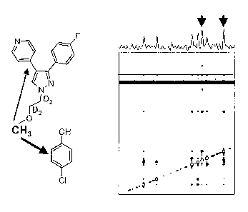
Figure 22 shows the structure of a 4-floro-
piridyl-pyrazole core ATP mimic and a 4-chlorophenol lead
fragment with a reverse NMR ACE urea antenna moiety, and,
the corresponding 2D IL-NOESY spectrum obtained with
these compounds in the presence of p38a.
DETAILED DESCRIPTION OF THE INVENTION
This invention provides a method for
identifying a compound that will bind to a macromolecule.
Using a method of the invention, the relative positions
of two or more ligands when bound to a macromolecule in a
multipartite complex can be determined. Based on this
determination, the ligands or portions of the ligands can
be covalently linked to form a binding compound. An
advantage of the invention is that atomic-resolution
CA 02559711 2006-09-12
WO 2004/083814 PCT/US2004/007471
8
structural data for a macromolecule, although useful in
some aspects of the invention, is not necessary in order
to obtain a compound that binds to the macromolecule.
The methods can be used to design a compound
that binds to a particular macromolecular target. The
compound can be designed by determining the relative
positions of two or more ligands when bound to a
macromolecular target in a multipartite complex. The
methods involve attaching an antenna moiety to a known
ligand (to produce a ligand-probe), incubating the
macromolecular target;ligand-probe and a candidate
ligand under conditions wherein the ligand-probe and
macromolecular target form a bound complex; and detecting
whether the ligand-probe and. candidate ligand are in
proximity to each other. In addition, the distance
between the antenna moiety of the ligand-probe and
candidate ligand and orientation of a candidate ligand
with respect to a ligand-probe can be determined.
The methods can also be used to design a
library that is focused toward members of a particular
protein family having a common ligand site (CLS). The
focused library can include compounds with various
combinations of linked moieties, where the moieties are
structurally similar to each ligand observed in a
multipartite complex and the linker between the moieties
is selected based on the relative positions of the
ligands in the multipartite complex. A focused library
can be designed by determining the relative positions of
two or more ligands when bound to a macromolecule in a
multipartite complex, identifying which of the ligands is
CA 02559711 2006-09-12
WO 2004/083814 PCT/US2004/007471
9
a common ligand capable of binding to the CLS and
building the library to contain members having the common
ligand linked to various moieties that are structurally
similar to the other ligand. An advantage of the
invention is that screening with one or a representative
subset of proteins in a family can be used to design a
'library that is focused with respect to other proteins in
the family.
Nuclear Magnetic Resonance (NMR) can be used in
a method of the invention to determine the relative
proximity or positions of ligands in a multipartite
complex with a macromolecule. In particular, proximal
ligands can be identified from NMR-based observation of
magnetization transfer between the ligands. Although NMR
methods have been previously used to predict or determine
the structure of ligands bound to macromolecules, these
methods have relied upon detection of magnetic
interactions between. the ligand and the macromolecule.
Isotopic labeling can be required for macromolecules in
order to detect magnetic interactions with a bound
ligand. Furthermore, for many large or membrane bound
macromolecules signal broadening, due in part to low
rotational mobility,.renders detection of magnetic
interactions with ligands impractical. Because the
methods of the present invention are based on detection
of interactions between ligands and do not require
detection of interactions with macromolecule,
isotopically labeled macromolecules are not necessary.
The methods are further advantageous for use with large
or membrane bound macromolecules because observation of
CA 02559711 2006-09-12
WO 2004/083814 PCT/US2004/007471
magnetization transfer between ligands can be enhanced
when the ligands experience low rotational mobility.
In several embodiments, the methods of the
5 invention advantageously involve using an isotope filter
to select or reject magnetization that originates solely
from a portion of a ligand molecule, such as a ligand
core structure or antenna moiety. Thus, the resulting
spectra contain a reduced number of resonances. The
10 application of isotope-editing or filtering of NMR
experiments in the methods of the invention allows for
reduced data complexity and:a corresponding simplified
data analysis. Simplified NMR data analysis in turn
allows for faster data analysis, as well as the reduced
training time required for individuals to interpret
experimental data. Moreover, isotope-edited or filtered
NMR experiments, such as those described in Example VII,
can be performed in short time frames that make feasible
high sample throughput.
A further advantage of the invention is that
ligands having relatively low affinities for a
macromolecule can be,identified and linked to form a
compound having substantially increased affinity for the
macromolecule. Such increased affinity is expected to
occur, for example, due to the chelate effect (for a
description of the chelate effect see Page et al., Proc.
Natl. Acad. Sci. USA 68:1678-1683 (1971)) and is
demonstrated in the Examples below. Another advantage of
the invention is that a compound having increased
specificity for a particular macromolecule, compared to a
ligand from which it is assembled, can be identified. In
CA 02559711 2011-08-05
11
particular, members of a CLS-containing protein family often
have a different specificity ligand site adjacent to the common
ligand binding site which provides a potential source of binding
specificity (as described, for example, in WO 99/60404). By
linking the common ligand to a particular specificity ligand, a
compound can be obtained that has increased affinity due to the
presence of both ligands and increased specificity for a
particular member of a protein family compared to the common
ligand.
Yet another advantage of the invention is that once a
specificity ligand is identified, it can be used in methods for
identifying other proximal ligands, including other common or
specificity ligands. The methods of the invention further can be
used to refine, or optimize, an identified ligand to select a
ligand with increased or decreased proximity to another ligand,
with respect to the originally identified ligand.
A ligand can include an antenna moiety that extends
from the core structure of the ligand to interact with a
proximal ligand. An antenna moiety can extend the range within
which a proximal ligand is identified. The use of a multi-probe
ligand having multiple antenna moieties or comparison of ligands
having antennas of different composition or point of attachment
on the ligand moiety can provide information on the relative
orientation of the proximal ligands and their binding sites. In
a method of the invention that employs isotope-edited NOESY, an
antenna moiety can contain one or more NMR-visible nuclei such
as 13c 15N 19F 31P 113Cd
r r ~ r r
CA 02559711 2006-09-12
WO 2004/083814 PCT/US2004/007471
12
and the like. An antenna moiety also can contain NMR-
invisible nuclei, such as 2H, for some applications.
In one embodiment, the invention provides a
compound comprising a molecule attached to an antenna
moiety that contains both a 13C nucleus and several 2H
nuclei.
Because the methods of the invention provide
not only a functional identification that a ligand binds
to a macromolecule, but also identify the relative
positions of two ligands when bound to the macromolecule,
the invention provides structural information. Use of a
method of the invention in a screening format provides a
way to increase the throughput at which structural
information can be obtained on the relative orientation
of the proximal ligands and their binding sites.
In the following description, for the purposes
of explanation, specific details are set forth in order
to provide a thorough understanding of the present
invention. Those skilled in the art will understand that
the present invention can be practiced without these
specific details and can be applied to any of a variety
of related systems. For example, although the methods
are described in the context of ligands that bind to a
protein, it is understood that the methods can be applied
to other macromolecules including, for example, synthetic
polymers, DNA, RNA or polysaccharides that interact with
ligands.
CA 02559711 2006-09-12
WO 2004/083814 PCT/US2004/007471
13
As used herein, the term "ligand" is intended
to mean a molecule that can form a specific, non-covalent
ass.ociation with a macromolecule. A molecule included in
the term can be a small molecule, a binding compound or a
macromolecule. A molecule included in the term can be
naturally occurring such as a DNA, RNA, polypeptide,
protein, lipid, carbohydrate, amino acid, nucleotide,
metabolite or hormone; a synthetic molecule; or a
derivative of a naturally occurring molecule. A
derivative can have, for example, an added moiety, a
removed moiety or a rearrangement in the relative
location of moieties compared to a naturally occurring
molecule. As used herein, the term "binding compound" is
intended to mean a ligand having a covalent structure
that includes at least two moieties that interact with a
macromolecule.
As used herein, the term "binding site" is
intended to mean a portion of a macromolecule or complex
of macromolecules that associates' specifically and non-
covalently with a ligand or portion of a ligand. A non-
covalent association'included in the term can be due to a
hydrogen bond, ionic interaction, van der Waals
interaction, or hydrophobic interaction or a combination
thereof.
As used herein, the term "competitive binding"
is intended to mean binding of a first ligand to a
binding site of a macromolecule in a manner that prevents
a second ligand from binding to the binding site.
Accordingly, a first and second ligand that bind to a
binding site of a macromolecule in a mutually exclusive
CA 02559711 2006-09-12
WO 2004/083814 PCT/US2004/007471
14
manner are understood to be competitive inhibitors of
each other for the macromolecule.
As used herein, the term "bound complex" is
intended to mean a specific non-covalent association
between 2 or more molecules. The term can include a
reversible association so long as the association is
sufficiently stable to be observed by a binding assay.
As used herein, the term "common ligand" or
"CL" is intended to mean a molecule that specifically
binds at a site conserved in a family of 2 or more
macromolecules. The term can therefore extend to
molecules that bind to members of a 'protein family or
gene family. Examples of common ligands include a
natural common ligand which is normally found in
biological systems or a common ligand mimic which has
sufficient structural similarity to a natural common
ligand that it can competitively inhibit binding of the
natural common ligand to its common-ligand binding site.
Accordingly, a "common ligand site" is intended to mean a
location in or on a macromolecule where a common ligand
binds. A common ligand site is also referred to as a
conserved site.
The term "mimic," when used in reference to a
ligand, is intended to mean a molecule that binds to a
protein at the same site as the ligand. The term can
encompass molecules having portions similar to
corresponding portions of the ligand in terms of
structure or function. The term can also encompass the
original ligand itself.
CA 02559711 2006-09-12
WO 2004/083814 PCT/US2004/007471
An example of a useful CL is a cofactor or a
cofactor mimic. A "cofactor" is any small molecule that
binds in the CL site and participates in catalysis when
5 bound to an enzyme. Cofactors often contain a nucleotide
such as adenine mononucleotide or nicotinamide
mononucleotide. Examples of such cofactors include ATP,
ADP and SAM (S-adenosyl methionine). Another group of
cofactors that contain a nucleotide is the group NAD+,
10 NADH, NADP+ and NADPH. Other such cofactors include
FMNH2, FMN, FAD, FADH2, CoA, GTP and GDP. Still other
cofactors include THF, DHF, TPP, biotin, dihydropterin,
heme, pyridoxal phosphate and thiamine pyrophosphate.
Other common ligands include conserved ligands such as
15 farnesyl, farnesyl-pyrophosphate, geranyl, geranyl-
pyrophosphate or ubiquitin~.
As used herein, the term "macromolecular
target" is intended to mean a macromolecule to which a
ligand-probe specifically binds. A macromolecular target
can be., for example, a polypeptide; a nucleic acid
molecule or nucleic acid molecule, complex; a
polysaccharide; a lipid; or a combination thereof.
As used herein, the term "family," when used in
reference to a macromolecule, is intended to mean a group
of at least 2 macromolecules exhibiting structure
homology and at least one function in common. An
exemplary function included in the term is the ability to
bind a common ligand such as NADH or ATP. Examples of
enzyme families include kinases, dehydrogenases,
oxidoreductases, GTPases, carboxyl transferases, acyl
CA 02559711 2006-09-12
WO 2004/083814 PCT/US2004/007471
16
transferases, decarboxylases, transaminases, racemases,
methyl transferases, formyl transferases, and c-
ketodecarboxylases. As used herein, the term "enzyme"
refers to a molecule that binds a substrate ligand and
carries out a catalytic reaction by converting the
substrate ligand to a product.
Enzymes can also be classified based on Enzyme
Commission (EC) nomenclature recommended by the
Nomenclature Committee of the International Union of
Biochemistry and Molecular Biology (IUBMB) and available
from the ENZYME database. (available on the internet at
expasy.ch/enzyme/; administered by The Swiss Institute
for Bioinformatics, Switzerland; see, for example,
15' Bairoch, Nucl. Acid. Res. 28:304-305 (2000)). For
example, oxidoreductases are classified as
oxidoreductases acting on the CH-OH group of donors with
NAD+ or NADP+ as an acceptor (EC 1.1.1); oxidoreductases
acting on the aldehyde or oxo group of donors with NAD+ or
NADP+ as an acceptor (EC 1.2.1); oxidoreductases acting on'
the CH-CH group of donors with NAD+ or NADP+ as an
acceptor (EC 1.3.1); oxidoreductases acting on the CH-NH2
group of donors with NAD+ or NADP+ as an acceptor '(EC
1.4.1); oxidoreductases acting on the CH-NH group of
donors with NAD+ or NADP+ as an acceptor (EC 1.5.1);
oxidoreductases acting on NADH or NADPH (EC 1.6); and
oxidoreductases acting on NADH or NADPH with NAD+ or NADP+
as an acceptor (EC 1.6.1).
Additional oxidoreductases include
oxidoreductases acting on a sulfur group of donors with
NAD+ or NADP+ as an acceptor (EC 1.8.1); oxidoreductases
CA 02559711 2006-09-12
WO 2004/083814 PCT/US2004/007471
17
acting on diphenols and related substances as donors with
NAD+ or NADP+ as an acceptor (EC 1.10.1); oxidoreductases
acting on hydrogen as donor with NAD+ or NADP+ as an
acceptor (EC 1.12.1); oxidoreductases acting on paired
donors with incorporation of molecular oxygen with NADH
or NADPH as one donor and incorporation of two atoms
(EC 1.14.12) and with NADH or NADPH as one donor and
incorporation of one atom (EC 1.14.13);, oxidoreductases
oxidizing metal ions, with NAD+ or NADP+ as an acceptor
(EC 1.16.1); oxidoreductases acting on -CH2 groups with
NAD+ or NADP+ as an acceptor (EC 1.17.1); and
oxidoreductases acting on reduced ferredoxin as donor,
with NAD+ or NADP+ as an acceptor (EC l..18.1).
Other enzymes include transferases classified
as transferases transferring one-carbon groups (EC 2.1);
methyltransferases (EC 2.1.1); hydroxymethyl-, formyl-
and related transferases (EC 2.1.2); carboxyl- and
carbamoyltransferases (EC 2.1.3); acyltransferases (EC
2.3); and transaminases (EC 2.6.1). Additional enzymes
include phosphotransferases such as: phosphotransferases
transferring phosphorous-containing groups with an
alcohol as an acceptor (kinases) (EC 2.7.1);
phosphotransferases with a carboxyl group as an acceptor
(EC 2.7.2); phosphotransfer with a nitrogenous group as
an acceptor (EC 2.7.3); phosphotransferases with a
phosphate group as an acceptor (EC 2.7.4); and
diphosphotransferases (EC 2.7.6).
Protein or gene family members can often be
identified by the presence of a conserved structural
motif as described, for example, in Branden and Tooze
CA 02559711 2006-09-12
WO 2004/083814 PCT/US2004/007471
18
Introduction to Protein Structure, Garland Publishing
Inc., New York (1991). A structural motif can be
identified at the primary structure level according to a
particular nucleotide or amino acid sequence or at the
tertiary structure level due to a particular combination
or orientation of secondary structure elements.
Identification of structural motifs using structural
alignments is described in further detail below.
Several large protein and gene families have
been identified, including families having as many as 20
or more, 50 or more, 100 or.more and even 200 or more
members. Two particular examples of a protein or gene
family are kinases and oxidoreductases. The term
"kinase" herein means any enzyme that catalyzes.. the
transfer of a phosphoryl group from ATP or other
nucleoside triphosphate to another compound. The term
"oxidoreductase" herein means any enzyme that catalyzes
an oxidation-reduction reaction. Still other gene
families include transaminases, decarboxylases and
methyltransferases.
Another particular gene family is the
dehydrogenase gene family. The term "dehydrogenase"
herein means any enzyme that catalyzes the removal of
hydrogen from a substrate using a compound other than
molecular oxygen as an acceptor. Typically the hydrogen
is transferred to the coenzyme NAD+ (nicotinamide adenine
dinucleotide) or NADP+ (nicotinamide adenine dinucleotide
phosphate). The dehydrogenase gene family is large,
containing approximately 17% of all enzymes (You, Kwan-
sa, "Stereospecificity for Nicotinamide Nucleotides in
CA 02559711 2006-09-12
WO 2004/083814 PCT/US2004/007471
19
Enzymatic and Chemical Hydride Transfer Reactions," CRC
Crit. Rev. Biochem. 17:313-451 (1985)). Thus, the
dehydrogenase family is likely to be a rich source of
drug targets.
As used herein, the term "specificity" refers
to the ability of a ligand to selectively bind to one
macromolecule over another. For example, the term can
include selective binding of a ligand to one member of a
protein family compared to other proteins outside of or
within the protein family. The selective binding of a
particular ligand to a macromolecule is measurably higher
than the binding of the ligand to at least one other
molecule. Specificity can also be exhibited over two, or
1.5 more, three or more, four or more, five or more, six ar
more, seven or more, ten or more, or even twenty or more
other macromolecules.
As used herein, the term "structure model" is
intended to mean a representation of the relative
locations of atoms of a molecule. A representation
included in the term can be defined by a coordinate
system that is preferably in 3 dimensions, however,
manipulation or computation of a model can be performed
in 2 dimensions or even 4 or more dimensions in cases
where such methods are desired. The location of atoms in
a molecule can be described, for example, according to
bond angles, bond distances, relative locations of
electron density, probable occupancy of atoms at points
in space relative to each other, probable occupancy of
electrons at points in space relative to each other or
combinations thereof. A representation included in the
CA 02559711 2006-09-12
WO 2004/083814 PCT/US2004/007471
term can contain information for all atoms of a
particular molecule or a subset of atoms thereof.
Examples of representations.included in the term that
contain a subset of atoms are those commonly used for
5 polypeptide structures such as ribbon diagrams, and the
like, which show the coordinates of the polypeptide
backbone while omitting coordinates for all or a portion
of the side chain moieties of the polypeptide.
Representations for other macromolecules and small
10 molecules included in the term can similarly contain all
or a subset of atoms.
A structure model can include a representation
that is determined from empirical data derived from, for
15 example, X-ray crystallography or nuclear magnetic
resonance spectroscopy. -.A representation included in the
term can include one that is derived from a theoretical
calculation including, for example, a structure obtained
by homology modeling or ab initio modeling. A
20 representation of a structure model can include, for
example, an electron density map, atomic coordinates, x-
ray structure model, ball and stick model, density map,
space filling model, surface map, Connolly surface, Van
der Waals surface or CPK model.
As used herein, the term "docking" is intended
to mean using a model of a first and second molecule to
simulate association of the first and second molecule at
a proximity sufficient for at least one atom of the first
molecule to be within bonding distance of at least one
atom of the second molecule. The term is intended to be
consistent with its use in the art pertaining to
CA 02559711 2006-09-12
WO 2004/083814 PCT/US2004/007471
21
molecular modeling. A model included in the term can be
any of a variety of known representations of a molecule
including, for example, a graphical representation of its
three-dimensional structure, a set of coordinates, set of
distance constraints, set of bond angle constraints or
set of other physical or chemical properties or
combinations thereof.
As used herein, the term "magnetization
transfer" is intended to mean a through-space alteration
of the nuclear magnetic resonance properties of an atomic
nucleus of a first atom due to a proximal atomic nucleus
or at least one electron of a proximal atom. An
alteration included in the term can occur due to the
Nuclear Overhauser Effect (NOE.)..or cross saturation.
Proximal atomic nuclei included in the term are those
that are within a distance sufficient to cause a magnetic
interaction detectable by a nuclear magnetic resonance
spectroscopy measurement used in the methods of the
invention. Examples of magnetic effects included in the
term are a relaxation effect which can be detected for
atoms.that are about 10 A apart or closer, the Nuclear
Overhauser Effect which can be detected for atoms that
are about 6 A apart or closer or chemical shift due to
shielding or de-shielding which can be detected for atoms
that are about 10 A or closer. Atoms that are about 5 A
apart or closer, 4 A apart or closer, 3 A apart or
closer, 2 A apart or closer or 1 A apart or closer are
also proximal atoms that are included in the term.
CA 02559711 2006-09-12
WO 2004/083814 PCT/US2004/007471
22
As used herein, the term "linker" is intended
to mean one or more atoms that covalently connect a first
moiety to a second moiety. A moiety included in the term
can be a ligand such as a common ligand, or fragment
thereof; a specificity ligand, or fragment thereof; or a
mimic of a common ligand or specificity ligand. A linker
can provide positioning and orientation of a first moiety
relative to a second moiety such that one moiety can bind
to a first ligand site and the other moiety can bind to a
second, proximal site on a macromolecule.
As used herein, a "library" is intended to mean
a population of different molecules. The library is
chemically synthesized and contains primarily the
'components generated during the synthesis. A population
included in the term can include two-or more different
molecules. A population can be as large as the number of
individual molecules currently available to the user or
able to be made by one skilled in the art. A population
can be as small as two molecules and as large as 1010
molecules. Generally, a population will contain two or
more, three or more, five or'more, nine or more, ten or,
more, twelve or more, fifteen or more, or twenty or more
different molecules. A population can also contain tens
or hundreds of different molecules or even thousands of
different molecules. For example, a population can
contain about 20 to about 100,000 different molecules or
more, for example about 25 or more, 30 or more, 40 or
more, 50 or more, 75 or more, 100 or more, 150 or more,
200 or more, 300 or more, 500 or more, or 1000 or more
different molecules, and particularly about 10,000,
100,000 or even 1x10⁶ or more different molecules. A
CA 02559711 2006-09-12
WO 2004/083814 PCT/US2004/007471
23
population of synthetic compounds can be derived, for
example, by chemical synthesis and is substantially free
of naturally occurring substances.
As used herein, the term "homolog" is intended
to mean a molecule or moiety of a molecule that has
similar structure in comparison to a reference molecule
or moiety. A moiety is a group of atoms that form a part
or portion of a larger molecule. A moiety can consist of
any number of atoms in a portion of a molecule and can
correlate with a physical or chemical property conferred
upon the molecule by the combined atoms..
As used herein, the term "ligand-probe" is
intended to mean a molecule: that can selectively bind a
protein and that has an antenna moiety-and a ligand
moiety. A "ligand moiety" is a fragment of a ligand-
probe, that when lacking the antenna moiety, is capable
of selectively binding to the protein. An "antenna
moiety" is a structure containing an NMR-visible nucleus
that is attached to a ligand moiety by bonding to at
least 1intervening atoms. A larger number of atoms can
intervene between an NMR-visible nucleus and ligand
moiety including, for example, at least 2, 3, 4, 5, 6, 7,
8, 9, 10 or more intervening atoms. The intervening
atoms can form an aliphatic chain that, when attached to
a ligand moiety having aromatic rings, allows selective
excitation due to differences in frequency for excitation
or saturation of aliphatic and aromatic protons. An NMR-
visible nucleus of an antenna moiety can be a proton that
is isolated from vicinal proton coupling. Isolation from
vicinal proton coupling provides for selective
CA 02559711 2006-09-12
WO 2004/083814 PCT/US2004/007471
24
observation of direct NOE transfer at short mixing times
compared to indirect NOE transfer via spin diffusion and
also reduces signal loss due to relaxation effects that
occur for vic.inal coupled protons. A proton can be
isolated from vicinal proton coupling by being attached
to a carbon that is adjacent to an atom that lacks
protons including, for example, an ether oxygen; carbonyl
carbon;.thioether, sulfone or sulfoxide sulfur;
deuterated carbon or selenium, or by being attached to a
carbon that is adjacent to an atom having fast exchanging
protons such as a nitrogen,at high pH. An NMR-visible
nucleus of an antenna moiety also can be, for example,
13C, 15N, 19F, 31P, or 113Cd. Such a nucleus can be isolated
for selective observation of direct NOE transfer using a
15. variety of isotope-editing NMR methods. As shown ins
example VII, 13C-isotope editing~lwa-s used to isolate
coupling of a carbon in an antenna moiety of a ligand-
probe to a portion of a ligand. A probe ligand or
antenna moiety can contain two or more NMR-visible
nuclei, such as two or more 13C, 15N, 19F, 31P, or 713Cd -
enriched heteroatoms. The presence of such heteroatoms
can be useful for the application of methods for
obtaining 3D X-nucleus separated NOESY spectra, such as
3D HMQC/HSQC-NOESY or NOESY-HMQC/HSQC spectra to separate
IL-NOE peaks corresponding to each isotope enriched and
attached proton into different planes in the spectrum.
Such methods can be used to resolve signal over-lap in
the two proton dimensions. For use in isotope edited
methods, the choice of where to incorporate a particular
nuclei into a ligand-probe or antenna moiety and the
choice of the number of enriched heteroatoms to include,
can be determined by those skilled in the art based on
CA 02559711 2006-09-12
WO 2004/083814 PCT/US2004/007471
physical properties of the particular ligand-probes or
antenna moieties.
The invention provides a method for assembling
5 a binding compound. The method includes the steps of (a)
obtaining a sample containing a macromolecule, a first
ligand and a second ligand under conditions wherein a
bound complex is formed containing the first ligand, the
second ligand and the macromolecule; (b) providing a
10 sample comprising the protein, the first ligand and a
second ligand under conditions wherein the first ligand,
the second ligand and the protein form a bound complex;
(c) detecting a subset of magnetization transfer signals
between the first ligand and the second ligand in the
1.5 bound complex, wherein the signals areobtained from an
isotope edited NOESY spectrum of the sample; thereby
determining that the two ligands are proximal in the
bound complex; and (d) obtaining a population of
candidate binding compounds comprising.the first ligand,
20 or a fragment thereof, linked to one of a plurality of
second ligand homologs, whereby the population contains
binding compounds that bind to members of the protein
family.
25 A schematic overview that exemplifies a method
for assembling a binding compound is shown in Figure 1.
At step 1, a ligand, shown as F1, is identified based on
the observation that it binds to a protein. The binding
can be observed based on magnetization transfer between
the protein binding site and the F1 ligand. The F1 ligand
can be obtained from a screen in which a library of
candidate ligands are tested for the ability to bind the
CA 02559711 2006-09-12
WO 2004/083814 PCT/US2004/007471
26
protein. At step 2, the F2 ligand is identified as
binding to the protein at a location that is proximal to
the F1 ligand. Proximal ligands can be identified based
on the observation of magnetization transfer between the
F1 ligand and F2 ligand when in a complex with the
protein. The F2 ligand can be identified from a screen
using a library of candidate ligands. Based on the
observed magnetization transfer a bi-ligand compound can.
be obtained in which the F1 and F2 ligands are linked.
Alternatively, fragments or homologs of either ligand can
be linked to forma bi-ligand. As shown in step 2', the
bi-ligand compound can be used to identify a third
proximal ligand, shown as ligand F3, and a tri-ligand can
be subsequently obtained in which the F1, F2 and F3
ligands ar_e. linked: Similarly, an F1 ligand can. be
identified in step 1, while an F2 ligand is identified in
step 2.
A schematic overview that exemplifies another
method for assembling a binding' compound is shown in
Figure 21. Figure 21 showsa previously identified
binding compound (A) and subsequent separation of the
binding compound into F1 and F2 fragments (B). In Figure
21C, the F2 fragment is used to identify a different
proximal Fl ligand. In Figure 21D, the F2 fragment is
used to identify an F2' proximal ligand that binds to a
different binding site. The distance between a newly
identified F2' proximal ligand and F2 or F1 can be
determined if the binding sites are close enough to allow
inter-ligand NOEs to be observed between them, and based
on knowledge of the position of the original F1. Thus,
the position of one or more new F2' fragments from the
CA 02559711 2006-09-12
WO 2004/083814 PCT/US2004/007471
27
inter-ligand NOEs between the an F2 fragment and a new F2'
binding site remote from the original F1 can be determined
by "triangulation." As described herein, a variety of
strategies can be used for identifying distances between
ligand fragments, which can include "Forward NMR ACE,"
used when a common ligand is used to probe for another
ligand, such as a specificity ligand; "Reverse NMR ACE,"
used when a specificity ligand is used to probe for a
proximal ligand, such as a common ligand;
"Triangulation," used to identify a third ligand (F2'
fragment) from two known Fl fragments, two known F2
fragments, or one of each; and "Extended NMR ACE," used
to identify a new F2' that binds remote from the F1 from
an F2 ligand-probe. These and related methods are set
forth in further detail below.
Initially, a macromolecule target such as a
protein is identified for the development of a binding
compound. In one embodiment, a macromolecule target for
development of a therapeutic agent can be identified
based on its presence in a pathogen or. its association
with a disease or condition. For example, a protein
target present in a pathogen can be selected as the
target to develop drugs effective in combating a disease
caused by that pathogen. Any pathogen can be selected as
a target organism. Examples of pathogens include, for
example, bacteria, fungi or protozoa.
Pathogenic bacteria useful as target organisms
include Staphylococcus, Mycobacteria, Mycoplasma,
Streptococcus, Haemophilus, Neisseria, Bacillus,
Clostridium, Corynebacteria, Salmonella, Shigella,
CA 02559711 2006-09-12
WO 2004/083814 PCT/US2004/007471
28
Vibrio, Campylobacter, Helicobacter, Pseudomonas,
Legionella, Bordetella, Bacteriodes, Fusobacterium,
Yersinia, Actinomyces, Brucella, Borrelia, Rickettsia,
Ehrlichia, Coxiella, Chlamydia, and Treponema.
Pathogenic strains of Escherichia coli can also be target
organisms.
Binding compounds targeted to macromolecules in
these pathogenic bacteria are useful for treating a
variety of diseases including bacteremia, sepsis,
nosocomial infections, pneumonia, pharyngitis, scarlet
fever, necrotizing fasciitis, abscesses, cellulitis,
rheumatic fever, endocarditis, toxic shock syndrome,
osteomyelitis, tuberculosis, leprosy, meningitis,
pertussis, food poisoning,. enteritis, enterocolitis,
diarrhea, gastroenteritis, shigellosis, dysentery,
botulism, tetanus, anthrax, diphtheria, typhoid fever,
cholera, actinomycosis, Legionnaire's disease, gangrene,
brucellosis, lyme disease, typhus, spotted fever, Q
fever, urethritis, vaginitis, gonorrhea and syphilis.
For example, Staphylococcus aureus is a major
cause of nosocomial infections and has become
increasingly resistant to a variety of antibiotics over
recent years. Similarly, Mycobacteria tuberculosis has
become increasingly resistant to multiple antibiotics in
recent years. M. tuberculosis infects almost one third
of the world population, with active tuberculosis found
in almost 10 million people worldwide and in AIDS
patients as a common opportunistic infection.
Streptomyces has also become increasingly resistant to
antibiotics over recent years. Therefore, these
CA 02559711 2006-09-12
WO 2004/083814 PCT/US2004/007471
29
pathogenic bacteria with known resistance and target
macromolecules required for their growth or pathogenesis
are particularly desirable as target organisms for which
therapeutic binding compounds can be identified.
In another embodiment, target organisms are
selected from yeast and fungi. Pathogenic yeast and
fungi useful as target organisms include Aspergillus,
Mucor, Rhizopus, Candida, Cryptococcus, Blastomyces,
Coccidioides, Histoplasma, Paracoccidioides, Sporothrix,
and Pneumocystis. Binding compounds targeted to
macromolecules in these pathogenic yeast and fungi are.
useful for treating a variety of diseases including
aspergillosis, zygomycosis, candidiasis, cryptococcoses,
blastomycosis, coccidioidomycosis,: histoplasmosis,
paracoccidioidomycosis,-sporotrichosis, and pneuomocystis
pneumonia.
In still another embodiment, target organisms
are selected from protozoa. Pathogenic protozoa useful
as target organisms include Plasmodium, Trypanosoma,
Leishmania, Toxoplasma, Cryptosporidium, Giardia, and
Entamoeba. Binding compounds targeted to macromolecules
in these pathogenic protozoa are useful for treating a
variety of diseases including malaria, sleeping sickness,
Chagas' disease, leishmaniasis, toxoplasmosis,
cryptosporidiosis, giardiasis, and amebiasis.
In addition, a target cell such as a cancer
cell can be selected to identify drugs effective for
treating cancer. Examples of such target cells include,
for example, breast cancer, prostate cancer, and ovarian
CA 02559711 2006-09-12
WO 2004/083814 PCT/US2004/007471
cancer cells as well as leukemia, lymphomas, melanomas,
sarcomas and gliomas. Binding compounds directed to a
target macromolecule in a cancer cell are useful for
targeted delivering of a chemotherapeutic agent or for
5 inhibition of unregulated growth. Diagnosis and
identification of causative factors or pathogens for a
targeted disease can be determined using methods known in
the art as described for example in The Merck Manual,
Sixteenth Ed, (Berkow, R., Editor) Rahway, N.J., 1992.
A macromolecule family to which a target
macromolecule belongs can be identified according to
structural or functional similarities using methods known
in the art. Structural similarity can be identified, for
example, by sequence analysis at the nucleotide or amino
acid level. One method for determining if two
macromolecules are related is BLAST, Basic Local
Alignment Search Tool. (available on the internet at
ncbi.nlm.nih.gov/BLAST/; administered by The National
Center for Biotechnology Information, Bethesda Maryland).
BLAST is a set of similarity search programs designed to
examine all available sequence databases and can function
to search for similarities in protein or nucleotide
sequences., A BLAST search provides search scores that
have a well-defined statistical interpretation.
Furthermore, BLAST uses a heuristic algorithm that seeks
local alignments and is therefore able to detect
relationships among sequences which share only isolated
regions of similarity (Altschul et al., J. Mol. Biol.
215:403-410 (1990)).
CA 02559711 2006-09-12
WO 2004/083814 PCT/US2004/007471
31
In addition to the originally described BLAST
(Altschul et al., supra, 1990), modifications to the
algorithm have been made (Altschul et al., Nucleic Acids
Res. 25:3389-3402 (1997)). One modification is Gapped
BLAST, which allows gaps, either insertions or deletions,
to be introduced into alignments. Allowing gaps in
alignments tends to reflect biologic relationships more
closely. A second modification is PSI-BLAST, which is a
sensitive way to search for sequence'homologs. PSI-BLAST
performs an initial Gapped BLAST search and uses
information from any significant alignments to construct
a position-specific score matrix, which replaces the
.query sequence for the next round of database searching.
A PSI-BLAST search is often more sensitive to weak but
biologically relevant sequence similarities.
A second resource for identifying members of a
protein family is PROSITE. (Available on the internet at
expasy.ch/sprot/prosite.html; administered by The Swiss
Institute for Bioinformatics, Switzerland). PROSITE is a
method of determining the function of uncharacterized
proteins translated from genomic or cDNA sequences
(Bairoch et al., Nucleic Acids Res. 25:217-221 (1997)).
PROSITE consists of a database of biologically
significant sites and patterns that can be used to
identify which known family of proteins, if any, the new
sequence belongs. In some cases, the sequence of an
unknown protein is too distantly related to any protein
of known structure to detect its resemblance by overall
sequence alignment. However, related proteins can be
identified by the occurrence in its sequence of a
particular cluster of amino acid residues, which can be
CA 02559711 2011-08-05
32
called a pattern, motif, signature or fingerprint. PROSITE uses
a computer algorithm to search for motifs that identify
proteins as family members. PROSITE also maintains a
compilation of previously identified motifs, which can be used
to determine if a newly identified protein is a member of a
known protein family.
Members of a protein family can also be identified by
clustering binding site structures or bound ligand
conformations as described, for example, in WO 02/059715. A
sequence model such as a Hidden Markov Model, representing the
frequency and order with which specific amino acids or gaps
occur in the binding sites of protein family members can be
used to search a sequence database and identify other members
as described, for example, in WO 02/059715. Members of a
protein family can also be identified by clustering their
sequence comparison signatures, where a sequence comparison
signature for a protein is a string of pairwise comparison
scores for the protein compared to the other proteins in a
database as described, for example, in WO 03/060807.
Another resource for identifying members of a protein
family is Structural Classification of Proteins (SCOP),
administered by Medical Research council, Cambridge, England.
SCOP maintains a compilation of previously determined protein
tertiary folds from which structural comparison can be made to
identify protein
CA 02559711 2006-09-12
WO 2004/083814 PCT/US2004/007471
33
family members having similar motifs (Murzin et al.,
J. Mol. Biol. 247:536-540 (1995)).
Table 1. Databases for Identifying Protein Family Motifs
SEARCHABLE MOTIF AND
PATTERN DATABASES WEBSITES
PROSITE expasy.hcuge.ch/sprot/prosite.html
BLOCKS, blocks.fhcrc.org/blocks_search.html
PRINTS
biochem.ucl.ac.uk/bsm/dbbrowser/
PRINTS/PRINTS.html
PIMA.:. dot.imgen.bcm.tmc.edu:9331/seq-
search/protein- search.html
PRODOM protein.toulouse.inra.fr/prodom.html
REGULAR EXPRESSION SEARCH ibc.wustl.edu/fpat/
PROFILESEARCH
seqnet.dl.ac.uk/h
hg/PROFILESE.html
PATSCAN
c.mcs.anl.gov/hom
e/overbeek/PatScan/HTM
L/patscan.html
PATTERNFIND
ulrec3.unil.ch/so
ftware/PATFND-
mailform.html
PROFILE
lenti.med.umn.edu
/MolBio man/chp-
10.html#HDR1
CA 02559711 2006-09-12
WO 2004/083814 PCT/US2004/007471
34
PMOTIF
alces.med.umn.edu
/pmotif.html
HMMER
genome.wustl.edu/
eddy/HMMER/
WWW AND FTP SERVERS FOR
SINGLE SEQUENCE EXHAUSTIVE
DATABASE SEARCHES WEBSITES
BLAST ~ncbi.nlm.nih.gov/BLAST/
BLITZ
ebi.ac.uk/searches/bli
tz input.html
FASTA genome.ad.jp/ideas/fasta
/fasta_genes.html
FTP ADDRESSES FOR MOTIF
AND PROFILE SEARCH PROGRAMS WESITES
BARTON'S FLEXIBLE PATTERNS geoff.biop.ox.ac.uk/
PROPAT mdc-berlin.de/
SON ftp.mdc-
berlin.de/pub/neural
SEARCHWISE
sable.ox.ac.uk/pu
b/users
PROFILE
ftp.ebi.ac.uk/pub
/software/unix/
TPROFILESEARCH
ftp.ebi.ac.uk/pub
/softare/vax/egcg
CAP
ncbi.nlm.nih.gov/
pub/koonin/cap
CA 02559711 2006-09-12
WO 2004/083814 PCT/US2004/007471
Additional resources for identifying motifs of
a protein family are shown in Table 1. The websites
cited therein are incorporated by reference.
5 Conserved amino acids are evolutionarily
conserved and carry out a common function. For example,
the Rossman fold is a tertiary structural motif that
includes GXXGXXG or GXGXXG and is present in enzymes that
bind nucleotides (Brandon and Tooze, in Introduction to
10 Protein Structure, Garland Publishing, New York (1991)).
Enzymes that bind nucleotides such as NAD, NADP, FAD,
ATP, ADP, AMP and FMN contain the Rossman fold sequence
motif (Creighton, Proteins: Structures and Molecular
Principles,, p.368, W.H. Freeman, New York (1984)).
15 Additional conserved residues as well as different
protein structures:distinguish protein families that
bind, for example, NAD from those that bind, for example,
ATP.
20 An example of a recognizable protein motif or
fingerprint is found in dinucleotide binding proteins
such. as dehydrogenases (Rossman et al., in The Enzymes
Vol 11, Part A, 3rd ed., Boyer, ed., pp. 61-102,.Academic
Press, New York (1975); Wierenga et al., J. Mol. Biol.
25 187:101-107 (1986); and Ballamacina, FASEB J. 10:1257-
1269 (1996)). The fingerprint region contains a
phosphate binding consensus sequence GXXGXXG or GXGXXG, a
hydrophobic core of six small hydrophobic residues, a
conserved, negatively charged residue that binds to the
30 ribose 2' hydroxyl of adenine and a conserved positively
charged residue (Bellamacina, supra).
CA 02559711 2006-09-12
WO 2004/083814 PCT/US2004/007471
36
Protein kinases also have recognizable motifs
conserved among all known protein kinases (Hanks and
Quinn, Methods Enzymol. 200:28-62 (1991)). Eight
invariant amino acid residues are conserved throughout
the protein kinase family, including a conserved GXGXXG
motif similar to that seen in dinucleotide binding
proteins. A crystallographic molecular model of cyclic;
AMP-dependent protein kinase as well as other protein
kinases showed that these conserved residues are nearly
all associated with essential, conserved functions such
as ATP binding and catalysis (Knighton et al., Science
253:407-414 (1991); and Knighton et al., Science 253:414-
420 (1991)). Thus, conserved amino acid residues,-which
are common to members of a protein family, are
'recognizable as a motif. cr..itical for the structure,
function or activity of a protein.
Pyridoxal binding proteins also have
recognizable motifs. One motif is GXGGXXXG, a second
motif is KXEX6SXKX5-6M, and a third motif is PXNPTG (Suyama
et al., Protein Engineering 8:1075-1080 (1995)).
A macromolecule family can be selected based on
a conserved and recognizable structural motif such as a
primary sequence motif, tertiary structure motif, or
both. Members of a macromolecule family can also be
recognized based on similar function. For example, a
protein family can be identified based on the ability of
its members to bind a natural common ligand that is
already known. For example, it is known that
dehydrogenases bind to dinucleotides such as NAD or NADP.
Therefore, NAD or NADP are natural common ligands to a
CA 02559711 2006-09-12
WO 2004/083814 PCT/US2004/007471
37
number of dehydrogenase family members. Similarly,
kinases bind ATP, which is therefore a natural common
ligand to kinases. Other natural common ligands of a
macromolecule family can be the coenzymes and cofactors
described above.
After a target macromolecule is selected, the
selected macromolecule or a functional fragment thereof
can be isolated for use in the methods. A functional
fragment of a macromolecule is a fragment that is capable
of binding at least one ligand that is.bound by the full
length=macromolecule. The macromolecule or fragment can
be isolated from a native tissue or organism, from a
population of cells maintained in culture, or from a
recombinant organism or cell culture. Methods for
isolating a protein are known in the art and are
described, for example, in Scopes, Protein Purification:
Principles and Practice, 3rd Ed., Springer-Verlag, New
York (1994); Duetscher, Methods in Enzymology, Vol 182,
Academic Press, San Diego (1990); and Coligan et al.,
Current protocols in Protein Science,-John Wiley and
Sons, Baltimore, MD (2000).
A target macromolecule can be cloned and
expressed in a recombinant organism using methods that
are known to those skilled in the art including, for
example, polymerase chain reaction (PCR) and other
molecular biology techniques (Dieffenbach and Dveksler,
eds., PCR Primer: A Laboratory Manual, Cold Spring Harbor
Laboratory Press, Plainview, NY (1995); Sambrook et al.,
Molecular Cloning: A Laboratory Manual, 2nd ed., Cold
Spring Harbor Laboratory Press, Plainview, NY (1989);
CA 02559711 2006-09-12
WO 2004/083814 PCT/US2004/007471
38
Ausubel et al., Current Protocols in Molecular Biology,
Vols. 1-3, John Wiley & Sons (1998)). The gene or cDNA
encoding the target macromolecule is cloned into an
appropriate expression vector for expression in an
organism such as bacteria, insect cells, yeast or
mammalian cells.
Appropriate expression vectors include those
that are replicable in eukaryotic cells and/or
prokaryotic cells and can remain episomal or be
integrated into the host cell genome. Suitable vectors
for expression in prokaryotic or eukaryotic cells are
well known to those skilled in the art as described, for
example, in Ausubel et al., supra. Vectors useful for
expression in eukaryotic.cells can include, for example,.
regulatory elements. including the SV40 early promoter,
the cytomegalovirus (CMV) promoter, the mouse mammary
tumor virus (MMTV) steroid-inducible promoter, Moloney
murine leukemia virus (MMLV) promoter, and the like. A
vector useful in the methods of the invention can
include, for example, viral vectors such as a
bacteriophage, a baculovirus or a retrovirus; cosmids or
plasmids; and, particularly for cloning large nucleic
acid molecules, bacterial artificial chromosome vectors
(BACs) and yeast artificial chromosome vectors (YACs).
Such vectors are commercially available, and their uses
are known in the art. One skilled in the art will know
or can readily determine an appropriate promoter for
expression in a particular host cell.
CA 02559711 2006-09-12
WO 2004/083814 PCT/US2004/007471
39
If desired, a target protein can be expressed
as a fusion with an affinity tag that facilitates
purification of the target protein. For example, the
target protein can be expressed as a fusion with a poly-
His tag, which can be purified by metal chelate
chromatography. Other useful affinity purification tags
which can be expressed as fusions with the target protein
and used to affinity purify the protein include, for
example, a biotin, polyhistidine tag (Qiagen; Chatsworth,
CA), antibody epitope such as the flag peptide (Sigma; St
Louis, MO), glutathione-S-transferase (Amersham
Pharmacia; Piscataway, NJ), cellulose binding domain
(Novagen; Madison, WI), calmodulin (Stratagene; San
Diego, CA), staphylococcus protein A (Pharmacia; Uppsala,
Sweden), maltose binding protein (New England BioLabs;
Beverley, MA) or strep-tag (Genosys;. Woodlands, .-TX) or
minor modifications thereof.
A target macromolecule'can be validated as a
representative member of a macromolecule family. In some
cases, the target macromolecule is well characterized
with respect to its binding properties to a natural
common ligand. However, if the target macromolecule is
encoded by a new, relatively uncharacterized gene, the
expressed target macromolecule can be tested to confirm
that it binds the natural common ligand. Other common
ligands of related macromolecule families, for example,
other nucleotide binding macromolecules, or known ligand
mimics can also be tested for binding to the target
macromolecule.
CA 02559711 2006-09-12
WO 2004/083814 PCT/US2004/007471
A target macromolecule can be further validated
as a useful therapeutic target by determining if the
selected target macromolecule is known to be required for
normal growth, viability or infectivity of the target
5 organism or cell. If it is unknown whether the target
macromolecule is required for normal growth, viability,
or infectivity, the target macromolecule can be
specifically inactivated by gene knockout, in a model
organism to determine if the macromolecule performs a
'10 critical function required for survival or infectivity of
the organism or cell. Such a macromolecule providing a
critical function is a good target for developing
therapeutic agents.
15 Methods for disrupting::a gene to generate .a
:knockout are well known in the art (Ausubel et al.,
Current Protocols in Molecular Biology, Vols 1-3, John
Wiley & Sons (1998)). For example, transposable elements
can be used to knockout a gene and test for the effect of
20 the knockout on cell growth, viability or infectivity
(Benson and Goldman, J. Bacteriol. 174:1673-1681 (1992);
Hughes and Roth, Genetics 119:9-12 (1988); and Elliot and
Roth, Mol. Gen. Genet. 213:332-338 (1988)). Methods for
gene knockouts in protozoa have also been previously
25 described (Wang, Parasitology 114:531-544 (1997); and Li
et al, Mol. Biochem. Parasitol. 78:227-236 (1996)).
Although use of the methods of the invention is
exemplified herein with regard to proteins, it is
30 understood that a method of the invention can be used for
any other macromolecule that is capable of binding two or
more ligands in proximity. Other macromolecules include,
CA 02559711 2006-09-12
WO 2004/083814 PCT/US2004/007471
41
for example, biological polymers such as polysaccharides
or polynucleotides or synthetic polymers such as plastics
and mimics of biological polymers. A polynucleotide can
be, for example, a ribozyme, ribosomal RNA or other RNA
that is capable of binding a ligand such as a nucleotide.
A method of the invention can include a step of
identifying a common ligand. In some cases, a common
ligand to a macromolecule family is already known. For
example, NAD is a natural common ligand for
dehydrogenases, and ATP is a natural common ligand for
kinases. However, natural common ligands such as the
coenzymes and cofactors often have limitations regarding
their usefulness as a starting compound. Substrates and
cofactors often undergo a chemical reaction, for example,
,transfer of a group to another substrate. or reduction or
oxidation during the enzymatic reaction. However, it is
desirable that a ligand to be used as a drug is not
metabolizable. Therefore, a natural common ligand or-a
derivative thereof that is non-metabolizable is generally
preferred as a commonligand. Examples of mimetics to
the common ligand NADH, for example cibacron blue, are
described in Dye-Ligand Chromatography, Amicon Corp.,
Lexington MA ('1980). Numerous other examples of NADH-
mimics, including useful modifications to obtain such
mimics, are described in Everse et al. (eds.), The
Pyridine Nucleotide Coenzymes, Academic Press, New York
NY (1982).
A ligand that binds a macromolecule can be
identified or characterized using a binding assay
including, for example, an equilibrium binding analysis,
CA 02559711 2006-09-12
WO 2004/083814 PCT/US2004/007471
42
competition assay, or kinetic assay as described in
Segel, Enzyme Kinetics John Wiley and Sons, New York
(1975), and Kyte, Mechanism in Protein Chemistry Garland
Pub. (1995). A common ligand can be identified by a
competitive binding assay. For example, a macromolecule
can be incubated in the presence of a known common ligand
and a candidate common ligand, and the rate or extent to
which the common ligand binds the macromolecule can be
determined. Competitive binding between the common
ligand and candidate ligand can be identified from a
reduction in the rate or extent of binding of the common
ligand to the macromolecule in the presence of the
candidate ligand, compared to,in the absence of the
candidate ligand (see, for example, Segel, Enzyme
Kinetics John Wiley and Sons,.New York (1975)). A.
candidate ligand that competes with the known common
ligand for binding to the common ligand site on the
macromolecule is identified as a new common ligand.
Alternatively, absence of competitive binding
of a ligand for the site on a protein to which a common
ligand binds can be used to determine that a ligand binds
to a different location on the protein from the common
ligand such as a site that is external to the common
ligand binding site. A site that is external to the
common ligand binding site on a protein can be, for
example, a specificity ligand binding site. Binding at a
specificity ligand binding site can be determined by
competitive binding with a specificity ligand or mimic
thereof. Even if the location where a ligand binds is
not known, a determination that the ligand binds to a
different location of a protein compared to a second
CA 02559711 2006-09-12
WO 2004/083814 PCT/US2004/007471
43
ligand -combined with information regarding proximity of
the two ligands can be used to map the protein binding
site, as demonstrated in Example III. A ligand site.on a
protein that is identified as external to a common ligand
binding site provides a specificity target for which a
binding compound can be designed. Such a binding
compound designed to interact with the external site will
typically show selective binding to the protein compared
to other proteins that bind the same common ligand.
In some cases, a common ligand has an intrinsic
property that is useful for detecting whether it is
bound. For example, the natural common ligand for
dehydrogenases, NAD,,has intrinsic fluorescence.
Therefore, increased fluorescence in the presence of=
candidate common ligands due to displacement of NAD can
be used to detect competition for binding of NAD to a
target NAD binding macromolecule (Li and Lin, Eur. J.
Biochem. 235:180-186 (1996); and Ambroziak and
Pietruszko, Biochemistry 28:5367-5373 (1989)).
In other cases, when the common ligand does not
have an intrinsic property useful for detecting ligand
binding, it can be labeled with a detectable moiety. For
example, the natural common ligand for kinases, ATP, can
be radiolabeled with 32P, and the displacement of
radioactive ATP from an ATP binding protein in the
presence of a candidate ligand can be used to identify
the candidate as a common ligand. Any detectable moiety,
for example a radioactive or fluorescent label, can be
added to a ligand so long as the labeled ligand can bind
to its binding site on a macromolecule.
CA 02559711 2006-09-12
WO 2004/083814 PCT/US2004/007471
44
A library of candidate ligands can be screened
to identify a ligand that binds to a macromolecule, for
example, at a common ligand site. Thus, a method of the
invention can include assaying a population of candidate
first ligands for the ability to bind to a target
macromolecule and identifying from the population of
candidate first ligands a first ligand that binds to the
macromolecule. A first ligand identified from such a
screen can then be used to form a complex with the
macromolecule and a second ligand that binds proximal to
the first ligand can be identified. The screen can be
performed by a competitive binding assay on a sample
containing the macromolecule, a candidate first ligand
and a.known common ligand.such that a.first.ligand can be
identified as a common ligand by its ability to displace
the known common ligand.
A library of candidate ligands can contain a
broad range of compounds of various structures. However,
the library of candidate ligands can also be focused on
compounds that are more likely to bind~to a particular
site in a macromolecule. A focused library can be
designed, for example, to have members that are
structural homologs of a natural common ligand or that
contain moieties found in the common ligand. A library
of candidate common ligands can also be chosen to include
members having structural features that are commonly
found in a particular class of ligands including, for
example, a MOTIF library as described in Example II. The
library of candidate common ligands can be a group of
analogs or mimetics of the natural common ligand.
CA 02559711 2006-09-12
WO 2004/083814 PCT/US2004/007471
One approach to identify a common ligand from a
library of candidate ligands is to perform high
throughput screening on a large library of molecules.
5 The molecules can be obtained from an existing source
such as a commercial or proprietary library or can be
synthesized using a combinatorial synthetic method. The
iterative approach to combinatorial synthesis is well-
known in the art and is set forth, in general, in
10 Houghten et al., Nature, 354, 84-86 (1991); and Dooley et
al., Science, 266, 2019-2022 (1994). In the iterative
approach, for example, sublibraries of a molecule having
.three variable groups are made wherein the first variable
is defined. Each of the compounds with the defined
15. variable group,is reacted with all of the other
possibilities at the other two variable groups. These
sub-libraries are each tested for binding to the target
macromolecule to define the identity of the second
variable in the sub-library having the highest affinity.
20 A new sub-library with the first two variable positions
defined is reacted again with all the other possibilities
at the remaining undefined variable position. As before,
the identity of the third variable position in the sub-
library having the highest activity is determined with a
25 binding assay using the target macromolecule. If more
variables exist, this. process is repeated for all
variables, yielding the compound with each variable
contributing to the desired binding affinity in the
screening process. Promising compounds from this process
30 can then be synthesized on larger scale in traditional
single-compound synthetic methods for further biological
investigation.
CA 02559711 2006-09-12
WO 2004/083814 PCT/US2004/007471
46
The positional-scanning approach has been
described for various organic libraries and for various
peptidd libraries (see, for example, R. Houghten et al.
PCT/US91/08694 and U.S. Patent 5,556,762). In the
positional scanning approach, sublibraries are made
defining only one variable with each set of sublibraries
and all possible sublibraires with each single variable
defined (and all other possibilities at all of the other
variable positions) are made and tested. For example,
one skilled in the art could synthesize libraries wherein
2 fixed positions are defined at a time. From the
testing,of each single-variable defined library for
binding to the target macromolecule, the optimum
substituent at that position is determined, pointing to
the optimum or at least a series of compounds having a
maximum affinity. `Thus, the number of sublibraries for
compounds with a single position defined will be the
number of different substituents desired at that
position, and the number of all the compounds in each
sublibrary will be the product of the number of
substituents at each of the other variables.
Once a library of candidate common ligands is
selected, the library is screened, for example, by
competition with a natural common ligand for binding to a
target macromolecule, to identify at least one common
ligand in the library that binds to a conserved site in
the target macromolecule. A common ligand identified by
the screen is then further characterized with respect to
affinity for the target macromolecule. In some cases it
is desirable to identify a common ligand that is not a
high affinity ligand. Since the common ligand binds to
CA 02559711 2006-09-12
WO 2004/083814 PCT/US2004/007471
47
multiple members of a macromolecule family, a high
affinity common ligand would likely bind to other members
of the family in addition to the target macromolecule.
It can therefore be desirable in such cases to identify a
common ligand having modest affinity, preferably at or
below the affinity of the natural common ligand that
binds to the same conserved site. Such a common ligand
having modest affinity is then used as a starting
compound for identifying a binding compound. Generally,
a modest affinity ligand will have affinity for a
macromolecule with an equilibrium dissociation constant
of about 10-2 to 10-7 M, or,about 10-3 to 10-6 M. The
equilibrium dissociation constant of a common ligand or
other ligand for a target macromolecule can be greater
than 1 x 10⁻⁶ M.
Another approach to identify a common ligand is
to use the three-dimensional structure of a natural
common ligand and search a database of commercially
available molecules such as the Available Chemicals
Directory (MDL Information Systems, Inc.; San Leandro CA)
to identify candidate common ligands having similar shape
or electrochemical properties of the natural common
ligand. Methods for identifying similar molecules are
well known in the art and are commercially available
(Doucet and Weber, in Computer-Aided Molecular Design:
Theory and Applications, Academic Press, San Diego CA
(1996); software is available from Molecular Simulations,
Inc., San Diego CA). A database can be searched, for
example, by querying based on chemical property
information or on structural information. In the latter
approach, an algorithm based on finding a match to a
CA 02559711 2006-09-12
WO 2004/083814 PCT/US2004/007471
48
template can be used as described, for example, in
Martin, "Database Searching in Drug Design," J. Med.
Chem. 35:2145-2154 (1992).
Furthermore, if structural information is
available for the conserved site in the macromolecule,
particularly with a known ligand bound, compounds that
fit the conserved site can be identified through
computational methods (Blundell, Nature 384 Supp:23-26
(1996)). Using such an approach a common ligand can be
identified by obtaining a structure model for the binding
site of the macromolecule and, docking a structure model
of a candidate ligand with the structure model of the
binding site. Algorithms available in the art for
,fitting a ligand structure to a protein binding site
.include, for example, DOCK (Kuntz et al., J. Mol. Biol.
161:269-288 (1982)) and INSIGHT98 (Molecular Simulations
Inc., San Diego, CA).
A molecular structure can be conveniently
stored in a computer readable medium and manipulated in a
computer system using structural coordinates. Structural
coordinates can occur.in any format known in the art so
long as the format can provide an accurate reproduction
of the observed structure. For example, crystal
coordinates can occur in a variety of file types such as
.fin, df, phs, or_.pdb as described for example in
McRee et al., Practical Protein Crystallography, Academic
Press, San Diego (1993). Although the examples above
describe structural coordinates derived from X-ray
crystallographic analysis, one skilled in the art will
recognize that structural coordinates can be in any
CA 02559711 2006-09-12
WO 2004/083814 PCT/US2004/007471
49
format derived from or used in a method known in the art
for determining molecular structure.
A ligand that binds to a target protein can be
identified using nuclear magnetic resonance methods. For
example, ligand binding can be characterized
qualitatively or quantitatively by measuring cross-
saturation between the ligand and macromolecule when
bound in a complex. An example of a cross-saturation
method is WaterLOGSY in which selective water excitation
is followed by NOE mixing such that magnetization is
effectively transferred via the protein-ligand complex to
the free ligand in a selective manner. Under these
conditions the resonance of non-bound molecules have an
.15- opposite sign and tend to be weaker than the resonances
for bound ligands. The macromolecule resonances can be
suppressed with a double spin echo scheme, which also
suppresses water, and for small and medium sized
proteins, where double spin echo may not sufficiently
suppress protein sequences, a T1p filter can be introduced
into the pulse sequence prior to the acquisition period.
Thus, the resonances for bound ligands can be readily
resolved from unbound molecules and the target
macromolecule. Accordingly, WaterLOGSY can be used to
screen mixtures of potential ligands to identify those
that bind to a target madromolecule, for example, in a
screening format. WaterLOGSY is described in further
detail in Dalvit et al., J. Biomol. NMR 21:349-359
(2001). Nuclear magnetic resonance can also be used to
identify a ligand that binds a macromolecule by observing
changes in line widths, relaxation rates or NOE values
for a ligand upon binding to a macromolecule, as
CA 02559711 2006-09-12
WO 2004/083814 PCT/US2004/007471
described, for example, in Ni et al., Prog. Nucl. Magn.
Reson. Spectrosc. 26:517-606 (1994).
Two ligands that bind simultaneously to a
5 macromolecule and in close proximity to each other can be
identified in a method of the invention. One of the
ligands can be a common ligand, as set forth above. The
second ligand can be any molecule that is capable of
binding to the macromolecule, in proximity with the first
10 ligand at a site that is external to the common ligand
binding site. In the case of an enzyme target, a
substrate that is acted upon by a,cofactor usually
provides a reasonable candidate as a second ligand. In
particular, the common ligand site and substrate site are
=_15 most-likely located in physical proximity to each other
in an enzyme'-sthree-dimensional structure to facilitate
catalysis. In particular, the three-dimensional
geometric relationship between the common ligand site and
substrate ligand sites has been shown to be conserved in
20 evolutionarily related dehydrogenases (Sem and Kasper,
Biochemistry 31:3391-3398 .(1992)).. Although the
relationship between the sites is conserved, the
substrate site itself imparts molecular properties that
distinguish the protein from other proteins in the same
25 protein family. Thus, the substrate site is referred to
as a "specificity site." The specificity site of a
macromolecule provides a binding site for a ligand that
selectively associates with the macromolecule compared to
other macromolecules that are in the same common ligand-
30 binding family. A site that is external to a common
ligand binding site such as a substrate specificity site
can be exploited as a potential binding site for the
CA 02559711 2006-09-12
WO 2004/083814 PCT/US2004/007471
51
identification of a ligand that has specificity for one
macromolecule over another member of the same
macromolecule family. A site that is external to a
common ligand binding site such as a specificity site is
distinct from the common ligand binding site in that the
natural common ligand does not bind to the specificity
site.
A second ligand such as a specificity ligand
can be identified using the above-described methods
including, for example, a binding assay, a structural
characterization or database search. In the case where
one or both ligands are known to bind to a macromolecule,
a method of the invention can be used to determine that
. the two ligands bind in proximity to each other.
Furthermore, as set forth below the relative orientation
of or distance between the two ligands can be determined
and used to design a binding compound or a library of
candidate binding compounds.
A method of the invention can also be used in a
screening format to identify a second ligand that is
capable of binding to a macromolecule simultaneously with
a first ligand and in proximity to the first ligand.
Thus, a second ligand that has not been previously shown
to bind to the macromolecule can be identified as being
capable of binding the macromolecule based on detection
of an interaction with another ligand. A second ligand
can be identified from a library of candidate ligands in
a screening method. A second ligand can be any type of
ligand in proximity to a first ligand, including a common
ligand or specificity ligand.
CA 02559711 2006-09-12
WO 2004/083814 PCT/US2004/007471
52
Accordingly, the invention provides a method
for obtaining a focused library of candidate binding
compounds, wherein the members of the protein family bind
a common ligand. The method includes the steps of a
method for obtaining a focused library of candidate
binding compounds, wherein the members of the protein
family bind a common ligand, comprising the steps of: (a)
providing a. plurality of samples comprising the protein
and a first ligand under conditions wherein the first
ligand and the protein form a bound complex, wherein the
protein is a member of a family of proteins that bind a
common ligand; (b) assaying a population of candidate
second ligands for the ability to transfer magnetization
to the first ligand in a sample from the plurality,
whe-rEein: the ability to transfer magnetization is assessed
by determining a subset of magnetization signals of an
isotope-edited NOESY spectrum of the sample; (c)
identifying, from the population of candidate second
ligands, a second ligand that transfers magnetization to
the first ligand, thereby determining that the two
ligands are proximal to each other in a ternary bound
complex with the protein; (d) observing competitive
binding between one of the two ligands and the common
ligand, thereby determining that the competitive binding
ligand binds to the common ligand binding site of the
protein; and (e) obtaining a population of candidate
binding compounds comprising the competitive binding
ligand, or a fragment thereof, linked to one of a
plurality of homologs of the other ligand, whereby the
population of candidate binding compounds contains
CA 02559711 2006-09-12
WO 2004/083814 PCT/US2004/007471
53
binding compounds that bind to members of the protein
family.
A library of second ligands can be obtained as
set forth above. In the case of an enzyme target, the
library can be designed based on the structure of a
natural specificity ligand since a substrate that is
acted upon by a cofactor is proximal to a common ligand,
as set forth above. A population of second ligands can
1.0 also be designed to include members having structural
features that are commonly found in a particular class of
ligands including, for example, a MOTIF library as
described in Example II. A population of second ligands
to be used in a method of the invention can be
synthesized using combinatorial methods similar to those
set forth above.
Thus, ligands that bind proximal to each other
in a complex with a macromolecule can be identified by
screening a library of candidate first ligands and a
library of candidate second ligands.
An advantage of designing a bi-ligand binding
compound based on screening candidate ligand libraries is
that the number of bi-ligand compounds that need to be
synthesized and tested compared with a classic structure
activity relationship (SAR) approach is reduced. For
example, two libraries of 1000 ligands can be rapidly
screened to identify a small number of ligands that bind
a macromolecule. Pairs of identified ligands can then be
combinatorially assayed for the ability to simultaneously
bind the macromolecule. If two ligands are found to
CA 02559711 2006-09-12
WO 2004/083814 PCT/US2004/007471
54
simultaneously bind the macromolecule, proximal atoms on
each ligand can be determined to guide chemistry to link
the two ligands, with a small number of different
linkers, for example, 5. Thus, only 5 compounds need to
be synthesized. In contrast, a more traditional SAR
approach would be to synthesize all possible pairs,
resulting in a library of about 1000 x 1000 x 5 = 5
million compounds.
Two ligands that bind simultaneously to a
macromolecule and in close proximity to each other can be
identified in a method of the invention by detecting
magnetization transfer between the two ligands when bound
in a ternary complex with the macromolecule. For
example, as demonstrated in Example I, interactions
between proximal ligands can be-identified based on NOE
crosspeaks observed in a 2D (1H,1H) NOESY spectrum. In
the case of a (1H,1H) NOESY spectrum obtained for ligands
in a ternary complex with a macromolecule, observation of
cross peaks occurring at the chemical shift positions of
the atoms from separate ligands indicate that the atoms
are proximal. Such inter-ligand NOE peaks can be
resolved from intra-ligand NOE signals by adjusting the
mixing time in the NOESY pulse sequence. Because the
strength of an NOE interaction between two protons is
dependent on 1/r6, where r is the distance between the two
protons, and because most inter-ligand proximal protons
will be further apart than intra-ligand proximal protons,
the mixing time can be increased to allow selective
detection of inter-ligand NOE peaks compared to most
intra-ligand and intra-protein NOE peaks. As another
example, as demonstrated in Example VII, interactions
CA 02559711 2006-09-12
WO 2004/083814 PCT/US2004/007471
between proximal ligands can be identified based on NOE
crosspeaks observed in a 13C-edited 2D NOESY spectrum. In
the case of a 13C-edited NOESY spectrum obtained for
ligands in a ternary complex with a macromolecule,
5 observations of cross peaks occurring at the chemical
shift positions of the NMR-visible atoms from separate
ligands indicate that the atoms are proximal.
Alternatively, where the resonance frequency of the
protons within one ligand are sufficiently removed from
10 other signals of interest selective excitation, gradient
sculpted excitation with shaped pulsesdan be used to
excite only this resonance of interest (see, for example,
Figure 17).
15 Typically magnetization transfer between_
proximal ligands is observed in a sample having a molar
excess of ligands compared to the protein to which they
bind. Because the lifetime of.an alteration to a nucleus
due to a magnetization transfer, such as an NOE
20 interaction, is usually longer than the residence time
for ligands in a complex with a protein, the number of
ligand nuclei for which magnetization transfer is
observed exceeds the number of protein molecules in the
sample. Thus, the protein acts to turn over altered
25 ligands to amplify the observed signal in a condition
where the ligand is in excess. The amplified signals can
be readily distinguished from signals arising from the
protein.
CA 02559711 2006-09-12
WO 2004/083814 PCT/US2004/007471
56
In cases where ligand is not in excess over the
protein, a signal that arises from a ligand atom can be
identified using known methods f.or assigning resonances.
Such signals can be differentiated from signals arising
from other atoms in a sample using isotope enrichment.
For example, where protons of a ligand are to be observed
the protein to which the ligand binds can be labeled with
deuterium (2H) to remove signals arising from the protein.
A ligand enriched with an NMR detectable isotope at an
observed atom position can be used to enhance detection
of a signal arising from theligand atom. In addition, a
signal can be selectively detected when an isotope filter
or relaxation filter is used such as any of those
described in Cavanaugh et al., Protein NMR Spectroscopy:
:::F.rinciples and Practice, ch. 7, Academic Press, San Diego
;cA (1996). Examples of isotope-filtered or edited NMR
methods include isolate-filtered experiments that detect
1H signals attached to 12C/14N nuclei and remove 13C/15N-
attached 1H signals, and isotope-edited (isotope-
separated) experiments that detect 1H signals attached to
13C /-15N nuclei and remove 12C/14N-attached 1H signals for
selective observation of interactions between 13 C/ 15 N
isotope-labeled and unlabeled molecules. Commonly used
isotope edited or filtered methods include 2D X-edited
TOCSY, or the analogs 3D TOCSY-HMQC and 2D TOCSY-HSQC; 2D
X-edited NOESY, or the analogs 3D-NOESY-HMQC and 3D
NOESY-HSQC, and 3D and 4D HMQC-NOESY-HMQC. Such methods
can employ a variety of well-known filters to allow the
study of intramolecule NOES between a ligand and a
protein. Exemplary filters include 13C ((ot) -
filtered/ 12C (wl) -selected experiments and 13C (0)1) -
filtered/ 12C (co2) -selected experiments.
CA 02559711 2006-09-12
WO 2004/083814 PCT/US2004/007471
57
The 1D(wl)-X edited (filtered or selected)
NOESY experiment produces a 1D NOESY spectrum in which 1H
directed attached to the X nucleic are efficiently
filtered or selected before evolution during the variable
t1 period. 2D(w1) filtered NOESY can be performed using
presaturation, using decoupling, or using both
presaturation and decoupling. Similar approaches can be
used, for example, by employing 13C/15N half filters and
time-shared 13C-filters.
To assist either rapid manual or automated
analysis of spectra the wl-13C-filtered NOESY was recorded
in the same measurement time as a 1D experiment (again
the two 'IL-NOE cross-peaks are marked with the two
arrows). This simplified spectrum facilities rapid
analysis by comparison (over-lay) with 1D reference
spectra of the two compounds in isolation. Upon
identification of an IL-NOE, a 2D NOESY experiment can be
recorded with the same sample to characterize in detail
the binding mode of the second ligand.
A protein or ligand-probe, such as an antenna
moiety of a ligand-probe, can be isotopically labeled
with 2H atoms to simplify spectra by replacing NMR-visible
1H atoms. For example, 2H atoms can be incorporated at
both exchangeable and non-exchangeable positions in a
macromolecule by growing an organism expressing the
macromolecule in the presence of D20 (2H20) ; and 2H atoms
can be incorporated at both exchangeable and non-
exchangeable positions in a ligand or ligand probe by
chemical synthesis methods such as those described in
CA 02559711 2006-09-12
WO 2004/083814 PCT/US2004/007471
58
Example V. A ligand-probe can contain 2H atoms at
positions only in an antenna moiety, at positions in the
ligand, or both. 2H atoms can be incorporated or
maintained at exchangeable positions, such as at amides
or hydroxyls of a protein, by carrying out steps in the
isolation of the macromolecule in deuterated solvent.
For protein labeling, acetate or glucose can be provided
as the sole carbon source in the presence of D20 if
.complete deuteration on carbon is desired. If pyruvate
is used as the sole carbon source, there will be protons
only on the methyl groups of Ala, Val, Leu and Ile (Kay,
Biochem. Cell Biol. 75:1-15 (1997). For ligand labeling,
a .variety of methods can be used, including those
described in Examples VII, which show deuteration of
:15 specific positions of small molecules.
When NOE methods are used to identify proximal
ligands, the measurements can be performed at low
temperature to increase NOE build-up rates and therefore
enable the observation of inter-ligand NOES at shorter
mixing time. As temperature decreases, mixing time can
be decreased resulting in (1H,1H) NOESY spectra with
increased sensitivity, thereby allowing observation of
peaks that are not visible or that are difficult to
distinguish at higher temperatures. Furthermore,
measurement of inter-ligand NOES at shorter mixing times
and lower temperature also decreases spin-diffusion and
protein mediated magnetization transfer which often have
deleterious effects on the intensity of NOE signals. In
general, higher sensitivity NOE measurements can be
obtained at temperatures below 10 C. Thus, a method of
the invention can include detecting magnetization
CA 02559711 2006-09-12
WO 2004/083814 PCT/US2004/007471
59
transfer at temperatures below 8 C, below 5 C or below
2 C, so long as the sample is in a liquid state.
Further, two ligands that bind simultaneously
to a macromolecule and in close proximity to each other
can be identified in a method of the invention by
detecting magnetization transfer between the two ligands
when bound simultaneously with the macromolecule. For
example, interactions between proximal ligands can be
identified based on measuring cross-saturation between
the ligands when bound in a complex with the
macromolecule. A saturation transfer difference (STD)
method can be applied in which selective excitation of a
particular resonance of one ligand is followed by
polarization transfer such that magnetization is
.effectively transferred, in a selective manner to a
proximal ligand when bound in a complex. Under these
conditions the resonances of non-bound molecules have an
opposite sign and tend to be weaker than the resonances
for bound ligands. Accordingly, STD between ligands can
be used to screen individual compounds or mixtures of
potential ligand to identify those that bind to a target-.
macromolecule. The cross saturation achieved via inter-
ligand magnetization transfer can be achieved with
natural abundance of NMR-visible isotopes. However, it
can be advantageous to use deuterium labeled protein to
effectively remove the effects of the proton mediated
magnetization transfer from the protein. The STD method
is particularly useful for use in a screening format
because the NMR signals that are used for identifying
proximal ligands can be collected on a relatively short
time frame compared to other methods of determining
CA 02559711 2006-09-12
WO 2004/083814 PCT/US2004/007471
ligand binding. In addition to providing a functional
identification that the ligand binds to the protein, the
STD method when used to identify proximal ligands
provides structural information regarding the relative
5 location of ligands when bound to the protein.
Because the proximity of ligands is determined
based on detection of interactions between ligands and
does not require detection of interactions with the
10 macromolecule to which they are bound, isotopically
labeled macromolecules are not necessary. Thus, a
macromolecule used'in a method of the invention can
contain a natural abundance of NMR-visible isotopes for
the atoms it contains. Examples of NMR-visible isotopes
15 are 1H which is present in-a natural abundance of 99.980,
13C which is present in a natural abundance of 1.11% and
15N which is present in a natural abundance of 0.37%. A
macromolecule can contain at most about 1% of the non-
NMR-visible hydrogen isotope 2H, at most 1.5% of 13C or at.
20 most about 0.5% of 15N.
Although labeled macromolecules are not
required, a labeled macromolecule can be used in a method
of the invention. For example, once proximal ligands are
25 identified, the orientation of one or more ligands can be
confirmed or further investigated by identifying NMR
interactions with a labeled macromolecule. In
applications where labeling of a macromolecule is desired
in order to further investigate the orientation of one or
30 more ligand when bound to the macromolecule or to
investigate structural properties of the macromolecule
binding site, strategies and methods known in the art for
CA 02559711 2006-09-12
WO 2004/083814 PCT/US2004/007471
61
introducing one or more isotopic label can be used (see,
for example, Laroche, et al., Biotechnology 12:1119-1124
(1994); LeMaster Methods Enzymol. 177:23-43 (1989);
Muchmore et al., Methods Enzymol. 177:44-73 (1989);
Reilly and Fairbrother, J. Biomolecular NMR 4:459-462
(1994); Ventors et al., J. Biomol. NMR 5:339-344 (1995);
and Yamazaki et al., J. Am. Chem. Soc. 116:11655-11666
(1994)).
A method of the invention is well suited for
use with large macromolecules because proximal ligands in
a complex with a macromolecule can be identified absent
knowledge of the structure of the macromolecule or
assignment of resonances for atoms of the macromolecule.
In particular, large macromolecules having a monomeric.
molecular weight greater than 20 kDa,, which often are not
completely NMR assigned, or for which complete structure
models are not available, can be characterized with
respect to pairs of ligands that bind thereto. Because
observation of magnetization transfer between ligands can
be enhanced when the ligands experience low rotational
mobility, macromolecules having,monomeric molecular
,weights greater than 25 kDa, 30 kDA, 40 kDa, 50 kDa, 75
kDa, 100 kDa or 150 kDa can be used. Furthermore, a
method of the invention can be used to identify proximal
ligands for other macromolecules with low rotational
mobility such as membrane bound proteins or multimeric
proteins having at least 2, at least 3, or at least 4
monomers, wherein the monomers can have monomeric
molecular weights in the range described above.
CA 02559711 2006-09-12
WO 2004/083814 PCT/US2004/007471
62
Because structural analysis of the
macromolecule itself is not required to identify or
characterize proximal. ligands in a method of the
invention, a macromolecule can be used for which
resonance assignments have not been made for a majority
of the atoms in the macromolecule. Thus, a method of the
invention can use a macromolecule for which less than
90%, 80%, 70%, 60%, 50%, 40%, 30%, 20% or 10% of the
atoms have been assigned a resonance.
Proximal ligands are identified in the methods
under conditions where a majority of the macromolecule is
bound in a complex with two ligands. A condition in
which a majority of the macromolecule is bound in a
complex with two ligands can be achieved-.when the
macromolecule is present at relatively low concentrations
and excess ligand is present. Thus, although a method of
the invention can be performed with millimolar
concentrations of a macromolecule, as is often required
for structure determination by NMR, lower concentrations
such as concentrations below 200 micromolar can be used.
The use of low concentrations of a ,macromolecule is
advantageous when the target macromolecule is available
in limited supplies or where screening procedures require
a large number of samples containing the macromolecule.
In such cases, concentrations of the macromolecule below
100 micromolar, 50 micromolar, 40 micromolar, 25
micromolar, or 10 micromolar can be used.
Ligands can be added to a macromolecule-
containing sample in molar excess such that a majority of
the macromolecule in the sample will be bound in a
CA 02559711 2006-09-12
WO 2004/083814 PCT/US2004/007471
63
complex with the ligands. The extent of the molar excess
can be determined using known methods for determining
percent occupancy based on equilibrium binding equations,.
a known or predicted affinity constant of a ligand for a
macromolecule and the concentration of the macromolecule
in a sample (see, for example, Segel, supra).
Alternatively, excess ligands can be added and the amount
sufficient to result in a majority of the macromolecule
being bound to the ligands, can be determined
empirically, for example, by titration.
Proximal ligands are identified in the methods
under conditions where the ligands bound in a complex
with the macromolecule are inert to catalysis by the
macromolecule. In cases whe.rd.the macromolecule is a
catalyst, a ligand mimic .can be chosen. that does not
undergo catalysis or that undergoes catalysis at a rate
that is slow compared to the timeframe in which ligand
interactions are measured. In cases where a reactive
ligand is used with an enzyme, conversion of the ligand
to a product can be prevented by altering conditions such
that catalytic activity of the enzyme is inhibited. For
example, anaerobic conditions can be employed to inhibit
reactions requiring oxygen, pH can be adjusted to inhibit
reactions requiring a particular protonation state of a
catalytic residue, or a noncompetitive inhibitor can be
added.
Once a pair of proximal protons from separate
ligands is identified for a particular ternary complex,
the distance between the ligands can be estimated. In
particular, an atom of a first ligand that is proximal to
CA 02559711 2006-09-12
WO 2004/083814 PCT/US2004/007471
64
an atom of a second ligand in a ternary complex can be
identified. For example, the distance between the
ligands can be estimated based on the distance separating
the proximal protons as determined by measurements of NOE
build-up rates using methods described in Cavanaugh et
al., supra (1996).
The distance determined to separate the
proximal protons can then be used in combination with the
average bond lengths separating other ligand atoms from
the protons to estimate inter-ligand atomic distance in
the ternary complex.. For example, the distance between
the atoms from each ligand that are directly bonded to
the proximal protons can be estimated from the sum of the
., 1N?OE measured distance and the theoretical lengths of both"'
atom-proton bonds. Similarly, by, summing the bond
lengths separating other atoms from the proximal protons
and considering bond angles, the distance separating
these other atoms can be estimated. Even when distances
are not measured, two ligands can be identified as
proximal based on observation of magnetic interactions,
when spin diffusion is absent or otherwise accounted for.
Spin diffusion can be eliminated using QUIET
NOESY (Quenching Undesirable Indirect External Trouble in
NOESY, Neuhaus et al. "The Nuclear Overhauser Effect in
Structural and Conformational Analysis", Wiley-VCH, New
York, 2000) or NOE build-up curves. QUIET NOESY
measurements can be performed to avoid artificial NOE
cross-peaks arising from spin diffusion. These
measurements differ from a conventional NOESY
measurements by the presence in the middle of the mixing
CA 02559711 2006-09-12
WO 2004/083814 PCT/US2004/007471
time of a selective (or a combination of selective) 180
degree pulse(s) to invert only the signals of the two
protons for which the length of separation is to be
determined. NOE build-up curves can be used to plot NOE
5 vs. mixing time such that signals due to direct NOE
transfer can be differentiated from those that are
indirect or due to spin diffusion based on the shapes of
the curves as described, for example, in Cavanaugh et
al., supra (1996) .
Those skilled in the art will understand that
depending upon the degree of conformational freedom for
the ligands in an observed ternary complex and the number
of observed inter-ligand interactions, the estimation of,
distance-9 between atoms that are increasingly removed
from the-proximal ligands can have different levels of
precision. For example, in the case of two proximal
aromatic ring structures for which two pairs of
'interactions are observed, the relative orientations of~
20, the rings can be estimated with relatively high precision
and the distance separating any of the atoms in the two
ring system can be determined with a relatively high
level of confidence due to two-point anchoring between
the planar rings. As set forth below, the estimated
distance separating two ligands can be used to guide the
selection of a linker to attach the proximal ligands in
designing a bi-ligand binding compound. Depending upon
the level of confidence with which the distance is
determined, the variety of linker types and ligand
attachment points represented in a library of potential
binding compounds can be adjusted.
CA 02559711 2006-09-12
WO 2004/083814 PCT/US2004/007471
66
A ligand-probe having an attached antenna
moiety can be used in a method of the invention. An
antenna moiety provides an NMR-detectable nucleus that
can occupy a position away from the ligand moiety of the
ligand-probe such that a magnetic interaction between the
nucleus and a proximal ligand can be used to identify the
relative location of the proximal ligand even if it is
too distant to magnetically interact with the ligand
moiety of the ligand-probe. Thus, an antenna moiety can
extend the range within which proximal ligands can be
identified.
An antenna moiety used in a method of the
invention can contain one'or more NMR-visible nuclei,
A5 such as one or more 130, 15N, 19F, 31P, or 113Cd. molecules.
An antenna moiety also can contain one or more NMR--
invisible nuclei, such as 2H; and can contain NMR both
visible and invisible nuclei. The invention provides a
compound that contains an antenna moiety useful in a
method of the invention. The compound has the formula:
D D
R _p O13CH3
D
wherein R is any chemical moiety which
binds to the macromolecular target and where the point of
attachment does not influence binding of the ligand by
methods which will be familiar to a person skilled in the
art (as judged either by NMR direct binding, activity
assay or biophysical, spectroscopic method or
computational ligand docking into a structural model).
CA 02559711 2006-09-12
WO 2004/083814 PCT/US2004/007471
67
An exemplary R group is:
F
_N
N-~
I
N
A compound of the invention can be synthesized
using a variety of methods well known to those skilled in
the art. As described in Example VI, introduction of 13C
carbon into the terminal methyl position by reaction of
an -OH precursor ligarid compound with 13C-methyl iodide
yields the 13C-enriched molecule in a one-step reaction.
This chemistry can be applied to any position into which
an alcohol group can be introduced. Additionally many
other high-yield routes with similar simple one-step
chemical synthesis known to those skilled in the art can
be employed, such as reaction of 13C-precusors with,-COOH,
NH2 etc. chemical groups in ligands chosen for study.
Accordingly, the invention provides a method
for obtaining a focused library of candidate binding
compounds for a protein family, wherein the members of
the protein family bind a common ligand. The method
includes the steps of: (a) providing a ligand-probe
having an antenna moiety, wherein the ligand-probe binds
to the common ligand binding site of a protein, wherein
the protein is a member of the protein family; (b)
providing a sample comprising the protein, the ligand-
probe and a second ligand under conditions wherein the
CA 02559711 2006-09-12
WO 2004/083814 PCT/US2004/007471
68
ligand-probe, the second ligand and the protein form a
bound complex; (c) detecting a subset of magnetization
transfer signals between antenna moiety of the ligand-
probe and the second ligand in the bound, wherein the
signals are obtained from an isotope-edited NOESY
spectrum. of the sample, thereby determining that the
antenna moiety and second ligand are proximal in the
bound complex; and (d) obtaining a population of
candidate binding compounds comprising the ligand-probe,
or a fragment thereof, linked to one of aplurality of
second ligand homologs, whereby the population contains
binding compounds that bind to members of the protein
family.
Also provided is.a method for obtaining a
focused library of candidate binding compounds, wherein
the members of the protein family bind a common ligand.
The method includes the steps of: (a) providing a ligand-
probe having an antenna moiety, wherein the ligand-probe
20, binds to the common ligand binding site of a protein,
wherein the protein is a member of the protein family;
(b) providing a plurality of samples comprising the
protein and the ligand-probe under conditions wherein the
ligand-probe and the protein form a bound complex,
wherein the protein is a member of a family of proteins
that bind a common ligand; (c) assaying a population of
candidate second ligands for the ability to transfer
magnetization to the antenna moiety of the ligand-probe
in a sample from the plurality, wherein the ability to
transfer magnetization is assessed by determining a
subset of magnetization signals of a an isotope-edited
NOESY spectrum of the sample (d) identifying, from the
CA 02559711 2006-09-12
WO 2004/083814 PCT/US2004/007471
69
population of candidate second ligands, a second ligand
that transfers magnetization to the antenna moiety of the
ligand-probe, thereby determining that the two ligands
are proximal to each other in a ternary bound complex
with the protein; and (e) obtaining a population of
candidate binding compounds comprising the ligand-probe,
or a fragment thereof, linked to one of a plurality of
homologs of the other ligand, whereby the population of
candidate binding compounds contains binding compounds
that bind to members of the protein family.
Based on the length of an antenna moiety and
its point of attachment to a ligand moiety, the relative
location of a proximal ligand can be determined.
Depending on where the antenna moiety is attached in.the
ligand-probe, the direction and approximate location of
the other proximal ligand relative to the ligand moiety
can be determined. Because the ligands are bound at a
particular orientation in their respective binding sites,
an antenna moiety can provide information regarding the
relative structural relationships of proximal binding
sites.
An antenna moiety can have any of a variety of
structures that extend from a ligand moiety including,
for example, those described below with respect to
linkers, so long as an NMR-visible nucleus is included.
An antenna moiety can have a structure that is selected
based on a particular distance desired for separating an
NMR-visible nucleus and the ligand moiety to which it is
attached or based on a particular orientation for the
NMR-visible nucleus relative to the ligand moiety. The
CA 02559711 2006-09-12
WO 2004/083814 PCT/US2004/007471
relative distance and orientation can be determined based
on visual inspection of a structure model for the protein
to be tested or of a homolog of the protein. The
distance and orientation can also be empirically
5 determined, for example, by iteration of a method of the
invention where the composition of the antenna moiety is
altered until a desired or diagnostic interaction is
observed.
10 An antenna moiety can have a structure that is
selected based on magnetic properties to be observed.
For example, an antenna moiety can contain an NMR-visible
nucleus that is magnetically isolated from other atoms in
the ligand probe to facilitate or improve a particular
15 NMR measurement. As demonstrated in Example IV use of an
ether linkage favored'observation of directNOE
interactions between a terminal methyl and a proximal
ligand at short mixing times compared to indirect NOE
interactions due to magnetization transfer in the ligand
20 probe. The ether linkage further provided an environment
for the terminal methyl protons:where relaxation effects
due to vicinal proton coupling did not occur, thereby
providing a stronger signal for the methyl protons.
Isolation of an NMR-visible nucleus can also be achieved
25 by providing an adjacent ether oxygen; carbonyl carbon;
thioether, sulfone or sulfoxide sulfur; deuterated
carbon; selenium; or nitrogen. An NMR-visible nucleus
used in an antenna moiety can be at an internal position
or at a terminal position. An example of an internal
30 position that is useful is a proton in a phenyl or other
aromatic ring structure that is deuterated at the other
CA 02559711 2006-09-12
WO 2004/083814 PCT/US2004/007471
71
positions. The use of an exemplary antenna moiety having
an NMR-visible 13C nucleus is described in Example VII.
An antenna moiety can also have an NMR-visible
nucleus that is in an environment that'differs from that
of other nuclei in the ligand-probe such that the nucleus
for which observation is desired can be selectively
excited. As demonstrated in Example V, methyl protons at
a position terminal to an aliphatic, ether-containing
antenna moiety were selectively saturated compared to the
aromatic protons of the attached ligand moiety. Those
skilled in the art will understand that antenna moieties
of different lengths, composition or point of attachment
can be routinely tested using a binding assay with the
:. , -15 target macromolecule.
A ligand-probe can contain a plurality of
antenna moieties, such as 2, 3, 4, 5, or more antenna
moieties attached to a ligand moiety, thereby forming a
ligand multi-probe. The composition, length and point of
attachment for each antenna moiety of aligand multi-
probe can be determined as described above. The antenna
moieties included in a ligand multi-probe can be selected
such that the nuclei of each antenna that is to be
observed will resonate at a frequency that is readily
distinguished from the other antenna nuclei that are to
be observed. Thus, the nuclei of the antenna moieties
can be separated in a single spectrum to facilitate
identification of a proximal ligand and determination of
its orientation relative to the ligand multi-probe.
CA 02559711 2006-09-12
WO 2004/083814 PCT/US2004/007471
72
Comparison of signals arising from antenna
moieties attached at different points in a ligand multi-
probe can provide information regarding the direction and
distance that separates it from one or more proximal
ligands. Thus, the ligand multi-probe can be used to
determine the direction and approximate location of
proximal ligands bound to different sites on the protein.
A ligand multi-probe can also be used to determine the
orientation of the ligand moiety of the ligand multi-
probe relative to the proximal ligand based on the points
on the ligand moiety at which each antenna moiety is
attached and the atoms of the proximal ligand that the
antenna moiety interacts with. Thus, using the methods
described below, a linker can be designed to connect the
two ligands, or fragments thereof or homologs thereof,
such that their relative positions are in accordance with.
the observed orientations. It is understood that an
antenna moiety can be attached to any of a variety of
ligands including, for example; a common ligand or
20, specificity ligand.
Once proximal ligands of ,a macromolecule are
identified they can be used to design a binding compound
or a library of candidate binding compounds for the
macromolecule. A binding compound can contain a moiety
formed by a first ligand, fragment of the first ligand or
homolog of the first ligand, attached by a linker to any
of a second ligand, fragment of the second ligand or
homolog of the second ligand. A fragment of a ligand
included in a linked binding compound can be any portion
of the ligand that interacts with the target protein in
such a way as to participate in specific binding. For
CA 02559711 2006-09-12
WO 2004/083814 PCT/US2004/007471
73
example, a fragment of a ligand-probe that can be linked
in a binding compound can be a ligand moiety, or fragment
thereof.
A portion of a ligand that interacts with a
protein can be identified according to magnetic
interactions of atoms of the particular portion of the
ligand with atoms of the protein. Such interactions can
be observed using methods known in the art such as those
described in WO 99/60404 and U.S. Pat. No. 5,698,401. A
portion of a ligand that interacts with a protein can
also be identified by visual inspection of a structure
model of a complex of the ligand and protein, such as an
X-ray crystallographic or NMR structure; docking of a
structure model of the ligand to a structure. model of the
protein;- or comparison to other ligands -r-hat-bind to the,
protein.
A library of candidate binding compounds can be
obtained in which a moiety formed by a first ligand,
fragment of the first ligand or homolog of the first
ligand is linked to a variety of homologs of the other
ligand. Where the library is directed to one or more
protein in a common ligand binding family, diversity of
the library occurs at the portion of the binding compound
that will interact with the specificity ligand binding
site (specificity portion), thereby providing specificity
for particular members of the family. The common ligand
portion of the bi-ligand provides favorable interactions,
thereby improving affinity of the binding compound for
its target compared to the affinity that would be
provided by the specificity portion alone.
CA 02559711 2006-09-12
WO 2004/083814 PCT/US2004/007471
74
Diversity of the library can be further
increased by using a variety of linkers or diverse
combinations of homologs of both ligands. A homolog or
population of homologs can be selected based on
structural similarity to a particular ligand. A
population of homologs can also be produced by a
combinatorial approach in which a core structure of a
ligand is modified or in which moieties found in a
particular ligand are combined.
A linker is selected based on the ability to
provide sufficient length and conformational freedom for
the ligands, or homologs thereof, to associate with their'
respective sites-.on the macromolecule. A linker can.
include any number of atoms that can attain a
conformation resulting in the desired length between
linked moieties including, for example, 1, 2, 3, 4, 5, 6,
7, 8, 9, 10 or more atoms that are linearly connected.
Linear connection is used to describe the positions of
the atoms relative to each other in a linker and is not
intended to limit the linker to a linear structure.
Accordingly, a linker can have atoms that form branch
structures off of linearly connected atoms or a linker
can be formed by one or more cyclic structure.
A linker can be directly attached to a ligand
at one of the atoms in the proximal pair. The linker can
also be attached to a ligand, or homologs thereof, at the
position of one of the atoms in the proximal pair whether
or not the same atom occupies the position in the
original ligand and in the linked compound. The linker
CA 02559711 2006-09-12
WO 2004/083814 PCT/US2004/007471
is designed to have at least two positions for attaching
at least two ligands, or homologs thereof.
The point of attachment for a linker on each
5 ligand, or homologs thereof, can be chosen to result in a
binding compound having the ligands, or homologs thereof,
separated by a distance similar to that observed for the
ligands in the macromolecule complex. The distance
between the two linked portions of a binding compound can
10 be determined based on the positions of the proximal
atoms in each portion. The position of one or both atoms
can be occupied by another atom that is, for example,
present due to the chemistry, selected for attachment.
The distance between the portions can also be determined
15 based on the positions of-other atoms where the relative. .
positions in the bound.-complex are known. . . .
Those skilled in the art will understand that
linkers of different lengths, composition or points of
20 attachment can be routinely tested using a binding assay
with the target macromolecule. The number of variations
to be tested can be determined, for example, based on the
degree of confidence in the distance estimate for the two
ligands to be joined. Variations can be individually
25 tested in a binding assay with the target macromolecule
or a library of variants can be screened for the ability
to bind to the target macromolecule. Thus, a method of
the invention can be carried out in an iterative fashion
wherein the steps of the method are repeated with linkers
30 of different lengths or compositions until a binding
compound having a desired linkage is obtained. Similar
iterations can be performed with different linked
CA 02559711 2006-09-12
WO 2004/083814 PCT/US2004/007471
76
moieties until a binding compound having a desired
affinity or specificity or both is identified.
In another embodiment a common ligand is linked
to each of a plurality of homologs or a proximal ligand
to create a focused library of candidate-binding
compounds. The use of a natural common ligand, or mimic
thereof, as a partner in a linked bi-ligand can be
advantageous because natural common ligands can be more
effective in crossing biological membranes such as
bacterial or eukaryotic cell membranes. For example, a
transport system actively transports the,nicotinamide
mononucleotide half of the NAD molecule (Zhu et al., J.
Bacteriol. 173:1311-1320 (1991)). Therefore, it is
possible that a bi-ligand comprising a common ligand, or
derivative thereof, that is-actively transported into a
cell will facilitate the transport of the bi-ligand
across the membrane.
Linkers that are useful for generating a
binding compound include, for example, substituted
phosgene, urea, furane and salicylic acid. However, any
chemical group with two reactive sites that can be used
to position a first ligand and a second ligand in an
25= optimized position for binding to their respective sites
can be used as a linker.
Another group of linkers includes molecules
containing phosphorous. These phosphorus-containing
molecules include, for example, substituted phosphate
esters, phosphonates, phosphoramidates and
phosphorothioates. The chemistry of substitution of
CA 02559711 2006-09-12
WO 2004/083814 PCT/US2004/007471
77
phosphates is well known to those skilled in the art
(Emsley and Hall, The Chemistry of Phosphorous:
Environmental, Organic, Inorganic and Spectroscopic
Aspects, Harper & Row, New York (1976); Buchwald et al.,
Methods Enzymol. 87:279-301 (1982); Frey et al., Methods
Enzymol. 87:213-235 (1982); Khan and Kirby, J. Chem. Soc.
B:1172-1182 (1970)). A related category of linkers
.includes phosphinic acids, phosphonamidates and
phosphonates, which can function as transition state
analogs for cleavage of peptide bonds and esters as
described previously (Alexander et al., J. Am. Chem. Soc.
112:933-937 (1990)). The phosphorous-containing
molecules useful as linkers can have various oxidation
states, both higher and lower, which have been well
characterized by= NMR spectroscopy : (Mark et-al., Progress
in NMR Spectroscopy 16:227--489 (1983)).
The reactive groups on a linker and the
ligands, or homologs thereof, to be attached should be
reactive with each other to generate a covalent
attachment of the ligands; or homologs thereof; to the
linker at a sufficient distance for binding to their
respective binding sites on the macromolecule. A
preferred reaction is that of a nucleophile reacting with
an electrophile. Many of the above described linkers
have electrophilic groups available for attaching
ligands. Electrophilic groups useful for attaching
ligands include electrophiles such as carbonyls, alkenes,
activated esters, acids and alkyl and aryl halides.
CA 02559711 2006-09-12
WO 2004/083814 PCT/US2004/007471
78
The linkers having electrophilic groups are
preferably attached to ligands, or homologs thereof,
having nucleophilic groups including, for example,
alcohols, amines, or mercaptans. However, if a ligand,
or homolog thereof, is identified that does not have
appropriate reactive groups for attaching a linker, it
can be modified to incorporate a reactive group at or
near the position, of an atom that was identified as one
of the proximal atoms. If the ligand, or homolog
thereof, cannot be modified to generate an appropriate
:reactive group in a desired position, an additional
screen can be performed, as described above, to identify
a homolog having desired binding characteristics as well
as a chemical group in the proper position for attachment
of a linker.
A compound that binds a protein can be obtained
by screening a library of binding compounds for the
ability to bind to a target macromolecule and identifying
20, a member of the library that binds to the protein. The
screen can be performed using the methods described above
for determining binding of a ligand to a macromolecule.
The compound can have specificity for a first protein
over asecond protein. For example, a compound can have
specificity for a first protein that binds a common
ligand compared to a second ligand that binds the same
common ligand. A binding compound obtained by a method
of the invention can have specificity for one or more
protein of a common-ligand binding family compared to a
non-family protein. Such specificity can be due to more
favorable interactions of the specificity portion of a
compound with the first protein compared to its
CA 02559711 2006-09-12
WO 2004/083814 PCT/US2004/007471
79
interactions with the second protein. Specificity can be
characterized as at least about 2 fold higher affinity,
at least about 3 fold higher affinity, at least about 4
fold higher affinity, at least about 5 fold higher
affinity, at least about 10 fold higher affinity, at
least about 25 fold higher affinity, at least about 50
fold higher affinity, at least about 100 fold higher
affinity or at least about 1000 fold higher affinity.
A binding compound obtained by a method of the
invention, by combining moieties from two ligands that
bind proximal to each other in a complex'with a protein,
will have higher affinity or specificity for the protein
than the affinity or specificity of either ligand alone.
The affinity of a compound, obtained by a methods 'f the..
invention, for a protein can have an equilibrium
dissociation constant of at most about 10-6 M, 10-8 M, 10-9
M, 10-10 M, 10-11 M or 10-12 M.
Although the methods of the invention have been
described above with respect to a complex in which two
ligands bind a macromolecule and in which a bivalent
binding compound is designed, a method of the invention
can also be used to identify 3 or more ligands that are
proximal when bound to a macromolecule and to link the
ligands using 2 or more linkers in order to form a multi-
ligand binding compound. A method of the invention can
also be used to design a multi-ligand binding compound by
sequentially adding ligands to a binding compound. Thus,
a method of the invention can include the steps of (a)
obtaining a sample containing a macromolecule, a first
ligand and a second ligand under conditions wherein a
CA 02559711 2006-09-12
WO 2004/083814 PCT/US2004/007471
bound complex is formed containing the first ligand, the
second ligand and the macromolecule; (b) detecting a
subset of magnetization transfer signals between the
first ligand and the second ligand in the bound complex,
5 wherein the signals are obtained from an isotope edited
NOESY spectrum of the sample; (c) determining from the
magnetization transfer the distance between an atom of
the first ligand and an atom of the second ligand in the
bound complex; (d) obtaining a binding compound including
10 the first ligand, or a fragment thereof, the second
ligand, or a fragment thereof, and a linker, whereby the
binding compound is capable of binding the macromolecule;
and (e) repeating steps (b) through (d), wherein the
first ligand is replaced by the binding compound obtained
15 instep (d) and the second ligand is r_.eplaced.by another
ligand.
Once a binding compound has been obtained its
specificity for a particular member of a macromolecule
20 family can be determined by determining the affinity of
the compound for the macromolecule compared to other
macromolecules in the family. If the compound binds to a
first macromolecule with higher affinity or at a faster
rate than a second macromolecule it will be identified as
25 being specific for the first macromolecule. Although
absence of binding to the second molecule in such an
assay-is preferable in many situations, any increase in
specificity for the first macromolecule over the second
can be exploited in applications were specificity is
30 desired. Furthermore, determination that a compound has
specificity for one macromolecule over another, even if
moderate, can identify the compound as a candidate for
CA 02559711 2006-09-12
WO 2004/083814 PCT/US2004/007471
81
iterative improvement in a method of the invention. In
particular, the compound so identified can be bound in a
complex with the target macromolecule and used to
identify a ligand that binds in a proximal location.
Covalent linkage of the compound and proximal ligand can
yield a subsequent binding compound with higher affinity
and improved specificity for the macromolecule compared
to other macromolecules in the same family.
The invention further provides a method for
identifying a compound having specificity for a
particular member of a protein family, compared to other
members of the protein family, prior to synthesizing the
compound. The method includes the steps of (a) observing
15- competitive binding of the common ligand and :a first:
ligand to a first protein, (b) observing competitive
binding of the common ligand and a first ligand to a
second protein, wherein the first and second proteins are
members of the protein family, thereby determining that
the first ligand binds to the common ligand binding site
of the first and second proteins; (c) providing a.sample
comprising the first protein, the first ligand and a
second ligand; (d) providing,a sample comprising the
second protein, the first ligand and the second ligand;
(e) comparing the degree of magnetization transfer
between the first ligand and the second ligand for the
samples of parts (b) and (c), wherein magnetization
transfer is determined by detecting a subset of
magnetization transfer signals from an isotope-edited
NOESY spectrum of the sample; and (f) obtaining a binding
compound comprising the first ligand, or a fragment
thereof, linked to the second ligand, or a fragment
CA 02559711 2006-09-12
WO 2004/083814 PCT/US2004/007471
82
thereof, whereby the binding compound selectively binds
the first protein compared to the second protein.
Two or more ligands to be linked, or for which
homologs can be identified, in order to produce a binding
compound with specificity for a first macromolecule over
a ,second macromolecule can be identified by comparison of
magnetization transfer between the ligands when bound to
the different macromolecules. Absence of magnetization
transfer between ligands,in the presence of the second
macromolecule will indicate that at least one of the
ligands does not bind the second macromolecule or that,
if the ligands both bind they are relatively distal from
each other. Thus, a binding compound in which the
ligands, or homclogs thereof, are linked according to
distances observed in the first macromolecule will have
reduced affinity for the second macromolecule either
because one of the ligands, or homologs thereof, does not
contribute to a favorable binding interaction or because
the ligands, or homologs thereof, are sterically
constrained from binding to both sites on the second
macromolecule. Similarly, a library of candidate binding
compounds will have a higher probability of containing a
compound that is specific for the first macromolecule.
A greater degree of magnetization transfer
between two ligands when bound to a first macromolecule
compared to when bound to a second macromolecule can
indicate a shorter distance between the ligands in the
first macrolecule. Based on the distance measured
between the ligands in both macromolecule complexes the
length of a linker can be chosen to favor binding to the
CA 02559711 2006-09-12
WO 2004/083814 PCT/US2004/007471
83
first macromolecule by being long enough to allow the two
ligands, or homologs thereof, to bind to both sites on
the first macromolecule but too short to allow both
ligands, or homologs thereof, to bind their respective
sites on the second macromolecule.
The following examples are intended to
illustrate but not limit the present invention.
EXAMPLE I
Design of a Potent and Specific Bi-Ligand for p38a MAP
Kinase
This Example demonstrates use of the methods of
thee- invention to design ..a potent and selective inhibitor
for-activated p38a MAP'kinase, starting from relatively
weak binding fragments.
A library of 29 PBBA structure analogs was
screened against unlabeled p38a MAP kinase (p38a) as'
follows. Samples containing 10 to 30 micromolar
concentration of unactivated p38a MAP kinase and 0.1 to
1,.0 millimolar concentration of one of the PBBA structure
analogs were obtained. The samples were screened using
WaterLOGSY with saturations times of 2s (frequency
selective excitation via a train of 232 6ms it pulses with
a GAUSS profile at an 80Hz RF field strength) with
solvent suspension using WATERGATE at 4 C. WaterLOGSY is
further described in Dalvit et al. J. Biomol. NMR 21:349-
59 (2001) Dalvit et al. J. Magn. Res. B112:282-288 (1996)
and Dalvit et al., J. Biomol. NMR 11:437-444 (1998).
Among the compounds screened, p-butyl benzoic acid (PBBA)
CA 02559711 2006-09-12
WO 2004/083814 PCT/US2004/007471
84
showed cross-saturation effects with p38a indicating
binding to the protein.
Potential inhibitors designed to mimic the
natural cofactor, as determined by visual inspection of
commercially available compounds and assessment of the
scientific literature pertaining to kinase medicinal
chemistry, were screened against a complex of p38a and
PBBA as follows. Samples containing 10 to 30 micromolar
concentration of unactivated p38a MAP kinase and 0.1 to
1.0 millimolar concentration of PBBA and 0.1 to 1.0
millimolar concentration of one of the potential
inhibitors were obtained. For each sample a 2D ['H,'H]
NOESY experiment was performed at 4 C. This process
identified 7 molecules that bcund.proximal to the
terminal methyl of PBBA. Figu=re 2 shows the structure of
PBBA, with the terminal methyl represented by an asterisk
and structures of the 7 molecules where arrows indicate
the regions of each molecule that contained atoms having
NOE interactions with the terminal methyl of PBBA.
Figure 3 shows exemplary NMR,NOESY data for the
ternary complex formed by p38a, PBBA and the
TTM2001.082.B09 inhibitor molecule shown in the first
panel of Figure 2a. As shown in Figure 3, NOE crosspeaks
were observed for atoms of the aliphatic moiety of PBBA
with atoms of the fluoro-phenyl moiety of the inhibitor
TTM2001.082.B09. The crosspeaks indicated that the atoms
of the aliphatic moiety of PBBA, identified as atoms 1 to
4 in the right hand panel of Figure 3, were proximal to
the atoms of the fluoro-phenyl moiety of the inhibitor
CA 02559711 2006-09-12
WO 2004/083814 PCT/US2004/007471
TTM2001.082.B09 that are labeled as atoms a and b in the
right hand panel of Figure 3.
NOE buildup experiments were performed on the
5 sample containing p38a MAP kinase, PBBA and the inhibitor
TTM2001.082.B09 and used to determine the distance
between atoms 1 to 4 of PBBA and atoms a and b of the
inhibitor TTM2001.082.B09. Based on the NOE buildup
experiments, the distance between the terminal methyl of
10 PBBA and atom a of the inhibitor TTM2001.082.B09 was
determined to be 0.5 A.
The TTM2001.101.A09 bi-ligand compound (shown
in Figure 4) was designed based on the NOE determined
15 distances to contain a moiety similar to the PBBA
molecule and a moiety similar to the._tnhibitor
TTM2001.082.B09 joined by a thioether (-CH2-CH2-S-CH2-)
linker. The TTM2001.101.A09 bi-ligand compound was
synthesized as follows. 4-(6-(Acetylsulfanyl)hexyl)
20 benzoic acid methyl ester (0.985 mmol) was deprotected,
removing the thioacetate group, in a biphasic mixture
containing potassium carbonate (4.34 mmol) in nitrogen
purged methanol (4 ml), water (2 ml) and tetrahydrofuran
(2 ml) that was stirred at room temperature for 1.5 hrs
25 under nitrogen. Esterified TTM2001.101.A09 (1-(4-Fluoro-
3-(6-((4-methoxycarbonyl-
phenyl)hexylsulfanyl)methyl)phenyl)-1H-benzoimidazole-5-
carboxylic acid) was synthesized by then adding 1-(3-
Cloromethyl-4-fluor-phenyl)-1H-benzimidazole-5-carboxylic
30 acid (0.820 mmol) to the stirred deprotection mixture.
Following work up by removing volatile solvent in vacuo,
diluting with water, acidifying with 2N HC1 to pH < 1,
CA 02559711 2006-09-12
WO 2004/083814 PCT/US2004/007471
86
diluting with brine and extracting with ethyl acetate,
the esterified TTM2001.101.A09 product was purified with
flash chromatography (gradient 95:5 dichloromethane/
methanol to 90:10 dichloromethane/methanol). The Ester
was removed from the purified product by stirring at room
temperature for 15 hours with lithium hydroxide (1.25
mmol) in methanol (1 ml) and water (1 ml), followed by
addition of another 1.25 mmol of lithium hydroxide
hydroxide and stirring at room temperature for another 24
hours. TTM2001.10l.A09 was purified from the mixture by
acidification with 2N HC1 until pH < 2 and collection of
the white precipitate by filtration and washing with
water and ether.
r.15 The ability.,~of-.TTM2001.1O1.A09:to bind to p38a
was determined by comparing the degree of line broadening
in 1D 1H NMR spectra for the compound in the presence of
p38a compared to in the absence of p38a. As shown in
Figure 4, significant line-broadening was observed in the
1D 1H NMR spectrum of 50 micromolar of TTM2001.101.A09 in
the presence of 10 mM of p38a,-(Figure 4b) compared to
the spectrum obtained for 50 micromolar of
TTM2001.101.A09 in the absence of p38a (Figure 4a),
indicative of tight binding between p38a and
TTM2001.101.A09.
The TTM2001.101.A09 bi-ligand compound and the
fragments from which it was constructed were tested as
inhibitors of p38a enzymatic activity in an assay
measuring phosphorylation of myelin basic protein (MBP)
by p38a. The results of the assay are shown in Figure 5,
and indicated that TTM2001.101.A09 bound to p38a with an
CA 02559711 2006-09-12
WO 2004/083814 PCT/US2004/007471
87
IC50 of 1.7 micromolar, which was greater than 100 fold
tighter than either of the starting fragments.
Correction of the IC50 of TTM2001.101.A09 for high ATP
concentration indicated that the Kd for binding between
p38a and TTM2001.101.A09 was about 300 nanomolar.
EXAMPLE II
Combinatorial Matching of Fragments with the MOTIF
Library
This example demonstrates the creation of a
library of molecules, termed a MOTIF library, having sub-
structural features or moieties that are commonly found
in marketed drugs or other compounds that have been
`15 evaluated in a clinical setting. This example further
describes the use of NMR ACE-to screen a MOTIF library to
obtain a bi-ligand that specifically binds a protein.
A number of PBBA related compounds were
screened with WaterLOGSY to identify those that bound to
p38a. WaterLOGSY screening identified the ligands shown
in Figure 6. Of the compounds shown in Figure 6, two were
identified by cross-saturation experiments (with 10 jiM
unactivated p38a, 1.5 s to 3.0 s saturation with a train
of IBURP pulses) to interact with TTM2001.082.A10 as
indicated by the arrows.
A library, referred to as the MOTIF library,
containing molecules having sub-structural features or
moieties that are commonly found in marketed drugs, as
well as other compounds that have been evaluated in a
clinical setting was constructed as follows. A database
CA 02559711 2006-09-12
WO 2004/083814 PCT/US2004/007471
88
of compounds that are either marketed as drugs or that
have undergone clinical trials was created. The database
contained over 3500 different chemical entities. The
prevalence of particular fragments in the database was
analyzed. For example, the diphenyl amine moiety was
found in 96 out of 3882 compounds, or 2.5% of the
compounds. Moreover, this moiety was found in multiple
pharmacological classes. Thus, diphenylamine was
identified as a small molecular weight molecule that can
be used as part of a screening library for NMR ACE.
Small molecules that. contain the diphenyl amine
moiety were included in the MOTIF library. Other
criteria for determining whether a molecule was to be
included in the MOTIF library included a maximum.
m.G.tecular weight of 220 Daltons and chemical inertness
under the assay conditions. Based on these criteria a
diverse MOTIF library of 160 small drug-like compound
fragments was arrayed in multiwell plates for screening.
The MOTIF library members were characterized in
terms of solubility and 1D and 2D COSY NMR spectroscopy
to obtain proton assignments. The members of the MOTIF
-library that were found to have favorable characteristics
such as solubility and resolved assignable protons were
then screened against samples containing a complex of
p38a and one of the 6 PBBA-related compounds shown in
Figure 6. The screen was repeated such that each member
of the MOTIF library, that was found to have favorable
characteristics, was screened against six different
samples containing p38a and, respectively, each of the
PBBA-related compounds shown in Figure 6. Samples found
CA 02559711 2006-09-12
WO 2004/083814 PCT/US2004/007471
89
to contain ternary complexes were further analyzed by NOE
buildup experiments to determine distances between pairs
of MOTIF library members and PBBA-related compounds in
the ternary complexes. One pair of ligands that bound
proximal to each other with p38a was TTM2001.082.A10 and
TTE2001.084.47A.
Bi-ligand binding compounds. are synthesized to
have covalently attached moieties based on the.chemical
identities of and distances between the pairs of MOTIF
library members and PBBA-related compounds that are found
to be proximal in the ternary complexes.
Example III
>Gene Family Focused Libraries with.N ACE
This example demonstrates the use of
competition experiments and structure analysis combined
with NMR ACE to design focused libraries targeted to a
particular protein or family of proteins.
Using the methods described in Example I, PBBA
and SB203580 were found to bind to p38a to form a ternary
complex where PBBA was proximal to SB203580. As shown in
the upper panel Figure 7, NOE interactions were observed
between atom 4 of PBBA and atoms a and b of SB203580.
Based on NOE buildup experiments the distance between
atom 4 of PBBA and atom b of SB203580 was determined to
be 3 . 0.5 A. Thus, PBBA bound to p38a at a site that
was proximal to the ATP common ligand binding site.
CA 02559711 2006-09-12
WO 2004/083814 PCT/US2004/007471
Specificity pocket
The location where PBBA binds to p38a was
predicted as follows. SB203580 is a known inhibitor of
5 p38a that shows competitive binding with ATP in enzymatic
assays, indicating that it binds in the ATP site.
Binding of SB203580 to the ATP site of p38a has also been
observed with a crystal structure of the SB203580/ p38a
complex shown in the lower panel of-Figure 7(see Wang et
10 al.sStructure 6:1117-1128 (1998) and Protein Data Bank
entry 1BL6.pdb). Cross-saturation competition studies
indicated that SB203580 was not able to displace PBBA.
Furthermore, PBBA was not able to displace a
fluorescently tagged staurosporine in a fluorescence
15 polarization experiment. Staurosporine is known to bind
in the ATP site of p38a. Thus, both NMR and traditional
displacement experiments,indicated that PBBA did not bind
in the ATP site.
20 A peptide having the sequence IPTTPITTTYFFFKKK
(SEQ ID NO:1) is a known phosphorylation substrate for
p38. as described, for example, in Chen et al.
Biochemistry 39:2079-2087 (2000). This peptide could not
displace PBBA from p38a in WaterLOGSY competition
25 experiments, indicating that the peptide and.PBBA
occupied different binding sites on p38a. However, PBBA
was shown to inhibit phosphorylation of MBP protein by
p38a as described in Example I and shown in Figure 5.
Since the Mbp binding site includes, but extends beyond
30 the peptide binding site, these various competition
experiments suggest that PBBA binds in a part of the MBP
CA 02559711 2006-09-12
WO 2004/083814 PCT/US2004/007471
91
substrate binding site that extends beyond the peptide
binding site. This site is shown in the lower panel of
Figure 7, and is referred to as the SL (specificity
ligand) site, since extending a bi-ligand library into
this site off of an ATP mimic might provide additional
specificity.
Based on the distances determined between PBBA
and SB203580, the location where SB203580 binds to p38a,
and the results of the competitive binding assays
described above, the relative locations of the binding
regions of p38a for ATP, MBP, the peptide of SEQ ID NO:1
and PBBA were determined. A'schematic diagram showing
the relative locations of these binding regions is
provided in Figure 8. 'As shown in Figure 8, the ATP
binding region (dark shaded region) is adjacent to the
=MBP binding region (white region) and within the MBP
region is a region where PBBA binds (indicated by
brackets) as well as a region where the peptide (SEQ ID
NO:1, lightly shaded region) binds. As shown in Figure
8, the region of p38a where PBBA binds is separate from
the region of p38a where phosphorylation occurs.
The locations of the binding regions of p38a
for ATP, Mbp, the peptide of SEQ ID NO:1 and PBBA were
further defined based on a structural comparison of p38a-
like proteins as follows. Figure 9A shows a portion of
the model of the p38a structure from Wang et al. supra
(1998)(Protein Data Bank entry 1BL6.pdb) which includes
the regions diagramed in Figure 8 and where residues are
color coded based on the degree of conservation between
the residues of the p38a -like proteins. The degree of
CA 02559711 2006-09-12
WO 2004/083814 PCT/US2004/007471
92
conservation was determined using PrISM (Yang and Honig
Proteins 37:66-72 (1999)) and Psi-blast (Altschul et al.,
Nucleic Acids Res.. 25:3389-3402 (1997)). Residue
conservation scores were obtained from the multiple
structure and sequence alignments, which range from
highly homologous (blue) to distantly related (red)
residues. As shown in Figure 9, the region of p38a that
binds PBBA is variable. Because the region where PBBA
binds is variable it is termed a specificity ligand site
of p38a. The ATP binding site has a conserved structure
and.is referred to as a common ligand site of the p38a-
like proteins.
The peptide (SEQ ID NO:1) was modeled into the
p38a structure based.on itslccation,in the PKC. crystal
structures described-in Nishikawa et al. J. Biol. Chem.
272:952-960 (1997); Nair et al. J. Med. Chem. 38:4276-
4283 (1995); Songyang et al. Cur. Biol. 4:973-982
(1994); Songyang et al. Mol. and Cell. Biol. 16:6486-
'20 6493 (1996). Incorporating residue conservation scores
as the starting point, computational docking simulations
were performed with small molecules using GOLD and other
known methods as described, for example, in Doucet and
Weber, "Computer-Aided Drug Design" Academic Press
(1996). The simulations showed that molecules, such as
PBBA, docked into the specificity region indicated by the
circle in Figure 9B. The location of the SB203580 ATP
mimic is represented with a pentagon in Figure 9 and the
relationship between PBBA and SB203580 is indicated with
arrows. The arrows span the cone of area that can be
occupied by the benzoic acid moiety of PBBA, when
constrained to have the terminal methyl proximal to
CA 02559711 2006-09-12
WO 2004/083814 PCT/US2004/007471
93
SB203580 according to the NOE of Figure 7. The
electrostatic surface potential map of p38a was
calculated using the Grasp algorithm (the Grasp algorithm
is described, for example, in Nicholl et al. Proteins:
Strut. Func. and Genet., 11:281-296, (1991)).
Design of a focused library
Based on the results described above, the
region of p38a that binds PBBA is predicted to be a
target for binding compounds having specificity for a
particular member of this gene sub-family. The proximity
of this specificity ligand site to the relatively
conserved ATP site indicates that a bi-ligand library can
be constructed in..which a common ATP.-or ATP---like moiety
is linked to one of a variety of moieties that are
similar to PBBA or that bind to the same site as PBBA. A
moiety that binds to the same site as PBBA is determined
by docking a model of the mioety to the PBBA binding
site, by structural comparison to PBBA or by identifying
ligands that bind to p38a in an in vitro binding assay.
In order to create a focused library that is
specific to p38a and related protein kinases, moieties
can be chosen based on specificity for the PBBA binding
site of p38a compared to other p38a-like proteins.
CA 02559711 2006-09-12
WO 2004/083814 PCT/US2004/007471
94
EXAMPLE IV
Identifying Ligand Location with a Ligand-Probe
Containing an Antenna Moiety
This example demonstrates the use of a common
ligand-probe having an antenna moiety capable of
detecting a proximal second ligand. This example further
demonstrates discrimination of the relative position and
orientation of second ligands using a common ligand-probe
having an antenna moiety.
Ligand-probe TTM2002.143.A27 contains an ATP
mimic core moiety covalently-attached via an amine
linkage to a 3-oxabutyl antenna-moiety as shown in Figure
10. The TTM2002.143.A27.ligand.probe was designed based
on the binding orientation and position of the parental
common ligand in a 3-dimensional structure of the p38a
protein kinase. The antenna moiety was placed such that
it can extend from the core structure toward a proximal
binding site.
The ether linkage in the antenna moiety allows
the terminal methyl group to be relatively isolated from
the other protons in the ligand probe, thereby favoring
observation of direct NOE transfer from the methyl to a
proximal ligand. The ether linkage allows greater
differentiation of direct NOE interactions between the
methyl and a proximal ligand compared to indirect NOE
interactions from a proximal ligand through the core
moiety of the ligand probe to the methyl. Isolation of
the methyl group due to distance from the other protons
allows direct NOE transfer to be selectively observed by
CA 02559711 2006-09-12
WO 2004/083814 PCT/US2004/007471
obtaining spectra at relatively short mixing times.
Furthermore, the absence of vicinal protons minimizes
relaxation effects for the methyl protons, thereby
providing a stronger signal.
5
From the MOTIF library, described in Example
II, 25 compounds were identified that bound to the p38a
protein kinase at sites different from the common ligand.
These 25 compounds were screened for proximal binding
10 near the core of the parental common ligand as follows.
Samples were obtained containing 100 to 1000 micromolar
concentrations of the TTM2002.143.A27 ligand-probe,,l0 to
50 micromolar concentrations of activated p38a protein
kinase, and one of the 25 compounds at a concentration of
15 100 to 1000 micromolar. The samples were screened for
proximal ligand interactions -by ('H, 1H) 2D NOESY
acquisitions using mixing times of 100-1200 msec at 4 C.
Among the 25 compounds screened, p-chloro-
20 phenol (PCP, TTE0020.003.A05) exhibited NOEs to protons
located within the antenna moiety of the TTM2002.143.A27
ligand-probe. As summarized in Figure 10, for the p38a-
TTM2002.143.A27-PCP ternary complex, NOEs were identified
between the aromatic hydrogen protons of PCP and the
25 aliphatic protons in the antenna of TTM2002.143.A27. No
NOE crosspeaks of significant intensity were observed
from PCP to aromatic protons in the core of the parental
common ligand in NOESY spectra. Thus, the binding site of
PCP appeared to be restricted to a location on the
30 surface of p38a that was within 6.0 A of the antenna
moiety, but at a distance greater than 6.0 A from the
aryl rings in the core moiety of TTM2002.143.A27. Inter-
CA 02559711 2006-09-12
WO 2004/083814 PCT/US2004/007471
96
ligand distance between the PCP ligand and antenna moiety
are determined based on inspection of the build-up of
intensity in NOESY interactions as a function of mixing
time (im)
Also among the 25 compounds screened, the
TTE0020.003.A09 ligand was found to bind proximal to the
TTM2002.143.A27 ligand-probe. As summarized in Figure
11, NOE cross peaks were observed. between aliphatic
protons of the TTE0020.003.A09 second ligand and the core
moiety of the ligand probe. However, no significant
inter-ligand NOE cross-peaks were observed to the
antenna-probe. Thus, the binding site of TTE0020.003.A09
appeared to be at a location of p38a that was within 6.0
A >f the aryl rings in the core moiety of
TTM2002.143.A27, but at a distance' greater than 6.0 A
from the antenna moiety.
Comparison of the NOEs observed between the
ligand-probe and PCP with the NOEs observed between the
ligand-probe and TTE0020.003.A09, indicates that addition
of the antenna moiety provides information discriminating
between the locations of the differing binding sites for
second ligands.
EXAMPLE V
Identification of Proximal Ligands by Selective Cross
Saturation of an Antenna Moiety
This example demonstrates identification of
proximal ligands using selective cross saturation of
protons of an antenna moiety attached to a ligand probe.
CA 02559711 2006-09-12
WO 2004/083814 PCT/US2004/007471
97
Ligand-probe TTM2002.143.A27 was obtained as
described in Example IV. The protons of the terminal
methyl group of the antenna moiety can be selectively
saturated compared to other protons of the ligand probe
because the methyl is isolated by the adjacent ether
group and because the frequency of saturation for the
methyl protons is different from that of the aromatic
ring protons.
The 25 compounds from the MOTIF library
described in Example IV were screened for proximal
binding near the ligand-probe as follows. Samples were
obtained containing 100 to 1000 micromolar concentrations
of the TTM2002.143.A27 ligand-probe, 10 to 50.micromola_r
concentrations of activated p38a protein kinase, and one
of the 25 compounds at a concentration of 100 to 1000
micromolar. Samples were screened for the presence of
second ligands binding proximal to the common ligand-
probe by ('H) 1 dimensional saturation transfer difference
experiments using saturations times of 2s (frequency
selective excitation via a train of 232 6ms it pulses with
a GAUSS profile at an 80Hz RF field strength) with
solvent suspension using WATERGATE at 4 C.
Among the compounds screened, the aromatic
protons of p-pentyl-aniline (PPA) showed a reduction in
the intensity on selective saturation of the methyl
protons located within the terminus of the antenna-probe
(2s saturation at 3.0 ppm), relative to a control
experiment (with off-resonance saturation 5000Hz up-
field). As summarized in Figure 12 intensity changes
CA 02559711 2006-09-12
WO 2004/083814 PCT/US2004/007471
98
were observed for two aromatic protons in a second ligand
(TTE0020.003.A09) indicating that they were proximal to
the antenna probe when bound to p38a.
These results demonstrate that proximally bound
ligands can be identified by observing reduced intensity
of resonances for protons in a second ligand that bind
close to an antenna moiety for which the proton
resonances have been selectively saturated. Such one-
dimensional NMR experiments can be performed in minutes,
allowing a roughly ten-fold reduction in the screening
time per compound compared to 2D NOESY based methods.
Such a pre-selection approach can be applied prior to
detailed characterization by 2D ('H, 'H) NOESY or as an
alternative to 2D. approaches, to increase screening',
throughput and reduce instrumentation demands.-
EXAMPLE VI
Use of Antenna Distance Measurements, to Guide Linkage of
F1 and F2 Fragments
This example describes the use of antenna
moiety distance measurements for determining the linkage
of a specificity ligand, or portion thereof, also
referred to as an F, fragment, and a common ligand, or
portion thereof, also referred to as an F2 fragment, and
shows that deuteration of CH2 groups in the antenna
moiety advantageously allows acquisition of accurate
distance measurements.
CA 02559711 2006-09-12
WO 2004/083814 PCT/US2004/007471
99
Distance measurements from an NMR-visible
nucleus of an antenna moiety to common ligand and
specificity ligand portions of a binding compound were
used for guiding rational compound design. Figure 18
shows a representation of the distances between the
specificity ligand fragment, termed Fl, and the common
ligand fragment, termed F2.
Using 1H-1H NOES intensity measurement, spin
diffusion effects can mask the difference in distances
measured between moieties of a ligand-probe. For
example, the methyl-group at the terminus of the antenna
moiety (distance A) and the proximal F2 fragment and the,
protons with the core of the Fl fragment (distance B) of
the'ligand probe shown in Figure 18 are subject to spin
diffusion effects. Specifically, spin diffusion
processes lead to similar NOE intensities for CH3->F1
core NOES and CH3->F2 (4-chlorophenol) NOES. This effect
is seen in the results of an experiment shown in Figure
19. The experiment was a 300ms 2D NOESY using 39iM
Protein activated human p38a kinase, in 50mM Potassium
Phosphate buffer pH 7.6; 20%.H20/ 80% D20 with ligand
concentrations of 500pM at 277K.
To remove these spin diffusion effects, the two
CH2 groups in the antenna moiety were deuterated, and
NOESY of the binding compound in the presence of p38a
was performed. The experiment was a 300ms 2D NOESY using
39pM Protein activated human p38a kinase, in 50mM
Potassium Phosphate buffer pH 7.6; 20% H20/ 80% D20 with
ligand concentrations of 500pM at 277K. Results from
this experiment are shown in Figure 20.
CA 02559711 2006-09-12
WO 2004/083814 PCT/US2004/007471
100
Deuteration of the two CH2 groups in the
antenna moiety removes the spin diffusion pathway through
the ligand, although that through the protein remains,
and the distances extracted from the NOESY spectrum
consequently reflect the true distances more faithfully.
IL-NOE cross peaks in the row are marked with arrows in
Figure 19.
Thus, distance measurements between an antenna
moiety and other portions of a binding compound can be
accurately determined using an antenna moiety in which 1H
atoms have been replaced by 2H atoms.
EXAMPLE-',,,VII
Simplification of IL-NOE Spectra Using Isotope-Editing
This example shows simplification of IL-NOE
spectra using isotope-editing methods.
2D spectra are highly, complex and are difficult
to interpret either manually or automatically'(see, for
example, Figure 15, right panel). To simplify analysis
of 2D spectra, isotope-editing methods can be used. For
example, using these methods, the identification of
proximal F2 binders to the CH3 group in the terminus of
the antenna moiety to a region of the protein targeted
for "scanning/probing" (two NOE cross-peaks indicated by
arrows in the right panel) was performed using only one
row of signals.
CA 02559711 2006-09-12
WO 2004/083814 PCT/US2004/007471
101
A 13C carbon was introduced into the terminal
methyl position by reaction of an -OH precursor Fl-
compound with 13C-methyl iodide. With this 13C-enriched
terminal methyl-group and the introduction of a 13C-half
filter at the beginning of the 2D Inter-Ligand NOESY (IL-
NOESY) experiment the spectrum was simplified
dramatically to a single row of NOE cross-peaks which
contain relative distance and orientation information
describing the binding of a second fragment in relation
to 'the position of the terminal methyl group in'the
antenna moiety. Figure 15 shows 13C-filtered 2D NOESY,
left panel, inter-ligand NOE cross within this single row
marked with arrows.
To assist either rapid manual or automated
analysis of spectra the cal-13C-filt'erE:d' NOESY was recorded
in the same measurement time as a 1D experiment (again
the two IL-NOE cross-peaks are marked with the two
arrows). This simplified spectrum facilities rapid
analysis by comparison (over-lay) with 1D reference
spectra of the two compounds in isolation. Upon
identification of an IL-NOE, a 2D NOESY experiment can be
recorded with the same sample to characterize in detail
the binding mode of the second ligand.
Experiments shown in Figure 16 are 300ms 2D
NOESYs using 39iN Protein activated human p38a kinase, in
50mM Potassium Phosphate buffer pH 7.6; 20% H20/ 80% D20
with ligand concentrations of 5OOpM at 277K with a 4 hour
acquisition time.
CA 02559711 2006-09-12
WO 2004/083814 PCT/US2004/007471
102
The experiment shown in Figure 17 is a 300ms 1D
NOESY using 12pM Protein activated human p38a kinase, in
50mM Potassium Phosphate buffer pH 7.6; 20% H20/ 80% D20
with ligand concentrations of 500pM at 277K with a 4 hour
acquisition time. The shaped pulse was a 40ms pulse with
a truncated Gaussian profile.
CA 02559711 2006-09-12
WO 2004/083814 PCT/US2004/007471
103
EXAMPLE VIII
Synthesis of a Ligand-Probe for Isotope-Edited NMR
Assembly of Chemical Entities
This example shows synthesis of a ligand probe
useful in the methods of the inventions.
Scheme 1.
OH
No \ + EtOOC \ F KBNIDS F H2N-NH
THF, r.t. O EtOH, AcOH (cat.)
F reflux
OH
r-j
N-NH DMFDMA N,~iOH
N\ - &F EtOH, reflux N
O N
NaH,13CH3I NaH, CH3I
F THF F TBF
N CH3 N N i,,,,OCH3
N~ N~
KHMDS N~
Nt + EtOOC F
THF, r.t. 0
To a solution of 4-Picoline (9.3 g, 0.10 mol)
and ethyl 4-fluorobenzoate (16.8 g, 0.10 mol.) in
anhydrous THF (300 mL) was added potassium
bis(trimethylsilyl)amide (KHMDS, 20 g, 0.10 mmol) at 0 C.
CA 02559711 2006-09-12
WO 2004/083814 PCT/US2004/007471
104
The reaction mixture was warmed up to room temperature
and stirred for overnight at room temperature. The
reaction product of yellow precipitate was filtered and
washed with THF. Additionally, the filtrate was
concentrated then water was added. More yellow crude
product was formed upon addition of water. The filtrate
was then extracted with ethyl acetate. The organic layer
was dried over MgSO4r and concentrated to give more
products. The total amount of product is 15.0 g (710).
1H NMR (300 MHz, DMSO-d6) 8 4.49 (s, 2H, -CH2CO-) , 7.29
(d, J = 5. 1 Hz, 2H, 2 =CH of Py) , 7.39 (dd, J1 = J2 = 8. 3
Hz, 2H, 2 =CH of FPh) , 8.14 (dd, J1 = 8. 3 Hz, J2 = 5. 6 Hz,
2H, 2 =CH of FPh), 8.51 (d, J = 5.1 Hz, 2H, 2 =CH of Py)
ppm
OH OH
N~ \ - F H2N-NH N-NH
\ _ N -
O EtOH, AcOH (cat.) - \ / F
reflux 0
F
DMFDMA NN -,,,,OH
EtOH, reflux
N
The mixture of 4-fluorobenzoyl-4'-pyridyl
methane (11.84 g, 55.1 mmol) and hydroxyethyl hydrazine
(90%, 4.5 mL, 59.5 mmol) in ethyl alchol (50 mL) containg
acetic acid (0.5 mL) was refluxed for 2 h, then cooled to
room temperature. Dimethylforamide dimethylacetal
CA 02559711 2006-09-12
WO 2004/083814 PCT/US2004/007471
105
(DMFDMA, 27 mL, 202 mmol) was added and the reaction
mixture was refluxed overnight. The reaction was cooled
down to roomperature and concentrated to remove ethyl
alcohol. Water was then added and pale yellow solid was
formed. The solid was filtered and washed with 10%
ethanol-water, the pale yellow solid was dried and
obtained as the correct product. The filterate was
concentrated and more products were precipitated out.
The total amount was 15.2 g (970). 1H NMR (300 MHz, DMSO-
d6) 8 3.85 (m, 2H, CH2OH), 4.25 (t, J = 5.4 Hz, 2H, -
CH2N=) , 5.06 (t, J = 5. 4 Hz, 1H, -OH), 7.24 (m, 4H, 2 =CH
of Py & 2 =CH of FPh), 7.47 (dd, J1 = 8.8 Hz, J2 = 5.8 Hz,
2H, 2 =CH of FPh), 8.23 (s, 1H, 1H of pyrazole), 8.50 (d,
J = 4.6 Hz, 2H, 2 =CH of Py) ppm. FAB-MS (m/z) 284.27
(M+1, 100).
F F
N ~iQH NaH, THE , N CH3I N OCH
N ~~ 3
N N
To a solution of 3-(4-Fluorophenyl)-4-(4-pyridinyl)-
1H-pyrazole-l-ehtanol (0.20 g, 0.72 mmol) in anhydrous
THE (15 mL) was added NaH (56 mg, 2.33 mmol) followed by
the addition of methyl iodide (45 pL, 0.74 mmol). The
reaction mixture was stirred overnight. Water was added
to quench the reaction and the reaction mixture was
extracted with dichloromethane (2x30 mL). The organic
layer was dried over MgSO4r filtered and concentrated to
CA 02559711 2006-09-12
WO 2004/083814 PCT/US2004/007471
106
give the crude product which was purified by silica gel
chromatography (eluent: 20/1 CH2C12/MeOH) to give the
product (0.12 g, 57%) as pale yellow solid. 1H NMR (300
MHz, CDC13) 8 3.40 (s, 3H, -OMe) , 3.82 (t, J = 4. 4 Hz, 2H,
-CH2N=) , 4.40 (t, J = 4. 4 Hz, 2H, -CH2OCH3) , 7.15 (dd, J1 =
J2 = 8.3 Hz, 2H, 2 =CH of FPh, 7.42 (dd, J1 = 8.3 Hz,' J2 _
5.3 Hz, 2H, 2 =CH of FPh), 7.66 (brs, 2H, 2 =CH of Py),
8.03 (s, 1H, 1H of pyrazole), 8.63 (brs, 2H, 2 =CH of Py)
ppm. FAB-MS (m/z): 298.3 (M+1, 100).
F F
N, ~~OH NaH, 13 CH31 NN~O13CH3
N THE
To a solution of 3-(4-Fluorophenyl)-4-(4-pyridinyl)-
1H-pyrazole-l-ehtanol (0.20 g, 0.72 mmol) in anhydrous
THE (15 mL) was added NaH (56 mg, 2.33 mmol) followed by
the addition'of 13CH3I (45 pL, 0.74 mmol). The reaction
mixture was stirred overnight. Water was added to quench
the reaction and the reaction mixture was extracted with
dichloromethane (2x30 mL). The organic layer was dried
over MgSO4r filtered and concentrated to give the crude
product which was purified by silica gel chromatography
(eluent: 20/1 CH2C12/MeOH) to give the product (0.11 g,
52%) as pale yellow solid. 1H NMR (300 MHz, CDC13) 8 3.42
(d, J = 141.6 Hz, 3H, -OMe), 3.85 (m, 2H, -CH2N=), 4.39
(t, J = S. 0 Hz, 2H, -CH2OCH3) , 7.08 (dd, J1 = J2 = 8. 6 Hz,
2H, 2 =CH of FPh, 7.20 (d, '2H, J = 4.6 Hz, 2 =CH of Py),
CA 02559711 2006-09-12
WO 2004/083814 PCT/US2004/007471
107
7.49 (dd, J1 = 8.6 Hz, J2 = 5.4 Hz, 2H, 2 =CH of FPh),
7.75 (s, 1H, 1H of pyrazole), 8.51 (brs, 2H, 2 =CH of Py)
ppm. FAB-MS (m/z): 299.3 (M+1, 100).
Scheme 2
F
D /
D D D D D N
DO KOD TBDMSO\ KOH TBDMSO KOTs + NH D DJJ~`D D D
I
2 S N
4
F F
_N D D _N D D
NO TBDMS \ N__ OH
N D D N D D
5 6
F F
D D D D
II2'N__ OMe N~O13CH3
N D D N D D
8 7
D D
TBDMSO~OH
D D
To a solution of ethylene glycol-d6 (1.00 g,
14.7 mmol) in 20 mL of methylene chloride was added tert-
butyldimethylsilyl chloride (2.21g, 14.7 mmol). The
solution was stirred at room temperature for 3 h and
CA 02559711 2006-09-12
WO 2004/083814 PCT/US2004/007471
108
washed with water (2 x 5 mL). The organic phase was
dried and concentrated to dryness. Chromatography
(EtOAc/hexane 1:7) gave 1.16 g (44.8 % yield) of title
compound. 1H NMR (CDC13) 0.93 (s, 9 H) , 0.12 (s, 6 H)
ppm.
D D
TBDMSOXI<OTs
D D
To a solution of compound 2 (1.600 g, 9.1
mmol), Et3N (1.840 g, 22.8 mmol) and 4-
dimethylaminopyridine (20 mg) in 15 mL of methylene
chloride was added toluenesulfonyl anhydride (5.000 g,
15.2 mmol). The solution was stirred overnight and
concentrated. Chromatography (EtOAc/hexane 1:10) gave
2.75 g (91.4 % yield) of tosylate compound. 1H NMR
(CDC13) : 6 7.84 (d, J = 7. 9 Hz, 2 H) , 7.37 (d, J = 7. 9 Hz,
1 H),--2.49 (s, 3 H), 0.90 (s, 9 H), 0.08 (s, 6 H) ppm.
F
_N D 4-TBDMS
I \ D D
N
A solution of 3-(4-fluorophenyl)-4-(pyridin-4-
yl)pyrazole (0.600 g, 2.5 mmol) and NaH (0.180 g, 7.5
mmol) was heated to 70 C for 0.5 h, followed by addition
CA 02559711 2006-09-12
WO 2004/083814 PCT/US2004/007471
109
of a solution of compound 3 in 3 mL of DMF. The solution
was kept at 70 C for 3 h, then diluted with EtOAc (50 mL)
and washed with water. The separated organic phase was
dried and concentrated. Chromatography (EtOAc/hexane
3:1) gave 0.250 g (24.9 %) of title compound. 1H NMR
(CDC13): S 8.49 (d, J = 5.2 Hz, 2 H), 7.76 (s, 1 H), 7.41
(m, 2 H), 7.25 (m, 2 H), 7.07 (t, J = 8.6 Hz, 2 H), 0.83
(s, 9 H) , -0.02 (s, 6 H) ppm; 13C NMR (CDC13) : S 164.02,
160.74, 149.69, 148.39, 140.86, 131.08, 130.02, 129.92,
129.01, 128.97, 122.19, 121.23, 117.19, 115.36, 115.08,
25.54, 17.99 ppm; MS (M+1)+ 402.
F
N D D
N__% DOH
D
Nom/
A mixture of compound 5 (0.25 g, 0.62 mmol) and
1 M solution of tetrabutylammonium floride (0.7 mL) was
stirred for 0.5 h. Concentration and chromatography
(EtOAc/hexane 10:1) gave 0.168 g (94.5% yield) of title
compound. 1H NMR (CDC13): 6 8.54 (d, J = 5.0 Hz, 2 H),
7.80 (s, 1 H), 7.41 (m, 2 H), 7.29 (d, J = 5.0 Hz, 2 H),
7.12 (t, J = 8.5 Hz, 2 H) ppm; 13C NMR (CDC13): 8 164.21,
160.93, 149.32, 148.53, 141.19, 130.94, 130.12, 130.02,
128.73, 128.69, 122.38, 117.26, 115.58, 115.29 ppm; MS
(M+1)+288.
CA 02559711 2006-09-12
WO 2004/083814 PCT/US2004/007471
110
F
N D D
N_ DOMe
D
N
To a solution of compound 6 (0.040g, 0.14 mmol)
and NaH (0.010 g, 0.42 mmol) in 5 mL of DMF was added a
solution of iodomethane (0.029 g, 0.21 mmol) in 0.5 mL of
DMF. The solution was stirred for 2 h and concentrated.
The resultant residue was dissolved in 30 mL of EtOAc and
washed with water (5 mL). The organic phase was dried
and concentrated. Chromatography (EtOAc/hexane 10:1)
gave 0.020 mg (47.5 % yield) of title compound. 1H NMR
(CDC13) : 6 8.54 (d, J = 5. 0 Hz, 2 H) , 7.76 (s, 1 H) , 7.48
(m, 2 H), 7.20 (d, J = 5.0 Hz, 2 H), 7.08 (t, J = 8.5 Hz,
2 H), 3.42 (s, 3 H) ppm; 13C NMR (CDC13) : 6 164.31,
161.03, 149.79, 148.50, 141.15, 141.13, 130.79, 130.28,
130.17, 129.04, 128.99, 122.55, 117.75, 115.63, 115.34,
58.95 ppm; MS (M+1)+ 302.
F
_N D D
NO13CH3
D D
N
To a solution of compound 6 (0.'064g, 0.22 mmol)
and NaH (0.016 g, 0.66 mmol) in 5 mL of DMF was added a
solution of 13 CH3I (0.029 g, 0.21 mmol) in 0.5 mL of DMF.
CA 02559711 2006-09-12
WO 2004/083814 PCT/US2004/007471
111
The solution was stirred for 2 h and concentrated. The
resultant residue was dissolved in 30 mL of EtOAc and
washed with water (5 mL). The organic phase was dried
and concentrated. Chromatography (EtOAc/hexane 10:1)
gave 0.044 mg (66.2 % yield) of title compound. 'H NMR
(CDC13) : 5 8.42 (d, J = 5.3 Hz, 2 H), 7.65 (s, 1 H), 7.37
(m, 2 H), 7.11 (d, J = 5.3 Hz, 2 H), 6.97 (t, J = 8.7 Hz,
2 H), 3'.54, 3.07 (d, J = 140 Hz, 3 Hj ppm; 13C NMR
(CDC13): 6 164.25, 160.97, 149.59, 148.45, 141.21, 130.78,
130.23, 130.12, 128.99, 128.95, 122.52, 117.65, 115.57,
115.29, 58.84 ppm; MS (M+1)+ 303.
Comparison of the NOEs observed between the
ligand-probe and PCP with the NOEs observed between the
ligand-probe and TTE0020.003.A09, indicates that addition
of the antenna moiety provides information discriminating
between the locations of the differing binding sites for
second ligands.
EXAMPLE IX
Identifying Ligand Location with a Ligand-Probe
Containing an Antenna Moiety
This example demonstrates the use of a
specificity ligand-probe having an antenna moiety capable
of detecting a proximal second ligand.
As shown in Figure 22, a 4-chlorophenol F2 lead
fragment containing a urea antenna moiety was used to
probe the core protein of F1 fragment 4-fluoro-piridyl-
pyrazole core ATP-mimic. The antenna moiety was placed
such that it can extend from the core structure toward a
CA 02559711 2011-08-05
112
proximal binding site. Isolation of the methyl group of the F2
fragment due to distance from the other protons allows direct
NOE transfer to be selectively observed by obtaining spectra at
relatively short mixing times. Furthermore, the absence of
vicinal protons minimizes relaxation effects for the methyl
protons, thereby providing a stronger signal.
A sample containing the Fl and F2 fragments together
with p38a was prepared. The sample was screened for proximal
ligand interactions by 2D NOESY using 24 micromolar activated
human p38 a kinase, in 50 mM potassium phosphate buffer pH 7.6;
20% H20/80o D20, ligand concentrations were 50 micromolar at
277K with a 4 hour acquisition time.
As shown in Figure 22, for the p38a-Fl/F2 ternary
complex, NOEs were identified between the aromatic hydrogen
protons of the Fl fragment and the aliphatic protons of the
urea antenna of the F2 fragment.
Although the invention has been described with
reference to the examples provided above, it should be
understood that various modifications can be made without
departing from the spirit of the invention. Accordingly, the
invention is limited only by the claims.