Note: Descriptions are shown in the official language in which they were submitted.
DEMANDES OU BREVETS VOLUMINEUX
LA PRESENTE PARTIE DE CETTE DEMANDE OU CE BREVETS
COMPREND PLUS D'UN TOME.
CECI EST LE TOME DE _2
NOTE: Pour les tomes additionels, veillez contacter le Bureau Canadien des
Brevets.
JUMBO APPLICATIONS / PATENTS
THIS SECTION OF THE APPLICATION / PATENT CONTAINS MORE
THAN ONE VOLUME.
THIS IS VOLUME 1 OF 2
NOTE: For additional volumes please contact the Canadian Patent Office.
CA 02560946 2009-09-10
COMPOSITIONS FOR USE IN IDENTIFICATION OF
VIRAL HEMORRHAGIC FEVER VIRUSES
STATEMENT OF GOVERNMENT SUPPORT
[0002] This invention was made with United States Government support under
DARPA/SPO
contract BAAOO-09. The United States Government may have certain rights in the
invention.
FIELD OF THE INVENTION
[0003] The present invention relates generally to the field of genetic
identification and
quantification of viruses in the Filoviridae, Flaviviridae, Bunyaviridae and
Arenaviridae families
and provides methods, compositions and kits useful for this purpose, as well
as others, when
combined with molecular mass analysis.
BACKGROUND OF THE INVENTION
A. Viral Hemorrhagic Fever
[0004] Viral hemorrhagic fevers (VHFs) are a group of febrile illnesses caused
by RNA
viruses from several viral families. These highly infectious viruses lead to a
potentially lethal
disease syndrome characterized by fever, malaise, vomiting, mucosal and
gastrointestinal (GI)
bleeding, edema and hypotension. The four viral families known to cause VHF
disease in
humans include Arenaviridae, Bunyaviridae, Filoviridae and Flaviviridae.
[0005] In acute VHF, patients are extremely viremic, and mRNA evidence of
multiple events
cytokine activation exists. In vitro studies reveal these cytokines lead to
shock and increased
vascular permeability, the basic pathophysiologic processes most often seen
with VHF. Multi-
system organ failure affecting the hematopoietic, neurologic and pulmonary
systems often
accompanies the vascular involvement. Another prominent pathologic feature is
pronounced
macrophage involvement. Inadequate or delayed immune response to these novel
viral antigens
may lead to rapid development of overwhelming viremia. Extensive infection and
necrosis of
affected organs also are described. Hemorrhagic complications are
multifactorial and are related
to hepatic damage, consumptive coagulopathy and primary marrow injury to
megakaryocytes.
CA 02560946 2006-09-21
WO 2005/092059 PCT/US2005/009557
-2-
Aerosol transmission of some VHF viruses is reported among nonhuman primates
and likely is a
mode of transmission in patients with severe infection. Specific symptoms of
VHF and modes of
transmission vary depending on the particular viral pathogen.
B. Filoviruses
[0006] Filoviruses are enveloped viruses with a genome consisting of one
linear single-
stranded RNA segment of negative polarity. The viral genome encodes 7
proteins.
Nucleoprotein (NP), virion protein 35 kDa (VP35) and virion protein 30 kDa
(VP30) are
associated with the viral ribonucleoprotein complex. VP35 is known to be
required for virus
replication and is thought to function as a polymerase cofactor. The viral RNA-
dependent RNA
polymerase is termed L (for large protein). The matrix protein (VP40) is the
major protein of the
viral capsid. The remaining proteins include virion glycoprotein (GP) and
membrane-associated
protein (VP24), which is thought to form ion channels. The Ebola viruses have
one additional
protein, small secreted glycoprotein (SGP).
[0007] Members of the filovirus genus include Zaire Ebola virus, Sudan Ebola
virus, Reston
Ebola virus, Cote d'Ivoire Ebola virus and Marburg virus. Ebola and Marburg
viruses can cause
severe hemorrhagic fever and have a high mortality rate. Ebola virus (Zaire
and Sudan species)
was first described in 1976 after outbreaks of a febrile, rapidly fatal
hemorrhagic illness were
reported along the Ebola River in Zaire (now the Democratic Republic of the
Congo) and Sudan.
Sporadic outbreaks have continued since that time, usually in isolated areas
of central Africa. In
1995, eighteen years after the first outbreak was reported, Zaire Ebola
reemerged in Kikwit,
Zaire with 317 confirmed cases and an 81% mortality rate. The natural host for
Ebola viruses is
still unknown. Marburg virus, named after the German town where it was first
reported in 1967,
is primarily found in equatorial Africa. The host range of Marburg virus
includes non-human and
human primates. Marburg made its first appearance in Zimbabwe in 1975 and was
later
identified in other African countries, including Kenya (1980 & 1987) and
Democratic Republic
of the Congo (1999). Marburg hemorrhagic fever is characterized by fever,
abdominal pain,
hemorrhage, shock and a mortality rate of 25% or greater ("The Springer Index
of Viruses," pgs.
296-303, Tidona and Darai eds., 2001, Springer, New York).
C. Flaviviruses
[0008] Flaviviridae is a family of viruses that includes the genera
flavivirus, hepacivirus and
pestivirus. Viruses in the genus flavivirus are known to cause VHFs.
Flaviviruses are enveloped
CA 02560946 2006-09-21
WO 2005/092059 PCT/US2005/009557
-3-
viruses with a genome consisting of one linear single-stranded RNA segment of
positive
polarity. The RNA genome has a single open reading frame and is translated as
a polyprotein.
The polyprotein is co- and post-transcriptionally cleaved by cell signal
peptidase and the viral
protease to generate individual viral proteins. Viral structural proteins
include capsid (C),
precursor to M (prM), minor envelope (M) and major envelope (E). Flavivirus
non-structural
proteins include NS 1, NS2A, NS2B, NS3, NS4A, NS4B and NS5. NS 1, NS2A, NS3
and NS4A
are found in the viral replicase complex. In addition, NS3 is known to
function as the viral
protease, helicase and NTPase. NS2B is a co-factor for the protease function
of NS3. NS5 is the
viral RNA-dependent RNA polymerase and also has methyltransferase activity.
[0009] Members of the flavivirus genus include yellow fever virus, Apoi virus,
Aroa virus,
Bagaza virus, Banzi virus, Bouboui virus, Bukalasa bat virus, Cacipacore
virus, Carey Island
virus, Cowbone Ridge virus, Dakar bat virus, dengue virus, Edge Hill virus,
Entebbe bat virus,
Gadgets Gully virus, Ilheus virus, Israel turkey meningoencephalomyelitis
virus, Japanese
encephalitis virus, Jugra virus, Jutiapa virus, Kadam virus, Kedougou virus,
Kokobera virus,
Koutango virus, Kyasanur Forest disease virus, Langat virus, Louping ill
virus, Meaban virus,
Modoc virus, Montana myotis leukoencephalitis virus, Murray Valley
encephalitis virus, Ntaya
virus, Omsk hemorrhagic fever virus, Phnom Phenh bat virus, Powassan virus,
Rio Bravo virus,
Royal Farm virus, Saboya virus, Sal Vieja virus, San Perlita virus, Saumarez
Reef virus, Sepik
virus, St. Louis encephalitis virus, Tembusu virus, tick-borne encephalitis
virus, Tyuleniy virus,
Uganda S virus, Usutu virus, Wesselsbron virus, West Nile virus, Yaounde
virus, Yokose virus,
Zika virus, cell fusing agent virus and Tamana bat virus.
[0010] A number of flaviviruses cause human disease, particularly hemorrhagic
fevers and
encephalitis. Each species of flavivirus has a unique geographic distribution;
however, taken
together, flaviviruses, and flavivirus-induced disease, can be found world-
wide. One of the more
commonly known diseases is dengue fever, or dengue hemorrhagic fever/shock,
which was first
described as a virus-induced illness in 1960. Dengue fever occurs in tropical
and temperate
climates and is spread by Aedes mosquitoes. The mortality rate is 1-10% and
symptoms include
febrile headache, joint pain, rash, capillary leakage, hemorrhage and shock.
Another common
flavivirus-induced disease is yellow fever. Yellow fever is found in tropical
Africa and America
and is transmitted by mosquitoes. The mortality rate is approximately 30% and
symptoms
include febrile headache, myalgia (muscle pain), vomiting and jaundice.
Examples of some of
the other diseases caused by flavivirus species include Japanese encephalitis,
Kyasanur Forest
CA 02560946 2006-09-21
WO 2005/092059 PCT/US2005/009557
-4-
disease, Murray Valley encephalitis, Omsk hemorrhagic fever, St. Louis
encephalitis and West
Nile fever. The mortality rate of these diseases ranges from 0-20%. These
diseases share many
of the same symptoms, which may include headache, myalgia, fever, hemorrhage,
encephalitis,
paralysis and rash ("The Springer Index of Viruses," pgs. 306-319, Tidona and
Darai eds., 2001,
Springer, New York).
D. Bunyaviridae
[00111 Bunyaviridae is a family of viruses that includes the genera
bunyavirus, phlebovirus,
nairovirus, hantavirus and tospovirus. Viruses in three of these genera,
hantavirus, phlebovirus
and nairovirus, are known to cause VHFs. Members of the Bunyaviridae family
are enveloped
viruses with a genome that consists of 3 single-stranded RNA segments of
negative polarity. The
genome segments are designated S (small), M (medium) and L (large). The S
segment encodes
the nucleocapsid protein (N). The two viral glycoproteins (G1 and G2) are
encoded by the M
segment and the L segment encodes the viral RNA-dependent RNA polymerase (L).
For some
Bunyaviridae species, additional viral non-structural proteins are encoded by
the S and/or M
segment ("The Springer Index of Viruses," pgs. 141-174, Tidona and Darai eds.,
2001, Springer,
New York).
[00121 Members of the hantavirus genus include, Hantaan virus, Seoul virus,
Dobrava-
Belgrade virus, Thailand virus, Puumala virus, Prospect Hill virus, Tula
virus, Khabarovsk virus,
Topografov virus, Isla Vista virus, Sin Nombre virus, New York virus, Black
Creek virus, Bayou
virus, Caflo Delgadito virus, Rio Mamore virus, Laguna Negra virus, Muleshoe
virus, El Moro
Canyon virus, Rio Segundo virus, Andes virus and Thottapalayam virus.
Hantaviruses have a
wide geographic distribution and typically cause either hemorrhagic fever with
renal syndrome
(HFRS) or hantavirus pulmonary syndrome (HPS). Symptoms of HFRS include fever,
hemorrhage and renal damage, with a mortality rate up to 15%, depending on the
hantavirus
species. The first documented case of HFRS occurred in 1934 with a notable
epidemic among
United Nations soldiers during the Korean War (1951). However, the causative
agent of HFRS,
Hantaan virus, was not isolated until 1978 (Lee et al. J.. Inf. Dis., 1978,
137, 298-308).
Symptoms of HPS include fever, pulmonary edema, shock and interstitial
pneumonitis (a type of
pneumonia involving connective tissue). Sin Nombre virus and Andes virus are
two of the
hantaviruses that cause a severe form HPS, with an approximately 40% mortality
rate. A
significant outbreak of pulmonary syndrome occurred in the Southwestern United
States in 1993.
The etiologic agent of the outbreak was later identified as a hantavirus (Sin
Nombre) (Nichol et
CA 02560946 2006-09-21
WO 2005/092059 PCT/US2005/009557
-5-
al. Science, 1993, 262,914-917). The typical route of transmission for
hantaviruses is through
rodent excreta aerosols, however, Andes virus has been associated with person-
to-person
transmission ("The Springer Index of Viruses," pgs. 141-174, Tidona and Darai
eds., 2001,
Springer, New York; Wells et al. Emerg. Infect. Dis., 1997, 3, 171-174).
[0013] Members of the phlebovirus genus include Bujaru virus, Chandiru virus,
Chilibre virus,
Frijoles virus, Punta Toro virus, Rift Valley Fever virus, Salehebad virus,
Sandfly fever Naples
virus, Uukuniemi virus, Aguacate virus, Anhanga virus, Arboledas virus,
Arumowot virus,
Caimito virus, Chagres virus, Corfou virus, Gabek Forest virus, Gordil virus,
Itaporanga virus,
Odrenisrou virus, Pacui virus, Rio Grande virus, Sandfly fever Sicilian virus,
Saint-Floris virus
and Urucuri virus. Several phleboviruses (e.g., Sandfly fever Naples virus,
Sandfly fever Sicilian
virus, Chandiru virus and Chagres virus) cause phlebotomus fever, which is
typically found in
America and the Mediterranean region. Phlebotomus fever, a non-fatal disease,
is transmitted by
phlebotomines (sand flies) and induces fever, myalgia (muscle pain) and other
flu-like
symptoms. Rift Valley fever virus, transmitted by mosquitoes, causes a disease
of the same name
in Africa. Rift Valley fever is characterized by hemorrhagic fever, hepatitis
and encephalitis.
[0014] Members of the nairovirus genus include Crimean-Congo hemorrhagic fever
virus,
Dera Ghazi Khan virus, Dugbe virus, Hughes virus, Nairobi sheep disease virus,
Qalyub virus,
Sakhalin virus and Thiafora virus. Nairoviruses are primarily found in Africa,
Asia, Europe and
the Middle East. In humans, nairoviruses can cause hemorrhagic fever (Crimean-
Congo
hemorrhagic fever), Nairobi sheep disease and Dugbe disease. Nairoviruses are
typically
transmitted to humans by ticks. The first recognized description of Crimean-
Congo hemorrhagic
fever dates back to the year 1110. This disease is characterized by sudden
onset of fever, nausea,
severe headache, myalgia and hemorrhage. The mortality rate is approximately
30%. Nairobi
sheep disease symptoms include fever, joint pains and general malaise, while
Dugbe disease
results in fever and prolonged thrombocytopenia (abnormal reduction in
platelets) ("The
Springer Index of Viruses," pgs. 141-174, Tidona and Darai eds., 2001,
Springer, New York).
E. Arenaviruses
[0015] Arenavirus is the sole genus of the family Arenaviridae. Arenaviruses
are enveloped
viruses with a genome that consists of 2 single-stranded RNA segments of
negative polarity. The
negative-sense RNA of the arenavirus genome serves as both a template for
transcription of
complementary RNA as well as a template for protein synthesis (ambisense RNA).
The genome
CA 02560946 2006-09-21
WO 2005/092059 PCT/US2005/009557
-6-
segments are designated S, which encodes the nucleocapsid protein (NP) and the
precursor
glycoprotein (GPC), and L, which encodes the zinc-binding protein (Z) and the
RNA-dependent
RNA polymerase (L).
[00161 Members of the arenavirus genus include lymphocytic choriomeningitis
virus
(LCMV), Lassa virus, Ippy virus, Mobala virus, Mopeia virus, Amapari virus,
Flexal virus,
Guanarito virus, Junin virus, Latino virus, Machupo virus, Parana virus,
Pichinde virus, Pirital
virus, Oliveros virus, Sabia virus, Tacaribe virus, Tamiami virus, Whitewater
Arroyo virus and
Pampa virus. A number of arenaviruses are known to cause disease in humans,
including
LCMV, Lassa virus, Junin virus, Machupo virus, Guanarito virus and Sabia
virus. LCMV has a
world-wide geographic distribution and infection with LCMV leads to fever,
malaise, weakness,
myalgia and severe headache. The remaining disease-causing arenaviruses are
more limited in
their distribution. Lassa fever is found in West Africa and is characterized
by fever, headache,
dry cough, exudative pharyngitis and hemorrhage. Sabia fever is found is
Brazil with symptoms
including fever, headache, myalgia (muscle pain), nausea, vomiting and
hemorrhage. Junin
virus, Machupo virus and Guanarito virus are the causative agents of
Argentinean hemorrhagic
fever, Bolivian hemorrhagic fever and Venezuelan hemorrhagic fever,
respectively, and as their
names suggest, are found only in Argentina, Bolivia and Venezuela. Symptoms of
these
hemorrhagic fevers include malaise, fever, headache, arthralgia (joint pain),
nausea, vomiting,
hemorrhage and CNS involvement ("The Springer Index of Viruses," pgs. 36-42,
Tidona and
Darai eds., 2001, Springer, New York).
F. Bioagent Detection
[00171 A problem in determining the cause of a natural infectious outbreak or
a bioterrorist
attack is the sheer variety of organisms that can cause human disease. There
are over 1400
organisms infectious to humans; many of these have the potential to emerge
suddenly in a
natural epidemic or to be used in a malicious attack by bioterrorists (Taylor
et al., Philos. Trans.
R. Soc. London B. Biol. Sci., 2001, 356, 983-989). This number does not
include numerous
strain variants, bioengineered versions, or pathogens that infect plants or
animals.
[00181 Much of the new technology being developed for detection of biological
weapons
incorporates a polymerase chain reaction (PCR) step based upon the use of
highly specific
primers and probes designed to selectively detect individual pathogenic
organisms. Although this
approach is appropriate for the most obvious bioterrorist organisms, like
smallpox and anthrax,
CA 02560946 2006-09-21
WO 2005/092059 PCT/US2005/009557
-7-
experience has shown that it is very difficult to predict which of hundreds of
possible pathogenic
organisms might be employed in a terrorist attack. Likewise, naturally
emerging human disease
that has caused devastating consequence in public health has come from
unexpected families of
bacteria, viruses, fungi, or protozoa. Plants and animals also have their
natural burden of
infectious disease agents and there are equally important biosafety and
security concerns for
agriculture.
[0019] An alternative to single-agent tests is to do broad-range consensus
priming of a gene
target conserved across groups of bioagents. Broad-range priming has the
potential to generate
amplification products across entire genera, families, or, as with bacteria,
an entire domain of
life. This strategy has been successfully employed using consensus 16S
ribosomal RNA primers
for determining bacterial diversity, both in environmental samples (Schmidt et
al., J. Bact., 1991,
173, 4371-4378) and in natural human flora (Kroes et al., Proc Nat Acad Sci
(USA), 1999, 96,
14547-14552). The drawback of this approach for unknown bioagent detection and
epidemiology is that analysis of the PCR products requires the cloning and
sequencing of
hundreds to thousands of colonies per sample, which is impractical to perform
rapidly or on a
large number of samples.
[0020] Conservation of sequence is not as universal for viruses, however,
large groups of viral
species share conserved protein-coding regions, such as regions encoding viral
polymerases or
helicases. Like bacteria, consensus priming has also been described for
detection of several viral
families, including coronaviruses (Stephensen et al., Vir. Res., 1999, 60, 181-
189), enteroviruses
(Oberste et al., J. Virol., 2002, 76, 1244-51); Oberste et al., J. Clin.
Virol., 2003, 26, 375-7);
Oberste et al., Virus Res., 2003, 91, 241-8), retroid viruses (Mack et al.,
Proc. Natl. Acad. Sci. U.
S. A., 1988, 85, 6977-81); Seifarth et al., AIDS Res. Hum. Retroviruses, 2000,
16, 721-729);
Donehower et al., J. Vir. Methods, 1990, 28, 33-46), and adenoviruses
(Echavarria et al., J. Clin.
Micro., 1998, 36, 3323-3326). However, as with bacteria, there is no adequate
analytical method
other than sequencing to identify the viral bioagent present.
[0021] In contrast to PCR-based methods, mass spectrometry provides detailed
information
about the molecules being analyzed, including high mass accuracy. It is also a
process that can
be easily automated. DNA chips with specific probes can only determine the
presence or absence
of specifically anticipated organisms. Because there are hundreds of thousands
of species of
CA 02560946 2009-09-10
-8-
benign pathogens, some very similar in sequence to threat organisms, even
arrays with 10,000
probes lack the breadth needed to identify a particular organism.
[0022] There is a need for a method for identification of bioagents which is
both specific and
rapid, and in which no culture or nucleic acid sequencing is required.
Disclosed in U.S. Patent
Application Publication Nos. 2003-0027135, 2003-0082539, 2003-0228571, 2004-
0209260,
2004-0219517 and 2004-0180328, and in U.S. Application Serial Nos. 10/660,997,
10/728,486,
10/754,415 and 10/829,826,
are methods for identification of bioagents (any organism, cell, or
virus, living or dead, or a nucleic acid derived from such an organism, cell
or virus) in an
unbiased manner by molecular mass and base composition analysis of "bioagent
identifying
amplicons" which are obtained by amplification of segments of essential and
conserved genes
which are involved in, for example, translation, replication, recombination
and repair,
transcription, nucleotide metabolism, amino acid metabolism, lipid metabolism,
energy
generation, uptake, secretion and the like. Examples of these proteins
include, but are not limited
to, ribosomal RNAs, ribosomal proteins, DNA and RNA polymerases, RNA-dependent
RNA
polymerases, RNA capping and methylation enzymes, elongation factors, tRNA
synthetases,
protein chain initiation factors, heat shock protein groEL, phosphoglycerate
kinase, NADH
dehydrogenase, DNA ligases, DNA gyrases and DNA topoisomerases, helicases,
metabolic
enzymes, and the like.
[0023] To obtain bioagent identifying amplicons, primers are selected to
hybridize to
conserved sequence regions which bracket variable sequence regions to yield a
segment of
nucleic acid which can be amplified and which is amenable to methods of
molecular mass
analysis. The variable sequence regions provide the variability of molecular
mass which is used
for bioagent identification. Upon amplification by PCR or other amplification
methods with the
specifically chosen primers, an amplification product that represents a
bioagent identifying
amplicon is obtained. The molecular mass of the amplification product,
obtained by mass
spectrometry for example, provides the means to uniquely identify the bioagent
without a
requirement for prior knowledge of the possible identity of the bioagent. The
molecular mass of
the amplification product or the corresponding base composition (which can be
calculated from
the molecular mass of the amplification product) is compared with a database
of molecular
masses or base compositions and a match indicates the identity of the
bioagent. Furthermore, the
method can be applied to rapid parallel analyses (for example, in a multi-well
plate format) the
CA 02560946 2006-09-21
WO 2005/092059 PCT/US2005/009557
-9-
results of which can be employed in a triangulation identification strategy
which is amenable to
rapid throughput and does not require nucleic acid sequencing of the amplified
target sequence
for bioagent identification.
[0024] The result of determination of a previously unknown base composition of
a previously
unknown bioagent (for example, a newly evolved and heretofore unobserved
virus) has
downstream utility by providing new bioagent indexing information with which
to populate base
composition databases. The process of subsequent bioagent identification
analyses is thus greatly
improved as more base composition data for bioagent identifying amplicons
becomes available.
[0025] The present invention provides, inter alia, methods of identifying
unknown viruses,
including viruses of the Filoviridae, Flaviviridae, Bunyaviridae and
Arenaviridae families. Also
provided are oligonucleotide primers, compositions and kits containing the
oligonucleotide
primers, which define viral bioagent identifying amplicons and, upon
amplification, produce
corresponding amplification products whose molecular masses provide the means
to identify
viruses of the Filoviridae, Flaviviridae, Bunyaviridae and Arenaviridae
families at the sub-
species level.
SUMMARY OF THE INVENTION
[0026] The present invention provides primers and compositions comprising
pairs of primers,
and kits containing the same, and methods for use in identification of viruses
in the Filoviridae,
Flaviviridae, Bunyaviridae and Arenaviridae families. The primers are designed
to produce viral
bioagent identifying amplicons of DNA encoding genes essential to virus
replication. The
invention further provides compositions comprising pairs of primers and kits
containing the
same, which are designed to provide species and sub-species characterization
of members of the
Filoviridae, Flaviviridae, Bunyaviridae and Arenaviridae families.
[0027] In some embodiments, an oligonucleotide primer 23 to 35 nucleobases in
length
comprising at least 70% sequence identity with SEQ ID NO: 129, or a
composition comprising
the same is provided. In other embodiments, an oligonucleotide primer 22 to 35
nucleobases in
length comprising at least 70% sequence identity with SEQ ID NO: 164 is
provided. In some
embodiments, a composition comprising both primers is provided. In some
embodiments, either
or both of the primers comprises at least one modified nucleobase, such as a 5-
propynyluracil or
5-propynylcytosine. In some embodiments, either or both of the primers
comprises at least one
CA 02560946 2006-09-21
WO 2005/092059 PCT/US2005/009557
-10-
universal nucleobase, such as inosine. In some embodiments, either or both of
the primers further
comprises a non-templated T residue on the 5'-end. In some embodiments, either
or both of the
primers comprises at least one non-template tag. In some embodiments, either
or both of the
primers comprises at least one molecular mass modifying tag. In some
embodiments, the
forgoing composition(s) are present within a kit. The kit may also comprise at
least one
calibration polynucleotide, and/or at least one ion exchange resin linked to
magnetic beads.
[0028] In some embodiments, methods for identification of an unknown filovirus
are
provided. In some embodiments, nucleic acid from the filovirus is amplified
using the
composition described above to obtain an amplification product. The molecular
mass of the
amplification product is measured. Optionally, the base composition of the
amplification product
is determined from the molecular mass. The molecular mass or base composition
is compared
with a plurality of molecular masses or base compositions of known filoviral
bioagent
identifying amplicons, wherein a match between the molecular mass or base
composition and a
member of the plurality of molecular masses or base compositions identifies
the unknown
filovirus. In some embodiments, the molecular mass is measured by mass
spectrometry.
[0029] In some embodiments, methods of determining the presence or absence of
a filovirus in
a sample are provided. Nucleic acid from the sample is amplified using the
composition
described above to obtain an amplification product. The molecular mass of the
amplification
product is determined. Optionally, the base composition of the amplification
product is
determined from the molecular mass. The molecular mass or base composition of
the
amplification product is compared with the known molecular masses or base
compositions of
one or more known filoviral bioagent identifying amplicons, wherein a match
between the
molecular mass or base composition of the amplification product and the
molecular mass or base
composition of one or more known filoviral bioagent identifying amplicons
indicates the
presence of the filovirus in the sample. In some embodiments, the molecular
mass is measured
by mass spectrometry.
[0030] In some embodiments, methods for determination of the quantity of an
unknown
filovirus in a sample are provided. The sample is contacted with the
composition described
above and a known quantity of a calibration polynucleotide comprising a
calibration sequence.
Nucleic acid from the unknown filovirus in the sample is concurrently
amplified with the
composition described above and nucleic acid from the calibration
polynucleotide in the sample
CA 02560946 2006-09-21
WO 2005/092059 PCT/US2005/009557
-11-
is concurrently amplified with the composition described above to obtain a
first amplification
product comprising a filoviral bioagent identifying amplicon and a second
amplification product
comprising a calibration amplicon. The molecular mass and abundance for the
filoviral bioagent
identifying amplicon and the calibration amplicon is determined. The filoviral
bioagent
identifying amplicon is distinguished from the calibration amplicon based on
molecular mass,
wherein comparison of filoviral bioagent identifying amplicon abundance and
calibration
amplicon abundance indicates the quantity of filovirus in the sample. In some
embodiments, the
base composition of the filoviral bioagent identifying amplicon is determined.
BRIEF DESCRIPTION OF THE DRAWINGS
[0031] The foregoing summary of the invention, as well as the following
detailed description
of the invention, is better understood when read in conjunction with the
accompanying drawings
which are included by way of example and not by way of limitation.
[0032] Figure 1 is a process diagram illustrating a representative primer
selection process.
[0033] Figure 2 is a graph of the inverse figure of merit cp plotted for a
master list of 16
primer sets in a Yersinia pestis target biocluster.
[0034] Figure 3 is a graph showing the base compositions of the 229E Human
Coronavirus,
OC43 Human Coronavirus and the SARS Coronavirus.
[0035] Figure 4 shows the phylogenetic relationship between a number of animal
coronavirus
species.
[0036] Figure 5A is a flow chart illustrating a method of training an
embodiment of a
polytope pattern classifier; Figure 5B is a flow chart illustrating a method
of identifying an
unknown sample using an embodiment of a trained polytope pattern classifier.
[0037] Figure 6A is a flow chart illustrating a method of training an
embodiment of a
polytope pattern classifier of a lower dimension when the sample space is
reduced in dimension
by imposing a constraint. Figure 6B and Figure 6C are flow charts illustrating
the method of
identifying a unknown bioagent using different embodiments of a trained
polytope pattern
classifier.
CA 02560946 2006-09-21
WO 2005/092059 PCT/US2005/009557
-12-
[0038] Figure 7A is a three dimensional representation of a polytope defined
by applying the
three unary inequality constraints; Figure 7B and Figure 7C are three
dimensional
representations of polytopes defined by additionally applying a unary
inequality on A, equivalent
to a trinary inequality on the three dimensions shown.
[0039] Figure 8A and Figure 8B are three dimensional representations of
polytopes defined
by applying the C+T (pyrimidine/purine) binary inequality.
[0040] Figure 9A and Figure 9 B are three dimensional representations of
polytopes defined
by applying the G+T (keto/amino preference) binary inequality.
[0041] Figure 10 is a three dimensional representation of polytopes defined by
applying the
G+C (strong/weak base paring constraints).
[0042] Figure 11A shows the three dimensional representation of the
Neisseriales polytope
along with its population, volume and density; Figure 11B shows the addition
of the three
dimensional representation of the Nitrosomonades polytope along with its
population, volume
and density to the polytope of Figure 11A; Figure 11C shows the addition of
the three
dimensional representation of the Burkholderiales polytope along with its
population, volume
and density to the polytope of Figure 11B; Figure 11D shows the addition of
the three
dimensional representation of the Hydrogenophilales polytope along with its
population, volume
and density; to the polytope of Figure 11C; Figure HE shows the addition of
the three
dimensional representation of the Rhodocyclales polytope along with its
population, volume and
density to the polytope of Figure 11D; Figure 11F outlines the polytope for
betaproteobacteria
order in relationship to the five exemplary taxons.
[0043] Figure 12 is a comparison of the individual probabilities of detecting
a bioagent using
individual amplicons as compared to the overall probability of classifying the
bioagent using
multiple amplicons.
[0044] Figure 13 is an graph illustrating the reliability of phylogenetic
assignment made using
one embodiment of the polytope pattern classifier.
[0045] Figure 14 is a process diagram illustrating an embodiment of the
calibration method.
CA 02560946 2006-09-21
WO 2005/092059 PCT/US2005/009557
-13-
DETAILED DESCRIPTION OF EMBODIMENTS
[0046] In the context of the present invention, a "bioagent" is any organism,
cell, or virus,
living or dead, or a nucleic acid derived from such an organism, cell or
virus. Examples of
bioagents include, but are not limited, to cells, including but not limited to
human clinical
samples, cell cultures, bacterial cells and other pathogens), viruses,
viroids, fungi, protists,
parasites, and pathogenicity markers (including, but not limited to:
pathogenicity islands,
antibiotic resistance genes, virulence factors, toxin genes and other
bioregulating compounds).
Samples may be alive or dead or in a vegetative state (for example, vegetative
bacteria or spores)
and may be encapsulated or bioengineered. In the context of this invention, a
"pathogen" is a
bioagent which causes a disease or disorder.
[0047] As used herein, "intelligent primers" are primers that are designed to
bind to highly
conserved sequence regions of a bioagent identifying arnplicon that flank an
intervening variable
region and yield amplification products which ideally provide enough
variability to distinguish
each individual bioagent, and which are amenable to molecular mass analysis.
By the term
"highly conserved," it is meant that the sequence regions exhibit between
about 80-100%, or
between about 90-100%, or between about 95-100% identity among all or at least
70%, at least
80%, at least 90%, at least 95%, or at least 99% of species or strains.
[0048] As used herein, "broad range survey primers" are intelligent primers
designed to
identify an unknown bioagent as a member of a particular division (e.g., an
order, family, class,
Glade, genus or other such grouping of bioagents above the species level of
bioagents). In some
cases, broad range survey primers are able to identify unknown bioagents at
the species or sub-
species level. As used herein, "division-wide primers" are intelligent primers
designed to
identify a bioagent at the species level and "drill-down" primers are
intelligent primers designed
to identify a bioagent at the sub-species level. As used herein, the "sub-
species" level of
identification includes, but is not limited to, strains, subtypes, variants,
and isolates.
[0049] As used herein, a "bioagent division" is defined as group of bioagents
above the
species level and includes but is not limited to, orders, families, classes,
clades, genera or other
such groupings of bioagents above the species level.
[0050] As used herein, a "sub-species characteristic" is a genetic
characteristic that provides
the means to distinguish two members of the same bioagent species. For
example, one viral
CA 02560946 2009-09-10
-14-
strain could be distinguished from another viral strain of the same species by
possessing a
genetic change (e.g., for example, a nucleotide deletion, addition or
substitution) in one of the
viral genes, such as the RNA-dependent RNA polymerase. In this case, the sub-
species
characteristic that can be identified using the methods of the present
invention, is the genetic
change in the viral polymerase.
[0051] As used herein, the term "bioagent identifying amplicon" refers to a
polynucleotide
that is amplified from a bioagent in an amplification reaction and which 1)
provides enough
variability to distinguish each individual bioagent and 2) whose molecular
mass is amenable to
molecular mass determination.
[0052] As used herein, a "base composition" is the exact number of each
nucleobase (A, T, C
and G) in a given sequence.
[0053] As used herein, a "base composition signature" (BCS) is the exact base
composition
(i.e., the number of A, T, G and C nucleobases) determined from the molecular
mass of a
bioagent identifying amplicon.
[0054] As used herein, a "base composition probability cloud" is a
representation of the
diversity in base composition resulting from a variation in sequence that
occurs among different
isolates of a given species. The "base composition probability cloud"
represents the base
composition constraints for each species and is typically visualized using a
pseudo four-
dimensional plot.
[0055] As used herein, a "wobble base" is a variation in a codon found at the
third nucleotide
position of a DNA triplet. Variations in conserved regions of sequence are
often found at the
third nucleotide position due to redundancy in the amino acid code.
[0056] In the context of the present invention, the term "unknown bioagent"
may mean either:
(i) a bioagent whose existence is known (such as the well known bacterial
species
Staphylococcus aureus for example) but which is not known to be in a sample to
be analyzed, or
(ii) a bioagent whose existence is not known (for example, the SARS
coronavirus was unknown
prior to April 2003). For example, if the method for identification of
coronaviruses disclosed in
commonly owned U.S. Patent Serial No. 10/829,826
CA 02560946 2009-09-10
-15-
was to be employed prior to April 2003 to identify the SARS coronavirus in a
clinical
sample, both meanings of "unknown" bioagent are applicable since the SARS
coronavirus was
unknown to science prior to April, 2003 and since it was not known what
bioagent (in this case a
coronavirus) was present in the sample. On the other hand, if the method of
U.S. Patent Serial
No. 10/829,826 was to be employed subsequent to April 2003 to identify the
SARS coronavirus
in a clinical sample, only the first meaning (i) of "unknown" bioagent would
apply since the
SARS coronavirus became known to science subsequent to April 2003 and since it
was not
known what bioagent was present in the sample.
[0057] As used herein, "triangulation identification" means the employment of
more than one
bioagent identifying amplicons for identification of a bioagent.
[0058] In the context of the present invention, "viral nucleic acid" includes,
but is not limited
to, DNA, RNA, or DNA that has been obtained from viral RNA, such as, for
example, by
performing a reverse transcription reaction. Viral RNA can either be single-
stranded (of positive
or negative polarity) or double-stranded.
[0059] As used herein, the term "etiology" refers to the causes or origins, of
diseases or
abnormal physiological conditions.
[0060] As used herein, the term "nucleobase" is synonymous with other terms in
use in the art
including "nucleotide," "deoxynucleotide," "nucleotide residue,"
"deoxynucleotide residue,"
"nucleotide triphosphate (NTP)," or deoxynucleotide triphosphate (dNTP).
[0061] The present invention provides methods for detection and identification
of bioagents in
an unbiased manner using bioagent identifying amplicons. Intelligent primers
are selected to
hybridize to conserved sequence regions of nucleic acids derived from a
bioagent and which
bracket variable sequence regions to yield a bioagent identifying amplicon
which can be
amplified and which is amenable to molecular mass determination. The molecular
mass then
provides a means to uniquely identify the bioagent without a requirement for
prior knowledge of
the possible identity of the bioagent. The molecular mass or corresponding
base composition
signature (BCS) of the amplification product is then matched against a
database of molecular
masses or base composition signatures. Furthermore, the method can be applied
to rapid parallel
multiplex analyses, the results of which can be employed in a triangulation
identification
CA 02560946 2006-09-21
WO 2005/092059 PCT/US2005/009557
-16-
strategy. The present method provides rapid throughput and does not require
nucleic acid
sequencing of the amplified target sequence for bioagent detection and
identification.
[0062] Despite enormous biological diversity, all forms of life on earth share
sets of essential,
common features in their genomes. Since genetic data provide the underlying
basis for
identification of bioagents by the methods of the present invention, it is
necessary to select
segments of nucleic acids which ideally provide enough variability to
distinguish each individual
bioagent and whose molecular mass is amenable to molecular mass determination.
[0063] Unlike bacterial genomes, which exhibit conversation of numerous genes
(i.e.
housekeeping genes) across all organisms, viruses do not share a gene that is
essential and
conserved among all virus families. Therefore, viral identification is
achieved within smaller
groups of related viruses, such as members of a particular virus family or
genus. For example,
RNA-dependent RNA polymerase is present in all single-stranded RNA viruses and
can be used
for broad priming as well as resolution within the virus family.
[0064] In some embodiments of the present invention, at least one viral
nucleic acid segment
is amplified in the process of identifying the bioagent. Thus, the nucleic
acid segments that can
be amplified by the primers disclosed herein and that provide enough
variability to distinguish
each individual bioagent and whose molecular masses are amenable to molecular
mass
determination are herein described as bioagent identifying amplicons.
[0065] In some embodiments of the present invention, bioagent identifying
amplicons
comprise from about 45 to about 200 nucleobases (i.e. from about 45 to about
200 linked
nucleosides). One of ordinary skill in the art will appreciate that the
invention embodies
compounds of 45, 46, 47, 48, 49, 50, 51, 52, 53, 54, 55, 56, 57, 58, 59, 60,
61, 62, 63, 64, 65, 66,
67, 68, 69, 70, 71, 72, 73, 74, 75, 76, 77, 78, 79, 80, 81, 82, 83, 84, 85,
86, 87, 88, 89, 90, 91, 92,
93, 94, 95, 96, 97, 98, 99, 100, 101, 102, 103, 104, 105, 106, 107, 108, 109,
110, 111, 112, 113,
114, 115, 116, 117, 118, 119, 120, 121, 122, 123, 124, 125, 126, 127, 128,
129, 130, 131, 132,
133, 134, 135, 136, 137, 138, 139, 140, 141, 142, 143, 144-, 145, 146, 147,
148, 149, 150, 151,
152, 153, 154, 155, 156, 157, 158, 159, 160, 161, 162, 163 , 164, 165, 166,
167, 168, 169, 170,
171, 172, 173, 174, 175, 176, 177, 178, 179, 180, 181, 181, 183, 184, 185,
186, 187, 188, 189,
190, 191, 192, 193, 194, 195, 196, 197, 198, 199, and 200 -mucleobases in
length, or any range
therewithin.
CA 02560946 2006-09-21
WO 2005/092059 PCT/US2005/009557
-17-
[0066] It is the combination of the portions of the bioagent nucleic acid
segment to which the
primers hybridize (hybridization sites) and the variable region between the
primer hybridization
sites that comprises the bioagent identifying amplicon. In some embodiments,
bioagent
identifying amplicons amenable to molecular mass determination which are
produced by the
primers described herein are either of a length, size or mass compatible with
the particular mode
of molecular mass determination or compatible with a means of providing a
predictable
fragmentation pattern in order to obtain predictable fragments of a length
compatible with the
particular mode of molecular mass determination. Such means of providing a
predictable
fragmentation pattern of an amplification product include, but are not limited
to, cleavage with
restriction enzymes or cleavage primers, for example. Thus, in some
embodiments, bioagent
identifying amplicons are larger than 200 nucleobases and are amenable to
molecular mass
determination following restriction digestion. Methods of using restriction
enzymes and cleavage
primers are well known to those with ordinary skill in the art.
[0067] In some embodiments, amplification products corresponding to bioagent
identifying
amplicons are obtained using the polymerase chain reaction (PCR) which is a
routine method to
those with ordinary skill in the molecular biology arts. Other amplification
methods may be used
such as ligase chain reaction (LCR), low-stringency single primer PCR, and
multiple strand
displacement amplification (MDA) which are also well known to those with
ordinary skill.
[00681 Intelligent primers are designed to bind to highly conserved sequence
regions of a
bioagent identifying amplicon that flank an intervening variable region and
yield amplification
products which ideally provide enough variability to distinguish each
individual bioagent, and
which are amenable to molecular mass analysis. In some embodiments, the highly
conserved
sequence regions exhibit between about 80-100%, or between about 90-100%, or
between about
95-100% identity, or between about 99-100% identity. The molecular mass of a
given
amplification product provides a means of identifying the bioagent from which
it was obtained,
due to the variability of the variable region. Thus design of intelligent
primers requires selection
of a variable region with appropriate variability to resolve the identity of a
given bioagent.
Bioagent identifying amplicons are ideally specific to the identity of the
bioagent.
[0069] Identification of bioagents can be accomplished at different levels
using intelligent
primers suited to resolution of each individual level of identification. Broad
range survey
intelligent primers are designed with the objective of identifying a bioagent
as a member of a
CA 02560946 2006-09-21
WO 2005/092059 PCT/US2005/009557
-18-
particular division (e.g., an order, family, class, Glade, genus or other such
grouping of bioagents
above the species level of bioagents). As a non-limiting example, members of
the filovirus genus
may be identified as such by employing broad range survey intelligent primers
such as primers
which target the viral RNA-dependent RNA polymerase. As another non-limiting
example,
members of the hantavirus genus may be identified as such by employing broad
range survey
intelligent primers such as primers which target the viral RNA-dependent RNA
polymerase. In
some embodiments, broad range survey intelligent primers are capable of
identification of
bioagents at the species or sub-species level.
[0070] Division-wide intelligent primers are designed with an objective of
identifying a
bioagent at the species level. As a non-limiting example, Zaire Ebola virus,
Sudan Ebola virus
and Marburg virus, species of the filovirus genus, can be distinguished from
each other using
division-wide intelligent primers. As another non-limiting example, Hantaan,
Sin Nombre and
Andes virus, species of the hantavirus genus, can be distinguished from each
other using
division-wide intelligent primers. Division-wide intelligent primers are not
always required for
identification at the species level because broad range survey intelligent
primers may provide
sufficient identification resolution to accomplishing this identification
objective.
[0071] Drill-down intelligent primers are designed with the objective of
identifying a bioagent
at the sub-species level (including strains, subtypes, variants and isolates)
based on sub-species
characteristics. As one non-limiting example, the Mayinga, Zaire and Eckron
isolates of Zaire
Ebola can be distinguished from each other using drill-down primers. As
another non-limiting
example, the NMR1 1, NMH1 0 and CC 107 isolates of Sin Nombre virus can be
distinguished
from each other using drill-down primers. Drill-down intelligent primers are
not always required
for identification at the sub-species level because broad range survey
intelligent primers may
provide sufficient identification resolution to accomplishing this
identification objective.
[0072] A representative process flow diagram used for primer selection and
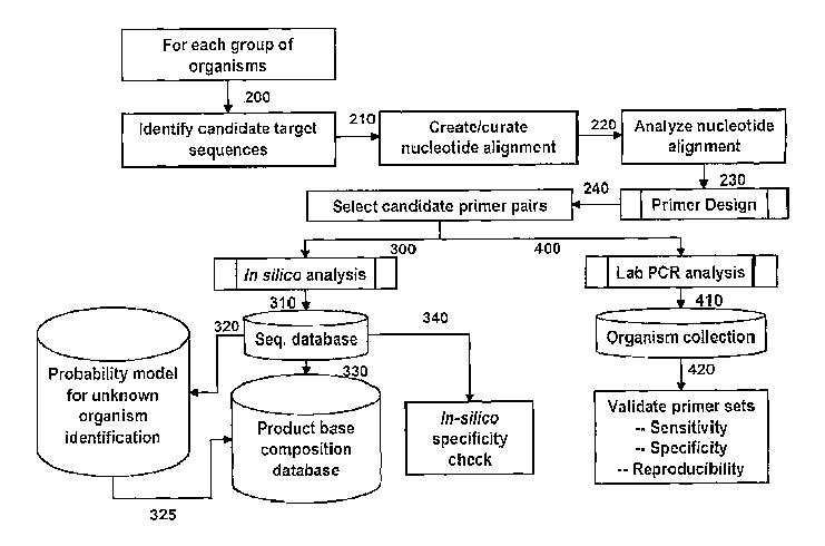
validation process
is outlined in Figure 1. For each group of organisms, candidate target
sequences are identified
(200) from which nucleotide alignments are created (210) and analyzed (220).
Primers are then
designed by selecting appropriate priming regions (230) which then makes
possible the selection
of candidate primer pairs (240). The primer pairs are then subjected to in
silico analysis by
electronic PCR (ePCR) (300) wherein bioagent identifying amplicons are
obtained from
sequence databases such as GenBank or other sequence collections (310) and
checked for
CA 02560946 2009-09-10
-19-
specificity in silico (340). Bioagent identifying amplicons obtained from
GenBank sequences
(310) can also be analyzed by a probability model (320) which predicts the
capability of a given
amplicon to identify unknown bioagents such that the base compositions of
amplicons with
favorable probability scores are then stored in a base composition database
(325). Alternatively,
base compositions of the bioagent identifying amplicons obtained from the
primers and
GenBank sequences can be directly entered into the base composition database
(330). Candidate
primer pairs (240) are validated by in vitro amplification by a method such as
PCR analysis
(400) of nucleic acid from a collection of organisms (410). Amplification
products thus obtained
are analyzed to confirm the sensitivity, specificity and reproducibility of
the primers used to
obtain the amplification products (420).
[0073] Many of the important pathogens, including the organisms of greatest
concern as
biological weapons agents, have been completely sequenced. This effort has
greatly facilitated
the design of primers and probes for the detection of unknown bioagents. The
combination of
broad-range priming with division-wide and drill-down priming has been used
very successfully
in several applications of the technology, including environmental
surveillance for biowarfare
threat agents and clinical sample analysis for medically important pathogens.
[0074] Synthesis of primers is well known and routine in the art. The primers
may be
conveniently and routinely made through the well-known technique of solid
phase synthesis.
Equipment for such synthesis is sold by several vendors including, for
example, Applied
Biosystems (Foster City, CA). Any other means for such synthesis known in the
art may
additionally or alternatively be employed.
[0075] The primers are employed as compositions for use in methods for
identification of viral
bioagents as follows: a primer pair composition is contacted with nucleic acid
(such as, for
example, DNA from a DNA virus, or DNA reverse transcribed from the RNA of an
RNA virus)
of an unknown viral bioagent. The nucleic acid is then amplified by a nucleic
acid amplification
technique, such as PCR for example, to obtain an amplification product that
represents a
bioagent identifying amplicon. The molecular mass of each strand of the double-
stranded
amplification product is determined by a molecular mass measurement technique
such as mass
spectrometry for example, wherein the two strands of the double-stranded
amplification product
are separated during the ionization process. In some embodiments, the mass
spectrometry is
electrospray Fourier transform ion cyclotron resonance mass spectrometry (ESI-
FTICR-MS) or
CA 02560946 2006-09-21
WO 2005/092059 PCT/US2005/009557
20 -
electrospray time of flight mass spectrometry (ESI-TOF-MS). A list of possible
base
compositions can be generated for the molecular mass value obtained for each
strand and the
choice of the correct base composition from the list is facilitated by
matching the base
composition of one strand with a complementary base composition of the other
strand. The
molecular mass or base composition thus determined is then compared with a
database of
molecular masses or base compositions of analogous bioagent identifying
amplicons for known
viral bioagents. A match between the molecular mass or base composition of the
amplification
product and the molecular mass or base composition of an analogous bioagent
identifying
amplicon for a known viral bioagent indicates the identity of the unknown
bioagent. In some
embodiments, the primer pair used is one of the primer pairs of Tables 4-7. In
some
embodiments, the method is repeated using a different primer pair to resolve
possible
ambiguities in the identification process or to improve the confidence level
for the identification
assignment.
[0076] In some embodiments, a bioagent identifying amplicon may be produced
using only a
single primer (either the forward or reverse primer of any given primer pair),
provided an
appropriate amplification method is chosen, such as, for example, low
stringency single primer
PCR (LSSP-PCR). Adaptation of this amplification method in order to produce
bioagent
identifying amplicons can be accomplished by one with ordinary skill in the
art without undue
experimentation.
[0077] In some embodiments, the oligonucleotide primers are broad range survey
primers
which hybridize to conserved regions of nucleic acid encoding the RNA-
dependent RNA
polymerase of all (or between 80% and 100%, between 85% and 100%, between 90%
and 100%
or between 95% and 100%) known filoviruses and produce bioagent identifying
amplicons. In
some embodiments, the oligonucleotide primers are broad range survey primers
which hybridize
to conserved regions of nucleic acid encoding nucleocapsid of all (or between
80% and 100%,
between 85% and 100%, between 90% and 100% or between 95% and 100%) known
filoviruses
and produce bioagent identifying amplicons.
[0078] In some embodiments, the oligonucleotide primers are broad range survey
primers
which hybridize to conserved regions of nucleic acid encoding the RNA-
dependent RNA
polymerase (NS5) of all (or between 80% and 100%, between 85% and 100%,
between 90% and
100% or between 95% and 100%) known flaviviruses and produce bioagent
identifying
CA 02560946 2006-09-21
WO 2005/092059 PCT/US2005/009557
-21-
amplicons. In some embodiments, the oligonucleotide primers are broad range
survey primers
which hybridize to conserved regions of nucleic acid encoding the
protease/helicase (NS3) of all
(or between 80% and 100%, between 85% and 100%, between 90% and 100% or
between 95%
and 100%) known flaviviruses and produce bioagent identifying amplicons.
[0079] In some embodiments, the oligonucleotide primers are broad range survey
primers
which hybridize to conserved regions of nucleic acid encoding the RNA-
dependent RNA
polymerase of all (or between 80% and 100%, between 85% and 100%, between 90%
and 100%
or between 95% and 100%) known hantaviruses and produce bioagent identifying
amplicons. In
some embodiments, the oligonucleotide primers are broad range survey primers
which hybridize
to conserved regions of nucleic acid encoding nucleocapsid of all (or between
80% and 100%,
between 85% and 100%, between 90% and 100% or between 95% and 100%) known
hantaviruses and produce bioagent identifying amplicons.
[0080] In some embodiments, the oligonucleotide primers are broad range survey
primers
which hybridize to conserved regions of nucleic acid encoding the RNA-
dependent RNA
polymerase of all (or between 80% and 100%, between 85% and 100%, between 90%
and 100%
or between 95% and 100%) known phleboviruses and produce bioagent identifying
amplicons.
[0081] In some embodiments, the oligonucleotide primers are broad range survey
primers
which hybridize to conserved regions of nucleic acid encoding nucleocapsid of
all (or between
80% and 100%, between 85% and 100%, between 90% and 100% or between 95% and
100%)
known nairoviruses and produce bioagent identifying amplicons.
[0082] In some embodiments, the oligonucleotide primers are broad range survey
primers
which hybridize to conserved regions of nucleic acid encoding the RNA-
dependent RNA
polymerase (L) of all (or between 80% and 100%, between 85% and 100%, between
90% and
100% or between 95% and 100%) known arenaviruses and produce bioagent
identifying
amplicons. In some embodiments, the oligonucleotide primers are broad range
survey primers
which hybridize to conserved regions of nucleic acid encoding nucleocapsid
(NP) of all (or
between 80% and 100%, between 85% and 100%, between 90% and 100% or between
95% and
100%) known arenaviruses and produce bioagent identifying amplicons.
CA 02560946 2006-09-21
WO 2005/092059 PCT/US2005/009557
-22-
[0083] As used herein, the term broad range survey primers refers to primers
that bind to
nucleic acid encoding genes essential to filovirus, flavivirus, hantavirus,
phlebovirus, nairovirus
or arenavirus replication (e.g., for example, RNA-dependent RNA polymerase or
nucleocapsid)
of all (or between 80% and 100%, between 85% and 100%, between 90% and 100% or
between
95% and 100%) known species of filovirus, flavivirus, hantavirus, phlebovirus,
nairovirus or
arenavirus.
[0084] In some embodiments, the broad range survey primer pairs comprise
oligonucleotides
ranging in length from 13-35 nucleobases, each of which have from 70% to 100%
sequence
identity with primer pair number 853, which corresponds to SEQ ID NOs:
129:164. In some
embodiments, the broad range survey primer pairs comprise oligonucleotides
ranging in length
from 13-35 nucleobases, each of which have from 70% to 100% sequence identity
with primer
pair number 858, which corresponds to SEQ ID NOs: 124:159. In some
embodiments, the broad
range survey primer pairs comprise oligonucleotides ranging in length from 13-
35 nucleobases,
each of which have from 70% to 100% sequence identity with primer pair number
856, which
corresponds to SEQ ID NOs: 134:169. In some embodiments, the broad range
survey primer
pairs comprise oligonucleotides ranging in length from 13-35 nucleobases, each
of which have
from 70% to 100% sequence identity with primer pair number 864, which
corresponds to SEQ
ID NOs: 138:174.
[0085] In some cases, the molecular mass or base composition of a viral
bioagent identifying
amplicon defined by a broad range survey primer pair does not provide enough
resolution to
unambiguously identify a viral bioagent at the species level. These cases
benefit from further
analysis of one or more viral bioagent identifying amplicons generated from at
least one
additional broad range survey primer pair or from at least one additional
division-wide primer
pair. The employment of more than one bioagent identifying amplicon for
identification of a
bioagent is herein referred to as triangulation identification.
[0086] In other embodiments, the oligonucleotide primers are division-wide
primers which
hybridize to nucleic acid encoding genes of species within a genus of viruses.
In other
embodiments, the oligonucleotide primers are drill-down primers which enable
the identification
of sub-species characteristics. Drill down primers provide the functionality
of producing
bioagent identifying amplicons for drill-down analyses such as strain typing
when contacted with
nucleic acid under amplification conditions. Identification of such sub-
species characteristics is
CA 02560946 2006-09-21
WO 2005/092059 PCT/US2005/009557
-23-
often critical for determining proper clinical treatment of viral infections.
In some embodiments,
sub-species characteristics are identified using only broad range survey
primers and division-
wide and drill-down primers are not used.
[0087] In some embodiments, the primers used for amplification hybridize to
and amplify
genomic DNA, DNA of bacterial plasmids, DNA of DNA viruses or DNA reverse
transcribed
from RNA of an RNA virus.
[0088] In some embodiments, the primers used for amplification hybridize
directly to viral
RNA and act as reverse transcription primers for obtaining DNA from direct
amplification of
viral RNA. Methods of amplifying RNA using reverse transcriptase are well
known to those with
ordinary skill in the art and can be routinely established without undue
experimentation.
[0089] One with ordinary skill in the art of design of amplification primers
will recognize that
a given primer need not hybridize with 100% complementarity in order to
effectively prime the
synthesis of a complementary nucleic acid strand in an amplification reaction.
Moreover, a
primer may hybridize over one or more segments such that intervening or
adjacent segments are
not involved in the hybridization event. (e.g., for example, a loop structure
or a hairpin
structure). The primers of the present invention may comprise at least 70%, at
least 75%, at least
80%, at least 85%, at least 90%, at least 95% or at least 99% sequence
identity with any of the
primers listed in Tables 4-7. Thus, in some embodiments of the present
invention, an extent of
variation of 70% to 100%, or any range therewithin, of the sequence identity
is possible relative
to the specific primer sequences disclosed herein. Determination of sequence
identity is
described in the following example: a primer 20 nucleobases in length which is
identical to
another 20 nucleobase primer having two non-identical residues has 18 of 20
identical residues
(18/20 = 0.9 or 90% sequence identity). In another example, a primer 15
nucleobases in length
having all residues identical to a 15 nucleobase segment of primer 20
nucleobases in length
would have 15/20 = 0.75 or 75% sequence identity with the 20 nucleobase
primer.
[0090] Percent homology, sequence identity or complementarity, can be
determined by, for
example, the Gap program (Wisconsin Sequence Analysis Package, Version 8 for
Unix, Genetics
Computer Group, University Research Park, Madison WI), using default settings,
which uses the
algorithm of Smith and Waterman (Adv. Appl. Math., 1981, 2, 482-489). In some
embodiments,
complementarity of primers with respect to the conserved priming regions of
viral nucleic acid,
CA 02560946 2006-09-21
WO 2005/092059 PCT/US2005/009557
-24-
is between about 70% and about 80%. In other embodiments, homology, sequence
identity or
complementarity, is between about 80% and about 90%. In yet other embodiments,
homology,
sequence identity or complementarity, is at least 90%, at least 92%, at least
94%, at least 95%, at
least 96%, at least 97%, at least 98%, at least 99% or is 100%.
[0091] In some embodiments, the primers described herein comprise at least
70%, at least
75%, at least 80%, at least 85%, at least 90%, at least 92%, at least 94%, at
least 95%, at least
96%, at least 98%, or at least 99%, or 100% (or any range therewithin)
sequence identity with
the primer sequences specifically disclosed herein. Thus, for example, a
primer may have
between 70% and 100%, between 75% and 100%, between 80% and 100%, and between
95%
and 100% sequence identity with SEQ ID NO: 129. Likewise, a primer may have
similar
sequence identity with any other primer whose nucleotide sequence is disclosed
herein.
[0092] One with ordinary skill is able to calculate percent sequence identity
or percent
sequence homology and able to determine, without undue experimentation, the
effects of
variation of primer sequence identity on the function of the primer in its
role in priming synthesis
of a complementary strand of nucleic acid for production of an amplification
product of a
corresponding bioagent identifying amplicon.
[0093] In some embodiments of the present invention, the oligonucleotide
primers are 13 to 35
nucleobases in length (13 to 35 linked nucleotide residues). These embodiments
comprise
oligonucleotide primers 13, 14, 15, 16, 17, 18, 19, 20, 21, 22, 23, 24, 25,
26, 27, 28, 29, 30, 31,
32, 33, 34 or 35 nucleobases in length, or any range therewithin.
[0094] In some embodiments, any given primer comprises a modification
comprising the
addition of a non-templated T residue to the 5' end of the primer (i.e., the
added T residue does
not necessarily hybridize to the nucleic acid being amplified). The addition
of a non-templated T
residue has an effect of minimizing the addition of non-templated A residues
as a result of the
non-specific enzyme activity of Taq polymerise (Magnuson et al.,
Biotechniques, 1996, 21, 700-
709), an occurrence which may lead to ambiguous results arising from molecular
mass analysis.
[0095] In some embodiments of the present invention, primers may contain one
or more
universal bases. Because any variation (due to codon wobble in the 3rd
position) in the conserved
regions among species is likely to occur in the third position of a DNA (or
RNA) triplet,
CA 02560946 2011-10-21
-25-
oligonucleotide primers can be designed such that the nucleotide corresponding
to this position is
a base which can bind to more than one nucleotide, referred to herein as a
"universal
xiucleobase." For example, under this "wobble" pairing, inosine (I) binds to
U, C or A; guano
(G) binds to U or C, and uxidine (U) binds to U or C. Other examples of
universal nucleobases
include nitroindoles such as 5-nitroindole or 3-nitropyrrole (Loakes et al.,
Nucleosides and
Nucleotides, 1995, 14, 1001-1003), the degenerate nucleotides d.P or dK (Hill
et a1.), an acyclic
nucleoside analog containing 5-nitroindazole (Van Aerschot at al,, Nucleosides
and Nucleotides,
1995, 14, 1053-1056) or the purine analog 1-(2-deoxy-~-))-ribof1 ranosyl)-
imidazole-4-
earboxana_ide (Sala at al., Nucl. Acids Res., 1996, 24, 3302-3306).
{.0096) In some embodixaxents, to compensate for the somewhat weaker binding
by the wobble
base, the oligorlucleatid.e primers are designed such that the first and
second positions of each
triplet are occupied by nucleotide analogs which bind with greater affinity
than the unmodified
nucleotide. Examples of these analogs include, but are not limited to, 2,6-
diamiuopurine which
binds to thymine, 5-propynyluracil which binds to adenine and 5
propynylcytosine and
plaenoxazines', including 0-clamp, which binds to G. Propynylated pyrimidines
are described in
U.S. Patent Nos, 5,645,985, 5,830,653 and 5,484,908, each of which is commonly
owned and
incorporated herein by reference in its entirety. lropynylated primers are
described in U.S Pre-
Grant Publication No. 2003-0170682, which is also commonly owned and
incorporated herein
by reference in its entirety. Phexxoxazines are described in U.S. Patent Nos.
5,502,177, 5,763,588,
and 6,005,096. G-olarnps are
described in U.S. Patent Nos. 6,007,992 and 6,025,183.
100971 In some estab6din-1eiits, to enable broad priming of rapidly evolving
RNA viruses,.
primer hybridization is enhanced using primers and probes containing 5-
propynyl deoxy-cytidine
and deoxyrthyxriidiize Nucleotides. These modified primers and probes offer
increased affinity
and base pairing selectivity.
[00981 In some embodiments, non-template primer tags are used to increase the
melting
temperature (T1,) of a primer-template duplex in order to improve
amplification efficiency. A.
non-template tag is at least three consecutive A or T nucleotide residues on a
primer which are
not complementary to the template, In any given non-template tag, A can be
replaced by C or G
and T can also be replaced by C or G. Although Watson-Crick hybridization is
not expected to
CA 02560946 2006-09-21
WO 2005/092059 PCT/US2005/009557
-26-
occur for a non-template tag relative to the template, the extra hydrogen bond
in a G-C pair
relative to an A-T pair confers increased stability of the primer-template
duplex and improves
amplification efficiency for subsequent cycles of amplification when the
primers hybridize to
strands synthesized in previous cycles.
[0099] In other embodiments, propynylated tags may be used in a manner similar
to that of the
non-template tag, wherein two or more 5-propynylcytidine or 5-propynyluridine
residues replace
template matching residues on a primer. In other embodiments, a primer
contains a modified
internucleoside linkage such as a phosphorothioate linkage, for example.
[0100] In some embodiments, the primers contain mass-modifying tags. Reducing
the total
number of possible base compositions of a nucleic acid of specific molecular
weight provides a
means of avoiding a persistent source of ambiguity in determination of base
composition of
amplification products. Addition of mass-modifying tags to certain nucleobases
of a given
primer will result in simplification of de novo determination of base
composition of a given
bioagent identifying amplicon from its molecular mass.
[0101] In some embodiments of the present invention, the mass modified
nucleobase
comprises one or more of the following: for example, 7-deaza-2'-deoxyadenosine-
5-triphosphate,
5-iodo-2'-deoxyuridine-5'-triphosphate, 5-bromo-2'-deoxyuridine-5'-
triphosphate, 5-bromo-2'-
deoxycytidine-5'-triphosphate, 5-iodo-2'-deoxycytidine-5'-triphosphate, 5-
hydroxy-2'-
deoxyuridine-5'-triphosphate, 4-thiothymidine-5'-triphosphate, 5-aza-2'-
deoxyuridine-5'-
triphosphate, 5-fluoro-2'-deoxyuridine-5'-triphosphate, 06-methyl-2'-
deoxyguanosine-5'-
triphosphate, N2-methyl-2'-deoxyguanosine-5'-triphosphate, 8-oxo-2'-
deoxyguanosine-5'-
triphosphate or thiothymidine-5'-triphosphate. In some embodiments, the mass-
modified
nucleobase comprises 15N or 13C or both 15N and 13C.
[0102] In some cases, a molecular mass of a given bioagent identifying
amplicon alone does
not provide enough resolution to unambiguously identify a given bioagent. The
employment of
more than one bioagent identifying amplicon for identification of a bioagent
is herein referred to
as triangulation identification. Triangulation identification is pursued by
analyzing a plurality of
bioagent identifying amplicons selected within multiple core genes. This
process is used to
reduce false negative and false positive signals, and enable reconstruction of
the origin of hybrid
or otherwise engineered bioagents. For example, identification of the three
part toxin genes
CA 02560946 2006-09-21
WO 2005/092059 PCT/US2005/009557
-27-
typical of B. anthracis (Bowen et al., J. Appl. Microbiol., 1999, 87, 270-278)
in the absence of
the expected signatures from the B. anthracis genome would suggest a genetic
engineering
event.
[0103] In some embodiments, the triangulation identification process can be
pursued by
characterization of bioagent identifying amplicons in a massively parallel
fashion using the
polymerase chain reaction (PCR), such as multiplex PCR where multiple primers
are employed
in the same amplification reaction mixture, or PCR in multi-well plate format
wherein a different
and unique pair of primers is used in multiple wells containing otherwise
identical reaction
mixtures. Such multiplex and multi-well PCR methods are well known to those
with ordinary
skill in the arts of rapid throughput amplification of nucleic acids.
[0104] In some embodiments, the molecular mass of a given bioagent identifying
amplicon is
determined by mass spectrometry. Mass spectrometry has several advantages, not
the least of
which is high bandwidth characterized by the ability to separate (and isolate)
many molecular
peaks across a broad range of mass to charge ratio (m/z). Thus mass
spectrometry is intrinsically
a parallel detection scheme without the need for radioactive or fluorescent
labels, since every
amplification product is identified by its molecular mass. The current state
of the art in mass
spectrometry is such that less than femtomole quantities of material can be
readily analyzed to
afford information about the molecular contents of the sample. An accurate
assessment of the
molecular mass of the material can be quickly obtained, irrespective of
whether the molecular
weight of the sample is several hundred, or in excess of one hundred thousand
atomic mass units
(amu) or Daltons.
[0105] In some embodiments, intact molecular ions are generated from
amplification products
using one of a variety of ionization techniques to convert the sample to gas
phase. These
ionization methods include, but are not limited to, electrospray ionization
(ES), matrix-assisted
laser desorption ionization (MALDI) and fast atom bombardment (FAB). Upon
ionization,
several peaks are observed from one sample due to the formation of ions with
different charges.
Averaging the multiple readings of molecular mass obtained from a single mass
spectrum affords
an estimate of molecular mass of the bioagent identifying amplicon.
Electrospray ionization
mass spectrometry (ESI-MS) is particularly useful for very high molecular
weight polymers such
as proteins and nucleic acids having molecular weights greater than 10 kDa,
since it yields a
CA 02560946 2006-09-21
WO 2005/092059 PCT/US2005/009557
-28-
distribution of multiply-charged molecules of the sample without causing a
significant amount of
fragmentation.
[0106] The mass detectors used in the methods of the present invention
include, but are not
limited to, Fourier transform ion cyclotron resonance mass spectrometry (FT-
ICR-MS), time of
flight (TOF), ion trap, quadrupole, magnetic sector, Q-TOF, and triple
quadrupole.
[0107] Although the molecular mass of amplification products obtained using
intelligent
primers provides a means for identification of bioagents, conversion of
molecular mass data to a
base composition signature is useful for certain analyses. As used herein, a
base composition
signature (BCS) is the exact base composition determined from the molecular
mass of a bioagent
identifying amplicon. In one embodiment, a BCS provides an index of a specific
gene in a
specific organism.
[0108] In some embodiments, conversion of molecular mass data to a base
composition is
useful for certain analyses. As used herein, a base composition is the exact
number of each
nucleobase (A, T, C and G).
[0109] RNA viruses depend on error-prone polymerases for replication and
therefore their
nucleotide sequences (and resultant base compositions) drift over time within
the functional
constraints allowed by selection pressure. Base composition probability
distribution of a viral
species or group represents a probabilistic distribution of the above
variation in the A, C, G and
T base composition space and can be derived by analyzing base compositions of
all known
isolates of that particular species.
[0110] In some embodiments, assignment of base compositions to experimentally
determined
molecular masses is accomplished using base composition probability clouds.
Base
compositions, like sequences, vary slightly from isolate to isolate within
species. It is possible to
manage this diversity by building base composition probability clouds around
the composition
constraints for each species. This permits identification of organisms in a
fashion similar to
sequence analysis. A pseudo four-dimensional plot can be used to visualize the
concept of base
composition probability clouds. Optimal primer design requires optimal choice
of bioagent
identifying amplicons and maximizes the separation between the base
composition signatures of
individual bioagents. Areas where clouds overlap indicate regions that may
result in a
CA 02560946 2006-09-21
WO 2005/092059 PCT/US2005/009557
-29-
misclassification, a problem which is overcome by a triangulation
identification process using
bioagent identifying amplicons not affected by overlap of base composition
probability clouds.
[0111] In some embodiments, base composition probability clouds provide the
means for
screening potential primer pairs in order to avoid potential
misclassifications of base
compositions. In other embodiments, base composition probability clouds
provide the means for
predicting the identity of a bioagent whose assigned base composition was not
previously
observed and/or indexed in a bioagent identifying amplicon base composition
database due to
evolutionary transitions in its nucleic acid sequence. Thus, in contrast to
probe-based techniques,
mass spectrometry determination of base composition does not require prior
knowledge of the
composition or sequence in order to make the measurement.
[0112] The present invention provides bioagent classifying information similar
to DNA
sequencing and phylogenetic analysis at a level sufficient to identify a given
bioagent.
Furthermore, the process of determination of a previously unknown base
composition for a given
bioagent (for example, in a case where sequence information is unavailable)
has downstream
utility by providing additional bioagent indexing information with which to
populate base
composition databases. The process of future bioagent identification is thus
greatly improved as
more base composition indexes become available in base composition databases.
[0113] Existing nucleic acid-based tests for bioagent detection are primarily
based upon
amplification methods using primer and probes designed to detect specific
organisms. Because
prior knowledge of nucleic acid sequence information is required to develop
these probe-based
tests they cannot be used to identify unanticipated, newly emergent, or
previously unknown
infections organisms. Thus, the discovery of new bioagents still relies
largely on traditional
culture methods and microscopy.
[0114] Methods of the present invention, however, allow rapid identification
of new bioagent
species without the need for prior knowledge of nucleotide sequence. This is
achieved by
applying a mathematical and/or probabilistic model for sequence variation
developed based on
known bioagent amplicon base composition (the "training set" of data) and
matching the
unknown bioagent data ("test data") to the model.
CA 02560946 2006-09-21
WO 2005/092059 PCT/US2005/009557
-30-
[0115] For unambiguous detection and identification of bioagents, it would be
ideal if every
isolate of a given species of bioagent (E. coli, for example) had exactly the
same base count in
any particular amplified region. However, due to naturally occurring mutations
and/or
deliberately engineered changes, isolates of any species might have some
variation in the base
count of a particular region. Because of naturally occurring variation and
because engineered
threat bioagents may differ slightly in particular regions from their
naturally occurring
counterparts, it is useful to "blur" the expected base count for a given
species to allow for this
variation so that the system does not miss detections. The more the expected
base count is
blurred, the less likely it is that a particular species will escape
detection; however, such blurring
will cause more overlap between the expected base counts of different species,
contributing to
misclassifications.
[0116] To solve this problem, expected base counts can be blurred according to
the natural
principles of biological mutations, customizing the specific blurring to the
biological constraints
of each amplified region. Each amplified region of a particular bioagent is
constrained in some
fashion by its biological purpose (i.e., RNA structure, protein coding, etc.).
For example, protein
coding regions are constrained by amino acid coding considerations, whereas a
ribosome is
mostly constrained by base pairing in stems and sequence constraints in
unpaired loop regions.
Moreover, different regions of the ribosome might have significant preferences
that differ from
each other.
[0117] One embodiment of application of the cloud algorithm is described in
Example 1. By
collecting all likely species amplicons from a primer set and enlarging the
set to include all
biologically likely variant amplicons using the cloud algorithm, a suitable
cluster region of base
count space is defined for a particular species of bioagent. The regions of
base count space in
which groups of related species are clustered are referred to as
"bioclusters."
[0118] When a biocluster is constructed, every base count in the biocluster
region is assigned a
percentage probability that a species variant will occur at that base count.
To form a probability
density distribution of the species over the biocluster region, the entire
biocluster probability
values are normalized to one. Thus, if a particular species is present in a
sample, the probability
of the species biocluster integrated over all of base count space is equal to
one.
CA 02560946 2006-09-21
WO 2005/092059 PCT/US2005/009557
-31-
[0119] At this point in the ranking procedure, proposed target species to be
detected are taken
into account. These generally are the bioagents that are of primary importance
in a particular
detection scenario. For example, if Yersinia pestis (the causative agent of
bubonic and
pneumonic plague) were the target, the Yersinia pestis species biocluster
identified as described
above, would be the "target biocluster." To complete the example, assume that
all other database
species serve as the scenario background. The discrimination metric in this
case is defined as the
sum total of all the biocluster overlap from other species into the Yersinia
pestis biocluster.
[0120] In this example, the Yersiniapestis biocluster overlap is calculated as
follows. A
probability of detection of 99% (PD=0.99) is defined, although this value can
be altered as
needed. The "detection range" is defined as the set of biocluster base counts,
of minimal
number, that encloses 99% of the entire target biocluster. For each additional
bacterial species in
the database, the amount of biocluster probability density that resides in the
base counts in the
defined detection range is calculated and is the effective biocluster overlap
between that
background species and the target species. The sum of the biocluster overlap
over all
background species serves as the metric for measuring the discrimination
ability of a defined
target by a proposed primer set. Mathematically, because the most
discriminating primer sets
will have minimal biocluster overlap, an inverse figure of merit cp is
defined, 1 = i = all
bioclusters i where the sum is taken over the individual biocluster overlap
values [6; from all N
background species bioclusters (i=1, . . . , N ). For example, Figure 2 shows
the inverse figure of
merit cp plotted for a master list of 16 primer sets using Yersinia pestis as
the target biocluster.
Using the inverse figure of merit minimization criteria defined above, the
result is that primer set
number 4 provides the best discrimination of any of the individual primer sets
in the master list.
[0121] This set of discrimination criteria also can be applied to combinations
of primer sets.
The respective four-dimensional base count spaces from each primer set can be
dimensionally
concatenated to form a (4 x N)-dimensional base count space for N primer sets.
Nowhere in the
biocluster definition is it necessary that the biocluster reside in a four-
dimensional space, thus the
biocluster analysis seamlessly adapts to any arbitrary dimensionality. As a
result, a master list of
primer sets can be searched and ranked according to the discrimination of any
combination of
primer sets with any arbitrary number of primer sets making up the
combination.
[0122] Using again the example of Yersinia pestis as the target, improved
discrimination is
achieved through use of an increasing number of primers. For each number of
primers value on
CA 02560946 2006-09-21
WO 2005/092059 PCT/US2005/009557
-32-
the x-axis, the plotted inverse figure of merit value is that obtained from
the most discriminating
group (that group with the minimum figure of merit for that number of primer
sets
simultaneously used for discrimination). The result is that after the best
groups of 3 and 4 primer
sets are found, the inverse figure of merit approaches one and goes no
further. That means that
there is the equivalent of one background species biocluster overlapping into
the target
biocluster. In this example it is the Yersiniapseudotuberculosis species
biocluster, which cannot
be discriminated from Yersinia pestis by any combination of the 16 primer sets
in the example.
Thus, using the "best" 3 or 4 primer sets in the master list, Yersinia pestis
is essentially
discriminated from all other species bioclusters.
[0123] Thus, one the one hand, probability clouds can be used to detect
variants of known
bioagents. On the other hand, this method of the present invention can be used
to
unambiguously determine that an unknown bioagent is not a likely variant of a
known bioagent
and at the same time, classify the bioagent in terms of similarity to the
known bioagents in the
database.
[0124] RNA viruses depend on an error-prone polymerase for replication and
therefore their
nucleotide sequences (and the resultant base compositions) drift over time
within the functional
constraints allowed by selection pressure. Base composition probability
distribution of a viral
species or group represents a probabilistic distribution of the above
variations in the {A, G, C,
and T} base composition space and can be derived by analyzing base
compositions of all known
isolates of that particular species.
[0125] In one embodiment of the invention, a model organism, such as the
positive strand
RNA virus, hepatitis C virus (HCV), can be used to model these sequence
variations. Mutation
probabilities can be derived from the observed variations among, e.g., a
number of HCV
sequences. Table 1 below, lists mutation probabilities that were derived from
the observed
variations among 50 HCV-I b sequences. Six different regions within the genome
of 120
nucleotide (nt) average length, were picked based on priming considerations
and a maximum
amplicon length criterion of -150 nt. Base composition probability
distributions for a species
were determined in two steps. In the first step, mutation probabilities, i.e.,
the probabilities of
occurrence of each type of substitution, insertion, or deletion, were derived
by pairwise
comparisons of all known HCV isolates in each target region, and an estimate
of the maximum
number of mutations that a sequence may undergo were calculated. In the second
step, the
CA 02560946 2006-09-21
WO 2005/092059 PCT/US2005/009557
-33-
mutation probabilities and maxima derived from the model organism were used to
estimate
variations in base compositions for each test species and to calculate
mutation probability
distances (Om) between the species in base composition space, which is
calculated as the negative
base 10 logarithm (-login P) of the cumulative probabilities of all possible
mutations of the A, G
C, and T base counts of one species that would lead to the other.
[0126] There are several approaches to classifying an unknown organism based
on the base
composition of certain amplicons. To illustrate these approaches, the
classification technique for
exemplary primer pairs is shown. The method can be applied to other primer
pairs.
Table 1. Position Independent, Nucleotide Mutation Probabilities Over 6
Training
Sequences For HCV-lb
Mutation Seq. 1 Seq. 2 Seq. 3 Seq. 4 Seq. 5 Seq. 6 All Seq.
A -> A 91.82% 88.42% 91.98% 92.51% 91.08% 89.89% 93.30%
A -> C 1.54% 1.22% 0.56% 2.25% 0.14% 0.61% 0.80%
A -> G 6.28% 9.57% 7.16% 5.08% 8.52% 8.61% 5.59%
A -> T/U 0.36% 0.79% 0.30% 0.15% 0.26% 0.90% 0.30%
A -> 0.00% 0.00% 0.00% 0.00% 0.00% 0.00% 0.00%
C -> A 1.00% 0.64% 0.40% 1.29% 0.10% 0.22% 0.46%
C -> C 89.91% 93.27% 89.89% 93.87% 93.84% 93.87% 94.68%
C -> G 1.26% 0.61% 0.76% 0.13% 0.00% 0.71% 0.37%
C -> T/U 7.83% 5.48% 8.95% 4.71% 6.06% 5.20% 4.49%
C -> 0.00% 0.00% 0.00% 0.00% 0.00% 0.00% 0.00%
G -> A 3.97% 6.93% 3.96% 4.29% 7.10% 2.52% 3.47%
G -> C 1.22% 0.85% 0.60% 0.19% 0.00% 0.57% 0.41%
G -> G 94.41% 91.93% 95.29% 94.96% 92.72% 96.77% 95.93%
G -> T/U 0.41% 0.29% 0.15% 0.56% 0.18% 0.13% 0.19%
G -> 0.00% 0.00% 0.00% 0.00% 0.00% 0.00% 0.00%
T -> A 0.49% 0.77% 0.22% 0.21% 0.22% 0.58% 0.29%
T -> C 16.21% 10.23% 9.61% 11.40% 7.68% 9.17% 7.67%
T -> G 0.88% 0.39% 0.20% 0.93% 0.18% 0.30% 0.30%
T -> T/U 82.42% 88.61% 89.96% 87.46% 91.92% 89.95% 91.75%
T -> 0.00% 0.00% 0.00% 0.00% 0.00% 0.00% 0.00%
-> A 0.00% 0.00% 0.00% 0.00% 0.00% 0.00% 0.00%
-> C 0.00% 0.00% 0.00% 0.00% 0.00% 0.00% 0.00%
-> G 0.00% 0.00% 0.00% 0.00% 0.00% 0.00% 0.00%
-> T/U 0.00% 0.00% 0.00% 0.00% 0.00% 0.00% 0.00%
Total -> 100.00% 0.00% 0.00% 0.00% 0.00% 0.00% 0.00%
[0127] There are several approaches to classifying an unknown organism based
on the base
composition of certain amplicons. To illustrate these approaches, the
classification technique for
exemplary primer pairs is shown. The method can be applied to other primer
pairs.
CA 02560946 2006-09-21
WO 2005/092059 PCT/US2005/009557
-34-
[0128] To develop a pattern classifier, the known base composition counts of
amplicons of
known organisms are used to construct the pattern classifier as a training
set. In one embodiment
of the pattern classifier, for each pattern class a base organism serves as a
central point. For that
pattern class, a distance is calculated from each organism in the training set
to the base organism.
The maximum distance found in this manner defines the class within the pattern
classifier; all
organisms less than the maximum distance to the base organism fall within the
class.
[0129] Once the pattern classifier has been trained the unknown organism can
be classified by
determining the distance between the unknown organism and the base organism
for each pattern.
If the unknown organism falls within the maximum distance determined in the
training process,
the organism is classified as belonging to the same pattern class as the base
organism. If the
unknown organism falls outside the maximum distance, a probability that the
organism belongs
to the class can be derived as a function of the distance from the unknown
organism to the base
organism.
[0130] In an alternate embodiment of the pattern classifier, rather than
identifying a base
organism, a pattern is defined by selecting a centroid, which may not
correspond to an actual
organism, but serves as a center for the pattern class. During the training
process, the centroid
and the maximum distance is determined. Once trained, the classification of an
unknown
organism follows much the same as described above.
[0131] Several criteria for measuring the distance between organisms can be
employed. For a
particular primer-pair, the distance between the base compositions can be
used. That is, if the
base counts are treated as a mathematical vector, the distance between the
vectors is the measure
of distance.
[0132] As an example, the 229 E Human Coronavirus has a base count in the RdRp
target
region of A25,G24,C11,T28 and the SARS Coronavirus has a base count of
A27,G19,C14,T28.
Using the first example of distance (a Euclidean distance), the distance
between them is 6.164.
[0133] An alternative measure of distance is to use the probability of
mutation to derive
distance. There are a number of mutation pathways between two polynucleotide
sequences,
which comprises a series of one or more mutation events. Based on empirical
finding, the
probability of individual mutations is known. Table 1 shows a list of typical
individual
CA 02560946 2006-09-21
WO 2005/092059 PCT/US2005/009557
-35-
mutations with their associated probabilities. The probability of a specific
mutation pathway is
the product of the probabilities of the individual mutations. One method of
defining distance is
to take the sum of all probabilities of all mutations pathways, P. The
mutational distance
between the two polynucleotide sequences can be defiried as -loglo P. In the
above example, the
distance between the 229E Human Coronavirus and the SARS coronavirus is 8.8.
It should be
noted that since longer mutation pathways are less like; ly, only certain
mutations are needed to
get from 229E to SARS, and thus the longer pathways can be discarded.
[0134] Figure 3 is a graph showing the base compositions of the 229E Human
Coronavirus,
OC43 Human Coronavirus and the SARS Coronavirus. In this graph, the A, G, and
C base
counts are plotted on the axes and the T base count is represented by using
rotation.
[0135] Figure 4 shows a number of animal coronaviTus species. The branches on
the tree
represent the phylogenetic relationship between the various taxons. For each
taxonomic
grouping, an oval represents the maximal distance between any two members of
the group
represented by A. next to the oval. For example, the bovine isolates (BCoV-
Quebec and BCov-
Lun) are clustered together (Am < 2.0), and are closer t each other than to
their nearest neighbor
on the phylogenetic tree, HCoV-OC43. The bovine and the OC43 species form a
closely related
cluster with a relatively high probability of misclassification ((Am <4.5).
Similarly, the murine
and rat coronavirus isolates are closely related species that can not be
distinguished from each
other using just two target regions (Am < .9), yet the ro dent viruses are
easily distinguished from
the bovine/OC43 group (A,,, < 6.8). Similarly, many of the group 1 animal
coronaviruses (CCoV,
FCoV, TGEV) clustered together and were very close to each other in mutation
and base
composition distance. These, therefore, could potentially be misclassified at
the species level
(Am < 4.7). This is consistent with previous reports that suggest that CCoV
are serologically and
genetically related to other group 1 animal coronaviruses. However, this group
was clearly
resolved from other members of group 1 coronaviruses such as 229E and PEDV (Am
<11.6). In
contrast to the group 1 and group 2 species clusters, the two target regions
chosen here did not
cluster the group 3 species together. The three known isolates of avian
coronaviruses were as far
away from each other as they were from members of group 1 coronaviruses.
Overall, the
mutation-distance analysis suggests that the previously known members of group
2
coronaviruses represent a clearly delineated group, well resolved from groups
1 and 3. In
contrast, no clear delineation between groups 1 and 3 'svas observed.
CA 02560946 2006-09-21
WO 2005/092059 PCT/US2005/009557
-36-
[0136] Further refinement to the classification can be made by assigning a
match probability
of an unknown for each pattern class by calculating the distance to each
pattern class. By
applying additional pattern classifiers based on other primer pairs, the
ability to resolve
unknowns is enhanced. In the example described above, it would be difficult to
distinguish an
unknown in group 1 from group 3 for the given primer pair. Applying the
pattern classifier with
other primer pairs may yield a greater distance between group 1 and group 3
coronaviruses. This
triangulation approach is described further below.
[0137] In alternate embodiments of the mutational probability model, a
centroid is not chosen
and restrictions among strains were compared to one another. Using best
estimates of the
phylogenetic tree, only descendants were compared to their direct forebears,
for a direct estimate
of a mutational probability. This comparison had the effect of reducing the
magnitude of the
mutation probabilities.
[0138] Because it is known that DNA triplets code for a single amino acid, in
some
embodiments, for primer regions that are in a protein-coding region of the
sequence, the
mutational probabilities are determined in a position-dependent way, so that
the 20 types of
mutations (12 substitutions, 4 deletions, and 4 insertions) are now expanded
to a set of 60 (20
types x 3 positions). It is well known that the first position of a triplet is
highly conserved, while
the third position is the least conserved (and it is referred to as a wobble
position because of this)
and this is reflected in the different mutation probabilities per position.
[0139] In other embodiments, the mutational probability model incorporates
both the
restrictions among strains and position dependence of a given nucleobase
within a triplet.
In one embodiment of the invention, a polytope pattern classifier is used to
classify test or
unknown organism according to its amplicon base composition. The polytope
pattern classifier
of the present invention defines the bounds of a pattern class by a convex
polytope. The
polytope pattern classifier is trained by defining a minimal polytope which
contains all the
samples in the training set.
[0140] Generally, a polytope can be expressed by a system of linear
inequalities. Data
supplied to the pattern classifier are typically expressed as an n-dimensional
vector.
Accordingly, an n-dimensional polytope can be expressed as a system of
inequalities of the form:
alxl+a2x2+a3x3+... +axn<C
CA 02560946 2006-09-21
WO 2005/092059 PCT/US2005/009557
-37-
and of the form,
D< blxl+b2x2+b3x3+... +bõx,,.
According to one embodiment of the present invention, the components of the
data vectors are
integers. Thus, the polytopes can be reduced to a system of linear
inequalities of the following
form,
D < alxI + a2x2 + a3x3 + ... + a,ax7z < C, where each al is either 0 or 1.
To define a minimal polytope, all inequalities of the form equation shown
above can be used for
all combinations of a;. During the training process the constants C and D are
determined for
each inequality.
[0141] In certain aspects of the invention, a density is defined for each
polytope by taking the
total number of samples in the training set residing in the polytope and
dividing by the total
volume of the polytope. Once the polytopes are calculated for each pattern
class identified in the
training set, the polytope pattern classifier is trained and can be applied to
test or unknown data.
In classifying an unknown represented by a data vector, the distance to each
pattern class is
calculated. A point density of the data vector to a polytope is defined to be
the density of the
polytope multiplied by a decay factor which is a function of distance of the
data vector to the
polytope. A match probability to each of the classes is calculated based on
the point density. In
one embodiment of the invention, for example, the match probability can be the
normalized
average of all point densities for that particular data sample.
[0142] It should be noted that the measure of volume and distance described in
the density and
point density calculations need not be standard Euclidean-based measures of
distance and
volume. For example, if the data vectors have integer components, the volume
of a polytope can
be defined as a lattice volume that is the number of integer lattice points
within a given polytope.
Similarly, the distance from a point to a polytope can be defined as a lattice
distance that is the
minimum number of lattice points traversed between a point and any point
within the polytope.
[0143] Figure 5A is a flow chart illustrating a method of training an
embodiment of a polytope
pattern classifier. At step 1202, a training sample is received from a
training set. Associated with
each training sample is the pattern class it is a member of. At step 1204, the
pattern class is
determined. At step 1206, if necessary that pattern class' polytope is
modified so as to
incorporate the training sample. If the training sample lies within the
current version of the
pattern class' polytope, no modification is required. This modification
typically takes the form of
CA 02560946 2006-09-21
WO 2005/092059 PCT/US2005/009557
-38-
comparing the training sample to the existing inequalities that defined the
polytope. If the
training sample falls outside an inequality, the inequality is modified to
incorporated the training
sample. In the modification process, the inequality is modified to expand the
polytope as little as
possible. At step 1208, the process iterates to the next training sample, if
any remain. Otherwise,
the training is complete.
[0144] One should note that though the flowchart describes an iteration
through the training
samples and in polytope modification, an iteration through the inequalities
which defined the
polytope, the order of iteration could be equivalently transposed. That is,
rather than considering
each training sample first, each inequality is considered. For each
inequality, the training sample
is compared against the inequality and the inequality is modified to
accommodate the training
sample if necessary. Then the iteration can continue to the next inequality.
[0145] Figure 5B is a flow chart illustrating the method of identifying an
unknown sample
using an embodiment of a trained polytope pattern classifier. At step 1222, an
unknown sample
is received by the polytope pattern classifier. At step 1224, a pattern class
is selected. At step
1226, the distance between the pattern class' polytope and the unknown sample
is calculated.
Based on the distance, at step 1228, the point density of the unknown sample
with respect to the
pattern class is calculated. At step 1230, the process repeats for the next
pattern class. When all
point densities with respect to all the pattern classes are calculated, a
match probability is
generated by normalizing the point densities at step 1232.
[0146] To simplify the complexity of higher dimensional polytope pattern
classifiers, a
plurality of lower dimensional polytope pattern classifiers can be used.
According to this
embodiment of the invention, all data including unknowns and the data in the
training set, are
divided into a plurality of subspaces having the lower dimension. A polytope
pattern classifier is
associated with each subspace. Each polytope pattern classifier is trained on
the subset of the
training set that resides within the associated subspace. Once trained, the
one of the plurality of
subspaces to which an unknown belongs is first applied, then the polytope
pattern classifier
associated with that subspace is applied to the data.
[0147] In certain aspects of the invention, subspaces are defined by the
length of the data, e.g.
the amplicon length. When the components of the data vectors are integers, the
subspaces
determined in this manner can yield a finite if not small number of subspaces.
CA 02560946 2006-09-21
WO 2005/092059 PCT/US2005/009557
-39-
[0148] In an alternative embodiment of the polytope pattern classifier,
contributions from all
polytopes are considered, regardless of which subspace the unknown data
belongs to. For
example, the point density of an unknown to a given pattern class can be a
function of the
distance of the unknown data vector to every polytope associated with a given
pattern class. In
order to simplify this calculation, the distance can be broken into two
components, the distance
between the unknown data vector to the subspace containing the polytope, and
the distance
between a projected data vector, i.e., the data vector when projected onto the
subspace
containing the polytope, and the polytope. These two components of the data
vector can be into
different decay factors.
[0149] Figure 6A is a flow chart illustrating the method of training an
embodiment of a
polytope pattern classifier of a lower dimension when the sample space is
reduced in dimension
by imposing a constraint. At step 1302, a training sample is received from the
training set. The
constraint is applied to determine which subspace the training sample belongs
to at step 1304.
The training sample is placed into a training subset corresponding to that
subspace, at step 1306.
At step 1308, the process is made to repeat, until all training samples have
been grouped into
corresponding subspaces. Then at step 1310, a subspace is selected along with
the corresponding
subset of the training samples. At step 1312 the pattern classifier
corresponding to that subspace
is trained. It can be trained using a method like that described in Figure 6A.
At step 1314, the
process is made to repeat, until all subspaces derived from the constraint
have fully trained
pattern classifiers. It should be noted that in another method of training the
order can be changed.
For example, after the subspace of a training sample is identified, it can be
used to train the
corresponding pattern classifier immediately rather than waiting until all
training samples are
sorted. The flow chart is intended to clearly describe an example of a
training method.
[0150] Figure 6B is a flow chart illustrating a method of identifying a
unknown sample in a
manner similar to that of Figure 5B. At step 1332, an unknown sample is
received by the pattern
classification system. At step 1334, the constraint is applied and the
subspace to which the
sample belongs is determined. Steps 1336, 1338, 1340, 1342, and 1344 apply a
similar same
pattern identification algorithm to that described in steps 1224, 1226, 1228,
1230, and 1232
respectively, where the polytope associated with each pattern class used is
the polytope
contained in the subspace to which the sample belongs. It should be noted that
depending on the
members of the various pattern classes, a pattern class can have more than one
polytope, but in
different subspaces.
CA 02560946 2006-09-21
WO 2005/092059 PCT/US2005/009557
-40-
[0151] The method described in Figure 6B does not account for the polytopes
for a given class
in subspaces other than that to which the sample belongs. Figure 6B is a flow
chart illustrating
an alternative method of identifying an unknown sample using polytope
classifiers trained by a
process such as that described in Figure 6A. At step 1352, an unknown sample
is received by
the pattern classification system. At step 1354, a pattern class is selected.
At step 1356, a
subspace is selected which contains one of the pattern class' polytopes. If no
polytope for that
pattern class exists in that subspace, another subspace can be selected. At
step 1358, a gap
distance is calculated, i.e. the distance between the unknown sample and the
selected subspace.
At step 1360, the mutation distance is calculated, i.e. the distance between a
"projection" of the
unknown sample and the pattern class' polytopes. In practice, the distance is
actually the
minimum distance between all possible minimal insertions (or deletions)
sufficient to mutate the
sample to the given subspace. At step 1362, the point density of the unknown
sample with
respect to the pattern class' polytope is calculated as a function of either
the gap distance, the
mutation distance or both. At step 1364, the process is made to repeat until
all subspaces with the
specific pattern class' polytopes have be selected. Once all the point
densities have been
calculated, at step 1366, the point probabilities are all combined to produce
a composite point
probability for the unknown sample with respect to the entire pattern class.
At step 1368, the
process is made to repeat until all pattern classes have been selected. When
all point densities
with respect to all the pattern classes are calculated, a match probability is
generated by
normalizing the point densities at step 1370.
[0152] Specifically, as applied to the classification of an unknown organism,
the polytope
pattern classifier is applied to data vectors representing the amplicon base
composition of
organisms. The polytope pattern classifiers are trained on the amplicon base
compositions of
known organisms using a database of known organism amplicon mass spectra that
has been
indexed for key parameters of amplicon DNA sequence, including amplicon
length, base
composition and ratios of key nucleotides (e.g., C + T, G +T, G+C). In one
aspect of the
invention, the amplicon database is organized according to taxonomic
identification of the
known organisms. In certain aspects of the invention, the database includes
amplicon data for all
known organisms in a given genus, order, class, phyla, or kingdom.
[0153] In one embodiment of the present invention, each amplicon is analyzed
separately. For
each amplicon, a taxon is associated with at least one pattern class. When
considering a given
amplicon, the data used in classification lies within the theoretical maximum
base composition
CA 02560946 2006-09-21
WO 2005/092059 PCT/US2005/009557
-41-
space defined by the content of A, G, C and T bases. Thus, the data used in
classification can be
represented by a four dimensional vector. Furthermore, these base counts
result in integer values.
[0154] To further simplify the classifier models, the data are subdivided into
potential pattern
subclasses based on amplicon length. By applying a constraint to the length of
the data vectors,
three dimensional pattern classifiers can be employed.
[0155] For example, Table 2 (below) represents a set of known organisms
belonging to the
Neisseriales taxon. The base compositions for bioagent identifying amplicons
obtained with a
broad range bacterial primer pair are shown. Within the known taxons of
Neisseriales, for
example, the amplicons are either 55 or 56 nucleotides in length. In
accordance with the use of
three dimensional polytope classification, the data are broken into two groups
where each
member has the same amplicon length. For illustrative purposes, the training
of a three-
dimensional classifier on a training set comprising data of amplicon length 56
is considered. In
the figures, the polyhedra (3-dimensional polytopes) are shown in the G, C,
and T axis. First
unary inequalities are applied to first define the polyhedron, these
inequalities are derived
selecting a smallest unary inequality ranges for which the data in the
training sets still reside
within the polyhedron. For the given example, these inequalities are 16:5G
<18, 13:5 C <16, and
7 < T <11. As illustrated in Figure 7A, these inequalities define a polyhedron
of volume 60. It
should be noted that the A composition value was not used since the value of A
is governed by
the amplicon length. However, it should be noted that from the training set, a
minimal unary
inequality of 15:5 A < 17 can be derived. Because of the constraint on
amplicon length, this is
equivalent to the trinary inequality of 39:5 G+C+T < 41. Figure 7B shows the
result of
boundaries of this inequality and Figure 7C shows the resultant polyhedron
when the inequality
is applied, resulting in a polyhedron with the volume of 31.
Table 2. Neisseriales Base Compositions for a Representative
Broad Range Bacterial Survey Primer Pair
Bioagent Base Composition
A G C T A+G+C+T
Neisseria gonorrhoeae FA1090 16 16 13 10 55
Neisseria meningitidis A 16 16 15 8 55
Neisseria meningitidis B 16 16 15 8 55
Neisseria meningitidis C 16 16 15 8 55
Chromobacterium violaceum 16 18 15 6 55
Neisseria gonorrhoeae B 5025 16 16 13 11 56
Neisseria weaveri 16 16 13 11 56
Formivibrio citricus 17 16 16 7 56
CA 02560946 2006-09-21
WO 2005/092059 PCT/US2005/009557
-42-
Aquaspirillum delicatum 15 17 15 9 56
Aquaspirillum sinuosum 15 17 15 9 56
Aquaspirillum gracile 15 17 16 8 56
Microvigula aerodenitrificans 16 18 14 8 56
[0156] In addition, individual binary inequalities can be applied. While
within the A, G, C, T
space, there are six possible binary inequalities, there are only three in the
G, C, T space as the
binary inequalities involving A are accounted for because of the constraint on
amplicon length.
[0157] Figure 8A illustrates the application of the 22< C+T 5 24 binary
inequality and shows
the boundaries imposed by the inequality to the existing polyhedron. Figure 8B
shows the
resultant polyhedron, which has a volume of 26. This inequality is a
constraint on the
composition of purines (C+T) in the amplicons determined. As will be apparent
to the skilled
artisan, constraining the polyhedron according to pyrimidine composition can
be considered
complementary to the purine constraint, because of the constraints on amplicon
length. Figures
9A and 9B show the result of applying the keto/amino preference (G+T binary
inequality).
Figure 10 shows the result of applying the strong/weak base pairing
constraints (G+C binary
inequality). In this example, the resulting polyhedral pattern class is
reduced to a minimum
volume of 23.
[0158] A density calculation can also be performed based on the number of
amplicons that
occupy the taxon. For this example, the 7 amplicons occupy a volume of 23 in
base
compositional space giving a density of 0.304.
[0159] Though not shown, similar classification training results a pattern
classifier where the
amplicons of length 55 generate a polyhedron of volume 9. With 5 exemplars in
the training set,
a density of 0.556 can be calculated.
[0160] The skilled artisan will recognize that the polytopes thus generated
can be generated or
represented in various forms, including but not limited to, 4 dimensions
rather than 3, and the
minimum volume of base compositions space may be observed by varying the
parameters used
to constrain the polyhedrons.
[0161] For a given amplicon length, in one embodiment of the invention, the
multidimensional
polyhedron space and the density thereof can be determined for all taxonomic
groups. As shown
CA 02560946 2006-09-21
WO 2005/092059 PCT/US2005/009557
-43-
in Figures 1 1A-E, the polyhedrons for each individual taxon can be
superimposed, while the
constraints imposed by the sum of all the taxons in, for example, a given
class can be
independently applied to define the overall base compositional space occupied.
It will be
apparent to the skilled artisan that the polyhedrons for each taxon may
overlap, while the overall
base compositional space of the larger class taxon may occupy space for which
no model
organism has been observed (Figure 11F).
[0162] Shown in Figure 11F, an unknown bioagent is determined to have a 346
base
composition of A=15, G=18, C=16, T=7, which has a total length of 56.
Accordingly, the
polytope pattern classifier trained on amplicons of length 56 is used. As
shown in Figure 11F,
the base composition resides in the polytope for the Birkholderiales Taxon and
Hydrogenophilales Taxon and has a distance of 1 (determined by lattice hops)
to the remain
taxons. The point densities for each taxon are determined by applying a decay
factor of 1/256
raised to the power of the distance. The resultant match probabilities are
then calculated by
normalizing the point densities. In the example only 5 bacteriological orders
are shown, but the
results are normalized to all 71 bacteriological orders, but most are not
shown for clarity.
[0163] In an alternate embodiment of the pattern classifier, the point
densities can be
calculated by combining the density values derived from polytopes all
representing a specific
taxon. In the example shown above, the Neisseriales pattern class comprises
amplicons of both
length 55 and length 56, as a result in the training of the pattern classifier
there is a polytope in
the "55 length subspace" associated with the Neisseriales pattern class
(henceforth the
Niesserales-55 polytope) and a polytope in the "56 length subspace" also
associated with the
Neisseriales pattern class (henceforth the Niesseriales-56 polytope). The
alternate pattern
classifier uses both polytopes for identification of the unknown sample. In
the preceding
example, there is a distance of 1 between the unknown sample and the
Neisseriales-56. In
deriving the distance between the unknown sample and the Niesseriales-55
polytope, the
distance measure can be broken into two distance components, the distance
between the sample
and the "55 length subspace" which is 1 and the distance between the sample
projected onto the
55 length subspace to the Neisseriales-55 polytope is 1. The first component
of distance is
referred to as the "gap distance" and the second component of the distance is
referred to as the
"mutation distance." In this case, the projection is the point in the 55
length subspace which lies
closest to the Neisseriales-55 polytope with only one change in A, G, C, or T.
If the gap distance
were 2, the projection would be the point in the subspace which lies closest
to the polytope have
CA 02560946 2009-09-10
-44-
at most two changes in A, G, C, or T. It should be noted that since the
unknown sample resides
in the 56 length subspace, the gap distance between the unknown sample and the
Neisseriales-56
polytope is 0.
[0164] However, the match probability based on a single primer pair may not
provide accurate
results. According to the present invention, the assignment of an unknown
bioagent to a taxon
can be further refined by comparing the base compositional space occupied by
additional
amplicons (Figure 12). Using this "triangulation" approach, the normalized
product of the
individual primer pair probabilities yields a global assignment probability
for each taxon. Thus,
in certain embodiments of the invention, an unknown bioagent is matched in
base compositional
space to the 1, 2, 3, 4 or more polyhedrons representing the base
compositional space of different
amplicons from known bioagents (the "training set").
[0165] Probability calculations can be applied to determine reliability of the
method, as
summarized in Table 3 below, wherein the primer pair numbers refer to primer
pairs disclosed in
commonly owned U.S. application Serial No.: 11/060,135.
Table 3. Reliability of Taxonomic Assignment of Bacteria using the Polytope
Pattern
Model.
Assign- Primer % of correct
of assignment above threshold
ment Pair assignment
Threshold Comb. Phylum Class Order Family Genus Phylum Class Order Family
Genus
346 48.6% 32.8% 32.4% 33.1% 31.7% 70.6% 70.0% 67.6% 60.4% 57.1%
347 86.2% 79.8% 65.2% 61.7% 56.6% 84.8% 73.0% 74.3% 70.7% 71.3%
348 92.4% 71.6% 66.4% 62.4% 65.3% 79.9% 82.4% 78.2% 73.8% 76.0%
361 97.1% 97.4% 97.4% 97.9% 95.9% 87.7% 94.7% 87.3% 83.6% 75.2%
346 +
85.7% 77.4% 79.3% 80.9% 80.3% 87.1% 91.1% 83.9% 88.3% 85.2%
347
346+
50% 96.4% 82.8% 86.4% 88.1% 85.3% 83.5% 91.0% 82.8% 83.0% 83.8%
348
346+
87.6% 64.5% 71.4% 73.3% 75.5% 81.1% 87.4% 85.5% 80.9% 84.0%
361
CA 02560946 2006-09-21
WO 2005/092059 PCT/US2005/009557
-45-
347+
97.2% 94.7% 93.6% 91.7% 91.0% 90.4% 92.2% 89.7% 89.1% 86.9%
348
347+
92.8% 89.3% 90.7% 84.7% 86.0% 91.1% 91.9% 87.1% 87.8% 83.0%
361
348+
96.9% 86.7% 84.5% 82.9% 87.9% 85.1% 94.6% 87.8% 85.4% 85.7%
361
346+
347 + 94.1% 92.9% 92.9% 95.0% 92.9% 89.6% 95.2% 91.3% 90.9% 86.6%
348
346+
347 + 90.5% 87.9% 89.0% 90.5% 89.3% 90.9% 94.5% 90.10%0 92.8% 89.6%
361
346+
348 + 95.7% 87.4% 87.4% 91.9% 89.7% 87.0% 95.7% 91.9% 88.9% 89.2%
361
347+
348+ 97.8% 94.7% 92.8% 95.9% 94.0% 93.50%0 96.5% 92.8% 91.70%0 90.8%
361
346 +
347+
95.9% 95.5% 93.3% 96.0% 92.8% 89.4% 96.60% 93.2% 94.3% 91.4%
348 +
361
60% 88.4% 88.8% 88.1% 91.6% 88.6% 94.3% 97.5% 96.3% 95.5% 93.2%
70% 81.7% 81.9% 82.1% 86.2% 84.5% 96.8% 97.9% 96.8% 95.8% 94.1%
0 346 + o 0 0 0 0 0 0 0 0 0
80 /0 66.9 /0 72.2/0 76.0% 81.6% 77.4% 97.9% 98.6% 98,0% 96.8% 96.0%
347+
90% 348 + o 0 0 0 0 0 0 0 0 0
90/0 55.3% 61.2% 66.6% 69.1% 70.7% 99.1% 99.2% 98.7% 98.0% 96.1%
361
[0166] Table 3 provides a summary of the polytope analysis of 580 test
bioagents (sample set)
compared to 3413 individual known species in the training set. To date, 14/19
Phyla, 22/28
Classes, 56/71 Orders, 119/170 Families, 229/466 Genera have been analyzed.
Figure 13
illustrates that reliable phylogenetic assignment can be made using the
polytope pattern model.
In certain embodiments of the invention alternate compatible assignments may
be suggested.
CA 02560946 2006-09-21
WO 2005/092059 PCT/US2005/009557
-46-
The present invention contemplates that in some circumstances the present
invention will
generate multiple possible phylogenetic assignments in parallel at different
levels, allowing at
least a partial assignment of unknown bioagents.
[0167] In some embodiments, the identity and quantity of an unknown bioagent
can be
determined using the process illustrated in Figure 14. Primers (500) and a
known quantity of a
calibration polynucleotide (505) are added to a sample containing nucleic acid
of an unknown
bioagent. The total nucleic acid in the sample is then subjected to an
amplification reaction (510)
to obtain amplification products. The molecular masses of amplification
products are determined
(515) from which are obtained molecular mass and abundance data. The molecular
mass of the
bioagent identifying amplicon (520) provides the means for its identification
(525) and the
molecular mass of the calibration amplicon obtained from the calibration
polynucleotide (530)
provides the means for its identification (535). The abundance data of the
bioagent identifying
amplicon is recorded (540) and the abundance data for the calibration data is
recorded (545),
both of which are used in a calculation (550) which determines the quantity of
unknown bioagent
in the sample.
[0168] A sample comprising an unknown bioagent is contacted with a pair of
primers which
provide the means for amplification of nucleic acid from the bioagent, and a
known quantity of a
polynucleotide that comprises a calibration sequence. The nucleic acids of the
bioagent and of
the calibration sequence are amplified and the rate of amplification is
reasonably assumed to be
similar for the nucleic acid of the bioagent and of the calibration sequence.
The amplification
reaction then produces two amplification products: a bioagent identifying
amplicon and a
calibration amplicon. The bioagent identifying amplicon and the calibration
amplicon should be
distinguishable by molecular mass while being amplified at essentially the
same rate. Effecting
differential molecular masses can be accomplished by choosing as a calibration
sequence, a
representative bioagent identifying amplicon (from a specific species of
bioagent) and
performing, for example, a 2-8 nucleobase deletion or insertion within the
variable region
between the two priming sites. The amplified sample containing the bioagent
identifying
amplicon and the calibration amplicon is then subjected to molecular mass
analysis by mass
spectrometry, for example. The resulting molecular mass analysis of the
nucleic acid of the
bioagent and of the calibration sequence provides molecular mass data and
abundance data for
the nucleic acid of the bioagent and of the calibration sequence. The
molecular mass data
obtained for the nucleic acid of the bioagent enables identification of the
unknown bioagent and
CA 02560946 2006-09-21
WO 2005/092059 PCT/US2005/009557
-47-
the abundance data enables calculation of the quantity of the bioagent, based
on the knowledge
of the quantity of calibration polynucleotide contacted with the sample.
[0169] In some embodiments, construction of a standard curve where the amount
of
calibration polynucleotide spiked into the sample is varied, provides
additional resolution and
improved confidence for the determination of the quantity of bioagent in the
sample. The use of
standard curves for analytical determination of molecular quantities is well
known to one with
ordinary skill and can be performed without undue experimentation.
[0170] In some embodiments, multiplex amplification is performed where
multiple bioagent
identifying amplicons are amplified with multiple primer pairs which also
amplify the
corresponding standard calibration sequences. In this or other embodiments,
the standard
calibration sequences are optionally included within a single vector which
functions as the
calibration polynucleotide. Multiplex amplification methods are well known to
those with
ordinary skill and can be performed without undue experimentation.
[0171] In some embodiments, the calibrant polynucleotide is used as an
internal positive
control to confirm that amplification conditions and subsequent analysis steps
are successful in
producing a measurable amplicon. Even in the absence of copies of the genome
of a bioagent,
the calibration polynucleotide should give rise to a calibration amplicon.
Failure to produce a
measurable calibration amplicon indicates a failure of amplification or
subsequent analysis step
such as amplicon purification or molecular mass determination. Reaching a
conclusion that such
failures have occurred is in itself, a useful event.
[0172] In some embodiments, the calibration sequence is comprised of DNA. In
some
embodiments, the calibration sequence is comprised of RNA.
[0173] In some embodiments, the calibration sequence is inserted into a vector
which then
itself functions as the calibration polynucleotide. In some embodiments, more
than one
calibration sequence is inserted into the vector that functions as the
calibration polynucleotide.
Such a calibration polynucleotide is herein termed a "combination calibration
polynucleotide."
The process of inserting polynucleotides into vectors is routine to those
skilled in the art and can
be accomplished without undue experimentation. Thus, it should be recognized
that the
calibration method should not be limited to the embodiments described herein.
The calibration
CA 02560946 2006-09-21
WO 2005/092059 PCT/US2005/009557
-48-
method can be applied for determination of the quantity of any bioagent
identifying amplicon
when an appropriate standard calibrant polynucleotide sequence is designed and
used. The
process of choosing an appropriate vector for insertion of a calibrant is also
a routine operation
that can be accomplished by one with ordinary skill without undue
experimentation.
[0174] Bioagents that can be identified by the methods of the present
invention include RNA
viruses. The genomes of RNA viruses can be positive-sense single-stranded RNA,
negative-
sense single-stranded RNA or double-stranded RNA. Examples of RNA viruses with
positive-
sense single-stranded genomes include, but are not limited to members of the
Caliciviridae,
Picornaviridae, Flaviviridae, Togaviridae, Retroviridae and Coronaviridae
families. Examples of
RNA viruses with negative-sense single-stranded RNA genomes include, but are
not limited to,
members of the Filoviridae, Rhabdoviridae, Bunyaviridae, Orthomyxoviridae,
Paramyxoviridae
and Arenaviridae families. Examples of RNA viruses with double-stranded RNA
genomes
include, but are not limited to, members of the Reoviridae and Birnaviridae
families.
[0175] In some embodiments of the present invention, RNA viruses are
identified by first
obtaining RNA from an RNA virus, or a sample containing or suspected of
containing an RNA
virus, obtaining corresponding DNA from the RNA by reverse transcription,
amplifying the
DNA to obtain one or more amplification products using one or more pairs of
oligonucleotide
primers that bind to conserved regions of the RNA viral genome, which flank a
variable region
of the genome, determining the molecular mass or base composition of the one
or more
amplification products and comparing the molecular masses or base compositions
with
calculated or experimentally determined molecular masses or base compositions
of known RNA
viruses, wherein at least one match identifies the RNA virus. Methods of
isolating RNA from
RNA viruses and/or samples containing RNA viruses, and reverse transcribing
RNA to DNA are
well known to those of skill in the art.
[0176] Members of the Filoviridae, Flaviviridae, Bunyaviridae and Arenaviridae
families
represent RNA virus examples of bioagents which can be identified by the
methods of the
present invention. Filoviruses, flaviviruses, arenaviruses and three genera of
the Bunyaviridae
family (hantavirus, phlebovirus and nairovirus) are known to cause to VHF.
[0177] In one embodiment of the present invention, the target gene is
filovirus RNA-
dependent RNA polymerase. In another embodiment, the target gene is filovirus
nucleocapsid.
CA 02560946 2006-09-21
WO 2005/092059 PCT/US2005/009557
-49-
[0178] In one embodiment of the present invention, the target gene is
flavivirus NS5, the viral
RNA-dependent RNA polymerase. In another embodiment, the target gene is
flavivirus NS3,
the viral protease, helicase and NTPase.
[0179] In one embodiment of the present invention, the target gene is
hantavirus RNA-
dependent RNA polymerase. In another embodiment, the target gene is hantavirus
nucleocapsid.
In another embodiment, the target gene is phlebovirus RNA-dependent RNA
polymerase. In
another embodiment, the target gene is nairovirus nucleocapsid.
[0180] In one embodiment of the present invention, the target gene is the
arenavirus gene L,
which is the viral RNA-dependent RNA polymerase. In another embodiment, the
target gene is
arenavirus NP, the viral nucleocapsid.
[0181] In other embodiments of the present invention, the intelligent primers
produce bioagent
identifying amplicons within stable and highly conserved regions of
hantaviral, phleboviral or
nairoviral genomes. The advantage to characterization of an amplicon in a
highly conserved
region is that there is a low probability that the region will evolve past the
point of primer
recognition, in which case, the amplification step would fail. Such a primer
set is thus useful as a
broad range survey-type primer. In another embodiment of the present
invention, the intelligent
primers produce bioagent identifying amplicons in a region which evolves more
quickly than the
stable region described above. The advantage of characterization bioagent
identifying amplicon
corresponding to an evolving genomic region is that it is useful for
distinguishing emerging
strain variants.
[0182] The present invention also has significant advantages as a platform for
identification of
diseases caused by emerging viruses. The present invention eliminates the need
for prior
knowledge of bioagent sequence to generate hybridization probes. Thus, in
another embodiment,
the present invention provides a means of determining the etiology of a virus
infection when the
process of identification of viruses is carried out in a clinical setting and,
even when the virus is a
new species never observed before. This is possible because the methods are
not confounded by
naturally occurring evolutionary variations (a major concern for
characterization of viruses
which evolve rapidly) occurring in the sequence acting as the template for
production of the
bioagent identifying amplicon. Measurement of molecular mass and determination
of base
composition is accomplished in an unbiased manner without sequence prejudice.
CA 02560946 2009-09-10
-50-
[0183] Another embodiment of the present invention also provides a means of
tracking the
spread of any species or strain of virus when a plurality of samples obtained
from different
locations are analyzed by the methods described above in an epidemiological
setting. In one
embodiment, a plurality of samples from a plurality of different locations are
analyzed with
primers which produce bioagent identifying amplicons, a subset of which
contain a specific
virus. The corresponding locations of the members of the virus-containing
subset indicate the
spread of the specific virus to the corresponding locations.
[0184] The present invention also provides kits for carrying out the methods
described herein.
In some embodiments, the kit may comprise a sufficient quantity of one or more
primer pairs to
perform an amplification reaction on a target polynucleotide from a bioagent
to form a bioagent
identifying amplicon. In some embodiments, the kit may comprise from one to
fifty primer pairs,
from one to twenty primer pairs, from one to ten primer pairs, or from two to
five primer pairs.
In some embodiments, the kit may comprise one or more primer pairs recited in
Tables 4-7.
[0185] In some embodiments, the kit may comprise one or more broad range
survey primer(s),
division wide primer(s), or drill-down primer(s), or any combination thereof.
A kit may be
designed so as to comprise particular primer pairs for identification of a
particular bioagent. For
example, a broad range survey primer kit may be used initially to identify an
unknown bioagent
as a member of the filovirus genus. Another example of a division-wide kit may
be used to
distinguish Zaire Ebola virus, Sudan Ebola virus and Marburg virus from each
other. A drill-
down kit may be used, for example, to distinguish different subtypes of Zaire
Ebola virus, or to
identify genetically engineered filoviruses. In some embodiments, any of these
kits may be
combined to comprise a combination of broad range survey primers and division-
wide primers
so as to be able to identify the species of an unknown bioagent.
[0186] In some embodiments, the kit may contain standardized calibration
polynucleotides for
use as internal amplification calibrants. Internal calibrants are described in
commonly owned
U.S. Patent Application Serial No: 60/545,425.
[0187] In some embodiments, the kit may also comprise a sufficient quantity of
reverse
transcriptase (if an RNA virus is to be identified for example), a DNA
polymerase, suitable
nucleoside triphosphates (including any of those described above), a DNA
ligase, and/or reaction
CA 02560946 2011-10-21
buffer, or any combination thereof, for the amplification processes des.cribed
above- A kit may
further include instructions pertinent for the particular embodiment of the
kit, such instructions
describing the primer pairs and amplification conditions for operation of the
method. A kit may
also comprise amplification reaction containers such as microcentrifuge tubes
and the like. A kit
may.also comprise reagents or other materials for isolating bioagent nucleic
acid or bioagent
identifying.amplicons from amplification, including, for example, detergents,
solvents, or ion
exchange resins which may be linked to magnetic beads. A kit may also comprise
a table of
measured or calculated molecular masses and/or base: compositions of bioagents
using the primer
pairs of the kit.
101.881 "While the present invention has been described with specificity in
accordance with
certain of its embodiments, the following examples serve only to illustrate
the invention and are
not intended to limit the same. In order that the invention disclosed herein
may be more
efficiently understood, examples are provided below. It should be understood
that these
examples are for illustrative purposes only and are not to be construed as
limiting the invention
in any manner.
EXAMPLES
Example 1: Selection of Primers that Define Bioagent Identifying Aniplicons
for VHF
Viruses
10189] -For design of primers that define viral-hemorrhagic fever virus
bioagent identifying
amplicons, relevant sequences from, for example, GenBank were obtained,
aligned and scanned
for regions where pairs of PCR, primers would amplify products of about 45 to
about 200
nucleotides in length and distinguish species and/or sub-species from each
other by their
molecular-iuasses or base compositions. A typical process shown in Figure 1.
is employed:
[0190] 'A database of expected base compositions for eaaa primer region is
generated using an -
in silica PCR search algorithm, such as (eFCR). An existing RNA structure
search algorithm
(Macke et al., Nucl, Acids Res., 2001, 29, 4724-4735')
has been modified to include P CR par.eters such as hybridization conditions,
mismatches, and thermodynamic calculations (SautaLucia, Proc. Nati. Acad. Sci.
U.S.A., 1998,
95, 1460-1465 .). This also provides
information on primer specificity of the selected primer pairs.
CA 02560946 2006-09-21
WO 2005/092059 PCT/US2005/009557
-52-
[0191] Tables 4-7 represent collections of primers (sorted by forward primer
name) designed
to identify, flaviviruses (Table 4), filoviruses (Table 5), bunyaviruses
(Table 6) and arenaviruses
(Table 7) using the methods described herein. Primer sites were identified on
essential filoviral,
flaviviral, hantaviral, phleboviral, nairoviral and arenaviral genes, such as,
for example, RNA-
dependent RNA polymerase and nucleocapsid genes. The forward or reverse primer
name shown
in Tables 4-7 indicates the gene region of the viral genome to which the
primer hybridizes
relative to a reference sequence. In Table 4, for example, the forward primer
name
FLAY NC_001474 10032_10056 F indicates that the forward primer hybridizes to
residues
10032-10056 of a flavivirus reference sequence represented by GenBank
Accession No.
NC_001474 (SEQ ID NO: 1). In Tables 4-7, Ta = 5-propynyluracil; Ca = 5-
propynylcytosine; I
= inosine. The primer pair number is an in-house database index number.
Table 4: Primer Pairs for Identification of Flaviviruses
Primer For. Rev.
Pair For. Primer For. Primer SEQ ID Rev. Primer Rev. Primer SEQ ID
Number Name Sequence NO: Name Sequence NO:
FLAV_NC_001474_ TGCAGAGTGGGCC FLAV_NC_001474_ TGCTCTCCAGTTTGA
2194 10032 10056 F AAGAACATCTGG 2 10138 10159 R GCTCCCAGTG 62
FLAV_NC_001474_ TAGGAGACACAGC FLAV_NC_001474_ TTGCGTGATCCAGGA
2243 2084 2108 F TTGGGACTTTGG 3 2185 2208 R CATTCCTCC 63
FLAV_NC_001474_ TGGAGAGAGGTCT FLAV_NC_001474_ TGCTCCTCCACATGG
2242 2389 2411 F CAGAGTGGTA 4 2518 2536 R CCAT 64
FLAV_NC_001474_ TGAAATTGGCTGG FLAY NC 001474_ TCATTCCTTGGTCTC
2241 2658 2679 F AAGGCCTGG 5 2731 2754 R CGGTCCATC 65
FLAV_NC_001474_ TGGAATGTTCACG FLAV_NC_001474_ TCTCTCTATCCAGTA
2240 2805 2829 F ACCAACATATGG 6 2914 2937 R ACCCATGTC 66
FLAV_NC001474_ TGTGACACAGGAG FLAV_NC_001474_ TCATCCAGAGACTCT
2239 2860 2879 F TCATGGG 7 2908 2932 R GATCTGTGTG 67
FLAV_NC_001474_ TGCTGACATGGGT FLAV_NC_001474_ TCCAGCACTCCATTG
2238 2910 2933 F TACTGGATAGA 8 3019 3038 R CTCCA 68
FLAV_NC_001474_ TCTGTGAGGAGCA FLAV_NC_001474_ TACCGGCCTTATTTC
2237 3214 3239 F CCACAGAGAGTGG 9 3313 3339 R CATGGCATACCA 69
FLAV_NC_001474_ TGCTGTCAATATG FLAV_NC_001474_ TCCTGAAGAACGCGA
2246 33 56 F CTGAAACGCGG 10 143 166 R AAAGAGCCA 70
TGGCTGCTGGTAT
FLAV_NC_001474_ GGAATGGAGATTA FLAV_NC_001474_ TGGCCAGGAACATGA
2236 3306 3335 F GACC 11 3412 3436 R CCAGAAGGCC 71
FLAV_NC_001474_ TTCCACACTCTAT FLAV_NC_001474_ TCCTCTTTCACACTG
2235 4561 4583 F GGCACACAAC 12 4627 4649 R CCCCAGTA 72
TCATGGATGAAGC
FLAV_NC_001474_ ACATTTCACAGAT FLAV_NC_001474_ TGAAGATCGCAGCTG
2234 5270 5297 F CC 13 5341 5365 R CCTCTCCCAT 73
TGGATGAAGCTCA
FLAV_NC_001474_ TTTCACCGATCCA FLAV_NC_001474_ TCCCGGCGGGGTGGC
2233 5273 5300 F GC 14 5365 5385 R TGTCAT 74
FLAV_NC_001474_ TGGACTGATCCCC FLAV_NC_001474_ TATGGCTCCGTTGGA
2232 5287 5309 F ACAGCATAGC 15 5401 542 1 R CTCCGG 75
TAGCGTGAAAATG
FLAV_NC_001474_ GGGAATGAGATTG FLAV_NC_001474_ TGTCAGTTGTGATGA
2231 5514 5540 F C 16 5629 5653 R CAAAGTCCCA 76
FLAV_NC_001474_ TCACACCGTGGCT FLAV_NC_001474_ TCCTCTGGGCCTTCC
2230 6050_6071F GGCATGGCA 17 6109 6131 R CATGTCCA 77
FLAV_NC_001474_ TGGGTCTTGGCAA FLAV_NC_001474_ TCCTGGGCCTATTAT
2229 6947 6968 F AGGATGGCC 18 7069-7092 R GGCATAATG 78
FLAV_NC_001474_ TGAGGACAACATG FLAV_NC_001474_ TGGACACGGCTATGG
2228 7271 7295 F GGCCTTGTGTGA 19 7357 7381 R TGGTGTTCCA 79
FLAV_NC_001474_ TGGCGGCTGTTCT FLAVNC_001474_ TGCTCATTCCCAGGC
2245 749 773 F TGGTTGGATGCT 20 835 862 R AGTTAAAGCTGTA 80
FLAV_NC_001474_ TCGGCTGTGGAAG FLAV_NC_001474_ TGGTTCTTCATGTCC
2227 7703 7727 F AGGAGGCTGGTC 21 7777 7803 R TGGTCCTCCTTT 81
CA 02560946 2006-09-21
WO 2005/092059 PCT/US2005/009557
-53-
FLAV_NC_001474_ TAGAGGCGGCTGG FLAV_NC_001474_ TGTTCCAACCGAGGC
2226 7713 7736 F TCCTACTATGC 22 7808 7831 R TTGTTACCA 82
FLAV_NC_001474_ TACAGCTTCAACT FLAV_NC_001474_ TCAAATCCACCCAAG
2244 835 858 F GTCTGGGAATG 23 889 910 R TGGCTCC 83
FLAV_NC_001474_ TCCATACAGGACA FLAV_NC_001474_ TCTTCCCGTGCATTC
2225 8358 8382 F TGGCAGTACTGG 24 8452 8474 R CATGGCCA 84
FLAV_NC_001474_ TACCACGGAAGTT FLAV_NC_001474_ TCAGTCATGGCCATG
2224 8377 8400 F ATGAGGTGAAG 25 8479 8498 R GTGGT 85
TCAAAGAGAAGGT
FLAV_NC_001474_ TGACACGAAAGCT FLAV_NC_001474_ TAGGCCCACAACCAG
2223 8528 8555 F CC 26 8593 8615 R TTGGTGGT 86
TGTCACACGTGTG
FLAV_NC_001474_ TCTACAACATGAT FLAV_NC_001474_ TGCTCCCAGCCACAT
2222 8803 8831 F GGG 27 8887 8907 R GTACCA 87
FLAV_NC_001474_ TCACCTGCATCTA FLAV_NC_001474_ TGGTCTTCATTGAGG
2221 8807 8831 2 F CAACATGATGGG 28 8926 8951 R AATCCCAGAGC 88
FLAV_NC_001474_ TCCATTGCGTGTA FLAV_NC001474_ TACTCCCCAGCCACA
2220 8807 8831 F CAACATGATGGG 29 8887-89'68 R TGTACCA 89
FLAV_NC_001474_ TACTTGCGTCTAC FLAV_NC_001474_ TGCTCCCAGCCACAT
2219 8808 8831 F AACATGATGGG 30 8887 8907 R GTACCA 87
TGTGTGTACAACA
FLAV_NC_001474_ TGATGGGGAAGAG FLAV_NC_001474_ TGCCCAGCCACATGT
2218 8812 8840 2 F AGA 31 8881 8905 R ACCAGATGGC 90
TGTGTCTACAACA
FLAV_NC001474_ TGATGGGAAAGAG FLAV_NC_001474_ TGCTCCCAGCCACAT
2217 8812 8840 F AGA 32 8887 8907 R GTACCA 87
FLAV_NC_001474_ TGCCAAGGGAAGC FLAV_NC_001474_ TGGTCTTCATTGAGG
2216 8865 8885 F AGGGCCAT 33 8926 8951 R AATCCCAGAGC 88
FLAV_NC_001474 TAGCCGAGCCATC FLAV_NC_001474_ TCTCTGGAAAGCCAG
2215 8874 8898 F TGGTACATGTGG 34 8941 8966 R TGGTCTTCATT 91
FLAV_NC_001474_ TGCCATCTGGTAC FLAV_NC_001474_ TTCCCTCAACTCCAG
2213 8880 8903 F ATGTGGCTGGG 35 8971 8992 R CTCCACT 92
FLAV_NC_001474_ TGCCATCTGGTAC FLAV_NC_001474_ TCTCTGGAAAGCCAG
2214 8880 8903 F ATGTGGCTGGG 35 8941 8966 R TGGTCTTCATT 91
FLAV_NC_001474_ TCTGGTTCATGTG FLAV_NC_001474_ TCTGCCCAGCCAGTG
2211 8885 8906 2 F GCTGGGAGC 36 8941 8964 R GTCTTCATT 93
FLAV_NC_001474_ TCTGGTACATGTG FLAV_NC001474_ TGCAGACCTTCTCCT
2210 8885-89-66 F GCTGGGAGC 37 8977-90'62 R TCCACTCCACT 94
FLAV_NC_001474 TCTGGTACATGTG FLAV_NC001474 TCTGCCCAGCCAGTG
2212 8885-89-66 F GCTGGGAGC 37 8941-8964 R GTCTTCATT 93
FLAVNC001474 TGGGATTCCTGAA FLAVNC001474 TGTGTCCCAGCCGGC
2209 8930 8955 F TGAAGACCACTGG 38 9061 9084 R TGTGTCATC 95
FLAV_NC_001474_ TCATTGAGTGGAG FLAV_NC_001474 TCCCAGCCGGCTGTG
2208 8971 8996 F TGGAAGGAGAAGG 39 9061 9080 2 R TCATC 96
TGGAAGGCATTGG
FLAV_NC_001474_ CTTACAATACCTA FLAV_NC_001474_ TCCCATCCAGCGGTG
2207 8984 9011 2 F GG 40 9061 9080 R TCATC 97
TGGAGGGAATCAG
FLAV_NC001474_ CCTGAACTACCTG FLAV_NC_001474 TCGTGTCCCAGCCAG
2206 8984-9011 F GG 41 9064 9085 R CTGTGTC 98
TCCAGAAGCTGGG
FLAV_NC_001474 ATACATCCTGCGT FLAV_NC001474_ TAGCAACTCCAGCAC
2205 8999 9026 F - GA 42 9112 9135 R CTTAGCTTC 99
FLAV_NC_001474_ TCATAAGTCGACG FLAV_NC_001474_ TGCTTCTGCCATTCT
2204 9239 9263 F AGACCAGAGAGG 43 9313 9339 R GATCAATTGGAC 100
FLAV_NC_001474_ TGAGGCTCAGGTC FLAV_NC_001474_ TATGACACCCTCCCC
2203 9259 9281 F AGGTTGTGAC 44 9328 9351 R CTCCATCAT 101
FLAV_NC001474_ TGGCGGTGAGTGG FLAV_NC_001474_ TAACCTTGGACATGG
2202 9437 9458 F AGACGACTG 45 9501 9526 R CGTTGAGATGG 102
FLAV_NC_001474_ TCTGTCAGCGGAG FLAV_NC_001474_ TGGATGTCTTTTCGG
2201 9439 9461 F ATGACTGTGT 46 9514-9539 R ACCTTTGACAT 103
TGCCCTTTACTTC
FLAV_NC_001474_ CTGAATGACATGG FLAV_NC001474_ TGTGAGCAGAAGGGG
2200 9492 9518 F C 47 9571-9596 R ACCTCTTCCCA 104
FLAV_NC_001474 TAAGGCCTACGGA FLAY NC 001474 TGCACCACTGGCATG
2199 9729 9752 F CAGATGTGGCT 48 9847 9870 R GATGCTCCA 105
FLAV_NC001474_ TGTGGCTGCTGCT FLAV_NC_001474 TGTCTTCTGTTGTCA
2198 9746 9767 F GTACTTCCA 49 9864-98-69 R TCCACTCTCCT 106
FLAV_NC_001474_ TCCCAACAAGCCG FLAV_NC_001474_ TAGCATGTCTTCCGT
2197 9827 9851 F AACAACCTGGTC 50 9871 9894 R GGTCATCCA 107
FLAV_NC_001474_ TGGATGACGACGG FLAV_NC001474 TCCTCAATCCAGACC
2196 9871 9891 F AAGACATG 51 9901 9923 R CTGTTCCA 108
TGTCTGGATTGAG
FLAV_NC_001474_ GAGAATGAATGGA FLAV_NC_001474_ TGAGGCTTCCACACC
2195 9909 9936 F TG 52 9994 10015 R AGATGTC 109
CA 02560946 2006-09-21
WO 2005/092059 PCT/US2005/009557
-54-
FLAV_N0001474_5 TGATaGT'GTCAT FLAV_N0001474_5 TAATGGGCTT'CATa
526 201 5216P F GC'CAC 53 272 5288P R C'CaAT 110
FLAVN0001474_8 TGTATGTaACAAC FLAVNCO014748 TCCCAGCCACATGTA
524 812 8831P F AT'GATaGGG 54 887 8904P R CaCaA 111
FLAV_N0001474_8 T'ACAACATaGATa FLAV_N0001474_8 TAGCCACATGTACaCa
525 818 8831P F GGG 55 887 8901P R A 112
FLAV_N0001474_8 TACAACATaGAT FLAV_NCO01474_8 TCCCAGCCACATGTA
523 818 8840 2P F aGGGGAAGIGIGA 56 887 8904P R C'CaA 111
FLAVNCO01474_8 TaACAACaATaGAT FLAVNCO014748 TCCCAGCCACATGTA
522 818 88409 F aGGGAAAGAGAGA 57 887 8904P R C'CaA 111
FLAVNCO014748 TGGTACAT'GT'GG FLAVN00014748 TC'CAGTGGTCTTaC
569 887 8903P F C'TGGG 58 941 8956P R aAT'T 113
FLAV_N0001474_8 T'GAATGAAGATC' FLAV_N0001474_9 TCaCaCAICCIGC= G
528 939 8955 2P F ACT'GG 59 061 9080P R TGTCITC 114
FLAV_N0001474_8 T'GAATGAAGATC' FLAV_N0001474_9 TC'CaCAICCIGC= G
530 939 8955 2P F ACTaGG 59 061 9080P R TGTCITC 114
FLAV_N00014748 TaGAATGAAGATCa FLAV_N0001474_9 TCaC'CAICCIGC= G
566 939 8955 3P F- ACT'GG 59 061 9080 2P R TGTCATC 115
FLAV_N0001474_8 T'GAATGAAGACCa FLAV_NCO01474_9 TCCCAICCIGCIGT G
567 939 8955 4P F ATTaGG 60 061 9080 3 R TCATC 116
FLAV_NCO01474_8 TaGAATGAAGACCa FLAV_N0001474_9 TCCCAICCIGCIGT G
529 939 8955P F ATTaGG 60 061 9080 R TCITC 117
FLAV_N0001474_8 TaGAATGAAGACC' FLAV_N0001474_9 TCCCAICCIGCIGT G
527 939 8955P F ATT'GG 60 061 9080 R TCITC 117
FLAV_N0001474_9 TACGCGCAGATaG FLAV_NC001474_9 TGTCTTCTGTTGTCA
568 736 9750P F TaGG 61 871 9889P R TaCaCaA 118
[0192] Reference Sequence NC_001474 (SEQ ID NO: 1) represents the genome of
the Dengue
virus.
Table 5: Primer Pairs for Identification of Filoviruses
Primer For. Rev.
pair For. primer SEQ ID Rev. primer SEQ ID
number name For. sequence NO: name Rev. sequence NO:
FILO N0002549 TGGACaACaAU'GATG FILONC002549_1 TGGCATaCATGACCA
504 1051 1072P F GT'AATaTTaT'C 119 131 1151 2P R GCCACaCaA 152
FILONCO02549 TGGACaACaATaGATG FILO_NC002549_1 TGGCATaCaATGGCCG
503 1051 1072P F GTAATaTT'T'C 120 131 1151P R GCCAC'CaA 153
FILO_NCO02549
1330913331 TGTGAAGCTCTGTTA FILO_N0002549_1 TGGTGCCATGATGCC
747 F GCAGATGG 121 3399 13418 R TGATG 154
FILO_NC002549
1331113331P TGAAGCTCaT'GTT'A FILO_NC002549_1 TTaCaAGTaGAC'TAC'
508 F GCaAGATaGG 122 3351-13374 2P R CAT'CaATAT'T'GCT 155
FILONCO02549
13311 13331P TGAAGCTCaT'GTTA FILO_NC002549_1 TTaC'AGTaGAC'TAC'
825 F GCaAGAT'GG 122 3351 13374 2P R CaAT'CATATaT'GCT 155
FILONCO02549
13311 13331P TGAAGCTC'TaGTTA FILO_NC002549_1 TTaCaAGTaGACaTAC'
825 F GCaAGAT'GG 122 3351 13374P R CaATaCaATGT'TaACT 156
FILONCO02549
133'1113331P TGAAGCTC'T'GTTA FILO_NCO02549_1 TT'C'AGT'GACaTAC'
507 F GC'AGATaGG 122 3351 13374P R CaATaCaATGTaT'ACT 156
FILO_NCO02549
1331113331P TGAAGCTC'T'GTT'A FILO_NCO02549_1 TCGTTaCaAGTaGAC'
509 F GC'AGAT'GG 122 3357-13377P R TACaC'AT'C'AT 157
FILO_N0002549 TGGCGAGATTGTAT T
_1339713418_ TGCATCAGGCATCTT FILO_NC002549_1 TCTCTAGATCAGTGA
859 2 F GGCACCA 123 3465 13498 R CAAA 158
FILONCO02549
1339713418 TTCATCAGGCATCAT FILO_NC002549_1 TCGGCGAGGTTGTAT
858 F GGCACCA 124 3471 13499 R TTCTCTAGATCAGT 159
FILONCO02549
1340113420 TCAGGCATCATGGCA FILO_NC002549_1 TGCAAGGTTGTATT T
746 F CCACA 125 3471 13497 R CTCTAGATCAGT 160
FILO_NC002549
1340113420P TCAGGCTTaC'ATGG FILO_NC002549_1 TAGATaTaGTATTaT'
510 F CAC'C'ACA 126 3471 13494P R CaTaC'TAGAT'CaAGT 161
FILO_NCO02549
_1346113490 TAGCCCTGTCACTGA FILONCO025491 TGCGGTAATCACTGA
857 F TCTAGAGAAATACAA 127 3594_13621 3 R CATGCATATAACA 162
CA 02560946 2006-09-21
WO 2005/092059 PCT/US2005/009557
-55-
FILO_NCO02549 TATGGTGTGAGGAAT
1354613577 GTCTTTGATTGGATG FILO_N0002549_1 TGCAAAAATCACTGA
852 F CA 128 3594 13621 2 R CATGCATGTAACA 163
FILO_NCO02549 TATaGGTaGT'IIIIA TGC'T'AT'AAIIITC
13546 13577P ATGTCTTTGATTGGA FILO_NCO02549_1 ACTGACATGCATGTA
853 F TGCA 129 3594 13624P R ACA 164
FILO_N0002549
1355113577 TGTGCGGAATGTCTT FILO N0002549_1 TATGCCAATCACTGA
850 F TGATTGGATGCA 130 359413621 R CATGCATGTAACA 165
FILONCO02549 TGTGCGGAATGTCTa TATGCCAAT'CAC'T
1355113577P TaTGATaT'GGATaGC FILO_NCO02549_1 aGACAT'GCaATGTA
851 F A 130 3594-13621P R ACA 165
FILO_NC002549
13557_13579P TAATaGTCT'TaTaGA FILO_NCO02549_1 TT'CAC'TaGACaAT'
511 F T'T'GGATaGCaATT 131 3594 13614P R GC'AT'ATaAACaA 166
FILO_NCO02549 TIICGTTACATGCAT
1359113621 GTCAGTGACTATTAT FILO_NC002549_1 TTTGIGCACAGGAIA
855 F A 132 3726 13750 R TGCTTGTCCA 167
FILO_NCO02549 TIICGTaTaACATaGC
13591 13621P 'AT'GTCAGTGACTAT FILO_NC002549_1 TTT'GIGCAC'AGGA
854 F TATA 132 3726 13750P R IATGCTTGTCCA 167
FILO_N0002549
1359413613P TGTaTaACATaGCaATa FILO_NC002549_1 TGTAGTCCCTCTATC
565 F GTCAGTGA 133 3696 13715P R CC'T'C'C 168
FILONCO02549
1359413621 TGCCGCATGCATGTC FILO_NC002549_1 TTTGAGCACAGGATA
856 F AGTGATTATTATA 134 3726 13750 2 R TGCTTGTCCA 169
FILO_NCO02549
_1372213745 TCTGTGGACAAGTAT FILO_NCO02549_1 TACACTGATTGTCAC
866 2 F ATCATGTGC 135 3795 13816 2 R CCATGAC 170
FILONCO02549
1372213745 TCTGTGGACAAGTAT FILO_N0002549_1 TACACTGATTGTCAC
745 F ATCATGTGC 135 3795 13816 R CCATCAC 171
FILONCO02549
1372213745P TCTGTGGACaAAGTA FILO_NCO02549_1 TACACTGAT'T'GTaC
865 F TaATaCaATaGTaGC 135 3795 13816P R aAC,C,CaATGAC 170
FILO_NCO02549 TCCGTGGACAAGTA TCTGTGATACACTGA
13722_13751 T'AT'C'AT'GT'GCTC FILO_NCO02549_1 TaTaGTaCaACaCaCaA
861 2P F AAAT 136 3795 13823 2P R TGAC 172
FILONCO02549
1372213751 TCCGTGGACAAGTAT FILO_NCO02549_1 TCTGTGATACACTGA
862 F ATCATGTGCTCAAAT 136 3795 13823 R TTGTCACCCATGAC 172
FILO_NCO02549 TCCGTaGGACAAGTA TCTGTGATACACTGA
13722.13751P TATaCATGTaGCTCAA FILO_NCO02549_1 T'TGTCACCaCATGA
860 F AT 136 3795 13823P R C 172
FILO_N0002549
137-613745P TGGACaAAGTAT'ATa FILO_NC002549_1 TTACACTGATaTaGTa
512 F C'ATaGTaGC 137 3799 13817P R CaACaCaCaA 173
FILO_NCO02549
_1372613751 TGGACAAGTATATCA FILO_NCO025491 TCTGTGATACACTGA
864 F TGTGCTCAAAT 138 3798-13823 R TTGTCACCCAT 174
FILO_NCO02549 TCTGTGATACACTGA
137-613751P TGGACaAAGTATaATa FILO_NCO02549_1 T'T'GTaCaACaC,CaA
863 F C'ATaGT'GCTCAAAT 138 3798 13823P R T 174
FILO_NCO02549 TGGAGTGGC'CACAG FILO_NCO025491 TCTaGTT'CaTC'C'AA
506 143- 1449P F CACA 139 466 1487P R CATaTaGACTCC 175
FILO_NCO02549 TGGAGTGGCaCACAG FILO_NC002549_1 TTGATACT'GTT'C'T
505 1432 1449P F CACA 139 472 1492P R C'CaAACATT 176
FILONCO02549
14644_14666P TCaATaCaAAATCaC'T FILO_NCO02549_1 TCC'T'TaC'AAGGTA
514 F 'GTaTATaGAGTCG 140 4720 14736P R TC'C'T'A 177
FILONCO02549
14652_14672P TCaC'TaGT'AAT'GAG FILO_NCO02549_1 TGTTCC'T'T'CaAAG
513 F TC'GCTaT'T'GC 141 4720 14739P R ATATCaC'T'A 178
FILO_NCO02549 GAGACAACGGAAGCT FILONCO02549_1 AACGGAAGATCACCA
867 890 909 F AATGC 142 057 1076 R TCATG 179
FILO_NCO02549 GGTCAGTTTCTATCC FILO_NCO02549_1 CATGTGTCCAACTGA
868 911 930 F TTTGC 143 041 1060 R TTGCC 180
FILO_NCO02549 TTTCTACCCAAACTT FILONCO02549_1 TTCAAACGGAAGATC
871 938 963 2 F GTCGTTGGGGA 144 055 1080 2 R ACCATCATGTG 181
FILONCO02549 TTCCTTCCCAAACTG FILONCO02549_I TAGGCGGAAAATTAC
870 938 963 F GTCGTTGGAGA 145 052 1078 R CATCATGTGTCC 182
FILO_NCO02549 TTCCTTCCCAAACTG FILONCO02549_1 TGCAACCGGAAAATT
869 938-963 F GTCGTTGGAGA 145 055 1080 R ACCATCATGTG 183
CA 02560946 2006-09-21
WO 2005/092059 PCT/US2005/009557
-56-
FILO N0002549 TCCCAAAACTTGTCG FILO NCO02549 1 TTCATGTGGCCTGTG
873 942963 F TCGGAGA 146 040 1062 R GTAAGCCA 184
FILONCO02549 TCaCaGAAACTGGTaC FILO NCO02549 J TGAAAATTACCTTaCa
501 943 963 2P F GTaGGGAGA 147 052 1073 2P R ATaGTaGTCC 185
FILONCO02549 TCaCaGAAACTGGTaT FILO N0002549 1 TGAAAATTACTTTaCa
502 943 963 3P F GTaCGGAGA 148 052 1073 3P R ATaGTaGTCC 186
FILO_NCO02549 TCCGAAACTGGTAGT FILO_NC002549_ L TCATGTGTCCTACTG
748 943 963 F GGGAGA 149 040 1061 R ATTGCCA 187
FILONCO02549 TCaCaGAAACTGGTaC FILONCO02549_J TGAAAATTACCATaCa
500 943 963P F GTaAGGAGA 150 052 1073P R ATaGTaGTCC 188
FILONCO02549 TTCAGAGGCAAATTC FILO N0002549 1. TACCATCATGTGTCC
872 984 1011 F AGGTACATGCAGA 151 040 1066 R TACTGATTGCCA 189
[0193] Reference sequence NC_002549 (SEQ ID NO: 268) represents the genome of
Ebola
Zaire virus.
Table 6: Primer Pairs for Identification of Bunyaviruses
Primer For. Rev.
Pair For. Primer For. Primer SEQ ID Rev. Primer Rev. Primer SEQ ID
Number Name Sequence NO: Name Sequence NO:
HVLGENEX5590 TACAGCCACATGGTT HVLGENEX55901_ TCAAAGATTGCACAT
592 1 1740 1760 F CCAATA 190 1849 18719 R AGTTTaCaAT 216
HVLGENE_X5590 TaGGAGAAATaATAGA HVLGENE_X55901 TGACCAGTCATGCTT
591 1 2 23P F GAGATTCA 191 125 144 R TATCA 217
HVLGENE_X5590 TCAACTGTCGGTGCA HVLGENE_X55901_ TTCCCATGCAGACCC
593 1 2077 2096 F AGTGG 192 2182 2201P R TaTaTTC 218
HVLGENE_X5590 TAAGGCACTCAGATG HVLGENE_X55901_ TGGCATaCaTGCACTA
594 1 2820 2840 F GGCATC 193 2899 29202 R ACATACAT 219
HVLGENE_X5590 TCATGTATGTTAGTG HVLGENE_X55901_ TGAATTATCTCCTGG
373 1 2897 2918 F CTGATGC 194 2926 2946 R TGACCA 220
HVLGENE_X5590 TCATGTATGTTAGTG HVLGENE_X55901_ TGCTGAATTATCTCC
374 1 2897 2918 F CTGATGC 194 2928 2949 R TGGTGAC 221
HVLGENEX5590
132795299P TGCTCATCATTaCaAG HVLGENE_X55901_ TAACaCaAATCAGTTC
595 F ATGATGC 195 3331 3351P R aCaATCATC 222
HVLGENEX5590
13279 3299P TGCTCATCATTaCaAG HVLGENE_X55901_ TTAAACATGCTCTaTa
596 F ATGATGC 195 3412 3431P R CaCaACAT 223
HVLGENE_X5590 TAGATGATGGAACTG HVLGENE_X55901_ TAGATTAAACATGCT
370 1 3329 3350 F ACTGGTT 196 3412 3435 R TTTCCACAT 224
HVLGENE_X5590 TGAGATGTGGAAAAG HVLGENE_X55901_ TATTGATACAGCACA
371 1 3408 3430 F CATGTTTA 196 3526 3549 R ACCTTCAAA 225
HVLGENE_X5590 TATTGTAACAGCTAT HVLGENE_X55901, TTCATGTGTTGCTTT
372 1 4173 4195 F GACCATGC 197 4224 4244 R GCTTGC 226
HVLGENEX5590 TGCTATGACAATGCA HVLGENE_X55901_ TACCTCCCTGAATGT
597 1 4182 4202 F GTCACC 198 4267 4287 R TACCCA 227
HVLGENE X5590
1_5329_53482_ TaGTGGGATaGAGATa HVLGENE_X55901_ TTAGGCTTTCCaCaCA
598 F TaTAAAAC 199 5401 5420P R TTCAAA 228
HVSGENENCO03
46610501070 TATGCGGAATACCAT HVSGENE_N00034 6 TGGTCCAGTTGTATT
375 F CATGGC 200 6 1153 1172 R CCCAT 229
HVSGENE_N0003
466_11431163 TACACAATCGATGGG HVSGENE_NCO034 6 TCAGGATCCATATCA
599 F AATACA 201 6 1234 1253 R TCACC 230
NAIRONU88410 TGGCTCTACATGCAC NAIRONU88410 1 TACAGGGATAGTCCa
605 1169 1187 F CCTG 202 226 12462 R CaAAAGCA 231
NAIRON_U88410 TACGTGCCGCTTTCG NAIRON_U88410 1 TCACAGAAGGAGGCG
601 12 29 F CCC 203 46 168 R GAGTTTGT 232
NAIRON_U88410 TCAAAGACACACGTG NAIRON_U88410_5 TGCCTCGATTTGGTT
600 3 20P F CaCG 204 6 76 R CTCCAT 233
NAIRONU88410 TGGCTGCCCTAAAGT NAIRON_U884105 TCGCCAGGGACTTTG
602 438 456 F GGAG 205 18 537 R TACTC 234
NAIRON_U88410 TGAGTaACAAAGTCCa NAIRON_U88410_ 5 TTCCaTaGCTCCTAAT
603 517 5342 F CaTGG 206 69 588P R CATGTC 235
NAIRON_U88410 TGACATaGATaTAGGA NAIRON_U88410 6 TCCaCaAAGGAGGGTT
604 568 5882 F GCAGGAA 207 92 7089 R GAA 236
PHLEBOL N0002
04327692789 TGGTaCaTaGAGAGAG PHLEBOL N00020 4 TGCCGTGTGTTTCAG
583 P F ATCTATGT 208 32899 2917 R GAAT 237
CA 02560946 2006-09-21
WO 2005/092059 PCT/US2005/009557
-57-
PHLEBOL_N0002
04328942915 TGACGATTCCTGAAA PHLEBOL N000204 TCCACTTGCTAGCAT
584 F CACATGG 209 3 2965 2986 R CATCTGA 238
PHLEBOL_NCO02
04329582975 TGCGACATaCaAGATG PHLEBOL N000204 TCGAGCATCaCaTCaT
585 P F ATaGC 210 3 3058 3076P R aAATaGAT 239
PHLEBOLNCO02
043_2965_2985 TCAGATGATGCTAGC PHLEBOL_N000204 TACATCGAGCATCCT
590 F AAGTGG 211 3 3058 3080 R CTAATGAT 240
PHLEBOL_N0002
04332433260 TACTGGGATaGATaGC PHLEBOL N000204 TATCATaCaTaGAGCa
586 P F CAGGG 212 3 3386 3403P R CaCTGCA 241
PHLEBOL_N0002
04332873302 TTCACACaCaCTGCAC PHLEBOL NCO0204 TATCATaCaTaGAGCa
587 P F CA 213 3 3386 3403P R CaCTGCA 241
PHLEBOL_N0002
043_33863401 TaGCaAAGGCTaCaAG PHLEBOL N000204 TGATGGGTaAAATaGC
588 P F ATGA 214 3 3509 3525P R CAA 242
PHLEBOL_N0002
043_41034122 TGAACATACaCaAGAG PHLEBOL N000204 TCCTGGACTATGGAC
589 P F AACaTaGG 215 3 4201 4221P R CTaTaCTC 243
[01941 Reference sequence X55901 (SEQ ID NO: 269) represents the L genome
segment of
the Hantaan virus. Reference sequence NC_004366 (SEQ ID NO: 270) represents
the S genome
segment of the Andes virus. Reference sequence U88410 (SEQ ID NO: 271)
represents the
sequence coding for the nucleoprotein gene of Crimean-Congo hemorrhagic fever
virus.
Reference sequence NC_002043 (SEQ ID NO: 272) represents the L genome segment
of the Rift
Valley fever virus.
Table 7: Primer Pairs for Identification of Arenaviruses
Primer For. Rev.
Pair For. Primer For. Primer SEQ ID Rev. Primer Rev. Primer SEQ ID
Number Name Sequence NO: Name Sequence NO:
ARENALNCO042
97_38663884 TTCTTGACATaGGGT ARENALN0004297 TCTGGTCATaCaACTA
576 2P F CAGGG 244 3979 4000 2P R GAGGTATA 257
ARENAL_N00042
97_38663884P TTCTTGATATaGGGC ARENAL_NCO04297 TCTGGTCATaCaACTA
575 F CAGGG 245 3979 4000P R GAAGTGTA 258
ARENAL_N00042
97_38693884 TaTGACATaGGGTCaA ARENAL_NCO04297 TGAGATCTGGTCATa
578 2P F GGG 246 3988 4005P R CaACT 259
ARENALN00042
97_3869_3884P TaTGATATaGGGCCaA ARENAL_N0004297 TGAGATCTGGTCATa
577 F GGG 247 3988 4005P R CaACT 259
ARENAL_N00042
97 39763995P TCTTaACACCTaCaAA ARENALN0004297 TTAGGGCaTaGACAAA
574 F GTGATaGA 248 4099 4118P R CTaTaGTT 260
ARENAL_N00042
97 3979 4001P TACACTT'CaTAGTGA ARENAL_NCO04297 TTAGGGCaTaGACAAA
573 F TaGATCAGAT 249 4099 4118P R CTaTaGTT 260
ARENAL_NCO042
97 4105 4124P TaTaTGTCAGCCaCaT ARENAL_NCO04297 TCTaTaTaGCACTTTA
570 F AAAAGTGT 250 4216 4235P R CATaTaGTG 261
ARENALNCO042
97 4105 4124P TaTaTGTCAGCCaCaT ARENAL_N0004297 TGTGTAGCGCTGCAG
571 F AAAAGTGT 251 4201 4219 R CAAC 262
ARENAL_N00042
97 4812 4829P TAACAAATaCaAGCAT ARENAL_N0004297 TCaCaTaATAAAGC Ca
572 F aTaCCA 252 4841 4857P R AGATG 263
ARENAN_N00042
96474494_2_ TGGTGTTGTGAGAGT ARENAN_NCO02496 TGGCATTGACCCAAA
582 F CTGGGA 253 520 540 2 R CTGGTT 264
ARENAN_N00042 TGGTGTTGTGAAGGT ARENAN_N0002496 TGGCATTGACCCGAA
581 96 474 494 F CTGGGA 254 520 540 R CTGATT 265
ARENAN_N00042 TaCaAGGTGAAGGTTa ARENAN_NCO02496 TGTGTTGTCCCAAGC
580 96 937 953 2P GGCaC 255 982 1002_2_R CCTTCC 266
CA 02560946 2009-09-10
-58-
F
ARENAN_N00042 T C AGGTGATGGAT ARENANN0002496 TGTGTTGTCCCAAGC
579 96 937-953P F GGC C 256 982_1002 R TCTC CC 267
[01951 Reference sequence NC004297 (SEQ ID NO: 273) represents the L genome
segment
of the Lassa virus. Reference sequence NC004296 (SEQ ID NO: 274) represents
the S genome
segment of the Lassa virus.
Example 2: One-Step RT-PCR of RNA Virus Samples
[0196] RNA was isolated from virus-containing samples according to methods
well known in
the art. To generate bioagent identifying amplicons for RNA viruses, a one-
step RT-PCR
protocol was developed. All RT-PCR reactions were assembled in 50 l reactions
in the 96 well
microtiter plate format using a Packard MPH liquid handling robotic platform
and MJ Dyad
thermocyclers (MJ research, Waltham, MA). The RT-PCR reaction consisted of 4
units of
Amplitaq Gold , 1.5x buffer II (Applied Biosystems, Foster City, CA), 1.5 mM
MgCl,, 0.4 M
betaine, 10 mM DTT, 20 mM sorbitol, 50 ng random primers (Invitrogen,
Carlsbad, CA), 1.2
units Superasin (Ambion, Austin, TX), 100 ng polyA DNA, 2 units Superscript TM
III (Invitrogen,
Carlsbad, CA), 400 ng T4 Gene 32 Protein (Roche Applied Science, Indianapolis,
IN), 800 M
dNTP mix, and 250 nM of each primer.
[01971 The following RT-PCR conditions were used to amplify the sequences used
for mass
spectrometry analysis: 60 C for 5 minutes, 4 C for 10 minutes, 55 C for 45
minutes, 95 C for 10
minutes followed by 8 cycles of 95 C for 30 seconds, 48 C for 30 seconds, and
72 C for 30
seconds, with the 48 C annealing temperature increased 0.9 C after each cycle.
The PCR
reaction was then continued for 37 additional cycles of 95 C for 15 seconds,
56 C for 20
seconds, and 72 C for 20 seconds. The reaction concluded with 2 minutes a_t 72
C.
Example 3: Solution Capture Purification of PCR Products for Mass Spectrometry
with
Ion Exchange Resin-Magnetic Beads
[0198] For solution capture of nucleic acids with ion exchange resin linked to
magnetic beads,
25 l of a 2.5 mg/mL suspension of BioClon amine terminated supraparaniagnetic
beads were
added to 25 to 50 ttl of a PCR (or RT-PCR) reaction containing approximately
10 pM of a
typical PCR amplification product. The above suspension was mixed for
approximately 5
minutes by vortexing or pipetting, after which the liquid was removed after
using a magnetic
separator. The beads containing bound PCR amplification product were then
washed 3x with
CA 02560946 2009-09-10
-59-
50mM ammonium bicarbonate/50% MeOH or 100mM ammonium bicarbonate/50% MeOH,
followed by three more washes with 50% MeOH. The bound PCR amplicon was eluted
with
25mM piperidine, 25mM imidazole, 35% MeOH, plus peptide calibration standards.
Example 4: Mass Spectrometry and Base Composition Analysis
[0199] The ESI-FTICR mass spectrometer is based on a Bruker Daltonics
(Billerica, MA)
Apex II 70e electrospray ionization Fourier transform ion cyclotron resonance
mass spectrometer
that employs an actively shielded 7 Tesla superconducting magnet. The active
shielding
constrains the majority of the fringing magnetic field from the
superconducting magnet to a
relatively small volume. Thus, components that might be adversely affected by
stray magnetic
fields, such as CRT monitors, robotic components, and other electronics, can
operate in close
proximity to the FTICR spectrometer. All aspects of pulse sequence control and
data acquisition
were performed on a 600 MHz Pentium II data station running Bruker's XmassTM
software under
Windows NT 4.0 operating system. Sample aliquots, typically 15 l, were
extracted directly
from 96-well microtiter plates using a CTC HTS PAL autosampler (LEAP
Technologies,
Carrboro, NC) triggered by the FTICR data station. Samples were injected
directly into a 10 l
sample loop integrated with a fluidics handling system that supplies the 100
l /hr flow rate to
the ESI source. Ions were formed via electrospray ionization in a modified
AnalyticaTM (Branford,
CT) source employing an off axis, grounded electrospray probe positioned
approximately 1.5 cm
from the metalized terminus of a glass desolvation capillary. The atmospheric
pressure end of
the glass capillary was biased at 6000 V relative to the ESI needle during
data acquisition. A
counter-current flow of dry N2 was employed to assist in the desolvation
process. Ions were
accumulated in an external ion reservoir comprised of an rf-only hexapole, a
skimmer cone, and
an auxiliary gate electrode, prior to injection into the trapped ion cell
where they were mass
analyzed. Ionization duty cycles > 99% were achieved by simultaneously
accumulating ions in
the external ion reservoir during ion detection. Each detection event
consisted of 1M data points
digitized over 2.3 s. To improve the signal-to-noise ratio (S/N), 32 scans
were co-added for a
total data acquisition time of 74 s.
[0200] The ESI-TOF mass spectrometer is based on a Bruker Daltonics
MicroTOFTM. Ions
from the ESI source undergo orthogonal ion extraction and are focused in a
reflectron prior to
detection. The TOF and FTICR are equipped with the same automated sample
handling and
fluidics described above. Ions are formed in the standard MicroTOFTM ESI
source that is
equipped with the same off-axis sprayer and glass capillary as the FTICR EST
source.
- -- -------- - ---
CA 02560946 2009-09-10
-60-
Consequently, source conditions were the same as those described above.
External ion
accumulation was also employed to improve ionization duty cycle during data
acquisition. Each
detection event on the TOF was comprised of 75,000 data points digitized over
75 s.
[0201] The sample delivery scheme allows sample aliquots to be rapidly
injected into the
electrospray source at high flow rate and subsequently be electrosprayed at a
much lower flow
rate for improved ESI sensitivity. Prior to injecting a sample, a bolus of
buffer was injected at a
high flow rate to rinse the transfer line and spray needle to avoid sample
contamination/carryover. Following the rinse step, the autosampler injected
the next sample and
the flow rate was switched to low flow. Following a brief equilibration delay,
data acquisition
commenced. As spectra were co-added, the autosampler continued rinsing the
syringe and
picking up buffer to rinse the injector and sample transfer line. In general,
two syringe rinses
and one injector rinse were required to minimize sample carryover. During a
routine screening
protocol a new sample mixture was injected every 106 seconds. More recently a
fast wash
station for the syringe needle has been implemented which, when combined with
shorter
acquisition times, facilitates the acquisition of mass spectra at a rate of
just under one
spectrum/minute.
[0202] Raw mass spectra were post-calibrated with an internal mass standard
and
deconvoluted to monoisotopic molecular masses. Unambiguous base compositions
were derived
from the exact mass measurements of the complementary single-stranded
oligonucleotides.
Quantitative results are obtained by comparing the peak heights with an
internal PCR calibration
standard present in every PCR well at 500 molecules per well. Calibration
methods are
commonly owned and disclosed in U.S. Provisional Patent Application Serial No.
60/545,425.
Example 5: De Novo Determination of Base Composition of Amplification Products
using
Molecular Mass Modified Deoxynucleotide Triphosphates
[0203] Because the molecular masses of the four natural nucleobases have a
relatively narrow
molecular mass range (A = 313.058, G = 329.052, C = 289.046, T = 304.046 - See
Table 8), a
persistent source of ambiguity in assignment of base composition can occur as
follows: two
nucleic acid strands having different base composition may have a difference
of about 1 Da
when the base composition difference between the two strands is G " A (-
15.994) combined
with C H T (+15.000). For example, one 99-mer nucleic acid strand having a
base composition
CA 02560946 2006-09-21
WO 2005/092059 PCT/US2005/009557
-61-
of A27G30C21T21 has a theoretical molecular mass of 30779.058 while another 99-
mer nucleic
acid strand having a base composition of A26G31C22T20 has a theoretical
molecular mass of
30780.052. A 1 Da difference in molecular mass may be within the experimental
error of a
molecular mass measurement and thus, the relatively narrow molecular mass
range of the four
natural nucleobases imposes an uncertainty factor.
[0204] The present invention provides for a means for removing this
theoretical 1 Da
uncertainty factor through amplification of a nucleic acid with one mass-
tagged nucleobase and
three natural nucleobases. The term "nucleobase" as used herein is synonymous
with other terms
in use in the art including "nucleotide," "deoxynucleotide," "nucleotide
residue,"
"deoxynucleotide residue," "nucleotide triphosphate (NTP)," or deoxynucleotide
triphosphate
(dNTP).
[0205] Addition of significant mass to one of the 4 nucleobases (dNTPs) in an
amplification
reaction, or in the primers themselves, will result in a significant
difference in mass of the
resulting amplification product (significantly greater than 1 Da) arising from
ambiguities arising
from the G H A combined with C H T event (Table 8). Thus, the same the G A (-
15.994)
event combined with 5-Iodo-C H T (-110.900) event would result in a molecular
mass
difference of 126.894. If the molecular mass of the base composition A27G30 5-
Iodo-C21T21
(33422.958) is compared with A26G315-Iodo-C22T20, (33549.852) the theoretical
molecular mass
difference is +126.894. The experimental error of a molecular mass measurement
is not
significant with regard to this molecular mass difference. Furthermore, the
only base
composition consistent with a measured molecular mass of the 99-mer nucleic
acid is A27G305-
Iodo-C21T21. In contrast, the analogous amplification without the mass tag has
18 possible base
compositions.
Table 8: Molecular Masses of Natural Nucleobases and the Mass-Modified
Nucleobase 5-
Iodo-C and Molecular Mass Differences Resulting from Transitions
Nucleobase Molecular Mass Transition A Molecular Mass
A 313.058 A-->T -9.012
A 313.058 A-->C -24.012
A 313.058 A-->5-Iodo-C 101.888
A 313.058 A-->G 15.994
T 304.046 T-->A 9.012
T 304.046 T-->C -15.000
T 304.046 T-->5-Iodo-C 110.900
T 304.046 T-->G 25.006
C 289.046 C-->A 24.012
CA 02560946 2009-09-10
-62-
C 289.046 C-->T 15.000
C 289.046 C-->G 40.006
5-Iodo-C 414.946 5-Iodo-C-->A -101.888
5-Iodo-C 414.946 5-Iodo-C-->T -110.900
5-Iodo-C 414.946 5-Iodo-G-->G -85.894
G 329.052 G-->A -15.994
G 329.052 G-->T -25.006
G 329.052 G-->C -40.006
G 329.052 G-->5-Iodo-C 85.894
Example 6: Data Processing
[0206] Mass spectra of bioagent-identifying amplicons are analyzed
independently using e.g.,
a maximum-likelihood processor, such as is widely used in radar signal
processing. This
processor, referred to as GenXTM, first makes maximum likelihood estimates of
the input to the
mass spectrometer for each primer by running matched filters for each base
composition
aggregate on the input data. This includes the GenXTM response to a calibrant
for each primer.
[0207] The algorithm emphasizes performance predictions culminating in
probability-of-
detection versus probability-of-false-alarm plots for conditions involving
complex backgrounds
of naturally occurring organisms and environmental contaminants. Matched
filters consist of a
priori expectations of signal values given the set of primers used for each of
the bioagents. A
genomic sequence database is used to define the mass base count matched
filters. The database
contains the sequences of known bacterial bioagents and includes threat
organisms as well as
benign background organisms. The latter is used to estimate and subtract the
spectral signature
produced by the background organisms. A maximum likelihood detection of known
background
organisms is implemented using matched filters and a running-sum estimate of
the noise
covariance. Background signal strengths are estimated and used along with the
matched filters to
form signatures which are then subtracted. the maximum likelihood process is
applied to this
"cleaned up" data in a similar manner employing matched filters for the
organisms and a
running-sum estimate of the noise-covariance for the cleaned up data.
[0208] The amplitudes of all base compositions of bioagent-identifying
amplicons for each
primer are calibrated and a final maximum likelihood amplitude estimate per
organism is made
based upon the multiple single primer estimates. Models of all system noise
are factored into this
two-stage maximum likelihood calculation. The processor reports the number of
molecules of
each base composition contained in the spectra. The quantity of amplification
product
CA 02560946 2009-09-10
-63-
corresponding to the appropriate primer set is reported as well as the
quantities of primers
remaining upon completion of the amplification reaction.
10209] Base count blurring can be carried out as follows. "Electronic PCR" can
be conducted
on nucleotide sequences of the desired bioagents to obtain the different
expected base counts that
could be obtained for each primer pair.
In one illustrative
embodiment, one or more spreadsheets, such as Microsoft Excel workbooks
contains a plurality
of worksheets. First in this example, there is a worksheet with a name similar
to the workbook
name; this worksheet contains the raw electronic PCR data. Second, there is a
worksheet named
"filtered bioagents base count" that contains bioagent name and base count;
there is a separate
record for each strain after removing sequences that are not identified with a
genus and species
and removing all sequences for bioagents with less than 10 strains. Third,
there is a worksheet,
"Sheet 1" that contains the frequency of substitutions, insertions, or
deletions for this primer pair.
This data is generated by first creating a pivot table from the data in the
"filtered bioagents base
count" worksheet and then executing an Excel VBA macro. The macro creates a
table of
differences in base counts for bioagents of the same species, but different
strains. One of
ordinary skill in the art may understand additional pathways for obtaining
similar table
differences without undo experimentation.
102101 Application of an exemplary script, involves the user defining a
threshold that specifies
the fraction of the strains that are represented by the reference set of base
counts for each
bioagent. The reference set of base counts for each bioagent may contain as
many different base
counts as are needed to meet or exceed the threshold. The set of reference
base counts is defined
by taking the most abundant strain's base type composition and adding it to
the reference set and
then the next most abundant strain's base type composition is added until the
threshold is met or
exceeded. The current set of data were obtained using a threshold of 55%,
which was obtained
empirically.
(0211] For each base count not included in the reference base count set for
that bioagent, the
script then proceeds to determine the mamier in which the current base count
differs from each
of the base counts in the reference set. This difference may be represented as
a combination of
substitutions, Si=Xi, and insertions, Ii=Yi, or deletions, Di=Zi. If there is
more than one
reference base count, then the reported difference is chosen using rules that
aim to minimize the
CA 02560946 2009-09-10
-64-
number of changes and, in instances with the same number of changes, minimize
the number of
insertions or deletions. Therefore, the primary rule is to identify the
difference with the
minimum sum (Xi+Yi) or (Xi+Zi), e.g., one insertion rather than two
substitutions. If there are
two or more differences with the minimum sum, then the one that will be
reported is the one that
contains the most substitutions.
[02121 Differences between a base count and a reference composition are
categorized as either
one, two, or more substitutions, one, two, or more insertions, one, two, or
more deletions, and
combinations of substitutions and insertions or deletions. The different types
of changes and
their probabilities of occurrence have been delineated in U.S. Patent
Application Publication No.
2004209260 (U.S. Application Serial No. 10/418,514).
Example 7: Identification of Five Different Strains of Filoviruses
[02131 Four primer pairs from Table 5 -primer pair nos. 853 (SEQ ID NOs:
129:164), 856
(SEQ ID NOs: 134:169), 858 (SEQ ID NOs: 124:159) and 864 (SEQ ID NOs: 138:174)
were
selected as candidate primer pairs for providing broad coverage of all known
viral bioagents in
the filoviridae family after amplification tests of 24 primer pairs wherein
efficiency of primer
pair amplification was assessed by gel electrophoresis. Each of these four
primer pairs targets the
L polymerase gene region. Samples of isolates of Zaire Ebola virus (Mayinga
strain), Sudan
Ebola virus (Boniface strain), Reston Ebola virus (Reston strain), and two
isolates of Marburg
virus (M/KenyalKitum/Cave/1987/Ravn strain and Voege strain) were obtained
from the Center
for Disease Control (CDC). RNA was isolated and reverse transcribed from these
isolate samples
according to Example 2. For each different reaction wherein a different primer
pair used (primer
pair numbers 853, 856, 858 and 864), the resulting cDNA was diluted by a
factor of 10 3 to 10-6
and 100 copies of a calibration polynucleotide (SEQ ID NO: 275) contained
within the pCR
Blunt vector (Invitrogen, Carlsbad, CA) was spiked into the sample. The
calibration
polynucleotide is based upon a portion of sequence of the Zaire Ebola virus
(Mayinga) genome
(SEQ ID NO: 268) and contains a series of deletions 5 nucleobases in length
which, for each
amplification product produced by primer pair numbers 853, 856, 858 and 864,
provide enough
of a difference in molecular mass to distinguish each calibration amplicon
from the
corresponding filovirus identifying amplicon. The 5 nucleobase deletions are
located at the
following coordinates with respect to the reference sequence (SEQ ID NO: 268):
15339-15343,
15441-15445, 15583-15587, 15641-15645, and 15772-15776.
CA 02560946 2006-09-21
WO 2005/092059 PCT/US2005/009557
-65-
[02141 The viral isolate cDNA and the calibrant were amplified and
amplification products
were purified by magnetic solution capture according to Example 3 followed by
mass
spectrometric analysis according to Example 4. Base compositions were
deconvolved from the
molecular masses of the filovirus identifying amplicons and are shown in Table
9 along with the
expected base compositions based on known sequence information. It should be
noted that
primer pair number 858 was not expected to prime the Sudan Ebola virus and, as
expected, an
amplification product was not observed.
[0215] This example indicates that the four primer pairs investigated are
functional in their
intended purpose for producing filovirus identifying amplicons with base
compositions that can
identify different filovirus strains.
Table 9: Expected and Observed Base Compositions of Filovirus Identifying
Amplicons
Produced with Primer Pair Nos: 853 (SEQ ID NOs: 129:164), 856 (SEQ ID NOs:
134:169),
858 (SEQ ID NOs: 124:159) and 864 (SEQ ID NOs: 138:174)
Virus Strain Sequence Primer Expected Base Observed Base
Available Pair Composition Composition
[A G C T] [A G C T]
Zaire Ebola Mayinga Yes 853 [20 19 14 26] [20 19 14 26]
virus
Sudan Ebola Boniface Yes 853 [18 19 15 27] [18 19 15 27]
virus
Reston Ebola Reston Yes 853 [18 20 13 28] [18 20 13 28]
virus
Marburg M/Kenya/Kitum/Cave/198 No 853 - [19 18 13 29]
virus 7/Ravn
Marburg Voege No 853 - [19 18 13 29]
virus
Calibrant Based on Ebola Virus Yes 853 [17 19 13 25] [17 19 13 25]
Zaire
Zaire Ebola Mayinga Yes 856 [50 35 40 32] [50 35 40 32]
virus
Sudan Ebola Boniface Yes 856 [47 36 34 40] [47 36 34 40]
virus
Reston Ebola Reston Yes 856 [48 35 36 38] [48 35 36 38]
virus
Marburg M/Kenya/Kitum/Cave/ No 856 - [41 35 30 51]
virus 1987/Ravn
Marburg Voege No 856 - [50 33 34 40]
virus
Calibrant Based on Ebola Virus Yes 856 [49 34 38 31] [49 34 38 31]
Zaire
Zaire Ebola Mayinga Yes 858 [32 24 22 25] [32 24 22 25]
virus
Sudan Ebola Boniface Yes 858 Amplification Amplification
virus not expected not observed
Reston Ebola Reston Yes 858 [30 25 22 26] [30 25 22 26]
CA 02560946 2009-09-10
-66-
virus
Marburg M/Kenya/Kitum/Cave/ No 858 - [34 24 20 25)
virus 1987/Rave
Marburg Voege No 658 - [34 24 22 233
virus
Calibrant Based on Ebola Virus Yes 858 [30 23 21 24] [30 23 21 241
Zaire
Zaire Ebola Mayinga Yes 864 [29 22 14 331 [29 22 14 333
virus
Sudan Ebola Boniface Yes 864 [32 23 17 26] [32 23 17 26]
virus
Reston Ebola Reston Yes 864 [31 22 16 29] [31 22 16 291
virus
Marburg M/Kenya/Kitum/Cave/ No 864 - [36 20 16 26]
virus 1987/Ravn
Marburg Voege No 864 - [38 17 15 281
virus
Calibrant Based on Ebola Virus Yes 864 [28 20 13 321 [28 20 13 321
Zaire
[0216] Various modifications of the invention, in addition to those described
herein, will be
apparent to those skilled in the art from the foregoing description. Such
modifications are also
intended to fall within the scope of the appended claims.
Those skilled in the art will appreciate that numerous changes and
modifications may be made to
the embodiments of the invention and that such changes and modifications may
be made without
departing from the spirit of the invention. It is therefore intended that the
appended claims cover
all such equivalent variations as fall within the true spirit and scope of the
invention.
DEMANDES OU BREVETS VOLUMINEUX
LA PRESENTE PARTIE DE CETTE DEMANDE OU CE BREVETS
COMPREND PLUS D'UN TOME.
CECI EST LE TOME 1 DE 2
NOTE: Pour les tomes additionels, veillez contacter le Bureau Canadien des
Brevets.
JUMBO APPLICATIONS / PATENTS
THIS SECTION OF THE APPLICATION / PATENT CONTAINS MORE
THAN ONE VOLUME.
THIS IS VOLUME 1 OF 2
NOTE: For additional volumes please contact the Canadian Patent Office.