Note: Descriptions are shown in the official language in which they were submitted.
CA 02561034 2013-11-05
FLEXIBLE ENDOSCOPIC CATHETER WITH AN END
EFFECTOR FOR COAGULATING AND TRANSECTING TISSUE
BACKGROUND
Technical Field
[0002] The present disclosure relates to electrosurgical instruments and,
more particularly, to flexible endoscopic bipolar electrosurgical forceps for
sealing
and/or cutting tissue.
Discussion of Related Art
[0003] Electrosurgical forceps utilize both mechanical clamping action and
electrical energy to affect hemostasis by heating the tissue and blood vessels
to
coagulate, cauterize and/or seal tissue. As an alternative to open forceps for
use
with open surgical procedures, many modern surgeons use endoscopes and
endoscopic instruments for remotely accessing organs through smaller,
1
CA 02561034 2006-09-27
puncture-like incisions. As a direct result thereof, patients tend to benefit
from
less scarring and reduced healing time.
[0004] Many surgical procedures may be completed through intra-luminal
techniques, where a flexible endoscope is accessed through a puncture into a
vascular branch or through one end of the gastrointestinal tract (e.g., the
mouth
or the rectum). These flexible endoscopes may contain lumens for purposes of
irrigation, suction or passage or surgical instruments (e.g., snares, organ
catheters, biopsy devices, etc.).
[0005] Many other surgical procedures utilize endoscopic instruments
which are often inserted into the patient through a cannula, or port, which
has
been made with a trocar. Typical sizes for cannulas range from three
millimeters
to twelve millimeters. Smaller cannulas are usually preferred, which, as can
be
appreciated, ultimately presents a design challenge to instrument
manufacturers
who attempt to find ways to make endoscopic instruments that fit through the
smaller cannulas.
[0006] Many endoscopic surgical procedures require cutting or ligating
blood vessels or vascular tissue. Due to the inherent spatial considerations
and
accessibility of the indoluminal sight, surgeons often have difficulty
suturing
vessels or performing other traditional methods of controlling bleeding, e.g.,
clamping and/or tying-off transected blood vessels. By utilizing an endoscopic
electrosurgical forceps, a surgeon can either cauterize, coagulate/desiccate
and/or simply reduce or slow bleeding simply by controlling the intensity,
2
CA 02561034 2006-09-27
frequency and duration of the electrosurgical energy applied through the jaw
members to the tissue. Most small blood vessels, i.e., in the range below two
millimeters in diameter, can often be closed using standard electrosurgical
instruments and techniques. However, if a larger vessel is ligated, it may be
necessary for the surgeon to convert the endoscopic procedure into an open-
surgical procedure and thereby abandon the benefits of endoscopic surgery.
Alternatively, the surgeon can seal the larger vessel or tissue.
[0007] It is thought that the process of coagulating vessels is
fundamentally different than electrosurgical vessel sealing. For the purposes
herein, "coagulation" is defined as a process of desiccating tissue wherein
the
tissue cells are ruptured and dried. "Vessel sealing" or "tissue sealing" is
defined
as the process of liquefying the collagen in the tissue so that it reforms
into a
fused mass. Coagulation of small vessels is sufficient to permanently close
them, while larger vessels need to be sealed to assure permanent closure.
[0008] In order to effectively seal larger vessels (or tissue) two
predominant mechanical parameters are accurately controlled - the pressure
applied to the vessel (tissue) and the gap distance between the electrodes -
both
of which are affected by the thickness of the sealed vessel. More
particularly,
accurate application of pressure is important to oppose the walls of the
vessel; to
reduce the tissue impedance to a low enough value that allows enough
electrosurgical energy through the tissue; to overcome the forces of expansion
during tissue heating; and to contribute to the end tissue thickness which is
an
indication of a good seal. It has been determined that a typical fused vessel
wall
3
CA 02561034 2006-09-27
is optimum between 0.001 and 0.006 inches. Below this range, the seal may
shred or tear and above this range the lumens may not be properly or
effectively
sealed.
[0009] With respect to smaller vessels, the pressure applied to the
tissue
tends to become less relevant whereas the gap distance between the
electrically
conductive surfaces becomes more significant for effective sealing. In other
words, the chances of the two electrically conductive surfaces touching during
activation increases as vessels become smaller.
[0010] As mentioned above, in order to properly and effectively seal
larger
vessels or tissue, a greater closure force between opposing jaw members is
required. It is known that a large closure force between the jaws typically
requires a large moment about the pivot for each jaw. This presents a design
challenge because the jaw members are typically affixed with pins which are
positioned to have small moment arms with respect to the pivot of each jaw
member. A large force, coupled with a small moment arm, is undesirable
because the large forces may shear the pins. As a result, designers compensate
for these large closure forces by either designing instruments with metal pins
and/or by designing instruments which at least partially offload these closure
forces to reduce the chances of mechanical failure. As can be appreciated, if
metal pivot pins are employed, the metal pins should be insulated to avoid the
pin acting as an altemate current path between the jaw members which may
prove detrimental to effective sealing.
4
CA 02561034 2006-09-27
[0011] Increasing the closure forces between electrodes may have other
undesirable effects, e.g., it may cause the opposing electrodes to come into
close contact with one another which may result in a short circuit and a small
closure force may cause pre-mature movement of the tissue during compression
and prior to activation. As a result thereof, providing an instrument which
consistently provides the appropriate closure force between opposing electrode
within a preferred pressure range will enhance the chances of a successful
seal.
As can be appreciated, relying on a surgeon to manually provide the
appropriate
closure force within the appropriate range on a consistent basis would be
difficult
and the resultant effectiveness and quality of the seal may vary. Moreover,
the
overall success of creating an effective tissue seal is greatly reliant upon
the
user's expertise, vision, dexterity, and experience in judging the appropriate
closure force to uniformly, consistently and effectively seal the vessel. In
other
words, the success of the seal would greatly depend upon the ultimate skill of
the
surgeon rather than the efficiency of the instrument.
[0012] It has been found that the pressure range for assuring a
consistent
and effective seal is between about 3 kg/cm2 to about 16 kg/cm2 and,
desirably,
within a working range of 7 kg/cm2 to 13 kg/cm2. Manufacturing an instrument
which is capable of providing a closure pressure within this working range has
been shown to be effective for sealing arteries, tissues and other vascular
bundles.
[0013] Various force-actuating assemblies have been developed in the
past for providing the appropriate closure forces to affect vessel sealing.
For
CA 02561034 2013-11-05
example, one such actuating assembly has been developed by Valleylab, Inc. of
Boulder, Colorado, a division of Tyco Healthcare LP, for use with Valleylab's
vessel sealing and dividing instrument commonly sold under the trademark
LIGASURE ATLAS . This assembly includes a four-bar mechanical linkage, a
spring and a drive assembly which cooperate to consistently provide and
maintain tissue pressures within the above working ranges. The LIGASURE
ATLAS is presently designed to fit through a lOmm cannula and includes a bi-
lateral jaw closure mechanism which is activated by a foot switch. A trigger
assembly extends a knife distally to separate the tissue along the tissue
seal. A
rotating mechanism is associated with distal end of the handle to allow a
surgeon
to selectively rotate the jaw members to facilitate grasping tissue. U.S.
Patent Nos.
7,101,371 and 7,083,618 describe in detail the operating features of the
LIGASURE
ATLAS and various methods relating thereto.
[0014]
Electrosurgical snares are used in endoscopic electrosurgical
procedures of the removal of intestinal polyps and the like. Electrosurgical
snares are predominantly monopolar, are used typically without any feedback to
the electrosurgical generator, and typically lack control over the amount of
cauterization of tissue. During a poly removal procedure, power applied to a
stem of the polyp must be carried away through the wall of the underlying
tissue
(i.e., intestinal wall or other body lumen).
6
CA 02561034 2006-09-27
[0015] It would be desirous to develop an endoscopic vessel sealing
instrument which reduces the overall amount of mechanical force necessary to
close the jaw members and to clamp tissue therebetween. It would also be
desirous for the instrument to provide a variable-ratio mechanical advantage
for
manipulating the jaw members and clamping tissue, such that, for example, the
jaw members can be closed on tissue, easier, quicker and with less user force
than previously envisioned to clamp the tissue.
[0016] Additionally, it would be desirous for the instrument to include a
blade for cutting tissue following electrosurgical sealing.
[0017] Additionally, it would be desirous for the instrument to be a
bipolar
instrument capable of reducing or limiting the effect to tissue captured
between
the jaw members.
[0018] Additionally, one must consider the ability to manipulate the
position of the surgical end effector. Controls are available to bend the
flexible
endoscope to position the view angle and the ports relative to the surgical
target.
It is then additionally desirable to manipulate the surgical effector within
the view
field of the endoscope. This may be accomplished by any number of means,
such as, for example, pull wires, thermally active memory wire, or micro-
machines.
SUMMARY
7
CA 02561034 2006-09-27
[0019] The present disclosure relates to flexible endoscopic bipolar
electrosurgical forceps for sealing and/or cutting tissue.
[0020] According to an aspect of the present disclosure, an endoscopic
forceps for vessel sealing is provided. The endoscopic forceps includes a
housing; a shaft extending from the housing and including a distal end
configured
and adapted to support an end effector assembly; and an end effector assembly
operatively supported on the distal end of the shaft.
[0021] The end effector assembly includes two jaw members movable
from a first position in spaced relation relative to one another to at least a
second
position closer to one another for grasping tissue therebetween. Each of the
jaw
members is adapted to connect to an electrosurgical energy source such that
the
jaw members are capable of conducting energy through tissue held
therebetween to affect a tissue seal. The end effector assembly further
includes
an outer sleeve translatably disposed about the shaft. The sleeve has a first
position in which the sleeve does not cover the jaw members, and a plurality
of
second positions in which the sleeve covers at least a portion of the two jaws
to
approximate the jaws at least partially toward one another. The end effector
assembly includes a linkage operatively connected to at least one of the jaw
members for pivoting both jaw members about a common pivot axis.
[0022] The endoscopic forceps includes a movable handle operatively
associated with the housing. Accordingly, actuation of the movable handle
relative to the housing results in movement of the outer sleeve relative the
jaw
8
CA 02561034 2006-09-27
members to actuate the end effector assembly between the first and second
positions.
[0023] The jaw members may be biased to the first position. The jaw
members are either unilateral or bilateral. The end effector assembly includes
at
least one stop member disposed on an inner facing surface of at least one of
the
jaw members. The end effector assembly may deliver a working pressure of
about 3 kg/cm2 to about 16 kg/cm2, preferably of about 7 kg/cm2 to about 13
kg/cm2.
[0024] In an embodiment, the jaw members are pivotable to a substantially
orthogonal orientation relative to a longitudinal axis of the shaft. The
linkage
desirably actuates the jaw members from the first position to a second
position.
The linkage may be operatively connected to one of the jaw members.
[0025] The shaft and outer sleeve may be at least partially flexible.
[0026] According to another aspect of the present disclosure, the
endoscopic forceps includes a housing; a shaft extending from the housing and
including a distal end configured and adapted to support an end effector
assembly; and an end effector assembly operatively supported on the distal end
of the shaft. The end effector assembly includes two jaw members movable from
a first position in spaced relation relative to one another to at least a
second
position closer to one another for grasping tissue therebetween. Each of the
jaw
members is adapted to connect to an electrosurgical energy source such that
the
jaw members are capable of conducting energy through tissue held
9
CA 02561034 2006-09-27
therebetween to affect a tissue seal. The jaw members are biased to the first
position. The end effector assembly of the endoscopic forceps further includes
a
wire having a proximal end connectable to an electrosurgical energy source and
a distal end translatably extending out of one of the jaw members and
operatively
associated with the other of the jaw members. Accordingly, in use, withdrawal
of
the proximal end of the wire results in movement of the jaw members from the
first position to a second position and cinching of the wire onto and/or
around the
tissue.
[0027] The jaw members may be unilateral or bilateral.
[0028] The distal end of the wire may translatably extend through the
other
of the jaw members and may be secured to itself. The wire may be fabricated
from shape-memory alloys.
[0029] It is envisioned that at least a portion of the shaft is flexible.
In an
embodiment, a distal most end of the shaft is rigid.
[0030] The end effector assembly may further include a scissor blade
operatively supported on a distal end of the shaft and movable from a first
position in which the scissor blade is substantially aligned with one of said
jaw
members and a plurality of second positions in which the scissor blade is out
of
alignment with the one jaw member and extends across to the other of the jaw
members thereby severing tissue grasped between the jaw members.
CA 02561034 2006-09-27
[0031] In an embodiment, the end effector assembly may still further
include a scissor blade linkage operatively connected to the scissor blade.
Accordingly, in use, movement of the scissor linkage results in actuation of
the
scissor blade between the first position and any number of second positions.
[0032] According to still a further aspect of the present disclosure, the
endoscopic forceps includes a housing; a shaft extending from the housing and
including a distal end configured and adapted to support an end effector
assembly; and an end effector assembly operatively supported on the distal end
of the shaft. The end effector assembly includes a cutting blade supported on
the distal end of the shaft, the cutting blade including a cutting edge
extending in
a distal direction; a movable jaw member translatably supported on the shaft,
the
movable jaw member including a tissue contacting portion extending across a
longitudinal axis of the shaft; and an anvil member slidably supported on the
movable jaw member between the tissue contacting portion of the movable jaw
member and the cutting blade, the anvil member defining a blade slot formed
therein for selectively receiving the cutting blade therethrough. The
endoscopic
forceps further includes a movable handle operatively associated with the
housing, wherein actuation of the movable handle relative to the housing
results
in movement of the movable jaw member relative to the shaft.
[0033] The end effector assembly may further include a biasing member
disposed between the anvil member and the cutting blade for maintaining the
anvil member biased a distance away from the cutting blade such that the
cutting
blade does not extend through the anvil member.
11
CA 02561034 2006-09-27
[0034] The end effector assembly may include a first position wherein the
tissue contacting portion of the movable jaw member is spaced a distance from
the anvil member for receiving a target tissue therein, and the anvil member
is
spaced a distance from the cutting blade such that the cutting blade does not
extend through the blade slot formed therein. The end effector assembly may
further include a second position wherein the tissue contacting portion of the
movable jaw member is approximated toward the anvil member to grasp the
tissue therebetween, and the anvil member is spaced a distance from the
cutting
blade such that the cutting blade does not extend through the blade slot
formed
therein. The end effector assembly may include a third position wherein the
tissue contacting portion of the movable jaw member is approximated toward the
anvil member to grasp the tissue therebetween, and the anvil member is
approximated toward the cutting blade such that the cutting edge of the
cutting
blade extends through the blade slot formed therein severs the tissue
extending
thereacross.
[0035] For a better understanding of the present disclosure and to show
how it may be carried into effect, reference will now be made by way of
example
to the accompanying drawings.
BRIEF DESCRIPTION OF THE DRAWINGS
[0036] Various embodiments of the subject instrument are described
herein with reference to the drawings wherein:
12
CA 02561034 2006-09-27
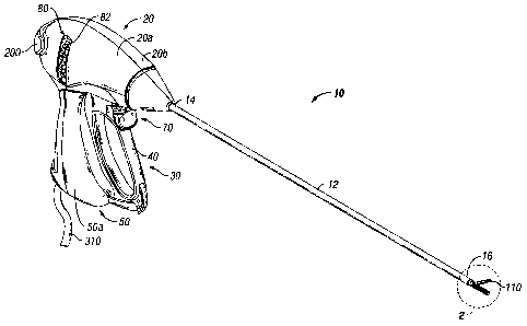
[0037] FIG. 1 is a perspective view of an endoscopic bipolar forceps
showing a housing, a shaft and an end effector assembly according to the
present disclosure;
[0038] FIG. 2 is an enlarged perspective view of the end effector assembly
of FIG. 1, with the jaw members in an open configuration;
[0039] FIG. 3 is a schematic, side elevational view of an end effector
according to an embodiment of the present disclosure, with the jaw members in
an open configuration;
[0040] FIG. 4 is a schematic, side elevational view of the end effector of
FIG. 3 with the jaw members in a closed configuration;
[0041] FIG. 5 is a schematic, side elevational view of an end effector
according to another embodiment of the present disclosure, in a first closed
configuration;
[0042] FIG. 6 is a schematic, side elevational view of the end effector
of
FIG. 5, in a second closed configuration for transmitting clamping force to
tissue
interposed therebetween;
[0043] FIG. 7 is a schematic, side elevational view of an end effector
according to yet another embodiment of the present disclosure, with the jaw
members in an open configuration;
13
CA 02561034 2006-09-27
[0044] FIG. 8 is a schematic, side elevational view of the end effector of
FIG. 7 with the jaw members in a closed configuration;
[0045] FIG. 9 is a schematic, side elevational view of an end effector
according to still another embodiment of the present disclosure, with the jaw
members in an open configuration;
[0046] FIG. 10 is a schematic, side elevational view of the end effector
of
FIG. 9 with the jaw members in a closed configuration;
[0047] FIG. 11 is a schematic, side elevational view of an end effector
according to another embodiment of the present disclosure, with the jaw
members in an open configuration;
[0048] FIG. 12 is a schematic, side elevational view of the end effector
of
FIG. 11 with the jaw members in a closed configuration;
[0049] FIG. 13 is a schematic, side elevational view of an end effector
according to yet another embodiment of the present disclosure, illustrating a
scissor blade in an unactuated condition;
[0050] FIG. 14 is a schematic, distal end view of the end effector of
FIG.
13, including tissue interposed between the jaw members;
[0051] FIG. 15 is a schematic, side elevational view of the end effector
of
FIGS. 13 and 14, illustrating the scissor blade in an actuated condition;
14
CA 02561034 2006-09-27
[0052] FIG. 16 is a schematic, distal end view of the end effector of
FIG.
15;
[0053] FIG. 17 is a schematic, top plan view of the end effector of FIGS.
13-16;
[0054] FIG. 18 is a schematic, perspective view of an end effector
according to yet another embodiment of the present disclosure, shown in a
first
condition;
[0055] FIG. 19 is a schematic, perspective view of the end effector of
FIG.
18, shown in a second condition; and
[0056] FIG. 20 is a schematic, perspective view of the end effector of
FIGS. 18 and 19, shown in a third condition.
DETAILED DESCRIPTION
[0057] Turning now to FIGS. 1 and 2, an embodiment of an endoscopic
bipolar forceps 10 is shown for use with various surgical procedures and
generally includes a housing 20, a handle assembly 30, a rotating assembly 80,
a trigger assembly 70 and an end effector assembly 100 that operates to grasp,
seal, divide, cut and dissect corporal tissue and the like. Although the
majority of
the figure drawings depict a bipolar forceps 10 for use in connection with
endoscopic surgical procedures, the present disclosure may be used for more
traditional open surgical procedures. For the purposes herein, the forceps 10
is
described in terms of an endoscopic instrument, however, it is contemplated
that
CA 02561034 2013-11-05
an open version of the forceps may also include the same or similar operating
components and features as described below.
[0058] In the drawings and in the descriptions that follow, the term
"proximal," as is traditional, will refer to the end of the forceps 10 which
is closer
to the user, while the term "distal" will refer to the end that is farther
from the
user.
[0059] Forceps 10 includes a shaft 12 that has a distal end 16
dimensioned to mechanically engage end effector assembly 100 and a proximal
end 14 that mechanically engages the housing 20. Proximal end 14 of shaft 12
is received within housing 20 and appropriate mechanical and electrical
connections relating thereto are established.
[0060] As best seen in FIG. 1, forceps 10 also includes an electrosurgical
cable 310 that connects the forceps 10 to a source of electrosurgical energy,
e.g., a generator (not shown). It is contemplated that generators such as
those
sold by Valleylab - a division of Tyco Healthcare LP, located in Boulder
Colorado
are used as a source of electrosurgical energy, e.g., FORCE EZTM
Electrosurgical Generator, FORCE FX114 Electrosurgical Generator, FORCE
1C1-m, FORCE 2TM Generator, SurgistatTM II. One such system is described in
commonly-owned U.S. Patent No. 6,033,399 entitled "ELECTROSURGICAL
GENERATOR WITH ADAPTIVE POWER CONTROL". Other systems have been
described in commonly-owned U.S. Patent No. 6,187,003 entitled "BIPOLAR
16
CA 02561034 2013-11-05
ELECTROSURGICAL INSTRUMENT FOR SEALING VESSELS".
[0061] In one embodiment, the generator includes various safety and
performance features including isolated output, independent activation of
accessories. It is envisioned that the electrosurgical generator includes
Valleylab's Instant ResponseTM technology features that provides an advanced
feedback system to sense changes in tissue 200 times per second and adjust
voltage and current to maintain appropriate power. The Instant ResponseTM
technology is believed to provide one or more of the following benefits to
surgical
procedure:
[0062] Consistent clinical effect through all tissue types;
[0063] Reduced thermal spread and risk of collateral tissue damage;
[0064] Less need to "turn up the generator"; and
[0065] Designed for the minimally invasive environment.
[0066] Cable 310 is internally divided into several cable leads (not
shown)
which each transmit electrosurgical energy through their respective feed paths
through the forceps 10 to the end effector assembly 100.
[0067] Handle assembly 30 includes a fixed handle 50 and a movable
handle 40. Fixed handle 50 is integrally associated with housing 20 and handle
40 is movable relative to fixed handle 50. In one embodiment, rotating
assembly
17
CA 02561034 2006-09-27
80 is integrally associated with housing 20 and is rotatable approximately 180
degrees about a longitudinal axis.
[0068] As mentioned above, end effector assembly 100 is attached at
distal end 16 of shaft 12 and includes a pair of opposing jaw members 110 and
120. Movable handle 40 of handle assembly 30 is ultimately connected to a
drive assembly (not shown) which, together, mechanically cooperate to impart
movement of jaw members 110 and 120 from an open position wherein jaw
members 110 and 120 are disposed in spaced relation relative to one another,
to
a clamping or closed position wherein jaw members 110 and 120 cooperate to
grasp tissue therebetween.
[0069] It is envisioned that forceps 10 may be designed such that it is
fully
or partially disposable depending upon a particular purpose or to achieve a
particular result. For example, end effector assembly 100 may be selectively
and
releasably engageable with distal end 16 of the shaft 12 and/or the proximal
end
14 of shaft 12 may be selectively and releasably engageable with the housing
20
and the handle assembly 30. In either of these two instances, the forceps 10
would be considered "partially disposable" or "reposable", i.e., a new or
different
end effector assembly 100 (or end effector assembly 100 and shaft 12)
selectively replaces the old end effector assembly 100 as needed. As can be
appreciated, the presently disclosed electrical connections would have to be
altered to modify the instrument to a reposable forceps.
18
CA 02561034 2006-09-27
[0070] As shown best in FIG. 2, end effector assembly 100 includes
opposing jaw members 110 and 120 that cooperate to effectively grasp tissue
for
operative purposes. End effector assembly 100 may be designed as a unilateral
assembly, i.e., jaw member 120 is fixed relative to the shaft 12 and jaw
member
110 pivots about a pivot pin 103 to grasp tissue and the like or as a
bilateral
assembly, i.e., both jaw members pivot relative to one another.
[0071] More particularly, and with respect to the particular embodiments
shown in FIG. 2, the unilateral end effector assembly 100 includes one
stationary
or fixed jaw member 120 mounted in fixed relation to shaft 12 and pivoting jaw
member 110 mounted about a pivot pin 103 attached to the stationary jaw
member 120. A reciprocating sleeve 60 is slidingly disposed within the shaft
12
and is remotely operable by a drive assembly. The pivoting jaw member 110
includes a detent or protrusion 117 that extends from jaw member 110 through
an aperture (not shown) disposed within the reciprocating sleeve 60. Pivoting
jaw member 110 is actuated by sliding the sleeve 60 axially within shaft 12
such
that a distal end of the aperture abuts against detent 117 on pivoting jaw
member
110. Pulling sleeve 60 proximally closes jaw members 110 and 120 about tissue
and the like, and pushing sleeve 60 distally opens jaw members 110 and 120.
[0072] As illustrated in FIG. 2, a knife channel 115b runs through the
center of the jaw member 120 (a complementary knife channel is formed in jaw
member 110) such that a blade from a knife assembly (not shown) may cut
through the tissue grasped between jaw members 110 and 120 when jaw
members 110 and 120 are in a closed position. Details relating to the knife
19
CA 02561034 2013-11-05
channel 115 and the knife actuating assembly including trigger assembly 70 are
explained in limited detail herein and explained in more detail with respect
to
commonly-owned U.S. Patent Publications US 2004/0254573 and US 2005/0107785.
[0073] With continued reference to FIG. 2, jaw member 110 also includes
a jaw housing has an insulative substrate or insulator 114 and an electrically
conducive
sealing surface 112. In one embodiment, insulator 114 is dimensioned to
securely
engage the electrically conductive sealing surface 112. This may be
accomplished by
stamping, by overmolding, by overmolding a stamped electrically conductive
sealing
plate and/or by overmolding a metal injection molded seal plate. Movable jaw
member
110 also includes a wire channel 113 designed to guide a cable lead 311 into
electrical
continuity with electrically conducive sealing surface 112 as described in
more detail
below.
[0074] Desirably, jaw member 110 has an electrically conducive sealing
surface 112 which is substantially surrounded by an insulating substrate 114.
Insulating substrate 114, electrically conductive sealing surface 112 and the
outer, non-conductive jaw housing 116 can be dimensioned to limit and/or
reduce
many of the known undesirable effects related to tissue sealing, e.g.,
flashover,
thermal spread and stray current dissipation. Alternatively, it is envisioned
that
jaw members 110 and 120 may be manufactured from a ceramic-like material
CA 02561034 2013-11-05
and the electrically conducive sealing surface(s) 112 thereof may be coated
onto
the ceramic-like jaw members 110 and 120.
[0075] It is envisioned that the electrically conductive sealing surface
112
may also include an outer peripheral edge that has a pre-defined radius and
the
insulating substrate 114 meets the electrically conductive sealing surface 112
along an adjoining edge of the sealing surface 112 in a generally tangential
position. In one embodiment, at the interface, the electrically conducive
sealing
surface 112 is raised relative to the insulating substrate 114. These and
other
envisioned embodiments are discussed in commonly assigned WO 02/080786
entitled
"ELECTROSURGICAL INSTRUMENT WHICH REDUCES COLLATERAL DAMAGE
TO ADJACENT TISSUE" by Johnson et al. and commonly assigned WO 02/080785
entitled "ELECTROSURGICAL INSTRUMENT WHICH IS DESIGNED TO REDUCE
THE INCIDENCE OF FLASHOVER" by Johnson et al.
[0076] In one embodiment, the electrically conducive sealing surface 112
and the insulating substrate 114, when assembled, form a longitudinally-
oriented
slot (not shown) defined therethrough for reciprocation of the knife blade. It
is
envisioned that knife channel (not shown) of jaw member 110 cooperates with a
corresponding knife channel 115b defined in stationary jaw member 120 to
facilitate longitudinal extension of the knife blade along a preferred cutting
plane
to effectively and accurately separate the tissue.
21
CA 02561034 2013-11-05
[0077] Jaw member 120 includes similar elements to jaw member 110
such as a jaw housing having an insulating substrate 124 and an electrically
conductive sealing surface 122 which is dimensioned to securely engage the
insulating substrate 124. Likewise, the electrically conductive surface 122
and
the insulating substrate 124, when assembled, include a longitudinally-
oriented
channel 115a defined therethrough for reciprocation of the knife blade. As
mentioned above, when the jaw members 110 and 120 are closed about tissue,
the knife channels of jaw members 110, 120 form a complete knife channel to
allow longitudinal extension of the knife blade in a distal fashion to sever
tissue.
It is also envisioned that the knife channel may be completely disposed in one
of
the two jaw members, e.g., jaw member 120, depending upon a particular
purpose.
[0078] As best seen in FIG. 2, jaw member 120 includes a series of stop
members 750 disposed on the inner facing surfaces of the electrically
conductive
sealing surface 122 to facilitate gripping and manipulation of tissue and to
define
a gap between opposing jaw members 110 and 120 during sealing and cutting of
tissue. It is envisioned that the series of stop members 750 may be employed
on
one or both jaw members 110 and 120 depending upon a particular purpose or to
achieve a desired result. A detailed discussion of these and other envisioned
stop members 750 as well as various manufacturing and assembling processes
for attaching and/or affixing the stop members 750 to the electrically
conductive sealing
surfaces 112, 122 are described in commonly-assigned WO 02/080796 entitled
"VESSEL SEALER AND DIVIDER WITH NON-CONDUCTIVE STOP MEMBERS" by
Dycus et al.
22
CA 02561034 2013-11-05
[0079] Jaw members 110 and/or 120 may be designed to be fixed to the
end of a tube 60 (see FIG. 2) extending from handle assembly 30 and configured
for rotation about a longitudinal axis thereof. In this manner, rotation of
tube 60
may impart rotation to jaw members 110 and/or 120 of end effector assembly
100.
[0080] Turning now to FIGS. 3 and 4, an alternate embodiment of end
effector assembly 300, in accordance with the present disclosure, is shown and
will be described. It is envisioned that end effector assembly 300 may include
some, if not all, of the features and elements provided and/or associated with
end
effector assembly 100.
[0081] As seen in FIGS. 3 and 4, end effector assembly 300 includes a
central shaft 302 supporting a pair of jaws 310, 320 at a distal end thereof
in a
unilateral arrangement. End effector assembly 300 includes a first or fixed
jaw
member 320 supported on a distal end 302a of central shaft 302, and a second
or movable jaw member 310 pivotably supported at distal end 302a of central
shaft 302 by a pivot pin 103. First and second jaw members 320, 310 are in
juxtaposed relation to one another and are movable between an open condition,
wherein tissue may be positioned between jaw members 320, 310, and a closed
configuration, wherein jaw members 320, 310 grasp and/or clamp onto tissue.
23
CA 02561034 2006-09-27
Jaw members 320, 310 are biased to the open condition by a biasing member,
e.g., spring, or the like (not shown).
[0082] End effector assembly 300 further includes an outer catheter
sleeve
304 defining a front or distal edge 304a and a lumen 306 therethrough. Lumen
306 of outer sleeve 304 is configured and dimensioned to translatably receive
central shaft 302 and jaw members 320, 310 therein.
[0083] In operation, as central shaft 302 is withdrawn into outer sleeve
304, as indicated by arrow "A" in FIG. 4, distal edge 304a of outer sleeve 304
abuts against movable jaw member 310 and forces movable jaw member 310
towards fixed jaw member 320. In so doing, tissue disposed between jaw
members 310, 320 is clamped or grasped therebetween. It is understood that, in
certain embodiments, that the greater the degree of withdrawal of central
shaft
302 and jaw member 310, 320 into lumen 306 of outer sleeve 304, the greater
the clamping force exerted on the tissue disposed between jaw members 310,
320.
[0084] It is envisioned and within the scope of the present disclosure
for
central shaft 302 and/or outer sleeve 304 to be fabricated from a flexible
material
or the like. Central shaft 302 and/or outer sleeve 304 may be fabricated from
any
one of or a combination of materials including and not limited to, NITINOL
(e.g.,
nickel-titanium alloys), polyurethane, polyester, and/or polymethylsiloxane
material (PDMS), fluorinated ethylene-propylene (FEP), polytetrafluoroethylene
(PTFE), nylon, etc.
24
CA 02561034 2006-09-27
[0085] Turning now to FIGS. 5 and 6, an end effector assembly, according
to another embodiment of the present disclosure, is generally designated as
300a. It is envisioned that end effector assembly 300b may include some, if
not
all, of the features and elements provided and/or associated with end effector
assembly 100.
[0086] End effector assembly 300a includes a pair of jaw members 310a,
320a each pivotably supported at a distal end of a central shaft 302a via a
pivot
pin 103. End effector assembly 300a further includes an outer catheter sleeve
304a defining a lumen 306a therethrough. Lumen 306a of outer sleeve 304a is
configured and dimensioned to translatably receive central shaft 302a and jaw
members 310a, 320a therein.
[0087] As seen in FIGS. 5 and 6, a linkage 330 or the like may be provided
for actuating one of jaw members 310a, 320a relative to the other thereby
effectuating opening and closing of end effector assembly 300a. A distal end
330a of linkage 330 is desirably connected to second jaw member 310a at a
location distal of pivot pin 103 when jaw members 310a, 320a are disposed
within outer sleeve 304a. Linkage 330 is desirably operatively connected to
second jaw member 310a in such a manner so as to effectuate rotation of
second jaw member 310a toward first jaw member 320a upon movement of
linkage 330 in a proximal direction.
[0088] In use, with jaw members 310a, 320a in a closed condition, jaw
members 310a, 320a are advanced through lumen 306a of outer sleeve 304, as
CA 02561034 2006-09-27
indicated by arrow "B" of FIG. 5. After jaw members 310a, 320a have cleared
the distal end or edge of outer sleeve 304a (i.e., pivot pin 103 has cleared
or
advanced beyond the distal end or edge of outer sleeve 304a), jaw members
310a, 320a may both be pivoted about pivot pin 103 to a substantially
orthogonal
orientation relative to central shaft 302a, as seen in FIG. 6. In order to
pivot or
rotate jaw members 310a, 320a about pivot pin 103, linkage 330 is moved in a
proximal direction, as indicated by arrow "A".
[0089] With jaw members 310a, 320a oriented in an orthogonal direction,
jaw members 310a, 320a may be opened and closed by moving linkage 330 in a
distal or proximal direction. For example, by moving linkage 330 in a distal
direction, second jaw member 310a is rotated about pivot pin 103 thereby
spacing second jaw member 310a from first jaw member 320a. In so doing, end
effector assembly 300a is configured to an open condition and the tissue
contacting surface of first jaw member 320a is oriented approximately 90
relative
to a longitudinal axis of outer sleeve 304a. With end effector assembly 300a
in
an open condition, tissue may be placed between jaw members 310a, 320a or
jaw members 310a, 320a may be placed over the tissue.
[0090] Following placement of tissue between jaw members 310a, 320a,
linkage 330 may be moved in a proximal direction thereby rotating second jaw
member 310a about pivot pin 103 to approximate second jaw member 310a
toward first jaw member 320a. In so doing, end effector assembly 300a is moved
to a closed condition to grasp the tissue interposed between first and second
jaw
members 320a, 310a. Since jaw members 310b, 320b are in an orthogonal
26
CA 02561034 2006-09-27
configuration, retraction of linkage 330 in a proximal direction results in
application of the clamping force in a substantially linear direction relative
to
central shaft 302b.
[0091] Following treatment of the tissue, linkage 330 may be reactuated
to
release the treated tissue from between first and second jaw members 320a,
310a. With the treated tissue released from between first and second jaw
members 320a, 310a, central shaft 302a is withdrawn through outer sleeve 304a.
In so doing, first and second jaw members 320a, 310a are re-oriented to an
axially aligned orientation due to a camming action between the distal edge of
outer sleeve 304a and first jaw member 320a.
[0092] It is envisioned and within the scope of the present disclosure
for
central shaft 302a and/or outer sleeve 304a to be fabricated from a flexible
material or the like.
[0093] Turning now to FIGS. 7 and 8, an end effector assembly, according
to an alternate embodiment of the present disclosure, is generally shown as
300b. It is envisioned that end effector assembly 300b may include some, if
not
all, of the features and elements provided and/or associated with end effector
assembly 100.
[0094] As seen in FIGS. 7 and 8, end effector assembly 300b includes a
central shaft 302b supporting a pair of jaws 310b, 320b at a distal end
thereof in
a unilateral arrangement. End effector assembly 300b includes a first or fixed
jaw member 320b supported on a distal end of central shaft 302b, and a second
27
CA 02561034 2006-09-27
or movable jaw member 310b pivotably supported at distal end of central shaft
302b by a pivot pin 103. First and second jaw members 320b, 310b are in
juxtaposed relation to one another and are movable between an open condition,
wherein tissue may be positioned between jaw members 320b, 310b, and a
closed configuration, wherein jaw members 320b, 310b grasp and/or clamp onto
tissue.
[0095] As seen in FIGS. 7 and 8, a linkage 330b or the like may be
provided for actuating second jaw member 310b relative to first jaw member
320b. A distal end 330a of linkage 330 is desirably connected to second jaw
member 310. In particular, as seen in FIG. 7, distal end 330a of linkage 330
is
connected to second jaw member 310a in such a manner so as to effectuate
rotation of second jaw member 310b toward first jaw member 320b upon
movement of linkage 330 in a proximal direction, as indicated by arrow "A", or
away from first jaw member 320b upon movement of linkage 330 in a distal
direction, as indicated by arrow "B".
[0096] As disclosed above, it is envisioned and within the scope of the
present disclosure that central shaft 302b may be fabricated from a flexible
material or the like.
[0097] Turning now to FIGS. 9 and 10, an end effector assembly,
according to a further embodiment of the present disclosure, is generally
designated as 300c. End effector assembly 300c is substantially identical to
end
28
CA 02561034 2006-09-27
effector assembly 300b and will only be discussed in detail to the extent
necessary to identify differences in construction and operation.
[0098] As seen in FIGS. 9 and 10, a central body portion 302c of end
effector assembly 300c includes a rigid distal portion 301c and a flexible
proximal
portion 303c. Jaw members 310c, 320c are arranged in a unilateral
configuration
and are actuatable by any of the methods described above or known by one
having skill in the art. Jaw members 310c, 320c are desirably biased to an
open
condition by a biasing member, e.g., spring, or the like (not shown), or by
the
wire snare 340.
[0099] As seen in FIGS. 9 and 10, end effector assembly 300c includes a
wire snare 340 extending out of one of jaw members 310c, 320c and anchored to
the other of jaw members 310c, 320c. In particular, wire snare 340 is disposed
within central body portion 302c and includes a proximal end (not shown) which
connects to an electrosurgical energy source, and a distal end 340a that
extends
out through fixed jaw member 320c and attaches to a distal end or tip of
movable
jaw member 310c.
[00100] It is envisioned that wire 340 may be fabricated from a shape
memory alloy, such as, for example, NITINOL, or the like. Accordingly, as seen
in FIG. 9, when end effector assembly 300c is in the open condition, wire 340
has a substantially arcuate shape or configuration.
[00101] In use, in order to close end effector assembly 300c, wire 340 is
withdrawn in a proximal direction thereby approximating the distal tip of
movable
29
CA 02561034 2006-09-27
jaw member 310c toward the distal tip of fixed jaw member 320c. In so doing
jaw
members 310c, 320c are approximated toward one another and desirably clamp
onto tissue "T".
[00102] In one mode of operation, with end effector assembly 300c in an
open condition and with wire 340 in an expanded condition, as seen in FIG. 9,
end effector assembly 300c is placed over tissue "T" to be excised, e.g., a
polyp
or the like, such that tissue "T" is interposed and/or disposed within the
space or
area "S" defined between jaw members 310c, 320c and wire 340. With tissue "T"
positioned in space "S", the proximal end of wire 340 is drawn in a proximal
direction thereby closing end effector assembly 300c (e.g., approximating jaw
members 310c, 320c) onto tissue "T" and cinching wire 340 about tissue "T".
[00103] Wire 340 is withdrawn an amount sufficient to tightly close end
effector assembly 300c onto and/or about tissue "T" and to apply pressure to
tissue "T" between the jaw members 310c, 320c. At such a time, electrical
current or electrical energy is transmitted through wire 340 and/or to the
electrically conducive sealing surface(s) of jaw members 310c, 320c. The
electrical current or energy is transmitted at a level and for a time
sufficient to
heat wire 340 to cut through tissue "T" and remove tissue "T" from the
underlying
or remaining tissue.
[00104] It is envisioned that wire 340 may or may not be insulated.
Additionally, distal portion 301c of central shaft 300c may be fabricated from
a
rigid, electrically conductive material. In so doing, an electrical lead 311c
may
CA 02561034 2006-09-27
extend through flexible proximal portion 303c of central shaft 302c and
electrically connect to a proximal end of rigid portion 301c.
[00105] In another mode of operation, with end effector assembly 300c in
an open condition and with wire 340 in an expanded condition, end effector
assembly 300c is placed over tissue "T" to be excised, e.g., a polyp or the
like,
such that tissue "T" is interposed and/or disposed between jaw members 310c,
320c. With tissue "T" so positioned, the proximal end of wire 340 is drawn in
a
proximal direction thereby cinching wire 340 and closing end effector assembly
300c (e.g., approximating jaw members 310c, 320c) onto tissue "T".
[00106] Wire 340 is withdrawn an amount sufficient to tightly close end
effector assembly 300c onto tissue "T" and to apply pressure to tissue "T"
between the jaw members 310c, 320c. It is envisioned that in the current mode
of operation, further withdrawal of wire 340 may result in pivoting of end
effector
assembly 300c about pivot pin 103 to improve the visibility at the surgical
site.
[00107] Turning now to FIGS. 11 and 12, an end effector assembly,
according to a further embodiment of the present disclosure, is generally
designated as 300d. End effector assembly 300d is substantially identical to
end
effector assembly 300c and will only be discussed in detail to the extent
necessary to identify differences in construction and operation.
[00108] As seen in FIGS. 11 and 12, end effector assembly 300d includes a
wire 340 extending out of one of jaw members 310d, 320d and into the other of
jaw members 310d, 320d. In particular, wire 340 is disposed within central
body
:31
CA 02561034 2006-09-27
portion 302d and includes a proximal end (not shown) which connects to an
electrosurgical energy source, and a distal end 340a which extends out through
a
distal tip of first jaw member 320d and back into a distal tip of second jaw
member 310d. Distal end 340a of wire 340 is anchored or secured to itself
according to any known method, including and not limited to use of a junction
block 342. In this manner, as will be described in greater detail below,
withdrawal of wire 340 in a proximal direction results in withdrawal of wire
340
through both jaw members 310b, and 320d.
[00109] While end effector assembly 300d is shown as having bilateral jaw
member arrangement, it is envisioned and within the scope of the present
disclosure for end effector assembly 300d to have a unilateral jaw member
arrangement. It is envisioned that when end effector assembly 300d is in the
open condition, wire 340 has a substantially arcuate shape or configuration.
Wire 340 includes a nipple region 340b formed along a length thereof. In use,
when cinching wire 340 it is desired for tissue "T" to be positioned within
nipple
region 340b of wire 340.
[00110] In use, in order to close end effector assembly 300d, wire 340 is
withdrawn in a proximal direction, by pulling on the proximal end of wire 340,
thereby approximating the distal tips of jaw members 310d, 320d toward one
another. Since distal end 340a of wire 340 is secured to itself by junction
block
342, by pulling on the proximal end of wire 340, distal end 340a of wire 340
is
drawn into both jaw members 310d, 320d substantially equally.
32
CA 02561034 2006-09-27
[00111] In operation, with end effector assembly 300d in an open condition
and with wire 340 in an expanded condition, as seen in FIG. 11, end effector
assembly 300d is placed over tissue "T" to be excised, e.g., a polyp or the
like,
such that tissue "T" is interposed and/or disposed within the space or area
"S"
defined between jaw members 310d, 320d and wire 340. With tissue "T"
positioned in space "S", the proximal end of wire 340 is drawn in a proximal
direction thereby closing end effector assembly 300d (e.g., approximating jaw
members 310d, 320d simultaneously) onto tissue "T" and cinching wire 340
about tissue "T".
[00112] Wire 340 is withdrawn an amount sufficient to tightly close end
effector assembly 300d onto and/or about tissue "T" and to apply pressure to
tissue "T" between jaw members 310d, 320d. At such a time, electrical current
or
electrical energy is transmitted through wire 340 and/or to the electrically
conducive sealing surface(s) of jaw members 310d, 320d. The electrical current
or energy is transmitted at a level and for a time sufficient to heat wire 340
to cut
through tissue "T" and remove tissue "T' from the underlying or remaining
tissue.
[00113] In accordance with the present disclosure, the rigid nature of jaw
members 310, 320 provides greater support and/or control of wire 340 as
compared to conventional wire snare instruments and the like.
[00114] Turning now to FIGS. 13-17, an end effector assembly, according
to a further embodiment of the present disclosure, is generally designated as
300e. End effector assembly 300e is substantially identical to end effector
33
CA 02561034 2006-09-27
assembly 300c and will only be discussed in detail to the extent necessary to
identify differences in construction and operation.
[00115] End effector assembly 300e further includes a knife or scissor
blade 350 pivotably connected to a distal end of central shaft 302e. Scissor
blade 350 may be pivotably connected to the distal end of central shaft 302e
via
pivot pin 103. Scissor blade 350 defines a cutting edge 350a or the like.
[00116] As seen in FIGS. 13-17, a linkage 352 or the like may be provided
for actuating scissor blade 350 relative to jaw members 310e, 320e of end
effector assembly 300e to sever tissue "T" and the like. A distal end 352a of
linkage 352 is desirably connected to scissor blade 352 at a location
desirably
distal of pivot pin 103. Linkage 352 is desirably operatively connected to
scissor
blade 350 in such a manner so as to effectuate rotation of scissor blade 350
upon movement of linkage 352 in a proximal direction.
[00117] As seen in FIGS. 13 and 14, scissor blade 350 has a first position
in which cutting edge 350a thereof is in substantial registration with gap "G"
between jaw members 310e, 320e, or, alternatively, cutting edge 350a of
scissor
blade 350 is in substantial registration with and/or substantially aligned
with the
sealing surface 122e of jaw member 310e. As seen in FIGS. 15 and 16, scissor
blade 350 has a second position in which cutting edge 350a thereof has been
rotated past or beyond gap "G" between jaw members 310e, 320e, to thereby
sever or cut tissue "T" extending from therebetween.
34
CA 02561034 2006-09-27
[00118] End effector assembly 300e may further include a wire 340
extending out of one of jaw members 310e, 320e and anchored to the other of
jaw members 310e, 320e. In particular, wire 340 is disposed within central
body
portion 302e and includes a proximal end (not shown) which connects to an
electrosurgical energy source, and a distal end 340a which extends out through
fixed jaw member 320e and attaches to a distal end or tip of movable jaw
member 310e.
[00119] In operation, either prior to, during or following severing of
tissue
"T" with wire 340, as described above with regard to end effector assemblies
300c or 300d, linkage 352 is actuated (e.g., moved in a proximal direction) to
pivot scissor blade 350 about pivot pin 103 and severing tissue "T" along the
sides of jaw members 310e, 320e.
[00120] Desirably, scissor blade 350 has a length substantially equal to
the
length of jaw members 310e, 320e. However, it is envisioned that scissor blade
350 may have any length necessary or desired in order to perform the operative
procedure.
[00121] It is envisioned and within the scope of the present disclosure
for
the proximal portions of any of the jaw members disclosed above and the distal
end of the respective central shafts to be covered by a resilient or flexible
insulating material or boot (not shown) to reduce stray current concentrations
during electrosurgical activation especially in a monopolar activation mode.
As
can be appreciated, when jaw members 310, 320 are opened, the boot flexes or
CA 02561034 2013-11-05
expands in certain areas in order to accommodate the movement of jaw
members 310, 320. Further details relating to one envisioned insulating boot
are
described in commonly-owned U.S. Publication US 2007/0078458.
[00122] Turning now to FIGS. 18-20, an end effector assembly, according
to yet another embodiment of the present disclosure, is generally designated
as
400. As seen in FIGS. 18-20, end effector assembly 400 includes a central
shaft
402 having a distal end 402a configured and adapted to support a cutting blade
404 thereon. It is envisioned that central shaft 402 may be either flexible or
rigid
along at least a portion of its length.
[00123] Cutting blade 404 includes a cutting edge 404a extending in a
substantially distal direction. Desirably, cutting edge 404a of cutting blade
404
lies along the central longitudinal axis of central shaft 402.
[00124] End effector assembly 400 includes a jaw member 406 movably
associated with central shaft 402. In an embodiment, movable jaw member 406
is configured and adapted to translate longitudinally along and/or relative to
central shaft 402. Movable jaw member 406 includes a leg portion 406a
extending substantially longitudinally along central shaft 402 and a tissue
contacting portion 406b extending in a substantially orthogonal direction from
a
distal end of leg portion 406a. In particular, tissue contacting portion 406b
of
36
CA 02561034 2013-11-05
movable jaw member 406 extends across the central longitudinal axis of central
shaft 402 and, more particularly, across cutting blade 404. Reference may be
made to commonly-owned and concurrently-filed U.S. Patent No. 6,267,761; and
U.S.
Patent Publication US 2006/0020265 for exemplary embodiments and modes of
operation of end effector assembly 400.
[00125] Jaw member 406 is movable from a position in which tissue contact
portion 406b is spaced a distance from cutting edge 404a of cutting blade 404
to
a position in which tissue contacting portion 406b is in contact with cutting
edge
404a of cutting blade 404.
[00126] End effector assembly 400 further includes a floating anvil member
408 interposed between cutting blade 404 and tissue contacting portion 406b of
jaw member 406. Anvil member 408 is slidably supported on leg portion 406a of
jaw member 406 so that anvil member 408 is translatable along leg portion
406a.
In one embodiment, anvil member 408 include a first slot 408a configured and
dimensioned to slidably receive leg portion 406a of jaw member 406
therethrough. Anvil member 408 further includes a second or blade slot 408b
formed therein that is configured and dimensioned to permit reciprocal
movement
of cutting blade 404 into and out of blade slot 408b (i.e., through anvil
member
408).
37
CA 02561034 2006-09-27
[00127] End effector assembly 400 further includes a biasing member or
spring 410 interposed between cutting blade 404 and anvil member 408. Biasing
member 410 is configured so as to maintain anvil member 408 spaced a distance
from cutting blade 404. Desirably, biasing member 408 maintains anvil member
408 spaced from cutting blade 404 by an amount sufficient that cutting edge
404a of cutting blade 404 does not extend through blade slot 408b of anvil
member 408.
[00128] It is envisioned that each of tissue contacting portion 406b and
anvil
member 408 may be electrically connected to an electrosurgical energy source
(not shown) and are provided with elements (not shown) for delivering and/or
receiving electrosurgical energy.
[00129] With continued reference to FIGS. 18-20, an exemplary method of
using a surgical instrument including an end effector assembly 400 is
provided.
As seen in FIG. 18, with jaw member 406 positioned such that tissue contact
portion 406b is spaced a distance from anvil member 408, tissue "T" (e.g., a
polyp or the like) in introduced therebetween, either by placing end effector
assembly 400 over tissue "T", as shown, or by drawing tissue "T" into the
space
therebetween.
[00130] As seen in FIG. 19, with tissue "T" interposed between tissue
contacting portion 406b of jaw member 406 and anvil member 408, jaw member
406 is moved in a proximal direction relative to central shaft 402, as
indicated by
arrow "A". In so doing, tissue "T" is clamped or grasped between tissue
38
CA 02561034 2006-09-27
contacting portion 406b of jaw member 406 and anvil member 408. Desirably, a
sufficient force is applied to jaw member 406 so as to clamp tissue "T"
between
tissue contacting portion 406b thereof and anvil member 408 and so as not to
substantially move anvil member 408 to compress biasing member 410. As
discussed above, biasing member 410 maintains anvil member 408 spaced a
distance from cutting blade 404 such that cutting edge 404a does not extend
beyond blade slot 408b.
[00131] With
tissue "T" clamped between tissue contacting portion 406b of
jaw member 406 and anvil member 408, an effective amount of electrosurgical
energy (e.g., for an effective time period at an effective energy level) is
delivered
to tissue contacting portion 406b of jaw member 406 and/or anvil member 408 to
achieve a desired effect in tissue "T". Desirably, bipolar current is applied
to seal
the base of the tissue.
[00132] As
seen in FIG. 20, with tissue "T" treated, jaw member 406 is
further advanced in a proximal direction, as indicated by arrow "A", to
overcome
the bias of biasing member 410 and advance anvil member 408 over cutting
blade 404. In so doing, cutting edge 404a of cutting blade 404 severs tissue
"T"
from the remaining underlying tissue.
[00133] In
accordance with the present disclosure, any of the end effectors
disclosed herein may be configured and adapted to deliver a working pressure
of
about 3 kg/cm2 to about 16 kg/cm2 and, preferably, of about 7 kg/cm2 to about
13
kg/cm2, to the tissue. By controlling the intensity, frequency and duration of
the
39
CA 02561034 2013-11-05
electrosurgical energy applied to the tissue by the end effector assemblies,
the
user can cauterize, coagulate/desiccate, seal and/or simply reduce or slow
bleeding.
[00134] From the foregoing and with reference to the various figure
drawings, those skilled in the art will appreciate that certain modifications
can also be
made to the preferred embodiments disclosed.
[00135] It is also contemplated that the forceps 10 (and/or the
electrosurgical generator used in connection with the forceps 10) may include
a
sensor or feedback mechanism (not shown) that automatically selects the
appropriate amount of electrosurgical energy to effectively seal the
particularly-
sized tissue grasped between the jaw members. The sensor or feedback
mechanism may also measure the impedance across the tissue during sealing
and provide an indicator (visual and/or audible) that an effective seal has
been
created between the jaw members. Examples of such sensor systems are
described in commonly-owned U.S. Patent No. 7,137,980.
[00136] It is envisioned that the outer surface of any of the end effector
assemblies disclosed herein may include a nickel-based material, coating,
stamping, metal injection molding which is designed to reduce adhesion between
CA 02561034 2006-09-27
the jaw members with the surrounding tissue during activation and sealing.
Moreover, it is also contemplated that the conductive surfaces of the jaw
members may be manufactured from one (or a combination of one or more) of
the following materials: nickel-chrome, chromium nitride, MedCoat 2000
manufactured by The Electrolizing Corporation of OHIO, inconel 600 and tin-
nickel. The tissue conductive surfaces may also be coated with one or more of
the above materials to achieve the same result, i.e., a "non-stick surface".
As
can be appreciated, reducing the amount that the tissue "sticks" during
sealing
improves the overall efficacy of the instrument.
[00137] One
particular class of materials disclosed herein has
demonstrated superior non-stick properties and, in some instances, superior
seal
quality. For example, nitride coatings which include, but not are not limited
to:
TiN, ZrN, TiAIN, and CrN are preferred materials used for non-stick purposes.
CrN has been found to be particularly useful for non-stick purposes due to its
overall surface properties and optimal performance. Other classes of materials
have also been found to reducing overall sticking. For
example, high
nickel/chrome alloys with a Ni/Cr ratio of approximately 5:1 have been found
to
significantly reduce sticking in bipolar instrumentation. One particularly
useful
non-stick material in this class is Inconel 600. Bipolar instrumentation
having
sealing surfaces 112 and 122 made from or coated with Ni200, Ni201 (-100% Ni)
also showed improved non-stick performance over typical bipolar stainless
steel
electrodes.
41
CA 02561034 2013-11-05
[00138] The scope
of the claims should not be limited by the preferred
embodiments set forth herein, but should be given the broadest interpretation
consistent with the description as a whole.
42