Note: Descriptions are shown in the official language in which they were submitted.
WO 2005/102182 CA 02563474 2006-10-17 PCT/NL2005/000290
Title: Devices and methods for anastomosis
The invention relates to medical devices, in particular condoms and
drains, that find use in anastomosis. The condoms/drains of the present
invention are for example made of biodegradable polyurethanes, in particular
polyurethanes comprising poly(ether)ester pre-polymer soft segments and
polyurethane hard segments. Thus the present invention relates to these
medical devices per se, to a medical kit comprising these medical devices, to
the use of these medical devices, to a method for medical treatment involving
application of these medical devices to an organism who is in need thereof as
well as to the use of certain biocompatible, biodegradable synthetic polymers
in
the preparation of a medical device for the treatment of a disease or a
condition which requires anastomosis.
Anastomosis, the operation of joining two ends of biological vessels,
such as the oesophagus, colon or other parts of the gastro-digestive channel,
is
often accompanied by complications. For instance, the most important
complication and cause of death following low anterior resection involving the
colorectal segment is anastomotic leakage. In many cases re-interventions are
needed to treat the complications. Leakage may occur as the result of
dehiscence of the anastomosis itself or of the tissue of the digestive channel
just proximal or particularly distal of the anastomosis. This may occur
typically approximately at around five days after the creation of the
anastomosis. Furthermore, there is an increasing use of preoperative
radiotherapy of rectal cancer and of the digestive channel in general causing
additional problems in anastomotic healing and making the tissue proximal
and particularly distal more vulnerable. Quite often a temporary diverting
stoma is created to reduce or prevent the complications resulting from an
anastomotic leakage. A stoma does not prevent leakage but it drains the gastro
intestinal contents before it can get near the newly formed anastomosis. The
creation, use and removal of a stoma, restoring the "normal" situation is a
burden for the patient and a very costly procedure. To avoid severe
CA 02563474 2012-08-10
2
complications like peritonitis and septic shock it is important to prevent
anastomotic leaks and to take all measures that might eventually be
responsible for that complication.
In the art, condoms have been used for the protection of coloanal
anastomosis, see e.g. Yoon et al. ("Intraluminal bypass technique using a
condom for protection of coloanal anastomosis"; Dis. Colon Rectum. 37(1994)
10464047). According to this known technique, a sterilized (latex rubber)
condom is used to protect a handsewn coloanal anastomosis. Although this
technique is said to be safe, there are still instances of colonic necrosis
reported.
It is desirable to provide an improvement over prior art materials and
techniques for protection of anastomoses.
In one aspect, the invention provides a device having a tubular shaped
15part, such as a condom or drain, of a biocompatible, biodegradable synthetic
polymer.
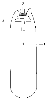
Figure 1 schematically shows a cross-section of a condom of the
present invention prior to application to the site of anastomosis.
Figure 2 schematically shows a cross-section condom of the present
invention at the time it is applied to the site where the anastomosis is to be
performed.
Figure 3 schematically shows a cross-section of a condom of the
present invention at the time it is brought into place in the proximal part of
a
lumen, prior to stapling.
Figure 4 schematically shows a cross-section of a condom of the
present invention just after the stapling has been carried out but prior to
the
cutting of an annulus.
Figure 5 schematically shows a cross-section of a condom of the
present invention at the time the stapling is carried out and the cap is
removed from the stapling site.
CA 02563474 2006-10-17
WO 2005/102182 PCT/NL2005/000290
3
Figure 6 schematically shows a cross-section of a condom of the
present invention at the time it is stretched to its final length and
position.
Figure 7 is a schematic cross-sectional representation, illustrating
the position of the staple relative to the device of the present invention and
the
two ends of the connected lumen.
Figure 8 schematically shows the use of a circular stapler as it is
known in the art.
Figure 9 schematically shows a staple for use known in the art and
also with the present invention. It also shows a pattern in which the staples
may be applied.
Figure 10 is an impression of a condom according to the present
invention.
In a first aspect, the present invention is directed to a medical device
for the protection of an anastomosis, which device comprises a tubular shaped
part whichis made of a biocompatible, biodegradable polymer. The term
"biodegradable" as used herein, means that the material decomposes or looses
structural integrity under the conditions it is applied (i.e., in the body).
It may
thus fragment and leave the body via the natural pathway.
The device of the present invention is preferably in the shape of a
condom, viz, having one open end and one closed end. Optionally, a small
opening or perforation may be provided in the closed end of the condom of the
present invention so as to facilitate the introduction of a stapling cap, as
will
explained in more detail hereinbelow.
It is a very important property of the devices of the present
invention that composition of the biodegradable polymer can be chosen such
that a desired and controllable fragmentation behavior, in particular
degradation rate, can be obtained. Suitable materials for the condoms or
otherwise shaped devices of the present invention are described in
WO-A-2004/039424. Particularly suitable polymer compositions are
biodegradable polyurethanes .The preferred polyurethane is composed of a
WO 2005/102182 CA 02563474 2006-10-17 PCT/NL2005/000290
4
poly(ether)ester pre-polymer soft segment and a polyurethane hard segment
with a structure -BDI-BDO-BDI-BDO-BDI- (BDI being 1,4-
butanediisocyanate and BDO being 1,4-butanediol). The preferred polyether is
a polyethyleneglycol. The rate of degradation of the polyurethane will depend
on the initial molecular weight (measured by the intrinsic viscosity) and the
chemical composition of the pre-polymer. The pre-polymer for this application
is preferably based on DL-lactide and c-caprolactone and has a molecular
weight of preferably 1500-2300, more preferably 2000. It may be obtained by a
ring opening polymerisation initiated by 1,4-butanediol combined with the
polyether compound. The preferred monomer ratio is from 50/50 to 70/30
(mol/mol). The PEG content in the polyurethane is preferably between 1-25
wt.% for applications in the digestive tract, more preferably from 5 to 20
wt.%.
In particular, for coloanal anastomosis the PEG content is preferably between
2-10 wt.%. The molecular weight of PEG is preferably between 600-2000 and is
mast preferably 1000.
Preferably the device of the present invention is manufactured by a
spray-coating process comprising the steps of providing a solution of a
biocompatible, biodegradable polymer, preferably of the above-mentioned type
in a suitable solvent. Suitable solvents are organic solvents, in particular
halogenated (in particular chlorated) or non-halogenated lower (typically C1-
C4) hydrocarbons (in particular ethers), such as chloroform, dichloromethane,
tetrahydrofuran, dioxane and the like. The concentration of the solution is
typically from 1 to 10 wt.%, preferably from 2 to 7 wt.%, e.g. about 4 wt.%.
The
solution is then spraycoated using a known device to a rotating mandrel,
which is made from a hard material, e.g. a ceramic material such as glass. The
mandrel may have a roughened surface. Subsequently, the solvent is allowed
= to evaporate. The wall thickness of the device will depend on the amount of
polymer solution sprayed on the mandrel. The mandrel and the polymeric
layer are submerged in a suitable liquid, such as (distilled) water. Next, the
devices of the present invention can be obtained by removing them from the
CA 02563474 2006-10-17
WO 2005/102182 PCT/NL2005/000290
5
mandrel and cutting them to the desired shape and size and after drying for
some time, typically several hours, up to 24 hours, preferably under vacuum at
slightly elevated temperatures, such as 25 to 50 C, preferably at about 40
C.
The device of the present invention is suitably manufactured by the
following typical spraycoating process wherein the polyurethane as described
above is dissolved in a suitable solvent, e.g. chloroform (or dichloromethane,
tetrahydrofuran, dioxane, and the like or combinations thereof), ca. 4 wt. %.
The polymer solution is spray coated on a horizontally placed and rotating
glass mandrel (surface is roughened). After spray coating, the solvent is
allowed to evaporate, e.g. during 30 minutes while rotating. The mandrel with
the polyurethane layer will be placed in distilled water. The devices will be
removed from the mandrel and will be cut to the appropriate shape. The device
will be dried e.g. during 24 h under vacuum at 40 C in order to remove the
organic solvent.
The device of the present invention, in particular a condom,
preferably has one or more of the following dimensions: a length of 15 to 35
cm
(more preferably 25 2 cm); a diameter of 15 to 50 mm (more preferably 35 5
mm); and a wall thickness of 50 to 90 p.m (more preferably 70 15 p.m).
The biodegradable devices of the present invention may be used to
perform anastomosis. To this end it is highly preferred that it is used in
combination with a stapler, in particular with a so-called circular stapler.
Staplers have been widely accepted for use in colorectal surgery since their
introduction in 1975 as an alternative to hand made sutures and are
responsible for the increase in the number of procedures in ultra low anterior
rectum resection (see Thiede A.; "Rectal stapler. A simple instrument for
alleviating reconstruction of the colorectal passage in the pelvis"; Chirurg.
63(1992) 72-3). The devices of the present invention can be used in
combination with these circular staplers. In Figure 8 the use of these known
circular stapler devices is illustrated: after the cap and stapler are
assembled,
and the two sections of intestine are aligned, the stapler is closed and
fired.
WO 2005/102182 CA 02563474 2006-
10-17 PCT/NL2005/000290
6
Before proceeding to remove the circular stapler, the adjusting knob should be
slightly opened to facilitate the extraction of the stapler.
The staples that are suitable for use in the present invention may be
made of metal, such as stainless steel, titanium, titanium alloys or cobalt-
base
alloys, as known in the art. It may also be possible to provide these staples
from biodegradable materials, such as biodegradable polymers having the
suitable mechanical properties, in particular a suitable ductility.
In principle, any glue may be used to glue the condom to cap (2),
since the glue does not contact the body. Preferably the glue is a
cyanoacrylate
based glue, which is a typical instant glue for skin applications, e.g.
DermabondTM, (JNJ, USA) or IndermilTm (Tyco, USA).
The device of the present invention may thus be used as a drain for
different purposes, such as a colorectal drain. The device of the present
invention typically supports the newly formed anastomosis for 10 to 15 days by
preventing contact between the anastomosis and the contents of the lumen
(e.g. the colorectal contents) so that the fresh wound can heal, thereby
reducing the chance of complications related to leakage. In accordance with
the
present invention the device may be stapled together with the distal and
proximal section of the lumen e.g. the intestine, using the known stapling
processes e.g. involving the use of a circular stapler device. The temporary
drain thus provided, supports the newly formed anastomosis for a sufficient
period of time e.g. from 10 to 15 days, after which it will fragment and leave
the body via the natural pathway. The device may be synthetic, making it
100% biologically safe.The application of the device of the present invention
will now be
described in more detail with particular reference to the figures, which show
the application of a condom according to the present invention. In Figure 1,
the
open end (top in Figure 1) of a drain or condom of the invention (1) is fixed
to a
stapler cap (2), e.g. using glue (3) that is applied to the top of the cap
(2). Note
that Figure 1 is a cross section and therefore the flaps, as depicted in
Figure
WO 2005/102182 CA 02563474 2006-10-17 PCT/NL2005/000290
7
10, are not visible. Preferably the condom has flaps, which are attached to
cap
(2), as discussed in more detail hereinbelow. Fixation can be achieved e.g. by
using a cyanoacrylate glue. Because the glue does not come in contact with the
body, these skin-glues as well as other types of glue can be used safely as
long
as the glue connection is strong enough to pull out the condom (see also
Figures 5 and 6). . The cap and device according to the present invention are
preferably dry before the application of glue for optimal adhesion.
The cap (2) is moved in the direction indicated by the arrow in
Figure 1. The pin shaped bottom end of the cap is pierced through the closed
end (bottom in Figure 1) of the condom. A hole or a perforation may be present
in the condom to facilitate puncturing of the condom.
The assembly (condom (1) glued to cap (2)) is then inserted in the
proximal section of the intestine (5), see Figure 2. It is ensured that the
drain
is placed upward and not folded at the site of the pin.
Next, the proximal intestine (5) is closed around the connecting pin
(2), see Figure 3 by means of a so called purse string, known to the skilled
person. A stapler device, in particular a circular stapler (7) is inserted in
the
distal end of the intestine (6). The stapler (7) also has a connecting pin,
which
fits on the connecting pin of the cap (2), e.g. by the stapler (7) having a
female
pin and the cap (2) having a male pin, so that both cap (2) and stapler (7)
can
be connected. The stapler (7) is further provided with staples (8) and an
annular knife (9) to cut the intestine and the condom, see also Figures 8 and
9.
In Figure 4, the pin (2) is connected to the stapler device (7).
Subsequently both ends of the intestine and the condom (2) are connected in
one single staple action. Also a circular portion of the intestine and the
condom
is cut out leaving an opening as indicated by the dashed line (9'). When the
two
ends of the intestine are connected by stapling using a circular stapler, the
drain according to the present invention is firmly attached at the proximal
end
of the intestinal wall by the staples.
WO 2005/102182 CA 02563474 2006-10-17 PCT/NL2005/000290
8
After the stapling procedure the cap with the condom still fixed to it
is pulled through the opening indicated by (9') in a direction further
proximal
to the anastomosis, as illustrated in Figure 5. The drain is thereby pulled
inside out.
Figure 6 illustrates the situation wherein the condom is in its proper
position to protect the anastomosis. The condom (1) is cut to remove the cap
(2). It may be cut at a suitable position, indicated by dashed line (10').
This
may be at a position before or after the anal sphincter. Depending on the
position of the anastomosis and, upon the discretion of the operator, the
loose
end may be drawn through the anal sphincter. In case it is too long, or the
operator may want to leave it in place on the inside only, it can be cut to
the
appropriate length. To avoid obstruction of the drain, e.g. due to torsion,
the
patency of the drain should be checked directly after placement.
In Figure 7, the position of a staple is illustrated, showing that the
two ends of the intestine (5) and (6) are connected together with the condom
(1)
by staple (8). At two weeks, or when the patient is released from the
hospital,
the part of the drain, which is extra-corporal should be removed.
Then the remaining part of the drain will stay in the lumen of the
rectum until it degrades and is removed from the body via the natural
pathway.
A medical kit according to the comprises a device as defined above
and typically instructions for use. It may further comprise one or more
staples,
preferably biodegradable staples. It may further comprise a stapling cap, a
stapler and/or glue (e.g. a cyanoacrylate glue as mentioned hereinabove). It
is
also possible to provide the glue already on the cap, and/or on the (flaps of
the)
condom, while sealing this glue from the environment e.g. by a covering sheet
which can be removed upon use so that the glue remains sticky.
The known staplers typically apply a double-staggered circular line
of staples, as is illustrated in Figure 9b, showing a typical double-staggered
pattern. Figure 9a shows how the staple height may vary in the course of
WO 2005/102182 CA 02563474 2006-10-17PCT/NL2005/000290
9
application. The cutting line is cut out (transected) by knives (9) upon which
the tissue within the circle is removed.
The condoms of the present invention may have one or more flaps on
the open end to facilitate gluing to the cap. Figure 10 shows such a preferred
embodiment, wherein the condom is provided with two flaps on the top end.
These flaps may have a length that is typically about the same size as the
diameter of the condom, e.g. 30 mm. Typically, the condom is shaped at the
open end as a sinusoid, making two full periods around the circumference of
the condom, as illustrated in Figure 10.
