Note: Descriptions are shown in the official language in which they were submitted.
CA 02564240 2006-10-25
WO 2005/104943
PCT/US2005/013968
MEDICAL IMAGING SYSTEM FOR ACCURATE MEASUREMENT
EVALUATION OF CHANGES IN A TARGET LESION
FIELD OF THE INVENTION
The present invention relates generally to analysis of medical imaging data,
and,
more particularly to an automated computer process for accurate measurement
evaluation
of changes in a target lesion or multiple target lesion as imaged by a medical
imaging
system
BACKGROUND OF THE INVENTION
The pharmaceutical industry develops a variety of products that require
approval
from the FDA often based on measurements derived from medical images. One of
the
most expensive and time-consuming aspects of drug development relates to
clinical trials
for getting anticancer agents such as anticancer drugs approved. This is
particularly
evident in the field of oncology, although it is also applicable to other
medical fields.
In the field of oncology, the use of medical images for assessing response to
an
anticancer agent treatment is now commonplace. Many clinical trials use
measurements of
variations in the size of an abnormality or lesion, such as a tumor, as the
prime indicator of
treatment effect. Although change in patient survival is considered to be the
primary
endpoint in making the evaluation of drug effectiveness, this metric, by
necessity, is
evaluated less frequently than the surrogate endpoint of change in tumor size
as a means of
receiving FDA approval. For example, a drug used to treat lung cancer might be
evaluated
using criteria based on the rate of reduction in size of a tumor or other
lesion in the lung.
RECIST (Response Evaluation Criteria In Solid Tumors) criterion is a formal
method that has been established to measure change in tumor size. RECIST
comprises a
set of published rules that define when cancer patients improve ("respond"),
stay the same
("stable"), or worsen ("progression") during treatments. The criteria were
published by an
international collaboration including the European Organization for Research
and
Treatment of Cancer (EORTC), National Cancer Institute (NCI) of the United
States, and
the National Cancer Institute of Canada Clinical Trials Group. (See Therasse,
et al., "New
CA 02564240 2006-10-25
WO 2005/104943
PCT/US2005/013968
Guidelines to Evaluate the Response to Treatment in Solid Tumors," Journal of
the
National Cancer Institute, Vol. 92, No. 3, Feb. 2, 2000, 205-216.) Today, the
majority of
clinical trials evaluating cancer treatments for objective response in solid
tumors are using
RECIST.
The essence of the RECIST criterion is the use of a single dimensional
measurement wherein an image containing the largest cross-sectional diameter
of the
tumor is selected and the largest measurement in one dimension is obtained
from that
image. The one dimensional measurement is then compared at a specific time to
a
comparable image of the same tumor to assess for response. According to
RECIST,
complete response is defined as disappearance of the tumor, partial response
is defined as a
30% decrease in size, and progression is defined as greater than a 20%
increase in tumor
size. RECIST does not consider lesions smaller than 1 cm.
In taking any measurement, accuracy is a critical issue. Unfortunately, the
current
RECIST approach for assessment of tumor response to treatment is severely
limited
because it does not consider measurement accuracy. As a result, it suffers
from the need to
observe large changes in single dimensional measurements in order to determine
if there
has been a response to treatment. The need for such large changes relates to
an inability to
reliably make measurements of a tumor size.
In previous standard practice, caliper measurements made by radiologists have
been
used to measure tumor size. The accuracy of measurements has been estimated by
measuring variability of expert radiologists in measuring either phantom or
actual nodules.
Errors related to manually measuring tumor lengths can be quite large.
Similarly, the
inability to reliably select comparable imaging planes on temporally separated
scans
necessitates reliance on large changes in order to be certain that the change
is genuine and
not one of measurement error.
Generally, current methods do not offer a process for accurate measurement
evaluation having steps in accordance with the present invention that use
volumetric
methods for size determination. Current methods measure the extent of the
tumor in one
slice and in only one or two directions, rather than measuring all voxels
associated with the
tumor.
2
CA 02564240 2013-09-06
77501-31
SUMMARY OF THE INVENTION
The present invention provides an automated method for determining a bound
on the error of a volume change measurement. A body part is scanned to produce
a first set of
imaging data. A target lesion in the imaging data is identified. The body part
is rescanned
at a subsequent time so as to produce a second set of imaging data. The target
lesion is
identified in the second set of imaging data and the size of the target lesion
is measured in the
first and second sets of imaging data to determine two apparent image volumes
corresponding
to the first and second sets of imaging data. A change in size is estimated by
comparing the
first and second apparent lesion sizes. A variance on the change in size is
estimated so as to
determine a bound on the change in size measurement.
In one aspect, the present invention offers a method for shortening the length
of clinical trials by providing an accurate method for learning whether a
tumor is responding
or not in shorter time intervals.
In another aspect, the present invention offers a method for confidently
measuring smaller degrees of change in a tumor.
According to one aspect of the present invention, there is provided an
automated method for determining a bound on the error of a size change
measurement, the
method comprising the steps of: (a) scanning a body part with an imaging
system to produce a
first set of imaging data; (b) identifying at least one target lesion in the
imaging data; (c)
rescanning the body part so as to produce a second set of imaging data; (d)
identifying the at
least one target lesion in the second set of imaging data; measuring the at
least one target
lesion as imaged in both the first set of imaging data and the second set of
imaging data to
determine a first apparent target lesion size corresponding to the first set
of imaging data and a
second apparent target lesion size corresponding to the second set of imaging
data; (e)
estimating a change in size by comparing the first and second apparent lesion
sizes; and (f)
estimating a variance on the change in size by at least using a measure of a
plurality of
features of the at least one target lesion, features of adjacent structures,
features of the imaging
3
CA 02564240 2013-09-06
77501-31
system including its inherent resolution, and the amount of noise present in
the image, so as to
determine a bound on the change in size measurement.
BRIEF DESCRIPTION OF THE DRAWINGS
While the novel features of the invention are set forth with particularity in
the
appended claims, the invention, both as to organization and content, will be
better understood
and appreciated, along with other objects and features thereof, from the
following detailed
description taken in conjunction with the drawings, in which:
FIG. 1 shows a simplified block diagram of a system for accurate measurement
evaluation of changes in a target lesion as imaged by an imaging system
constructed in
accordance with one embodiment of the present invention;
FIG. 2 is a high-level functional block diagram of an automated method for
determining a bound on the error of an image measurement constructed in
accordance with
one embodiment of the present invention;
FIG. 3 is a high-level functional block diagram of an alternate embodiment of
a
method for determining a bound on the error of an image measurement
constructed in
accordance with an alternate embodiment of the present invention;
FIG. 4 schematically shows a CT image slice through a large pulmonary;
3a
CA 02564240 2006-10-25
WO 2005/104943
PCT/US2005/013968
FIG. 5 schematically shows a nodule boundary visualization method superimposed
on a CT image;
FIG. 6 schematically shows an alternate embodiment of a nodule boundary
visualization method superimposed on a CT image;
FIG. 7A and FIG. 7B schematically show an alternate embodiment of a nodule
boundary visualization method superimposed on CT images acquired at different
times;
FIG. 8A and FIG. 8B schematically show another alternate embodiment of a
nodule
boundary visualization method superimposed on CT images acquired at different
times;
FIG. 9A and FIG. 9B schematically show another alternate embodiment of a
nodule
boundary visualization method superimposed on CT images acquired at different
times;
FIG. 10 schematically shows another alternate embodiment of a nodule boundary
visualization method superimposed on a CT image; and
FIG. 11 schematically shows yet another alternate embodiment of a nodule
boundary visualization method superimposed on a CT image.
DESCRIPTION OF THE PREFERRED EMBODIMENTS
Preliminarily, it should be noted that, while a particular system and method
is
described in detail herein for analyzing medical imaging data, such as
radiology data, this
is not by way of limitation, but solely for the purposes of illustration, and
the invention
may also be employed for analyzing data of other types.
The present invention builds on advances in imaging technology that have now
made it possible to scan tumors such that the entire tumor volume is imaged.
There have
been significant improvements in the methods for the measurement of tumor size
from CT
images over the last decade by using 3D volumetric computer algorithms. In
addition,
images are now obtained isotropically, meaning that the resolution is nearly
the same in the
x, y, and z dimensions. Advanced image processing allows for improved
segmentation of
the tumor from surrounding structures, with better definition of the tumor
boundaries, thus
leading to improved measurements.
The present invention uses a combination of higher resolution imaging
techniques
and advanced image processing to compare tumors more accurately. In this way,
smaller
degrees of change can be measured while maintaining confidence in measuring
tumor size
4
CA 02564240 2006-10-25
WO 2005/104943
PCT/US2005/013968
changes. In addition, changes can be measured volumetrically rather than using
a simple
one-dimensional measurement. In this way, a more complete assessment of the
data can be
made.
Measurement accuracy depends upon a large number of factors. By estimating the
error associated with each of these factors an accuracy for any specific tumor
measurement
can be determined. As a result of knowing the measurement accuracy, a much
tighter
bound can be provided on the size change of a tumor to indicate a significant
event. Thus,
by using an accuracy analysis, a significant event can be identified earlier
and more reliably
while using a smaller, but more accurate, size change in a tumor, than by
using the existing
RECIST criterion.
An existing clinical trials data management system called the ELCAP Management
System (EMS) may advantageously be used in connection with the method of the
invention. EMS provides for all aspects of trial management system including
remote
radiologist reading and computer analysis of image data. The innovative
capabilities of
EMS allow for more efficient and timely management of clinical trails,
resulting in a
shorter time to conduct a trial while using more accurate data measurements
and superior
quality control. The amount of data loss characteristic of clinical trails due
to poor patient
protocol monitoring at participating sites is also improved by the use of a
web-based
system with real-time feedback and reporting. In addition to automated
methods, a semi-
automated method that allows the radiologist to manually draw certain
delineating
boundaries to set limits for the area or volume measurements will improve
overall
reproducibility and accuracy.
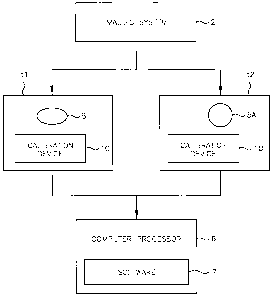
Referring now to FIG. 1, there shown is a simplified block diagram of an
automated system for accurate measurement evaluation of changes in a target
lesion as
imaged by an imaging system as constructed in accordance with one embodiment
of the
present invention. An imaging system 2 produces image data at differing times
ti and t2. A
target lesion 5 in the image data at time ti also appears in the image data at
subsequent time
t2 as target lesion 5A. That is lesion 5 is the same lesion as lesion 5A, but
differences in the
volume sizes of the target lesion at different times are here presumed due to
anticancer
agent treatment for exemplary purposes. The image data is processed in
computer
processor 6 running image processing software 7. Target lesions may include
cancerous
5
CA 02564240 2006-10-25
WO 2005/104943
PCT/US2005/013968
tumors, nodules and the like. The images may also include a calibration device
10,
discussed further in detail below.
The medical imaging system 2 may advantageously include any known medical
imaging system. Some useful known imaging systems include computerized
tomography
scanners, magnetic resonance imagers, positron emission imaging systems, X-ray
imaging
systems vascular interventional and angiogram/angiography procedures,
ultrasound
imaging systems and equivalent medical imaging systems. Scanned target lesions
5 may
advantageously include tumor types specified for application of World Health
Organization (WHO) and RECIST criteria including breast, lung, melanoma,
colon, ovary,
and sarcoma tumors.
In one useful embodiment of the invention, the software 7 automatically
operates to
accurately measure size and volume of the target lesion 5. In this way a
change in volume
can then be estimated given a time difference between acquiring image data of
the target
lesion 5. The method of the present invention determines the degree of error
associated
with each measurement in order to estimate the volume and ultimately the
proportional
.
change in the volume. Automatic methods implemented under computer control
provide
precise repeatability. Calibration methods estimate the measurement error due
to scanner
artifacts. Modeling, simulation and actual nodules function to characterize
the
measurement accuracy with respect to different nodules and their corresponding
appearances in images, such as CT images.
Numerous features of lesions are assessed to determine the variance in the
measurement of a given volume based on its apparent volume. Measurements will
vary
depending on the differences in the signal of the lesion versus background.
Measurement
error variance may advantageously include an estimate reflecting various
portions of a
lesions, such as a nodule that may have a particular edge characteristic.
Thus, for a given definition of boundary for a particular edge, an estimate
can be
made regarding the variance of the measurement. As discussed below, other
factors that
may advantageously be estimated include the extent to which adjacent
structures are
attached to a target lesion, and the influence adjacent structures may have on
volume
estimates. Characteristics of the measuring device may also be included as
factors
influencing error variance. Volume measurements may also be affected by the
inherent
6
CA 02564240 2006-10-25
WO 2005/104943
PCT/US2005/013968
resolution of the imaging system itself as well as by the amount of noise
present in the
image.
Spatial calibration
Standard calibration methods include the scanning of phantoms and measuring
the
quantities of noise, scanner artifacts and image distortion. Phantoms are
synthetic objects
having known dimensions. Due to the physical properties of an imaging system,
such as,
for example, a CT seamier, these factors are spatially dependent. That is, the
measurement
error varies according to the location of the measurement within the body and
the position
of the body within the scanner. Current practice does not take advantage of
such factors,
but instead uses a conservative global distortion figure as provided by the
manufacture.
By conducting phantom studies, maps that characterize the degree of image
distortion, image artifacts and noise for all pertinent regions of the human
body may
advantageously be established for a given imaging system. Once established,
the maps can
be used to determine a more accurate bound for the measurement error of the
measurement
of a tumor.
Error correction
Accurate computer measurement of tumor size from CT images employs an
algorithm for determining the exact location of junctions between a lesion and
other tissue.
The algorithm may process many different types of lesions and use different
strategies to
resolve different situations. An error estimate may be made based on the form
of the image
and the details of the specific algorithmic process for that image. In one
aspect of the
invention a database is created for each identifiable image distortion and a
measurement
error estimate is made from the statistical variation within the database.
In accordance with the present invention, approaches for system error
estimation
include (a) measurements from CT images of calibrated phantoms, and (b)
measurements
from multiple scans of actual lesions from patients. In one useful embodiment,
measurements from slow growing lesions may advantageously be obtained with
short
intervals between scans.
As another example, repeated images of lesions may be scanned at very short
intervals regardless of growth rate to provide error estimates based on a
substantially
unchanged lesion. Such repeated images may be obtained during a biopsy wherein
multiple
7
CA 02564240 2006-10-25
WO 2005/104943
PCT/US2005/013968
images of a lesion are obtained within a few seconds. In addition, when a
human observer
is involved in the measurement process the variation or error due to the human
interaction
can be obtained by human observer trials involving either phantoms or
repeatedly scanned
lesions.
Error associated with specific geometric situations (e.g. a nodule attached to
the
chest wall) can be estimated by taking multiple images of a set of phantoms
that mimic the
situation. The variation between the scans of a synthetic phantom can be used
to
characterize the error boundary for each specific situation tested.
By taking multiple measurements of phantom images, good characterization of
the
scanner system variation parameters can be obtained. For example, scanner
reconstruction
properties such as the point spread function can be accurately determined by
the analysis of
phantom studies and experiments. However, phantom data cannot imitate all
situations
because some nodules exhibit subtle changes in density that are difficult to
model. In such
cases multiple scans of a number of such nodules that do not exhibit apparent
growth can
be used to create a nodule database. One way to do this is by comparing two
scans of the
same lesion taken within a short time interval. The nodule database can then
be applied to
measure the measurement variation between scans in order to estimate the
measurement
error for a given class of difficult nodules.
Specific imaging problems may give rise to specific image artifacts. For
example,
heart motion produces a ripple in the z-dimension of the three-dimensional
image shape.
As a further example, bones in the apical region can produce excessive amounts
of noise.
These and other particular conditions can be identified and error bounds may
be estimated
from a database of similar cases.
Error estimates when manual intervention is required
In some difficult imaging situations a radiologist may intervene in the nodule
segmentation process. Further processing by computer algorithms may then
reconcile the
differences between the radiologist's decisions between scans. An estimate of
the
measurement variation due specifically to the radiologist's intervention by
establishing a
database of such cases. Once all sources of measurement error have been
determined an
overall measurement error can be computed.
8
CA 02564240 2006-10-25
WO 2005/104943
PCT/US2005/013968
Referring now to Fig. 2, there shown is a high level functional block diagram
of an
automated method for determining a bound on the error of a size change
measurement, in
accordance with one embodiment of the present invention. The automated method
for
determining a bound on the error of a volume change measurement comprises the
steps of:
scanning a body part with an imaging system to produce a first set of imaging
data
at step 20;
identifying at least one target lesion in the imaging data at step 30;
rescanning the body part so as to produce a second set of imaging data at step
40;
identifying the at least one target lesion in the second set of imaging data
at step 50;
measuring the at least one target lesion as imaged in both the first set of
imaging
data and the second set of imaging data to determine a first apparent target
lesion
size corresponding to the first set of imaging data and a second apparent
target
lesion size corresponding to the second set of imaging data at step 60;
estimating a change in size by comparing the first and second apparent lesion
sizes
at step 70; and
estimating a variance on the change in size so as to determine a bound on the
change in size measurement at step 80.
The step of estimating a variance on the change in size at step 80 may
advantageously include results from assessing a plurality of factors that
affect
measurement accuracy. Standard statistical methods may be employed to estimate
or
otherwise determine the image measurement variance and other error
measurements
discussed herein. Such techniques include, for example, linear regression,
random effects
models and the like.
Factors that affect measurement accuracy include primary sources of error like
nodule form, scanner parameters, patient factors, algorithm and operator
factors. Many of
these are interrelated. For example, the definition of the boundary of the
nodule will
depend upon the nodule tissue, the point spread function of the scanner,
patient motion,
and other factors. Estimates of error variation are obtained using image
models for the
error factors and obtaining the parameters for these models from measurements
on image
phantoms and patients and also by computer simulations. Paired observations of
the same
patient may be used to reduce error.
9
CA 02564240 2006-10-25
WO 2005/104943
PCT/US2005/013968
Examples of nodule form factors include:
a. Density distribution characteristics, such as
i. homogeneous or variable distribution characteristics, and/or
ii. solid tissue or diffuse tissue characteristics.
b. Geometric shape characteristics of the nodule such as
i. spherical or complex shapes, where complexity may be estimated,
for example, as a ratio of surface area to volume normalized to a
sphere (=.1),
ii. shapes of multiple components,
iii. cavities, and/or
iv. small features close to the reconstruction resolution.
c. Surface characteristics such as whether the nodule is rough (i.e.
exhibiting a
complex surface) or smooth, where a rough surface implies high average
curvature.
Examples of scanner parameters include:
a. Reconstruction resolution further including slice thickness, overlap,
and/or
in-plane pixel size,
b. X-ray energy (dose): kVp and mAs,
c. Reconstruction filter,
d. Gantry rotation speed,
e. Table speed (pitch),
f. Spatially varying point spread function, and/or
g. Calibration.
Examples of patient factors include the following:
a. Location of the scanned area in the body,
b. Size of the body,
c. Degree of inspiration,
d. Respiration motion (especially at the base of the lungs),
e. Small muscle spasms,
f. Lungs apical region, for example, streaking artifacts,
and/or
CA 02564240 2006-10-25
WO 2005/104943
PCT/US2005/013968
g. Health of the lung tissue adjacent to the nodule, noting the presence of
=
scars, emphysema, or other health-related conditions.
Operator factors result from operators that assist in the nodule measurement
process. For example, an operator may manually modify the estimated nodule
boundary
resulting in a measurable contribution to the measurement error that can be
characterized
by observer studies.
Completely automated algorithms typically have situations that are close to
intrinsic decision points. For example, an automated algorithm may consider a
peripheral
bump on a lesion to be an attached vessel or a part of the nodule. Algorithms
may be
instrumented to indicate how close to decision points they operate and hence
factor in the
error associated with falling on the other side of the decision point.
Once an image region has been determined to represent a nodule the variance of
the
measurement may be estimated by considering, for example, the following image
model
factors.
1. Density: Low variance is associated with homogeneous solid tissue density
distribution. High variance is associated with high image noise an low or
spatially
varying density distribution.
2. Shape: Low variance is associated with a spherical shape form and high
variation is
associated with a highly irregular shape containing many bumps or cavities.
3. Surface characteristics: At the boundary (edge) of the nodule region low
variation
is associated with a high image gradient and high variation is associated with
low
image gradient. Further low variation is associated with a smooth surface
while
high variation is associated an irregular surface with high curvatures. The
boundary
region between the nodule and other relatively solid structures such as
vessels or
the chest wall (where there is little or no image gradient evidence of a
boundary)
must be treated in a different manner. For low variation these boundary
regions
should be matched between the two scans in the image segmentation algorithm.
Since these regions are less accurately determined than gradient edges, the
ratio of
non-gradient to gradient edge surface areas is directly related to the
variation.
11
CA 02564240 2006-10-25
WO 2005/104943
PCT/US2005/013968
Assignment of boundaries and incorporation of boundary accuracies using the
method of the invention is discussed herein with reference to FIG. 4-FIG. 11
below.
4. Size: In general the larger the nodule, the smaller the proportion
of partial voxels
the more accurate is the volume estimate. Low variance is associated with
large
nodules (or very fine scanner resolution) while large variance is usually
associated
with a smaller nodule (given a similar structural complexity (shape)).
Situations in which the estimated variance may be used include:
A. When two scans are available, all image data and parameters are considered
to
IA) provide bounds on the estimated growth rate.
B. When a single scan is available, the estimated variance is used to
determine the
minimum time to wait for taking the second scan in order to obtain a
clinically
significant decision. That is the time to measure a malignant growth rate
within the
measurement error bound.
In some situations size will be measured on a two dimensional (2D) area of a
single image
instead a volume estimated from a set of images.
In a preferred embodiment of the present invention each step is carried out by
adaptive software that allows for interaction of a medical professional. One
useful
embodiment of the invention further includes a step of defining the edge of
the at least one
target lesion in the imaging data. Edge definition may be determined by
applying a
threshold and/or a gradient function to the at least one target lesion to
determine a
boundary for the edge. To further aid diagnosis, the adaptive software applies
automatic
segmentation and classification techniques as are well known in the art to
identify
boundaries and segment features, including abnormalities, from body parts,
such as lungs
that are imaged by the imaging system.
In yet another useful embodiment, the method of the invention includes the
step of
automatically estimating a degree of motion for a particular structure. In yet
another useful
embodiment, the method of the invention includes the step of automatically
estimating a
degree of motion for a particular structure includes measuring a degree of
variation of
surface structures and structures outside of the target lesion. In the lung
this will vary
markedly with the location of the target lesion relative to the heart.
12
CA 02564240 2006-10-25
WO 2005/104943
PCT/US2005/013968
In yet another useful embodiment, the method of the invention includes the
step of
automatically matching corresponding images of the at least one target lesion
acquired at
differing times. For example, the software may select the target lesion having
the maximal
size in an image and compare it with the comparable target lesion in a second,
subsequently acquired image. Size measurements may advantageously include
length, area
and three-dimensional volume of the lesion.
In yet another useful embodiment, the method of the invention includes the
step of
selecting the at least one target lesion as an object having a maximal area
and finding a
comparable object obtained at a subsequent time.
In yet another useful embodiment, the method of the invention includes the
step of
spatially calibrating an imaging system using at least one phantom and
measuring noise,
scanner artifacts and image distortion.
Referring now to Fig. 3, there shown is a high-level functional block diagram
of a
method for determining a bound on the error of an image measurement. Process
steps, in
accordance with one embodiment of the present invention, include:
scanning a body part with an imaging system to produce a set of imaging data
at
step 120;
measuring at least one target lesion imaged in the set of imaging data to
determine
an apparent target lesion size corresponding to the set of imaging data at
step 130;
estimating at least one error variance on the first apparent target lesion
size so as to
determine an estimate of overall measurement accuracy at step 140;
using the estimate of overall accuracy measurement to determine a bound on the
target lesion size at step 150; and
determining a time frame based on the estimate of overall accuracy measurement
for performing a second measurement indicative of a clinical change at step
160.
The method of this aspect of the invention is preferably carried out by
adaptive
software residing on a personal computer. In a preferred embodiment of the
invention, the
size change of an target lesion to indicate a significant event is smaller
than specified by
the RECIST criterion. The step of estimating at least one error parameter
advantageously
includes (a) calculating error measurements from computerized tomography
scanner
13
CA 02564240 2006-10-25
WO 2005/104943 PCT/US2005/013968
images of calibrated phantoms, and (b) calculating error measurements from
multiple scans
of patient lesions.
In another useful embodiment of the invention, the adaptive software further
includes a process module for obtaining a variation due to human interaction
using data
from human observer trials conducted with phantoms or repeatedly scanned
lesions of
known size.
In one example embodiment, the set of error factors comprises at least one
factor
selected from the group consisting of:
a point spread function of the imaging device and associated reconstruction
filters;
scanner parameters;
image artifacts caused by high density objects in the same image plane as the
nodule;
patient motion;
change in patient orientation between scans;
when scanning the lung, change in body situation or amount of inspiration;
size of the nodule;
confounding structures attached to the nodule;
nodule density variation;
scanner calibration;
nodule boundary definition; and
operator variation, when a human expert interacts with the measurement
process.
The scanner point spread function may be estimated by a set of test scans with
calibration phantoms. The scanner point spread function may also be estimated
by
scanning a 3D calibration phantom with the patient. Since the phantom
dimensions are
known, the scan provides information for estimating any bias due to scanner
parameters.
The bias information may then be applied to the image data so as to reduce
error due to
scanner parameters.
Differing scanner parameters between at least two scans may advantageously be
measured using a set of phantom scans using both parameter settings to
estimate the
volume bias due to parameter differences. Ideal practice is to use two scans
having the
same parameters.
14
CA 02564240 2006-10-25
WO 2005/104943
PCT/US2005/013968
Image artifacts may advantageously be characterized by computing an image
noise
index based on the spatial frequency content in the region of the object
interest, as for
example, a nodule or tumor. Image artifacts may also be characterized by data
obtained
from consistency studies with phantoms having a similar noise index and other
parameters
provide an estimate of the variation.
Patient motion during scanning may affect results. Common types of patient
motion
include heart motion, patient muscle spasm, respiration, pulsatile motion or
other types of
patient motion during the scan of an target lesion, such as a nodule. Patient
motion error
characterized by heart motion, for example, is detected by repetitive z-axis
variation in the
imaged nodules surface. In addition to patient motion, patient orientation can
affect
imaging results. Changes in patient orientation between scans is measured by
comparing
the orientation of a 3D rigid body matching between at least two scans at
differing times.
Changes in patient situation can be measured by a 3D registration between any
two
scans. Changes in inspiration error may be estimated using studies on a
dataset of scan
pairs. For large changes in inspiration studies on a dataset of scan pairs can
be used to
estimate the bias and variation that this causes.
Where the target lesion is a nodule, nodule size error may usefully be
characterized
by phantom studies using different sized phantoms to determine the intrinsic
measurement
variation for a selected nodule size. Similarly, error due to attached
structures may be
characterized by phantom data using multiple scans and measure variation of
the attached
structures under different conditions. Attached structures may include, for
example, organ
abutments or attachments to similar density organs. Error due to attached
structures may
advantageously characterized by data from nodules of known size where there
are
attachments and multiple scans compared for segmentation consistency.
Error due to scanner calibration may also be characterized by using histogram
matching of image noise from local image statistics. Error due to scanner
calibration may
also be characterized by using calibration phantoms scanned with the body
part.
In one example embodiment, error due to nodule boundary definition may be
characterized by comparing a nodule boundary profile with the point-spread
function. Error
due to nodule boundary definition may also be characterized by conducting
phantom
studies to determine the variation in volume estimate under different
conditions. Error due
CA 02564240 2006-10-25
WO 2005/104943
PCT/US2005/013968
to nodule density is characterized by comparing multiple scans of slow growing
lesions of
known size.
In another example embodiment, error due to operator variation may be measured
by conducting human observer studies with a number of radiologists and
evaluating their
variation under different image quality conditions.
As discussed hereinabove, there are several sources of error in making
measurements. Selecting certain operational modes when performing a scan, such
as
keeping the slice thickness constant, can control some error factors. Other
factors are
intrinsic to the scanning machine such as the Modulation Transfer Function
(MTF) of the
scanning system. In some cases such intrinsic factors, like MTF, may be
specified by the
scanning system manufacturer. Currently there is no universally recognized
standard for
taking images for cancer related measurements. However, the effect of error
factors on the
measurement accuracy enough to raise the level of confidence about a given
measurement
using error variance and measurement accuracy measurements can be estimated or
otherwise derived as discussed herein. Another way to achieve higher
confidence about the
accuracy of a measurement is to scan the patient with a calibration device
each time.
Referring again to FIG. 1, the present invention optionally includes using a
calibration device 10 whenever a patient is scanned where there is
consideration of
performing volume assessment. The calibration device may comprise a synthetic
phantom
scanned simultaneously during the patient scan. In this way the synthetic
phantom will be
subject to the same scanning parameters as the patient. The calibration device
can be made
available at a scanning center, and/or, in addition, a calibration device can
also be given to
a patient so that they can carry the device with them. The calibration device
may
advantageously contain a set of synthetic phantoms of varying size. The
synthetic
phantoms may include a set of highly calibrated spherical shaped objects as
well as a set of
more complex structures.
In one example embodiment, the calibration device set may be held inside an
acrylic or plastic casing and be quite small. For example, any easily
transportable device
ranging in size from about a 2 cm x 2 cm x 2 cm can up to the size of a
standard envelope,
typical book or similar items may be used depending on the scale desired.
Larger or
smaller devices may also be appropriate in some scanning situations. Other
calibration
16
CA 02564240 2006-10-25
WO 2005/104943
PCT/US2005/013968
devices may include wires, beads, rods and similar items of known size and/or
density. The
set may be placed on the patient at the time of the scanning and be subject to
identical scan
parameters. The objects inside the phantom can then be measured. Using
multiple objects
of different sizes and types, that are highly calibrated for size and density,
a measure of
variance can be obtained to account for both bias and reproducibility. In this
way, the
measurement accuracy for a given scanner, using a particular instrument setup
when a scan
is performed on a given patient, can be estimated. Measurement accuracy may
further be
enhanced by using additional information known about the scanning device, such
as
intrinsic factors like MTF as discussed above.
An alternative embodiment of the method of the invention may use an in vivo
calibration device or set. For example, wires, beads, catheters, implantable
devices or
similar items of known dimensions may be in the patient's body for reasons of
calibration
or other medical reasons. These in vivo devices or elements may be used to
calibrate the
scan and correlate scanning results and errors at different times, between
different scanning
situations or both.
Referring now to FIG. 4, a CT image slice through a large pulmonary nodule is
shown. A CT image 214 shows a pulmonary nodule 202 comprising a mass
substantially
bounded within region 208 within lung portion 204. Other body features include
a spinal
portion 206 and other features 210 and 212 adjacent the lung. The pulmonary
nodule 202
typically will include spicules that emanate from the nodule as best shown in
FIG. 6. Those
skilled in the art will understand that typical CT images often do not exhibit
clearly defined
boundaries for lesions such as nodules and surrounding features.
Referring now to FIG. 5, a nodule boundary visualization method is
schematically
shown superimposed on a CT image. In a preferred embodiment color-coded
boundaries .
are represented by differing dashed lines 218, 220 and 222 indicate error
source regions. In
one example, dashed line 220 may correspond to a light green boundary
indicating a region
where there is a well-defined nodule margin, (e.g. having a high image
gradient); therefore,
the expected error of the green boundary will be small. Dashed line 222 may
correspond to
a light red boundary that indicates a region where the image gradient is low,
or where there
are small detailed features (called spicules) of the lesion that may be
discarded from the
volume estimation. The presence of either of a low image gradient or spicules
will
17
CA 02564240 2006-10-25
WO 2005/104943
PCT/US2005/013968
decrease measurement accuracy. Dashed line 218 may correspond to a light blue
boundary
that indicates a region where there is little or no image gradient evidence of
a boundary.
For these situations a radiologist may be permitted as by interactive software
to make a
manual decision as to the location of the boundary. Areas exhibiting low image
gradient
provide the largest source of boundary location error. In this way, placement
accuracy of a
boundary of a nodule may be visualized to indicate the source of error and the
potential
size of the error.
In one embodiment of the invention color-coded boundaries may be automatically
drawn on the display using known graphic software techniques in combination
with
information from the teachings herein. For example, colors may be selected
based on the
error associated with a given boundary as determined by edge-finding software
and the
associated error variance or other parameters as determined in accordance with
the above
teachings. Appropriate keys or legends may also be displayed to aid the
operator in
interpreting the display or images.
Referring now to FIG. 6, an alternate embodiment of a nodule boundary
visualizationk
method is schematically shown superimposed on a CT image. In the alternate
embodiment
the nodule may advantageously include colored boundary lines comprising, for
example,
yellow 224, light blue 218, light green 220 and light red 222, as colors are
here indicated
by dashed lines of varying types. Dual boundary lines encompassing the region
208 may be
used to indicate the estimated bounds of error. That is, the true boundary of
the nodule is
expected to be located within the dual boundary lines. In this example, yellow
224 is used
to outline fine detailed features, such as spicules, which are expected to be
components of
the nodule but are discarded in the measurement process with respect to the
nodule volume
calculation since they are also large sources of measurement error.
Such spicules include complex, but medically insignificant structures, that
are
treated statistically as outliers in accordance with the methods of the
invention. Such
structures tend to be long and thin, but have minute volume. In general,
structures having
small volume in relation to a high degree of error can be discounted so as not
to skew
measurement accuracy results.
Referring now to FIG. 7A and FIG. 7B, an alternate embodiment of a nodule
boundary visualization method is schematically shown superimposed on CT images
18
CA 02564240 2006-10-25
WO 2005/104943
PCT/US2005/013968
acquired at different times. Here a visualization scheme similar to those
described
hereinabove may be used, where at least two scans of a lesion are available
and where the
difference between the scans can also be visualized. FIG. 7A shows a first CT
image 214A
of a nodule 202A acquired at a first time and FIG. 7B shows a second CT image
214B of
the same nodule 202B acquired at a second time. Color coded boundaries 218A,
220A, and
222A are applied to the first image 214A following the techniques described
with
reference to FIGs. 5 and 6 above. Color coded boundaries 218B, 220B, and 222B
are
applied to the second image 214B also following the techniques described with
reference
to FIGs. 5 and 6 above.
Referring now to FIG. 8A and FIG. 8B, another alternate embodiment of a nodule
boundary visualization method is schematically shown superimposed on CT images
acquired at different times showing one example in which the growth or other
size change
of a nodule may be visualized. Here boundaries 218A, 220A, and 222A from the
first
image are superimposed on boundaries 218B, 220B, and 222B obtained from the
second
image. The resulting overlays are displayed, as on a computer monitor or other
suitable
display, to provide a visualization of the change in nodule size as well as an
indication of
measurement accuracy corresponding to the color coded boundaries.
Those skilled in the art having the benefit of this disclosure will recognize
that the
boundary techniques described herein are not limited to the examples. There
are many
possible variations of the these visualization methods including:
1. Applying markings to 3-dimensional renderings of the nodule from all image
slices,
2. Using translucent (e.g. colored) markings so that the underlying structure
can still
be observed,
3. Using line shaded markings,
4. Using graduated markings so that distances can be quantitatively viewed,
5. Adding distance scale and text annotations so that quantitative
measurements are
presented, and/or
6. Any combination of the above.
Referring now to FIG. 9A and FIG. 9B, another alternate embodiment of a nodule
boundary visualization method is schematically shown superimposed on CT images
acquired at different times showing another example in which the growth or
other size
19
CA 02564240 2013-09-06
77501-31
change of a nodule may be visualized. Here the crosshatched areas 301A and
301B
= indicate the region of nodule size change between the two CT images 214A
and 214B. The
crosshatched area may advantageously be displayed on a color monitor as bright
red, for
example. Other colors may also be used.
= 5 Referring now to FIG. 10, another alternate embodinient
of a nodule boundary
visuali7ation method is schematically shown superimposed on a CT image showing
another example in which the growth or other size change of a nodule may be
visualized.
Here the various crosshatched areas 303, 305, 307 and 309 may be displayed as
various
colors to indicate change has occurred and with what degree of certainty. In
one example,
to area 303 may correspond to a yellow region representing higher degree of
change related to=
the original tumor size estimate. Area 307 may correspond to a green region
relating to
uncertainty in size related to the second measurement. Area 307 may correspond
to a red
region representing areas where there is a high probability of change. Area
309 may
correspond to a blue region representing areas where an overlap in uncertain
measurements
= 15 occurs. This map also sets a model for how we might want to
measure response.
Additionally a central point 320 may advantageously be selected so that an
estimate of
change can be made for various quadrants 320A, 320B, 320C and 320D of the
mass. In
some volumes, the change may be quite large compared to' others, and the
degree of
certainty may also be different.
20 Referring now to FIG. 11, another alternate embodiment of a nodule
boundary
visualization method is schematically shown superimposed on a CT image showing
yet
another example in which the growth or other size change of a nodule may be
visualized.
FIG. 11 is substantially the same as FIG. 10 with the addition of boundary
line 313 placed
along that portion of the nodule where change cannot be reliably measured, but
where no
25 change can be reliably determined. This allows= for the remainder of the
nodule to be
analyzed.
While specific embodiments of the invention have been illustrated and
described
herein, it is realized that numerous modifications and changes will occur to
those skilled in
the art. It is therefore to be understood that the appended claims are not
limited to the
30 specific embodiments described in the detailed description.