Note: Descriptions are shown in the official language in which they were submitted.
CA 02565581 2006-11-02
WO 2004/096975
PCT/1B2004/001724
APPARATUS AND METHODS FOR AMPLIFICATION OF
BLOOD STEM CELL NUMBERS
FIELD OF THE INVENTION
[0001] The present invention relates to an apparatus and methods for expanding
stem or progenitor cells in a controllable bioprocess system, providing for
expansion
of the stem or progenitor cells, controlling endogenous factor production, and
providing cell populations (mixtures of stem, progenitor, and mature cells)
that are
useful for transplantation (hematopoietic rescue) and other therapeutic
treatments.
BACKGROUND OF INVENTION
[0002] Hematopoietic stem cells are rare cells that have been identified in
fetal bone
marrow, umbilical cord blood, adult bone marrow, and peripheral blood, which
are
capable of differentiating into each of the myeloerythroid (red blood cells,
granulocytes, monocytes), megakaryocyte (platelets) and lymphoid (T-cells, B-
cells,
and natural killer cells) lineages. In addition these cells are long-lived,
and are
capable of producing additional stem cells, a process termed self-renewal.
Stem cells
initially undergo commitment to lineage restricted progenitor cells, which can
be.
assayed by their ability to form colonies in semisolid media. Progenitor cells
are
restricted in their ability to undergo multi-lineage differentiation and have
lost their
ability to self-renew. Progenitor cells eventually differentiate and mature
into each
of the functional elements of the blood. The lifelong maintenance of mature
blood
cells results from the proliferative activity of a small number of pluripotent
hematopoietic stem cells that have a high, but perhaps limited, capacity for
self-
renewal. In culture, hematopoietic stem cells rapidly commit to differentiated
cell
types, which irreversibly predominate in the culture. This property, along
with their
relative scarcity in blood, presents challenges to the creation of long term,
stable
cultures of pluripotent hematopoietic stem cells.
1
CA 02565581 2006-11-02
WO 2004/096975
PCT/1B2004/001724
SUMMARY OF THE INVENTION
[0003] The present invention provides for an apparatus and methods for
expanding
undifferentiated pluripotent cells of the hematopoietic lineage in culture,
whereby
the cells proliferate in culture with little to no lineage commitment, and
differentiation. The undifferentiated hematopoietic cells generally have the
phenotypes of CD34+, CD34+Lin-, CD133+, NOD/SCID repopulating cells, and rapid
NOD/SCID repopulating cells.
[0004] This bioprocess includes in one aspect, a bioprocess device having a
first cell
culture chamber, a second cell culture chamber, and a conduit in regulatable
fluid
communication with the first chamber and the second chamber. In one
embodiment,
the interior surfaces of the first cell culture chamber, the second cell
culture chamber
and the conduit are substantially closed to the environment. In another
embodiment, one or more of the interior surfaces of the first cell culture
chamber, the
second cell culture chamber and the conduit are substantially open to the
environment. The device includes embodiments wherein the first cell culture
chamber or the second cell culture chamber is semipermeable to oxygen gas and
carbon dioxide gas, but substantially impermeable to liquids. In other
embodiments,
at least one of the first cell culture chamber or the second cell culture
chamber is
adapted to a pump device. In certain embodiments, the bioprocess system is
modular, and the first chamber, the second chamber, or the conduit are
detachable.
[0005] In one aspect, the conduit has a selection element. The selection
element is
used to segregate differentiated cells from undifferentiated cells, and in one
embodiment has affinity for one or more antigens expressed by differentiated
hematopoietic cells, for example but not limited to antigens selected from the
group
consisting of: link antigens, CD2, CD3, CD4, CD8, CD13, CD14, CD16, CD19,
CD24,
CD38, CD45, CD56, CD66b and glycophorin A. In one embodiment, the conduit
further includes a magnet or a magnetizable element, to facilitate segregation
of the
cell subpopulations.
2
CA 02565581 2006-11-02
WO 2004/096975
PCT/1B2004/001724
[0006] Also provided are methods of cell culture. A sample of hematopoietic
cells is
obtained, further including a subset of undifferentiated hematopoietic cells.
The
sample of hematopoietic cells are cultured in culture media and under
conditions
appropriate to cause proliferation of the undifferentiated hematopoietic
cells. The
undifferentiated hematopoietic cells are segregated from differentiated
hematopoietic cells or growth factors, and the segregated undifferentiated
hematopoietic cells are further cultured thereby causing proliferation of the
segregated undifferentiated hematopoietic cells. This method can be carried
out in
either closed or open culture conditions. In various embodiments, the growth
factors are segregated from the undifferentiated hematopoietic cells, by
exchange of
the culture media, by dilution, or by perfusion of the culture. In other
embodiments,
the differentiated hematopoietic cells are segregated from the
undifferentiated
hematopoietic cells by affinity separation, immunoaffinity separation, and the
immunoaffinity separation is performed using a selection element having an
antibody or fragment thereof, for example but not limited to anti-CD2, anti-
CD3,
anti-CD4, anti-CD8, anti-CD13, anti-CD14, anti-CD16, anti-CD19, anti-CD24,
anti-
CD38, anti-CD45, anti-CD56, anti-CD66b, and an anti-glycophorin A antibody.
[0007] The invention also provides methods of preserving cells. A sample of
hematopoietic cells is obtained further including a subset of undifferentiated
hematopoietic cells. The cells hematopoietic cells are cultured in culture
media and
under conditions appropriate to cause proliferation of the subpopulation of
undifferentiated hematopoietic cells; the undifferentiated hematopoietic cells
are
segregated from the differentiated hematopoietic cells and undesired growth
factors;
and the cells are cultured further, thereby causing proliferation of the
segregated
undifferentiated hematopoietic cells. The segregated undifferentiated
hematopoietic
cells are then frozen, e.g., in DMSO, in glycerin, or another suitable
cryopreservative.
These methods can be performed in closed system and open system embodiments.
In various other embodiments, the growth factors are segregated from the
undifferentiated hematopoietic cells, by exchange of the culture media, by
dilution,
3
CA 02565581 2006-11-02
WO 2004/096975
PCT/1B2004/001724
or by perfusion of the culture. In other embodiments, the differentiated
hematopoietic cells are segregated from the undifferentiated hematopoietic
cells by
affinity separation, immunoaffinity separation, and the immunoaffinity
separation is
performed using a selection element having an antibody or fragment thereof,
for
example but not limited to anti-CD2, anti-CD3, anti-CD4, anti-CD8, anti-CD13,
anti-
CD14, anti-CD16, anti-CD19, anti-CD24, anti-CD38, anti-CD45, anti-CD56, anti-
CD66b, and an anti-glycophorin A antibody.
[0008] In another aspect, the invention includes methods of treating a mammal.
A
mammal is first identified, having a disorder characterized by an insufficient
number of hematopoietic cells; a sample of hematopoietic cells is obtained,
e.g., from
a donor for an allograft transplant, or from the mammal for an autologous
transplant, the sample further including a subset of undifferentiated
hematopoietic
cells; the sample of hematopoietic cells is cultured in culture media and
under
conditions appropriate to cause proliferation of the undifferentiated
hematopoietic
cells; the undifferentiated hematopoietic cells are segregated from
differentiated
hematopoietic cells or growth factors; and the segregated undifferentiated
hematopoietic cells are cultured further, thereby causing further
proliferation of the
segregated undifferentiated hematopoietic cells. The mammal is providing with
a
suitable quantity of the cultured undifferentiated hematopoietic cells, and
the
cultured undifferentiated hematopoietic cells increase the number of
hematopoietic
cells in the mammal, thereby treating the disorder. Embodiments of the
invention
include open and closed systems. Disorders suitable for treatment include, for
example but not limited to a cytopenia or an anemia such as those induced by
cancer
treatments, or a genetic defect resulting in aberrant levels of blood cells,
or cancer,
for example a graft versus tumor approach. In one embodiment, cultures of
undifferentiated hematopoietic cells with long-term repopulating potential are
expanded at least a four-fold prior to transplantation in the mammal. In one
embodiment, the invention includes a method for providing a cell population of
4
CA 02565581 2006-11-02
WO 2004/096975
PCT/1B2004/001724
undifferentiated human hematopoietic cells; wherein the number of
undifferentiated
human hematopoietic cells increases by at least 20-fold to form the cell
population.
[0009] The invention also includes in one aspect, a method of providing a
therapeutic protein to a mammal. A mammal in need of a therapeutic protein is
identified; a sample of hematopoietic cells is obtained further including a
subset of
undifferentiated hematopoietic cells; a gene encoding the therapeutic protein
is
introduced into at least one undifferentiated hematopoietic cell; the
undifferentiated
hematopoietic cell having the gene is cultured in culture media and under
conditions
appropriate to cause proliferation of the undifferentiated hematopoietic cell;
the
undifferentiated hematopoietic cells having the gene are segregated from
differentiated hematopoietic cells or growth factors; and the segregated
undifferentiated hematopoietic cells having the gene are cultured thereby
causing
further proliferation of the hematopoietic cells having the gene; and a
suitable
quantity of the cultured undifferentiated hematopoietic cells having the gene
encoding the therapeutic protein, are provided to the mammal as a transplant.
The
cultured undifferentiated hematopoietic cells having the gene proliferate in
the
mammal, and express the therapeutic protein in the mammal. In one embodiment,
the mammal does not demonstrate a pathological immune response to the
transplant
after transplantation. In another embodiment, mammal does not demonstrate a
pathological immune response to the transgene, or its expression products.
[0010] In another aspect, the invention provides a method of providing blood
to a
mammal. A mammal is identified having an insufficient number of hematopoietic
cells; a sample of hematopoietic cells is obtained further comprising a subset
of
undifferentiated hematopoietic cells; the sample of hematopoietic cells is
cultured in
culture media and under conditions appropriate to cause proliferation of the
undifferentiated hematopoietic cells; the undifferentiated hematopoietic cells
are
segregated from differentiated hematopoietic cells or growth factors; the
segregated
undifferentiated hematopoietic cells are cultured further thereby causing
proliferation of the segregated undifferentiated hematopoietic cells; and the
mammal
CA 02565581 2006-11-02
WO 2004/096975
PCT/1B2004/001724
is provided with a suitable quantity of the cultured undifferentiated
hematopoietic
cells as a transplant, wherein the cultured undifferentiated hematopoietic
cells
increase the number of hematopoietic cells in the mammal following the
transplant.
In one embodiment, cultures of undifferentiated hematopoietic cells with long-
term
repopulating potential are expanded at least a four-fold prior to
transplantation in
the mammal. In another embodiment, the invention includes a method for
providing a cell population of undifferentiated human hematopoietic cells;
wherein
the number of undifferentiated human hematopoietic cells increases by at least
20-
fold to form the cell population. Culture of cells is performed using either
closed
systems or open systems. In various embodiments, the undifferentiated
hematopoietic stem cells do not cause graft versus host disease in the mammal
following transplantation.
[0011] The invention also provides in various aspects, method of controlling
cell
proliferation. Levels of one or more growth factors in a cell culture having a
subpopulation of undifferentiated hematopoietic cells, are reduced, wherein
reduction of the growth factor levels allows the undifferentiated
hematopoietic cells
to expand in number in the culture without substantial lineage commitment of
the
cells. The growth factors reduced are, for example but not limited to
hematopoietins, TGF-beta or MIP-1-alpha. In various embodiments, growth factor
levels are reduced by subpopulation segregation, or by media exchange or media
dilution, or by perfusion of the culture. Cell cultures may be closed systems
or open
systems.
[0012] In yet another aspect the invention provides a method of banking blood
for a
mammal. A sample of hematopoietic cells further comprising a subset of
undifferentiated hematopoietic cells is obtained from a mammal; the sample of
hematopoietic cells are cultured in culture media and under conditions
appropriate
to cause proliferation of the undifferentiated hematopoietic cells; the
undifferentiated hematopoietic cells are segregated from differentiated
hematopoietic cells or growth factors; the segregated undifferentiated
hematopoietic
6
CA 02565581 2011-11-21
WO 2004/096975
PCT/IB2004/001724
cells are cultured further thereby causing further proliferation of the
segregated
undifferentiated hematopoietic cells; and the mammal is provided with a
transplant,
including a suitable quantity of the cultured undifferentiated hematopoietic
cells,
wherein the cultured undifferentiated hematopoietic cells increase the number
of
hematopoietic cells in the mammal following the transplant. Culture of cells
is
performed using either dosed systems or open systems. In certain embodiments,
the
invention includes commercial processes for collecting, expanding, and banking
for
a patient, a sample of cultured undifferentiated hematopoietic cells suitable
for
transplant into the patient. The sample is provided by a donor and used in an
allograft, or the patient provides the initial sample, and the cultured
undifferentiated
hematopoietic cells are used in an autologous transplant.
[0013] In various other aspects, the invention provides for a transplant kit.
The kit
includes a population of undifferentiated hematopoietic cells, that have been
expanded at least four-fold in culture, and are suitable for transplant into a
mammal,
particularly a human. Cells provided in the kit are cultured in either closed
systems
or open systems. Also included in the kit are instructions for using the cells
in a
transplant procedure.
[0014] Various other embodiments will be apparent in view of the teaching
provided
herein, and are included in the scope of the invention.
BRIEF DESCRIPTION OF THE DRAWINGS
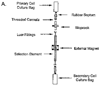
[0015] Figure 1 is a picture of the apparatus used for the bioprocess. The
cell culture
chamber shown in this illustration employs culture bags semipermeable to
gases.
The conduit as shown includes an enrichment element, which separates the CD34+
cells from differentiated and committed lint hematopoietic cells.
[0016] Figure 2 shows graphical representations comparing the kinetic growth
of
total cells (A), CD34+ cells (B), CD344CD38- cells (C) and CFCs (D) using
either
traditional culture dishes or using the present invention.
7
CA 02565581 2006-11-02
WO 2004/096975
PCT/1B2004/001724
[0017] Figure 3 is a graphical representation showing the extent of expansion
of total
cells, CD34+ cells, CD34+CD38- cells and CFCs, expanded by using either
traditional
culture dishes or using the present invention.
[0018] Figure 4A depicts RT-PCR analysis of cultured hematopoietic cells
showing
expression levels of various inhibitory factors. Beta-actin was used as a
control.
Figure 4B is a graphical representation of ELISA analysis showing the
secretion of
inhibitory factors in culture. The detection of TGF-beta1 and MIP4 alpha is
illustrated.
[0019] Figure 5A is a graphical representation of ELISA analysis showing
changes in
TGF-beta1 secretion rates in response to cell selection and enrichment. Figure
5B
depicts RT-PCR analysis showing that column isolated and FAGS sorted link
cells
express TGF-beta1. Beta-actin was used as a control.
[0020] Figure 6A is a graphical representation of ELISA analysis showing
changes in
MIP4 alpha secretion rates in response to media exchange. Figure 6B depicts
semi-
quantitative RT-PCR analysis showing that MIP-1 alpha expression is decreased
in
response to fresh media. Beta-actin was used as a control.
[0021] Figure 7A is a schematic of the closed-system bioprocess. The
bioprocess
consists of two cell culture bags (3 or 7 ml) which are joined through a
conduit
having a subpopulation selection element. The subpopulation selection element
is
used to remove contaminating lin+ cells from culture. Figure 7B illustrates
the effect
of the subpopulation selection element. Representative flow cytometric plots
showing the amount of lin+ cells present pre- (7Bii) and post-selection
(7Biii). A
negative control is also shown that was not labeled with the lin+ antibody
cocktail
(7Bi). Comparisons were made to the commercially available StemSepTM column
(7B
iv, ii, iii). The plots demonstrate the successful removal of lin+ cells when
such cells
are passed through the conduit having the subpopulation selection element.
8
CA 02565581 2011-11-21
, . , =
WO 2004/096975
PCT/1B2004/001724
[0022] Figure 8 shows the absolute numbers of hematopoietic cells generated in
the
bioprocess. Purified UCB lin- cells (1 x 10⁵ cells/1rd) were cultured for 8-
days in the
bioprocess with subpopulation selection and media dilution/exchange occurring
at
day 4. Kinetic growth profiles for total cells (A), CD34+ cells (A), CD34+CD38-
cells
(A), CFCs (B) and LTC-ICs (C) are shown over the 8-day culture period (n=4).
[0023] Figure 9 illustrates the calculated expansions of total cells, CD34+
cells,
CD34+CD38- cells, CFCs and LTC-ICs relative to input for cells grown using the
bioprocess (n=4). For comparison, results of cells grown using standard tissue
culture dishes are also provided. Also shown are the theoretical expansions
that
should be observed in the bioprocess with decreased cell loss (see Section
4.2.5).
This value was calculated by multiplying the observed expansion by a
correction
factor reflecting the percentage of non-specific cell loss. The value of the
correction
factor was 1.3449.
[0024] Figure 10 illustrates human cell engraftment in NOD/SCID mice following
intravenous injection. (A) Representative flow cytometric plots showing the
presence of human CD451-1LA-abe cells in NOD/SCID mice (Aiii). A control
NOD/SCID mouse that did not receive cells is also shown (Au) along with a
representative isotype control (Ai). (B) Limiting dilution analysis was
performed in
NOD/SCID mice to determine the frequency of LT-SRCs present in fresh UCB fin-
cells (n=24) and expanded cells (n=25). The resultant engraftment frequencies
for
fresh lin- cells (Bi) and cultured cells (Bii) are shown for each cell dose
and overlayed
with curves representing results from the maximum likelihood estimator.
Isotype
controls were established for each sample.
[0025] Figure 11A shows a bioprocess configuration that allows for flow rate
control.
A peristaltic pump is used to 'push' cells through the conduit having a
subpopulation selection element. Figure 11B shows the effect of increasing
flow rate
on the recovery and purity of lin- cells exiting the subpopulation selection
element.
Subpopulation selection was performed in which cells flowed through the
selection
9
=
CA 02565581 2006-11-02
WO 2004/096975
PCT/1B2004/001724
element at 0.45 0.03 (gravity induced flow rate; n=4), 0.61 0.04 (n=4),
0.76 0.06
(n=4) and 1.25 0.07 ml/min (n=5). At a flow rate of 1.25 0.07 ml/min, both
percent
recovery and percent purity of lin- cells were maximized. The gravity induced
flow
rate comparison is highlighted with a hashed column and a filled square.
[0026] Figure 12. is a schematic of the experimental protocol. The
experimental
design of all 4 test conditions is shown. Subpopulation selection and/or media
dilution were performed on day 4 with all cultures allowed to incubate for a
total of
8-days. Cultures were established with fresh UCB lin- cells in media
containing
TPO, SCF and FL. S: subpopulation selection; E: media dilution/exchange; NS:
no
selection; NE: no media dilution/exchange. ,
[0027] Figure 13. shows the effects of subpopulation selection and media
dilution on
the expansion of hematopoietic progenitor cell populations. Purified UCB lin-
cells (1
x 105 cells/nil) were cultured for 8-days using the four culture conditions
indicated in
Figure 1. At the end of the 8-day culture period, total cell (A) and
progenitor cell (A,
B, C) expansion was analyzed using phenotypic and in vitro functional assays.
The
fold expansion values shown are in comparison to fresh UCB lin- cells. (*)
Represents significant difference (p<0.05) in comparison to unmanipulated
control
cultures (NS/NE).
[0028] Figure 14. illustrates human cell engraftment in NOD/SCID mice
following
intrafemoral injection. These are so-call rapid repopulating stem cells. May
have
enhanced clinical utility by allowing for "rapid engraftment" following
transplantation. (A) A total of 3 x 10⁵ cells, grown using the four culture
conditions
indicated, were injected intrafemorally into NOD/SCID mice. Examination of
human
cell engraftment in both the right and left femurs was assessed after 2-weeks.
(B) A
representative mouse, transplanted with cells from the S/E condition, showing
engraftment in both the right femur (By) and left femur (Bvi) is represented
along
with a corresponding isotype control (Biv). Also shown is a non-engrafted
control
mouse that did not receive a transplant (isotype, Bi; right femur, Bii; left
femur, Biii).
CA 02565581 2006-11-02
WO 2004/096975
PCT/1B2004/001724
(C) Limiting dilution analysis was performed to determine the frequency of
migrating R-SRCs in fresh UCB Err cells and cultured cells using the
intrafemoral
NOD/SCID assay. Engraftment frequencies for fresh UCB Err (Ci) and expanded
(Cu) cells are shown for each cell dose. The frequency of migrating R-SRCs was
calculated using the maximum likelihood estimator with the overlayed curves
representing results from these analyses. The calculated frequencies of R-SRCs
are
shown on the plots. Isotype controls were established for each sample.
[0029] Figure 15. illustrates human cell engraftment in NOD/SCID mice
following
intravenous injection. (A) A typical flow cytometry plot showing the presence
of
human CD45-EHLA-abc+ cells in intravenously injected NOD/SCID mice (Aiii). A
control NOD/SCID mouse that did not receive cells is also shown (Au) along
with a
representative isotype control (Ai). (B) Limiting dilution analysis was
performed to
determine the frequency of LT-SRCs present in fresh UCB llir cells and
expanded
cells. The resultant engraftment frequencies for fresh En- cells (Bi) and
cultured cells
(Bii) are shown for each cell dose and overlayed with curves representing
results
from the maximum likelihood estimator. Isotype controls were established for
each
sample.
[0030] Figure 16 shows multilineage differentiation of cells engrafted into
NOD/SCID mice. Mice that were found to show human cell engraftment (CD45+)
were analyzed using flow cytometry for their ability to differentiate into
cells of both
lymphoid and myeloid lineages. (Au, Bii) Representative FACS analysis dot
plots
showing CD19 expression on engrafted cells from mice injected intrafemorally
(Au)
or intravenously (Bii). Positive staining indicated that cells were capable of
lymphoid
differentiation. (Aiii, Biii) CD33 expression on engrafted cells from mice
injected
intrafemorally (Aiii) or intravenously (Biii) showed that cells were also
capable of
myeloid differentiation. Corresponding isotype controls are also shown (Ai,
Bi).
[0031] Figure 17 illustrates the endogenous secretion of inhibitory factors is
modulated by subpopulation selection and media dilution. (A) ELISA analysis
11
CA 02565581 2006-11-02
WO 2004/096975 PCT/1B2004/001724
showing changes in TGF-I31 secretion rates in response to subpopulation
selection
and/or media dilution. Conditions undergoing subpopUlation selection resulted
in
significantly lower TGF-131 production levels. (B) RT-PCR analysis showing
that
column isolated and FACS sorted link cells express TGF-131. (C) ELISA analysis
showing changes in MIP-la secretion rates in response to subpopulation
selection
and/or media dilution. Media dilution significantly decreased the secretion of
MIP-
la. (D) Semi-quantitative RT-PCR analysis showing that MIP-la steady state
levels
are decreased in response to fresh media. For all experiments, f3-actin was
used as a
housekeeping gene. Serial dilutions (10-fold at each step) were done for all
RT-PCR
,
experiments as a means to quantify expression levels. Shown above the gel
images
are the corresponding fold-dilutions (neat' denotes undiluted samples). For
all
,
dilutions tested, no genomic contamination was observed. (*) Represents
significant
difference (p<0.05) in comparison to unmanipulated control cultures (NS/NE).
DETAILED DESCRIPTION
[0032] The development of ex vivo culture conditions that facilitate the
expansion of
hematopoietic stem cells (HSCs) would greatly accelerate the clinical
implementation of next generation therapeutics including cell transplantation,
gene
therapy and tissue engineering. In fact, the last few years have shown an
increase in
the clinical utility of such cells in transplantation therapies1-4.
Unfortunately, the
establishment of culture conditions capable of consistently and efficiently
growing
HSCs in vitro has been elusive. Current strategies aimed at expanding HSCs,
primarily through growth factor supplementation or stromal cell support,
generally
result in the expansion of mature progenitors, but are complicated by the
loss5-9 or
moderate expansion7'8,10-12 of more primitive cells following short-term
culture.
These results are somewhat surprising since, in vivo, HSCs have been shown to
have
extensive proliferative potential. Experiments demonstrating that long-term
engraftment in mice can be achieved by the progeny of a single murine 13-15 or
human
16 cell, and that the progeny of a single clone can repopulate multiple
secondary
recipients 17," provide evidence of this potential. Furthermore, serial
transplant
12
CA 02565581 2006-11-02
WO 2004/096975
PCT/1B2004/001724
studies in mice, in which input and output numbers of repopulating stem cells
were
monitored and quantified at each passage, have convincingly shown that
repopulating stem cells are capable of sustained in vivo expansion where
theoretical
150-8400 fold expansions have been calculated 19'20. Therefore, it is apparent
that
simple media supplementation is not sufficient to overcome the growth
inhibitory
effects seen in most in vitro systems.
[0033] Imm unophenotyping is a method that can be used to characterize
hematopoietic cells based on the expression of cell surface antigens. These
markers
are expressed on distinct sub-populations of cells and in combination with
systematic functional analysis of cells expressing particular cell surface
antigens has
led to their categorization based on lineage relationships.
[0034] As used herein, the terms "undifferentiated hematopoietic cell",
"undifferentiated cell", "hematopoietic stem cell (HSC)", and "primitive cell"
are
used interchangeably to describe a pluripotential hematopoietic stem cell that
is
capable of long term in vivo expansion and repopulation when transplanted into
a
mammal. It has been established that the most primitive cell types express the
cell
surface antigen CD34, which is a transmembrane glycophosphoprotein thought to
play an important role in stem and progenitor cell adhesion in BM 72. Cell
populations expressing CD34 and lacking the CD38 antigen (i.e. CD34+CD38-
cells)
have been shown to display primitive cell potentials. For example, the
majority of
SRCs can be found in the CD34+CD38- cell fractions and not in the CD34+CD381-
populations, which are thought to contain more differentiated cell types 7.
The
CD344CD38- phenotype has also been associated with an enrichment of cells
having
LTC-IC characteristics 73. The existence of murine and human CD34- HSCs that
are
capable of long-term multilineage repopulation illustrates that the CD34
antigen
may itself be regulated independently of HSC potential and that CD34
expression
itself is not a requisite HSC marker 14'74-77. Primitive cells have also been
identified
based on the expression of Thy-1, a T-cell related marker 78. Thy-1 expression
allows
for the recovery of LTC-ICs from UCB, BM and human fetal liver mononuclear
cells
13
CA 02565581 2006-11-02
WO 2004/096975
PCT/1B2004/001724
(MNCs) 75 and accounts for all repopulating cells (Thy-1.11o) present in mouse
BM ".
Another marker, CD133 (AC133), a transmembrane receptor glycoprotein has also
been shown to coincide with the enrichment of early hematopoietic progenitors
81.
CD34+CD133+ cell fractions isolated from UCB are highly enriched in primitive
progenitors 82 and SRCs that additionally have the capacity to engraft
secondary
recipients 83'84. Recently, vascular growth factor receptor 2 (KDR) has been
implicated as a marker for primitive cell types. Studies have shown that the
isolation
of BM derived CD34+KDR+ results in an enrichment of human LTC-ICs and SRCs 85.
[0035] The absence of specific antigens can also be used to characterize and
isolate
primitive hematopoietic stem cell populations. For example, human CD34k cells
lacking HLA-DR 86 or CD45RA/CD71 87 identify primitive multipotential
hematopoietic cells capable of self-renewal and differentiation into multiple
hematopoietic lineages. Additionally, isolating cells that lack markers
associated
with mature myeloid and lymphoid cells represents a method of enriching for
primitive cell types.
[0036] As used herein, the term "differentiated hematopoietic cell",
"differentiated
cell" , or "progenitor cell" refers to a lineage committed hematopoietic cell.
These
cells typically express one or more of the antigens CD2, CD3, CD14, CD16,
CD19,
CD24, CD56, CD66b, and glycophorin A, and are termed lineage markers (link).
The
detection of link antigens indicates the loss of pluripotential properties and
that the
cell has become differentiated, or lineage committed. Accordingly, these link
antigens also provide the appropriate antigens for targeted separation of
differentiated cells as described herein, and antibodies to these antigens are
widely
available for immunoseparation procedures.
[0037] As described, the majority of culture conditions investigated to date
result in
the dominance of differentiated cell types and the concomitant decrease in the
frequency and numbers of primitive cells, eventually resulting in culture
extinction.
This undesired end result is a consequence of several competing factors that
can
14
CA 02565581 2006-11-02
WO 2004/096975
PCT/1B2004/001724
influence culture dynamics. One such parameter is the effect of the endogenous
secretion of regulatory molecules, which can be stimulatory or inhibitory to
HSC
proliferation, by different subpopulations of hematopoietic cells in culture.
Using
gene expression and protein secretion analysis, a variety of factors known to
inhibit
HSC expansion were shown to be expressed and secreted by both progenitor and
mature cell types21-28. For example, monocytes are known to secrete
transforming
growth factor (TGF)-(31 and macrophage inflammatory protein (MIP)-la 21'28.
Neutrophils have been associated with the secretion of TGF-131, MIP-la and
tumor
necrosis factor (TNF)-a 21'25 while megakaryocytes secrete interleukin (IL)-3
23. Similar
findings have been documented for erythroid and megakaryocytic progenitors
which have been shown to secrete TGF-f31 27. Furthermore, research has shown
that a
number of these secreted factors can stimulate the secondary secretion of
inhibitory
factors by other cell types. For example, the production of IL-12, TNF-a, IL-
1, or IL-
by monocytes can stimulate lymphocytes to produce MIP-la 29,3 . These
inhibitory
factors are known to prevent HSC expansion in vitro by causing them to remain
quiescent, undergo apoptosis, and/or differentiate into mature cell types 31-
35.
[0038] This phenomenon is exacerbated by the fact that cytokine receptors are
not
specific to HSCs but instead can also be found on cells at different stages of
blood
development. It has been shown that c-kit, flk2/f1t3, c-mpl, IL-6R and GM-CSFR
(the
receptors for SCF, FL, TPO, IL-6 and GM-CSF respectively), can be
differentially
expressed not only on cells from the stem cell compartment but also on
progenitor
and mature cell populations 36-43. The presence of these receptors throughout
the
hematopoietic hierarchy implies that the actions of supplemented cytokines are
not
specific to HSCs but also target more differentiated cells. For example,
cytoldnes
have been shown to stimulate the terminal differentiation and proliferation of
megakaryocyte 44'45, granulocyte 46 and macrophage 47 progenitors. Because of
the
cellular heterogeneity of HSC expansion cultures, cytokine supplementation
would
stimulate the simultaneous proliferation and/or differentiation of stem cells
(and
progenitor cells) which would ultimately result in the formation of large
numbers of
CA 02565581 2006-11-02
WO 2004/096975
PCT/1B2004/001724
progenitor and terminally differentiated mature cell populations. In this
context,
these generated cell populations may then prevent HSC expansion in vitro
through
the secretion of inhibitory factors.
[0039] Further evidence showing that differentiated cells may inhibit stem
cell
growth comes from mouse transplant studies where it has been shown that the in
vivo expansion potential of mouse repopulating stem cells can actually be
limited by
the transplantation of increased numbers of stem cells 19'20. These reports
suggested
that the recovery and production of mature blood cells in recipient mice,
which arise
from the injected repopulating cells, may be responsible for activating
inhibitory
mechanisms which ultimately limit stem cell proliferation. Accordingly,
undifferentiated cells are segregated from these growth factors in culture,
which is
accomplished by for example but not limited to, media dilution, media
exchange,
perfusion, and the like, with the object being to reduce local concentrations
of
growth factors in the culture media.
[0040] A demonstration that endogenously secreted factors can negatively
influence
culture output, comes from studies in which blocking antibodies or agonists
(i.e.
oligonucleotides or competitive receptor blockers) specifically directed
against
individual inhibitory factors have been successful in reversing or preventing
the
effects of known inhibitors such as TGF-(31, MIP-la, (MCP)-1 and SDP-1 in both
in
vitro and in vivo models 48-52. Unfortunately, the use of such blocking
schemes has not
propagated into a higher expansion of repopulating HSCs, perhaps because
multiple
secreted factors are responsible for inhibiting this population 31. 31'48-55
[0041] One model for stem cell expansion involves a negative feedback control
mechanism whereby differentiated blood cells, generated in cytokine
supplemented
cultures, produce soluble factors that, directly or indirectly, prevent HSC
expansion.
This mechanism implies that the removal of these cells or the endogenous
factors
generated by these cells would remove the block to HSC expansion by shifting
the
balance of signals presented to the stem cells (i.e. from supplemented
cytokines and
16
CA 02565581 2006-11-02
WO 2004/096975
PCT/1B2004/001724
secreted cytokines) from those preventing expansion to those favoring
expansion.
The removal of these cells may also provide a mechanism to enrich for cells
that may
secrete stimulatory factors. Usable methods that control and modulate the
endogenous production of stimulatory and inhibitory factors thus overcome
limitations of current HSC expansion systems. The invention disclosed herein
describes an apparatus and processes for expanding HSCs ex vivo in part by
controlling the global effects of endogenously produced inhibitory and
stimulatory
factors.
[0042] The removal of specific target cells from culture, coupled with media
exchange, results in the concomitant decrease in the endogenous production and
overall concentration of inhibitory factors present in culture, which, in
turn, results
in greater expansion of the HSC population".
[0043] The HSCs generated as described herein can be used for a variety of
clinical
applications. For example, the expanded HSCs can be transplanted for
amelioration
of cytopenia and anemia induced by radiotherapy or chemotherapy using
anticancer
drugs, in order to enhance or accelerate immune and hematopoietic recovery
following intensive treatment. Alternatively, the invention can be used for
prevention and treatment of infectious diseases associated with lymphopenia,
such
as the CD4+ T cell depletion seen with chronic HIV infection. The HSCs can be
cultured with differentiating factors to produce specific blood cell types.
For
example, HSCs produced using this invention can be induced to differentiate
into
cells of a desired population and function using known biological agents. In
this
manner, the invention can generate "designer transplants" with a plurality of
functions established to provide the greatest patient care. The HSCs can also
be used
in gene therapy, to express a transgene in a recipient subject, taking
advantage of
their reduced immunogenicity and pluripotential properties.
[0044] The present invention provides for expanding stem or progenitor cells,
particularly of the hematopoietic lineage. The process generally includes
obtaining
17
CA 02565581 2011-11-21
WO 2004/096975
PCT/IB2004/001724
hematopoietic cells that are emichecl for hematopoietic stem and progenitor
cells; for
example lin- cells, and introducing them into a suitable growth medium. The
cells
are maintained in culture and allowed to proliferate. Differentiated cells and
endogenous growth factors are removed, either continuously during culture or
intermittently during the culture process, for example, through performing
media
exchange on the cells remaining in culture, and by targeted separation and
removal
of differentiated cells. The remaining undifferentiated stem cells are
cultured and
allowed to proliferate further. Multiple cycles of culture and selection/media
exchange are performed to expand the cells. Alternatively, differentiated
cells in
various phases of lineage commitment can be selected and propagated further in
accordance with the invention. Likewise, one or more hematopoietirts can be
added
to the culture to force differentiation or lineage commitment. It is preferred
that cell
expansion and selection be performed in a completely controllable,
environmentally
closed-system, in accordance with FDA and other regulations governing the
handling and processing of blood products, and to maintain sterility. These
methods and a representative apparatus for performing this bioprocess, are
discussed in detail below.
[0045] The bioprocess disclosed herein can be practiced as an open system or
as a
closed system as is illustrated in the Examples. Closed systems are generally
sealed
from the environment, and provide a more regulatable sterile microenvironment
for
the culture. Additional benefits to closed systems include increased safety
for
researchers and medical professionals in the handling of biological fluids.
Current
FDA and other administrative guidelines require closed systems for the
handling
and processing of blood cells and products designed to be used in humans, and
are
accordingly preferred. However, open systems exist for the expansion of
hematopoietic stem cells, such as those disclosed herein, and for example US
patents
5,674,750 and 5,925,567, and
other known systems can be modified in accordance with the teachings provided
herein to produce a suitable open system bioprocess.
18
CA 02565581 2006-11-02
WO 2004/096975
PCT/1B2004/001724
[0046] The invention consists of one or more cell culture chambers, capable of
receiving and containing a sample of cells. The cell culture chambers may be
substantially rigid, for example, as in the case of a cell culture flask or
dish, or may
be semi-rigid, for example, as in the case of a cell culture bag. There are
many types
and kinds of cell culture containers (chambers) that are commercially
available, such
as those produced by Corning Costar. Suitable materials are ones that can
withstand
a variety of sterilization techniques including autoclaving and gamma
irradiation
and, for those components which directly contact cells, should also be
biologically
inert. Selection of an appropriate cell culture chamber is made in view of
these and
such other factors as the volume desired, transparency, gas diffusion, open or
closed
design, and the particular selection of the type and kind of chamber would be
apparent to one of skill in the art in view of the teachings provided. A
currently
preferred embodiment employs cell culture bags that are semi-permeable to
oxygen
gas and carbon dioxide gas, but substantially impermeable to water vapor and
liquids such as cell culture media, thus ensuring no or little loss of growth
medium
during culture. Fluorinated ethylene polymers exemplify material suitable for
this
purpose. Other materials that are not gas permeable but meet the appropriate
criteria include polypropylene, stainless steel and other medical grade
materials,
particularly polymers.
[0047] The cell culture chambers may include one or more ports, replaceable
caps or
covers, self-sealing septa such as rubber stoppers, valves, or similar means
that allow
the user to add or remove materials from the chamber without substantial
exposure
of the interior of the bioprocess to the external environment. For example,
these
mechanisms permit the cells, media and other components, such as antibodies
and
growth factors, to be introduced into the chamber, and permit removal of
media,
cells, endogenous soluble growth factors and the like, from the chamber, while
maintaining an environmentally closed system. Vents, regulators or other ports
for
attaching external gas (e.g., oxygen or air) or liquid (e.g., culture media)
sources, or
for attaching pumps or pressure devices, may be provided.
19
CA 02565581 2006-11-02
WO 2004/096975
PCT/1B2004/001724
[0048] In a currently preferred embodiment, a first cell culture chamber is
used for
initial culture and expansion of cells. Following selection, a second cell
culture
chamber is used for subsequent expansion of the desired cells. To maintain the
closed system, this embodiment includes a conduit, which provides a means for
achieving fluid communication between the first cell culture chamber and the
second cell culture chamber. Fluid transfer between the cell culture chambers,
through the conduit, can be regulated by flow regulators, ports or valves,
pumps or
similar devices, as described above.
[0049] The conduit may include a selection element, which may be positioned
within the lumen of the conduit, or which may be external. A selection element
may
take the form of for example an enrichment matrix, such as microbeads
contained
inside the conduit, which have a specific affinity for a ligand, or have a
specific
charge, for example Affi-Gel beads with covalently bound anti-CD34 monoclonal
antibody, or the like.
[0050] The selection element targets and selects cells having particular
phenotypes,
for example, those characteristic of differentiated cells. One role of the
selection
element is to immobilize the differentiated cells to the selection surface,
thereby
reducing the number of differentiated cells in culture, and segregating them
from
undifferentiated cells. For example a selection element may include beads
having
antibodies immunospecific to pan-differentiated hematopoietic cells, such as
those
manufactured by StemCell Technologies. Antibodies provide an excellent means
for
affinity separation of differentiated cells, because many antigenic markers
for
differentiated cells exist, and antibodies to these antigens are commercially
available.
However, antibodies provide one means for targeted immunoseparation, and F(ab)
or F(ab)2 fragments, Fv fragments, and bispecific antibodies (or fragments)
can also
be used, and the descriptions herein of cellular segregation with whole
immunoglobulins is intended to be exemplary and non-limiting. Cells can be
separated by numerous other methods, such as FACS, lectin affinity, and other
methods know in the art.
CA 02565581 2006-11-02
W020041096975
PCT/1B2004/001724
_
[0051] The selection element targets and selects cells having antigenic
markers
characteristic of undifferentiated hematopoietic stem cells, providing for
their
removal from the heterogeneous culture. For example, a CD34+ expressing cell
may
be contacted with, and immobilized to an enrichment element having CD34
affinity.
In either embodiment, the designations positive selection and negative
selection will
apply with respect to the particular cells targeted, the selection element
used, and
whether the selection element targets differentiated cells or undifferentiated
cells,
e.g., if the target cell is CD34+ and the selection element has affinity for
CD34, then
isolation of CD34 + cells is an example of positive selection; but if the
target cell is
CD34 + and the selection element has affinity for pan-differentiated
hematopoietic
cells, isolation of CD34 + cells by binding to the matrix and removal from the
culture
of differentiated cells provides an example of negative selection.
[0052] Segregation of differentiated cells from undifferentiated cells can be
accomplished by many methods. Positive or negative selection methods are
preferred. Selection can be accomplished in a particular location within the
apparatus, such as within the conduit using a selection element, and cells can
be
segregated in one step, such as during passage through the conduit.
Alternatively,
segregation may take several steps. For example, bispecific antibodies are
added to
the cell culture along with a magnetic colloid, the bispecific antibodies
having
affinity for the magnetic colloid and for a lin+ antigen. This process
effectively
attaches a magnetic colloid to a lin+ cell. The magnetically labeled cells are
passed
through the conduit, which is itself placed in a magnetic field. Other
modifications
are described herein and will be apparent to those of skill in the art in view
of the
teachings provided. The segregation of undifferentiated cells and
differentiated cells
is thus believed to be routine.
[0053] The bioprocess described herein may employ a continuous process of
growth
and selection, or a discontinuous process of growth an selection. In a
continuous
process, cells are cultured and selection of target cells (either positive
selection or
negative selection) is effectuated without removing the cells from media or
21
CA 02565581 2006-11-02
WO 2004/096975
PCT/1B2004/001724
otherwise interrupting the cell culture process. In a discontinuous process,
culture
and selection proceed in a stepwise manner. Where a modular closed system
apparatus is used, it may be more convenient to employ a discontinuous
bioprocess
since the chambers can be removed from the conduit and placed in an incubator.
without having to keep the apparatus assembled.
[0054] Under most conditions, ex vivo HSC cultures will attempt to
recapitulate
hematopoiesis and as such will eventually form a heterogeneous population of
cells
containing components of the hematopoietic system. Insight into the overall
developmental potential and primitiveness of these cells would provide
information
about the ability of a specific culture methodology to expand primitive cell
types.
Developing this knowledge requires robust and quantitative monitoring of
cells, that
are at different stages of differentiation. Various assays have been developed
which
identify these cells based on distinct functional properties. Cell function
can be
queried in vitro using established retrospective assays that detect the
presence of
committed and multipotent progenitor cells based on the formation of
morphologically distinguishable colonies. Colony forming cells (CFCs) are
progenitor cells that can be detected by the formation of erythroid, myeloid
or mixed
(i.e. both erythroid and myeloid) cell containing colonies after 2-3 weeks of
culture in
semi-solid media (methycellulose). Long-term culture-initiating cells (LTC-
ICs) are
more primitive than CFCs and can be enumerated by their ability to give rise
to
CFCs after greater than 5-weeks of culture with stromal cells 57'58. The
stromal cell
elements, which are composed of mesenchymal cells including fibroblasts,
endothelial cells, adipocytes and osteogenic cells, produce a variety of
soluble factors
that support the long-term proliferation and maintenance of LTC-ICs. The
sensitivity
of this assay can be increased through the use of genetically engineered
murine
fibroblast (M2-10B4) cell lines that secrete factors known to enhance the
detection
and maintenance of LTC-ICs
[0055] In vivo functional assays offer the best indication of the
developmental
potential of a hematopoietic cell population. This is because they directly
test the
22
CA 02565581 2006-11-02
WO 2004/096975
PCT/1B2004/001724
potential for a stem cell population to contribute to the development or re-
development of a particular organ, tissue or system following intravenous
injection.
For example, murine HSCs have been identified based on their ability to
reconstitute
hematopoiesis after transplantation into an immunocompromised and
hematologically compromised host. Till and McCulloch 61 first reported the
existence
of such a cell type when they injected syngeneic BM cells into irradiated
mouse
recipients and observed the formation of multi-lineage colonies in the spleen.
Interestingly, these colonies contained cells that could form additional
colonies upon
transplantation into secondary hosts.
[0056] While many animal models have been developed to detect the presence of
human HSCs 62-65/ the most widely-used involves non-obese diabetic/severe
combined immurtodeficient (NOD/SCID) mice 66'67. Cells capable of engrafting
these
recipients have been termed NOD/SCID-repopulating cells (SRCs) 68 and are
considered to be human HSCs that home to and engraft the murine BM, where they
subsequently proliferate and differentiate into multiple blood cell lineages
69,7 . This
assay has been successfully used with standard limiting dilution analysis
(LDA) as a
means to quantify HSC content in a given cell sample 71.
[0057] The present invention provides an apparatus and methods for the
expansion
of hematopoietic stem and progenitor cells used, for example in a therapeutic
transplant to repopulate the blood of a mammal. Since these cells are
relatively rare,
a starting cell population is first obtained using methods known in the art.
Blood,
such as mobilized peripheral blood (PB) and bone Marrow (BM) are suitable
sources,
but umbilical cord blood (UCB) provides an enriched source of these
undifferentiated cells. Further enrichment of the hematopoietic stem or
progenitor
cell content from these sources can also be performed prior to culture, for
example
by purifying mononuclear cell (MNC) fractions. This can be accomplished by
using,
e.g., centrifugation such as through a Ficoll gradient. Isolation of more
enriched
populations of hematopoietic stem and progenitor cells can be accomplished
using
fluorescence-activated cell sorting, immobilization to glass wool, column
separation
23
CA 02565581 2006-11-02
WO 2004/096975
PCT/1B2004/001724
or bead separation techniques, or other known methods of enrichment. Many
suitable types are known in the art.
[0058] In accordance with the present invention, HSCs in culture are separated
from
inhibitory hematopoietins (through subpopulation selection and/or media
exchange
procedures) to prevent their differentiation and commitment to particular
lineages.
Hematopoietins are a generic name given to hematopoietic growth factors (HGF)
or
hematopoietic cytokines, which act on cells of the hematopoietic system. These
factors are active at all stages of development, and accordingly these
hematopoietins
will be removed from the bioprocess to prevent HSC differentiation.
[0059] Hematopoietic growth factors are produced by many different cell types
including those not belonging to the hematopoietic system. These factors are
either
secreted or they exist in membrane-bound or matrix-associated forms. They may
have different modes of action also, such as autocrine, paracrine, or
juxtacrine
growth control. Production of hematopoietic factors is regulated strictly, i.
e., they
are synthesized by activated cells under certain conditions rather than being
produced constitutively all the time. Many observations point to the existence
of an
ordered hierarchy and a concerted action of factors involved in the
development of
the hematopoietic system. These factors are required for the maintenance of
hematopoietic stem cells, their proliferation, their differentiation into
different
hematopoietic lineages, and for the maintenance of a stable equilibrium
between
proliferation and differentiation. These factors allow an organism to shift
this
equilibrium to one or the other side, as required, for example, under stress
conditions. Many of these factors overlap in their biological activities.
Teleologically
this guarantees a high efficiency and also allows substitution and/or
complementation of individual components the functions of which may have been
impaired, for example, under pathological conditions. In addition, responses
elicited
by these factors are usually contextual, i.e. these responses depend on the
presence
and concentration of other cytokines and/or factors in the environment of the
responding cells. The majority of studies aimed at stimulating HSC expansion
in
24
CA 02565581 2006-11-02
WO 2004/096975
PCT/1B2004/001724
vitro focus on the use of exogenous cytokine supplementation strategies.
Cytokines
interact with HSCs via three classes of transmembrane receptors; 1) those with
intrinsic tyrosine kinase activity, 2) those that interact with the gp130
subunit and 3)
those that interact with Janus kinases (JAKs) 88'89. Over the years, the use
of phenotypic
and functional assays has identified a number of cytokines which have distinct
stimulatory effects on primitive hematopoietic cells. These include flk2/f1t3
ligand
(FL), stem cell factor (SCF), interleukin (IL)-6, IL-6/soluble IL-6-receptor
(SIL-6R), IL-
11, thrombopoietin (TPO), IL-3, IL-1, IL-12, granulocyte- colony stimulating
factor
(G-CSF) and granulocyte- macrophage- colony stimulating factor (GM-CSF) 90-
101. The
first reported use of stroma-free cytokine supplemented cultures, which
contained
IL-1, IL-3, IL-6, G-CSF, GM-CSF and SCF, supported a significant expansion (66-
fold)
of colony forming unit- granulocyte- macrophage (CFU-GM) progenitor cells 182.
Accordingly these hematopoietins can be introduced to the bioprocess to
modulate
differentiation of HSC's, or can be removed.
[0060] Some factors, such as those mentioned earlier, negatively regulate
processes
of hematopoiesis. For example, they may selectively inhibit the proliferation
of some
types of hematopoietic cells and may even induce cell death. For example, it
has
been shown that the addition of TGF-13 to hematopoietic cell cultures directly
inhibits
the expansion of repopulating stem cells 103, LTC-ICs 48 and primitive CFCs
10455 but
has no effect on more mature progenitors 1". Similar is the finding that TGF-
13 preferentially inhibits the growth of CD34+CD38- cells whereas more mature
CD34+CD38+ cells are poorly affected 188. The functional effects of TGF-I3
have been
attributed to its ability to prevent cells from progressing through the cell
cycle. It has
been shown that in the presence of TGF-P primitive cell populations (including
CD34+ cells) are unable to transition from either Go to Gi or Gi to S phase
presumably
due to the up-regulation of the cyclin dependent kinase (cdk) inhibitors p15,
p27 and
p21 31'187. Finally, TGF-f3 may also elicit some of its actions by down-
regulating the
expression of receptor types whose signaling is important for the in vitro
growth of
HSCs including c-kit 1 8,1 9, c_mpi 100 and flogik2 107,110. MIP-la has been
shown to
CA 02565581 2006-11-02
WO 2004/096975
PCT/1B2004/001724
inhibit the proliferation of primitive hematopoietic cells including CFU-GEMM
and
CFU-GM even in the presence of stimulatory factors 33'111413. In vivo
administration of
MIP4a into mice (C3H/HeJ and BDF1 strains) significantly decreases the number
of
primitive progenitor cells in cycle as assayed using the thymidine kill assay
(which
effectively kills cycling cells) 114 and protects stem and progenitor cells
from the
cytotoxic effects of hydroxy urea 115. Interestingly, MIP-1a has little
effect, or even
stimulatory effects, on more mature progenitors 116 suggesting that MIP-1a may
be a
pleiotropic factor. IL-3 is another cytokine thought to have inhibitory
functions. It is
a controversial cytokine because of conflicting reports regarding its ability
to
stimulate or inhibit HSC expansion. IL-3 has been linked to the growth of
primitive
cells including LTC-ICs and CFCs, and is often found in cytokine combinations
reported to be effective in expansion cultures 117. Conversely, several
studies have
indicated that IL-3 can abrogate the expansion and self-renewal of primitive
stem
cells in a concentration dependent manner 118 and, in both human and murine
models, IL-3 has been shown to impair the reconstituting ability of HSCs "A".
These
observations are somewhat clarified by the recent finding that IL-3 may
prevent
HSCs from homing to the BM by impairing their chemotactic response to stromal
derived factor-1 (SDF-1) through the CXCR4 receptor 120/ thereby resulting in
the in
vivo clearance and destruction of potential engrafting cells in non-
hematopoietic
tissues. Additionally, the presence of TNF-a in cultures supplemented with
stimulatory cytokines including SCF 121 and FL 34,122 can potently inhibit the
proliferation of progenitor cells, likely by promoting apoptosis 1" through
Fas (a
member of the TNF receptor family) signaling 124. Examples of other inhibitory
cytokines include monocyte chemoattractant protein (MCP)-1 52 and SDF-1
125,126.
Those hematopoietins can be introduced into the bioprocess or removed as
described.
[0061] As a whole the action of many of these factors underscores the problems
associated with continuous culture of stem cells, since hematopoietins
generally
need to be removed to prevent lineage commitment. Alternatively, they may be
26
CA 02565581 2006-11-02
WO 2004/096975
PCT/1B2004/001724
added to specific cultures to force lineage commitment. In contrast, certain
hematopoetins can preserve the naive and undifferentiated state of CD34+
cells, and
their addition to or enrichment in the bioprocess may improve yield.
Modulation of
hematopoietins in the bioprocess is thus considered within the abilities of
one skilled
in the art of the teachings provided herein.
[0062] Exemplary culture conditions for growing HSCs are given in the
Examples,
but generally in accordance with the invention, a sample of cells containing a
subset
of HSCs is first obtained then cultured. The cultured cells are then
maintained for a
growth period suitable for allowing proliferation to occur, which may include
media
exchanges to remove soluble growth factors, after which time the HSCs are
segregated from the other differentiated cells. The HSCs are then allowed to
proliferate again as described. The segregation of differentiated cells can be
performed again if necessary. At the end of the culture period the expanded
HSCs
can be preserved by freezing after addition of, for example glycerin, DMSO or
a
suitable cryopreservative, or used directly in a therapeutic procedure. It is
important to note that the above steps can be performed with the entire
bioprocess
apparatus assembled or in separate parts in which cell culture is carried out
independent of cell segregation.
[0063] Clinical uses for HSCs include, for example, the therapeutic treatment
of
blood cancers treatment of anemia, treatment of hereditary blood disorders,
replenishment of blood cells following high dose radiation and chemotherapy in
the
treatment of cancer, graft-versus-tumor treatment of cancer, treatment of
autoimmune disorders, and in gene therapy approaches.
[0064] For the therapeutic treatment of blood cancers, including lymphoblastic
leukemia, acute myeloblastic leukemia, chronic myelogenous leukemia (CML)
Hodgkin's disease, multiple myeloma, and non-Hodgkin's and B-cell lymphomas, a
patient's own cancerous hematopoietic cells are first destroyed by high dose
radiation and chemotherapy. A matched donor (having similar HLA profiles)
27
CA 02565581 2006-11-02
WO 2004/096975
PCT/1B2004/001724
provides the source of transplantable HSCs, which are isolated and expanded
according to the methods provided herein. The transplant of undifferentiated
cells
provides for long term repopulation of the blood of the recipient. Non-
cancerous
blood disorders amenable to treatment by HSC therapy include aplastic and
other
types of anemia. The transplant of undifferentiated cells provides for long
term
repopulation of the blood of the recipient. Using UCB, multiple studies have
demonstrated that cell dose is an important determinant of patient survival in
stem
I cell transplantation scenarios 127. Wagner et al. (2002) reported that
the rate of
engraftment is decreased in patients receiving fewer than 1.7 x 10⁵ CD34+
cells/kg
body weight (72% versus 93% in patients who received larger doses) 128.
Likewise, it
was found that transplants of 3.7 x 10⁸MNC/kg resulted in more rapid
engraftment
than patients who received only 3.7 x 10⁷ MNC/kg (i.e. one log less), although
patients receiving the lower cell dose also showed good engraftment 127'129.
Based on
these results, it was recommended that the minimum cell dose for UCB
transplants
be 3.7 x 10⁷ MNC/kg 127. Because typical UCB collections contain an average of
approximately 1.4 x 10⁹MNC (Cairo, Blood, 1997, 90:4665), it was calculated
that an
average cord blood sample would be sufficient to transplant a patient weighing
a
maximum of approximately 37 kg (-81 lbs). Using similar calculations, it was
reported that 75% of the greater than 4000-banked samples at the Toronto
Umbilical
Cord Blood Bank contain only enough cells to be useful for pediatric bone
marrow
transplantations (i.e. patients weighing -24 kg or -53 lbs) 13/3. Therefore,
in order for
these, and other banked samples to be a useful source of HSCs for single or
multiple
adult transplants, or for multiple tissue regeneration therapies, their HSC
content
must be increased. The ex vivo expansion of HSCs described herein provides
such a
provides a solution. The bioprocess can also be used to expand
undifferentiated
cells from adult sources, for example a donor provides his or her own bone
marrow
or peripheral blood, thus eliminating immune mismatch in the event of a
transplant
of these cells back to the donor. Alternatively, the bioprocess can be used to
expand
pluripotential hematopoietic cells that are allogeneic but not immunogenic,
and thus
suitable for transplant purposes.
28
CA 02565581 2006-11-02
WO 2004/096975
PCT/1B2004/001724
[0065] The bioprocess may be used to express a therapeutic protein from the
undifferentiated cells, which have been genetically modified ex vivo to
incorporate a
transgene encoding the therapeutic protein. The hematopoietic cells are
obtained,
transfected with the transgene, and expanded in culture as described.
Differentiated
cells are removed from culture, and the undifferentiated cells are assayed for
expression of the transgene. Cells positively expressing the transgene are
transplanted into a mammal. The low immunogenicity of stem cells makes this
cell
type well-suited for gene therapy.
[0066] Other uses of the invention are contemplated. For example, the
bioprocess
can be used by commercial and non-profit blood banking organizations, to
expand a
subpopulation of undifferentiated cells, e.g., lin-, CD133+, CD34+, long-term
repopulating NOD/SCID and other undifferentiated cells, that can later be
frozen
and stored, or used in a transplant procedure. A donor may provide the sample
of
cells, or a patient about to undergo a medical procedure may provide the
source of
cells that will be expanded. In the latter case, the cells are a perfect
immunological
match for the recipient. Commercial methods of storing, processing and
providing
undifferentiated are thus included within the scope of this invention.
[0067] The invention is now described in specific terms by the foregoing
examples,
which are illustrative only and are intended to be non-limiting and specific
embodiments, whereas the full scope of the invention shall be determined
solely by
the claims.
EXAMPLES
1. CELL SAMPLE COLLECTION AND PURIFICATION
[0068] UCB samples were collected from consenting donors according to the
procedures accepted by the ethics board of Mt. Sinai Hospital (Toronto, ON,
Canada) and centrifuged over 10% pentastarch (Bristol-Myers Squibb Canada,
Montreal, QC, Canada) to obtain the mononuclear cell (MNC) fraction. Lineage
depleted (lin-) cells were isolated from the MNC fraction using the StemSepTM
29
CA 02565581 2006-11-02
WO 2004/096975
PCT/1B2004/001724
system according to the manufacturers protocol (Stem Cell Technologies,
Vancouver,
BC, Canada). Briefly, MNCs were collected in Hanks Buffered Saline Solution
(HBSS;
Gibco, Rockville, MD) containing 2% human serum (HS) at a concentration of 50
x
106 cells/ml. The selection antibody cocktail was then added at a
concentration of 100
111/m1 cell suspension. The antibody cocktail used removes cells expressing
CD2,
CD3, CD14, CD16, CD19, CD24, CD56, CD66b, and glycophorin A. The cells were
then incubated for 15 min at room temperature (RT) after which time a magnetic
colloid was added at a concentration of 60 0/m1 cell suspension. The cells
were then
allowed to incubate an additional 15 min at RT. These steps effectively attach
a
magnetic particle to target cells (lin+ cells) that are to be removed from the
initial
MNC sample. The cells were then passed through a magnetic column, containing
magnetic beads, to isolate the lin- cell fraction. The magnetic column was
placed in
an external magnetic field prior to the separation step.
2. BIOPROCESS ASSEMBLY
[0069] The bioprocess apparatus was assembled using the following procedure:
[0070] Two 3 ml FEP culture bags (VueLife , American Fluoroseal Corporation,
Gaithersburg, MD) were used as cell culture chambers to culture cells pre- and
post-
selection. Each bag was fitted with a self-sealing rubber septum (InterLink,
American Fluoroseal Corporation) at its inlet port.
[0071] An approximately 4-5 inch length of 1/8 inch internal diameter FEP
lined
Tygon tubing (Cole-Parmer, Vernon Hills, IL) was used as the conduit, and was
adapted to house the selection element, in the form of enrichment beads. The
beads
were held in the tubing by first punching out two 0.12"-0.15" diameter pieces
of 80
mesh 430 stainless steel screening (Stem Cell Technologies) and forming them
to fit
snugly into the inside diameter of the tubing (all done using a machined punch
and
form). One of the screens was then placed into the tubing (about half-way
down)
using a small metal plunger. Care was taken to insert the screen so that its
flat face
was perpendicular to the length of the tubing. The enrichment beads (Stem Cell
CA 02565581 2006-11-02
WO 2004/096975
PCT/1B2004/001724
Technologies) were then placed in the tubing until they filled a length or
approximately 1" after which the second screen was inserted into the tubing to
hold
the beads in place. These beads aid in the magnetic separation of cells as
described
above. 1/8" male luer fittings (Cole-Parmer) were then placed into each end of
the
tubing to which were attached two threaded lock cannulas (American Fluoroseal
Corporation). The carthula mates with the self-sealing rubber septums for
sterile
filling, sampling or emptying of culture bags. This selection element is also
referred
to as the 'enrichment element'.
[0072] The enrichment element was then connected between the two culture bags
via
the mating of the septum and cannula. The entire assembled product in its
entirety
is now called the "bioprocess apparatus" or "bioprocess". As mentioned above,
the
bioprocess can be used entirely assembled or in discontinuous sections, thus
permitting cell culture to be performed separately from cell segregation. In
one
modification, the use of the bioprocess as a single assembled product would
require
that a three-way stopcock (Cole-Parmer) be placed between one of the culture
bags
and the tubing. The stopcock would facilitate filling of the culture bag with
cells and
growth medium. In this case, the sterile septum would be attached to the
stopcock
inlet port.
3. CELL SEEDING AND CULTURE IN OPEN SYSTEM
[0073] Lin- cells were seeded at 1 x 10⁵ cells/m1 in StemSpariTM media (Stem
Cell
Technologies) containing Iscove's MDM, 1% BSA, 10 vg/m1 rh insulin, 200 jig/m1
human transferring, 10-4M 2-mercaptoethanol and 2 mM L-glutamine. The media
was supplemented with 100 ng/ml SCF (Amgen, Thousand Oaks, CA), 100 ng/ml FL
(Amgen) and 50 ng/ml TPO (R&D Systems, Minneapolis, MN). Approximately 1.5
ml of the cell suspension was then placed into the wells of a 24-well plate
and
maintained at 37 C in a humidified atmosphere of 5% CO2 in air for 8-days. Re-
selection after 4-days in culture (i.e. to remove link cells generated during
culture)
was performed.
31
CA 02565581 2006-11-02
WO 2004/096975
PCT/1B2004/001724
4. CELL SEEDING AND CULTURE IN BIOPROCESS
[0074] Lin- cells were seeded at 1 x 10⁵ cells/ml in StemSpanTM media (Stem
Cell
Technologies) containing Iscove's MDM, 1% BSA, 10 micrograms/m1 rh insulin,
200
micrograms/ml human transferrin, 10-4M 2-mercaptoethanol and 2 mM L-
glutamine. The media was further supplemented with 100 ng/ml SCF (Amgen), 100
ng/ml FL (Amgen) and 50 ng/ml TPO (R&D Systems). Approximately 2-3 ml of the
cell suspension was then injected into a 3 ml culture bag (through the septum)
using
a sterile syringe attached to a threaded cannula. The culture bag and cell
contents
were then maintained at 37 C in a humidified atmosphere of 5% CO2 in air. Re-
selection after 4-days in culture (i.e. to remove lin+ cells generated during
culture)
was performed as described.
5. CELL SELECTION AND MEDIA EXCHANGE IN THE BIOPROCESS
[0075] Lin- cell selection in the bioprocess was performed. Incubation of the
cells
with antibody cocktail and magnetic colloid was carried out inside the culture
bag
with the cells remaining in growth medium. The antibody cocktail and magnetic
colloid were used at half the amounts suggested by the manufacturer. Following
these steps, the cells were then allowed to flow through the enrichment
element
(either by directing flow via the stopcock or by attaching the bag to the
enrichment
element via septum/cannula mating). For flow rate experiments, a peristaltic
pump
was adapted to the primary cell culture container. Similar to the StemSepTM
system,
the conduit having an enrichment element was placed in an external magnetic
field
prior to separation. Upon passing through the conduit, and thus the enrichment
element, the now purified lirr cells flow into the second culture chamber and
were
collected for further processing.
[0076] Media exchange can be performed on enriched lin- cells in the culture
bags.
Briefly, the cells are centrifuged by placing the culture bag into a 15 or 50
ml conical
centrifuge tube (tissue paper can be used to stabilize the bag and prevent the
bag
from collapsing). The tube is then centrifuged for 5 min at approximately 1000
rpm
32
CA 02565581 2006-11-02
WO 2004/096975
PCT/1B2004/001724
after which time a cell pellet is visible at the bottom of the culture bag.
The media is
then carefully removed through the self-sealing septum using a sterile
syringe. Care
must be taken to prevent cells from being removed. Fresh media is then added
through the same septum. This process can be adapted for automated media
exchange.
6. PHENOTYPIC ANALYSIS
[0077] Phenotypic analysis was performed using flow cytometry. CD34, CD38,
CD19
and CD33 expression was analyzed by first collecting and washing cells in ice
cold
HBSS containing 2% HSA (HBSS-HS). 5 x 10⁴ to 100 x 105 cells were then
resuspended in 100 ul HBSS-HS containing saturating amounts of the appropriate
antibodies or isotype controls for 30 min on ice. Stained cells were then
washed
using HBSS-HS and resuspended in a 10% formalin solution (Fisher, Nepean, ON,
Canada). Fixed cells were then analyzed immediately using flow cytometry or
placed at 4 C for later analysis. CD45 and HLA-abc expression was assessed as
described below. Flow cytometric analysis was performed using a Coulter EPICS
XL
with 4-Color Expo software or a Coulter EPICS Elite with Elite software
(Beckman
Coulter, Miami, FL). CD34, CD38, CD19, CD33, CD45 and HLA-abc expression was
assessed using anti-CD34-PE, anti-CD38-FITC, anti-CD19-PE, anti-CD33-PE, anti-
CD45-PE/FITC and anti-HLA-abc-FITC (Beckman Coulter). Isotype controls were
tested using anti-IgGi-PE, anti-IgGi-FITC and anti-IgG2a-FITC (Beckman
Coulter).
7. COLONY FORMING CELL (CFC) AND LONG TERM CULTURE-INITIATING
CELL (LTC-IC) ASSAYS
[0078] Non-cultured or post-cultured cells were assayed for CFC content by
plating
500 cells into methylcellulose media (MethoCultTm, Stem Cell Technologies)
containing 1% methylcellulose in Iscove's MDM, 30% FBS, 1% BSA, 10-4 M 2-
mercaptoethanol, 2 m.M L-glutamine, 50 ng/ml rhSCF, 10 ng/ml rhGM-CSF, 10
ng/ml
rhIL-3 and 3 units/ml rhEPO. After 14 days of incubation, duplicate or
triplicate
cultures were scored for CFC content and frequency.
33
CA 02565581 2006-11-02
WO 2004/096975
PCT/1B2004/001724
[0079] LTC-IC assays were performed by initially seeding 2000 cells on to
irradiated
human stromal cells (M2-10B4) in 6-well plates. Cell were allowed to incubate
for 5
weeks at 37 C with weekly half media exchanges using MyeloCultTM media (Stem
Cell Technologies) containing alpha-MEM, 12.5% HS, 12.5% FBS, 0.2 mM inositol,
20
micromolar folic acid, 10-4M 2-mercaptoethanol, 2 m.M L-glutamine, and 10-6 M
freshly dissolved hydrocortisone. After 5-weeks, the entire contents of each
well
were harvested by trypsinization and plated into methycellulose media (see
above).
LTC-IC content and frequency was then determined by analysis of CFCs present
after 14-days.
8. TRANSPLANTATION OF CELLS INTO NON-OBESE DIABETIC/SEVERE
COMBINED IMMUNODEFICIENT (NOD/SCID) MICE
[0080] Either fresh lin- cells or the progeny of lin- cells grown using the
culture
conditions described were transplanted into sublethally irradiated (3.6 Gy) 8-
10
week old NOD/SCID mice using either intravenous (to test for LT-SRCs) or
intrafemoral (to test for R-SRCs) injection. NOD/SCID mice received
transplants of
either fresh lin- cells or the progeny of lin- cells grown using the
bioprocess (see
above). For intravenous studies, cells were introduced via a standardized tail
vein
injection method as previously reportedn. For intrafemoral studies, the mice
were
first anesthetized using a 2.5 % avertin solution. The right knee was then
bent and 25
Jul of the cell sample was injected through the knee joint directly into the
BM cavity
of the right femur131.
[0081] At the end of the experiment (6-8-weeks for IV; 2-weeks for IF), mice
were
sacrificed and the bone marrow from the right and left femurs were collected
and
analyzed for human cell engraftment. The bone marrow from mice receiving cells
IV
were pooled prior to analysis, while the bone marrow samples from mice
undergoing IF transplants were analyzed separately. Erythrocytes were removed
using ammonium chloride lysis (Stem Cell Technologies) and human engraftment
was determined by suspending cells in 100 microliters of HBSS-HS and
incubating
with saturating amounts of CD45/HLA-abc antibodies or isotype controls for 30
min
34
CA 02565581 2006-11-02
WO 2004/096975
PCT/1B2004/001724
on ice. Stained cells were then washed and re-suspended in HBSS-HS containing
propidium iodide (PI) to stain for dead cell content. Cells were then analyzed
for
human cell content (CD45+) using flow cytometry. Transplanted NOD/SCID mice
were scored positive if at least 0.1% of the BM cells collected expressed
human CD45
using FACS analysis.
9. REVERSE TRANSCRIPTASE-POLYMERASE CHAIN REACTION (RT-PCR)
[0082] TGF-beta and mRNA expression was determined using RT-PCR.
Briefly, total cellular mRNA was collected by lysing cells in 500 1.xl Trizol
Reagent
(Invitrogen, Groningen, The Netherlands) for 5 min followed by addition of 100
1
chloroform. After centrifugation at 12,000 g for 15 min, the aqueous phase was
collected and mixed with isopropanol to precipitate the mRNA. The mRNA pellet
was then obtained by centrifugation at 12,000 g for 10 min, washed in 70%
ethanol
and re-suspended in sterile water. mRNA concentration was then determined
using
a microQuant plate reader (Biotek Instruments, Winooski, VT).
[0083] Synthesis of mRNA into cDNA was performed by mixing 1-5 pg of total
mRNA with 1 microliter oligo-dT and sterile water to a final volume of 12
microliters
in a micro centrifuge tube. The mixture was then heated to 70 C for 10 min and
quickly chilled on ice. A mixture of 4 microliters 5x first strand buffer, 2
microliters
0.1 M DTT and 1 microliter of a 10mM dNTP mix (10 mM each dATP, dGTP, dCTP
and dTTP) was then added to the tube (Invitrogen) and the contents were
incubated
at 42 C for 2 min after which time 1 microliter of reverse transcriptase
enzyme
(SUPERSCRIPTTm, Invitrogen) was added. Genomic controls were established by
not adding the enzyme to specified samples. The mixture was then incubated for
an
additional 50 min at 42 C followed by inactivation at 70 C for 15 min. The
resultant
cDNA was then used for PCR.
[0084] Briefly, PCR was performed by mixing 2 microliters of the cDNA solution
with 23 microliters of a solution containing 10 microliters 10x PCR buffer, 2-
3
microliters 50 mM MgC12, 1 microliter 10 mM dNTP mix, 1 microliter sense
primer
CA 02565581 2006-11-02
WO 2004/096975
PCT/1B2004/001724
(25 micromolar), 1 microliter anti-sense primer (25 micromolar), 1 microliter
Tag
polymerase (5U/m1) and 80 ml autoclaved water (Invitrogen). The PCR reaction
was
carried out using optimized reagent concentrations and temperatures that were
specific to the primer of choice. The primers used were as follows:
[0085] TGF-beta/sense; GCGACTCGCCAGAGTGGTTAT, SEQ ID NO:1
10086j TGF-beta/anti-sense; ATAGTTGGTGTCCAGGGCTCG, SEQ ID NO:2
[0087] MIP-1 alpha/sense; TGCAACCAGTTCTCTGCATC, SEQ ID NO:3 and
[0088] MIP-1 alpha/anti-sense; ATCATGTTTGAGACCTTCAA, SEQ ID NO:4.
[0089] PCR products were then analyzed by running samples on a 1% agarose gel,
staining with an ethydium bromide solution and visualizing bands under a UV
source.
10. SEMI-QUANTITATIVE RT-PCR
[0090] Semi-quantitative RT-PCR was performed as previously described132 with
slight modifications. First, a dilution series was produced in which cDNA was
serially diluted by 10-fold (for a total of 4 samples). These samples were
then
subjected to PCR as described. For quantitative comparisons between
treatments,
only those samples that were found to be in the linear range of amplification
were
analyzed. 13-actin was used as an internal control.
11. ENZYME-LINKED IMMUNOSORBENT ASSAY (ELISA)
The secretion of TGF-beta and MIP-1 alpha was measured using Quantikine EL1SA
Kits (R&D Systems, Minneapolis, MN). Both were done according to the
manufacturer's protocols. The amount of protein in each sample was determined
by
comparison to a standard curve. Data acquisition and analysis was done using a
VERSAmax microplate reader (Molecular Devices, Sunnyvale, CA).
36
CA 02565581 2006-11-02
WO 2004/096975
PCT/1B2004/001724
12. STATISTICAL ANALYSIS
[0091] Data presented in this document are standard error of the mean (SEM).
Where reported, significant differences between test groups were determined
using
the Student's t-test.
[0092] The expansion of R-SRCs or LT-SRCs was quantified using limiting
dilution
analysis by applying Poisson statistics to the single hit mode171. The
frequency of R-
SRCs and LT-SRCs was calculated by using the maximum likelihood estimator as
previously described133. In all cases, calculated )(,2 values were not
significant (p>0.10)
indicating that pooling of data between experiments was valid.
13. TOTAL CELL AND PROGENITOR CELL EXPANSION IN BIOPROCESS:
SUBPOPULATION SELECTION WITHOUT MEDIA EXCHANGE
[0093] In initial studies implemented to test the bioprocess, cultured cells
were
subjected to subpopulation selection without media exchange to test the
effects the
enrichment element may have on overall cell growth. Phenotypic and functional
assays were used to compare the expansion of total cells, CD34+ cells,
CD34+CD38-
cells and CFCs grown using either normal culture dishes or using the
bioprocess. In
order to test these two systems, parallel cultures were established in which 3
x 105
lin- cells were isolated and plated at 1 x 10⁵ cells/ml into 1) two wells of a
24-well
plate (1.5 ml/well) or 2) injected into a 3 ml cell culture bag. The cells
were then
allowed to grow for 4-days after which time they were subjected to
subpopulation
selection (to remove any link cells generated during the first 4 days of
culture). The
cells cultured in the 24-well plate were enriched using the standard StemSepTM
column and protocol. After selection, the cells were then allowed to grow
undisturbed for an additional 4-days.
[0094] Figures 2 and 3 show the kinetic growth profiles for the various cell
types,
normalized to a starting cell sample of 150,000 (i.e. the number of input
cells in 1
well of a 24-well plate), and the overall expansion of the cells compared to
input
numbers, respectively. The results of these studies have shown that the
bioprocess
37
CA 02565581 2006-11-02
WO 2004/096975
PCT/1B2004/001724
can be used to grow total cells, CD34+ cells, CD34+CD38- cells and CFCs with
expansions equivalent to, if not greater than, those obtained using standard
culture
dishes. Cells grown using the bioprocess also maintain similar functional and
phenotypic characteristics as those cells grown in standard culture dishes.
The data
also shows that expansion of total and progenitor cell types can be maintained
in the
bioprocess over at least an 8-day culture period with minimal cell death
(assayed
using trypan blue dye exclusion). Importantly, these results demonstrate that
the
selection element and overall selection process (i.e. cells exiting and
entering culture
bags, flow through tubing, etc.) do not have a negative affect on overall
cellular
growth.
14. CONTROL OF ENDOGENOUS FACTOR PRODUCTION
[0095] With the knowledge that the culture manipulations were capable of
increasing primitive progenitor cell expansion, experiments were designed to
determine the effects of subpopulation selection and media exchange on the
endogenous secretion of inhibitory factors. In these experiments 4 different
culture
conditions were tested: 1) no-selection and no-media exchange, 2) no-selection
with
media exchange, 3) selection with no media exchange and 4) selection and media
exchange. Initial studies using RT-PCR and ELISA analysis showed that TGF-(31
and
MIP-la were expressed and secreted by cells in unmanipulated control cultures
(Fig.
4). Other inhibitory factors were tested including TNF-a, IL-3 and SDF-la none
of
which were detected in culture supernatants. Consequently, subsequent analysis
focused on determining how our culture manipulations were affecting the
concentration and production of TGF-(31 and MIP-1a.
[0096] The production of TGF-131 was found to be significantly affected by
subpopulation selection. Analysis of TGF-131 secretion rates (on a per cell
basis)
demonstrated that subpopulation selection resulted in a significantly lower
secretion
rate regardless of whether or not media exchange was performed or not (Fig.
5A).
This implies that the selection process removes a population(s) of cells which
generate and secrete TGF-(31 in vitro. In order to confirm this finding, lin+
cells were
38
CA 02565581 2006-11-02
WO 2004/096975
PCT/1B2004/001724
specifically analyzed for their ability to express and secrete TGF-131. In
these studies
link cells were collected either from the selection column (i.e. cells which
were
retained in column) or by FACS sorting. As shown in Figure 5B, it was found
that
link cells do have the capacity to express and secrete TGF-131 regardless of
the
method of collection. Lint cells were found to secrete TGF-f31 at a rate of
6.8 0.1
pg/cell/hr (x106). These findings demonstrated that the selection process does
indeed
remove cells which secrete TGF-131.
[0097] Analysis of MIP-1a production revealed a different phenomenon. It was
found that MIP-la secretion rates were compromised when media exchange was
performed, regardless of whether subpopulation selection was performed or not
(Fig. 6A). This finding indicated that the addition of fresh media to our
cultures
resulted in either the simple dilution of MIP-la already present in culture
supernatants or a direct or indirect downregulation of MIP-1a expression
and/or
secretion by those cells responsible for generating MIP-1a. Semi-quantitative
RT-
PCR of MIP-la expression was then used as a means to determine if MIP-1a
expression was affected by media exchange. In these experiments, cells were
grown
for 4-days, a time when MIP-la expression was known to be high (previous
experiments). The cells were then placed into fresh media for 24-hours in
order to
mimic a media exchange. After the 24-hour incubation time, the cells were then
collected and analyzed. As shown in Figure 6B, the expression of MIP-la
appeared
to be significantly lower in those cells which were placed in fresh media.
This
implies that the lower levels of MIP-1a production may be due to the decreased
expression of the MIP-la gene.
15. BIOPROCESS FOR UMBILICAL CORD BLOOD DERIVED HEMATOPOIETIC STEM
CELL EXPANSION FOR MAMMALIAN TRANSPLANTATION
[0098] In recent years, umbilical cord blood (UCB) has been established as an
important source of hematopoietic stem cells (HSCs) for use in bone marrow
transplantation (BMT) therapies to reconstitute hematopoiesis in patients with
hematological and non-hematological disorders 134'135. These cells have been
used for
39
CA 02565581 2006-11-02
WO 2004/096975
PCT/1B2004/001724
both allogeneic-related and unrelated hematopoietic transplantation and, to
date,
more than 2000 transplants have been performed in both children and adults
worldwide 127'136. The advantages of using UCB include low incidences of graft-
versus-host disease (GvHD), lack of risk to the donor, lack of donor
attrition, and
low viral transmission from donor to patient 137. Importantly, UCB cells can
be
cryopreserved and selected without loss of HSC number and proliferative
capacity,
thus providing the potential for cord blood banking as a means to augment the
donor pool 138.
[0099] The major disadvantage of UCB for transplantation is the limited number
of
stem cells that can be harvested from a typical cord blood collection 139,14 .
The
finding that a direct positive correlation exists between the dose of cells
transplanted
and patient recovery has led to intense investigation involving the ex vivo
expansion
of UCB derived HSCs 127-129. Recently, a number of human clinical trials have
shown
that ex vivo expanded HSCs may aid in immune and hematopoietic recovery
following intensive myeloablative therapy 14,141446. While promising, these
studies
have not yet consistently demonstrated advantageous hematological recovery in
comparison to studies performed with non-expanded cells suggesting that
repopulating stem cells may have only been maintained in culture 147. However,
there is a high demand for culture processes capable of robustly producing
increased
quantities of HSCs such as the bioprocess disclosed herein.
[00100] In general, a suitable process for production of undifferentiated
cells for
transplant must carefully consider both the efficacy and safety of the
cellular product
to be administered to the patient. As such, these processes should also meet
the
standards outlined by the Food and Drug Administration (FDA) which include the
requirement for the system to be closed (i.e. no exposure to the environment
or
environmental contaminants), that the process be designed with biocompatible
materials and that the process be sterile, sterilizable and pyrogen free (FDA,
2003).
Single-use bioprocesses ensure low incidences of cell contamination as they
avoid
repeated use. Closed-system culture configurations that meet these
requirements for
CA 02565581 2006-11-02
WO 2004/096975
PCT/1B2004/001724
expanding HSCs for transplant include culture chambers of gas permeable bags
148,
stirred/spinner flasks 149'150 and flat bed perfusion bioreactors 151.
[00101] We have shown that mature blood cells (expressing lineage related
markers;
lin+) produce soluble factors that prevent HSC expansion in vitro. By
decreasing the
concentration of these inhibitory hematopoietins in culture through
subpopulation
selection (i.e. the removal of mature cells secreting these factors) and media
dilution/exchange processes, culture conditions were established which could
support significant expansions of primitive progenitors as well as short- and
long-
term repopulating stem cells. In order to apply this culture technique in a
clinical
setting, we have designed a closed-system bioprocess for ex vivo HSC expansion
that
incorporates in-line subpopulation selection and media dilution/exchange
capacities.
Here we show that the bioprocess is capable of expanding UCB derived
hematopoietic stem and progenitor cells including CFCs, LTC-ICs and long-term
non-obese diabetic/severe combined immunodeficient (NOD/SCID) repopulating
stem cells (LT-SRCs). We also show that non-specific cell loss occurs during
the
subpopulation selection process, which ultimately lowers the overall expansion
potential of the bioprocess, and that these losses can be minimized by
controlling the
bioprocess operating conditions. Accordingly the closed-system bioprocess
described, is capable of expanding HSCs to clinically relevant levels, for
therapeutic
transplantation into a mammal.
[00102] We have designed a closed-system bioprocess that has the ability to
perform
subpopulation selection and media dilution/exchange processes for the
expansion of
HSCs. The bioprocess described consists of two cell culture chambers, more
particularly culture bags and a subpopulation selective element, which is
responsible
for removing mature blood cells (lin+) from culture. The selection element
comprises a conduit having a tube containing stainless steel beads and
connects the
two culture chamber bags to form the system (Figure 7A). Furthermore, the
bioprocess design is modular such that each component can be separated without
exposing cell contacting areas (i.e. within the selection element and cell
culture bags)
41
CA 02565581 2006-11-02
WO 2004/096975
PCT/1B2004/001724
to outside environmental contaminants. Finally, the bioprocess is a single-use
system, which makes it attractive for clinical applications as the risk of
cell
contamination due to repeated use is removed.
[00103] The application of the bioprocess involves first introducing UCB
derived
lineage depleted (lin-) undifferentiated cells into the primary cell culture
bag
through a sterile self-sealing rubber septum. The cells are then maintained in
culture
and after a specified time lin+ undifferentiated cells, which are generated
during
culture, are removed. This is accomplished by linking lin+ undifferentiated
cells to
dextran coated iron particles and subsequently allowing them to flow through
the
selection element. The selection element is placed in an external magnetic
field,
which allows the particle labeled cells to be retained in the selection
element. Flow
rates are established using gravity. The enriched lin- cell population that
flows
through the selection element is then channeled into the secondary culture bag
where media dilution/exchange is performed by centrifuging the container,
removing spent media through a sterile self-sealing rubber septum and re-
introducing fresh media through the same septum. The cells are then allowed to
proliferate. Additional segregation and media exchange may be performed. Other
methods of lin+ cell depletion can be envisioned including chemical targeting
of lin+
cells, centrifugal elutriation, electromagnetic separation, or other methods
described
in the literature.
[00104] Subpopulation Selection and Media Dilution/Exchange
[00105] Initial studies were done to test the performance of the bioprocess.
First, the
ability of the subpopulation selection element to remove lin+ cells from
culture was
tested. For comparison, selection was also performed using the commercially
available StemSepTM selection column, which efficiently removes lin+ cells
from a
heterogeneous cell sample. In these experiments, cultured (4-24 days) UCB
derived
lin- cells were exposed to the selection process (bioprocess and StemSepTM
column).
Flow cytometric analysis showed that during these culture times approximately
19.9
42
CA 02565581 2006-11-02
WO 2004/096975
PCT/1B2004/001724
2.8 % (range: 2.5 to 49.0) of the cells expressed lin+ cell surface markers.
The input
cell numbers ranged from 1.1 x 10⁶ - 3.5 x 10⁶ for the bioprocess and 1.7 x
106 - 3.5 x
106 for the StemSepTM column. Cells were stained for the presence of lin+
markers
both pre- and post- selection.
[00106] Flow cytometric analysis confirmed that the subpopulation selection
element
was capable of efficiently removing lin+ cells from the cultured cell samples
(Figure
7B). Cells exiting the selection element were highly enriched for lin- cells
with a
purity of 99.7 0.2 %. This was comparable to results with the StemSepTM
column
where purities of 99.6 0.1 % were observed. Furthermore, cells were not
damaged
during this process as trypan blue dye exclusion demonstrated that the cells
remained viable following subpopulation. selection.
[00107] Experiments were also initiated to test the media dilution/exchange
process.
To test this, a range of 5 x 10⁵ to 2 x 10⁶ cells were introduced into 3 ml or
7 ml
culture bags. The bags were then subjected to centrifugation at 1000 rpm after
which
spent media was removed and replaced with fresh media through a self-sealing
rubber septum. Cell counts of the now re-fed cells were then taken to
determine if
cell loss occurred during this process. The results conclusively demonstrated
that
media dilution/exchange could be performed without significant cell loss with
an
average cell recovery of 98.9 0.7 %. Finally, it is important to note that
the culture
containers were able to withstand centrifugation without noticeable damage.
[00108] Total Cell and Progenitor Cell Expansion in Bioprocess: Subpopulation
Selection and Media Exchange
[00109] To determine the overall effectiveness of the bioprocess to grow
hematopoietic progenitor cells, UCB lin- cells were subjected to 8-day
cultures in
which cells were exposed to the subpopulation selection and media
dilution/exchange processes at day 4. Phenotypic as well as in vitro
functional assays
were used to quantify the expansion of total cells and progenitor cells
including
43
CA 02565581 2006-11-02
WO 2004/096975
PCT/1B2004/001724
CD34+ cells, CD34+CD38- cells, CFCs and LTC-ICs at input as well as on days 4
and
8.
[00110] Analysis of the kinetic growth profiles of the various cell types
revealed that
the bioprocess could effectively increase the absolute numbers of total cells,
CD34+
cells, CD34+CD38- cells, CFCs and LTC-ICs over the 8-day culture period
(Figure 8).
Furthermore, the observed fold expansions of total cells, CFCs and LTC-ICs
were
comparable to those obtained using standard tissue culture dishes as described
above (Figure 9). Cells grown in the bioprocess resulted in expansions,
relative to
input values, of 32.0 2.7, 25.9 3.7 and 20.8 4.6 for total cells, CFCs
and LTC-ICs
respectively, while cells grown in tissue culture dishes showed expansions of
30.8
2.6, 20.7 5.2 and 14.6 1.6 for the same cell types. Interestingly, a
significant
(p<0.05) increase in CD34+ and CD34+CD38- cells were generated in the
bioprocess
over the 8-day culture period upon comparison to standard tissue culture
dishes
(Figure 9). In the bioprocess CD34+ and CD34+CD38- cells showed expansions of
26.0 5.8 and 77.0 4.3 respectively while in culture dishes values of 12.9
2.5 and
35.5 7.3 were observed.
[00111] The data also showed that the expansion of hematopoietic stem and
progenitor cell types can be maintained in the bioreactor with minimal cell
death
(assayed using 7-AAD exclusion), demonstrating that the subpopulation
selection
element and overall selection process (i.e. cells exiting and entering culture
bags,
flow through tubing, etc.) do not have a negative affect on overall cellular
viability.
[00112] Human Cell Engraftment in NOD/SCID Mice
[00113] In order to determine if cells grown in the bioreactor were capable of
long-
term repopulating potential, LDA using the NOD/SCID mouse model was
performed. Fresh UCB lin- cells (n=24) or cultured cells (n=25) were injected
intravenously into NOD/SCID mice and, after 8-weeks, the mice were sacrificed
and
examined for the presence of human cells (i.e. CD45+HLA-abc+) using flow
cytometry (Figure 10A). Engraftment was detected when 1.0 x 10⁵ (14/15 mice),
5.0 x
44
CA 02565581 2006-11-02
WO 2004/096975
PCT/1B2004/001724
104 (4/5 mice) and 2.0 x 10⁴ (1/4 mice) fresh En- cells were transplanted
(Figure 10Bi).
A higher inoculum range was used for transplantation of bioreactor expanded
cells
as the expanded population was no longer enriched for lin- cells. Engraftinent
was
observed with cell doses of 7.5 x 10⁵ (6/8 mice), 3.75 x 10⁵ (3/5 mice) and
1.5 x 10⁵ (3/8
mice) cells, however, no engraftment was observed with 5.0 x 10⁴ (0/4 mice)
cells
(Figure 10Bii). Based on this data, the frequency of repopulating HSCs in
fresh En-
cells was found to be approximately 1/37900 (95% confidence interval, 1/22076-
1/66961) while that in the expanded population was 1/464100 (95% confidence
interval, 1/266200-1/888300) both of which were calculated using the maximum
likelihood estimator. Coupled with the overall total cell expansion for these
experiments, a calculated expansion for LT-SRCs of approximately 4.2-fold was
observed.
[00114] Cell Loss in the Subpopulation Selection Element
[00115] In performing the expansion studies, it was found that the actual
numbers of
in- cells exiting the selection element did not correlate to the theoretical
numbers of
cells that were expected. Table 1 summarizes these findings and provides a
detailed
explanation of the calculations used to obtain these results. In all
instances, lower
numbers of cells were observed exiting the selection element (in comparison to
theoretical values) suggesting the occurrence of non-specific cell loss. This
non-
specific loss was also observed with the StemSepTM column. The extent of non-
specific cell loss was calculated to be 34.49 6.45 % and 32.38 12.99 % for
the
bioprocess and StemSepTM column respectively (Table 1).
[00116] In order to abrogate cell loss, flow rate was initially tested as a
possible
mechanism to decrease and/or prevent cell loss. Previous studies have
demonstrated that flow rate can significantly affect the performance of a
similar
immunomagnetic selection column. In order to control flow rate through the
bioreactor, a peristaltic pump was used to 'push' the cells out of the primary
cell
culture bag, through the conduit having the selection element and into the
secondary
CA 02565581 2006-11-02
WO 2004/096975
PCT/1B2004/001724
cell culture bag. By simply replacing the primary cell culture bag with one
fabricated with an additional port (Figure 11A), the peristaltic pump could be
attached upstream of the process and used to control flow rate. Average flow
rates
tested included 0.45 0.03 (gravity induced flow rate), 0.62 0.04, 0.76
0.06 and
1.25 0.07 ml/min.
[00117] Table 1: Experiments showing the extent of non-specific cell loss
occurring though the subpopulation selection element.
Total Input
A Lin Pre Theoretical Number Actual Number of
Experiment
Cell of Cells Exiting Cells Exiti % Cell
ng
No. Column Loss'
Number Selection Element Selection Element
Bioreactor system (gravity)
1 3412500 10.5 3054188 1968750 35.84
2 3500000 3.70 3370500 2500000 26.15
3 3500000 3.70 3370500 1612500 52.17
4 1109375 30.85 767133 589375 23.79
Avg. 34.49
SEM 6.45
StemSepTM column
1 3412500 34.15 2247131 1864125 17.04
2 3491250 10.8 3114195 1837605 40.99
3 3500000 2.5 3412500 1199760 64.84
4 2350000 29.1 1666150 1555476 6.64
Avg. 32.38
SEM 12.99
[00118] Percent cell loss is calculated by first taking the difference between
the
theoretical number of cells that should pass through the selection element
[i.e. total
input cell number ¨ (total input cell number * % link cells)] and the actual
number of
cells exiting the selection element, dividing this value by the theoretical
number of
cells and multiplying by 100.
[00119] For each flow rate, cell samples containing varying amounts of link
cells and
input cell numbers were placed into the primary cell culture bag. The cells
were then
46
CA 02565581 2006-11-02
WO 2004/096975
PCT/1B2004/001724
subjected to the subpopulation selection process. Post-selection cells were
collected,
counted and analyzed for the presence of link cells using flow cytometry. The
results
convincingly showed that increasing flow rate significantly decreases non-
specific
cell loss through the selection element (Table 2). At the maximum tested flow
rate of
1.25 0.07 ml/min, approximately 98.05 1.27 % of the lin- cell population
was
recovered which was significantly (p<0.05) better than all other flow rates
tested
where 65.51 6.45, 75.88 3.17 and 80.54 4.81 % recovery was observed at
flow
rates of 0.45 0.03, 0.61 0.04 and 0.76 0.06 nal/min, respectively
(Figure 11B).
[00120] In order to show that at this increased flow rate the purity of the
exiting lin-
cell population would not be compromised, cells that passed through the
selection
element were collected and stained for the presence of contaminating lin+
cells. The
results showed that at the flow rate of 1.25 0.70 the purity of lin- cells
exiting the
selection element was approximately 97.98 1.40 % (Figure 11B).
Interestingly, all
flow rates tested provided high levels of purity suggesting that while flow
rate
affected percent recovery it did not have a significant affect on cellular
purity. Taken
together, these studies allowed us to conclude that the bioreactor should be
operated
at an optimal flow rate of at least 1.25 ml/min in order to minimize the
effects of non-
specific cell loss.
[00121] Theoretical calculations were done to determine the expected expansion
of the
various cell types in response to the decreased cell loss. These values were
calculated
by multiplying the observed expansion by a correction factor reflecting the
percentage of non-specific cell loss (-34.49%). The value of the correction
factor was
1.3449. These corrected values are reflected in Figure 9.
47
CA 02565581 2006-11-02
WO 2004/096975
PCT/1B2004/001724
[00122] Table 2: Effects of flow rate on yield and purity of lin- cells
exposed to the subpopulation selection process.
Flow Rate
% Input % Output lint % Recovery A Purity
Tested
lin+ Cells Cells lin- Cells lin- Cells
(ml/min) -
10.50 0.46 64.16 99.54
0.45 3.70 0.43 73.85 99.57
0.39 3.70 0.03 47.83 99.97
0.51 30.85 0.81 76.21 99.19
Avg. 0.45 12.2 0.43 65.51 99.57
SEM 0.03 6.42 0.16 6.45 0.16
0.69 34.15 7.45 68.05 92.55
0.60 10.50 0.83 75.79 99.17
0.50 10.50 0.59 83.60 99.41
0.68 29.10 5.76 76.11 94.24
Avg. 0.62 21.06 3.66 75.88 96.34
SEM 0.04 6.18 1.74 3.17 1.74
0.86 3.70 0.05 86.28 99.95
0.64 3.70 0.03 77.41 99.97
0.90 3.70 0.02 68.48 99.98
0.71 30.85 0.88 89.98 99.12
Avg. 0.76 10.49 0.25 80.54 99.76
SEM 0.06 6.79 0.21 4.81 0.21
1.15 3.70 0.02 96.47 99.98
1.29 3.70 0.01 100.00 99.99
1.20 3.70 0.02 93.78 99.98
1.50 30.85 7.19 100.00 92.81
1.11 23.88 2.85 100.00 97.15
Avg. 1.25 13.17 2.52 98.05 97.98
SEM 0.07 5.90 1.52 1.27 1.40
[00123] The expansion of HSC in vitro is an area of active interest in the
field of stem
cell transplantation. The ability to expand hematopoietic progenitor cells and
repopulating stem cells using a novel methodology that incorporates
subpopulation
selection (i.e. removal of lin+ cells from culture) and media exchange
strategies to
reduce the concentration of inhibitory factors in culture, provides a source
for HSCs.
The results of studies with the closed-system bioprocess show that the
bioprocess
supports the expansion of UCB derived stem and progenitor cells including
CD34+
cells, CD34+CD38- cells, CFCs, LTC-ICs and LT-SRCs.
48
CA 02565581 2006-11-02
WO 2004/096975
PCT/1B2004/001724
[00124] The subpopulation selection element was shown to efficiently remove
contaminating lin+ cells from culture. However, it was observed that during
this
process a considerable amount of non-specific cell loss occurred (i.e. lin-
cells were
retained in the selection element) indicating that the full expansion
potential of the
bioprocess was not being realized. The studies presented here showed that non-
specific cell loss could be decreased as a function of increased flow rate. By
employing a peristaltic pump to increase flow rate, we were able to increase
overall
lin- cell recovery. Without being restricted to theory, an explanation for
this
observation may be the decreased residence time of the cells within the
subpopulation selection element. The shorter time period spent in the
selection
element would decrease the probability of cells contacting non-specific
binding sites
which may otherwise retain them within the element. It is also possible that
the
slower flow rates generated by simple gravity may allow for 'pooling' or
'trapping'
of the cell suspension within the pores of the selection element (possibly due
to
surface tension), in which case increased flow rate may simply allow the cell
suspension to progress through the selection element more efficiently. Using a
similar immunomagnetic selection column, we show that flow rate could
significantly affect non-specific cell loss. The decrease in cell loss is a
important as it
results in significantly higher expansions of hematopoietic stem and
progenitor cells
within the bioprocess. Theoretical values, corrected for cell loss, are
reflected in
Figure 9.
[00125] Interestingly, a significantly (p<0.05) greater number of CD34+ and
CD34+CD38- cells were consistently generated in the bioprocess apparatus in
comparison to standard tissue culture dishes without an accompanying increase
in
CFCs, LTC-ICs and LT-SRCs. A possible explanation comes from observations that
direct changes in phenotype can occur when cells are exposed to in vitro
culture
conditions (von Laer et al., 2000). However, because both systems represent in
vitro
cultures, the differences in the expansion of these phenotypically defined
cell
populations may further be attributed to changes in culture configuration.
49
CA 02565581 2006-11-02
WO 2004/096975
PCT/1B2004/001724
Configuration differences may manifest themselves as dissimilar culture
microenvironments involving parameters such as oxygen concentration, pH,
metabolic by-product concentration and local cytokine concentration.
Discrepancies
have been reported where the expansion of phenotypically defined cells
(including
CD34+CD38- cells) do not always produce proportional increases in functional
progenitor cell numbers (Danet et al., 2001; Dorrell et al., 2000; Mobest et
al., 1999;
Sp angrude et al., 1995).
[00126] While the main function of the selection element is to remove
differentiated
lin+ cells from culture, it is also general in its use such that any cell type
with a
characteristic phenotype can be removed. This characteristic makes the
bioprocess
versatile for use in a variety of alternative applications. For example, donor
T cells
are responsible for the onset of graft-versus-host disease (GvHD) (Ho et al.,
2001). It
has been suggested that the removal of subsets of donor T cells including
those
expressing CD8 can reduced the incidence of acute GvHD in patients undergoing
allogeneic stem cell transplantation (Baron et al., 2002). Because the
bioprocess
incorporates simple cell selection processing based on cell surface phenotype,
it is
possible to generate a culture strategy whereby contaminating T cells are
concomitantly removed during culture. In this manner, the resulting cell graft
generated using the bioprocess would not only be enriched for repopulating
stem
cells but also devoid of cells responsible for GvHD, increasing the
probability for
successful long-term engraftment.
[00127] A wide range of cell numbers can be obtained from typical cord blood
collections (Cairo et al., 1997), suggesting that the bioprocess can perform
over a
wide range of input cell numbers. The modularity of the bioprocess makes this
feasible. By keeping the culture concentration of cells constant for all cord
blood
samples (100,000 cells/ml was used in this study), the only alteration to the
bioprocess to accommodate the variability in cell numbers would be the use of
cell
culture bags with differing volumetric capacities. The fact that cell culture
bags can
be removed from the bioprocess without compromising cell-contacting areas
would
CA 02565581 2006-11-02
WO 2004/096975
PCT/1B2004/001724
make the interchange of bags tor this purpose possible. Furthermore, it has
been
previously reported that the expansion of primitive cells (i.e. CD34+)
obtained from
different UCB and bone marrow samples is highly variable (De Bruyn et al.,
1997;
Koller et al., 1996). Therefore, the bioprocess is able to conform to samples
that may
have high proliferative potentials, in which case it may be necessary to
transfer cells
into larger volume bags. The ability to control the flow of cells in the
bioprocess
from one bag to another (i.e. through the selection element) under closed
conditions
would make this possible. The versatility of the bioprocess regarding these
issues
allows the culture of cord blood samples with highly variable cell numbers and
growth potentials to be cultured in a standardized process.
[00128] The closed-system bioprocess described herein is capable of
efficiently and
robustly expanding hematopoietic stem and progenitor cells. As such, it should
prove to be a valuable tool for the development, implementation and success of
clinical transplantation therapies requiring these types of cells.
16. MODIFICATIONS OF THE METHOD OF THE PRESENT INVENTION
[00129] In the collection stage samples are collected, e.g., in a 250 ml
Baxter collection
bag by gravity flow. Collection is initiated within 15 minutes of delivery (or
on an
undelivered placenta). Samples are collected and stored temporarily at 25 C
with no
drop off in viability, for example, samples are stored for up to 72 hours
before
processing, and decrease in total cell viability, as assessed by FACS, is
negligible.
[003.30] In the processing stage samples are mixed with pentastarch by methods
known in the art for cell enrichment. Processing stage samples can also be
spun
without addition of starch or Ficoll (12 min. spin) and may be processed
directly in
bags. Sample are frozen with DMSO. The NYBC thawing process is also useful. It
is not necessary to culture the cells overnight. RBCs are lysed by ammonium
chloride. Cells are run through a StepSep column with all washes in HANKS
buffer
or PBS buffer and at a temperature from about 4 degrees Celsius to about 37
degrees
Celsius.
51
CA 02565581 2006-11-02
WO 2004/096975
PCT/1B2004/001724
[001311 Cells are cultured for 2 to 7 days (preferably 4 days) in serum-free
medium
plus growth factors, for example SCF [about 10Ong/m1], TPO [about 5Ong/m1],
Flt-3L
[about 10Ong/m1]. Cells are run through a StepSep column with all washes in
HANKS buffer or PBS buffer from about 4 degrees Celsius to about 37 degrees
Celsius. Cells are cultured from about 2 to about 7 days (preferably 4 days)
fresh
serum-free medium plus growth factors: SCF (about 10Ong/m1), TPO (about
5Ong/rn1), Flt-3L (about 10Ong/ml). Phenotypic analysis is performed, for
example
FACS detection of CD34 or CD38 expressing target cells or other such markers.
In
vitro detection and activity assays include CFC or LTC-IC. In vivo detection
and
activity assays are mouse NOD-SCID or IF. RBC depletion is typically performed
before freezing. Growth periods are preferably 4 day cycles plus media
exchange. A
rest period is not typically required.
[00132] The results disclosed herein demonstrate that the claimed bioprocess
apparatus and methods, provide a methodology to expand hematopoietic stem or
progenitor cells, based on cell enrichment and media exchange, in a
controllable,
closed-system. In its ability to expand total cells, CD34+ cells, CD34+CD38-
cells,
CFCs, and LTC-ICs, the bioprocess performs in an equivalent, if not better,
manner
when compared to standard systems known in the art (i.e. culture dishes). In
addition, the built-in cell enrichment process has been shown to be as
efficient as
commercially available columns in removing link cells from culture, and media
exchange can easily be performed without cell loss.
17. TOTAL CELL AND PROGENITOR CELL EXPANSION IN OPEN SYSTEM FOR
TRANSPLANTATION INTO A MAMMAL
[00133] Umbilical cord blood (UCB) derived hematopoietic stem cells (HSCs)
provide
a therapeutically efficacious source of cells to treat a variety of
hematological
disorders134,135. Unfortunately, the low numbers of HSCs isolated from a
typical UCB
donation limits this therapy to pediatric patients130,134. Effective HSC
expansion
represents an attractive solution, however this goal has remained elusive
despite >20
years of experimentation in animal models and human clinical trials147. Even
the
52
CA 02565581 2006-11-02
WO 2004/096975
PCT/1B2004/001724
seemingly attainable goal of using culture ¨ generated progenitors to shorten
neutrophil and platelet recovery times in patients following myeloablative
chemotherapy143145 has been generally ineffective147.
[00134] Virtually all prior attempts to stimulate stem cell expansion ex vivo
have
focused on the effects of various combinations of known (supplemented) or
unknown (provided by feeder cells or conditioned media) cytokines and growth
factors147,152. The challenge of this approach is to identify factors that
exclusively or
predominantly act on HSCs and not on their differentiated progeny, which will
otherwise ultimately outgrow the stem cell compartment. Fundamentally, such
factors must induce HSCs to undergo symmetrical self-renewal divisions (as
opposed to induction of differentiation) in a dynamic culture microenvironment
that
is, or is rapidly becoming, heterogeneous. Given the spectrum of molecules
expressed throughout the hematopoietic hierarchy26,27, success using this
approach
has been understandably difficult and, in fact, transplantation studies using
non-
obese diabetic/severe combined immunodeficient (NOD/SCID) mice has shown that
cytokine supplementation strategies generally result in the loss, maintenance
or
moderate expansion of human repopulating stem cells5,8-10,153,154. Even recent
studies
using cellular proteins that typically target HSC development (e.g.
hedgehog155,
H0XB4156 and Wntir proteins), have resulted in limited human HSC expansion.
[00135] Hematopoietic progenitor and mature blood cell populations secrete
regulatory proteins whose actions are known to inhibit HSC proliferation
and/or
induce stem cell differentiation 21,22,24,27,158. Thus, it is likely that the
generation of these
cells due to induced or "background" differentiation, which typically occurs
during
all stem cell culture, and their secreted products limits HSC expansion in
vitro.
However, the effects of these endogenously secreted negative regulators are
currently under investigated. Here we demonstrate that the secretion of such
factors
represents a feedback control mechanism through which differentiated blood
cells
can significantly affect the proliferative status of HSCs. We further
demonstrate that
the global control of negative regulator production represents a simple means
to
53
CA 02565581 2006-11-02
WO 2004/096975
PCT/1B2004/001724
overcome the block to HSC expansion observed in vitro. To show this, we
developed
a simple global culture manipulation (GCM) technique, based upon semi-
continuous
subpopulation selection and media dilution, to proactively control the
production of
'negative regulators in a mariner that is independent of exogenous factor
supplementation to produce a culture microenvironment capable of selectively
expanding primitive progenitor cells as well as short- and long- term
repopulating
human HSCs.
[00136] Global culture manipulation selectively targets the expansion of
primitive
progenitor cells
[00137] To determine the role of GCM using subpopulation selection (S), no
selection
(NS), media dilution/exchange (E), no exchange (NE) or a combination thereof
on
HSC output, lin- UCB cells were subjected to 8-day cultures under four
different
conditions. In the first condition cells remained unmartipulated during the 8-
day
culture period (Figure 12, NS/NE). For the second and third conditions, lin-
cells
were initially grown for 4-days and then subjected to either subpopulation
selection
(Figure 12, S/NE) or media dilution (Figure 12, NS/E) respectively.
Subpopulation
selection removed link cells (mature blood cells) generated during the first 4-
days of
culture. The fourth condition underwent both subpopulation selection and media
dilution at day 4 (Figure 12, S/E). At the end of the 8-day cultures,
phenotypic and
functional assays were performed to test the impact of GCM on the expansion of
total cells, CD34+ cells, CD344CD38- cells, CFCs and LTC-ICs. All cultures
were
grown in the same medium supplemented with the same growth factors (SCF, FL
and TPO) in order to isolate the effects of the individual manipulation.
[00138] Subpopulation selection, media dilution or a combination of both did
not
have a significant effect on the expansion of total cells, CD34+ cells or
CD34+CD38-
cells (Figure 13a). CFC expansion was also statistically similar between the
different
culture conditions although there was a trend towards a higher expansion of
CFCs
under the S/E condition (Figure 13b). In contrast, analysis of LTC-IC
expansion
54
CA 02565581 2006-11-02
WO 2004/096975
PCT/1B2004/001724
results revealed synergistic effects of subpopulation selection and media
dilution.
The S/E condition yielded significantly (p<0.05) greater LTC-IC expansions
(14.6 1.6
fold, relative to input) than unmanipulated control (NS/NE) cultures (6.6
1.8 fold)
(Figure 13c). Cultures undergoing only subpopulation selection or media
dilution
were statistically indistinguishable from control cultures indicating that
parameters
such as cytokine, glucose and/or metabolic byproduct depletion, all of which
are
known to inhibit overall cell proliferation, were unlikely to explain the
differences in
progenitor cell output. These findings demonstrate that GCM using
subpopulation
selection and media dilution preferentially target the growth of primitive
cell types
while having no effects on the production of mature progenitors.
[00139] Subpopulation selection and media dilution uniquely expand R-SRCs with
in
vivo migratory potential
[00140] To determine the effect of GCM on the expansion of early blood
progenitor
cell developmental potential independent of effects on homing, we used, for
the first
time in expansion studies, the recently developed intrafemoral NOD/SCID
assay131-'59. In these studies, the progeny of lin- cells, cultured under the
four culture
conditions, were injected directly into the right femur of mice. The size of
each
transplant was 3 x 10⁵ cells per mouse for all conditions. After 2-weeks,
examination
of human cell engraftment in both the right and left femurs was assessed by
flow
cytome try. The data show that all culture conditions were able to produce
cells with
the capacity for rapid repopulation (i.e. 2-weeks) into the injected right
femur
(Figure 14a). These R-SRCs can be considered, at a minimum, a short-term
repopulating blood stem cell population, although previous use of the
intrafemoral
assay has shown that these cells also possess long-term engrafting ability131.
Interestingly, higher engraftment levels of R-SRCs were measured in the
injected
right femur of mice receiving cells from the S/E condition with an average of
22.1
7.6 % (range: 2.8 to 54.9) of the total BM cells collected consisting of human
cells.
Cells from the S/E group were also uniquely capable of human cell engraftment
in
the non-injected left femur [6 of 6 mice were engrafted with an average
engraftment
CA 02565581 2006-11-02
WO 2004/096975
PCT/1B2004/001724
of 3.2 0.9 % (range: 0.1 to 5.9); Figure 14a, bvi]. This indicated that the
transplanted
cells were not only capable of engraftment but also migration into secondary
bone
sites. This migration of R-SRCs is an indicator of a highly primitive stem and
progenitor cell population and thus suggests that the S/E condition generates
cells
with the functional capacities required for HSC transplantation (i.e.
mobilization and
tissue homing).
[00141] To quantify the expansion of R-SRCs capable of migrating from the
injected
right femur to the non-injected left femur, limiting dilution analysis was
performed
on this output. NOD/SCID mice received intrafemoral transplants, over a range
of
doses of either fresh (non-cultured) lin- cells (n=28) or the progeny of
cultured lin-
cells (n=46) undergoing both subpopulation selection and media dilution. The
percentage of human cell engraftment as well as engraftment frequencies in the
left
femur were calculated for each cell dose. Only mice containing at least 0.1%
human
cell content were scored positive. Left femur engraftment from uncultured lin-
cells
was observed using 1.4 x 10⁵ (4/4 mice), 7.2 x 10⁴ (2/4 mice) and 3.6 x 10⁴
(1/4 mice)
cells; no engraftment was observed when 1.3 x 10³ to 1.1 x 10⁴ cells were
injected
(0/16 mice; Figure 14ci) When mice were transplanted with day 8 S/E expanded
cells, engraftment was observed with 3.0 x 10⁵ (6/6 mice), 2.5 x 10⁵ (6/16
mice) and 1.5
x 105 (9/20 mice) cells (Figure 14cii). No engraftment was detected using 7.5
x 104
cells (0/4 mice). Using the maximum likelihood estimator133, the calculated
frequencies of migrating R-SRCs in fresh lin- cells and in the S/E expanded
progeny
were 1/89700 (95% confidence interval, 1/43500 - 1/217000) and 1/291900
(1/198200 -
1/452400) respectively. When coupled with total cell expansion values for
these
experiments, subpopulation selection and media dilution supported a 12.1-fold
expansion of migrating R-SRCs above input values.
[00142] Global culture manipulations result in the expansion of LT-SRCs
[00143] In order to determine if subpopulation selection and media dilution
resulted
in significant expansions of LT-SRCs, limiting dilution analysis was performed
on
56
CA 02565581 2006-11-02
WO 2004/096975
PCT/1B2004/001724
intravenously injected NOD/SCID mice using standard methods71. The mice
received
transplants of either fresh lin- cells (n=24) or S/E cultured cells (n=25) and
after 8-
weeks were sacrificed and examined for the presence of human cells using flow
cytometry (Figure 15a). Engraftment was detected when 1.0 x 10⁵ (14/15 mice),
5.0 x
104 (4/5 mice) and 2.0 x 10⁴ (1/4 mice) fresh lin- cells were transplanted
(Figure 15bi).
Transplantation with S/E expanded cells resulted in a graft with cell doses of
7.5 x
105 (6/7 mice), 3.75 x 10⁵ (4/7 mice) and 1.5 x 10⁵ (3/7 mice) but not at 5.0
x 104 (0/4
mice) cells (Figure 15bii). The frequency of LT-SRCs in fresh lirr cells was
found to be
1/37900 (1/22100 - 1/67000) while that in the expanded population was 1/391500
(1/226500 - 1/734800). By combining total cell expansion and these
frequencies, the
results indicate that after 8-days of culture the S/E condition was capable of
delivering approximately 5.2-fold more LT-SRCs than input.
[00144] Cultured Cells Give Rise To Multilineage Differentiation In Vivo
[00145] To confirm that the engrafted cultured cells retained the capacity to
differentiate into cells of both the lymphoid and myeloid lineages, the BM of
engrafted NOD/SCID mice transplanted with S/E expanded cells were further
analyzed using flow cytometry. Analysis of lineage markers, gated on the CD45+
(human) population of engrafted mice following intrafemoral injection, showed
the
presence of CD45+CD19+ lymphoid cells (Figure 16aii) and CD45+CD33+ myeloid
cells
(Figure 16aiii), indicating that these short-term repopulating cells have
lymphomyeloid capacity. The reduced levels of B-cell engraftment is consistent
with
prior kinetic analysis showing the delayed engraftment at the two week time
point.
Similarly, cells introduced by intravenous injection and analyzed at week 8
also
demonstrated both lymphoid (Figure 16bii) and myeloid (Figure 16biii)
potential.
Together these data indicate that the expanded cell population contains both
short-
and long- term repopulating human blood stem cells with the capacity for
multilineage differentiation into mature blood cells.
57
CA 02565581 2006-11-02
WO 2004/096975
PCT/1B2004/001724
[00146] The secretion of endogenously produced negative regulators can be
controlled using global culture manipulations
[00147] To determine the mechanism that underlies HSC expansion following GCM,
we measured the secretion profiles of various known endogenously produced
negative regulators using ELISA and semi-quantitative RT-PCR. Analysis of
unmanipulated cultures revealed that TGF-131 and MIP-la were expressed and
secreted by cells during culture. Other inhibitory factors including tumor
necrosis
factor (TNF)-a, interleukin (IL)-3 and stromal derived factor (SDF)-1 were
below the
sensitivity of detection.
[00148] The secretion rate (on a per cell basis) of TGF-131 was found to be
significantly
lower in conditions that underwent subpopulation selection, regardless of
whether
media dilution was performed or not, resulting in an overall decrease in the
concentration of TGF-131 in culture supernatants (Figure 17a). Control
cultures
(NS/NE condition) produced TGF-f31 at a rate of 21.8 x 10⁻⁶ 1.1 x 10-
6pg/cell/hr (bulk
concentration: 2297 426 pg/ml) while cultures undergoing subpopulation
selection
alone (S/NE) or subpopulation selection and media dilution (S/E) resulted in
values
of 13.0 x 10⁻⁶ 1.4 x 10⁻⁶ (bulk concentration: 1528 167 pg/ml) and 13.0 x
10-6 1.7 x
10-6 (bulk concentration: 1002 281 pg/ml) pg/cell/hr respectively. These
findings
show that subpopulation selection removes a population(s) of cells that
secrete TGF-
131. Independently analyzed link cells collected either directly from the
selection
column or by fluorescence activated cell sorting (FACS) were shown to express
TGF-
131 mRNA and secrete TGF-131 at detectable levels (Figure 17b) confirming this
finding.
[00149] In contrast, MIP-la production was unaffected by subpopulation
selection
but was significantly impacted by media dilution. In control cultures,
cultures
undergoing media dilution alone (NS/E) and cultures undergoing media dilution
plus subpopulation selection (S/E), MIP-la was secreted at a rate of 3.3 x 10-
6 0.8 x
1(16 (bulk concentration: 206 36 pg/ml), 0.5 x 10⁻⁶ 0.2 x 10⁻⁶ (bulk
concentration: 70
58
CA 02565581 2006-11-02
WO 2004/096975
PCT/1B2004/001724
30 pg/ml) and 0.5 x 10⁻⁶ 0.2 x 10 (bulk concentration: 53 13 pg/ml)
pg/cell/hr
respectively (Figure 17c). These findings indicated that the addition of fresh
media
resulted in either a decrease in MIP-la transcript steady state levels and/or
a
downregulation of MIP-la secretory mechanisms. Semi-quantitative RT-PCR of
MIP-la expression was performed on cells grown for 4-days, when media dilution
was typically performed and when MIP-la expression was known to be present,
and
on cells which were placed into fresh media for 24-hours. The expression of
MIP-la
was found to be significantly lower in those cells which were placed in fresh
media
for 24 hours (Figure 17d). These results suggest that the lower levels of MIP-
la
production was due to the decreased steady state levels of MIP-la gene
expression
in response to media dilution. The S/E condition, which had the highest
expansion
of LTC-ICs and uniquely resulted in short- and long- term blood stem cell
expansion, was the only condition to have low production levels of both TGF-
131 and
MIP-la (Figures 17a and 17c). These findings suggest that a correlation may
exist
between the concentrations of inhibitory factors generated in culture and the
expansion of primitive cell types in which lower concentrations result in
higher
expansions.
[00150] Endogenous production of negative regulators acts as a feedback
control
mechanism that limits HSC proliferation in vitro. There are two independent
mechanisms by which these negative regulators can be produced: 1) secretion by
differentiating cells (e.g., TGF-f31) and 2) stimulation of cells by culture
conditioned
media (e.g., MIP-1a). The GCM strategies (i.e. subpopulation selection and
medium
dilution) we devised were able to control the influence of these feedback
mechanisms on HSCs resulting in expansions of both short- and long- term
repopulating stem cells that had not been achievable previously. Our
combination of
these culture manipulations, as well as the elucidation of the mechanisms
behind
their beneficial effects to yield HSC expansion, provides a new strategy for
in vitro
stem cell culture that should be widely applicable.
59
CA 02565581 2006-11-02
WO 2004/096975
PCT/1B2004/001724
[001.51] The studies described herein are the first to quantitatively analyze
HSC
expansion based not only on repopulation but also on normal HSC
trafficking/functioning following engraffinent. Using the recently developed
intrafemoral NOD/SCID assay131, we were able to identify a (short-term)
repopulating HSC with the capacity to migrate to secondary bone sites
following
engraftrnent. We further demonstrated that the expansion and maintenance of
these
cells could only be achieved when cells were exposed to both subpopulation
selection and media dilution. Because these cells possess the ability to
mobilize,
migrate and home to BM microenvironments in vivo, and because it has been
shown
that systemic HSC trafficking is a requisite for normal HSC function" , we
concluded
that these migrating cells may represent a more primitive cell population than
those
cells that are only capable of engrafting the transplant site. To our
knowledge, this is
the first reported use of this assay to quantify the expansion of stem cells
that have
this unique migratory ability.
[00152] These GCM strategies will enable elucidation of the true underlying
responses
of stem cells to supplemented cytokines and growth factors by reducing the
convoluting effects of contaminating cell types. Our results lend insight into
the
apparent contradiction between the observation that many different cytokines,
when
tested for their effects on HSC self-renewal, have stimulatory effects over
the first
few stem cell divisions, but rapidly lead to stem cell differentiation as
culture
complexity develops161. Our results support the hypothesis that HSC self-
renewal
may be elicited using a variety of proliferation-inducing cytokines, but that
the build
up of relevant levels of negative regulators, which inhibit stem cell
proliferation
and/or induce differentiation, during in vitro culture ultimately limits stem
cell
growth.
[00153] Finally, gene expression and protein secretion analysis has
convincingly
shown that a variety of known negative regulators are expressed and secreted
by
different classes of differentiated blood cells21,22,24,27,158, suggesting
that the types of
cells and cellular products that are generated during a typical expansion
period may
CA 02565581 2006-11-02
WO 2004/096975
PCT/1B2004/001724
negatively influence current HSC culture systems. Previous work has shown that
the
neutralization of individual negative regulators including TGF-13, MIP-la, SDF-
1 and
monocyte chemoattractant protein (MCP) -1 can initiate and support progenitor
cell
cycling and expansion in both in vitro and in vivo models48-51. Unfortunately,
the use
of such blocking schemes has not resulted in the expansion of the most
primitive cell
populations, including LT-SRCs31 such as has been reported here. These
findings
suggest that, in normal expansion cultures, the production of a variety of
negative
regulators is responsible for inhibiting stem cell expansion, and that
blocking only
individual factors would not abrogate the effects of this mechanism.
Therefore, the
GCM strategy described may represent the most efficient methodology to
overcome
this problem because it targets the global production of both known and
unknown
negative regulators from targeted populations of cells. It is important to
note that
medium dilution strategies have been documented as important for progenitor
cell
maintenance162, although the mechanism behind the observed effects have been
predominantly attributed to metabolic byproduct regulation163 and stromal-cell
derived factor secretion164. Ongoing microarray studies designed to examine
the
global changes in gene expression of families of regulatory factors upon
subpopulation selection and medium dilution (manipulations that can be
independently controlled) provide a robust strategy to optimize this
methodology,
with respect to which cells to remove, timing of selection and media dilution
frequency. This understanding represents an important step in the design of
controlled expansion bioprocesses capable of producing clinically relevant
quantities
of functional blood stem cells for therapeutic applications.
EQUIVALENTS
[00154] From the foregoing detailed description of the specific embodiments of
the
invention, it should be apparent that a unique bioprocess apparatus and
methods
has been described. Although particular embodiments have been disclosed herein
in
detail, this has been done by way of example for purposes of illustration
only, and is
not intended to be limiting with respect to the scope of the appended claims
which
61
CA 02565581 2011-11-21
=
WO 2004/096975
PCT/1B2004/001724
follow. For example, cell culture media and other culture conditions or
selection of
cells may be altered without departing from the inventive concepts described
herein.
BIBLIOGRAPHY
[00155] References:
1. Barnett, M. J., Eaves, C. J., Phillips, G. L., Gascoyne, R. D., Hogge,
D. E., Horsman,
D. E., Humphries, R. K., Klingemann, H. G., Lansdorp, P. M., Nantel, S. H. &
et al.
Autografting with cultured marrow in chronic myeloid leukemia: results of a
pilot
study. Blood 84, 724-32 (1994).
2. Bertolini, F., Battaglia, M., Pedrazzoli, P., Da Prada, G. A., Lanza,
A., Soligo, D.,
Caneva, L., Sarina, B., Murphy, S., Thomas, T. & della Cuna, G. R.
Megakaryocytic
progenitors can be generated ex vivo and safely administered to autologous
peripheral
blood progenitor cell transplant recipients. Blood 89, 2679-88 (1997).
3. Brugger, W., Heimfeld, S., Berenson, R. J., Mertelsmann, R. & Kanz, L.
Reconstitution of hematopoiesis after high-dose chemotherapy by autologous
progenitor cells generated ex vivo. N Engl .1 Med 333, 283-7 (1995).
4. Williams, S. F., Lee, W. J., Bender, J. G., Zimmerman, T., Swinney, P.,
Blake, M.,
Carreon, J., Schilling, M., Smith, S., Williams, D. E., Oldham, F. & Van Epps,
D.
Selection and expansion of peripheral blood CD34+ cells in autologous stem
cell
transplantation for breast cancer. Blood 87, 1687-91 (1996).
5. Williams, D. A. Ex vivo expansion of hematopoietic stem and progenitor
cells--
robbing Peter to pay Paul? Blood 81, 3169-72 (1993).
6. Gan, 0. I., Murdoch, B., Larochelle, A. & Dick, J. E. Differential
maintenance of
primitive human SCID-repopulating cells, clonogenic progenitors, and long-term
culture-initiating cells after incubation on human bone marrow stromal cells.
Blood
90, 641-50 (1997).
7. Bhatia, M., Wang, J. C., Kapp, U., Bonnet, D. & Dick, J. E. Purification
of primitive
human hematopoietic cells capable of repopulating immune-deficient mice. Proc
Natl
Acad Sci USA 94, 5320-5. (1997).
8. Ueda, T., Tsuji, K., Yoshino, H., Ebihara, Y., Yagasald, H., Hisakawa,
H., Mitsui, T.,
Manabe, A., Tanaka, R, Kobayashi, K., Ito, M., Yasukawa, K. & Nakahata, T.
Expansion of human NOD/SOD-repopulating cells by stem cell factor, F1k2/F1t3
ligand, thrombopoietin, IL-6, and soluble IL-6 receptor [see comments]. J Clin
Invest
105, 1013-21 (2000).
9. McNiece, I. K., Almeida-Porada, G., Shpall, E. 3. & Zanjani, E. Ex vivo
expanded
cord blood cells provide rapid engraftuient in fetal sheep but lack long-term
engrafting potential. Exp Hematol 30, 612-6 (2002).
10. Conneally, E., Cashman, J., Petzer, A. & Eaves, C. Expansion in vitro
of
transplantable human cord blood stem cells demonstrated using a quantitative
assay of
their lympho-myeloid repopulating activity in nonobese diabetic-scid/scid
mice. Proc
NatlAcadSci USA 94, 9836-41 (1997).
62
CA 02565581 2006-11-02
WO 2004/096975
PCT/1B2004/001724
11. Glimm, H. & Eaves, C. J. Direct evidence for multiple self-renewal
divisions of
human in vivo repopulating hematopoietic cells in short-term culture. Blood
94, 2161-
8 (1999).
12. Denning-Kendall, P. A., Evely, R., Singha, S., Chapman, M., Bradley, B.
A. & Hows,
J. M. In vitro expansion of cord blood does not prevent engraftment of severe
combined immunodeficient repopulating cells. Br J Haematol 116, 218-28 (2002).
13. Dick, J. E., Magli, M. C., Huszar, D., Phillips, R. A. & Bernstein, A.
Introduction of a
selectable gene into primitive stem cells capable of long-term reconstitution
of the
hemopoietic system of W/Wv mice. Cell 42, 71-9. (1985).
14. Osawa, M., Hanada, K., Hamada, H. & Nakauchi, H. Long-term
lymphohematopoietic reconstitution by a single CD34-low/negative hematopoietic
stem cell. Science 273, 242-5. (1996).
15. Szilvassy, S. J., Humphries, R. K., Lansdorp, P. M., Eaves, A. C. &
Eaves, C. J.
Quantitative assay for totipotent reconstituting hematopoietic stem cells by a
competitive repopulation strategy. Proc Nall Acad Sci USA87, 8736-40. (1990).
16. Guenechea, G., Gan, 0. I., Dorrell, C. & Dick, J. E. Distinct classes
of human stem
cells that differ in proliferative and self-renewal potential. Nat Imnzunol 2,
75-82.
(2001).
17. Keller, G. & Snodgrass, R. Life span of multipotential hematopoietic
stem cells in
vivo. J Exp Med 171, 1407-18. (1990).
18. Lemischka, I. R., Raulet, D. H. & Mulligan, R. C. Developmental
potential and
dynamic behavior of hematopoietic stem cells. Cell 45, 917-27. (1986).
19. Pawliuk, R., Eaves, C. & Humphries, R. K. Evidence of both ontogeny and
transplant
dose-regulated expansion of hematopoietic stem cells in vivo. Blood 88, 2852-8
(1996).
20. Iscove, N. N. & Nawa, K. Hematopoietic stem cells expand during serial
transplantation in vivo without apparent exhaustion. Curr Biol 7, 805-8
(1997).
21. Grotendorst, G. R., Smale, G. & Pencev, D. Production of transforming
growth factor
beta by human peripheral blood monocytes and neutrophils. J Cell Physiol 140,
396-
402 (1989).
22. Cluitmans, F. H., Esendam, B. H., Landegent, J. E., Willemze, R. &
Falkenburg, J. H.
Constitutive in vivo cytokine and hematopoietic growth factor gene expression
in the
bone marrow and peripheral blood of healthy individuals. Blood 85, 2038-44
(1995).
23. Wickenhauser, C., Lorenzen, J., Thiele, J., Hillienhof, A., Jungheim,
K., Schmitz, B.,
Hansmann, M. L. & Fischer, R. Secretion of cytokines (interleukins-1 alpha, -
3, and -
6 and granulocyte-macrophage colony-stimulating factor) by normal human bone
marrow megakaryocytes. Blood 85, 685-91 (1995).
24. Sautois, B., Fillet, G. & Beguin, Y. Comparative cytokine production by
in vitro
stimulated mononucleated cells from cord blood and adult blood. Exp Hematol
25,
103-8 (1997).
25. Scapini, P., Lapinet-Vera, J. A., Gasperini, S., Calzetti, F., Bazzoni,
F. & Cassatella,
M. A. The neutrophil as a cellular source of chemokines. Immunol Rev 177, 195-
203
(2000).
26. Phillips, R. L., Ernst, R. E., Brunk, B., Ivanova, N., Mahan, M. A.,
Deanehan, J. K.,
Moore, K. A., Overton, G. C. & Lemischka, I. R. The genetic program of
hematopoietic stem cells. Science 288, 1635-40. (2000).
27. Majka, M., Janowska-Wieczorek, A., Ratajczak, J., Ehrenman, K.,
Pietrzkowski, Z.,
Kowalska, M. A., Gewirtz, A. M., Emerson, S. G. & Ratajczak, M. Z. Numerous
growth factors, cytokines, and chemokines are secreted by human CD34(+) cells,
myeloblasts, erythroblasts, and megakaryoblasts and regulate normal
hematopoiesis in
an autocrine/paracrine manner. Blood 97, 3075-3085. (2001).
63
CA 02565581 2006-11-02
WO 2004/096975
PCT/1B2004/001724
28. Hariharan, D., Ho, W., Cutilli, J., Campbell, D. E. & Douglas, S. D. C-
C chemokine
profile of cord blood mononuclear cells: selective defect in RANTES
production.
Blood 95, 715-8 (2000).
29. Hornung, F., Scala, G. & Lenardo, M. J. TNF-alpha-induced secretion of
C-C
chemokines modulates C-C chemokine receptor 5 expression on peripheral blood
lymphocytes. J Innnunol 164, 6180-7 (2000).
30. Bluman, E. M., Bartynski, K. J., Avalos, B. R. & Caligiuri, M. A. Human
natural
killer cells produce abundant macrophage inflammatory protein-1 alpha in
response to
monocyte-derived cytokines. J Clin Invest 97, 2722-7 (1996).
31. Dao, M. A., Hwa, J. & Nolta, J. A. Molecular mechanism of transforming
growth
factor beta-mediated cell-cycle modulation in primary human CD34(+)
progenitors.
Blood 99, 499-506 (2002).
32. Fortune', N. 0., Hatzfeld, J. A., Monier, M. N. & Hatzfeld, A. Control
of
hematopoietic stem/progenitor cell fate by transforming growth factor-beta.
Oncol
Res 13, 445-53 (2003).
33. Broxmeyer, H. E., Sherry, B., Cooper, S., Lu, L., Maze, R., Beckmann,
M. P.,
Cerami, A. & Ralph, P. Comparative analysis of the human macrophage
inflammatory protein family of cytokines (chemokines) on proliferation of
human
myeloid progenitor cells. Interacting effects involving suppression,
synergistic
suppression, and blocking of suppression. J Innnunol 150, 3448-58 (1993).
34. Veiby, 0. P., Jacobsen, F. W., Cui, L., Lyman, S. D. & Jacobsen, S. E.
The flt3 ligand
promotes the survival of primitive hemopoietic progenitor cells with myeloid
as well
as B lymphoid potential. Suppression of apoptosis and counteraction by TNF-
alpha
and TGF-beta. J Innnunol 157, 2953-60. (1996).
35. Yonemura, Y., Ku, H., Hirayama, F., Souza, L. M. & Ogawa, M.
Interleukin 3 or
interleukin 1 abrogates the reconstituting ability of hematopoietic stem
cells. Proc
Nall Acad Sci USA 93, 4040-4 (1996).
36. Elliott, M. J., Moss, J., Dottore, M., Park, L. S., Vadas, M. A. &
Lopez, A. F.
Differential binding of IL-3 and GM-CSF to human monocytes. Growth Factors 6,
15-29 (1992).
37. Fukuda, T., Kamishima, T., Tsuura, Y., Suzuki, T., Kakihara, T., Naito,
M., Kishi, K.,
Matsumoto, K., Shibata, A. & Seito, T. Expression of the c-kit gene product in
normal
and neoplastic mast cells but not in neoplastic basophil/mast cell precursors
from
chronic myelogenous leukaemia. J Pathol 177, 139-46 (1995).
38. Lyman, S. D. & Jacobsen, S. E. c-kit ligand and Flt3 ligand:
stem/progenitor cell
factors with overlapping yet distinct activities. Blood 91, 1101-34. (1998).
39. Metcalf, D. & Nicola, N. A. Direct proliferative actions of stem cell
factor on murine
bone marrow cells in vitro: effects of combination with colony-stimulating
factors.
Proc Nall Acad Sci USA 88, 6239-43 (1991).
40. Rappold, I., Ziegler, B. L., Kohler, I., Marchetto, S., Rosnet, 0.,
Birnbaum, D.,
Simmons, P. J., Zannettino, A. C., Hill, B., Neu, S., Knapp, W., Alitalo, R.,
Alitalo,
K., Ullrich, A., Kanz, L. & Buhring, H. J. Functional and phenotypic
characterization
of cord blood and bone marrow subsets expressing FLT3 (CD135) receptor
tyrosine
kinase. Blood 90, 111-25 (1997).
41. Methia, N., Louache, F., Vainchenker, W. & Wendling, F.
Oligodeoxynucleotides
antisense to the proto-oncogene c-mpl specifically inhibit in vitro
megakaryocytopoiesis. Blood 82, 1395-401(1993).
42. Sato, N., Caux, C., Kitamura, T., Watanabe, Y., Arai, K., Banchereau,
J. & Miyajima,
A. Expression and factor-dependent modulation of the interleukin-3 receptor
subunits
on human hematopoietic cells. Blood 82, 752-61 (1993).
64
CA 02565581 2006-11-02
WO 2004/096975
PCT/1B2004/001724
43. Wognum, A. W., de Jong, M. 0. & Wagemaker, G. Differential expression
of
receptors for hemopoietic growth factors on subsets of CD34+ hemopoietic
cells.
Leuk Lymphoma 24, 11-25 (1996).
44. Debili, N., Masse, J. M., Katz, A., Guichard, J., Breton-Gorius, J. &
Vainchenker, W.
Effects of the recombinant hematopoietic growth factors interleukin-3,
interleukin-6,
stem cell factor, and leukemia inhibitory factor on the megakaryocytic
differentiation
of CD34+ cells. Blood 82, 84-95 (1993).
45. Grossi, A., Vannucchi, A. M., Bacci, P., Longo, G., Rafanelli, D.,
Alterini, R. & Rossi
Ferrini, P. In vivo administration of stem cell factor enhances both
proliferation and
maturation of murine megakaryocytes. Haematologica 80, 18-24 (1995).
46. Lindemann, A., Herrmann, F., Oster, W., Haffner, G., Meyenburg, W.,
Souza, L. M.
& Mertelsmann, R. Hematologic effects of recombinant human granulocyte colony-
stimulating factor in patients with malignancy. Blood 74, 2644-51(1989).
47. Metcalf, D. Control of granulocytes and macrophages: molecular,
cellular, and
clinical aspects. Science 254, 529-33 (1991).
48. Cashman, J. D., Clark-Lewis, I., Eaves, A. C. & Eaves, C. J.
Differentiation stage-
specific regulation of primitive human hematopoietic progenitor cycling by
exogenous and endogenous inhibitors in an in vivo model. Blood 94, 3722-9
(1999).
49. Dao, M. A., Taylor, N. & Nolta, J. A. Reduction in levels of the cyclin-
dependent
kinase inhibitor p27(kip-1) coupled with transforming growth factor beta
neutralization induces cell-cycle entry and increases retroviral transduction
of
primitive human hematopoietic cells. Proc Nall Acad Sci USA 95, 13006-11
(1998).
50. Eaves, C. J., Cashman, J. D., Kay, R. J., Dougherty, G. J., Otsuka, T.,
Gaboury, L. A.,
Hogge, D. E., Lansdorp, P. M., Eaves, A. C. & Humphries, R. K. Mechanisms that
regulate the cell cycle status of very primitive hematopoietic cells in long-
term human
marrow cultures. II. Analysis of positive and negative regulators produced by
stromal
cells within the adherent layer. Blood 78, 110-7 (1991).
51. Batard, P., Monier, M. N., Fortune!, N., Ducos, K., Sansilvestri-Morel,
P., Phan, T.,
Hatzfeld, A. & Hatzfeld, J. A. TGF-(beta)1 maintains hematopoietic immaturity
by a
reversible negative control of cell cycle and induces CD34 antigen up-
modulation. J
Cell Sci 113, 383-90 (2000).
52. Cashman, J. D., Eaves, C. J., Sarris, A. H. & Eaves, A. C. MCP-1, not
MIP-1 alpha, is
the endogenous chemokine that cooperates with TGF-beta to inhibit the cycling
of
primitive normal but not leukemic (CML) progenitors in long-term human marrow
cultures. Blood 92, 2338-44. (1998).
53. Guba, S. C., Sartor, C. I., Gottschalk, L. R., Jing, Y. H., Mulligan,
T. & Emerson, S.
G. Bone marrow stromal fibroblasts secrete interleukin-6 and granulocyte-
macrophage colony-stimulating factor in the absence of inflammatory
stimulation:
demonstration by serum-free bioassay, enzyme-linked immunosorbent assay, and
reverse transcriptase polymerase chain reaction. Blood 80, 1190-8 (1992).
54. Cashman, J., Clark-Lewis, I., Eaves, A. & Eaves, C. Stromal-derived
factor 1 inhibits
the cycling of very primitive human hematopoietic cells in vitro and in
NOD/SCID
mice. Blood 99, 792-9 (2002).
55. Fortunel, N., Hatzfeld, J., Kisselev, S., Monier, M. N., Ducos, K.,
Cardoso, A.,
Batard, P. & Hatzfeld, A. Release from quiescence of primitive human
hematopoietic
stem/progenitor cells by blocking their cell-surface TGF-beta type II receptor
in a
short-term in vitro assay. Stein Cells 18, 102-11(2000).
56. Madlambayan, G. J., Rogers, I., Casper, R. F. & Zandstra, P. W.
Manipulation of
endogenous factor production enables in vitro stem and progenitor cell
expansion:
effects of cell selection and medium supplementation. Blood 100, 643a (2002).
CA 02565581 2006-11-02
WO 2004/096975
PCT/1B2004/001724
57. Sutherland, H. J., Eaves, C. J., Eaves, A. C., Dragowska, W. &
Lansdorp, P. M.
Characterization and partial purification of human marrow cells capable of
initiating
long-term hematopoiesis in vitro. Blood 74, 1563-70 (1989).
58. Sutherland, H. J., Lansdorp, P. M., Henkelman, D. H., Eaves, A. C. &
Eaves, C. J.
Functional characterization of individual human hematopoietic stem cells
cultured at
limiting dilution on supportive marrow stromal layers. Proc Nall Acad Sci US A
87,
3584-8 (1990).
59. Sutherland, H. J., Eaves, C. J., Lansdorp, P. M., Thacker, J. D. &
Hogge, D. E.
Differential regulation of primitive human hematopoietic cells in long-term
cultures
maintained on genetically engineered murine stromal cells. Blood 78, 666-72
(1991).
60. Hogge, D. E., Lansdorp, P. M., Reid, D., Gerhard, B. & Eaves, C. J.
Enhanced
detection, maintenance, and differentiation of primitive human hematopoietic
cells in
cultures containing murine fibroblasts engineered to produce human steel
factor,
interleukin-3, and granulocyte colony-stimulating factor. Blood 88, 3765-73.
(1996).
61. Till, J. E. & Mc, C. E. A direct measurement of the radiation
sensitivity of normal
mouse bone marrow cells. Radiat Res 14, 213-22 (1961).
62. Horn, P. A., Thomasson, B. M., Wood, B. L., Andrews, R. G., Morris, J.
C. & Kiem,
H. P. Distinct hematopoietic stem/progenitor cell populations are responsible
for
repopulating NOD/SCID mice compared with nonhuman primates. Blood 102, 4329-
35 (2003).
63. Nolta, J. A., Hanley, M. B. & Kohn, D. B. Sustained human hematopoiesis
in
immunodeficient mice by cotransplantation of marrow stroma expressing human
interleukin-3: analysis of gene transduction of long-lived progenitors. Blood
83, 3041-
51(1994).
64. McCune, J. M., Namikawa, R., Kaneshima, H., Shultz, L. D., Lieberman,
M. &
Weissman, I. L. The SCID-hu mouse: murine model for the analysis of human
hematolymphoid differentiation and function. Science 241, 1632-9 (1988).
65. Zanjani, E. D. The human sheep xenograft model for the study of the in
vivo potential
of human HSC and in utero gene transfer. Stern Cells 18, 151 (2000).
66. Lapidot, T., Pflumio, F., Doedens, M., Murdoch, B., Williams, D. E. 8z
Dick, J. E.
Cytokine stimulation of multilineage hematopoiesis from immature human cells
engrafted in SCID mice. Science 255, 1137-41(1992).
67. Kamel-Reid, S. & Dick, J. E. Engraftment of immune-deficient mice with
human
hematopoietic stem cells. Science 242, 1706-9. (1988).
68. Dick, J. E. Normal and leukemic human stem cells assayed in SCID mice.
Semin
Immunol 8, 197-206 (1996).
69. Hogan, C. J., Shpall, E. J., McNulty, 0., McNiece, I., Dick, J. E.,
Shultz, L. D. &
Keller, G. Engraftment and development of human CD34(+)-enriched cells from
umbilical cord blood in NOD/LtSz-scid/scid mice. Blood 90, 85-96 (1997).
70. Cashman, J. D., Lapidot, T., Wang, J. C., Doedens, M., Shultz, L. D.,
Lansdorp, P.,
Dick, J. E. & Eaves, C. J. Kinetic evidence of the regeneration of
multilineage
hematopoiesis from primitive cells in normal human bone marrow transplanted
into
immunodeficient mice. Blood 89, 4307-16 (1997).
71. Wang, J. C., Doedens, M. & Dick, J. E. Primitive human hematopoietic
cells are
enriched in cord blood compared with adult bone marrow or mobilized peripheral
blood as measured by the quantitative in vivo SCID-repopulating cell assay.
Blood 89,
3919-24 (1997).
72. Krause, D. S., Fackler, M. J., Civin, C. I. & May, W. S. CD34:
structure, biology, and
clinical utility. Blood 87, 1-13. (1996).
73. Sauvageau, G., Lansdorp, P. M., Eaves, C. J., Hogge, D. E., Dragowska,
W. H., Reid,
D. S., Largman, C., Lawrence, H. J. & Humphries, R. K. Differential expression
of
66
CA 02565581 2006-11-02
WO 2004/096975
PCT/1B2004/001724
homeobox genes in functionally distinct CD34+ subpopulations of human bone
marrow cells. Proc Natl Acad Sci USA 91, 12223-7 (1994).
74. Goodell, M. A., Brose, K., Paradis, G., Conner, A. S. & Mulligan, R. C.
Isolation and
functional properties of murine hematopoietic stem cells that are replicating
in vivo. J
Exp Med 183, 1797-806 (1996).
75. Goodell, M. A., Rosenzweig, M., Kim, H., Marks, D. F., DeMaria, M.,
Paradis, G.,
Grupp, S. A., Sieff, C. A., Mulligan, R. C. & Johnson, R. P. Dye efflux
studies
suggest that hematopoietic stem cells expressing low or undetectable levels of
CD34
antigen exist in multiple species. Nat Med 3, 1337-45 (1997).
76. Bhatia, M., Bonnet, D., Murdoch, B., Gan, 0. I. & Dick, J. E. A newly
discovered
class of human hematopoietic cells with SCID-repopulating activity [see
comments].
Nat Med 4, 1038-45 (1998).
77. Zanjani, E. D., Almeida-Porada, G., Livingston, A. G., Flake, A. W. &
Ogawa, M.
Human bone marrow CD34- cells engraft in vivo and undergo multilineage
expression that includes giving rise to CD34+ cells. Exp Hematol 26, 353-60
(1998).
78. Baum, C. M., Weissman, I. L., Tsukamoto, A. S., Buckle, A. M. & Peault,
B.
Isolation of a candidate human hematopoietic stem-cell population. Proc Nat!
Acad
USASci 89, 2804-8. (1992).
79. Craig, W., Kay, R., Cutler, R. L. & Lansdorp, P. M. Expression of Thy-1
on human
hematopoietic progenitor cells. J Exp Med 177, 1331-42. (1993).
80. Uchida, N., Aguila, H. L., Fleming, W. H., Jerabek, L. & Weissman, I.
L. Rapid and
sustained hematopoietic recovery in lethally irradiated mice transplanted with
purified
Thy-1.110 Lin-Sca-1+ hematopoietic stem cells. Blood 83, 3758-79 (1994).
81. Pasino, M., Lanza, T., Marotta, F., Scarso, L., De Biasio, P., Amato,
S., Corcione, A.,
Pistoia, V. & Mori, P. G. Flow cytometric and functional characterization of
AC133+
cells from human umbilical cord blood. Br J Haemato1108, 793-800. (2000).
82. Goussetis, E., Theodosaki, M., Paterakis, G., Peristeri, J.,
Petropoulos, D., Kitra, V.,
Papassarandis, C. & Graphakos, S. A functional hierarchy among the CD34+
hematopoietic cells based on in vitro proliferative and differentiative
potential of
AC133+CD34(bright) and AC133(dim4-CD34+ human cord blood cells. J
Hematother Stein Cell Res 9, 827-40. (2000).
83. Handgretinger, R., Gordon, P. R., Leimig, T., Chen, X., Buhring, H. J.,
Niethammer,
D. & Kuci, S. Biology and plasticity of CD133+ hematopoietic stem cells. Ann
NY
Acad Sci 996, 141-51 (2003).
84. de Wynter, E. A., Buck, D., Hart, C., Heywood, R., Coutinho, L. H.,
Clayton, A.,
Rafferty, J. A., Burt, D., Guenechea, G., Bueren, J. A., Gagen, D., Fairbaim,
L. J.,
Lord, B. I. & Testa, N. G. CD34+AC133+ cells isolated from cord blood are
highly
enriched in long-term culture-initiating cells, NOD/SCID-repopulating cells
and
dendritic cell progenitors. Stein Cells 16, 387-96 (1998).
85. Ziegler, B. L., Valtieri, M., Porada, G. A., De Maria, R., Muller, R.,
Masella, B.,
Gabbianelli, M., Casella, I., Pelosi, E., Bock, T., Zanjani, E. D. & Peschle,
C. KDR
receptor: a key marker defining hematopoietic stem cells. Science 285, 1553-8
(1999).
86. Srour, E. F., Brandt, J. E., Briddell, R. A., Leemhuis, T., van Besien,
K. & Hoffman,
R. Human CD34+ HLA-DR- bone marrow cells contain progenitor cells capable of
self-renewal, multilineage differentiation, and long-term in vitro
hematopoiesis. Blood
Cells 17, 287-95 (1991).
87. Mayani, H. & Lansdorp, P. M. Proliferation of individual hematopoietic
progenitors
purified from umbilical cord blood. Exp Heinato123, 1453-62 (1995).
88. Audet, J., Zandstra, P. W., Eaves, C. J. & Piret, J. M. Advances in
hematopoietic stem
cell culture. Curr Opin Biotechnol 9, 146-51 (1998).
89. Ihle, J. N. Cytokine receptor signalling. Nature 377, 591-4 (1995).
67
CA 02565581 2006-11-02
WO 2004/096975
PCT/1B2004/001724
90. Haylock, D. N., Horsfall, M. J., Dowse, T. L., Ramshaw, H. S., Niutta,
S.,
Protopsaltis, S., Peng, L., Burrell, C., Rappold, I., Buhring, H. J. &
Simmons, P. J.
Increased recruitment of hematopoietic progenitor cells underlies the ex vivo
expansion potential of FLT3 ligand. Blood 90, 2260-72. (1997).
91. Broudy, V. C. Stem cell factor and hematopoiesis. Blood 90, 1345-64.
(1997).
92. Kimura, T., Wang, J., Minamiguchi, H., Fujiki, H., Harada, S., Okuda,
K., Kaneko,
H., Yokota, S., Yasukawa, K., Abe, T. & Sonoda, Y. Signal through gp130
activated
by soluble interleukin (IL)-6 receptor (R) and IL-6 or IL-6R/IL-6 fusion
protein
enhances ex vivo expansion of human peripheral blood-derived hematopoietic
progenitors. Stem Cells 18, 444-52 (2000).
93. Yagi, M., Ritchie, K. A., Sitnicka, E., Storey, C., Roth, G. J. &
Bartelmez, S.
Sustained ex vivo expansion of hematopoietic stem cells mediated by
thrombopoietin.
Proc Natl Acad Sci USA 96, 8126-31 (1999).
94. Ikebuchi, K., Wong, G. G., Clark, S. C., Ihle, J. N., Hirai, Y. &
Ogawa, M.
Interleukin 6 enhancement of interleukin 3-dependent proliferation of
multipotential
hemopoietic progenitors. Proc Natl Acad Sci USA 84, 9035-9 (1987).
95. Neben, S., Donaldson, D., Sieff, C., Mauch, P., Bodine, D., Ferrara,
J., Yetz-Aldape,
J. & Turner, K. Synergistic effects of interleukin-11 with other growth
factors on the
expansion of murine hematopoietic progenitors and maintenance of stem cells in
liquid culture. Exp Hematol 22, 353-9 (1994).
96. Ikebuchi, K., Clark, S. C., Ihle, J. N., Souza, L. M. & Ogawa, M.
Granulocyte colony-
stimulating factor enhances interleukin 3-dependent proliferation of
multipotential
hemopoietic progenitors. Proc Natl Acad Sci USA85, 3445-9 (1988).
97. Hirayama, F., Katayama, N., Neben, S., Donaldson, D., Nickbarg, E. B.,
Clark, S. C.
& Ogawa, M. Synergistic interaction between interleukin-12 and steel factor in
support of proliferation of murine lymphohematopoietic progenitors in culture.
Blood
83, 92-8 (1994).
98. Bagby, G. C., Jr. Interleukin-1 and hematopoiesis. Blood Rev 3, 152-61
(1989).
99. Metcalf, D., Burgess, A. W., Johnson, G. R., Nicola, N. A., Nice, E.
C., DeLamarter,
J., Thatcher, D. R. & Mermod, J. J. In vitro actions on hemopoietic cells of
recombinant murine GM-CSF purified after production in Escherichia coli:
comparison with purified native GM-CSF. J Cell Physiol 128, 421-31 (1986).
100. Ramsfjell, V., Borge, 0. J., Cui, L. & Jacobsen, S. E. Thrombopoietin
directly and
potently stimulates multilineage growth and progenitor cell expansion from
primitive
(CD34+ CD38-) human bone marrow progenitor cells: distinct and key
interactions
with the ligands for c-kit and flt3, and inhibitory effects of TGF-beta and
TNF-alpha.
J Immunol 158, 5169-77 (1997).
101. Shapiro, F., Pytowski, B., Rafii, S., Witte, L., Hicklin, D. J., Yao, T.
J. & Moore, M.
A. The effects of Flk-2/flt3 ligand as compared with c-kit ligand on short-
term and
long-term proliferation of CD34+ hematopoietic progenitors elicited from human
fetal
liver, umbilical cord blood, bone marrow, and mobilized peripheral blood. J
Hematother 5, 655-62 (1996).
102. Haylock, D. N., To, L. B., Dowse, T. L., Juttner, C. A. & Simmons, P. J.
Ex vivo
expansion and maturation of peripheral blood CD34+ cells into the myeloid
lineage.
Blood 80, 1405-12 (1992).
103. Sitnicka, E., Ruscetti, F. W., Priestley, G. V., Wolf, N. S. & Bartelmez,
S. H.
Transforming growth factor beta 1 directly and reversibly inhibits the initial
cell
divisions of long-term repopulating hematopoietic stem cells. Blood 88, 82-8
(1996).
104. Jacobsen, S. E., Keller, J. R., Ruscetti, F. W., Kondaiah, P., Roberts,
A. B. & Falk, L.
A. Bidirectional effects of transforming growth factor beta (TGF-beta) on
colony-
stimulating factor-induced human myelopoiesis in vitro: differential effects
of distinct
TGF-beta isoforms. Blood 78, 2239-47 (1991).
68
CA 02565581 2006-11-02
WO 2004/096975
PCT/1B2004/001724
105. Oftm- arm, O. G. & Pelus, L.M. Differential proliferative effects of
transforming
growth factor-beta on human hematopoietic progenitor cells. J linmunol 140,
2661-5
(1988).
106. Van Ranst, P. C., Snoeck, H. W., Lardon, F., Lenjou, M., Nijs, G., Weekx,
S. F.,
Rodrigus, I., Bememan, Z. N. & Van Bockstaele, D. R. TGF-beta and MIP-1 alpha
exert their main inhibitory activity on very primitive CD34+2CD38- cells but
show
opposite effects on more mature CD34+CD38+ human hematopoietic progenitors.
Exp Hematol 24, 1509-15 (1996).
107. Fortune!, N. 0., Hatzfeld, A. & Hatzfeld, J. A. Transforming growth
factor-beta:
pleiotropic role in the regulation of hematopoiesis. Blood 96, 2022-36 (2000).
108. Heinrich, M. C., Dooley, D. C. & Keeble, W. W. Transforming growth factor
beta 1
inhibits expression of the gene products for steel factor and its receptor (c-
kit). Blood
85, 1769-80 (1995).
109. Sansilvestri, P., Cardoso, A. A., Batard, P., Panterne, B., Hatzfeld, A.,
Lim, B.,
Levesque, J. P., Monier, M. N. & Hatzfeld, J. Early CD34high cells can be
separated
into KIThigh cells in which transforming growth factor-beta (TGF-beta)
downmodulates c-kit and KITlow cells in which anti-TGF-beta upmodulates c-kit.
Blood 86, 1729-35 (1995).
110. Fortunel, N., Batard, P., Hatzfeld, A., Monier, M. N., Panterne, B.,
Lebkowski, J. &
Hatzfeld, J. High proliferative potential-quiescent cells: a working model to
study
primitive quiescent hematopoietic cells. J Cell Sci 111, 1867-75 (1998).
111. Broxmeyer, H. E., Sherry, B., Lu, L., Cooper, S., Oh, K. 0., Tekamp-
Olson, P.,
Kwon, B. S. & Cerami, A. Enhancing and suppressing effects of recombinant
murine
macrophage inflammatory proteins on colony formation in vitro by bone marrow
myeloid progenitor cells. Blood 76, 1110-6 (1990).
112. Broxmeyer, H. E. & Kim, C. H. Regulation of hematopoiesis in a sea of
chemokine
family members with a plethora of redundant activities. Exp Hematol 27, 1113-
23
(1999).
113. Lu, L., Xiao, M., Grigsby, S., Wang, W. X., Wu, B., Shen, R. N. &
Broxmeyer, H. E.
Comparative effects of suppressive cytokines on isolated single CD34(3+)
stem/progenitor cells from human bone marrow and umbilical cord blood plated
with
and without serum. Exp Hematol 21, 1442-6 (1993).
114. Cooper, S., Mantel, C. & Broxmeyer, H. E. Myelosuppressive effects in
vivo with
very low dosages of monomeric recombinant murine macrophage inflammatory
protein-1 alpha. Exp Hematol 22, 186-93 (1994).
115. Lord, B. I., Dexter, T. M., Clements, J. M., Hunter, M. A. & Gearing, A.
J.
Macrophage-inflammatory protein protects multipotent hematopoietic cells from
the
cytotoxic effects of hydroxyurea in vivo. Blood 79, 2605-9 (1992).
116. Mayani, H., Little, M. T., Dragowska, W., Thombury, G. & Lansdorp, P. M.
Differential effects of the hematopoietic inhibitors MIP-1 alpha, TGF-beta,
and TNF-
alpha on cytokine-induced proliferation of subpopulations of CD34+ cells
purified
from cord blood and fetal liver. Exp Hematol 23, 422-7 (1995).
117. Zandstra, P. W., Eaves, C.J., and Piret, J.M. in Ex Vivo Cell Therapy
(ed. Schindhelm,
K. a. N., R) 245-272 (Academic Press, San Diego, 1999).
118. Zandstra, P. W., Conneally, E., Petzer, A. L., Piret, J. M. & Eaves, C.
J. Cytokine
manipulation of primitive human hematopoietic cell self-renewal. Proc Natl
Acad Sci
USA 94, 4698-703 (1997).
119. Piacibello, W., Gammaitoni, L., Bruno, S., Gunetti, M., Fagioli, F.,
Cavalloni, G. &
Agliefta, M. Negative influence of i13 on the expansion of human cord blood in
vivo
long-term repopulating stem cells. J Heinatother Stern Cell Res 9, 945-56.
(2000).
69
CA 02565581 2006-11-02
WO 2004/096975
PCT/1B2004/001724
120. Jo, D. Y., Rafii, S., Hamada, T. & Moore, M. A. Chemotaxis of primitive
hematopoietic cells in response to stromal cell-derived factor-1. J Clin
Invest 105,
101-11 (2000).
121. Rusten, L. S., Smeland, E. B., Jacobsen, F. W., Lien, E., Lesslauer, W.,
Loetscher, H.,
Dubois, C. M. & Jacobsen, S. E. Tumor necrosis factor-alpha inhibits stem cell
factor-
induced proliferation of human bone marrow progenitor cells in vitro. Role of
p55 and
p75 tumor necrosis factor receptors. J Clin Invest 94, 165-72. (1994).
122. Jacobsen, S. E., Veiby, 0. P., Myklebust, J., Okkenhaug, C. & Lyman, S.
D. Ability
of flt3 ligand to stimulate the in vitro growth of primitive murine
hematopoietic
progenitors is potently and directly inhibited by transforming growth factor-
beta and
tumor necrosis factor-alpha. Blood 87, 5016-26 (1996).
123. Jacobsen, F. W., Veiby, 0. P., Stokke, T. & Jacobsen, S. E. TNF-alpha
bidirectionally
modulates the viability of primitive murine hematopoietic progenitor cells in
vitro. J
Immunol 157, 1193-9. (1996).
124. Nagata, S. & Golstein, P. The Fas death factor. Science 267, 1449-56.
(1995).
125. Glimm, H., Tang, P., Clark-Lewis, I., von Kalle, C. & Eaves, C. Ex vivo
treatment of
proliferating human cord blood stem cells with stroma-derived factor-1
enhances their
ability to engraft NOD/SCID mice. Blood 99, 3454-7 (2002).
126. Peled, A., Petit, I., Kollet, 0., Magid, M., Ponomaryov, T., Byk, T.,
Nagler, A., Ben-
Hur, H., Many, A., Shultz, L., Lider, 0., Alon, R., Zipori, D. & Lapidot, T.
Dependence of human stem cell engraftment and repopulation of NOD/SCID mice on
CXCR4. Science 283, 845-8 (1999).
127. Rocha, V., Cornish, J., Sievers, E. L., Filipovich, A., Locatelli, F.,
Peters, C.,
Remberger, M., Michel, G., Arcese, W., Dallorso, S., Tiedemann, K., Busca, A.,
Chan, K. W., Kato, S., Ortega, J., Vowels, M., Zander, A., Souillet, G.,
Oakill, A.,
Woolfrey, A., Pay, A. L., Green, A., Gamier, F., Ionescu, I., Wemet, P.,
Sirchia, G.,
Rubinstein, P., Chevret, S. & Gluckman, E. Comparison of outcomes of unrelated
bone marrow and umbilical cord blood transplants in children with acute
leukemia.
Blood 97, 2962-71 (2001).
128. Wagner, J. E., Barker, J. N., DeFor, T. E., Baker, K. S., Blazar, B. R.,
Eide, C.,
Goldman, A., Kersey, J., Krivit, W., MacMillan, M. L., Orchard, P. J., Peters,
C.,
Weisdorf, D. J., Ramsay, N. K. & Davies, S. M. Transplantation of unrelated
donor
umbilical cord blood in 102 patients with malignant and nonmalignant diseases:
influence of CD34 cell dose and HLA disparity on treatment-related mortality
and
survival. Blood 100, 1611-8 (2002).
129. Rubinstein, P., Carrier, C., Scaradavou, A., Kurtzberg, J., Adamson, J.,
Migliaccio, A.
R., Berkowitz, R. L., Cabbad, M., Dobrila, N. L., Taylor, P. E., Rosenfield,
R. E. &
Stevens, C. E. Outcomes among 562 recipients of placental-blood transplants
from
unrelated donors [see comments]. N Engl J Med 339, 1565-77 (1998).
130. Rogers, I., Sutherland, Holt, D., Macpate, F., Lains, A., Hollowell, S.,
Cruickshank,
B. & Casper, R. F. Human UC-blood banking: impact of blood volume, cell
separation and cryopreservation on leukocyte and CD34N cell recovery.
Cytotherapy
3, 269-76 (2001).
131. Mazurier, F., Doedens, M., Gan, 0. I. & Dick, J. E. Rapid myeloerythroid
repopulation after intrafemoral transplantation of NOD-SCID mice reveals a new
class of human stem cells. Nat Med 9, 959-63 (2003).
132. Murphy, L. D., Herzog, C. E., Rudick, J. B., Fojo, A. T. & Bates, S. E.
Use of the
polymerase chain reaction in the quantitation of mdr-1 gene expression.
Biochemistry
29, 10351-6 (1990).
133. Taswell, C. Limiting dilution assays for the determination of
immunocompetent cell
frequencies. I. Data analysis. J Immunol 126, 1614-9 (1981).
CA 02565581 2006-11-02
WO 2004/096975
PCT/1B2004/001724
134. Gluckman, E., Rocha, V., Boyer-Chammard, A., Locatelli, F., Arcese, W.,
Pasquini,
R., Ortega, J., Souillet, G., Ferreira, E., Laporte, J. P., Fernandez, M. &
Chastang, C.
Outcome of cord-blood transplantation from related and unrelated donors.
Eurocord
Transplant Group and the European Blood and Marrow Transplantation Group. N
Engl J Med 337, 373-81 (1997).
135. Wagner, J. E., Rosenthal, J., Sweetman, R., Shu, X. 0., Davies, S. M.,
Ramsay, N. K.,
McGlave, P. B., Sender, L. & Cairo, M. S. Successful transplantation of HLA-
matched and BLA-mismatched umbilical cord blood from unrelated donors:
analysis
of engraftment and acute graft-versus-host disease. Blood 88, 795-802 (1996).
136. Wagner, J. E., Keman, N. A., Steinbuch, M., Broxmeyer, H. E. & Gluckman,
E.
Allogeneic sibling umbilical-cord-blood transplantation in children with
malignant
and non-malignant disease. Lancet 346, 214-9 (1995).
137. Barker, J. N. & Wagner, J. E. Umbilical-cord blood transplantation for
the treatment
of cancer. Nat Rev Cancer 3, 526-32 (2003).
138. Broxmeyer, H. E., Srour, E. F., Hangoc, G., Cooper, S., Anderson, S. A. &
Bodine, D.
M. High-efficiency recovery of functional hematopoietic progenitor and stem
cells
from human cord blood cryopreserved for 15 years. Proc Natl Acad Sci U S A100,
645-50 (2003).
139. Broxmeyer, H. E., Hangoc, G., Cooper, S., Ribeiro, R. C., Graves, V.,
Yoder, M.,
Wagner, J., Vadhan-Raj, S., Benninger, L., Rubinstein, P. & et al. Growth
characteristics and expansion of human umbilical cord blood and estimation of
its
potential for transplantation in adults. Proc Natl Acad Sci USA 89, 4109-13
(1992).
140. Wagner, J. E., Broxmeyer, H. E., Byrd, R. L., Zehnbauer, B.,
Schmeckpeper, B.,
Shah, N., Griffin, C., Emanuel, P. D., Zuckerman, K. S., Cooper, S. & et al.
Transplantation of umbilical cord blood after myeloablative therapy: analysis
of
engraftment [see comments]. Blood 79, 1874-81 (1992).
141. Zimmerman, T. M., Williams, S. F., Bender, J. G., Lee, W. J., Blake, M.,
Carreon, J.,
Swinney, P., Smith, S. J., Schilling, M., Oldham, F. & et al. Clinical use of
selected
and expanded peripheral blood CD34+ cells: a preliminary report of feasibility
and
safety. J Hematother 4, 527-9 (1995).
142. Bachier, C. R., Gokmen, E., Teale, J., Lanzkron, S., Childs, C.,
Franklin, W., Shpall,
E., Douville, J., Weber, S., Muller, T., Armstrong, D. & LeMaistre, C. F. Ex-
vivo
expansion of bone marrow progenitor cells for hematopoietic reconstitution
following
high-dose chemotherapy for breast cancer. Exp Hematol 27, 615-23 (1999).
143. McNiece, I., Jones, R., Bearman, S. I., Cagnoni, P., Nieto, Y., Franklin,
W., Ryder, J.,
Steele, A., Stoltz, J., Russell, P., McDermitt, J., Hogan, C., Murphy, J. &
Shpall, E. J.
Ex vivo expanded peripheral blood progenitor cells provide rapid neutrophil
recovery
after high-dose chemotherapy in patients with breast cancer. Blood 96, 3001-7
(2000).
144. Paquette, R. L., Dergham, S. T., Karpf, E., Wang, H. J., Slamon, D. J.,
Souza, L. &
Glaspy, J. A. Ex vivo expanded unselected peripheral blood: progenitor cells
reduce
posttransplantation neutropenia, thrombocytopenia, and anemia in patients with
breast
cancer. Blood 96, 2385-90 (2000).
145. Reiffers, J., Cailliot, C., Dazey, B., Attal, M., Caraux, J. & Boiron, J.
M. Abrogation
of post-myeloablative chemotherapy neutropenia by ex-vivo expanded autologous
CD34-positive cells. Lancet 354, 1092-3 (1999).
146. Stiff, P., Chen, B., Franklin, W., Oldenberg, D., Hsi, E., Bayer, R.,
Shpall, E.,
Douville, J., Mandalam, R., Malhotra, D., Muller, T., Armstrong, R. D. &
Smith, A.
Auto logous transplantation of ex vivo expanded bone marrow cells grown from
small
aliquots after high-dose chemotherapy for breast cancer. Blood 95, 2169-74
(2000).
147. Devine, S. M., Lazarus, H. M. & Emerson, S. G. Clinical application of
hematopoietic
progenitor cell expansion: current status and future prospects. Bone Marrow
Transplant 31, 241-52 (2003).
71
CA 02565581 2006-11-02
WO 2004/096975
PCT/1B2004/001724
148. Purdy, M. H., Hogan, C. J., Hami, L., McNiece, I., Franklin, W., Jones,
R. B.,
Bearman, S. I., Berenson, R. J., Cagnoni, P. J., Heimfeld, S. & et al. Large
volume ex
vivo expansion of CD34-positive hematopoietic progenitor cells for
transplantation. J
Hematother 4, 515-25 (1995).
149. Collins, P. C., Miller, W. M. & Papoutsakis, E. T. Stirred culture of
peripheral and
cord blood hematopoietic cells offers advantages over traditional static
systems for
clinically relevant applications. Biotechnol Bioeng 59, 534-43 (1998).
150. Zandstra, P. W., Eaves, C. J. & Piret, J. M. Expansion of hematopoietic
progenitor
cell populations in stirred suspension bioreactors of normal human bone marrow
cells.
Biotechnology (NY) 12, 909-14. (1994).
151. Koller, M. R., Bender, J. G., Miller, W. M. & Papoutsakis, E. T.
Expansion of
primitive human hematopoietic progenitors in a perfusion bioreactor system
with IL-
3, IL-6, and stem cell factor. Biotechnology (NY) 11, 358-63 (1993).
152. Heike, T. & Nakahata, T. Ex vivo expansion of hematopoietic stem cells by
cytokines. Biochim Biophys Acta 1592, 313-21 (2002).
153. Bhatia, M., Bonnet, D., Kapp, U., Wang, J. C., Murdoch, B. & Dick, J. E.
Quantitative analysis reveals expansion of human hematopoietic repopulating
cells
after short-term ex vivo culture. J Exp Med 186, 619-24 (1997).
154. Peters, S. 0., Kittler, E. L., Ramshaw, H. S. & Quesenberry, P. J. Ex
vivo expansion
of murine marrow cells with interleukin-3 (IL-3), IL-6, IL-11, and stem cell
factor
leads to impaired engraftment in irradiated hosts. Blood 87, 30-7 (1996).
155. Bhardwaj, G., Murdoch, B., Wu, D., Baker, D. P., Williams, K. P.,
Chadwick, K.,
Ling, L. E., Karanu, F. N. & Bhatia, M. Sonic hedgehog induces the
proliferation of
primitive human hematopoietic cells via BMP regulation. Nat Immunol 2, 172-80
(2001).
156. Antonchuk, J., Sauvageau, G. & Humphries, R. K. HOXB4-induced expansion
of
adult hematopoietic stem cells ex vivo. Cell 109, 39-45 (2002).
157. Reya, T., Duncan, A. W., Ailles, L., Domen, J., Scherer, D. C., Willert,
K., Hintz, L.,
Nusse, R. & Weissman, I. L. A role for Wnt signalling in self-renewal of
haematopoietic stem cells. Nature 423, 409-14 (2003).
158. Wickenhauser, C., Hillienhof, A., Jungheim, K., Lorenzen, J., Ruskowski,
H.,
Hansmann, M. L., Thiele, J. & Fischer, R. Detection and quantification of
transforming growth factor beta (TGF-beta) and platelet-derived growth factor
(PDGF) release by normal human megakaryocytes. Leukemia 9, 310-5 (1995).
159. Mazurier, F., Gan, 0. I., McKenzie, J. L., Doedens, M. & Dick, J. E.
Lentivector-
mediated clonal tracking reveals intrinsic heterogeneity in the human
hematopoietic
stem cell compartment and culture-induced stem cell impairment. Blood 103, 545-
552
(2004).
160. Wright, D. E., Wagers, A. J., Gulati, A. P., Johnson, F. L. & Weissman,
I. L.
Physiological migration of hematopoietic stem and progenitor cells. Science
294,
1933-6 (2001).
161. Lansdorp, P. M. & Dragowska, W. Maintenance of hematopoiesis in serum-
free bone
marrow cultures involves sequential recruitment of quiescent progenitors. Exp
Hematol 21, 1321-7 (1993).
162. Schwartz, R. M., Palsson, B. 0. & Emerson, S. G. Rapid medium perfusion
rate
significantly increases the productivity and longevity of human bone marrow
cultures.
Proc Natl Acad Sci USA 88, 6760-4 (1991).
163. Caldwell, J., Palsson, B. 0., Locey, B. & Emerson, S. G. Culture
perfusion schedules
influence the metabolic activity and granulocyte-macrophage colony-stimulating
factor production rates of human bone marrow stromal cells. J Cell Physio1147
, 344-
53 (1991).
72
CA 02565581 2006-11-02
WO 2004/096975
PCT/1B2004/001724
164. Koller, M. R., Palsson, M. A., Manchel, I. & Palsson, B. 0. Long-term
culture-
initiating cell expansion is dependent on frequent medium exchange combined
with
stromal and other accessory cell effects. Blood 86, 1784-93 (1995).
73