Note: Descriptions are shown in the official language in which they were submitted.
DEMANDES OU BREVETS VOLUMINEUX
LA PRESENTE PARTIE I)E CETTE DEMANDE OU CE BREVETS
COMPREND PLUS D'UN TOME.
CECI EST LE TOME DE _2
NOTE: Pour les tomes additionels, veillez contacter le Bureau Canadien des
Brevets.
JUMBO APPLICATIONS / PATENTS
THIS SECTION OF THE APPLICATION / PATENT CONTAINS MORE
THAN ONE VOLUME.
THIS IS VOLUME 1 OF 2
NOTE: For additional volumes please contact the Canadian Patent Office.
CA 02567748 2006-11-22
WO 2005/103084 PCT/US2005/013694
-1-
POLY-N-ACETYL GLUCOSAMINE (PNAG/dPNAG)-BINDING PEPTIDES AND
METHODS OF USE THEREOF
Government Support
This work was funded in part by grant number A146706, from the National
Institutes
of Health. Accordingly, the United States Government may have certain rights
to this
invention.
Related Applications
This application claims priority to U.S. Provisional Application Serial No.
60/564,105,
filed April 21, 2004, the entire contents of which are incorporated by
reference herein.
Field of the Invention
This invention relates generally to peptides that bind to poly-N-acetyl
glucosamine
(PNAG) and deacetylated PNAG (dPNAG) of bacteria such as Staphylococcus, and
their use
in the diagnosis and treatment of Staphylococcal and other PNAG-expressing
bacterial
infections.
Background of the Invention
Staphylococci are gram-positive bacteria which normally inhabit and colonize
the skin
and mucus membranes of humans. If the skin or mucus membrane becomes damaged
during
surgery or other trauma, the Staphylococci may gain access to internal tissues
causing
infection to develop. If the Staphylococci proliferate locally or enter the
lymphatic or blood
system, serious infectious complications such as those associated with
Staphylococcal
bacteremia may result. Complications associated with Staphylococcal bacteremia
include
septic shock, endocarditis, arthritis, osteomyelitis, pneumonia, and abscesses
in various
organs.
Staphylococci include both coagulase positive organisms that produce a free
coagulase
and coagulase negative organisms that do not produce this free coagulase.
Staphylococcus
aureus is the most common coagulase-positive form of Staphylococci. S. aureus
generally
causes infection at a local site, either extravascular or intravascular, which
ultimately may
CA 02567748 2006-11-22
WO 2005/103084 PCT/US2005/013694
-2-
result in bacteremia. S. aureus is also a leading cause of acute osteomyelitis
and causes
Staphylococcal pneumonia infections. Additionally, S aureus is responsible for
approximately 1-9% of the cases of bacterial meningitis and 10-15% of brain
abscesses.
There are at least twenty-one known species of coagulase-negative
Staphylococci,
including S. epidermidis, S. saprophyticus, S. hominis, S. warneri, S.
haernolyticus, S.
saprophiticus, S. cohnii, S. xylosus, S. simulans, and S. capitis. S. epidet
midis is the most
frequent infection-causing agent associated with intravenous access devices
and the most
frequent isolate in primary nosocomial bacteremias. S. epidermidis is also
associated with
prosthetic valve endocarditis.
Staphylococcus is also a common source of bacterial infections in animals. For
instance, Staphylococcal mastitis is a common problem in ruminants including
cattle, sheep,
and goats. The disease is generally treated with antibiotics to reduce the
infection but the
treatment is a costly procedure and still results in a loss of milk
production. The most
effective vaccines for livestock identified to date are live, intact S. aureus
vaccines
administered subcutaneously. The administration of live vaccines, however, is
associated
with the risk of infection and with toxic reactions. For that reason, many
researchers have
attempted to produce killed S. aureus vaccines and/or to isolate capsular
polysaccharides or
cell wall components which will induce immunity to S. aureus. None of these
attempts,
however, has been successful.
Summary of the Invention
The present invention relates generally to the identification and use of
peptides that
bind to poly-N-acetyl glucosamine (PNAG) such as Staphylococcal poly-N-acetyl
glucosamine (PNAG), and poorly acetylated or deacetylated PNAG (collectively
referred to
herein as dPNAG). These peptides are referred to herein as PNAG/dPNAG-binding
peptides.
Examples of such peptides include those having amino acid sequences derived
from
complementarity determining regions (CDRs) or variable regions of antibodies
described
herein or produced from hybridomas deposited with the ATCC on April 21, 2004,
under
Accession Nos. PTA-5931 (F598), PTA-5932 (F628) and PTA-5933 (F630). These
peptides
include but are not limited to polypeptides, monoclonal antibodies (such as
human
monoclonal antibodies) and antibody fragments. A common feature of the
peptides disclosed
herein is their ability to recognize and bind to Staphylococcal PNAG and/or
dPNAG
specifically. PNAG and/or dPNAG expressed by other bacterial strains may also
be
CA 02567748 2006-11-22
WO 2005/103084 PCT/US2005/013694
-3-
recognized and bound by the peptides of the invention. An important
characteristic of some
of the antibodies and antibody fragments provided by the invention is their
ability to enhance
opsonization and phagocytosis (i.e., opsonophagocytosis) of bacterial strains,
such as
Staphylococcal species, that express PNAG.
Thus, in one aspect, the invention provides a composition comprising an
isolated
peptide that selectively binds to Staphylococcal poly-N-acetyl glucosamine
(PNAG/dPNAG)
and comprises an amino acid sequence of a Staphylococcal PNAG/dPNAG-binding
CDR, or
functionally equivalent variant thereof.
Various embodiments are shared between this and other aspects of the
invention.
These embodiments will be recited once but it is to be understood that they
apply equally to
all aspects of the invention.
In one embodiment, the Staphylococcal PNAG/dPNAG-binding CDR is a
Staphylococcal PNAG/dPNAG-binding CDR3. The Staphylococcal PNAG/dPNAG-binding
CDR3 may comprise an amino acid sequence of a heavy cliain CDR3 selected from
the group
consisting of SEQ ID NO: 9, SEQ ID NO:15 and SEQ ID NO:21 or it may comprise
an
amino acid sequence of a heavy chain CDR3 derived from a deposited hybridoma
having
ATCC Accession No. PTA-5931, PTA-5932 or PTA-5933. The Staphylococcal
PNAG/dPNAG-binding CDR3 may comprise an amino acid sequence of a light chain
CDR3
selected from the group consisting of SEQ ID NO: 12, SEQ ID NO: 18, and SEQ ID
NO: 24 or
it may comprise an amino acid sequence of a light chain CDR3 derived from a
deposited
hybridoma having ATCC Accession No. PTA-593 1, PTA-5932 or PTA-5933.
In another embodiment, the Staphylococcal PNAG/dPNAG-binding CDR is a
Staphylococcal PNAG/dPNAG-binding CDR2. The Staphylococcal PNAG/dPNAG-binding
CDR2 may comprise an amino acid sequence selected from the group consisting of
SEQ ID
NO:8, SEQ ID NO:11, SEQ ID NO:14, SEQ ID NO:17, SEQ ID NO:20 and SEQ ID NO:23
or it may comprise an amino acid sequence of a CDR2 derived from a deposited
hybridoma
having ATCC Accession No. PTA-5931, PTA-5932 or PTA-5933.
In another embodiment, the Staphylococcal PNAG/dPNAG-binding CDR is a
Staphylococcal PNAG/dPNAG-binding CDRl. The Staphylococcal PNAG/dPNAG-binding
CDRl may comprise an amino acid sequence selected from the group consisting of
SEQ ID
NO:7, SEQ ID NO:10, SEQ ID NO:13, SEQ ID NO:16, SEQ ID NO:19 and SEQ ID NO:22
or it may comprise an amino acid sequence of a CDRl derived from a deposited
hybridoma
having ATCC Accession No. PTA-593 1, PTA-5932 or PTA-5933.
CA 02567748 2006-11-22
WO 2005/103084 PCT/US2005/013694
-4-
In one embodiment, the isolated peptide comprises an amino acid sequence
selected
from the group consisting of SEQ ID NO:1, SEQ ID NO:3 and SEQ ID NO:5 or an
amino
acid sequence of a heavy chain variable region derived from a deposited
hybridoma having
ATCC Accession No. PTA-593 1, PTA-5932 or PTA-5933.
In another embodiment, the isolated peptide comprises an amino acid sequence
selected from the group consisting of SEQ ID NO:2, SEQ ID NO:4 and SEQ ID NO:6
or an
amino acid sequence of a light chain variable region derived from a deposited
hybridoma
having ATCC Accession No. PTA-5931, PTA-5932 or PTA-5933.
In one embodiment, the isolated peptide is an isolated antibody or antibody
fragment,
such as but not limited to an isolated intact, preferably soluble, monoclonal
antibody or an
isolated monoclonal antibody fragment such as but not limited to an F(ab')2
fragment an Fd
fragment and an Fab fragment. The isolated antibody may be an antibody
produced from a
deposited hybridoma having ATCC Accession No. PTA-593 1, PTA-5932 or PTA-5933,
or an
antibody fragment thereof.
In one embodiment, the isolated antibody or antibody fragment enhances
opsonophagocytosis of PNAG-expressing bacterial strains (e.g., Staphylococci
such as but
not limited to S. aureus or S. epidermidis).
In one embodiment, the isolated antibody or antibody fragment comprises an
amino
acid sequence comprising a heavy chain variable region and selected from the
group
consisting of SEQ ID NO:1, SEQ ID NO:3 and SEQ ID NO:5, and an amino acid
sequence
comprising a light chain variable region and selected from the group
consisting of SEQ ID
NO:2, SEQ ID NO:4 and SEQ ID NO:6. In another embodiment, the isolated
antibody or
antibody fragment comprises an amino acid sequence comprising a heavy chain
variable
region derived from a deposited hybridoma having ATCC Accession No. PTA-593 1,
PTA-
5932 or PTA-5933, and an amino acid sequence comprising light chain variable
region
derived from a deposited hybridoma having ATCC Accession No. PTA-593 1, PTA-
5932 or
PTA-5933.
The isolated antibody or antibody fragment may comprise an amino acid sequence
of
SEQ ID NO: 1 and an amino acid sequence of SEQ ID NO:2, or an amino acid
sequence of
SEQ ID NO:3 and an amino acid sequence of SEQ ID NO:4, or an amino acid
sequence of
SEQ ID NO:5 and an amino acid sequence of SEQ ID NO:6.
The isolated antibody or antibody fragment may comprise an amino acid sequence
of a
heavy chain variable region derived from deposited hybridoma having Accession
No. PTA-
CA 02567748 2006-11-22
WO 2005/103084 PCT/US2005/013694
-5-
5931 (F598) and an amino acid sequence comprising light chain variable region
derived from
deposited hybridoma having Accession No. PTA-5931 (F598), or an amino acid
sequence of a
heavy chain variable region derived from deposited hybridoma having Accession
No. PTA-
5932 (17628) and an amino acid sequence comprising light chain variable region
derived from
deposited hybridoma having Accession No. PTA-5932 (F628), or an amino acid
sequence of a
heavy chain variable region derived from deposited hybridoma having Accession
No. PTA-
5933 (F630) and an amino acid sequence comprising light chain variable region
derived from
deposited hybridoma having Accession No. PTA-5933 (F630).
In one embodiment, the isolated peptide is conjugated to a detectable label.
The
detectable label may be an in vivo or an in.vitro detectable label.
In one embodiment, the composition further comprises a pharmaceutically
acceptable
carrier. In other embodiments, the isolated peptide such as the isolated
antibody or antibody
fragment is present in an effective amount for inhibiting an infection by a
bacterial strain
expressing PNAG (such as a Staphylococcal infection) or in an effective amount
for
detecting a bacterial strain expressing PNAG (such as Staphylococci) in a
sample in or from a
subject.
In one embodiment, the isolated peptide selectively binds to Staphylococcal
PNAG.
In another embodiment, the isolated peptide selectively binds to
Staphylococcal dPNAG.
In yet another aspect, the invention provides an isolated nucleic acid
molecule
comprising a nucleotide sequence encoding a Staphylococcal PNAG/dPNAG-binding
CDR.
In one embodiment, the nucleotide sequence is selected from the group
consisting of
SEQ ID NO:9, SEQ ID NO:12, SEQ ID NO:15, SEQ ID NO: 18, SEQ ID NO:21 and SEQ
ID
NO:24. In another embodiment, the nucleotide sequence is selected from the
group consisting
of SEQ ID NO:1, SEQ ID NO:2, SEQ ID NO:3, SEQ ID NO:4, SEQ ID NO:5 and SEQ ID
NO:6.
In one embodiment, the nucleic acid is a heavy chain variable region nucleic
acid
molecule derived from a hybridoma having Accession No. PTA-593 1, PTA-5932 or
PTA-
5933. In another embodiment, the nucleic acid is a light chain variable region
nucleic acid
molecule derived from a hybridoma having Accession No. PTA-5931, PTA-5932 or
PTA-
5933. In yet another embodiment, the nucleic acid is a heavy chain CDR nucleic
acid
molecule derived from a hybridoma having Accession No. PTA-593 1, PTA-5932 or
PTA-
5933 or it is a light chain CDR nucleic acid molecule derived from a hybridoma
having
Accession No. PTA-593 1, PTA-5932 or PTA-5933.
CA 02567748 2006-11-22
WO 2005/103084 PCT/US2005/013694
-6-
The invention further provides, in other aspects, expression vectors
comprising the
afore-mentioned isolated nucleic acid molecules, operably linked to a promoter
and cells
transformed or transfected with such expression vectors.
In other aspects, the invention provides an isolated cell producing an anti-
Staphylococcal PNAG/dPNAG monoclonal antibody (F598) and having ATCC Accession
No. PTA-5931, an isolated cell producing an anti-Staphylococcal PNAG/dPNAG
monoclonal
antibody (F628) and having ATCC Accession No. PTA-5932, and an isolated cell
producing
an anti-Staphylococcal PNAG/dPNAG monoclonal antibody (F630) and having ATCC
Accession No. PTA-5933. The invention further provides, in additional aspects,
the isolated
monoclonal antibody produced by the afore-mentioned deposited isolated cells,
or antibody
fragments thereof. The antibody fragment may be but it not limited to an
F(ab')2 fragment, an
Fd fragment or an Fab fragment. In a related embodiment, the fragment enhances
opsonophagocytosis of PNAG-expressing bacterial strains (e.g., Staphylococci
such as but not
limited to S. aureus or S. epidermidis).
In another aspect, the invention provides a method for detecting bacterial
strains
expressing PNAG (such as Staphylococci) in a subject or a sample from a
subject. The
method comprises determining a test level of binding of an isolated peptide or
a functionally
equivalent variant thereof to a sample in or from a subject, and comparing the
test level of
binding to a control, wherein the isolated peptide selectively binds to
Staphylococcal
PNAG/dPNAG and comprises a Staphylococcal PNAG/dPNAG-binding CDR, or a
fiulctionally equivalent variant thereof, and wlierein a test level of binding
that is greater than
the control is indicative of the presence of the bacterial strain (e.g.,
Staphylococci) in the
sample. The bacteria to be detected may be Staphylococci, E. coli,
Yersiniapestis (Y. pestis),
Y. entercolitica, Xanthomonas axonopodis (X. axonopodis), Pseudomonas
fluorescens (P.
fluorescens), Actinobacillus actinomycetemcomitans (A. actinomycetemcomitans),
A.
pleuropneumoniae, Bordetella pertussis (B. pertussis), B. parapertussis or B.
bronchiseptica.
The invention also provides methods for detecting and treating plant
infections by bacteria
expressing PNAG such as Ralstonia solanacearutn (R. solanacearum).
In one embodiment, the test level of binding is measured in vitro.
In another aspect, the invention provides a method for treating a subject
having, or at
risk of developing, an infection by a bacterial strain expressing PNAG (e.g.,
a Staphylococcal
infection). The method comprises administering to a subject in need of such
treatment an
isolated peptide that selectively binds to Staphylococcal PNAG/dPNAG, and
comprises a
CA 02567748 2006-11-22
WO 2005/103084 PCT/US2005/013694
-7-
Staphylococcal PNAG/dPNAG-binding CDR or a functionally equivalent variant
thereof, in
an amount effective to inhibit the infection. In another embodiment, the
isolated peptide is
conjugated to a cytotoxic agent.
In one embodiment, the subject has or is at risk of developing a
Staphylococcal
infection, such as but not limited to S. aureus or S. epidermidis infection.
In another
embodiment, the subject has or is at risk of developing an E. coli, Yersinia
pestis (Y pestis),
Y. entercolitica, Xanthomonas axonopodis (X axonopodis),
Pseudomonasfluorescens (P.
fluorescens), Actinobacillus actinomycetemcomitans (A. actinomycetemcomitans),
A.
pleuropneumoniae, Bordetella pertussis (B. pertussis), B. parapertussis or B.
bronchiseptica
infection.
The foregoing bacterial infections underlie conditions such as
gastroenteritis, urinary-
tract infections, plague, whopping cough, bloodstream infections and dental
infections
(periodontitis). The invention intends to treat these latter conditions by
treating underlying
the bacterial infection. The detection and treatment methods provided herein
are suitable for
human and non-human subjects that have or are at risk of developing such
infections. Non-
human subjects include agricultural animals such as cows and pigs, but are not
so limited.
Ralstonia solanacearum (R. solanacearum) is another PNAG expressing bacteria,
however it is considered a plant rather than an animal pathogen. The invention
contemplates
detection and treatment of plant species having such infections using the
binding peptides
provided herein, preferably conjugated to a detectable or cytotoxic label,
depending on the
method.
In yet another aspect, the invention provides a method for treating an
infection by a
bacterial strain that expresses PNAG (e.g., Staphylococcal infection)
comprising
administering to a subject in need thereof a PNAG/dPNAG-binding peptide that
reduces
bacterial load in a subject by at least 50% in at least 4 hours after exposure
to a bacterium that
expresses PNAG in an amount effective to treat the infection.
In one embodiment, the PNAG/dPNAG-binding peptide is an isolated antibody or
antibody fragment. In one embodiment, the infection is a Staphylococcal
infection. In one
embodiment, the Staphylococcal infection is an S. aureus infection or an S.
epidermidis
infection. In another embodiment, the infection is an E. coli, Yersiniapestis
(Y. pestis), Y.
entercolitica, Xanthomonas axonopodis (X. axonopodis), Pseudomonasfluorescens
(P.
fluorescens), Actinobacillus actinornycetemcomitans (A.
actinomycetemcomitans), A.
pleuropneurnoniae, Bordetella pertussis (B. pertussis), B. parapertussis or B.
bronchiseptica
CA 02567748 2006-11-22
WO 2005/103084 PCT/US2005/013694
-8-
infection. Ralstonia solanacearum (R. solanacearum) infections are also
contemplated by the
invention, although these affect plants rather than animals. In another
embodiment, the
PNAG/dPNAG-binding peptide is administered prior to exposure to the bacterium,
such as
but not limited to at least 24 hours prior to exposure to the bacterium.
In one embodiment, the PNAG/dPNAG-binding peptide reduces bacterial load in a
subject by at least 60% in at least 4 hours after exposure to the bacterium.
In another
embodiment, the PNAG/dPNAG-binding peptide reduces bacterial load in a subject
by at
least 50% in 2 hours after exposure to the bacterium. In yet another
embodiment, the
PNAG/dPNAG-binding peptide reduces bacterial load in a subject by at least 60%
in 2 hours
after exposure to the bacterium. Bacteria that express PNAG include but are
not limited to
Staphylococci, E. coli, Yersiniapestis (Y. pestis), Y. entercolitica,
Xanthomonas axonopodis
(X axonopodis), Pseudomonasfluorescens (P. fluorescens), Actinobacillus
actinomycetemcomitans (A. actinomycetemcomitans), A. pleuropneumoniae,
Bordetella
pertussis (B. pertussis), B. parapertussis and B. bronchiseptica, which affect
animals, and
Ralstonia solanacearum (R. solanacearum) which affects plants.
These and other einbodiments of the invention will be described in greater
detail
herein.
Brief Description of the Figures
FIG. 1 is a graph showing the binding affinities of monoclonal antibodies
(MAbs)
F598, F628 and F630 (in an IgG2 form) to native PNAG. MAb to P. aeruginosa MEP
is used
as a negative control.
FIG. 2 is a graph showing the binding affinities of MAbs F598, F628 and F630
(in an
IgG2 form) to dPNAG.
FIG. 3 is a graph showing the results of a competition ELISA using PNAG and
MAbs
F598, F628 and F630 (in an IgG2 form).
FIG. 4 is a graph showing the binding affinities of MAbs F598, F628 and F630
(in an
IgGl form) to native PNAG.
FIG. 5 is a graph showing the binding affinities of MAbs F598, F628 and F630
(in an
IgGl form) to dPNAG.
FIG. 6 is a graph showing complement fixation activity of MAbs F598, F628 and
F630 in both IgGl and IgG2 form on PNAG. MAb to P. aeruginosa MEP is used as a
negative control.
CA 02567748 2006-11-22
WO 2005/103084 PCT/US2005/013694
-9-
FIG. 7 is a graph showing the opsonophagocytic activity of MAbs F598, F628 and
F630 in IgGl and IgG2 form against S. aureus strain Mn8.
FIG. 8A is a bar graph showing averaged results comparing levels of
Staphylococci in
the blood of mice (8 per group) given either a control human IgGl MAb to P.
aeruginosa
alginate or MAb F598 specific to PNAG/dPNAG (in an IgGI form) and
demonstrating that
MAb F598 can provide passive protection against S. aureus challenge.
FIG. 8B is a graph showing the results of protection against S. aureus
challenge in
individual mice, reporting the CFU per ml of blood following administration of
a control
human IgGl MAb to P. aeruginosa alginate and MAb F598 specific for PNAG/dPNAG
(in
an IgGI form).
FIG. 8C is a graph showing the results of protection against S. aureus
challenge in
individual FVB mice using MAb F598 and control MAb to P. aeruginosa MEP.
FIG. 9 is an immunoblot showing PNAG expression by E. coli UTI strains labeled
D-
U and including an E. colipga over-expressing isolate (top right hand corner).
FIG. 10 is a bar graph showing the level of killing of E. coli isolates using
polyclonal
antiserum raised against S. aureus dPNAG.
FIGs. 11 A and 11B are graphs showing the level of killing of E. coli isolates
expressing relatively high (strain U) and intermediate (strain P) levels of
PNAG, respectively,
using polyclonal antiserum raised against dPNAG and PNAG.
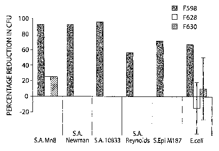
FIG. 12 is a bar graph showing reduction in CFU from different PNAG-expressing
bacterial strains using F598, F628 and F630.
FIG. 13 is a graph showing proportion of S. aureus bacteria killed by F598 and
F628
as a function of icaB gene presence or absence. FIG. 14 is a graph showing
proportion of S. aureus bacteria killed by F598 and F628
as a function of icaB gene over-expression.
It is to be understood that the Figures are not required for enablement of the
invention.
Brief Description of the Sequence Listing
SEQ ID NO: 1 is the amino acid sequence of antibody F598 heavy chain variable
region.
SEQ ID NO: 2 is the amino acid sequence of antibody F598 light chain variable
region.
CA 02567748 2006-11-22
WO 2005/103084 PCT/US2005/013694
-10-
SEQ ID NO: 3 is the amino acid sequence of antibody F628 heavy chain variable
region.
SEQ ID NO: 4 is the amino acid sequence of antibody F6281ight chain variable
region.
SEQ ID NO: 5 is the amino acid sequence of antibody F630 heavy chain variable
region.
SEQ ID NO: 6 is the amino acid sequence of antibody F6301ight chain variable
region.
SEQ ID NO: 7 is the amino acid sequence of CDRl of antibody F598 heavy chain.
SEQ ID NO: 8 is the amino acid sequence of CDR2 of antibody F598 heavy chain.
SEQ ID NO: 9 is the amino acid sequence of CDR3 of antibody F598 heavy chain.
SEQ ID NO: 10 is the amino acid sequence of CDR1 of antibody F5981ight chain.
SEQ ID NO: 11 is the amino acid sequence of CDR2 of antibody F5981ight chain.
SEQ ID NO: 12 is the amino acid sequence of CDR3 of antibody F5981ight chain.
SEQ ID NO: 13 is the amino acid sequence of CDRl of antibody F628 heavy chain.
SEQ ID NO: 14 is the amino acid sequence of CDR2 of antibody F628 heavy chain.
SEQ ID NO: 15 is the amino acid sequence of CDR3 of antibody F628 heavy chain.
SEQ ID NO: 16 is the amino acid sequence of CDRl of antibody F628 light chain.
SEQ ID NO: 17 is the amino acid sequence of CDR2 of antibody F6281ight chain.
SEQ ID NO: 18 is the amino acid sequence of CDR3 of antibody F6281ight chain.
SEQ ID NO: 19 is the amino acid sequence of CDRl of antibody F630 heavy chain.
SEQ ID NO: 20 is the amino acid sequence of CDR2 of antibody F630 heavy chain.
SEQ ID NO: 21 is the amino acid sequence of CDR3 of antibody F630 heavy chain.
SEQ ID NO: 22 is the amino acid sequence of CDR1 of antibody F630 light chain.
SEQ ID NO: 23 is the amino acid sequence of CDR2 of antibody F6301ight chain.
SEQ ID NO: 24 is the amino acid sequence of CDR3 of antibody F630 light chain.
SEQ ID NO: 25 is the nucleotide sequence of antibody F598 heavy chain variable
region.
SEQ ID NO: 26 is the nucleotide sequence of antibody F5981ight chain variable
region.
SEQ ID NO: 27 is the nucleotide sequence of antibody F628 heavy chain variable
region.
CA 02567748 2006-11-22
WO 2005/103084 PCT/US2005/013694
-11-
SEQ ID NO: 28 is the nucleotide sequence of antibody F628 light chain variable
region.
SEQ ID NO: 29 is the nucleotide sequence of antibody F630 heavy chain variable
region.
SEQ ID NO: 30 is the nucleotide sequence of antibody F630 light chain variable
region.
SEQ ID NO: 31 is the nucleotide sequence of CDR1 of antibody F598 heavy chain.
SEQ ID NO: 32 is the nucleotide sequence of CDR2 of antibody F598 heavy chain.
SEQ ID NO: 33 is the nucleotide sequence of CDR3 of antibody F598 heavy chain.
SEQ ID NO: 34 is the nucleotide sequence of CDR1 of antibody F598 light chain.
SEQ ID NO: 35 is the nucleotide sequence of CDR2 of antibody F598 light chain.
SEQ ID NO: 36 is the nucleotide sequence of CDR3 of antibody F598 light chain.
SEQ ID NO: 37 is the nucleotide sequence of CDR1 of antibody F628 heavy chain.
SEQ ID NO: 38 is the nucleotide sequence of CDR2 of antibody F628 heavy chain.
SEQ ID NO: 39 is the nucleotide sequence of CDR3 of antibody F628 heavy chain.
SEQ ID NO: 40 is the nucleotide sequence of CDR1 of antibody F628 light chain.
SEQ ID NO: 41 is the nucleotide sequence of CDR2 of antibody F628 light chain.
SEQ ID NO: 42 is the nucleotide sequence of CDR3 of antibody F628 light chain.
SEQ ID NO: 43 is the nucleotide sequence of CDR1 of antibody F630 heavy chain.
SEQ ID NO: 44 is the nucleotide sequence of CDR2 of antibody F630 heavy chain.
SEQ ID NO: 45 is the nucleotide sequence of CDR3 of antibody F630 heavy chain.
SEQ ID NO: 46 is the nucleotide sequence of CDR1 of antibody F630 light chain.
SEQ ID NO: 47 is the nucleotide sequence of CDR2 of antibody F630 light chain.
SEQ ID NO: 48 is the nucleotide sequence of CDR3 of antibody F6301ight chain.
SEQ ID NO: 49 is the nucleotide sequence of primer lambda constant.
SEQ ID NO: 50 is the nucleotide sequence of primer Hu lambda sig 5.
SEQ ID NO: 51 is the nucleotide sequence of primer Heavy chain constant.
SEQ ID NO: 52 is the nucleotide sequence of primer VH7LDRHU.
SEQ ID NO: 53 is the nucleotide sequence of primer Hu lambda sig 1.
SEQ ID NO: 54 is the nucleotide sequence of primer VHILDRHU.
SEQ ID NO: 55 is the amino acid sequence of F598 heavy chain variable region
including some constant region sequence.
CA 02567748 2006-11-22
WO 2005/103084 PCT/US2005/013694
-12-
SEQ ID NO: 56 is the nucleotide sequence of F598 heavy chain variable region
including some constant region sequence.
SEQ ID NO: 57 is the amino acid sequence of F5981ight chain variable region
including some constant region sequence.
SEQ ID NO: 58 is the amino acid sequence of F628 heavy chain variable region
including some constant region sequence.
SEQ ID NO: 59 is the nucleotide sequence of F628 heavy chain variable region
including some coristant region sequence.
SEQ ID NO: 60 is the amino acid sequence of F630 light chain variable region
including some constant region sequence.
SEQ ID NO: 61 is the nucleotide sequence of F630 light chain variable region
including some constant region sequence.
Detailed Description of the Invention
The invention provides compositions and methods useful, inter alia, for
immunization
of humans and animals against infection by bacterial strains that express poly-
N-acetyl
glucosamine (PNAG) as well as detection of such pathogens. Such bacterial
strains include '
but are not limited to coagulase-negative and coagulase-positive Staphylococci
such as S.
aureus and S. epidermis. The invention further provides peptides that bind to
various forms
of PNAG expressed by some bacterial strains.
The invention is based in part on the discovery, isolation and
characterization of a
number of human monoclonal antibodies that bind to various forms of PNAG
(including
highly acetylated forms, poorly acetylated forms and deacetylated forms, as
described below).
These antibodies are produced by hybridomas deposited with the ATCC under ATCC
Accession Nos. PTA-5931, PTA-5932 and PTA-5933 on April 21, 2004 in accordance
with
the Budapest Patent Treaty. The liybridomas and the antibodies they produce
are designated
F598, F628 and F630. These hybridomas are referred to herein repeatedly. It is
to be
understood that reference to hybridomas (or antibodies produced by hybridomas)
having
ATCC Accession Nos. PTA-593 1, PTA-5932 and PTA-5933 means the afore-mentioned
hybridomas. The deposited hybridomas were produced from B cells harvested from
a human
subject recovering from a Staphylococcal infection. The B cells were
transformed with the
Epstein-Barr virus and then fused with the human-mouse myeloma cell line HMMA
2.5 to
generate the deposited hybridomas.
CA 02567748 2006-11-22
WO 2005/103084 PCT/US2005/013694
-13-
PNAG exists in nature in various forms that differ according to the degree of
acetate
substitutions. Acetate substitutions can range from 0-100%. As used herein,
PNAG refers to
"native PNAG" corresponding to the naturally occurring mixture of PNAG with
the
aforementioned range of acetate substitutions. Poorly acetylated PNAG is a
subpopulation of
PNAG polysaccharides in which less than 50% of amino groups of glucosamine are
substituted with acetate. As used herein, the term dPNAG embraces both poorly
acetylated
PNAG as well as completely deacetylated PNAG (i.e., dPNAG refers to a subset
of PNAG
polysaccharides that comprise 0- less than 50% acetate substituents).
PNAG has the following structure:
0CHZ
I H
C~ 0
H\
C OH H C
~\ ~ '/\H
/ I
H R
n
where, n is an integer ranging from 2 to greater than or equal to 300, R is
selected from the
group consisting of -NH-CO-CH3 and -NH2. PNAG has a beta (0) 1-6 linkage
(i.e., it is
comprised of glucosamine monomer units linked together by beta (0) 1-6
linkages).
PNAG may be a homo-polymer. A homo-polymer is one in which the R groups of
the glucosamine residues are identical. The homo-polymer may comprise solely
unsubstituted R groups (i.e., R NH2). PNAG can also be a hetero-polymer with a
mixture of
-NH2 and -NH-CO-CH3 groups at the R position. dPNAG has the identical
structure as
PNAG with the exception that less than 50% of the R groups are -NH-CO-CH3.
PNAG and dPNAG can be naturally occurring and prepared from any bacterial
strain
carrying the ica locus (or a homologous locus such as the pga locus),
producing the
biosynthetic enzymes encoded by this locus, and using these enzymes to
synthesize PNAG or
dPNAG. Bacteria that express PNAG include Staphylococci such as S. aureus and
S.
epidermidis, E. coli such as E. coli strains 0157:H7 and CFT073, Yersinia
pestis, Yersinia
entercolitica, Xanthomonas axonopodis, Pseudomonas fluorescens (all of which
are
CA 02567748 2006-11-22
WO 2005/103084 PCT/US2005/013694
-14-
sequenced species with complete pgaABCD loci), and Actinobacillus
actinomycetemcomitans
(AA), Actinobacillus pleuropneurnoniae (Ap), Ralstonia solanacearum (e.g.,
megaplasmid
form), Bordetella pertussis, Bordetella parapertussis and Bordetella
bronchiseptica (all of
which contain pgaABC genes but apparently lack apgaD homologue). pgaD
apparently is
not required for PNAG expression as pgaABC encoding species such as AA and Ap
(listed
above) make PNAG.
Bacteria that express PNAG are bacteria that carry the ica locus or a
homologous
locus such as thepga locus. For example, PNAG-expressing Staphylococci are
Staphylococci
that carry the ica locus. PNAG-expressing bacterial strains include dPNAG-
expressing
bacterial strains. For example, PNAG-expressing Staphylococci include dPNAG-
expressing
Staphylococci. These strains include but are not limited to S. epidermis and
S. aureus, as well
as other strains (e.g., S. carnosus) that have been transformed with the genes
in the ica locus
or liomologous locus such as the pga locus. In particular, PNAG can be
prepared from
specific strains including S. epidermis RP62A (ATCC number 35984), S.
epidermis RP12
(ATCC number 35983), S. epidermis M187, S. carnosus TM300 (pCN27), S. aureus
RN4220
(pCN27), S. aureus MN8 mucoid, E.coli 0157:H7 and E. coli CFT073. dPNAG may
also be
synthesized de novo or via modification of native PNAG. PNAG and dPNAG can be
prepared according to the methods described in Maira-Litran et al. Infect
Immun. 2002
Aug;70(8):4433, and in U.S. Patent Application 10/713,790 filed on Noveinber
12, 2003.
PNAG is also expressed by other bacteria including but not limited to E. coli,
Yersinia
pestis (Y. pestis), Y. entercolitica, Xanthomonas axonopodis (X. axonopodis),
Pseudomonas
fluorescens (P. fluorescens), Actinobacillus actinomycetemcomitans (A.
actinomycetemcomitans), A. pleuropneunaoniae, Ralstonia solanacearum (R.
solanacearum),
Bordetellapertussis (B. pertussis), B. parapertussis and B. bronchiseptica. As
described in
the Examples, 17 out of 18 urinary tract infection E. coli isolates carried
the pga locus. Of
these, about one third expressed relatively high levels of PNAG, about one
third expressed
relatively intermediate levels of PNAG, and the remaining third expressed
relatively low
levels of PNAG. The above analyses were carried out by immunoblot using
antisera raised to
S. aureus PNAG. This is evidence that PNAG from one species can be used to
raise
antibodies (and accordingly binding peptides) to other species that express
PNAG.
Thus, in one aspect, the invention provides binding peptides and antibodies.
The
antibodies of the invention bind to Staphylococcal PNAG/dPNAG and enhance
opsonophagocytosis of species that elaborate PNAG (i.e., opsonophagocytic
human
CA 02567748 2006-11-22
WO 2005/103084 PCT/US2005/013694
- 15-
monoclonal antibodies specific for Staphylococcal PNAG/dPNAG). The antibodies
are
referred to herein as anti-Staphylococcal PNAG/dPNAG antibodies. It is to be
understood,
however, that such antibodies are able to bind PNAG/dPNAG regardless of its
source.
Accordingly, antibodies of the invention that are defined as binding to, for
example,
Staphylococcal PNAG/dPNAG and capable of detecting and/or enhancing
opsonophagocytosis of, for example, Staphylococcal species are also capable of
detecting
and/or enhancing opsonophagocytosis of non- Staphylococcal PNAG-expressing
bacteria.
An anti-Staphylococcal PNAG/dPNAG antibody is an antibody that a) binds to
both
PNAG and dPNAG, b) binds to PNAG but not dPNAG, or c) binds to dPNAG but not
PNAG.
Preferred antibodies bind to dPNAG.
Antibodies F598, F628 and F630 are all able to bind to native PNAG and some
are
also able to bind to dPNAG. Although not intending to be bound by any
mechanism or
theory, it is believed that antibodies that recognize dPNAG are more likely to
bind
specifically to parts of the PNAG molecule that do not contain acetate groups,
rather than to
parts of the molecule that include substituents such as the acetate
substitutions. For example,
antibodies that bind to dPNAG may recognize and bind to the backbone of PNAG
rather than
its acetate substituents. These antibodies are capable of mediating
opsonophagocytic killing
of PNAG-expressing bacteria such as but not limited to Staphylococcal or E.
coli isolates
from infected human subjects. When used in vivo in murine models of
Staphylococcal
infection, the antibodies provide protection to Staphylococcal challenge. The
conditions
under which each monoclonal antibody provides protection may vary. These and
other
findings are described in greater detail in the Examples.
Although not intending to be bound by any particular theory, it is believed
that
progression of infection by PNAG-expressing bacteria (such as Staphylococcal
infection) is
due to a failure to produce an adequate immune response that eliminates the
pathogen.
Specifically, one of the defects is a failure to produce opsonophagocytic
antibodies specific
for PNAG (such as that produced by Staphylococci.)
Opsonophagocytic antibodies are antibodies that deposit themselves onto an
antigen or
onto a bacterium with and without the ability to recruit additional deposition
of components
of the complement system and facilitate the phagocytosis of the antigen or
bacterium by
phagocytic cells such as antigen presenting cells (e.g., macrophages or
dendritic cells), or
polymorphonuclear neutrophils. Phagocytosis can proceed in an Fc-mediated
manner that
involves only the antibody bound to the antigen or bacterium. Phagocytosis can
also proceed
CA 02567748 2006-11-22
WO 2005/103084 PCT/US2005/013694
-16-
by binding of complement receptors on phagocytes to complement opsonins on
bacterial
surfaces to which antibodies have deposited. Phagocytosis can also proceed by
a combination
of these two mechanisms. The ability to provide opsonophagocytic antibodies to
the site of
infection should therefore contribute to the eradication of the infection more
effectively than
previously possible.
Both PNAG and dPNAG are highly immunogenic in vivo and are capable of
eliciting
antibodies that mediate opsonic killing and protection from infection, it is
hypothesized that
dPNAG preferentially elicits antibodies that mediate opsonic killing and
protection from
infection. The dPNAG polysaccharide is therefore useful, inter alia, in the
generation of
immune responses, including antibody dependent immune responses, to PNAG-
expressing
bacterial strains such as but not limited to Staphylococci. The antibodies
elicited following
dPNAG administration recognize dPNAG and in important embodiments also
recognize
highly acetylated forms of PNAG.
Thus, the invention relates to the identification and use of peptides that
bind to PNAG
and/or dPNAG. Peptides that bind to Staphylococcal PNAG and/or dPNAG are
referred to
herein as PNAG/dPNAG-binding peptides. Again, it is to be understood that such
binding
peptides are able to bind PNAG/dPNAG regardless of source. PNAG/dPNAG-binding
peptides include a) peptides that bind to botli PNAG and dPNAG, b) peptides
that bind to
PNAG and not to dPNAG (referred to herein as PNAG-binding peptides), and c)
peptides that
bind to dPNAG and not to PNAG (referred to herein as dPNAG-binding peptides).
In
preferred embodiments, the peptides at least bind to dPNAG (thereby embracing
afore-
mentioned categories (a) and (c)).
The peptides of the invention minimally comprise regions that bind to
PNAG/dPNAG
(i.e., Staphylococcal PNAG/dPNAG-binding regions). As used herein, a
Staphylococcal
PNAG/dPNAG-binding region is a region that a) binds to both PNAG and dPNAG, b)
binds
to PNAG but not dPNAG (referred to herein as a PNAG-binding region), or c)
binds to
dPNAG but not PNAG (referred to herein as a dPNAG-binding region), regardless
of the
source of PNAG/dPNAG. Preferably, the PNAG/dPNAG binding region is a region
that at
least binds dPNAG (and therefore embraces categories (a) and (c)).
Staphylococcal
PNAG/dPNAG-binding regions derive from the PNAG/dPNAG-binding regions of the
antibodies of the invention, or alternatively, they are functionally
equivalent variants of such
regions.
CA 02567748 2006-11-22
WO 2005/103084 PCT/US2005/013694
-17-
Accordingly, two particularly important classes of antibody-derived PNAG/dPNAG-
binding regions are variable regions and CDRs of the antibodies described
herein or produced
by hybridomas deposited with the ATCC under ATCC Accession Nos. PTA-593 1, PTA-
5932
and PTA-5933 on April 21, 2004. CDR and variable region nucleic acids can be
cloned from
antibody-producing cells such as those on deposit as described in the
Examples.
An antibody, as is well known in the art, is an assembly of polypeptide chains
linked
by disulfide bridges. Two principle amino acid chains, referred to as the
light chain and
heavy chain, make up all major structural isotypes of antibody. Both heavy
chains and light
chains are furtlier divided into subregions referred to as variable regions
and constant regions.
In some instances, the peptides encompass the antibody heavy and light chain
variable regions
of the foregoing antibodies. The heavy chain variable region is a peptide
which generally
ranges from 100 to 150 amino acids in length. The light chain variable region
is a peptide
which generally ranges from 80 to 130 amino acids in length.
As is also well-known in the art, CDRs of an antibody are the portions of the
antibody
variable region which are largely responsible for the binding specificity of
an antibody for a
given antigen or antigenic epitope. The CDRs directly interact with the
epitope of the antigen
(see, in general, Clark, 1986; Roitt, 1991): In both the heavy chain and the
light chain
variable regions of IgG immunoglobulins, there are four framework regions (FRl
through
FR4) separated respectively by three complementarity determining regions
(CDR1, CDR 2
and CDR3). The framework regions (FRs) maintain the tertiary structure of the
paratope,
which is the portion of the antibody which is involved in the interaction with
the antigen or
antigenic epitope. CDRs, and in particular CDR3, and more particularly heavy
chain CDR3,
contribute substantially to antibody specificity. Because CDRs, and in
particular CDR3,
confer a large proportion of antigenic specificity on the antibody, these
regions may be
incorporated into other antibodies or peptides to confer the identical
antigenic specificity onto
that antibody or peptide.
Preferably, the PNAG/dPNAG-binding peptides minimally encompass at least one
CDR from those described herein or those that can be derived from the
deposited hybridomas
(i.e., a Staphylococcal PNAG/dPNAG-binding CDR). As used herein, a
Staphylococcal
PNAG/dPNAG-binding CDR is a CDR described herein or is a CDR derived from
hybridomas deposited under ATCC Accession Nos. PTA-593 1, PTA-5932 and PTA-
5933.
Staphylococcal PNAG/dPNAG-binding CDRs include a) CDRs that bind to both PNAG
and
dPNAG, b) CDRs that bind to PNAG and not to dPNAG (referred to herein as PNAG-
binding
CA 02567748 2006-11-22
WO 2005/103084 PCT/US2005/013694
-18-
CDRs), and c) CDRs that bind to dPNAG and not to PNAG (referred to herein as
dPNAG-
binding CDRs), regardless of the source of the PNAG/dPNAG. These peptides
preferably
contain at least one Staphylococcal PNAG/dPNAG-binding CDR.
The Staphylococcal PNAG/dPNAG-binding region may be a Staphylococcal
PNAG/dPNAG-binding CDR1, a Staphylococcal PNAG/dPNAG-binding CDR2, or a
Staphylococcal PNAG/dPNAG-binding CDR3, all of which are derived from the
antibodies
and antibody variable chains disclosed herein.
As used herein, a "Staphylococcal PNAG/dPNAG-binding CDR1" is a CDR1 that
binds, preferably specifically, to Staphylococcal PNAG/dPNAG, and is derived
from either
the heavy or light chain variable regions of the antibodies described herein
or produced by
hybridomas deposited under ATCC Accession Nos. PTA-5931, PTA-5932 and PTA-
5933. It
may have an amino acid sequence selected from the group consisting of SEQ ID
NO: 7, SEQ
ID NO: 10, SEQ ID NO: 13, SEQ ID NO: 16, SEQ ID NO: 19 and SEQ ID NO: 22.
Similar
respective definitions apply to Staphylococcal PNAG/dPNAG-binding CDR2 and
CDR3.
A "Staphylococcal PNAG/dPNAG-binding CDR2" is a CDR2 that binds, preferably
specifically, to Staphylococcal PNAG/dPNAG, and is derived from either the
heavy or light
chain variable regions of the antibodies described herein or produced by the
hybridomas
deposited under ATCC Accession Nos. PTA-593 1, PTA-5932 and PTA-5933. It may
have an
amino acid sequence selected from the group consisting of SEQ ID NO: 8, SEQ ID
NO: 11,
SEQ ID NO: 14, SEQ ID NO: 17, SEQ ID NO: 20 and SEQ ID NO: 23.
A "Staphylococcal PNAG/dPNAG-binding CDR3" is a CDR3 that binds, preferably
specifically, to Staphylococcal PNAG/dPNAG, and is derived from either the
heavy or light
chain variable regions of the antibodies described herein or produced by the
hybridomas
deposited under ATCC Accession Nos. PTA-5931, PTA-5932 and PTA-5933. It may
have an
amino acid sequence selected from the group consisting of SEQ ID NO: 9, SEQ ID
NO: 12,
SEQ ID NO: 15, SEQ ID NO: 18, SEQ ID NO: 21 and SEQ ID NO: 24.
In addition to the sequences listed above, the invention intends to embrace
functionally equivalent variants of these sequences including conservative
substitution
variants in either the amino acid or nucleotide sequence, as described in
greater detail below.
The peptides of the invention, including but not limited to the
opsonophagocytic
antibodies discussed herein, are useful inter alia in diagnostic methods aimed
at detecting, in a
sample in or from a subject, the PNAG/dPNAG antigen or PNAG-expressing
bacteria (such
as but not limited to Staphylococcal bacteria that express PNAG). At a
minimum, peptides
CA 02567748 2006-11-22
WO 2005/103084 PCT/US2005/013694
-19-
useful in these methods need only recognize and bind to PNAG/dPNAG (such as
Staphylococcal PNAG/dPNAG) regardless of whether they also enhance
opsonization and
phagocytosis. In important embodiments, the antibodies and fragments thereof
bind to
PNAG/dPNAG selectively. Accordingly, they need only possess one or more of the
CDRs
derived from the antibody clones described herein or produced by the
hybridomas deposited
under ATCC Accession Nos. PTA-5931, PTA-5932 and PTA-5933. In preferred
embodiments, the peptides comprise a PNAG/dPNAG-binding CDR3, and even more
preferably, the peptides coinprise a heavy chain PNAG/dPNAG-binding CDR3. It
is to be
understood that not all of the CDRs are required in order to effect binding to
PNAG/dPNAG.
However, in some embodiments the peptides comprise all of the CDRs of a given
antibody
clone disclosed herein or produced by hybridomas deposited under ATCC
Accession Nos.
PTA-5931, PTA-5932 and PTA-5933.
In addition, it should be understood that the invention also embraces the
exchange of
CDRs between the variable regions provided herein. Preferably, a heavy chain
CDR is
exchanged with another heavy chain variable region CDR, and likewise, a light
chain CDR is
exchanged with another light chain variable region CDR.
The amino acid sequences of the CDRs of the variable chains disclosed in the
present
invention are as follows:
Clone Chain CDR SEQ ID NO: Sequence
F598 Hv CDR1 7 GYYWS
F598 Hv CDR2 8 YIHYSRSTNSNPALKS
F598 Hv CDR3 9 DTYYYDSGDYEDAFDI
F598 Lt CDR1 10 TLSSGHSNYAIA
F598 Lt CDR2 11 VNRDGSHIRGD
F598 Lt CDR3 12 QTWGAGIRV
F628 Hv CDR1 13 NYYWS
F628 Hv CDR2 14 YIHYSGSTNSNPSLKS
F628 Hv CDR3 15 DTYYESSGHWFDGLDV
F628 Lt CDR1 16 TLDSEHSRYTIA
F628 Lt CDR2 17 VKSDGSHSKGD
F628 Lt CDR3 18 QTWGPGIRV
CA 02567748 2006-11-22
WO 2005/103084 PCT/US2005/013694
-20-
F630 Hv CDR1 19 NFGIS
F630 Hv CDR2 20 WVSTYNGRTNYAQKFRG
F630 Hv CDR3 21 DYYETSGYAYDDFAI
F630 Lt CDR1 22 TLSSGHSTYAIA
F630 Lt CDR2 23 VNSDGSHTKGD
F630 Lt CDR3 24 QTWGPGIRV
The nucleotide sequences of the CDRs of the variable chains disclosed in the
present
invention are as follows:
Clone Chain CDR SEQ ID NO: Sequence
F598 Hv CDR1 31 GGT TAC TAC TGG AGT
F598 Hv CDR2 32 TAT ATT CAT TAT AGT AGG AGC ACC
AAC TCC AAC CCC GCC CTC AAG AGT
F598 Hv CDR3 33 GAT ACC TAT TAC TAT GAT AGT GGT
GAT TAT GAG GAT GCT TTT GAT ATT
F598 Lt CDR1 34 ACT CTG AGC AGT GGC CAC AGC AAC
TAC GCC ATC GCT
F598 Lt CDR2 35 GTT AAC AGA GAT GGC AGC CAC ATC
AGG GGG GAC
F598 Lt CDR3 36 CAG ACC TGG GGC GCT GGC ATT CGA
GTG
F628 Hv CDR1 37 AAT TAC TAC TGG AGT
F628 Hv CDR2 38 TAT ATC CAT TAT AGT GGG AGC ACC
AAC TCC AAT CCA TCC CTC AAG AGT
F628 Hv CDR3 39 GAT ACT TAC TAT GAA AGT AGT GGT
= CAT TGG TTC GAC GGT TTG GAC GTC
F628 Lt CDR1 40 ACT CTG GAC AGT GAA CAC AGC AGA
TAC ACC ATC GCA
F628 Lt CDR2 41 GTT AAG AGT GAT GGC AGT CAC AGC
AAG GGG GAC
F628 Lt CDR3 42 CAG ACT TGG GGC CCT GGC ATT CGA
GTG
F630 Hv CDR1 43 AAC TTT GGT ATC AGT
F630 Hv CDR2 44 TGG GTC AGC ACT TAC AAT GGT CGC
ACA AAT TAT GCA CAG AAG TTC CGG
GGC
F630 Hv CDR3 45 GAT TAC TAT GAG ACT AGT GGT TAC
GCC TAT GAT GAT TTT GCG ATC
CA 02567748 2006-11-22
WO 2005/103084 PCT/US2005/013694
-21 -
F630 Lt CDR1 46 ACT CTG AGC AGT GGG CAC AGC ACC
TAC GCC ATC GCG
F630 Lt CDR2 47 GTC AAC AGT GAT GGC AGC CAC ACC
AAG GGG GAC
F630 Lt CDR3 48 CAG ACG TGG GGC CCT GGC ATT CGA
GTG
The peptides may also comprise a Staphylococcal PNAG/dPNAG-binding variable
region. A Staphylococcal PNAG/dPNAG-binding variable region is a variable
region
(preferably an antibody variable region as described herein or as derived from
hybridomas
deposited under ATCC Accession Nos. PTA-593 1, PTA-5932 and PTA-5933) that a)
binds to
both PNAG and dPNAG, b) binds to PNAG but not dPNAG (referred to herein as a
PNAG-
binding variable region), or c) binds to dPNAG but not PNAG (referred to
herein as a
dPNAG-binding variable region), regardless of the PNAG/dPNAG source.
The present invention provides at least six different variable regions, at
least three of
which are heavy chain variable regions and at least three of which are light
chain variable
regions. SEQ ID NO: 1 and SEQ ID NO: 25 correspond to the amino acid and
nucleotide
sequence of the heavy chain variable region derived from antibody clone F598.
SEQ ID NO:
2 and SEQ ID NO: 26 correspond to the amino acid and nucleotide sequence of
the light chain
variable region derived from antibody clone F598. SEQ ID NO: 3 and SEQ ID NO:
27
correspond to the amino acid and nucleotide sequence of the heavy chain
variable region
derived from antibody clone F628. SEQ ID NO: 4 and SEQ ID NO: 28 correspond to
the
amino acid and nucleotide sequence of the light chain variable region derived
from antibody
clone F628. SEQ ID NO: 5 and SEQ ID NO: 29 correspond to the amino acid and
nucleotide
sequence of the heavy chain variable region derived from antibody clone F630.
SEQ ID NO:
6 and SEQ ID NO: 30 correspond to the amino acid and nucleotide sequence of
the light chain
variable region derived from antibody clone F630.
It is to be understood that the nucleic acids or peptides of the invention may
be derived
from the sequences provided herein or from the deposited hybridomas. These
sequences can
be cloned (e.g., by PCR) and inserted into a vector and/or cells in order to
produce peptides
corresponding to full length variable regions or fragments of full length
variable regions, and
antibodies comprising the variable regions. It is therefore possible to
generate antibodies or
fragments thereof that comprise a combination of light and heavy chain
variable regions. For
example, an antibody of the invention may comprise the heavy chain variable
region from
MAb F598 (or from the antibody produced by the deposited F598 hybridoma) and
the light
CA 02567748 2006-11-22
WO 2005/103084 PCT/US2005/013694
-22-
chain variable region of F630 (or from the antibody produced by the deposited
F630
hybridoma). It is to be understood that any combination of heavy and light
chain variable
regions (as disclosed herein or as comprised in antibodies produced by
hybridomas deposited
under ATCC Accession Nos. PTA-593 1, PTA-5932 and PTA-5933) can be used in the
synthesis of an antibody or antibody fragment according to the invention.
Accordingly, the invention embraces antibodies or antibody fragments that are
comprised of the following variable region combinations: SEQ ID NO:1 and
SEQ.ID NO:2;
SEQ ID NO:1 and SEQ ID NO:4; SEQ ID NO:1 and SEQ ID NO:6; SEQ ID NO:3 and SEQ
ID NO:2; SEQ ID NO:3 and SEQ ID NO:4; SEQ ID NO:3 and SEQ ID NO:6; SEQ ID NO:5
and SEQ ID NO:2; SEQ ID NO:5 and SEQ ID NO:4; and SEQ ID NO:5 and SEQ ID NO:6.
Similarly, the invention embraces antibodies or antibody fragments that are
comprised
of the following variable region combinations:
1. heavy chain variable region from hybridoma F598 having ATCC Accession No.
PTA-5931 and light chain variable region from hybridoma F598 having ATCC
Accession No.
PTA-593 1;
2. heavy chain variable region from hybridoma F598 having ATCC Accession No.
PTA-5931 and light chain variable region from hybridoma F628 having ATCC
Accession No.
PTA-5932;
3. heavy chain variable region from hybridoma F598 having ATCC Accession No.
PTA-5931 and light chain variable region from hybridoma F630 having ATCC
Accession No.
PTA-5933;
4. heavy chain variable region from hybridoma F628 having ATCC Accession No.
PTA-5932 and light chain variable region from hybridoma F598 having ATCC
Accession No.
PTA-593 1;
5. heavy chain variable region from hybridoma F628 having ATCC Accession No.
PTA-5932 and light chain variable region from hybridoma F628 having ATCC
Accession No.
PTA-5932;
6. heavy chain variable region from hybridoma F628 having ATCC Accession No.
PTA-5932 and light chain variable region from hybridoma F630 having ATCC
Accession No.
PTA-5933;
7. heavy chain variable region from hybridoma F630 having ATCC Accession No.
PTA-5933 and light chain variable region from hybridoma F598 having ATCC
Accession No.
PTA-5931;
CA 02567748 2006-11-22
WO 2005/103084 PCT/US2005/013694
- 23 -
8. heavy chain variable region from hybridoma F630 having ATCC Accession No.
PTA-5933 and light chain variable region from hybridoma F628 having ATCC
Accession No.
PTA-5932; and
9. heavy chain variable region from hybridoma F630 having ATCC Accession No.
PTA-5933 and light chain variable region from hybridoma F630 having ATCC
Accession No.
PTA-5933.
The invention intends to capture antibody and antibody fragments of various
isotypes.
The deposited hybridomas produce IgG2 isotype antibodies. However, the
recombined
immunoglobulin (Ig) genes, particularly the variable region genes, can be
isolated from the
deposited hybridomas, as described in the Examples, and cloned into an Ig
recombination
vector that codes for human Ig constant region genes of both heavy and light
chains. Using
this technique, IgGl isotype antibodies that bind to Staphylococcal PNAG/dPNAG
and
thereby enhance opsonophagocytosis of PNAG-expressing bacteria (such as
Staphylococci)
have been identified, synthesized and isolated.
The antibodies may be of an IgGl, IgG2, IgG3, IgG4, IgD, IgE, IgM, IgAl, IgA2,
or
sIgA isotype. The invention intends to capture isotypes found in non-human
species as well
such as but not limited to IgY in birds and sharks. Vectors encoding the
constant regions of
various isotypes are known and previously described. (See, for example,
Preston et al.
Production and characterization of a set of mouse-human chimeric
immunoglobulin G (IgG)
subclass and IgA monoclonal antibodies with identical variable regions
specific for P.
aeruginosa serogroup 06 lipopolysaccharide. Infect Immun. 1998 Sep;66(9):4137-
42;
Coloma et al. Novel vectors for the expression of antibody molecules using
variable regions
generated by polymerase chain reaction. J Immunol Methods. 1992 Jul
31;152(1):89-104;
Guttieri et al. Cassette vectors for conversion of Fab fragments into full-
length human IgGl
monoclonal antibodies by expression in stably transformed insect cells. Hybrid
Hybridomics.
2003 Jun;22(3):135-45; McLean et al. Human and murine immunoglobulin
expression
vector cassettes. Mol Immunol. 2000 Oct;37(14):837-45; Walls et al. Vectors
for the
expression of PCR-amplified immunoglobulin variable domains with human
constant regions.
Nucleic Acids Res. 1993 Jun 25;21(12):2921-9; Norderhaug et al. Versatile
vectors for
transient and stable expression of recombinant antibody molecules in mammalian
cells. J
Immunol Methods. 1997 May 12;204(1):77-87.)
CA 02567748 2006-11-22
WO 2005/103084 PCT/US2005/013694
-24-
As used herein, the term "peptide" includes monoclonal antibodies,
functionally active
and/or equivalent antibody fragments, and functionally active and/or
equivalent peptides and
polypeptides.
The peptides of the invention are isolated peptides. As used herein, the term
"isolated
peptides" means that the peptides are substantially pure and are essentially
free of other
substances with which they may be found in nature or in vivo systems to an
extent practical
and appropriate for their intended use. In particular, the peptides are
sufficiently pure and are
sufficiently free from other biological constituents of their hosts cells so
as to be useful in, for
example, producing pharmaceutical preparations or sequencing. Because an
isolated peptide
of the invention may be admixed with a pharmaceutically acceptable carrier in
a
pharmaceutical preparation, the peptide may comprise only a small percentage
by weight of
the preparation. The peptide is nonetheless substantially pure in that it has
been substantially
separated from the substances with which it may be associated in living
systems.
The peptides of the invention bind to PNAG and/or dPNAG, preferably in a
selective
manner. As used herein, the terms "selective binding" and "specific binding"
are used
interchangeably to refer to the ability of the peptide to bind with greater
affinity to PNAG
and/or dPNAG and fragments thereof than to non-PNAG derived compounds. That
is,
peptides that bind selectively to PNAG and/or dPNAG will not bind to non-PNAG
derived
compounds to the same extent and with the same affinity as they bind to PNAG
and/or
dPNAG and fragments thereof, with the exception of cross reactive antigens or
molecules
made to be mimics of PNAG/dPNAG such as peptide mimetics of carbohydrates or
variable
regions of anti-idiotype antibodies that bind to the PNAG/dPNAG-binding
peptides in the
same manner as PNAG/dPNAG. Antibodies that bind selectively to PNAG bind to
PNAG
with greater affinity than to dPNAG. Antibodies that bind to dPNAG may also
bind to
dPNAG with lesser, comparable or greater affinity than to PNAG. In preferred
embodiments,
the peptides of the invention bind solely to PNAG and/or dPNAG and fragments
thereof, and
even more preferably, they at least bind to dPNAG. As used herein, a binding
peptide that
binds selectively or specifically to Staphylococcal PNAG/dPNAG may also bind
PNAG/dPNAG from other sources and will bind with lesser affinity (if at all)
to non-
PNAG/dPNAG derived compounds. Lesser affinity may include at least 10% less,
20% less,
30% less, 40% less, 50% less, 60% less, 70% less, 80% less, 90% less, or 95%
less. Thus,
"selective" in this sense refers to the binding to PNAG/dPNAG rather than to
the
Staphylococcus-derived form of PNAG/dPNAG.
CA 02567748 2006-11-22
WO 2005/103084 PCT/US2005/013694
-25-
As stated earlier, the invention provides peptides e.g., antibodies or
antibody
fragments, that bind to Staphylococcal PNAG and/or dPNAG. Such antibodies
preferably
enhance opsonization and phagocytosis (i.e., opsonophagocytosis) of PNAG-
expressing
bacteria (such as PNAG-expressing Staphylococci), and as a result are useful
in the
prevention and therapy of some forms of bacterial infections in a subject.
Opsonization refers
to a process by which phagocytosis is facilitated by the deposition of
opsonins (e.g., antibody
and/or opsonic complement factors such as C4b or C3b or any other factor
capable of
promoting opsonophagocytosis) on the antigen. Phagocytosis and
opsonophagocytosis refer
to the process by which phagocytic cells (e.g., macrophages, dendritic cells,
and
polymorphonuclear leukocytes (PMNL)) engulf material and enclose it within a
vacuole (e.g.,
a phagosome) in their cytoplasm. Thus, antibodies or antibody fragments that
opsonize
bacteria and enhance phagocytosis are antibodies or antibody fragments that
recognize and
deposit onto an antigen, and in doing so, facilitate the uptake and
engulfinent of the antigen
(and the antigen-bearing substance, e.g., Staphylococcal bacteria) by
phagocytic cells.
Generally, in order to enhance phagocytosis and opsonization, the antibody
comprises an Fc
domain or region. The Fe domain is recognized by Fc receptor bearing cells
(e.g., antigen
presenting cells such as macrophages, or PMNL). As used herein, "to enhance
opsonophagocytosis" means to increase the likelihood that an antigen or an
antigen bearing
substrate will be recognized and engulfed-by a phagocytic cell, via antibody
deposition. This
enhancement can be measured by reduction in bacterial load in vivo or by
bacterial cell killing
in vitro using the in vitro methods described below.
Opsonization assays are standard in the art. Generally such assays measure the
amount of bacterial killing in the presence of an antibody, an antigen
(expressed on the target
bacterial cell), coinplement, and phagocytic cells. Serum from either animals
or humans is
commonly used as a source of complement, and polymorphonuclear cells from
animals or
humans are commonly used as a source of phagocytic cells. The target cell for
opsonophagocytic killing can be prokaryotic (as in the present invention) or
eukaryotic,
depending upon which cell type expresses the antigen. Cell killing can be
measured by viable
cell counts prior to and following incubation of the reaction components.
Alternatively, cell
killing can be quantitated by measuring cell contents in the supernatant of
the reaction
mixture (e.g., release of radioactive chromium or release of intracellular
enzymes such as
lactate dehydrogenase). Other assays will be apparent to those of skill in the
art, having read
the present specification, which are useful for determining whether an
antibody or antibody
CA 02567748 2006-11-22
WO 2005/103084 PCT/US2005/013694
-26-
fragment that binds to Staphylococcal PNAG and/or dPNAG also stimulates
opsonization and
phagocytosis.
The present invention provides, inter alia, PNAG/dPNAG-specific human
monoclonal
antibodies that enhance opsonic killing of PNAG-expressing bacteria such as
but not limited
to Staphylococci. These antibodies are named F598, F628 and F630. When used in
vivo in
humans, human monoclonal antibodies are far less likely to be immunogenic (as
compared to
antibodies from another species). As a result, these antibodies represent
novel agents useful
in the design of vaccines as well as passive immunotherapy targeting bacterial
strains that
express PNAG such as but not limited to Staphylococci.
The synthesis of these monoclonal antibodies is described in the Examples.
Briefly,
the antibodies were derived as follows: B cells were harvested from
individuals recovering
from a Staphylococcal infection. Harvested B cells were transformed using
Epstein-Barr
virus and, after a period of growth and screening for secretion of antibody to
PNAG/dPNAG,
fused with the immortalized human-mouse myeloma cell line partner designated
HMMA 2.5.
After an initial period of growth of the fused cells, single antibody
producing clones were
isolated, grown and analyzed separately using a binding assay (e.g., ELISA).
Three
hybridomas were selected based on the ability of their secreted antibody to
bind to
Staphylococcal PNAG and/or dPNAG. All three antibodies were of the IgG2
isotype and
were used as a source of antibody of the IgG2 isotype. Variable regions were
cloned from the
hybridomas by PCR as described above.
Variable region nucleic acids for the heavy and light chains of the antibodies
were
cloned into an human Ig expression vector (i.e., TCAE6) that contained the
IgGl (gamma 1)
constant region coding sequences for the heavy chain and the lambda constant
region for the
light chains. (See, for example, Preston et al. Production and
characterization of a set of
mouse-human chimeric immunoglobulin G (IgG) subclass and IgA monoclonal
antibodies
with identical variable regions specific for P. aeruginosa serogroup 06
lipopolysaccharide.
Infect Immun. 1998 Sep;66(9):4137-42.) The variable regions can be placed in
any vector
that encodes constant region coding sequences. For example, human Ig heavy-
chain constant-
region expression vectors containing genomic clones of the human IgG2, IgG3,
IgG4 and IgA
heavy-chain constant-region genes and lacking variable-region genes have been
described in
Coloma, et al. 1992 J. Immunol. Methods 152:89-104.)
These expression vectors were then transfected into cells (e.g., CHO DG44
cells), the
cells were grown in vitro, and IgGl was subsequently harvested from the
supernatant.
CA 02567748 2006-11-22
WO 2005/103084 PCT/US2005/013694
-27-
Resultant antibodies possessed human variable regions and human IgGl and
lambda constant
regions. Their ability to bind to PNAG and/or dPNAG and to enhance
opsonization and
phagocytosis of PNAG-expressing bacteria such as Staphylococci was evaluated
using
binding and opsonophagocytic killing assays such as those described herein.
"Isolated antibodies" as used herein refer to antibodies that are
substantially physically
separated from other cellular material (e.g., separated from cells which
produce the
antibodies) or from other material that hinders their use either in the
diagnostic or therapeutic
methods of the invention. Preferably, the isolated antibodies are present in a
homogenous
population of antibodies (e.g., a population of monoclonal antibodies).
Compositions of
isolated antibodies can however be combined with other components such as but
not limited
to pharmaceutically acceptable carriers, adjuvants, and the like.
"Isolated antibody producing cells" including isolated hybridomas and isolated
recombinant cells (such as those described herein), as used herein, refer to
antibody-
producing cells that are substantially physically separated from other cells,
other bodily
material (e.g., ascites tissue and fluid), and other material that hinders
their use in the
production of, for example, an isolated and preferably homogenous antibody
population. The
hybridomas deposited with the ATCC under the Budapest Treaty as ATCC Accession
Nos.
PTA-593 1, PTA-5932 and PTA-5933 on April 21, 2004 are considered to be
examples of
isolated antibody producing cells and more specifically isolated hybridomas.
Thus in one embodiment, the peptide of the invention is an isolated intact
soluble
monoclonal antibody specific for Staphylococcal PNAG and/or dPNAG. As used
herein, the
term "monoclonal antibody" refers to a homogenous population of
immunoglobulins that
specifically bind to an identical epitope (i.e., antigenic determinant). The
peptide of the
invention in one embodiment is, for example, a monoclonal antibody having a
heavy chain
variable region having an amino acid sequence of SEQ ID NO:1, SEQ ID NO:3 or
SEQ ID
NO:5. The monoclonal antibody can have a light chain variable region having an
amino acid
sequence of SEQ ID NO:2, SEQ ID NO:4 or SEQ ID NO:6. Monoclonal antibodies
having
any combination of light chain and heavy chain variable regions are embraced
by the
invention.
The invention intends to encompass antibodies other than, for example, clones
F598,
F628 and F630, provided that such antibodies have the binding characteristics
of the
monoclonal antibodies described herein. Optionally, these additional
antibodies also enhance
opsonophagocytosis of PNAG-expressing bacterial strains such as but not
limited to PNAG-
CA 02567748 2006-11-22
WO 2005/103084 PCT/US2005/013694
-28-
expressing Staphylococci. One of ordinary skill in the art can easily identify
antibodies
having the functional characteristics (e.g., binding, opsonizing and
phagocytosing attributes)
of these monoclonal antibody using the screening and binding assays set forth
in detail herein.
In other embodiments, the peptide is an antibody fragment. As is well-known in
the
art, only a small portion of an antibody molecule, the paratope, is involved
in the binding of
the antibody to its epitope (see, in general, Clark, W.R. (1986) The
Experimental Foundations
of Moder'n Immunology Wiley & Sons, Inc., New York; Roitt, I. (1991) Essential
Immunology, 7th Ed., Blackwell Scientific Publications, Oxford; and Pier GB,
Lyczak JB,
Wetzler LM, (eds). Immunology, Infection and Immunity (2004) 1st Ed. American
Society
for Microbiology Press, Washington D.C.). The pFc' and Fc regions of the
antibody, for
example, are effectors of the complement cascade and can mediate binding to Fc
receptors on
phagocytic cells, but are not involved in antigen binding. An antibody from
which the pFc'
region has been enzymatically cleaved, or which has been produced without the
pFc' region,
designated an F(ab')2 fragment, retains both of the antigen binding sites of
an intact antibody.
An isolated F(ab')2 fragment is referred to as a bivalent monoclonal fragment
because of its
two antigen binding sites. Similarly, an antibody from which the Fc region has
been
enzymatically cleaved, or which has been produced without the Fc region,
designated an Fab
fragment, retains one of the antigen binding sites of an intact antibody
molecule. Proceeding
further, Fab fragments consist of a covalently bound antibody light chain and
a portion of the
antibody heavy chain denoted Fd (heavy chain variable region). The Fd
fragments are the
major determinant of antibody specificity (a single Fd fragment may be
associated with up to
ten different light chains without altering antibody specificity) and Fd
fragments retain
epitope-binding ability in isolation.
The terms Fab, Fc, pFc', F(ab')2 and Fv are employed with either standard
immunological meanings [Klein, Immunology (John Wiley, New York, NY, 1982);
Clark,
W.R. (1986) The Experimental Foundations ofModern Immunology (Wiley & Sons,
Inc.,
New York); Roitt, I. (1991) Essential Immunology, 7th Ed., (Blackwell
Scientific
Publications, Oxford); and Pier GB, Lyczak JB, Wetzler LM, (eds). Immunology,
Infection
and Immunity (2004) 1st Ed. American Society for Microbiology Press,
Washington D.C.].
In other embodiments, the Fc portions of the antibodies of the invention may
be
replaced so as to produce IgM as well as human IgG antibodies bearing some or
all of the
CDRs of the monoclonal antibodies described herein or produced by the
hybridomas
deposited under ATCC Accession Nos. PTA-593 1, PTA-5932 and PTA-5933. Of
particular
CA 02567748 2006-11-22
WO 2005/103084 PCT/US2005/013694
-29-
importance is the inclusion of a Staphylococcal PNAG/dPNAG-binding CDR3 region
and, to
a lesser extent, the other CDRs and portions of the framework regions of the
monoclonal
antibodies described herein or produced by the hybridomas deposited under ATCC
Accession
Nos. PTA-5931, PTA-5932 and PTA-5933. Such human antibodies will have
particular
clinical utility in that they will recognize and bind, preferably selectively,
to Staphylococcal
PNAG and/or dPNAG, but will not evoke an immune response in humans against the
antibody itself.
The invention also intends to include functionally equivalent variants of the
Staphylococcal PNAG/dPNAG-binding peptides. A "functionally equivalent
variant" is a
compound having the same function (i.e., the ability to bind to Staphylococcal
PNAG and/or
dPNAG and in some embodiments to facilitate opsonization of PNAG-expressing
bacterial
strains) as the peptides of the invention. A functionally equivalent variant
may be peptide in
nature but it is not so limited. For example, it may be a carbohydrate, a
peptidomimetic, etc.
In important embodiments, the functionally equivalent variant is a peptide
having the amino
acid sequence of a variable region or a CDR with conservative substitutions
therein, that is
still capable of binding to Staphylococcal PNAG and/or dPNAG. An example of a
functionally equivalent variant of Staphylococcal PNAG/dPNAG-binding CDR3 from
the
heavy chain variable region of clone F598 (i.e., SEQ ID NO: 1) is a peptide
having
conservative substitutions in SEQ ID NO: 1 which bind, preferably
specifically, to
Staphylococcal PNAG and/or dPNAG, and optionally which enhances opsonization
of
PNAG-expressing bacterial strains such as PNAG-expressing Staphylococci. As
used herein,
"conservative substitution" refers to an amino acid substitution which does
not alter the
relative charge or size characteristics of the peptide in which the amino acid
substitution is
made. Conservative substitutions of amino acids include substitutions made
amongst amino
acids with the following groups: (1) M,I,L,V; (2) F,Y,W; (3) K,R,H; (4) A,G;
(5) S,T; (6)
Q,N; and, (7) E,D.
Functional equivalent variants can have identity to the peptides explicitly
recited
herein. That is, such variants may have at least 99% identity, at least 98%
identity, at least
97% identity, at least 96% identity, at least 95% identity, at least 94%
identity, at least 93%
identity, at least 92% identity, at least 91% identity, at least 90% identity,
at least 85%
identity, at least 80% identity, at least 75% identity, at least 70% identity,
at least 65%
identity, at least 60% identity, at least 55% identity, at least 50% identity,
at least 45%
identity, at least 40% identity, at least 35% identity, at least 30% identity,
at least 25%
CA 02567748 2006-11-22
WO 2005/103084 PCT/US2005/013694
-30-
identity, at least 20% identity, at least 10% identity, or at least 5%
identity to the amino acid
sequences provided herein.
Functional equivalence refers to an equivalent activity (e.g., binding to
Staphylococcal
PNAG and/or dPNAG, or enhancing opsonophagocytosis of PNAG-expressing bacteria
such
as PNAG-expressing Staplzylococci), however it also embraces variation in the
level of such
activity. For example, a functional equivalent is a variant that binds to
Staphylococcal PNAG
and/or dPNAG with lesser, equal, or greater affinity than the monoclonal
antibody clones
described herein, provided that the variant is still useful in the invention
(i.e., it binds to
Staphylococcal PNAG and/or dPNAG and optionally enhances opsonophagocytosis of
PNAG-expressing bacteria such as PNAG-expressing Staphylococci).
Such substitutions can be made by a variety of methods known to one of
ordinary skill
in the art. For example, amino acid substitutions may be made by PCR-directed
mutation,
site-directed mutagenesis according to the method of Kunkel (Kunkel, Proc.
Nat. Acad. Sci.
U.S.A. 82: 488-492, 1985), or by chemical synthesis of a gene encoding the
particular CDR or
a peptide comprising the CDR amino acid sequences described herein. These and
other
methods for altering a CDR containing peptide will be known to those of
ordinary skill in the
art and may be found in references which compile such methods, e.g. Sambrook
or Ausubel,
noted above. In some embodiments, however, due to the size of the CDRs, it may
be more
convenient to synthesize the variant peptides using a peptide synthesizer such
as those
commercially available. The activity of functionally equivalent variants of
the
Staphylococcal PNAG/dPNAG-binding CDR can be tested by the binding assays, and
in
some cases biological activity assays, discussed in more detail below. As used
herein, the
terms "functional variant", "functionally equivalent variant" and
"functionally active variant"
are used interchangeably.
As used herein the term "functionally active antibody fragment" means a
fragment of
an antibody molecule including a Staphylococcal PNAG-binding or dPNAG-binding
region
of the invention which retains the ability to bind to Staphylococcal PNAG or
dPNAG
respectively, preferably in a specific manner. Such fragments can be used both
in vitro and in
vivo. In particular, well-known functionally active antibody fragments include
but are not
limited to F(ab')2, Fab, Fv and Fd fragments of antibodies. These fragments
which lack the Fc
fragment of intact antibody, clear more rapidly from the circulation, and may
have less
non-specific tissue binding than an intact antibody (Wahl et al., J. Nucl.
Med. 24:316-325
(1983)). As another example, single-chain antibodies can be constructed in
accordance with
CA 02567748 2006-11-22
WO 2005/103084 PCT/US2005/013694
-31 -
the methods described in U.S. Patent No. 4,946,778 to Ladner et al. Such
single-chain
antibodies include the variable regions of the light and heavy chains joined
by a flexible linker
moiety. Methods for obtaining a single domain antibody ("Fd") which comprises
an isolated
variable heavy chain single domain, also have been reported (see, for example,
Ward et al.,
Nature 341:644-646 (1989), disclosing a method of screening to identify an
antibody heavy
chain variable region (VH single domain antibody) with sufficient affinity for
its target epitope
to bind thereto in isolated form). Methods for making recombinant Fv fragments
based on
known antibody heavy chain and light chain variable region sequences are known
in the art
and have been described, e.g., Moore et al., US Patent No. 4,462,334. Other
references
describing the use and generation of antibody fragments include e.g., Fab
fragments (Tijssen,
Practice and Theory of Enzyme Immunoassays (Elsevier, Amsterdam, 1985)), Fv
fragments
(Hochman et al., Biochemistry 12: 1130 (1973); Sharon et al., Biochemistry 15:
1591 (1976);
Ehrlich et al., U.S. Patent No. 4,355,023) and portions of antibody molecules
(Audilore-
Hargreaves, U.S. patent No. 4,470,925). Thus, those skilled in the art may
construct antibody
fragments from various portions of intact antibodies without destroying the
specificity of the
antibodies for Staphylococcal PNAG and/or dPNAG.
In important aspects of the invention, the functionally active antibody
fragment also
retains the ability to opsonize and phagocytose PNAG-expressing bacteria such
as PNAG-
expressing Staphylococci. In this latter instance, the antibody fragment
includes an Fc region
as well as an epitope binding domain. The Fc region allows the antibody
fragment to bind to
Fc receptor positive cells, which subsequently phagocytose the epitope bound
by the Fab
region of the antibody.
Additionally small peptides including those containing the Staphylococcal
PNAG/dPNAG-binding CDR3 region may easily be synthesized or produced by
recombinant
means to produce the peptide of the invention. Such methods are well known to
those of
ordinary slcill in the art. Peptides can be synthesized, for example, using
automated peptide
syntliesizers which are commercially available. The peptides can be produced
by
recombinant techniques by incorporating the DNA expressing the peptide into an
expression
vector and transforming cells with the expression vector to produce the
peptide.
Peptides, including antibodies, can be tested for their ability to bind to
Staphylococcal
PNAG and/or dPNAG using standard binding assays known in the art. As an
example of a
suitable assay, PNAG and/or dPNAG, such as Staphylococcal PNAG and/or dPNAG,
can be
immobilized on a surface (such as in a well of a multi-well plate) and then
contacted with a
CA 02567748 2006-11-22
WO 2005/103084 PCT/US2005/013694
-32-
labeled peptide. The amount of peptide that binds to the PNAG and/or dPNAG
(and thus
becomes itself immobilized onto the surface) may then be quantitated to
determine whether a
particular peptide binds to PNAG and/or dPNAG. Alternatively, the amount of
peptide not
bound to the surface may also be measured. In a variation of this assay, the
peptide can be
tested for its ability to bind directly to a PNAG-expressing colony grown in
vitro.
Peptide binding can also be tested using a competition assay. If the peptide
being
tested (including an antibody) competes with the monoclonal antibodies or
antibody
fragments described herein, as shown by a decrease in binding of the
monoclonal antibody or
fragment, then it is likely that the peptide and the monoclonal antibody bind
to the same, or at
least an overlapping, epitope. In this assay system, the antibody or antibody
fragment is
labeled and the PNAG and/or dPNAG is immobilized onto the solid surface. These
and other
assays are described in more detail herein. In this way, competing peptides
including
competing antibodies can be identified. The invention embraces peptides and in
particular
antibodies (and fragments thereof) that compete with antibody F598, F628 or
F630 for
binding to PNAG/dPNAG (i.e., antibodies that recognize and bind to the same
epitopes as
F598, F628 or F630).
Standard binding assays are well known in the art, and a number of these are
suitable
in the present invention including ELISA, competition binding assay (as
described above),
sandwich assays, radioreceptor assays using radioactively labeled peptides or
radiolabeled
antibodies, immunoassays, etc. The nature of the assay is not essential
provided it is
sufficiently sensitive to detect binding of a small number of peptides.
A variety of other reagents also can be included in the binding mixture. These
include
reagents such as salts, buffers, neutral proteins (e.g., albumin), detergents,
etc. which may be
used to facilitate optimal binding. Such a reagent may also reduce non-
specific or
background interactions of the reaction components. Other reagents that
improve the
efficiency of the assay may also be used. The mixture of the foregoing assay
materials is
incubated under conditions under which the monoclonal antibody normally
specifically binds
PNAG and/or dPNAG such as Staplzylococcal PNAG and/or dPNAG. Such conditions
will
preferably inimic physiological conditions. The order of addition of
components, incubation
temperature, time of incubation, and other parameters of the assay may be
readily determined.
Such experimentation merely involves optimization of the assay parameters, not
the
fundamental composition of the assay. Incubation temperatures typically are
between 4 C
and 40 C. Incubation times preferably are minimized to facilitate rapid, high
throughput
CA 02567748 2006-11-22
WO 2005/103084 PCT/US2005/013694
- 33 -
screening, and typically are between 0.1 and 10 hours. After incubation, the
presence or
absence of specific binding between the peptide and PNAG and/or dPNAG is
detected by any
convenient method available to the user.
Typically, a plurality of assay mixtures are run in parallel with different
peptides or
different peptide concentrations to obtain a different response to the various
concentrations.
One of these concentrations serves as a negative control, i.e., at zero
concentration of PNAG
and/or dPNAG or at a concentration of PNAG and/or dPNAG below the limits of
assay
detection.
A separation step is often used to separate bound from unbound peptide or
antibody.
The separation step may be accomplished in a variety of ways. Conveniently, at
least one of
the components (e.g., peptide or antibody) is immobilized on a solid substrate
via binding to
PNAG and/or dPNAG. The unbound components may be easily separated from the
bound
fraction. The solid substrate can be made of a wide variety of materials and
in a wide variety
of shapes, e.g., columns or gels of polyacrylamide, agarose or sepharose,
microtiter plates,
microbeads, resin particles, etc. The separation step preferably includes
multiple rinses or
washes. For example, when the solid substrate is a microtiter plate, the wells
may be washed
several times with a washing solution, which typically includes those
components of the
incubation mixture that do not participate in specific bindings such as salts,
buffer, detergent,
non-specific protein, etc. Where the solid substrate is a magnetic bead, the
beads may be
washed one or more times with a washing solution and isolated using a magnet.
The peptides can be used alone or in conjugates with other molecules such as
detection or cytotoxic agents in the detection and treatment methods of the
invention, as
described in more detail herein.
Typically, one of the components usually comprises, or is coupled or
conjugated to a
detectable label. A detectable label is a moiety, the presence of which can be
ascertained
directly or indirectly. Generally, detection of the label involves an emission
of energy by the
label. The label can be detected directly by its ability to emit and/or absorb
photons or other
atomic particles of a particular wavelength (e.g., radioactivity,
luminescence, optical or
electron density, etc.). A label can be detected indirectly by its ability to
bind, recruit and, in
some cases, cleave another moiety which itself may emit or absorb light of a
particular
wavelength (e.g., epitope tag such as the FLAG epitope, enzyme tag such as
horseradish
peroxidase, etc.). An example of indirect detection is the use of a first
enzyme label which
cleaves a substrate into visible products. The label may be of a chemical,
peptide or nucleic
CA 02567748 2006-11-22
WO 2005/103084 PCT/US2005/013694
-34-
acid molecule nature although it is not so limited. Other detectable labels
include radioactive
isotopes such as P32 or H3, luminescent markers such as fluorochromes, optical
or electron
density markers, etc., or epitope tags such as the FLAG epitope or the HA
epitope, biotin,
avidin, and enzyme tags such as horseradish peroxidase, (3-galactosidase, etc.
The label may
be bound to a peptide during or following its synthesis. There are many
different labels and
methods of labeling known to those of ordinary skill in the art. Examples of
the types of
labels that can be used in the present invention include enzymes,
radioisotopes, fluorescent
compounds, colloidal metals, chemiluminescent compounds, and bioluminescent
compounds.
Those of ordinary skill in the art will know of other suitable labels for the
peptides described
herein, or will be able to ascertain such, using routine experimentation.
Furthermore, the
coupling or conjugation of these labels to the peptides of the invention can
be performed
using standard techniques common to those of ordinary skill in the art.
Another labeling technique wliich may result in greater sensitivity consists
of coupling
the peptides to low molecular weight haptens. These haptens can then be
specifically altered
by means of a second reaction. For example, it is common to use haptens such
as biotin,
which reacts with avidin, or dinitrophenol, pyridoxal, or fluorescein, which
can react with
specific anti-hapten antibodies.
Conjugation of the peptides including antibodies or fragments thereof to a
detectable
label facilitates, among other things, the use of such agents in diagnostic
assays. Another
category of detectable labels includes diagnostic and imaging labels
(generally referred to as
in vivo detectable labels) such as for example magnetic resonance imaging
(MRI):
Gd(DOTA); for nuclear medicine: 201T1, gamma-emitting radionuclide 99mTc; for
positron-
emission tomography (PET): positron-emitting isotopes, (1 8)F-
fluorodeoxyglucose
((1 8)FDG), (18)F-fluoride, copper-64, gadodiamide, and radioisotopes of
Pb(II) such as
203Pb; 111In.
The conjugations or modifications described herein employ routine chemistry,
which
chemistry does not form a part of the invention and which chemistry is well
known to those
skilled in the art of chemistry. The use of protecting groups and known
linkers such as mono-
and hetero-bifunctional linkers are well documented in the literature and will
not be repeated
here.
As used herein, "conjugated" means two entities stably bound to one another by
any
physiochemical means. It is important that the nature of the attachment is
such that it does
not impair substantially the effectiveness of either entity. Keeping these
parameters in mind,
CA 02567748 2006-11-22
WO 2005/103084 PCT/US2005/013694
-35-
any covalent or non-covalent linkage known to those of ordinary skill in the
art may be
employed. In some embodiments, covalent linkage is preferred. Noncovalent
conjugation
includes hydrophobic interactions, ionic interactions, high affinity
interactions such as
biotin-avidin and biotin-streptavidin complexation and other affinity
interactions. Such
means and methods of attachment are well known to those of ordinary skill in
the art.
A variety of methods may be used to detect the label, depending on the nature
of the
label and other assay components. For example, the label may be detected while
bound to the
solid substrate or subsequent to separation from the solid substrate. Labels
may be directly
detected through optical or electron density, radioactive emissions,
nonradiative energy -
transfers, etc. or indirectly detected with antibody conjugates, streptavidin-
biotin conjugates,
etc. Methods for detecting the labels are well known in the art.
The monoclonal antibodies described herein can also be used to produce anti-
idiotypic
antibodies that can be used to screen and identify other antibodies having the
same binding
specificity as the monoclonal antibodies of the invention. An anti-idiotypic
antibody is an
antibody which recognizes unique determinants present on a monoclonal antibody
of the
invention. These determinants are located in the hypervariable region of the
antibody. It is
this region that binds to a given epitope and is thereby responsible for the
specificity of the
antibody. Such anti-idiotypic antibodies can be produced using well-known
hybridoma
techniques (Kohler and Milstein, Nature, 256:495, 1975). As an example, an
anti-idiotypic
antibody can be prepared by immunizing a subject with the monoclonal antibody.
The
immunized subject will recognize and respond to the idiotypic determinants of
the
immunizing monoclonal antibody and produce an antibody to these idiotypic
determinants.
By using the anti-idiotypic antibodies of the immunized animal, which are
specific for the
monoclonal antibody of the invention, it is possible to identify other clones
with the same
idiotype as the monoclonal antibody used for immunization. Idiotypic identity
between
monoclonal antibodies of two cell lines demonstrates that the two monoclonal
antibodies are
the same with respect to their recognition of the same epitopic determinant.
Thus, by using
anti-idiotypic antibodies, it is possible to identify other hybridomas
expressing monoclonal
antibodies having the same epitopic specificity. The invention intends to
embrace all the fore-
going antibody types.
The anti-idiotypic antibodies can also be used for active immunization
(Herlyn, et al.,
Science, 232:100, 1986), since it is possible to use the anti-idiotype
technology to produce
monoclonal antibodies that mimic an epitope. For example, an anti-idiotypic
monoclonal
CA 02567748 2006-11-22
WO 2005/103084 PCT/US2005/013694
-36-
antibody made to a first monoclonal antibody will have a binding domain in the
hypervariable
region which is the image of the epitope bound by the first monoclonal
antibody. Thus, the
anti-idiotypic monoclonal antibody can be used for immunization, since the
anti-idiotype
monoclonal antibody binding domain effectively acts as an antigen.
The invention further contemplates bi-specific antibodies that include a first
antigen-
binding domain specific for PNAG/dPNAG and a second antigen binding domain
specific for
another moiety. The first antigen binding domain specific for PNAG/dPNAG may
comprise
any of the PNAG/dPNAG binding peptides (including CDRs, variable regions, Fab
fragments
described herein or produced or derived from deposited hybridomas having ATCC
Accession
Nos. PTA-5931, PTA-5932 and PTA-5933). The second antigen binding domain may
be
specific for a moiety on a cell such as a bacterial cell or a host cell. Host
cells may be
immune system cells or cells from the infected tissue. Antibodies for cell
surface molecules
expressed by immune system cells or from various host tissue cells are
generally
commercially available from sources such as Sigma or BD Biosciences
Pharmingen. Those
of ordinary skill in the art will be able to generate such bi-specific
antibodies based on the
teaching herein and the knowledge in the art. In a similar manner, the
invention contemplates
tri-specific antibodies also. (See, for example, US 5,945,311 and 6,551,592
for bi-specific
and tri-specific antibody generation.)
The sequences responsible for the specificity of the monoclonal antibodies of
the
invention have been determined. Accordingly, peptides according to the
invention can be
prepared using recombinant DNA technology. There are entities in the United
States which
will perform this function commercially, such as Thomas Jefferson University
and the Scripps
Protein and Nucleic Acids Core Sequencing Facility (La Jolla, California). For
example, the
variable region cDNA can be prepared by polymerase chain reaction from the
deposited
hybridoma RNA using degenerate or non-degenerate primers (derived from the
amino acid
sequence). The cDNA can be subcloned to produce sufficient quantities of
double stranded
DNA for sequencing by conventional sequencing reactions or equipment.
With knowledge of the nucleic acid sequences of the heavy chain and light
chain
variable domains of the anti-Staphylococcal PNAG/dPNAG monoclonal antibody,
one of
ordinary skill in the art is able to produce nucleic acids which encode this
antibody or which
encode the various antibody fragments, humanized antibodies, or polypeptides
described
above. It is conteinplated that such nucleic acids will be operably joined to
other nucleic
acids forming a recombinant vector for cloning or for expression of the
peptides of the
CA 02567748 2006-11-22
WO 2005/103084 PCT/US2005/013694
-37-
invention. The present invention includes any recoiubinant vector containing
the coding
sequences, or part thereof, whether for prokaryotic or eukaryotic
transformation, transfection
or gene therapy. Such vectors may be prepared using conventional molecular
biology
techniques, known to those with skill in the art, and would comprise DNA
coding sequences
for the CDR region (and preferably the CDR3 region) and additional variable
sequences
contributing to the specificity of the antibodies or parts thereof, as well as
other non-specific
peptide sequences and a suitable promoter either with (Whittle et al., Protein
Eng. 1:499,
1987 and Burton et al., Science 266:1024-1027, 1994) or without (Marasco et
al., Proc. Natl.
Acad. Sci. (USA) 90:7889, 1993 and Duan et al., Proc. Natl. Acad. Sci. (USA)
91:5075-
5079,1994) a signal sequence for export or secretion. Such vectors may be
transformed or
transfected into prokaryotic (Hiuse et al., Science 246:1275, 1989, Ward et
al., Nature 341:
644-646, 1989; Marks et al., J. .Mol. Biol. 222:581, 1991 and Barbas et al.,
Proc. Natl. Acad.
Sci. (USA) 88:7978, 991) or eukaryotic (Whittle et al., 1987 and Burton et
al., 1994) cells or
used for gene therapy (Marasco et al., 1993 and Duan et al., 1994) by
conventional
techniques, known to those with skill in the art.
As used herein, a "vector" may be any of a number of nucleic acids into which
a
desired sequence may be inserted by restriction and ligation for transport
between different
genetic environments or for expression in a host cell. Vectors are typically
composed of
DNA although RNA vectors are also available. Vectors include, but are not
limited to,
plasmids and phagemids. A cloning vector is one which is able to replicate in
a host cell, and
which is further characterized by one or more endonuclease restriction sites
at which the
vector may be cut in a determinable fashion and into which a desired DNA
sequence may be
ligated such that the new recombinant vector retains its ability to replicate
in the host cell. In
the case of plasmids, replication of the desired sequence may occur many times
as the plasmid
increases in copy number within the host bacterium or just a single time per
host before the
host reproduces by mitosis. In the case of phage, replication may occur
actively during a lytic
phase or passively during a lysogenic phase. An expression vector is one into
which a desired
DNA sequence may be inserted by restriction and ligation such that it is
operably joined to
regulatory sequences and may be expressed as an RNA transcript. Vectors may
further
contain one or more marker sequences suitable for use in the identification of
cells which
have or have not been transformed or transfected with the vector. Markers
include, for
example, genes encoding proteins which increase or decrease either resistance
or sensitivity to
antibiotics or other compounds, genes which encode enzymes whose activities
are detectable
CA 02567748 2006-11-22
WO 2005/103084 PCT/US2005/013694
-38-
by standard assays known in the art (e.g., f3-galactosidase or alkaline
phosphatase), and genes
which visibly affect the phenotype of transformed or transfected cells, hosts,
colonies or
plaques. Preferred vectors are those capable of autonomous replication and
expression of the
structural gene products present in the DNA segments to which they are
operably joined.
The expression vectors of the present invention include regulatory sequences
operably
joined to a nucleotide sequence encoding one of the peptides of the invention.
As used
herein, the term "regulatory sequences" means nucleotide sequences which are
necessary for,
or conducive to, the transcription of a nucleotide sequence which encodes a
desired
polypeptide and/or which are necessary for or conducive to the translation of
the resulting
transcript into the desired polypeptide. Regulatory sequences include, but are
not limited to,
5' sequences such as operators, promoters and ribosome binding sequences, and
3' sequences
such as polyadenylation signals. The vectors of the invention may optionally
include 5' leader
or signal sequences, 5' or 3' sequences encoding fusion products to aid in
protein purification,
and various markers which aid in the identification or selection of
transformants. The choice
and design of an appropriate vector is within the ability and discretion of
one of ordinary skill
in the art. The subsequent purification of 'the peptides may be accomplished
by any of a
variety of standard means known in the art.
A preferred vector for screening peptides, but not necessarily preferred for
the mass
production of the peptides of the invention, is a recombinant DNA molecule
containing a
nucleotide sequence that codes for and is capable of expressing a fusion
polypeptide
containing, in the direction of amino- to carboxy-terminus, (1) a prokaryotic
secretion signal
domain, (2) a polypeptide of the invention, and, optionally, (3) a fusion
protein domain. The
vector includes DNA regulatory sequences for expressing the fusion
polypeptide, preferably
prokaryotic regulatory sequences. Such vectors can be constructed by those
with skill in the
art and have been described by Smith et al. (Science 228:1315-1317, 1985),
Clackson et al.
(Nature 3 52:624-628, 1991); Kang et al. (in "Methods: A Companion to Methods
in
Enzymology: Vol. 2", R.A. Lerner and D.R. Burton, ed. Academic Press, NY, pp
111-
118,1991); Barbas et al. (Proc. Natl. Acad. Sci. (USA) 88:7978-7982, 1991),
Roberts et al.
(Proc. Natl. Acad. Sci. (USA) 89:2429-2433, 1992)
A fusion polypeptide may be useful for purification of the peptides of the
invention.
The fusion domain may, for example, include a poly-His tail which allows for
purification on
Ni+ columns or the maltose binding protein of the commercially available
vector pMAL
(New England BioLabs, Beverly, MA). A currently preferred, but by no means
necessary,
CA 02567748 2006-11-22
WO 2005/103084 PCT/US2005/013694
-39-
fusion domain is a filamentous phage membrane anchor. This domain is
particularly useful
for screening phage display libraries of monoclonal antibodies but may be of
less utility for
the mass production of antibodies. The filamentous phage membrane anchor is
preferably a
domain of the cpIII or cpVIII coat protein capable of associating with the
matrix of a
filamentous phage particle, thereby incorporating the fusion polypeptide onto
the phage
surface, to enable solid phase binding to specific antigens or epitopes and
thereby allow
enrichment and selection of the specific antibodies or fragments encoded by
the phagemid
vector.
The secretion signal is a leader peptide domain of a protein that targets the
protein
membrane of the host cell, such as the periplasmic membrane of gram negative
bacteria. A
preferred secretion signal for E. coli is a pelB secretion signal. The
predicted amino acid
residue sequences of the secretion signal domain from two pelB gene producing
variants from
Erwinia carotova are described in Lei, et al. (Nature 381:543-546, 1988). The
leader
sequence of the pelB protein has previously been used as a secretion signal
for fusion proteins
(Better, et al., Science 240:1041-1043, 1988; Sastry, et al., Proc. Natl.
Acad. Sci (USA)
86:5728-5732, 1989; and Mullinax, et al., Proc. Natl. Acad. Sci. (USA) 87:8095-
8099, 1990).
Amino acid residue sequences for other secretion signal polypeptide domains
from E. coli
useful in this invention can be found in Oliver, In Neidhard, F.C. (ed.),
Escherichia coli and
Salmonella Typhimurium, American Society for Microbiology, Washington, D.C.,
1:56-69
(1987).
To achieve high levels of gene expression in E. coli, it is necessary to use
not only
strong promoters to generate large quantities of mRNA, but also ribosome
binding sites to
ensure that the mRNA is efficiently translated. In E. coli, the ribosome
binding site includes
an initiation codon (AUG) and a sequence 3-9 nucleotides long located 3-11
nucleotides
upstream from the initiation codon (Shine, et al., Nature 254:34, 1975). The
sequence,
AGGAGGU, which is called the Shine-Dalgarno (SD) sequence, is complementary to
the 3'
end of E. coli 16S rRNA. Binding of the ribosome to mRNA and the sequence at
the 3' end of
the mRNA can be affected by several factors: (i) the degree of complementarity
between the
SD sequence and 3' end of the 16S rRNA; (ii) the spacing and possibly the DNA
sequence
lying between the SD sequence and the AUG (Roberts, et al., Proc. Natl. Acad.
Sci. (USA)
76:760.,1979a: Roberts, et al., Proc. Natl. Acad. Sci. (USA) 76:5596, 1979b;
Guarente, et al.,
Science 209:1428, 1980; and Guarente, et al., Cell 20:543, 1980). Optimization
is achieved
by measuring the level of expression of genes in plasmids in which this
spacing is
CA 02567748 2006-11-22
WO 2005/103084 PCT/US2005/013694
-40-
systematically altered. Comparison of different mRNAs shows that there are
statistically
preferred sequences from positions -20 to +13 (where the A of the AUG is
position 0) (Gold,
et al., Annu. Rev. Microbiol. 35:365, 1981). Leader sequences have been shown
to influence
translation dramatically (Roberts, et al., 1979a, b supra); and (iii) the
nucleotide sequence
following the AUG, which affects ribosome binding (Taniguchi, et al., J. Mol.
Biol:, 118:533,
1978).
The 3' regulatory sequences define at least one termination (stop) codon in
frame with
and operably joined to the heterologous fusion polypeptide.
In preferred embodiments with a prokaryotic expression host, the vector
utilized
includes a prokaryotic origin of replication or replicon, i.e., a DNA sequence
having the
ability to direct autonomous replication and maintenance of the recombinant
DNA molecule
extra-chromosomally in a prokaryotic host cell, such as a bacterial host cell,
transformed
therewith. Such origins of replication are well known in the art. Preferred
origins of
replication are those that are efficient in the host organism. A preferred
host cell is E. coli.
For use of a vector in E. coli, a preferred origin of replication is ColE1
found in pBR322 and a
variety of other common plasmids. Also preferred is the p15A origin of
replication found on
pACYC and its derivatives. The ColEl and p15A replicons have been extensively
utilized in
molecular biology, are available on a variety of plasmids and are described by
Sambrook. et
al., Molecular Cloning: A Laboratory Manual, 2nd edition, Cold Spring Harbor
Laboratory
Press, 1989).
In addition, those embodiments that include a prokaryotic replicon preferably
also
include a gene whose expression confers a selective advantage, such as drug
resistance, to a
bacterial host transformed therewith. Typical bacterial drug resistance genes
are those that
confer resistance to ampicillin, tetracycline, neomycin/kanamycin or
chloramphenicol.
Vectors typically also contain convenient restriction sites for insertion of
translatable DNA
sequences. Exemplary vectors are the plasmids pUC 18 and pUC 19 and derived
vectors such
as pcDNAII available from Invitrogen (San Diego, CA).
When the peptide of the invention is an antibody including both heavy chain
and light
chain sequences, these sequences may be encoded on separate vectors or, more
conveniently,
may be expressed by a single vector. The heavy and light chain may, after
translation or after
secretion, form the heterodimeric structure of natural antibody molecules.
Such a
heterodimeric antibody may or may not be stabilized by disulfide bonds between
the heavy
and light chains.
CA 02567748 2006-11-22
WO 2005/103084 PCT/US2005/013694
-41-
A vector for expression of heterodimeric antibodies, such as the intact
antibodies of
the invention or the F(ab')2, Fab or Fv fragment antibodies of the invention,
is a recombinant
DNA molecule adapted for receiving and expressing translatable first and
second DNA
sequences. That is, a DNA expression vector for expressing a heterodimeric
antibody
provides a system for independently cloning (inserting) the two translatable
DNA sequences
into two separate cassettes present in the vector, to form two separate
cistrons for expressing
the first and second polypeptides of a heterodimeric antibody. The DNA
expression vector
for expressing two cistrons is referred to as a dicistronic expression vector.
Preferably, the vector comprises a first cassette that includes upstream and
downstream DNA regulatory sequences operably joined via a sequence of
nucleotides adapted
for directional ligation to an insert DNA. The upstream translatable sequence
preferably
encodes the secretion signal as described above. The cassette includes DNA
regulatory
sequences for expressing the first antibody polypeptide that is produced when
an insert
translatable DNA sequence (insert DNA) is directionally inserted into the
cassette via the
sequence of nucleotides adapted for directional ligation.
The dicistronic expression vector also contains a second cassette for
expressing the
second antibody polypeptide. The second cassette includes a second
translatable DNA
sequence that preferably encodes a secretion signal, as described above,
operably joined at its
3' terminus via a sequence of nucleotides adapted for directional ligation to
a downstream
DNA sequence of the vector that typically defines at least one stop codon in
the reading frame
of the cassette. The second translatable DNA sequence is operably joined at
its 5' terminus to
DNA regulatory sequences forming the 5' elements. The second cassette is
capable, upon
insertion of a translatable DNA sequence (insert DNA), of expressing the
second fusion
polypeptide comprising a secretion signal with a polypeptide coded by the
insert DNA.
The peptides of the present invention may also be produced by eukaryotic cells
such
as CHO cells, human hybridomas, immortalized B-lymphoblastoid cells, and the
like. In this
case, a vector is constructed in which eukaryotic regulatory sequences are
operably joined to
the nucleotide sequences encoding the peptide. The design and selection of an
appropriate
eukaryotic vector is within the ability and discretion of one of ordinary
skill in the art. The
subsequent purification of the peptides may be accomplished by any of a
variety of standard
means known in the art.
CA 02567748 2006-11-22
WO 2005/103084 PCT/US2005/013694
-42-
In another embodiment, the present invention provides host cells, both
prokaryotic and
eukaryotic, transformed or transfected with, and therefore including, the
vectors of the present
invention.
As used herein with respect to nucleic acids, the term "isolated" means: (i)
amplified
in vitro by, for example, polymerase chain reaction (PCR); (ii) recombinantly
produced by
cloning; (iii) purified, as by cleavage and gel separation; or (iv)
synthesized by, for example,
chemical synthesis. An isolated nucleic acid is one which is readily
manipulable by
recombinant DNA techniques well known in the art. Thus, a nucleotide sequence
contained
in a vector in which 5' and 3' restriction sites are known or for which
polymerase chain
reaction (PCR) primer sequences have been disclosed is considered isolated but
a nucleic acid
sequence existing in its native state in its natural host is not. An isolated
nucleic acid may be
substantially purified, but need not be. For example, a nucleic acid that is
isolated within a
cloning or expression vector is not pure in that it may comprise only a tiny
percentage of the
material in the cell in which it resides. Such a nucleic acid is isolated,
however, as the term is
used herein because it is readily manipulable by standard techniques known to
those of
ordinary skill in the art.
As used herein, a coding sequence and regulatory sequences are said to be
"operably
joined" when they are covalently linked in such a way as to place the
expression or
transcription of the coding sequence under the influence or control of the
regulatory
sequences. If it is desired that the coding sequences be translated into a
functional protein,
two DNA sequences are said to be operably joined if induction of a promoter in
the 5'
regulatory sequences results in the transcription of the coding sequence and
if the nature of
the linkage between the two DNA sequences does not (1) result in the
introduction of a
frame-shift mutation, (2) interfere with the ability of the promoter region to
direct the
transcription of the coding sequences, or (3) interfere with the ability of
the corresponding
RNA transcript to be translated into a protein. Thus, a promoter region would
be operably
joined to a coding sequence if the promoter region were capable of effecting
transcription of
that DNA sequence such that the resulting transcript might be translated into
the desired
protein or polypeptide.
The precise nature of the regulatory sequences needed for gene expression may
vary
between species or cell types, but shall in general include, as necessary, 5'
non-transcribing
and 5' non-translating sequences involved with initiation of transcription and
translation
respectively, such as a TATA box, capping sequence, CAAT sequence, and the
like.
CA 02567748 2006-11-22
WO 2005/103084 PCT/US2005/013694
- 43 -
Especially, such 5' non-transcribing regulatory sequences will include a
promoter region
which includes a promoter sequence for transcriptional control of the operably
joined gene.
Regulatory sequences may also include enhancer sequences or upstream activator
sequences,
as desired.
The invention also intends to embrace the use of the peptides described herein
in in
vivo and in vitro methods. The methods of the invention are useful for
diagnosing as well as
treating infections by PNAG-expressing bacteria such as Staphylococcal
infections.
"Staphylococci" as used herein refers to all Staphylococcal bacterial species
expressing the
PNAG antigen. Bacteria that are classified as Staphylococci are well known to
those of skill
in the art and are described in the microbiology literature. Staphylococci
expressing PNAG
include but are not limited to Staphylococcus epidermidis (including RP62A
(ATCC Number
35984), RP12 (ATCC Number 35983), and M187), Staphylococcus aureus (including
RN4220 (pCN27) and MN8 mucoid), and strains such as Staphylococcus carnosus
transformed with the genes in the ica locus (including TM300 (pCN27)). Other
bacterial
strains expressing PNAG naturally carry or are transformed with the pga locus.
Examples
include E. coli, Yersinia pestis (Y. pestis), Y. entercolitica, Xanthon2onas
axonopodis,
Pseudomonas fluorescens (all of which are sequenced species with complete
pgaABCD loci),
and Actinobacillus actinomycetemcomitans (A. actinomycetemcomitans), A.
pleuropneumoniae, Ralstonia solanacearum (e.g., megaplasmid form), Bordetella
pertussis
(B. pertussis), B. parapertussis and B. bronchiseptica. Other bacterial
strains expressing
PNAG can be identified easily by those of ordinary skill in the art. For
instance, bacteria that
carry the ica orpga locus can produce PNAG. One of ordinary skill in the art
can easily
screen for the expression of mRNA or protein related to the ica or pga locus
since the nucleic
acid sequences of the ica and pga locus are known (described in Heilmann, C.,
O. Schweitzer,
C. Gerke, N. Vanittanakom, D. Mack and F. Gotz (1996) Molecular basis of
intercellular
adhesion in the biofilm-forming Staphylococcus epidermidis. Molec. Microbiol.
20:1083 for
S. epidermidis and in Cramton SE, Gerke C, Schnell NF, Nichols WW, Gotz F. The
intercellular adhesion (ica) locus is present in Staphylococcus aureus and is
required for
biofilm formation. Infect Immun. 1999 Oct;67(10):5427-33) for S. aureus;
Blattner, F. R., G.
Plunkett III, C. A. Bloch, N. T. Perna, V. Burland, M. Riley, J. Collado-
Vides, J. D. Glasner,
C. K. Rode, G. F. Mayhew, J. Gregor, N. W. Davis, H. A. Kirkpatrick, M. A.
Goeden, D. J.
Rose, B. Mau, and Y. Shao. 1997. The complete genome sequence of Escherichia
coli K-12.
Science 277:1453-1474. The genes reported by Blattner et al. were designated
ycdSRQP in
CA 02567748 2006-11-22
WO 2005/103084 PCT/US2005/013694
-44-
and renamed pgaABCD in Wang et al., J. Bacteriology, May 2004, p. 2724-2734,
Vol. 186,
No. 9.) Additionally the capsule of bacterial strains can be isolated and
analyzed using liquid
chromatography and mass spectroscopy.
The detection or diagnosis methods provided by the invention generally involve
contacting one or more peptides of the invention with a sample in or from a
subject.
Preferably, the sample is first harvested from the subject, although in vivo
detection methods
are also envisioned. The sample may include any body tissue or fluid that is
suspected of
harboring the bacteria. For example, a Staphylococcal infection can occur in
essentially all
tissues, organs and fluids of the human body but are most commonly found
infecting the skin,
bones, joints lungs and blood. An E. coli infection can occur, for example, in
the genito-
urinary tract, as well as other tissues and locations. Y. pestis infection is
the cause of bubonic
plague in the skin and pneumonic plague in the lung. B. pertussis infection
causes whopping
cough in the respiratory tract. Essentially any bodily fluid, tissue or organ
such as skin, bone,
joints, lungs, mucous such as phlegm and blood can be tested for the presence
of the bacteria.
As used herein, the term "treatment" refers to the administration of peptides
to a
subject for the purpose of achieving a medically desirable benefit.
Accordingly, "treatment"
intends to embrace both "prophylactic" and "therapeutic" treatment methods.
Prophylactic
treatment methods refer to treatment administered to a subject prior to the
diagnosis of an
infection (such as a Staphylococcal infection). In other words, the subject
does not present
with symptoms of an infection (such as a Staphylococcal infection) although
the subject may
be at risk thereof. Therapeutic treatment methods refer to treatment
administered to a subject
after the diagnosis of an infection (such as a Staphylococcal infection). In
other words, the
subject has been diagnosed as having an infection (such as a Staphylococcal
infection) or
alternatively, the subject may exhibit symptoms associated with such an
infection.
The anti-PNAG/dPNAG antibodies of the invention are useful for inducing
passive
immunization in a subject to prevent or limit the development of systemic
infection and
disease in those subjects at risk of exposure to infectious agents. The method
for inducing
passive immunity to infection by PNAG-expressing bacteria, such as
Staphylococci such as S.
aureus, is performed by administering to a subject an effective amount of an
anti-
PNAG/dPNAG antibody (e.g., one that causes opsonization of Staphylococci such
as S.
aureus). "Passive immunity" as used herein involves the administration of
antibodies to a
subject, wherein the antibodies are produced in a different subject (including
subjects of the
CA 02567748 2006-11-22
WO 2005/103084 PCT/US2005/013694
- 45 -
same and different species), such that the antibodies attach to the surface of
the bacteria and
cause the bacteria to be phagocytosed.
The anti-PNAG/dPNAG antibody may be administered to any subject at risk of
developing an infection by bacteria that express PNAG (e.g., PNAG-expressing
Staphylococcal infection) to induce passive immunity, and in some embodiments
may be
particularly suited for subjects incapable of inducing active immunity to PNAG
and/or
dPNAG. A subject that is incapable of inducing an immune response is an
immunocompromised subject (e.g. patient undergoing chemotherapy, AIDS patient,
etc.) or a
subject that has not yet developed an immune system (e.g. pre-term neonate).
A "subject" as used herein is a warm-blooded mammal and includes but is not
limited
to humans, primates, agricultural animals such as horses, cows, swine, goats,
sheep and
chicken, and domestic animals such as dogs and cats. In some embodiments, the
subject is a
non-rodent subject. A non-rodent subject is any subject as defined above, but
specifically
excluding rodents such as mice, rats, and rabbits. In some embodiments, the
preferred subject
is a human. In some instances, the subject may be one that has or will receive
a prosthesis
such as a hip or knee replacement since such devices are especially prone to
colonization by
bacteria. As stated herein, some aspects of the invention provide for
detection and treatment
of infections in plants also.
The anti-PNAG/dPNAG antibody of the invention is administered to the subject
in an
effective amount for inducing immunity to PNAG-expressing bacteria (e.g.,
Staphylococci
such as S. aureus). An "effective amount for inducing immunity to PNAG-
expressing
bacteria" is an amount of anti-PNAG/dPNAG antibody that is sufficient to (i)
prevent
infection by such bacteria from occurring in a subject that is exposed to such
bacteria; (ii)
inhibit the development of infection, i.e., arresting or slowing its
development; and/or (iii)
relieve the effects of the infection, i.e., reduction in bacterial load or
complete eradication of
the bacteria in infected subjects. As an example, an "effective amount for
inducing immunity
to Staphylococci" as used herein is an amount of anti-PNAG/dPNAG antibody that
is
sufficient to (i) prevent infection by Staphylococci from occurring in a
subject that is exposed
to Staphylococci; (ii) inhibit the development of infection, i.e., arresting
or slowing its
development; and/or (iii) relieve the effects of the infection, i.e.,
reduction in bacterial load or
complete eradication of the bacteria in infected subjects.
Using routine procedures known to those of ordinary skill in the art, one can
determine
whether an amount of anti-PNAG/dPNAG antibody is an "effective amount for
inducing
CA 02567748 2006-11-22
WO 2005/103084 PCT/US2005/013694
-46-
immunity to infection" by using an in vitro opsonization assay which is
predictive of the
degree of opsonization of an antibody. An antibody that opsonizes PNAG-
expressing bacteria
such as PNAG-expressing Staphylococcal bacteria is one that when added to a
sample of such
bacteria causes phagocytosis of the bacteria. An opsonization assay may be a
colorimetric
assay, a chemiluminescent assay, a fluorescent or radiolabel uptake assay, a
cell mediated
bactericidal assay or other assay which measures the opsonic potential of a
material.
Antibody doses ranging from 1 ng/kg to 100 mg/kg, depending upon the mode of
administration, will be effective. The preferred range is believed to be
between 500 ng and
500 g/kg, and most preferably between 1-100 g/kg. The absolute amount will
depend upon
a variety of factors including whether the administration is performed on a
high risk subject
not yet infected with the bacteria or on a subject already having an
infection, the concurrent
treatment, the number of doses and the individual patient parameters including
age, physical
condition, size and weight. These are factors well known to those of ordinary
skill in the art
and can be addressed with no more than routine experimentation. It is
preferred generally that
a maximum dose be used, that is, the highest safe dose according to sound
medical judgment.
Multiple doses of the antibodies of the invention are also contemplated.
Generally
immunization schemes involve the administration of a high dose of an antibody
followed by
subsequent lower doses of antibody after a waiting period of several weeks.
Further doses
may be administered as well. The dosage schedule for passive immunization may
require
more frequent administration. Desired time intervals for delivery of multiple
doses of a
particular PNAG/dPNAG antibody can be determined by one of ordinary skill in
the art
employing no more than routine experimentation.
A variety of administration routes are available. The particular mode selected
will
depend, of course, upon the particular anti-PNAG/dPNAG antibody selected, the
particular
condition being treated and the dosage required for therapeutic efficacy. The
methods of this
invention, generally speaking, may be practiced using any mode of
administration that is
medically acceptable, meaning any mode that produces effective levels of
protection without
causing clinically unacceptable adverse effects. Preferred modes of
administration are
parenteral routes. The term "parenteral" includes subcutaneous, intravenous,
intramuscular,
intraperitoneal, and intrasternal injection, or infusion techniques. Other
routes include but are
not limited to oral, nasal, dermal, sublingual, and local. -
The anti-PNAG/dPNAG antibodies of the invention may be delivered in
conjunction
with other anti-bacterial drugs (e.g., antibiotics) or with other anti-
bacterial antibodies. The
CA 02567748 2006-11-22
WO 2005/103084 PCT/US2005/013694
-47-
use of antibiotics in the treatment of bacterial infection is routine. A
common administration
vehicle (e.g., tablet, implant, injectable solution, etc.) may contain both
the antibody of the
invention and the anti-bacterial drug and/or antibody. Alternatively, the anti-
bacterial drug
and/or antibody can be separately dosed. The anti-bacterial drug or antibody
can also be
conjugated to the anti-PNAG/dPNAG antibody.
Anti-bacterial drugs are well known and include: penicillin G, penicillin V,
ampicillin,
amoxicillin, bacampicillin, cyclacillin, epicillin, hetacillin, pivampicillin,
methicillin,
nafcillin, oxacillin, cloxacillin, dicloxacillin, flucloxacillin,
carbenicillin, ticarcillin, avlocillin,
mezlocillin, piperacillin, aindinocillin, cephalexin, cephradine, cefadoxil,
cefaclor, cefazolin,
cefuroxime axetil, cefamandole, cefonicid, cefoxitin, cefotaxime, ceftizoxime,
cefinenoxine,
ceftriaxone, moxalactam, cefotetan, cefoperazone, ceftazidme, imipenem,
clavulanate,
timentin, sulbactam, neomycin, erythromycin, metronidazole, chloramphenicol,
clindamycin,
lincomycin, vancomycin, trimethoprim-sulfamethoxazole, aminoglycosides,
quinolones,
tetracyclines and rifampin. (See Goodman and Gilman's, Pharmacological Basics
of
Therapeutics, 8th Ed., 1993, McGraw Hill Inc.).
According to the methods of the invention, the peptide may be administered in
a
pharmaceutical composition. In general, a pharmaceutical composition comprises
the peptide
of the invention and a pharmaceutically-acceptable carrier. Pharmaceutically-
acceptable
carriers for peptides, monoclonal antibodies, and antibody fragments are well-
known to those
of ordinary skill in the art. As used herein, a pharmaceutically-acceptable
carrier means a
non-toxic material that does not interfere with the effectiveness of the
biological activity of
the active ingredients, e.g., the ability of the peptide to bind to
Staphylococcal PNAG and/or
dPNAG and optionally to enhance opsonization and phagocytosis.
Pharmaceutically acceptable carriers include diluents, fillers, salts,
buffers, stabilizers,
solubilizers and other materials which are well-known in the art. Exemplary
pharmaceutically
acceptable carriers for peptides in particular are described in U.S. Patent
No. 5,211,657. Such
preparations may routinely contain salt, buffering agents, preservatives,
compatible carriers,
and optionally other therapeutic agents. When used in medicine, the salts
should be
pharmaceutically acceptable, but non-pharmaceutically acceptable salts may
conveniently be
used to prepare pharmaceutically-acceptable salts thereof and are not excluded
from the scope
of the invention. Such pharmacologically and pharmaceutically-acceptable salts
include, but
are not limited to, those prepared from the following acids: hydrochloric,
hydrobromic,
sulfuric, nitric, phosphoric, maleic, acetic, salicylic, citric, formic,
malonic, succinic, and the
CA 02567748 2006-11-22
WO 2005/103084 PCT/US2005/013694
-48-
like. Also, pharmaceutically-acceptable salts can be prepared as alkaline
metal or alkaline
earth salts, such as sodium, potassium or calcium salts.
The peptides of the invention may be formulated into preparations in solid,
semi-solid,
liquid or gaseous forms such as tablets, capsules, powders, granules,
ointments, solutions,
depositories, inhalants and injections, and usual ways for oral, parenteral or
surgical
administration. The invention also embraces pharmaceutical compositions which
are
formulated for local administration, such as by implants.
Compositions suitable for oral administration may be presented as discrete
units, such
as capsules, tablets, lozenges, each containing a predetermined amount of the
active agent.
Other compositions include suspensions in aqueous liquids or non-aqueous
liquids such as a
syrup, elixir or an emulsion.
When the compounds described herein (including peptide and non-peptide
varieties)
are used therapeutically, in certain embodiments a desirable route of
administration may be by
pulmonary aerosol. Techniques for preparing aerosol delivery systems
containing compounds
are well known to those of skill in the art. Generally, such systems should
utilize components
which will not significantly impair the biological properties of the peptides
(see, for example,
Sciarra and Cutie, "Aerosols," in Remington's Pharmaceutical Sciences, 18th
edition, 1990,
pp 1694-1712; incorporated by reference). Those of skill in the art can
readily determine the
various parameters and conditions for producing aerosols without resort to
undue
experimentation.
The methods of the invention also encompass the step of administering the
peptides of
the invention in conjunction with conventional therapies for treating the
underlying bacterial
infection. For example, the method of the invention may be practiced
simultaneously with a
conventional treatment, such as for example antibiotic therapy. In some
embodiments, the
peptides may be administered substantially simultaneously with the
conventional treatment.
By substantially simultaneously, it is meant that a peptide of the invention
is administered to a
subject close enough in time with the administration of the conventional
treatment (e.g.,
antibiotic), whereby the two compounds may exert an additive or even
synergistic effect. In
some instances, the peptide and the agent of the conventional treatment are
conjugated to each
other. In others, the compounds are physically separate.
The peptides of the invention may be administered directly to a tissue.
Preferably, the
tissue is one in which the bacterial infection exists. Alternatively, the
tissue is one in which
the infection is likely to arise. Direct tissue administration may be achieved
by direct
CA 02567748 2006-11-22
WO 2005/103084 PCT/US2005/013694
-49-
injection. The peptides may be administered once, or alternatively they may be
administered
in a plurality of administrations. If administered multiple times, the
peptides may be
administered via different routes. For example, the first (or the first few)
administrations may
be made directly into the affected tissue while later administrations may be
systemic.
Preparations for parenteral administration include sterile aqueous or non-
aqueous
solutions, suspensions, and emulsions. Examples of non-aqueous solvents are
propylene
glycol, polyethylene glycol, vegetable oils such as olive oil, and injectable
organic esters such
as ethyl oleate. Aqueous carriers include water, alcoholic/aqueous solutions,
emulsions or
suspensions, including saline and buffered media. Parenteral vehicles include
sodium
chloride solution, Ringer's dextrose, dextrose and sodium chloride, lactated
Ringer's, or fixed
oils. Intravenous vehicles include fluid asld nutrient replenishers,
electrolyte replenishers
(such as those based on Ringer's dextrose), and the like. Preservatives and
other additives
may also be present such as, for example, antimicrobials, anti-oxidants,
chelating agents, and
inert gases and the like. Lower doses will result from other forms of
administration, such as
intravenous administration. In the event that a response in a subject is
insufficient at the
initial doses applied, higher doses (or effectively higher doses by a
different, more localized
delivery route) may be employed to the extent that patient tolerance permits.
Multiple doses
per day are contemplated to achieve appropriate systemic levels of compounds.
In yet other embodiments, the preferred vehicle is a biocompatible
microparticle or
implant that is suitable for implantation into the mammalian recipient.
Exemplary bioerodible
implants that are useful in accordance with this method are described in PCT
International
Application No. PCT/US/03307 (Publication No. WO 95/24929, entitled "Polymeric
Gene
Delivery System", claiming priority to U.S. patent application serial no.
213,668, filed
March 15, 1994). PCT/US/0307 describes a biocompatible, preferably
biodegradable
polymeric matrix for containing a biological macromolecule. The polymeric
matrix may be
used to achieve sustained release of the agent in a subject. In accordance
with one aspect of
the instant invention, the agent described herein may be encapsulated or
dispersed within the
biocompatible, preferably biodegradable polymeric matrix disclosed in
PCT/US/03307. The
polymeric matrix preferably is in the form of a microparticle such as a
microsphere (wherein
the agent is dispersed throughout a solid polymeric matrix) or a microcapsule
(wherein the
agent is stored in the core of a polymeric shell). Other forms of the
polymeric matrix for
containing the agent include films, coatings, gels, implants, and stents. The
size and
composition of the polymeric matrix device is selected to result in favorable
release kinetics
CA 02567748 2006-11-22
WO 2005/103084 PCT/US2005/013694
-50-
in the tissue into which the matrix device is implanted. The size of the
polymeric matrix
device further is selected according to the method of delivery which is to be
used, typically
injection into a tissue or administration of a suspension by aerosol into the
nasal and/or
pulmonary areas. The polymeric matrix composition can be selected to have both
favorable
degradation rates and also to be formed of a material which is bioadhesive, to
further increase
the effectiveness of transfer when the device is administered to a vascular,
pulmonary, or
other surface. The matrix composition also can be selected not to degrade, but
rather, to
release by diffusion over an extended period of time.
Both non-biodegradable and biodegradable polymeric matrices can be used to
deliver
the agents of the invention to the subject. Biodegradable matrices are
preferred. Such
polymers may be natural or synthetic polymers. Synthetic polymers are
preferred. The
polymer is selected based on the period of time over which release is desired,
generally in the
order of a few hours to a year or longer. Typically, release over a period
ranging from
between a few hours and three to twelve months is most desirable. The polymer
optionally is
in the form of a hydrogel that can absorb up to about 90% of its weight in
water and further,
optionally is cross-linked with inultivalent ions or other polymers.
In general, the agents of the invention may be delivered using the bioerodible
implant
by way of diffusion, or more preferably, by degradation of the polymeric
matrix. Exemplary
synthetic polymers which can be used to form the biodegradable delivery system
include:
polyamides, polycarbonates, polyalkylenes, polyalkylene glycols, polyalkylene
oxides,
polyalkylene terepthalates, polyvinyl alcohols, polyvinyl ethers, polyvinyl
esters, poly-vinyl
halides, polyvinylpyrrolidone, polyglycolides, polysiloxanes, polyurethanes
and co-polymers
thereof, alkyl cellulose, hydroxyalkyl celluloses, cellulose ethers, cellulose
esters, nitro
celluloses, polymers of acrylic and methacrylic esters, methyl cellulose,
ethyl cellulose,
hydroxypropyl cellulose, hydroxy-propyl methyl cellulose, hydroxybutyl methyl
cellulose,
cellulose acetate, cellulose propionate, cellulose acetate butyrate, cellulose
acetate phthalate,
carboxylethyl cellulose, cellulose triacetate, cellulose sulphate sodium salt,
poly(methyl
methacrylate), poly(ethyl methacrylate), poly(butylmethacrylate),
poly(isobutyl
methacrylate), poly(hexylmethacrylate), poly(isodecyl methacrylate),
poly(lauryl
methacrylate), poly(phenyl methacrylate), poly(methyl acrylate),
poly(isopropyl acrylate),
poly(isobutyl acrylate), poly(octadecyl acrylate), polyethylene,
polypropylene, poly(ethylene
glycol), poly(ethylene oxide), poly(ethylene terephthalate), poly(vinyl
alcohols), polyvinyl
acetate, poly vinyl chloride, polystyrene and polyvinylpyrrolidone.
CA 02567748 2006-11-22
WO 2005/103084 PCT/US2005/013694
-51-
Examples of non-biodegradable polymers include ethylene vinyl acetate,
poly(meth)acrylic acid, polyamides, copolymers and mixtures thereof.
Examples of biodegradable polymers include synthetic polymers such as polymers
of
lactic acid and glycolic acid, polyanhydrides, poly(ortho)esters,
polyurethanes, poly(butic
acid), poly(valeric acid), and poly(lactide-cocaprolactone), and natural
polymers such as
alginate and other polysaccharides including dextran and cellulose, collagen,
chemical
derivatives thereof (substitutions, additions of chemical groups, for example,
alkyl, alkylene,
hydroxylations, oxidations, and other modifications routinely made by those
skilled in the
art), albumin and other hydrophilic proteins, zein and other prolamines and
hydrophobic
proteins, copolymers and mixtures thereof. In general, these materials degrade
either by
enzymatic hydrolysis or exposure to water in vivo, by surface or bulk erosion.
Bioadhesive polymers of particular interest include bioerodible hydrogels
described by
H.S. Sawhney, C.P. Pathak and J.A. Hubell in Macromolecules, 1993, 26, 581-
587, the
teachings of which are incorporated herein, polyhyaluronic acids, casein,
gelatin, glutin,
polyanhydrides, polyacrylic acid, alginate, chitosan, poly(methyl
methacrylates), poly(ethyl
methacrylates), poly(butylmethacrylate), poly(isobutyl methacrylate),
poly(hexylmethacrylate), poly(isodecyl methacrylate), poly(lauryl
methacrylate), poly(phenyl
methacrylate), poly(methyl acrylate), poly(isopropyl acrylate), poly(isobutyl
acrylate), and
poly(octadecyl acrylate).
Other delivery systems can include time-release, delayed release or sustained
release
delivery systems. Such systems can avoid repeated administrations of the
peptide, increasing
convenience to the subject and the physician. Many types of release delivery
systems are
available and known to those of ordinary skill in the art. They include
polymer base systems
such as poly(lactide-glycolide), copolyoxalates, polycaprolactones,
polyesteramides,
polyorthoesters, polyhydroxybutyric acid, and polyanhydrides. Microcapsules of
the
foregoing polymers containing drugs are described in, for example, U.S. Patent
5,075,109.
Delivery systems also include non-polymer systems that are: lipids including
sterols such as
cholesterol, cholesterol esters and fatty acids or neutral fats such as mono-
di- and tri-
glycerides; hydrogel release systems; silastic systems; peptide based systems;
wax coatings;
compressed tablets using conventional binders and excipients; partially fused
implants; and
the like. Specific examples include, but are not limited to: (a) erosional
systems in which the
platelet reducing agent is contained in a form within a matrix such as those
described in U.S.
Patent Nos. 4,452,775, 4,675,189, and 5,736,152 and (b) diffusional systems in
which an
CA 02567748 2006-11-22
WO 2005/103084 PCT/US2005/013694
-52-
active component permeates at a controlled rate from a polymer such as
described in U.S.
Patent Nos. 3,854,480, 5,133,974 and 5,407,686. In addition, pump-based
hardware delivery
systems can be used, some of which are adapted for implantation.
Use of a long-tenn sustained release implant may be particularly suitable for
prophylactic treatment of subjects at risk of developing an infection such as
a Staphylococcal
infection. Long-term release, as used herein, means that the implant is
constructed and
arranged to delivery therapeutic levels of the active ingredient for at least
30 days, and
preferably 60 days. Long-term sustained release implants are well-known to
those of ordinary
skill in the art and include some of the release systems described above.
The following examples are provided to illustrate specific instances of the
practice of
the present invention and are not intended to limit the scope of the
invention. As will be
apparent to one of ordinary skill in the art, the present invention will find
application in a
variety of compositions and methods.
Examples
S. aureus and S. epidermidis are associated with a wide range of hospital and
community acquired infections. The rise of antibiotic resistance drives the
development of
new therapies to treat and prevent these infections. Adhesion of the bacteria
to host tissues or
to implanted prosthetic devices is often important for a successful
Staphylococcal infection.
One such adhesion molecule expressed on the surface of Staphylococci in vivo
and found to '
be a target of protective antibodies is poly-N acetyl-glucosamine (PNAG). This
adhesion
molecule is expressed and employed by other bacteria such as but not limited
to E. coli.
Experimental Procedures
Hybridomas:
Blood was collected from patients after the onset of,S aureus infection and
peripheral
blood mononuclear cells (PBMC) were isolated from the blood samples using
Ficoll Hypaque
sedimentation. B cells were stimulated by overnight exposure to Epstein-Barr
virus (EBV)
produced from the B95.8 cell line as described by Posner et al. (Posner et al.
Epstein Barr
virus transformation of peripheral blood B cells secreting antibodies reactive
with cell surface
antigens. Autoimrnunity. 1990;8(2):149-58.) After 24 hours, the cells were
washed and
dispersed into 96 well plates at a concentration of 1x10⁶ PBMC/well in 100 l
of growth
media (RPMI1640 supplemented with 20% FBS) containing 10% Lymphocyte
Conditioned
CA 02567748 2006-11-22
WO 2005/103084 PCT/US2005/013694
- 53 -
Medium (LyCM, prepared from human PBMC stimulated for 48 hours with
phytohemagglutinin). After 5 days, an additional 100 l of growth media
supplemented with
10% LyCM was added. EBV stimulated cells were then fed weekly by removal of
100 l of
spent media and the addition of 100 l of growth media supplemented with 10%
LyCM.
When the wells were densely seeded (as evidenced by growth over 80% of the
bottom
of the well surface and the appearance of a pH change in the media indicative
of cellular
growth), the cultures were screened for production of specific antibody to
PNAG/dPNAG by
ELISA. The cells from single individual wells giving a positive reaction for
antibody were
then dispersed into 48 wells of a tissue culture plate and after several days
of growth the
supernates tested for reactivity with PNAG/dPNAG antigen.
Cultures that continued to test positive by ELISA were then fused with the
human-
mouse myeloma cell line HMMA 2.5 to generate hybridomas as previously
described (Posner
et al. The construction and use of a human-mouse myeloma analogue suitable for
the routine
production of hybridomas secreting human monoclonal antibodies. Hybridoma.
1987
Dec;6(6):611-25). After fusion, cells were cultured in microwell plates with
growth medium
(RPMI 1640 supplemented with 20% FBS and hypoxanthine-aminopterin-thymidine
(HAT)
and oubain) for selection of fused cells. These cultures were fed at weekly
intervals and
screened by ELISA for antibody production.
Hybridomas were cloned at a density of 1 cell/well, wells with positive growth
screened by ELISA for specific antibody, and wells containing positive
antibody-producing
hybridomas expanded into wells in tissue culture plates of increasing volume,
then flasks of
increasing volume to obtain cloned cell lines. Three hybridomas, designated
F598, F628 and
F630, producing human IgG2 antibodies that bound to either PNAG, dPNAG or both
were
recovered.
Chemical modification of PNAG:
To remove the majority of the N- and 0- substituents, purified PNAG was
dissolved in
5M NaOH to a final concentration of 0.5mg/ml and incubated for 18 hr at 37 C
with stirring.
The strong base solution was then neutralized with 5N HCI, to a final pH
between 6 and 8,
and dialyzed against dH2O for 24 hrs. The final product was obtained by freeze
drying the
sample.
CA 02567748 2006-11-22
WO 2005/103084 PCT/US2005/013694
-54-
ELISAs:
Immulon 4 microtiter plates were coated with 100 1 of the optimal binding
concentration of each antigen (0.6 g/hnl for native PNAG and 3 g/ml for dPNAG)
in
sensitizing buffer (0.2M NaH2PO4, 0.2M Na2HPO4, 0.02% azide) and incubated
overnight at
4 C. Plates were washed 3x with PBS/0.05% tween and blocked with 200 1 of 5%
skim milk
in PBS, and then incubated overnight at 4 C. The plates were washed and
purified MAbs
were added at various concentrations, diluted in 5% skim milk/0.05% tween in
PBS (dilution
buffer). The plates were then incubated for 1 hr at 37 C and washed. 100 1 of
secondary
antibody (anti-human IgQ whole molecule-conjugated to alkaline phosphatase
(AP) and
obtained from ICN) was added at a 1:1000 dilution made in the dilution buffer.
The plates
were incubated at 37 C for 1 hr and washed. 100 1 of p-Nitrophenyl Phosphate
at a
concentration of lmg/ml in substrate buffer (800mg NaHCO3, 1.46g Na2CO3, 10mg
MgCl,
20mg Na3N in 500m1 H20) was added and the plates were incubated at room
temperature for
30 min. The plates were read at 405nm. Purified human IgG from Sigma was used
as a
standard to quantify MAbs on plates sensitized with anti-human IgG.
Complement deposition assay:
Microtiter plates were prepared as for the ELISA assay. After incubation with
the
MAb and washing, normal human sera absorbed with three different S. aureus
strains was
used as a source of complement at a dilution of 1:50 in dilution buffer. The
plates were
incubated for 15 min at 37 C. After washing the plate, goat anti-human C3
antibody was
added at a concentration of 1:2000 and incubated for one hour at 37 C. An anti
goat IgG AP
conjugate was added at 1:2000 and incubated at 37 C for one hour. The plates
were
developed essentially as for the ELISA assay, except only for a 15 min
duration.
Opsonophagocytic assays:
Opsonophagocytic killing assays have been described previously. (See Ames et
al.
Infection and Immunity 49:281-285, 1985 and Maira-Litran et al. Infect Immun.
70(8):4433-
4440, 2002.) The target strain used is Mn8 (S: aureus). The target strain was
grown to an
optical density at a wavelength of 650 nm (OD650) of 0.4 and diluted to 1:100
for the assay.
Complement (obtained from an infant rabbit via a commercial source such as
Accurate
Chemical And Scientific Corp. Westbury, New York 11590, and used at a 1:15
dilution) was
absorbed for 1 hr with the Mn8m strain of S. aureus (resuspended to an OD650
of 1 .0).
CA 02567748 2006-11-22
WO 2005/103084 PCT/US2005/013694
-55-
Polymorphonuclear cells (PMNs) were separated from freshly drawn human blood
using
Heparin/dextran (1:1 mix). PMNs were used at a concentration of 5x10⁶
cells/ml. Solutions
witli the monoclonal antibodies at various concentrations are used as the
antibody source. One
hundred l of each component (monoclonal antibody solution, PMNs, complement,
target
bacteria) were added together and then incubated for 1.5 hr at 37 C, while
rotating.
Supernates were taken and dilutions made and then aliquots plated on trypic
soy agar (TSA),
generally using supernate dilutions of 1:100 and 1:1000. After incubating
overnight at 37 C,
bacterial colonies were counted and levels of killing calculated.
Cloning of antibody variable regions:
RNA extraction from each hybridoma was performed on -6x10⁶ cells using the
RNAeasy kit from Qiagen. 1 g of total RNA was reverse transcribed into cDNA
using a
Qiagen Omniscript kit. 1 l of cDNA product was used as a template for the PCR
reactions.
Each reaction consisted of 50 1 of Invitrogen Hi fi mix, 100pmoles of each
nucleotide primer
and 1 l of cDNA template. -30 PCR cycles were performed with the following
protocol:
94 C for 30 sec, cycle: 94 C for 30 sec, 65 C for 30 sec, 72 C for 1 min,
final extension 72 C
for 5 min. PCR products were sequenced and searched using the Ig BLAST program
against
known germline sequences available on the NCBI database.
Primers used to clone antibody variable regions from the hybridoma cell lines
deposited with the ATCC under Accession No. PTA-5931, PTA-5932 and PTA-5933 on
Apri121, 2004, are as follows: (5'-3' with restriction sites underlined and
starting ATGs in
bold):
F5981ight chain
lambda constant : GACCGAGGGGGCAGCCTTGGGCTGACCTAGG (SEQ ID NO: 49)
Hu lambda sig 5: AGATCTCTCACCATGGCATGGATCCCTCTCTTC (SEQ ID NO: 50)
F598 heavy chain
Heavy chain constant: TGGGCCCTTGGTGCTAGCTGAGGAGAC (SEQ ID NO: 51)
VH7LDRHU: GTCGACATGAAACATCTGTGGTTCTTC (SEQ ID NO: 52)
F6281ight chain
lambda constant : GACCGAGGGGGCAGCCTTGGGCTGACCTAGG (SEQ ID NO: 49)
CA 02567748 2006-11-22
WO 2005/103084 PCT/US2005/013694
-56-
Hu lambda sig 1: AGATCTCTCACCATGGCCRGCTTCCCTCTCCTC (SEQ ID NO: 53)
F628 heavy chain
Heavy chain constant: TGGGCCCTTGGTGCTAGCTGAGGAGAC (SEQ ID NO: 51)
VH7LDRHU: GTCGACATGAAACATCTGTGGTTCTTC (SEQ ID NO: 52)
F630 light chain
lambda constant : GACCGAGGGGGCAGCCTTGGGCTGACCTAGG (SEQ ID NO: 49)
Hu lambda sig 5: AGATCTCTCACCATGGCATGGATCCCTCTCTTC (SEQ ID NO: 50)
F630 heavy chain
Heavy chain constant: TGGGCCCTTGGTGCTAGCTGAGGAGAC (SEQ ID NO: 51)
VHILDRHU: GTCGACATGGACTGGACCTGGA (SEQ ID NO: 54)
In vivo bacterial challenge assays:
Mice were intravenously (IV) administered MAb F598 which binds to both PNAG
and dPNAG, or a control, non-PNAG/dPNAG binding human IgGl MAb to P.
aeruginosa
alginate or MEP (designated MAb F429) to induce passive immunity. Twenty-four
hours
later, mice were challenged with S. aureus (5 x 10⁷ CFU/mouse) by the same
route of
administration as the MAb.
CFU levels in blood 2 hours after infection were used as the measure of
efficacy of the
MAb administered for inducing passive immunity against S. aureus.
Results
MMb sequences:
The amino acid and nucleotide sequences for the variable regions and CDR of
the
MAbs F598, F628 and F630 are shown below. CDR regions are underlined and
constant
regions are italicized.
Ia. F598 HEAVY CHAIN VARIABLE REGION NUCLEOTIDE AND AMINO ACID
SEQUENCE ALIGNMENT
CAG GTG CAG CTG CAG GAG TCG GGC CCA GGA CTG GTG AAG CCT TCG
Q V Q L Q E S G P G L V K P S
CA 02567748 2006-11-22
WO 2005/103084 PCT/US2005/013694
-57-
GAG ACC CTG TCC CTC ACC TGC ACT GTT TCT GGT GGC TCC ATC AGT
E T L S L T C T V S G G S I S
GGT TAC TAC TGG AGT TGG ATC CGG CAG CCC CCA GGG AAG GGA CTG
G Y Y W S W I R Q P P G K G L
GAG TGG ATT GGG TAT ATT CAT TAT AGT AGG AGC ACC AAC TCC AAC
E W I G Y I H Y S R S T N S N
CCC GCC CTC AAG AGT CGA GTC ACC ATA TCA TCA GAC ACG TCC AAG
P A L K S R V T I S S D T S K
AAC CAG CTC TCC CTG AGA CTG AGC TCA GTG ACC GCT GCG GAC ACG
N Q L S L R L S S V T A A D T
GCC GTG TAT TAC TGT GCG AGA GAT ACC TAT TAC TAT GAT AGT GGT
A V Y Y C A R D T Y Y Y D S G
GAT TAT GAG GAT GCT TTT GAT ATT TGG GGC CAA GGG ACA ATG GTC
D Y E D A F D I W G Q G T M V
ACC GTC TCC TCA (SEQ ID N0: 25)
T V S S (SEQ ID NO: 1)
Ib. F598 HEAVY CHAIN VARIABLE REGION AMINO ACID SEQUENCE
QVQLQESGPGLVKPSETLSLTCTVSGGSISGYYWSWIRQPPGKGLEWIGYIHYSRSTNSNPA
LKSRVTISSDTSKNQLSLRLSSVTAADTAVYYCARDTYYYDSGDYEDAFDIWGQGTMVTVSS
(SEQ ID NO: 1)
QVQLQESGPGLVKPSETLSLTCTVSGGSISGYYWSWIRQPPGKGLEWIGYIHYSRSTNSNPA
LKSRVTISSDTSKNQLSLRLSSVTAADTAVYYCARDTYYYDSGDYEDAFDIWGQGTMVTVSS
AS (SEQ ID NO: 55)
Ic. F598 HEAVY CHAIN VARIABLE REGION NUCLEOTIDE SEQUENCE
CAG GTG CAG CTG CAG GAG TCG GGC CCA GGA CTG GTG AAG CCT TCG
GAG ACC CTG TCC CTC ACC TGC ACT GTT TCT GGT GGC TCC ATC AGT
GGT TAC TAC TGG AGT TGG ATC CGG CAG CCC CCA GGG AAG GGA CTG
GAG TGG ATT GGG TAT ATT CAT TAT AGT AGG AGC ACC AAC TCC AAC
CCC GCC CTC AAG AGT CGA GTC ACC ATA TCA TCA GAC ACG TCC AAG
AAC CAG CTC TCC CTG AGA CTG AGC TCA GTG ACC GCT GCG GAC ACG
GCC GTG TAT TAC TGT GCG AGA GAT ACC TAT TAC TAT GAT AGT GGT
CA 02567748 2006-11-22
WO 2005/103084 PCT/US2005/013694
-58-
GAT TAT GAG GAT GCT TTT GAT ATT TGG GGC CAA GGG ACA ATG GTC
ACC GTC TCC TCA
(SEQ ID NO: 25)
CAG GTG CAG CTG CAG GAG TCG GGC CCA GGA CTG GTG AAG CCT TCG
GAG ACC CTG TCC CTC ACC TGC ACT GTT TCT GGT GGC TCC ATC AGT
GGT TAC TAC TGG AGT TGG ATC CGG CAG CCC CCA GGG AAG GGA CTG
GAG TGG ATT GGG TAT ATT CAT TAT AGT AGG AGC ACC AAC TCC AAC
CCC GCC CTC AAG AGT CGA GTC ACC ATA TCA TCA GAC ACG TCC AAG
AAC CAG CTC TCC CTG AGA CTG AGC TCA GTG ACC GCT GCG GAC ACG
GCC GTG TAT TAC TGT GCG AGA GAT ACC TAT TAC TAT GAT AGT GGT
GAT TAT GAG GAT GCT TTT GAT ATT TGG GGC CAA GGG ACA ATG GTC
ACC GTC TCC TCA GCT AGC
(SEQ ID NO: 56)
IIa. F598 LIGHT CHAIN VARIABLE REGION AMINO ACID AND
NUCLEOTIDE SEQUENCE ALIGNMENT
CAG CTT GTG CTG ACT CAG TCG CCC TCT GCC TCT GCC TCC CTG GGA
Q L V L T Q S P S A S A S L G
GCC TCG GTC AAG CTC ACC TGC ACT CTG AGC AGT GGC CAC AGC AAC
A S V K L T C T L S S G H S N
TAC GCC ATC GCT TGG CAT CAG CAG CAG CCA GGG AAG GGC CCT CGC
Y A I A W H Q Q Q P G K G P R
TAC TTG ATG AAG GTT AAC AGA GAT GGC AGC CAC ATC AGG GGG GAC
Y L M K V N R D G S H I R G D
GGG ATC CCT GAT CGC TTC TCA GGC TCC ACC TCT GGG GCT GAG CGT
G I P D R F S G S T S G A E R
TAC CTC ACC ATC TCC AGT CTC CAG TCT GAA GAT GAG GCT GAC TAT
Y L T I S S L Q S E D E A D Y
TAC TGT CAG ACC TGG GGC GCT GGC ATT CGA GTG TTC GGC GGA GGG
Y C Q T W G A G I R V F G G G
ACC AAG CTG ACC GTC CTA GGT (SEQ ID NO: 26)
T K L T V L G (SEQ ID NO : 2)
CA 02567748 2006-11-22
WO 2005/103084 PCT/US2005/013694
-59-
IIb. F598 LIGHT CHAIN VARIABLE REGION AMINO ACID SEQUENCE
QLVLTQSPSASASLGASVKLTCTLSSGHSNYAIAWHQQQPGKGPRYLMKVNRDGSHIRGDGI
PDRFSGSTSGAERYLTISSLQSEDEA DYYCQTWGAGIRVFGGGTKLTVLG
(SEQ ID NO: 2)
QLVLTQSPSASASLGASVKLTCTLSSGHSNYAIAWHQQQPGKGPRYLMKVNRDGSHIRGDGI
PDRFSGSTSGAERYLTISSLQSEDEA DYYCQTWGAGIRVFGGGTKLTVLGQPKAAPSV
(SEQ ID NO: 57)
IIc. F598 LIGHT CHAIN VARIABLE REGION NUCLEOTIDE SEQUENCE
CAG CTT GTG CTG ACT CAG TCG CCC TCT GCC TCT GCC TCC CTG GGA
GCC TCG GTC AAG CTC ACC TGC ACT CTG AGC AGT GGC CAC AGC AAC
TAC GCC ATC GCT TGG CAT CAG CAG CAG CCA GGG AAG GGC CCT CGC
TAC TTG ATG AAG GTT AAC AGA GAT GGC AGC CAC ATC AGG GGG GAC
GGG ATC CCT GAT CGC TTC TCA GGC TCC ACC TCT GGG GCT GAG CGT
TAC CTC ACC ATC TCC AGT CTC CAG TCT GAA GAT GAG GCT GAC TAT
TAC TGT CAG ACC TGG GGC GCT GGC ATT CGA GTG TTC GGC GGA GGG
ACC AAG CTG ACC GTC CTA GGT
(SEQ ID NO: 26)
IIIa. F628 HEAVY CHAIN VARIABLE REGION AMINO ACID AND
NUCLEOTIDE SEQUENCE ALIGNMENT
CAG GTG CAG CTG CAG GAG TCG GGC CCA GGA CTG GTG AAG CCT TCG
Q V Q L Q E S G P G L V K P S
GAG ACC CTG TCC CTC ACG TGC ACT GTC TCT GGT GGC TCC ATC AGT
E T L S L T C T V S G G S I S
AAT TAC TAC TGG AGT TGG ATC CGG CAG TCC CCA GGG AGG GGA CTG
N Y Y W S W I R Q S P G R G L
GAG TGG ATT GGG TAT ATC CAT TAT AGT GGG AGC ACC AAC TCC AAT
E W I G Y I H Y S G S T N S N
CCA TCC CTC AAG AGT CGA GTC ACC ATA TCA GTT GAC ACG TCC AAG
P S L K S R V T I S V D T S K
CA 02567748 2006-11-22
WO 2005/103084 PCT/US2005/013694
-60-
AAC CAG GTC TCC CTG AAG CTG GGC TCT GTG ACC GCT GCG GAC ACG
N Q V S L K L G S V T A A D T
GCC ATA TAT TAC TGT GCG AGA GAT ACT TAC TAT GAA AGT AGT GGT
A I Y Y C A R D T Y Y E S S G
CAT TGG TTC GAC GGT TTG GAC GTC TGG GGC CAA GGG ACC TCG GTC
H W F D G L D V W G Q G T S V
ACC GTC TCC TCA (SEQ ID NO: 27)
T V S S (SEQ ID NO: 3)
IIIb. F628 HEAVY CHAIN VARIABLE REGION AMINO ACID SEQUENCE
QVQLQESGPGLVKPSETLSLTCTVSGGSISNYYWSWIRQSPGRGLEWIGYIHYSGSTNSNPS
LKSRVTISVDTSKNQVSLKLGSVTAADTAIYYCARDTYYESSGHWFDGLDVWGQGTSVTVSS
(SEQ ID NO: 3)
QVQLQESGPGLVKPSETLSLTCTVSGGSISNYYWSWIRQSPGRGLEWIGYIHYSGSTNSNPS
LKSRVTISVDTSKNQVSLKLGSVTAADTAIYYCARDTYYESSGHWFDGLDVWGQGTSVTVSS
ASTKGP (SEQ ID NO: 58)
IIIc. F628 HEAVY CHAIN VARIABLE REGION NUCLEOTIDE SEQUENCE
CAG GTG CAG CTG CAG GAG TCG GGC CCA GGA CTG GTG AAG CCT TCG
GAG ACC CTG TCC CTC ACG TGC ACT GTC TCT GGT GGC TCC ATC AGT
AAT TAC TAC TGG AGT TGG ATC CGG CAG TCC CCA GGG AGG GGA CTG
GAG TGG ATT GGG TAT ATC CAT TAT AGT GGG AGC ACC AAC TCC AAT
CCA TCC CTC AAG AGT CGA GTC ACC ATA TCA GTT GAC ACG TCC AAG
AAC CAG GTC TCC CTG AAG CTG GGC TCT GTG ACC GCT GCG GAC ACG
GCC ATA TAT TAC TGT GCG AGA GAT ACT TAC TAT GAA AGT AGT GGT
CAT TGG TTC GAC GGT TTG GAC GTC TGG GGC CAA GGG ACC TCG GTC
ACC GTC TCC TCA
(SEQ ID NO: 27)
CAG GTG CAG CTG CAG GAG TCG GGC CCA GGA CTG GTG AAG CCT TCG
GAG ACC CTG TCC CTC ACG TGC ACTSGTC TCT GGT GGC TCC ATC AGT
AAT TAC TAC TGG AGT TGG ATC CGG CAG TCC CCA GGG AGG GGA CTG
GAG TGG ATT GGG TAT ATC CAT TAT AGT GGG AGC ACC AAC TCC AAT
CA 02567748 2006-11-22
WO 2005/103084 PCT/US2005/013694
-61-
CCA TCC CTC AAG AGT CGA GTC ACC ATA TCA GTT GAC ACG TCC AAG
AAC CAG GTC TCC CTG AAG CTG GGC TCT GTG ACC GCT GCG GAC ACG
GCC ATA TAT TAC TGT GCG AGA GAT ACT TAC TAT GAA AGT AGT GGT
CAT TGG TTC GAC GGT TTG GAC GTC TGG GGC CAA GGG ACC TCG GTC
ACC GTC TCC TCA GCT AGC ACC
(SEQ ID NO: 59)
IVa. F628 LIGHT CHAIN VARIABLE REGION AMINO ACID AND
NUCLEOTIDE SEQUENCE ALIGNMENT
CAG CCT GTG CTG ACT CAG TCG CCC TCT GCC TCT GCC TCC CTG GGA
Q P V L T Q S P S A S A S L G
GCC TCG GTC AAG CTC ACC TGC ACT CTG GAC AGT GAA CAC AGC AGA
A S V K L T C T L D S E H S R
TAC ACC ATC GCA TGG CAT CAG CAG CAG CCA GAG AAG GGC CCT CGG
Y T I A W H Q .Q Q P E K G P R
TAC CTG ATG AAG GTT AAG AGT GAT GGC AGT CAC AGC AAG GGG GAC
Y L M K V K S D G S H S K G D
GGC ATT ACT GAT CGC TTC TCA GGC TCC AGC TCT GGG GCT GAG CGC
G I T D R F S G S S S G A E R
TAC CTC ACC ATC TCC AGC CTC CAG TCT GAG GAT GAG GCT GAC TAT
Y L T I S S L Q S E D E A D Y
TAC TGT CAG ACT TGG GGC CCT GGC ATT CGA GTG TTC GGC GGA GGG
Y C Q T W G P G I R V F G G G
ACC AAG CTG ACC GTC CTA (SEQ ID NO: 28)
T K L T V L (SEQ ID NO : 4)
IVb. F628 LIGHT CHAIN VARIABLE REGION AMINO ACID SEQUENCE
QPVLTQSPSASASLGASVKLTCTLDSEHSRYTIAWHQQQPEKGPRYLMKVKSDGSHSKGDGI
TDRFSGSSSGAERYLTISSLQSEDEA DYYCQTWGPGIRVFGGGTKLTVL
(SEQ ID NO: 4)
IVc. F628 LIGHT CHAIN VARIABLE REGION NUCLEOTIDE SEQUENCE
CAG CCT GTG CTG ACT CAG TCG CCC TCT GCC TCT GCC TCC CTG GGA
GCC TCG GTC AAG CTC ACC TGC ACT CTG GAC AGT GAA CAC AGC AGA
CA 02567748 2006-11-22
WO 2005/103084 PCT/US2005/013694
-62-
TAC ACC ATC GCA TGG CAT CAG CAG CAG CCA GAG AAG GGC CCT CGG
TAC CTG ATG AAG GTT AAG AGT GAT GGC AGT CAC AGC AAG GGG GAC
GGC ATT ACT GAT CGC TTC TCA GGC TCC AGC TCT GGG GCT GAG CGC
TAC CTC ACC ATC TCC AGC CTC CAG TCT GAG GAT GAG GCT GAC TAT
TAC TGT CAG ACT TGG GGC CCT GGC ATT CGA GTG TTC GGC GGA GGG
ACC AAG CTG ACC GTC CTA (SEQ ID NO: 28)
Va. F630 HEAVY CHAIN VARIABLE REGION AMINO ACID AND NUCLEOTIDE
SEQUENCE ALIGNMENT
CAG GTT CAG CTG GTG CAG TCT GGA GCT GAG ATG AAG AGG CCT GGG
Q V Q L V Q S G A E M K R P G
GCC TCA GTG AAG GTC TCC TGC AAG GCT TCT GGT TAC ACC TTT ACC
A S V K V S C K A S G Y T F T
AAC TTT GGT ATC AGT TGG GTG CGA CAG GCC CCT GGA CAA GGG CTT
N F G I S W V R Q A P G Q G L
GAG TGG ATA GGA TGG GTC AGC ACT TAC AAT GGT CGC ACA AAT TAT
E W I G W V S T Y N G R T N Y
GCA CAG AAG TTC CGG GGC AGA GTC ACC ATG ACC ACA GAC ACA TCC
A Q K F R G R V T M T T D T S
ACG AAC ACA GCG TAC ATG GAA CTG AGG AGC CTG GGA TCT GAC GAC
T N T A Y M E L R S L G S D D
ACG GCC GTC TTT TAC TGT GCG AGA GAT TAC TAT GAG ACT AGT GGT
T A V F Y C A R D Y Y E T S G
TAC GCC TAT GAT GAT TTT GCG ATC TGG GGC CAA GGG ACA TTG GTC
Y A Y D D F A I W G Q G T L V
ACC GTC TCC TCA (SEQ ID NO: 29)
T V S S (SEQ ID NO: 5)
Vb. F630 HEAVY CHAIN VARIABLE REGION AMINO ACID SEQUENCE
QVQLVQSGAEMKRPGASVKVSCKASGYTFTNFGISWVRQAPGQGLEWIGWVSTYNGRTNYAQ
KFRGRVTMTTDTSTNTAYMELRSLGSDDTAVFYCARDYYETSGYAYDDFAIWGQGTLVTVSS
(SEQ ID NO: 5)
CA 02567748 2006-11-22
WO 2005/103084 PCT/US2005/013694
-63-
Vc. F630 HEAVY CHAIN VARIABLE REGION NUCLEOTIDE SEQUENCE
CAG GTT CAG CTG GTG CAG TCT GGA GCT GAG ATG AAG AGG CCT GGG
GCC TCA GTG AAG GTC TCC TGC AAG GCT TCT GGT TAC ACC TTT ACC
AAC TTT GGT ATC AGT TGG GTG CGA CAG GCC CCT GGA CAA GGG CTT
GAG TGG ATA GGA TGG GTC AGC ACT TAC AAT GGT CGC ACA AAT TAT
GCA CAG AAG TTC CGG GGC AGA GTC ACC ATG ACC ACA GAC ACA TCC
ACG AAC ACA GCG TAC ATG GAA CTG AGG AGC CTG GGA TCT GAC GAC
ACG GCC GTC TTT TAC TGT GCG AGA GAT TAC TAT GAG ACT AGT GGT
TAC GCC TAT GAT GAT TTT GCG ATC TGG GGC CAA GGG ACA TTG GTC
ACC GTC TCC TCA
(SEQ ID NO: 29)
VIa. F630 LIGHT CHAIN VARIABLE REGION AMINO ACID AND
NUCLEOTIDE SEQUENCE ALIGNMENT
CAG CTT GTG CTG ACT CAA TCG CCC TCT GCC TCT GCT TCC CTG GGA
Q L V L T Q S P S A S A S L G
GCC TCG GTC AAG CTC ACC TGC ACT CTG AGC AGT GGG CAC AGC ACC
A S V K L T C T L S S G H S T
TAC GCC ATC GCG TGG CAT CAG CAG CAG CCA CTG AGG GGT CCT CGT
Y A I A W H Q Q Q P L R G P R
TTC TTG ATG AAA GTC AAC AGT GAT GGC AGC CAC ACC AAG GGG GAC
F L M K V N S D G S H T K G D
GGG ATC CCT GAT CGC TTC TCA GGC TCC AGT TCT GGG GCT GAG CGC
G I P D R F S G S S S G A E R
TAC CTC TCC ATC TCC AGC CTC CAG TCT GAA GAT GAG TCT GAC TAT
Y L S I S S L Q S E D E S D Y
TAC TGT CAG ACG TGG GGC CCT GGC ATT CGA GTG TTC GGC GGA GGG
Y C Q T W G P G I R V F G G G
ACC AAG CTG ACC GTC CTA GGT (SEQ ID NO: 30)
T K L T V L G (SEQ ID NO : 6)
VIb. F630 LIGHT CHAIN VARIABLE REGION NUCLEOTIDE SEQUENCE
QLVLTQSPSASASLGASVKLTCTLSSGHSTYAIAWHQQQPLRGPRFLMKVNSDGSHTKGDGI
PDRFSGSSSGAERYLSISSLQSEDESDYYCQTWGPGIRVFGGGTKLTVLG
CA 02567748 2006-11-22
WO 2005/103084 PCT/US2005/013694
-64-
(SEQ ID NO: 6)
QLVLTQSPSASASLGASVKLTCTLSSGHSTYAIAWHQQQPLRGPRFLMKVNSDGSHTKGDGI
PDRFSGSSSGAERYLSISSLQSEDESDY.YCQTWGPGIRVFGGGTKLTVLGQPKAAPSV
(SEQ ID NO: 60)
VIc. F630 LIGHT CHAIN VARIABLE REGION NUCLEOTIDE SEQUENCE
CAG CTT GTG CTG ACT CAA TCG CCC TCT GCC TCT GCT TCC CTG GGA
GCC TCG GTC AAG CTC ACC TGC ACT CTG AGC AGT GGG CAC AGC ACC
TAC GCC ATC GCG TGG CAT CAG CAG CAG CCA CTG AGG GGT CCT CGT
TTC TTG ATG AAA GTC AAC AGT GAT GGC AGC CAC ACC AAG GGG GAC
GGG ATC CCT GAT CGC TTC TCA GGC TCC AGT TCT GGG GCT GAG CGC
TAC CTC TCC ATC TCC AGC CTC CAG TCT GAA GAT GAG TCT GAC TAT
TAC TGT CAG ACG TGG GGC CCT GGC ATT CGA GTG TTC GGC GGA GGG
ACC AAG CTG ACC GTC CTA GGT (SEQ ID NO: 30)
CAG CTT GTG CTG ACT CAA TCG CCC TCT GCC TCT GCT TCC CTG GGA
GCC TCG GTC AAG CTC ACC TGC ACT CTG AGC AGT GGG CAC AGC ACC
TAC GCC ATC GCG TGG CAT CAG CAG CAG CCA CTG AGG GGT CCT CGT
TTC TTG ATG AAA GTC AAC AGT GAT GGC AGC CAC ACC AAG GGG GAC
GGG ATC CCT GAT CGC TTC TCA GGC TCC AGT TCT GGG GCT GAG CGC
TAC CTC TCC ATC TCC AGC CTC CAG TCT GAA GAT GAG TCT GAC TAT
TAC TGT CAG ACG TGG GGC CCT GGC ATT CGA GTG TTC GGC GGA GGG
ACC AAG CTG ACC GTC CTA GGT CAG CCC AAG GCT GCC CCA TCG GTC
ACC TGT TCC CGC CTC (SEQ ID NO: 61)
Characterization of IgG2 MAbs:
The hybridomas were named from their corresponding fusion numbers: F598, F628,
and F630. The antibodies produced from these hybridomas were all IgG2 and
lambda types.
After purification of the antibodies using protein G columns, ELISAs were used
to determine
differences in epitope specificities of the MAbs. Chemical modification of
native PNAG was
performed in order to remove certain substituents. Strong base treatment (5M
NaOH) results
in removal of most of the N-acetyl groups. As seen in FIG. 1 all of the MAbs
bind well to the
CA 02567748 2006-11-22
WO 2005/103084 PCT/US2005/013694
-65-
native form of PNAG, although with different binding curves. When PNAG is
treated with
5M NaOH to yield dPNAG, F598 MAb binds with the greatest activity (FIG. 2).
This result
suggests that F598 has specificity for the backbone epitopes of PNAG and does
not require N-
and 0-acetylated groups to bind to the PNAG polymer. MAbs F630 and F628 bind
poorly to
dPNAG suggesting that their specificities require the acetates found in the
native form of
PNAG.
Competition ELISAs were used to determine the relative binding activities of
the
MAbs. FIG. 3 shows that the relative binding activity of the MAbs are:
F598>F628>F630.
Creation and characterization of IgGl switched MAbs:
Human IgGl isotype antibodies can fix complement onto the surface of an
antigen
better than antibodies of the IgG2 isotype. Therefore, cloning of the MAb
variable regions
and production of the IgGl isotype was performed. Primers directed at the IgG2
constant
region and primers specific for the 5' end of the variable regions identified
in the original
hybridomas (see listing of primers herein under "Cloning of antibody variable
regions") were
used to obtain PCR products from cDNA preparations made from inRNA isolated
from the
original IgG2 hybridoma cell lines. PCR products were sequenced and analyzed
to determine
the gennline genes that most likely gave rise to each antibody. As shown in
Table 1 , there
are common germline genes that are used by the hybridomas in making antibodies
directed to
PNAG and/or dPNAG. As shown, MAbs F598, F628 and F630 use the same light chain
germline genes and heavy chain D regions. The only difference is the V gene
used to produce
the heavy chain of MAb F630 and the J gene used to produce the heavy chain of
MAb F628;
the remainder of the germline genes are identical for the MAbs.
Table 1.
Hybridoma Germline Genes
H or L chain
F598L IGLV4-69 or V5-6 IGLJ3 or IGLJ2
F598H IGHV4-59 IGHD3-22 IGHJ3
F628L IGLV4-69 or V5-6 IGLJ3 or IGLJ2
F628H IGHV4-59 IGHD3-22 IGHJ6
F630L IGLV4-69 or V5-6 IGLJ3 or IGLJ2
F630H IGHV1-18 IGHD3-22 IGHJ3
CA 02567748 2006-11-22
WO 2005/103084 PCT/US2005/013694
-66-
The DNA encoding the entire variable regions encompassing V, J and D segments
for
the heavy chain and V and J segments for the light chain of each of the MAbs
(F598, F728
and F630) was cloned into the TCAE6 vector, which contains the kappa light
chain and IgGl
heavy chain human constant regions. The initial constructs maintained the
original pairing of
the heavy and light chain genes obtained from the original hybridomas. Plasmid
DNA
containing each of the constructs were transfected into CHO cells and the
resulting IgGl
MAbs were -purified and characterized. As seen in FIGs. 4 and 5, all of the
IgGl MAbs have
identical binding curves to PNAG when compared to the original IgG2 MAbs,
however the
IgGl constructs of MAbs F628 and F630 have lost some of their ability to bind
to dPNAG
(e.g., compare with FIGs. 1 and 2).
To test whether the IgGl MAbs have more functional complement activating
activity
than the IgG2 parental MAbs, complement deposition assays were performed. The
complement deposition assay is essentially an ELISA assay that measures the
deposition of
complement protein C3 when human serum is added to the reaction mixture. As
shown in
FIG. 6, all of the IgGl MAbs have better complement fixing activity than the
parental IgG2
MAbs. The extent of the increase in complement fixation depends on the MAb.
For MAb
F598, which has the highest binding activity to PNAG and dPNAG, there is only
a slight
increase in activity of the IgGl over the IgG2 isotype. For MAbs F628 and
F630, the IgGl
MAbs have at least double the complement deposition activity than the parental
IgG2 MAbs.
Opsonophagocytic activity:
The opsonophagocytic activity of monoclonal antibodies F598, F628 and F630 in
both
the IgGl and IgG2 forms (6 g of MAb) was tested against S. aureus strain Mn8.
Monoclonal
antibody F598 showed the highest level of reduction (i.e., killing) in CFU
when the IgGl
form was used (FIG. 7).
Passive protection against infection:
Administration of the MAb F598 that binds to both PNAG and dPNAG to mice 24
hours prior to challenge with S. aureus strain Mn8 resulted in a 68% reduction
2 hours
following infection in the number of CFU/ml blood as compared to mice
receiving a MAb to
an irrelevant antigen, P. aeruginosa alginate (significance of P = 0.002)
(FIG. 8A). FIG. 8B
shows the CFU of S. aureus per ml of blood for each individual animal given
either the
control MAb or the MAb F598g1. Administration of 800 g MAb F598 per FVB mouse
4
CA 02567748 2006-11-22
WO 2005/103084 PCT/US2005/013694
-67-
hours prior to intraperitoneal (IP) challenge with 5 x 10⁸ CFU S. aureus (Mn8
strain) resulted
in increased survival compared to mice administered a control MAb (F429
specific to P.
aeruginosa). FIG. 8C shows the results of these experiments. At five days
after bacterial
challenge, all mice that received F598 and only about 20% of mice receiving
control MAb
were alive (8 mice per group).
E. coli urinary tract infection isolates:
Eighteen E. coli clinical urinary tract infection (UTI) isolates were isolated
and tested
for the presence of the pga locus by PCR and PNAG expression by immunoblot
using antisera
raised to S. aureus PNAG. The clinical isolates were grown in culture and
either DNA was
extracted by standard techniques for use in PCR or cells were subjected to
EDTA extraction
(boiling for 5 minutes) once cells were in stationary phase. Seventeen of the
eighteen isolates
carried pga genes as determined by PCR. Based on the immunoblot results, of
these
seventeen, about one third were characterized as expressing relatively high
levels of PNAG,
about one third were characterized as expressing relatively intermediate (or
moderate) levels
of PNAG and the remaining one third were characterized as expressing
relatively low levels
of PNAG. In addition, over-expression of the pga locus resulted in enhanced
production of
PNAG. FIG. 9 shows the results of this immunoblot. Strain "H" expresses
undetectable
levels of PNAG and does not have apga locus. The slot at the upper right hand
corner
represents the pga over-expressing strain of E. coli.
FIG. 10 shows the level of opsonic killing of the afore-mentioned E. coli
clinical UTI
isolates using a polyclonal antiserum raised against S. aureus dPNAG. BW
represents a wild
type E. coli strain, pga represents an E. coli strain- with the pga locus
deleted, and pga ++
represents apga over-expressing strain of E. coli. The level of killing
roughly correlates with
the level of PNAG expression by the E. coli isolate.
FIGs. 11 A and 11B show the level of opsonic killing of a high PNAG expressing
E.
coli strain (strain U) and an intermediate PNAG expressing E. coli strain
(strain P) by
polyclonal antiserum raised against dPNAG and PNAG. At all antiserum dilutions
tested, the
anti-dPNAG was more effective at killing either strain than the anti-PNAG
antiserum.
FIG. 12 shows the opsonophagocytic activity of MAb F598 against various
Staphylococcal strains and an E. coli strain by MAb F598, F628 and F630 (6
g/ml of MAb
per assay).
CA 02567748 2006-11-22
WO 2005/103084 PCT/US2005/013694
-68-
ica locus mutation:
FIGs. 13 and 14 shows the results of killing by MAbs F598 and F628 of S.
aureus
strains having mutant ica loci. S. aureus strain 10833 was deleted for the ica
locus
(10833Aica) then transformed with a plasmid carrying wild-type ica isolated
from S. aureus
Mn8m (pMuc, PNAG-over-producer) or with pMuc with the icaB gene deleted
(pMucAicaB),
as shown in FIG. 13. S. aureus strain 10833 (wild type) and 10833 (picaB) are
shown in FIG.
14. Strain 10833 (picaB) over-expresses the icaB gene from a plasmid using the
constitutive
promoter from the ica locus of S. aureus Mn8m (PNAG-over-producer). The icaB
gene is
the enzyme believed responsible for deacetylating PNAG. Deletion of the icaB
gene affects
killing by MAb F598 but not MAb F628. In the absence of the icaB gene, killing
by MAb
F598 is reduced (FIG. 13). Over-expression of the icaB gene results in
enhanced killing by
MAb F598 with little or no effect on MAb 628 killing.
Conclusions
ELISAs using chemically modified PNAG highlighted differences in the
specificity of
three fully human MAbs directed at the native form of PNAG. MAb F598 was found
to
recognize PNAG and dPNAG and so is specific for the backbone of the molecule.
MAbs
F628 and F630 apparently recognize acetate-specific epitopes. Competition
ELISAs reveal
that the relative binding activities of the MAbs ranks F598 with the highest
binding activity
followed by F628 and then F630. Cloning of the variable regions reveals that
there is gene
restriction usage for producing antibodies to PNAG and/or dPNAG. Changing the
constant
region of the MAbs from gamma 2 to gamma 1 resulted in identical binding to
PNAG, but
reduced the ability of 2 of the 3 MAbs to bind to dPNAG. Finally changing the
constant
region to gamma 1 resulted in an increased ability of the MAbs to fix
complement, however
this increase was most dramatic for MAbs F628 and F630 which have lower
binding activity.
Evaluation of the protective efficacy of MAb F598g1 showed administration this
product to
mice 24 hours before IV challenge with live S. aureus strain Mn8 resulted in a
68% reduction
in the levels of Staphylococci in the blood 2 hours after infection.
Administration of MAb
F598 to mice 4 hours before IP challenge with live S. aureus strain Mn8
resulted in increased
survival as compared to control MAb treated mice.
CA 02567748 2006-11-22
WO 2005/103084 PCT/US2005/013694
-69-
Eguivalents
The foregoing written specification is to be considered to be sufficient to
enable one
skilled in the art to practice the invention. The particular antibodies and
peptides disclosed
herein are not to be construed as limiting of the invention as they are
intended merely as
illustrative of particular embodiments of the invention as enabled herein.
Therefore, any
peptides, antibodies, and antibody fragments that are functionally equivalent
to those
described herein are within the spirit and scope of the claims appended
hereto. Indeed,
various modifications of the invention in addition to those shown and
described herein will
become apparent to those skilled in the art from the foregoing description and
fall within the
scope of the appended claims.
All references, patents and patent publications that are recited in this
application are
incorporated in their entirety herein by reference.
What is claimed is:
A. F598
LL 100 CA 02567748 2006-11-22 ~ F628
u F630
Z 80 ~.
z
tu=
LU
Z
w 0
V
"''
a 20 -
S.A. S.A.
S.A. Mn8 Newman S.A.10833 Reynolds S.Epi M187 E.co1i
A... REDUCTION DU POURCENTAGE EN CFU
DEMANDES OU BREVETS VOLUMINEUX
LA PRESENTE PARTIE DE CETTE DEMANDE OU CE BREVETS
COMPREND PLUS D'UN TOME.
CECI EST LE TOME 1 DE 2
NOTE: Pour les tomes additionels, veillez contacter le Bureau Canadien des
Brevets.
JUMBO APPLICATIONS / PATENTS
THIS SECTION OF THE APPLICATION / PATENT CONTAINS MORE
THAN ONE VOLUME.
THIS IS VOLUME 1 OF 2
NOTE: For additional volumes please contact the Canadian Patent Office.