Note: Descriptions are shown in the official language in which they were submitted.
CA 02567758 2006-11-22
WO 2006/004736 PCT/US2005/022902
~..=;, ,~ r r la I ,~ Q , s
UIID 6 i=::aF~I LYOPHILIZED FORMULATION OF PROTEINS
FIELD OF THE INVENTION
The present invention relates generally to the field of immunology and
pharmaceutical
formulations. In particular, it concerns stable liquid and lyophilized
pharmaceutical formulations
comprising a protein, such as an antibody or a fragment thereof or a peptide,
having one or more
thiol groups linked to a stabilizing molecule. The protein, e.g., antibody,
typically has a free
thiol group and additional stabilizing components or excipients.
BACKGROUND OF THE INVENTION
Antibodies and polypeptides are among the most important therapeutic proteins
in use
today for treating a variety of diseases including, but not limited to cancer,
autoinimune
diseases, heart failure, and infectious diseases.
A typical need in cancer treatment is for a treatment that is specific to
cancer tissue
while not harming normal tissue. Therefore, the specificity of antibodies and
antibody
fragments, e.g., antigen-binding Fab fragments, is highly desirable, as they
have a specificity
that is not typically provided by other molecules.
For example, growing tumors are characterized by a high level of angiogenesis
activity.
Angiogenic vasculature has a number of up-regulated cell surface markers,
e.g., integrins, that
are optionally targeted, by a chemotherapeutic molecule, to destroy or inhibit
tumor tissue and
leave normal tissue unharmed. For example, a chemotherapeutic molecule is
optionally attached
to an antibody or antibody fragment that specifically binds to a tumor cell
and leaves normal
tissue unharmed.
Small peptides are also used in the treatment of cancer, e.g., melanoma.
Peptides that
bind to the proteoglycan NG2/HM, a melanoma associated antigen, expression of
which
increases the proliferative capacity of melanoma cells, can be used to target
melanoma cells.
See, e.g., US Patent 6,528,481, describing non-antibody peptides that
selectively target
angiogenic vasculature, e.g., in a tumor.
Another method of inhibiting tumor growth involves a compound that blocks the
Protein
C system. For example, an anti-Protein C or anti-activated Protein C antibody
is optionally used
to disrupt the Protein C pathway. This blocks natural anticoagulant pathways
and leads to
microvascular thrombosis in tumor capillaries. In this pathway, the inhibitory
effect may need to
be reversed quickly in the event that thrombotic complications occur at sites
other than the
ta.mor. Therefore, a Fab or Fab' fragment that has a shorter half-life than a
full-length antibody
is preferable. See, e.g., US Patent 6,423,313, by Esmon.
1
CA 02567758 2006-11-22
WO 2006/004736 PCT/US2005/022902
EP,:,, ,,.I!,,, i 41 _, ,,:a! ;:4! ! 1! !i,,, 4
" = s~t~rCbf ~.1"f-'lif~e Y~ asb"' 'dsxrable in other treatments, e.g., when
preventing blood
clotting or coagulation during procedures such as angioplasty. For example,
cardiovascular
disease, a leading cause of death in the United States, is currently treated
used anti-thrombic
antibodies and polypeptides. Such medications include heparin, aspirin,
integrilin (a cyclic
heptapeptide), anti-GP-IIb/IIIa antibodies, and the like. Typically, a short
half-life is desirable in
these medications so that the effect can be reversed or terminated if too much
bleeding occurs.
Antibody Fab' fragments and small peptides are therefore useful for such
treatments because
they have a shorter half-life than full-length proteins or antibodies.
Naturally occurring antibodies (immunoglobulins) comprise two heavy chains
linked
together by disulfide bonds and two light chains, each light chain being
linked to one of the
heavy chains by disulfide bonds. Each chain has an N-terminal variable domain
(VH or VL) and
a constant domain at its C-terminus. The constant domain of the light chain is
aligned with and
disulfide bonded to the first constant domain of the heavy chain, and the
light chain variable
domain is aligned with the variable domain of the heavy chain. The heavy chain
constant region
includes (in the N- to C-terminal direction) the CH1, hinge, CH2 and CH3
regions.
Antibodies can be divided or fragmented into a variety of antigen-binding
fragments.
Papain digestion of most antibody molecules produces two Fab fragments
containing the
variable domain and the constant domain of the light chain dimerized with the
variable domain
and the first constant domain (CH1) of the heavy chain and a residual Fc
domain. Each Fab
fragment typically comprises a single antigen-binding fragment.
Fab' fragments differ from Fab fragments in that they include a few additional
residues at
the carboxy terminus of the heavy chain CH1 domain including one or more
cysteines from the
antibody hinge region. Fab'-SH is the designation used herein for a Fab'
fragment in which the
cysteine residue(s) of the constant domains contain a free thiol group.
F(ab')2 antibody fragments
produced by digestion of antibodies with papain, originally are produced as
pairs of Fab'-SH
fragments which are disulfide bonded via the hinge cysteines. As described
below, Fab'-SH
fragments are typically generated by papain digestion of antibodies, e.g.,
under certain
circumstances. Due to the presence of an exposed free thiol group, however,
the Fab'-SH
fragments typically are not stable in liquid formulations.
In fact, many protein and peptide preparations intended for human use require
stabilizers
to prevent denaturation, aggregation and other alterations to the protein
prior to using the
preparation. This is a particular problem with proteins containing one or more
free thiol groups
because such molecules are especially prone to oxidation and aggregation.
Oxidation of cysteine residues in a protein results in the formation of both
intra-and
intermolecular disulfide bonds and can give rise to disulfide linked protein
aggregates (see e.g.,
2
CA 02567758 2006-11-22
WO 2006/004736 PCT/US2005/022902
IP==ii õn, .,,l~,,. .. !1 II lG~=%~ I~,., ~ II:::, y u.,, .v
6~1:'P7'50i''-"'1''f3fi5"(1'992); Free Radical Biol. Med. 7:659-673(1989)).
Oxidation
of cysteine also results in the production of reactive oxygen species that can
cause further
oxidative damage to disulfide bonds as well as to other residues in the
protein.
Some strategies employed to inhibit cysteine oxidation in liquid formulations
include the
use of metal chelators such as EDTA that makes metal ions unavailable to
initiate the oxidation
process (see e.g., Pharm. Res. 10:649-659(1993)). Other commonly used
pharmaceutical
antioxidants may also inhibit cysteine oxidation (see e.g., Biotechnol. Appl.
Biochem. (2000)
32, 145-153; Adami, M et al., International Patent Application No. WO
92/01442). Cysteine
oxidation can also be reduced by lowering the pH of the protein containing
solution thereby
protonating sulfhydryl groups (pKa 8.5) which inhibits their reaction with
metal ions that initiate
the oxidation reaction (see e.g., Biophys. J. 68:2218-2223(1995)).
Addition of excipients that serve as mild reducing agents, for example,
cysteine, is also
optionally used to reduce disulfide linked aggregate formation, e.g.,
resulting from oxidation of
cysteines in the protein molecule. However, this approach has limited
applicability in the
development of liquid protein containing forinulations because mixed disulfide
bonds are often
formed between the reactive reducing agent and the free thiol residues in the
protein. Use of
cysteine as a mild reducing agent to prevent aggregation is further limited
due to the possible
oxidation of free cysteine to form cystine, which has very low water
solubility, and tends to
precipitate over time.
Another existing approach is to make stable derivatives of the proteins and
then
formulate the derivatives in appropriate pharmaceutical solutions. In one
example, the thiol
groups are attached to a hydrophilic polymer (U.S. Patent No. 6,210,707), or
linked to hydrazine
(U.S. Patent No. 6,576,746) to form stable derivatives. Antibody fragments
containing free thiol
groups, such as Fab fragments are stabilized by being linked to polyethylene
glycol (PEG)
molecules, e.g, PEGylated antibodies, (see e.g., Chapman, A.P., et al,
Advanced Drug Delivery
Reviews 54: 531-545 (2002)). Free thiol groups are also optionally stabilized
through
nitrosylation and/or s-nitrosation (see e.g., Sumbayev V.V. et al, FEBS
Letters: 535: 106-112
(2003)).
Given the limited options available to stabilize proteins with reactive free
thiols in a
liquid formulations, other options for stabilization, such as lyophilization,
are found in the
literature (see e.g., "Formulation, Characterization, and Stability of Protein
Drugs, Case
Histories," Eds. Rodney Pearlman and Y. John Wang, Pharmaceutical
Biotechnology, Volume
9, Plemum Press, 1996, NY). However, additional stabilization methods are
still needed for
biological pharmaceuticals.
3
CA 02567758 2006-11-22
WO 2006/004736 PCT/US2005/022902
~~
jj"'~ 4 i' Give}h-lb-~i~~iirt~iiM o~'rtide and antibody pharmaceuticals and
the limited options
available to stabilize proteins with free thiols, e.g., in liquid
formulations, a clear need for
additional agents and methods for stabilizing these proteins remains. See,
e.g., U.S. Patent
6,475,488, describing fibronectin binding polypeptides for the inhibition of
angiogenesis, which
asserts that a need exists for protein pharmaceuticals of increased biological
stability. The
present invention fulfills these needs and others as described in detail
below.
SUMMARY OF THE INVENTION
The present invention provides stable liquid and lyophilized protein
compositions and
methods of preparing such compositions. For example, proteins comprising a
free thiol group
are coupled to sulflrydryl reactive molecules, e.g., N-acetyl-L-cysteine, N-
ethyl-maleimide, or
cysteine, to stabilize the protein, e.g., in a liquid formulation.
In one aspect, the present invention provides compositions comprising a
protein, wherein
the protein comprises a thiol group coupled to N-acetyl-L-cysteine, N-ethyl-
maleimide, or
cysteine. In one embodiment, the protein comprises an antibody or an antibody
fragment, e.g., a
Fab' fragment. Typical antibodies of the invention comprise Fab' fragments of
IgG4 antibodies.
In other embodiments, the proteins of the invention comprise antibodies that
bind to integrins,
e.g., a501 or a401 integrin, or anticoagulation proteins or peptides, e.g.,
Reopro , Integrilin, or
the like, and peptides used for the treatment of heart failure, e.g.,
urodilatin, nesiritide, and the
like. In one embodiment, the present invention comprises an anti-a5(31
integrin antibody having
the amino acid sequence of SEQ ID NOs: 1 and/or 2, or a Fab' fragment thereof.
In another aspect, the present invention provides stable liquid or lyophilized
pharmaceutical formulations comprising a protein or protein derivative and a
pharmaceutically
acceptable carrier, wherein the protein comprises a thiol group coupled to N-
acetyl-L-cysteine,
N-ethyl-maleimide, or cysteine. Typical proteins of the invention include, but
are not limited to,
antibodies, e.g., IgG4 antibodies, antibody fragments, e.g., Fab' fragments,
anti-coagulation
proteins and peptides, and the like. For example, one pharmaceutical
formulation of the
invention comprises an antibody fragment that binds to a5(31 integrin, e.g.,
the antibody having
the heavy chain amino acid sequence provided in SEQ ID NO: 1 and the light
chain amino acid
sequence of SEQ ID NO: 2.
In another aspect, the present invention provides methods for preparing
protein
compositions e.g., proteins that are coupled to a stabilizing agent, e.g., N-
acetyl-L-cysteine, N-
ethyl-maleimide, or cysteine. The methods typically comprise incubating a
protein of the
invention, e.g., an antibody or anti-coagulation peptide with a free thiol
group, with N-acetyl-L-
4
CA 02567758 2006-11-22
WO 2006/004736 PCT/US2005/022902
. (...
~y~tue; oY 6~steine, e.g., in the presence of sodium tetrathionate, thereby
coupling the stabilizing agent to the thiol group of the protein.
For example, the present invention provides methods of coupling a Fab'
fragment of an
antibody to N-acetyl-L-cysteine. A typical method of the invention comprises
digesting the
antibody with papain, to produce a Fab' fragment, wherein the Fab' fragment
comprises a free
thiol group. The Fab' fragment is then typically incubated with N-acetyl-
cysteine in the presence
of sodium tetrathionate, thereby coupling the N-acetyl-cysteine to the Fab'
fragment via the free
thiol group. Additional steps, e.g., purifying the Fab' fragment, are also
provided herein.
. BRIEF DESCRIPTION OF THE DRAWING
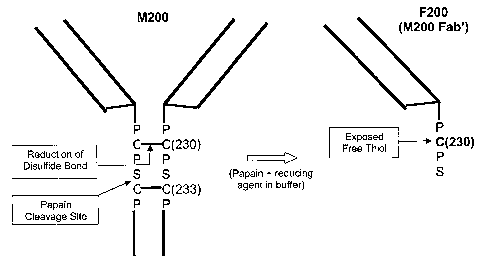
Figure 1 depicts a schematic of the papain digestion of M200 (an antibody
having a
heavy chain amino acid sequence of SEQ ID NO: 1 and a light chain amino acid
sequence of
SEQ ID NO: 2, or conservatively modified variations thereof) antibody to
produce a Fab'
fragment, F200, with an exposed free thiol.
DETAILED DESCRIPTIONS OF THE PREFERRED EMBODIMENTS
To address the problem of stability of proteins having free thiols such as
Fab'-SH
antibody fragments in liquid and lyophilized formulations, the present
invention utilizes a
stabilizing agent, e.g., a sulfliydryl reactive stabilizing molecule, for
coupling to free thiols. The
present invention therefore provides stabilized protein derivatives, e.g., for
use in
pharmaceuticals, and methods of making stabilized protein derivatives.
Preferred proteins of the invention include, but are not limited to,
antibodies, antibody
fragments, and peptides. The molecules of the invention are typically
stabilized by coupling a
free thiol in the molecule of interest to a stabilizing agent such as an N-
acetyl-L-cysteine (NAC)
molecule, a cysteine (CYS) molecule, or a N-ethylmaleimide (NEM) molecule. The
free thiol is
optionally at the terminus of a protein molecule and includes those that are
internal to the
polypeptide chain and those that are buried in the hydrophobic core of the
protein molecule.
Those that are buried in the core of the protein are partially unfolded, e.g.,
with denaturants such
as urea or guanidine hydrochloride to expose the buried thiol for coupling to
the stabilizing
agent.
5
CA 02567758 2006-11-22
WO 2006/004736 PCT/US2005/022902
tt~y4 !{õ. ,.,~~õ ., ~ tt tÃ'T:' !E" Ifi'!t a !t R U td,."'t6' C41
In a'tt=ef ! !(
~~d ~nib'o'tlrm~iit; 'the proteins are IgG4 antibodies and more preferably are
chimeric or humanized antibodies or fragments thereof. For example, the
protein is optionally an
antibody that binds to an integrin, e.g., a5(31 integrin, a4(31 integrin, or
the like, or a Fab'-SH
fragment of such antibodies. In other embodiments, the proteins are peptides,
such as urodilatin,
nesiritide, integrilin, and the like.
The stabilizing agents of the present invention include, but are not limited
to, N-acetyl-L-
cysteine (NAC), cysteine (CYS), and N-ethylmaleimide (NEM), or other
sulfhydryl reactive
molecules to which the proteins of the invention are coupled, e.g., via a
disulfide bond. N-
acetyl-L-cysteine (NAC), for example, is a molecule commonly used as an
additive in food. It is
a potent antioxidant and an approved inactive ingredient for nonparenteral
administration to
patients, such as in the form of tablets, capsules, powders, granules, or
suspensions in non-
aqueous solutions (see e.g., U.S. Patent Nos. 4,920,122; 6,207,190; and
6,689,385). Waterman
K., et al also disclose the use of NAC as an anti-oxidant in both liquid and
solid formulations
(Waterman K., et al, Pharmaceutical Development and Technology 7(1): 1-32
(2002)).
According to the present invention, NAC, NEM, and/or CYS are also optionally
used as
excipients to stabilize proteins in liquid or lyophilized formulations without
coupling to free
thiols. This approach allows the stabilization of the protein having a free
thiol in the liquid
formulation prior to the start of the lyophilization process, and also in the
lyophilized product by
reducing or inhibiting the formation of the disulfide-linked aggregates. The
methods and
compositions of the invention are described in more detail below.
As used herein, the phrase "protein derivative" refers to a protein having a
thiol group
coupled to NAC, NEM, CYS or other sulfhydryl reactive molecules. "Protein" as
used herein
includes, but is not limited to, proteins, antibodies, antibody fragments,
polypeptides, peptides,
and the like. For example, a peptide off the invention is typically about 5 to
about 50 amino
acids. Furthermore, the proteins of the invention are optionally naturally
occurring proteins or
non-naturally occurring proteins.
The term "pharmaceutical formulation" refers to physiologically acceptable
excipients
and carrier solutions well known to those of ordinary skill in the art.
Methods for developing
suitable dosing and treating regimens for using the particular pharmaceutical
formulations are
also well known to those of 'ordinary skill in the art. The pharmaceutical
formulations of the
present invention allow the proteins or protein derivatives to remain
physically, chemically and
biologically stable.
"Stable" (or "stability") as used in the context of the present invention
means that the
protein composition retains its physical stability and/or chemical stability
and/or biological
activity upon storage. Various analytical techniques for measuring protein
stability for
6
CA 02567758 2006-11-22
WO 2006/004736 PCT/US2005/022902
~pr Metermid: '"~t~e~sj'aiic# teieip~~~~~t~es stability are well known in the
art and are reviewed in
e.g., "Peptide and Protein Drug Delivery," 247-301, Vincent Lee Ed., Marcel
Dekker, Inc., New
York, N.Y., Pubs. (1991) and Jones, A. Adv. DrugDeliver.y Rev. 10:29-90
(1993). Stability is
optionally measured, for example, after exposure to a selected temperature for
a selected time
period.
A protein, e.g., an antibody, antibody fragment, polypeptide, or peptide,
"retains its
physical stability" in a pharmaceutical formulation if it shows,no significant
increase in
aggregation, precipitation and/or denaturation, e.g., upon visual examination
of color and/or
clarity, or as measured by UV light scattering, size exclusion chromatography
(SEC), SDS-
PAGE or other methods well known in the art. Protein denaturation is also
optionally evaluated
by fluorescence to determine the tertiary structure, by circular dicliroism
spectroscopy (CD
spectroscopy) that measures changes in secondary and tertiary structures,
and/or by FTIR to
determine the secondary structure.
A protein, e.g., an antibody, antibody fragment, or polypeptide, "retains its
chemical
stability", e.g., in a pharmaceutical formulation, if it shows no significant
chemical alteration.
Chemical stability is optionally assessed by detecting and/or quantifying
chemically altered
forms of the protein. Chemical alteration optionally involves size
modification (e.g. clips or
clipping) that is typically evaluated using size exclusion chromatography, SDS-
PAGE and/or
matrix-assisted laser desorption ionization/time-of-flight mass spectrometry
(MALDI/TOF MS)
of other analytical methods well known to one of ordinary skill in the art.
Other types of
chemical alteration include charge alteration (e.g. occurring as a result of
deamidation) which
can be evaluated by ion-exchange chromatography. Clipping/deamidation and/or
isomerization
may result in change in the CIEF profile. Deamidation and/or isomerization may
also result in
iso-aspartic acid formation, which is readily determined by well-known methods
in the art.
A protein, e.g., an antibody, antibody fragment, polypeptide, or peptide,
"retains its
biological activity" in a pharmaceutical formulation, if the biological
activity of theprotein at a
given time is within a predetermined range of the biological activity
exhibited at the time the
pharmaceutical formulation was prepared. Where the protein is an antibody, the
biological
activity of an antibody is optionally determined, for example, by an antigen-
binding assay.
A "stable liquid formulation" or "stable lyophilized formulation" comprises a
liquid
formulation or lyophilized formulation comprising a protein, e.g., an antibody
or fragment
thereof or protein derivative as described herein, that exhibits no
significant physical, chemical,
or biological changes in the protein when stored at a refrigerated
temperature, e.g., about 2 C to
about 8 C, for at least about 12 months, preferably about 2 years, and more
preferably about 3
years; or at room temperature, e.g., about 22 C to about 28 C, for at least
about 3 months,
7
CA 02567758 2006-11-22
WO 2006/004736 PCT/US2005/022902
~'r''~fcrabiy Ea~oitt' ~ 111ontlis,1faiiW'moie' preferably about 1 year. The
criteria for stability are as
follows: no more than about 10%, and preferably no more than about 5%, of
protein monomer is
degraded as measured by SEC-HPLC. Preferably, the solution remains colorless,
or clear to
slightly opalescent by visual analysis. The concentration, pH and osmolality
of the formulation
have no more than about +/- 10% change. Potency is typically within about 70-
130%, and
preferably 80-120 % of a control level. No more than about 10%, and preferably
no more than
about 5% clipping of the protein is observed. No more than about 10%, and
preferably no more
than about 5% of protein forms aggregates.
The term "buffer" encompasses those agents which maintain the pH value of a
solution,
e.g., in an acceptable range and includes, but is not limited to, sodium
citrate, succinate (sodium
or potassium), histidine, phosphate (sodium or potassium), TRIS (tris
(hydroxymethyl)
aminomethane), diethanolamine, and the like. A preferred buffer has a pH in
the range from
about 5.0 to about 8.0; and preferably has a pH of about 6.0 to 7Ø Examples
of buffers that will
control pH in this range include succinate (such as sodium succinate),
gluconate, histidine,
citrate, phospate and other organic acid buffers.
The terms "lyophilized," and "freeze-dried" refer to a material that is first
in a"pre-
lyophilized" liquid form and which is subsequently frozen and sublimed in a
vacuum
environment to remove the ice or frozen solvent. During the lyophilization
process an excipient
is optionally included in the pre-lyophilized liquid formulation, e.g., to
enhance the stability of
the lyophilized product upon storage.
The term "bulking agent" includes agents that can provide additional structure
to a
freeze-dried product (e.g., to provide a pharmaceutically acceptable cake).
Commonly used
bulking agents include mannitol, glycine, lactose, sucrose, and the like. In
addition to providing
a pharmaceutically acceptable cake, bulking agents also typically impart
useful qualities to the
lyophilized composition such as modifying the collapse temperature, providing
freeze-thaw
protection, further enhancing the protein stability over long-term storage,
and the like. These
agents can also serve as tonicity modifiers.
The term "cryoprotectants" generally includes agents that stabilize the
protein or protein
derivative against freezing-induced stresses. They also typically offer
protection during primary
and secondary drying, and long-term product storage. Examples of such
cryoprotectants are
polymers such as dextran and polyethylene glycol; sugars such as sucrose,
glucose, trehalose,
and lactose; surfactants such as polysorbates; and amino acids such as
glycine, arginine, serine,
and the like.
The term "lyphoprotectant" includes agents that provide stability to a protein
during a
drying or'dehydration' process (primary and secondary drying cycles),
presumably by providing
8
CA 02567758 2006-11-22
WO 2006/004736 PCT/US2005/022902
}F=_, k ~.. n ..!!. .' (( !} I! {I
~n'ameSrph~i~ '~~a s~ matrik and'b~'binding with the protein or protein
derivative through
hydrogen bonding, e.g., replacing the water molecules that are removed during
the drying
process. This helps to maintain protein conformation, minimi~e protein
degradation during a
lyophilization cycle, and improve the long-term stability of the protein or
protein derivative.
Examples include polyols or sugars such as sucrose and trehalose.
"Reconstitution time" is the time that is required to rehydrate a lyophilized
formulation
with a liquid, e.g., to provide a particle-free clarified solution.
The term "isotonic" means that the formulation of interest has essentially the
same
osmolarity as human blood. Isotonic formulations generally have an osmolarity
of about 270-
328 mOsm. Slightly hypotonic osmolarity in pressure is about250-269 mOsm and
slightly
hypertonic is about 328-350 mOsm. Osmolarity is measured, for example, using a
vapor
pressure or ice-freezing type osmometer.
Tonicity modifiers useful in the formulations of the present invention
include, for
example, salts, e.g., NaCl, KC1, MgCl2, CaC12, and the like, and are used to
control osmolarity.
In addition, cryprotecants/lyoprotectants and/or bulking agents such as
sucrose, mannitol,
glycine, and others can serve as tonicity modifiers.
1. Proteins and Methods for Producing Them
A protein is a polymer of amino acid residues. In the present invention, the
term
"protein" encompasses naturally occurring amino acids and polymers thereof as
well as amino
acid polymers in which one or more amino acid residues is an artificial
chemical mimetic of a
naturally occurring amino acid, as well as amino acid polymers containing
modified residues,
and non-naturally occurring amino acid polymers.
Amino acids include naturally occurring and synthetic amino acids, as well as
amino
acid analogs and amino acid mimetics that function similarly to the naturally
occurring amino
acids. Naturally occurring amino acids are those encoded by the genetic code,
as well as those
amino acids that are later modified, e.g., hydroxyproline, y-carboxyglutamate,
and 0-
phosphoserine. Amino acid analogs include compounds that have the same basic
chemical
structure as a naturally occurring amino acid, e.g., an a carbon that is bound
to a hydrogen, a
carboxyl group, an amino group, and an R group. Such analogs include, but are
not limited to,
homoserine, norleucine, methionine sulfoxide, methionine methyl sulfonium.
Such analogs
optionally include modified R groups (e.g., norleucine) or modified peptide
backbones, but
retain the same basic chemical structure as a naturally occurring amino acid.
"Amino acid
mimetic" refers to a chemical compound that has a structure that is different
from the general
chemical structure of an amino acid, but functions similarly to a naturally
occurring amino acid.
9
CA 02567758 2006-11-22
WO 2006/004736 PCT/US2005/022902
Pr telii~ L.Acdxripass~e db'jl~e-present invention include all types of
proteins including
secreted proteins, transmembrane proteins or intracellular proteins. Preferred
proteins comprise
antibodies or fragments thereof or peptides, e.g., for use in the treatment of
cancer or heart
failure.
Currently available antibody pharmaceuticals that can benefit from the
stabilizing
methods and compositions provided herein include, but are not limited to,
trastuzumab,
(Herceptin(V, Genentech, Inc); omalizumab, (Xolair(T) efalizumab (RaptivaTM,
Genentech, Inc);
bevacizumab (AvastinTM, Genentech, Inc); daclizumab (Zenapax(&, Roche);
palivizumab
(Synagis , Medlmmune, Inc); natalizumab (Tysabri(l), alemtuzumab (Campath ),
cetuximab
(Erbitux ), infliximab (Remicade ), rituximab (Rituxan ), basiliximab
(Simulect ),
palivizumab (Synagis(M), and gemtuzumab ozogamicin (Mylotarg , Wyeth). In
addition, the
following therapeutic products, which are in various stages of development,
are also optionally
used in the methods and compositions of the invention: epratuzumab, (Vitaxin
), apolizumab
(Zamyl ), and labetuzuma (CEA-Cide ).
Additional preferred proteins of the invention comprise polypeptides, e.g.,
anti-coagulant
polypeptides as described in, e.g., US Patent 6,239,101 (Esmon et al.). For
example,
Eptifibatide (Integrelin ) is an intravenous cyclical heptapeptide that
selectively blocks the
platelet glycoprotein IIb/ IIIa receptor. It reversibly binds to platelets and
has a short half-life. It
has demonstrated efficacy in the treatment of patients during coronary
angioplasty, myocardial
infarction and angina.
Abciximab (Reopro Centocor B.V.) is the Fab fragment of the chimeric human-
murine
monoclonal antibody 7E3. This antibody binds to glycoprotein IIb/IIIa receptor
of human
platelets and inhibits platelet aggregation. It also binds to a vitronection
avP3 receptor on
platelets. Reopro is multi-receptor antagonist that reduces complications
associated with
coronary angioplasty by preventing the formation of blood clots by inhibiting
platelet
aggregation.
A natural human peptide called human B-type natriuretic peptide (hBNP) that is
secreted
by the heart as part of the body's normal response to heart failure is the
basis for another peptide
pharmaceutical, e.g., Natrecor (nesiritide), a recombinant form of the
endogenous human
peptide. Natrecor is used in the treatment of acute heart failure.
Listed above are various peptides, polypeptides, and antibodies that are
optionally
stabilized using the methods described herein. It will be apparent to one
skilled in the art upon
review of the following detailed description that many other proteins are
optionally stabilized
using the compositions and methods provided herein.
CA 02567758 2006-11-22
WO 2006/004736 PCT/US2005/022902
~
~Sc~'urri'Fi~g'Pro iY~is of the present invention can be isolated and purified
with
the methods well known in the art, for example, hydroxylapatite
chromatography, gel
electrophoresis, dialysis, and affmity chromatography, with affmity
chromatography being the
preferred purification technique. Other purification techniques such as
fractionation on an ion-
exchange column, ethanol precipitation, reverse phase HPLC, chromatography on
silica,
chromatography on heparin SEPHAROSETT"' chromatography on an anion or cation
exchange
resin (such as a polyaspartic acid column), chromatofocusing, SDS-PAGE, and
ammonium
sulfate precipitation are also available.
Proteins of the present invention are also optionally produced recombinantly.
DNA
molecules encoding the proteins of the present invention are used together
with a variety of
expression vectors to express the proteins, for example, in prokaryotic or
eukaryotic cells.
Expression vectors and recombinant DNA technology are well known to those of
skill in the art
(see, e.g., Ausubel, supra, and Gene Expression Systems (Fernandez & Hoeffler,
eds, 1999)).
The proteins of the present invention are typically produced by culturing a
host cell transformed
with an expression vector containing nucleic acid encoding the proteinof
interest, e.g, an anti-
coagulant peptide, under appropriate conditions to induce or cause expression
of the protein.
Conditions appropriate for protein expression will vary with the choice of the
expression vector
and the host cell, and are easily ascertained by one skilled in the art
through routine
experimentation or optimization. Appropriate host cells include yeast,
bacteria, archaebacteria,
fungi, insect and animal cells, including mammalian cells. Of particular
interest are
Saccharomyces cerevisiae and other yeasts, E. coli, Bacillus subtilis, Sf9
cells, C129 cells, 293
cells, Neurospora, BHK, CHO, COS, HeLa cells, HUVEC (human umbilical vein
endothelial
cells), NSO cells, THP1 cells (a macrophage cell line) and various other human
cells and cell
lines. The recombinantly produced proteins are also optionally purified, e.g.,
by any techniques
discussed above or known in the art.
In a preferred embodiment, proteins of the present invention contain one or
more thiol
groups, which can be located in any domain or region of the protein. In one
aspect, the thiol
groups are exposed, i.e., on the surface of protein so that they may react,
e.g., with NAC, NEM
or CYS. In another aspect of the invention, the thiol groups are hidden, e.g.,
buried within any
folded three-dimensional structures of the protein. In that case, the proteins
are partially
unfolded with denaturants such as urea or guanidine hydrochloride, e.g., to
make the hidden
thiol group available to react with NAC, NEM or CYS, or the like. The
denaturant is then
typically removed, e.g., to allow the protein, such as an anti-integrin
antibody, to refold back to
its active (or native) three-dimensional structure.
11
CA 02567758 2006-11-22
WO 2006/004736 PCT/US2005/022902
ir , q.. . f... ,, 11 ,, ~u:õ ,,,,,~i
r~n~ilit~~ii~~s"~ild IVIdthd&'for Producing Them
A typical protein that is stabilized according to the present invention
comprises an
antibody. For the purpose of the present invention, the term "antibody"
includes an
immunoglobulin molecule immunologically reactive with a particular antigen,
and includes both
polyclonal and monoclonal antibodies. The term also includes genetically
engineered forms such
as humanized (e.g., humanized murine antibodies), primatized or chimeric
antibodies and
heteroconjugate antibodies (e.g., bispecific antibodies). The term "antibody"
also encompasses
antigen binding forms or parts of antibodies, including fragments with antigen-
binding
capability (e.g., Fab', Fab'-SH, F(ab')2, Fab, Fv and rIgG). See also, Pierce
Catalog and
Handbook, 1994-1995 (Pierce Chemical Co., Rockford, IL). See also, e.g., Kuby,
J.,
Immunology, 3ra Ed., W.H. Freeman & Co., New York (1998). The term also refers
to
recombinant single chain Fv fragments (scFv). In addition, the term "antibody"
also includes
bivalent or bispecific molecules, diabodies, triabodies, and tetrabodies.
Bivalent and bispecific
molecules are described in, e.g., Kostelny et al.. (1992) Jlmmunol 148:1547,
Pack and
Pluckthun (1992) Biochemistry 31:1579, Hollinger et-al., 1993, supra, Gruber
et al. (1994) J
Immunol :5368, Zhu et al. (1997) Protein Sci 6:781, Hu et al. (1996) Cancer
Res. 56:3055,
Adams et al. (1993) Cancer Res. 53:4026, and McCartney, et al. (1995) Protein
Eng. 8:301.
An antibody immunologically reactive with a particular antigen (i.e., that
binds to the
antigen) can be generated by recombinant methods such as selection from
libraries of
recombinant antibodies in phage or similar vectors, see, e.g., Huse et al.,
Science 246:1275-1281
(1989); Ward et al., Nature 341:544-546 (1989); and Vaughan et al., Nature
Biotech. 14:309-
314 (1996), or by immunizing an animal with the antigen or with DNA encoding
the antigen.
Typically, an immunoglobulin comprises a heavy and light chain. Each heavy and
light
chain contains a constant region and a variable region, (the regions are also
referred to as
"domains"). Light and heavy chain variable regions contain four "framework"
regions
interrupted by three hypervariable regions, also called "complementarity-
determining regions"
or "CDRs". Sequences of the framework regions of different light or heavy
chains are relatively
conserved within a species. The framework region of an antibody, typically the
combined
framework regions of the constituent light and heavy chains, serves to
position and align the
CDRs in three-dimensional space.
The tenn "VH" refers to the variable region of an immunoglobulin heavy chain
of an
antibody, including the heavy chain of an Fv, scFv, Fab'-SH or Fab. References
to "VL" refer to
the variable region of an immunoglobulin light chain, including the light
chain of an Fv, scFv,
dsFv, Fab'-SH or Fab.
12
CA 02567758 2006-11-22
WO 2006/004736 PCT/US2005/022902
Qy"respd~s~i~sle for binding of an antibody or fragment thereof to an
epitope of an antigen. The CDRs of each chain are typically referred to as
CDRl, CDR2, and
CDR3, numbered sequentially starting from the N-terminus, and are also
typically identified by
the chain in which the particular CDR is located. Thus, a VH CDR3 is located
in the variable
domain of the heavy chain of the antibody in which it is found, whereas a VL
CDR1 is the CDR1
from the variable domain of the light chain of the antibody in which it is
found.
The phrase "single chain Fv" or "scFv" refers to an antibody in which the
variable
domains of the heavy chain and of the light chain of a traditional two chain
antibody have been
joined to form one polypeptide chain. Typically, a linker peptide is inserted
between the two
chains to allow for proper foldirig and creation of an active antigen binding
site.
An antibody of the invention, e.g., an anti-integrin antibody, is optionally a
chimeric
antibody. A "chimeric antibody" is an immunoglobulin molecule in which (a) the
constant
region, or a portion thereof, is altered, replaced or exchanged so that the
antigen binding site
(variable region) is linked to a constant region of a different or altered
class, effector function
and/or species, or an entirely different molecule which confers new properties
to the chimeric
antibody, e.g., an enzyme, toxin, hormone, growth factor, drug, etc.; or (b)
the variable region,
or a portion thereof, is altered, replaced or exchanged with a variable region
having a different or
altered antigen specificity. In a preferred embodiment, the variable regions
of the chimeric
antibody are derived from mouse, while the constant regions are derived from
human. In order
to produce the chimeric antibodies, the portions derived from two different
species (e.g., human
constant region and murine variable or binding region) can be joined together
chemically by
conventional techniques or can be prepared as single contiguous proteins with
genetic
engineering techniques. The DNA molecules encoding the proteins of both the
light chain and
heavy chain portions of the chimeric antibody can be expressed as contiguous
proteins. The
method of making the chimeric antibody is disclosed in U.S. Patent No.
5,677,427; U.S. Patent
No. 6,120,767; and U.S. Patent No. 6,329,508, each of which is incorporated by
reference in its
entirety.
A preferred antibody of the present invention is a humanized antibody. A
"humanized
antibody" is an immunoglobulin molecule that contains minimal sequence derived
from non-
human immunoglobulin. Humanized antibodies include human immunoglobulins
(recipient
antibody) in which its native CDRs are replaced by residues from a CDR of a
non-human
species (donor antibody) such as mouse, rat, rabbit, or the like, having the
desired specificity,
affinity and capacity. In some instances, corresponding non-human residues
replace Fv
framework residues of the human immunoglobulin. Humanized antibodies also
optionally
comprise residues that are found neither in the recipient antibody nor in the
imported CDR or
13
CA 02567758 2006-11-22
WO 2006/004736 PCT/US2005/022902
frai~evt~ctrk's'e~ti~ifc~s: Iy;IAumanized antibody comprises substantially all
of at least
one, and typically two, variable domains, in which all or substantially all of
the CDR regions
correspond to those of a non-human immunoglobulin and all or substantially all
of the
framework (FR) regions are those of a human immunoglobulin consensus sequence.
The
humanized antibody will optimally also comprise at least a portion of an
immunoglobulin
constant region (Fc), typically that of a human immunoglobulin. For
humanization methods and
antibodies, see, Queen et al., U.S. Patents Nos. 5,530,101; 5,585,089;
5,693,762; and 6,180,370
(each of which is incorporated by reference in its entirety). See also, Jones
et al., Nature
321:522-525 (1986); Riechmann et al., Nature 332:323-329 (1988); and Presta,
Curr. Op.
Struct. Biol. 2:593-596 (1992)). Humanization of antibodies can also be
performed following the
methods of e.g. Winter and co-workers (Jones et al., Nature 321:522-525
(1986); Riechmann et
al., Nature 332:323-327 (1988); or Verhoeyen et al., Science 239:1534-1536
(1988)), by
substituting rodent CDRs or CDR sequences for the corresponding sequences of a
human
antibody. See also, U.S. Patent No. 5,585,089.
Antibodies useful in the practice of the present invention are also optionally
fully human
antibodies. Fully human antibodies are optionally produced by a variety of
techniques. One
example is trioma methodology. The basic approach and an exemplary cell fusion
partner,
SPAZ-4, for use in this approach have been described by Oestberg et al.,
Hybridoma 2:361-367
(1983); Oestberg, U.S. Patent No. 4,634,664; and Engleman et al., U.S. Patent
No. 4,634,666
(each of which is incorporated by reference herein in its entirety). Fully
human antibodies are
also optionally produced from non-human transgenic animals having transgenes
encoding at
least a segment of the human immunoglobulin locus. The production and
properties of animals
having these properties are described in detail by, e.g., Lonberg et al., WO
93/12227; U.S.
Patent No. 5,545,806; and Kucherlapati, et al., WO 91/10741; U.S. Patent No.
6,150,584, each
of which is incorporated herein by reference in its entirety.
Various recombinant antibody library technologies are also optionally utilized
to produce
fully human antibodies. For example, one approach is to screen a DNA library
from human B
cells according to the general protocol outlined by Huse et al., Science
246:1275-1281 (1989).
Antibodies or fragments thereof are selected from this library, typically by
binding to a
preselected antigen or a fragment thereof. Sequences encoding such antibodies
(or binding
fragments of an antibody) are then cloned and amplified. The protocol
described by Huse is
rendered more efficient in combination with phage-display technology. See,
e.g., Dower et al.,
WO 91/17271 and McCafferty et al., WO 92/01047; U.S. Patent No. 5,969,108,
(each of which
is incorporated by reference in its entirety). In these methods, libraries of
phage are produced in
which members display different antibodies on their outer surfaces. Antibodies
are usually
14
CA 02567758 2006-11-22
WO 2006/004736 PCT/US2005/022902
+1 h IF,,., ,. !, it,.,i~
displa,ec~ 'kFf -6r "'Fib'-SH~Uagments. Phage displaying antibodies with a
desired
specificity are selected by binding to the antigen or fragment thereof.
Eukaryotic ribosomes are optionally used as means to display a library of
antibodies and
which may be selected by screening against a target antigen, such as a5(31, as
described in Coia
G, et al., J. hnmunol. Methods 1: 254 (1-2):191-7 (2001); Hanes J. et al.,
Nat. Biotechnol. 18
(12):1287-92 (2000); Proc. Natl. Acad. Sci. U. S. A. 95 (24):14130-5 (1998);
Proc. Natl. Acad.
Sci. U. S. A. 94 (10):4937-42 (1997), each of which is incorporated by
reference in its entirety.
Antibody libraries are also optionally displayed on the surface of yeast cells
for the
purpose of obtaining the human antibodies and their encoding nucleic acid
against a target
antigen. This method is described by Yeung, et al., Biotechnol. Prog.
18(2):212-20 (2002);
Boeder, E. T., et al., Nat. Biotechnol. 15(6):553-7 (1997), each of which is
herein incorporated
by reference in its entirety. Alternatively, human antibody libraries are
expressed intracellularly
and screened via yeast two-hybrid system (W00200729A2, which is incorporated
by reference
in its entirety).
The antibodies of the present invention are optionally further purified, e.g.,
using,
hydroxylapatite chromatography, gel electrophoresis, dialysis, and affmity
chromatography,
with affmity chromatography, e.g., using protein A, being the preferred
purification technique.
The suitability of protein A as an affinity ligand typically depends on the
species and isotype of
any immunoglobulin Fc domain that is present in the antibody. Protein A is
optionally used to
purify antibodies that are based on human Y'1, Y'2, or Y'4 heavy chains
(Lindmark et al., J.
Immunol. Meth. 62:1-13 (1983)). Protein G is recommended as an affmity ligand
for all mouse
isotypes and for human Y'3 (Guss et al., EMBO J. 5:1567-1575 (1986)). The
matrix to which the
affinity ligand is attached is typically agarose, but other matrices are
optionally used. For
example, mechanically stable matrices such as controlled pore glass or
poly(styrenedivinyl)benzene allow for faster flow rates and shorter processing
times than can be
achieved with agarose. Where the antibody comprises a CH3 domain, the
Bakerbond ABXrM
resin (J. T. Baker, Phillipsburg, N.J.) is useful for purification. Other
techniques for protein
purification such as fractionation on an ion-exchange column, ethanol
precipitation, Reverse
Phase HPLC, chromatography on silica, chromatography on heparin SEPHAROSETT"'
chromatography on an anion or cation exchange resin (such as a polyaspartic
acid column),
chromatofocusing, SDS-PAGE, and ammonium sulfate precipitation are also
available
depending on the antibody to be recovered.
Antibodies of the present invention are typically derived from species
including, but not
limited to, human, chicken, goats, and rodents (e.g., rats, mice, hamsters and
rabbits), including
transgenic rodents genetically engineered to produce human antibodies (see,
e.g., Lonberg et al.,
CA 02567758 2006-11-22
WO 2006/004736 PCT/US2005/022902
!k":~i r~=eh ,, It.,> . " Ik Ik t! :'; !! =,tt 1l,~,. : ,. , ~'U . at ~k.,N
Ik,,,1~ ,,,at
... . ..-
W093/1222%','"and Kucherlapati, et al., W091/10741; U.S. Patent
No. 6,150,584, which are herein incorporated by reference in their entirety).
The antibodies of the present invention include antibodies having all types of
constant
regions, including IgM, IgG, IgD, IgA and IgE, and any isotype, including
IgGl, IgG2a, IgG2b,
IgG3 and IgG4, with IgG4 as a preferred isotype. The light chains of the
antibodies are
optionally either kappa light chains or lambda light chains. The antibodies
typically bind to their
epitopes at a binding affiuiity of at least 106M-1,107M-1, 108M-1,109Nr1, or
101oM"1
In a preferred embodiment, the antibodies or antibody fragment of the present
invention
are antibodies against a5(31 integrin which bind specifically to at least one
subunit of a5(31
integrin. The binding specificity of antibodies is optionally assessed by the
methods known in
the art such as concurrent immunoelectrophoresis, radioimmuno-assays,
radioinimuno-
precipitation, enzyme-linked immuno-sorbent assays (ELISA), dot blot or
Western blot assays,
inhibition or competition assays, and sandwich assays. For a review of
immunological and
immunoassay procedures, see, e.g., Basic and Clinical Inzmunology (Stites &
Terr eds., 7th ed.
1991).
Antibodies of the invention are optionally provided in a variety of forms,
such as
monoclonal, polyclonal, chimeric, humanized, fully human, and/or bispecific
antibodies, e.g.,
against a501 integrin or fragments thereof. These antibodies are typically
made by any method
known in the art and/or discussed above.
The anti-a5P 1 integrin antibodies of the present invention preferably
neutralize at least
one biological activity of an a5(31 integrin, such as receptor binding
activity, signaling
transduction, and cellular responses induced by a5(31. Preferably, such
neutralizing antibodies
are capable of competing with the binding of a5(31 to its signaling molecules,
or even block the
binding completely. Such antibodies preferably inhibit tumor angiogenesis
and/or induce death
of the proliferating endothelial cells.
In a preferred embodiment, the anti-a5(3lintegrin antibodies are those
disclosed in U.S.
Patent Application Serial No. 10/724,274, filed November 26, 2003,
(Publication No.: US
2005/0054834 Al,which is incorporated by reference in its entirety), which
discloses the anti-
a5(3lintegrin antibody M200, which is a high affinity chimeric IgG4 antibody
(with a human
IgG4 constant region). M200 comprises a heavy chain an amino acid sequence as
follows:
QVQLKESGPGLVAPSQSLSITCTISGFSLTDYGVHWVRQPPGKGLEWLVVIWSDGSSTYNSALK
SRMTIRKDNSKSQVFLIMNSLQTDDSAMYYCARHGTYYGMTTTGDALDYWGQGTSVTVSSASTK
GPSVFPLAPCSRSTSESTAALGCLVKDYFPEPVTVSWNSGALTSGVHTFPAVLQSSGLYSLSSV
VTVPSSSLGTKTYTCNVDHKPSNTKVDKRVESKYGPPCPSCPAPEFLGGPSVFLFPPKPKDTLM
ISRTPEVTCVVVDVSQEDPEVQFNWYVDGVEVHNAKTKPREEQFNSTYRVVSVLTVLHQDWLNG
16
CA 02567758 2006-11-22
WO 2006/004736 PCT/US2005/022902
~KGAV~I~GfLV~~IL"R!9l4 QPREPQVYTLPPSQEEMTKNQVSLTCLVKGFYPSDIAVE
WESNGQPENNYKTTPPVLDSDGSFFLYSRLTVDKSRWQEGNVFSCSVMHEALHNHYTQKSLSLS
LGK [SEQ ID NO: 1].
M200 also comprises a light chain amino acid sequence as follows:
QIVLTQSPAIMSASLGERVTMTCTASSSVSSNYLHWYQQKPGSAPNLWIYSTSNLASGVPARFS
GSGSGTSYSLTISSMEAEDAATYYCHQYLRSPPTFGGGTKLEIKRTVAAPSVFIFPPSDEQLKS
GTASVVCLLNNFYPREAKVQWKVDNALQSGNSQESVTEQDSKDSTYSLSSTLTLSKADYEKHKV
YACEVTHQGLSSPVTKSFNRGEC [SEQ ID NO: 2].
U.S. Patent Application Serial No. 10/724,274 also discloses F200, the Fab'
fragment of
M200.
IH. Fab'-SH Fragments and Methods for Producing Them
In one preferred embodiment, the proteins of the present invention comprise
Fab'-SH
fragments of antibodies. Novel methods of producing Fab'-SH fragments are also
provided
herein. In particular, a starting antibody is digested with either pepsin or
papain, either in an
immobilized form or preferably in a solution in the presence or absence of a
reducing agent,
preferably at a pH of about 6.0 to about 8.0, and more preferably about 7Ø
The reaction is
typically performed at about 15 C to about 50 C, preferably at about 30 C to
40 C, and most
preferably at about 37 C. Where the starting antibody is an IgG4 type, the
digestive enzyme
papain is typically preferred. The papain/antibody ratio (weight) is typically
from about 1:10 to
1:108, preferably from about 1:103 to 1:105, and more preferably about 1:104.
The digestion is
carried out for about 1-100 hours, preferably about 1-10 hours, and more
preferably about 3-4
hours. Various reducing agents known in the art are optionally used in the
digestion, including,
but not limited to, DTT, cysteine, f3-mercaptoethylamine, and N-actyl-L-
cysteine.
Concentrations of the reducing agents are typically about 0.1-100 mM,
preferably about 1-50
mM, and more preferably about 1-20 mM.
In a preferred embodiment, the starting antibody is an antibody of IgG4 class,
preferably
a chimeric or humanized IgG4 antibody. In a more preferred embodiment, the
antibody is M200
(as provided by SEQ ID NOS: 1-2) or HuMV833. HuMV833 is a humanized anti-VEGF
antibody. Figure 1 provides a schematic depiction of papain digestion of an
IgG4 antibody.
Papain cleaves between the two intra-heavy chain disulfide bonds. Reduction of
the C230-C230
disulfide bond is required for the release of the Fab' fragment that has an
exposed free thiol
group at position 230.
Preferably, soluble papain is utilized for digestion processes in the present
invention
instead of immobilized papain, which is typically used in the art. hnmobilized
papain often
contains sodium azide as a preservative, which is often problematic for
clinical manufacturing.
17
CA 02567758 2006-11-22
WO 2006/004736 PCT/US2005/022902
s Il 1, ..,, .,. ; , I} ~t II: .' 14,..J1 ;~ , ; . ..::It .:::I t
A
s{:::1d !P",S i A
hbf ~t~l'tYbTd'p~'oaif~"a~'tiigcl(~s~id in the present specification avoids
this problem. Another
advantage of using soluble papain is that antibodies are digested with a low
papain/antibody
ratio. For example, using soluble papain, with 1:10000 ratio (weight) (e.g.,
100 ppm) of papain
to antibody, M200 is 99% digested in 3 hours. In contrast, when using
immobilized papain, a
papain/antibody ratio of 1:5 is required to achieve the same digestion
efficiency. Another
advantage of using soluble papain is that it is easily removed by cation
exchange
chromatography (CEX). Further, when soluble papain is stored in sodium azide
free preservative
at 4-8 C in dark it loses less than 50% activity in 13 months.
Soluble papain sometimes causes proteolytic digestion of the linkage between
protein A
and the matrix used in antibody purification, thus releasing protein A into
the solution. As a
consequence, the methods of the present invention typically further comprise a
step of purifying
the post digestion mixture before a potential protein A affmity chromatography
step. For
example, cation exchange chromatography is optionally used to remove papain,
the residue
reducing agents, undigested starting antibodies, Fc and other impurities.
Protein A affmity
chromatography is then typically used as an additional subsequent step to
remove trace
undigested antibodies. The antibody fragments after Protein A purification are
optionally
subjected to ultrafiltration/diafiltration buffer exchange and fonnulation,
e.g., using methods
well known in the art.
The Fab' fragments, e.g., natalizumab fragments or M200 fragments, produced as
described above are in condition to be derivatized with a stabilizing agent as
described in more
detail below.
IV. Protein Derivative and Methods for Producing Them
The present invention provides compositions comprising stable protein
derivatives
having at least one thiol group that is coupled to a NAC molecule, NEM
molecule or CYS
molecule via a disulfide bond. Methods of preparing these stable protein
derivatives are also
provided. The methods typically comprise coupling the free thiol group of a
protein to a
molecule such as NAC, NEM or CYS, preferably in the presence of sodium
tetrathionate.
In some embodiments, the derivatized proteins comprise antibodies or fragments
thereof.
In a preferred embodiment, the antibodies bind specifically to integrin
molecules, e.g., a501
integrin. A preferred antibody is M200, as described above. More preferably,
the proteins are
Fab' -SH fragments of antibodies, and more preferably of antibodies that bind
to integrins, e.g.,
the Fab'-SH fragment of the M200 antibody which binds to a501 integrin or the
Fab'-SH
fragment of natalizumab which binds to the a401 integrin. Methods of making
stable derivatives
18
CA 02567758 2006-11-22
WO 2006/004736 PCT/US2005/022902
6f 8thd1 6ntibddi~,"A~d~'frdg'&611ts, e.g., antibody fragments that inhibit
angiogenesis, and
peptides, e.g., anti-coagulant peptides, are also provided.
The protein derivatives of the present invention are optionally generated by
incubating a
protein having a free thiol group with NAC, CYS, or NEM for at least about 1
minute, about 5
minutes, about 10 minutes, or about 30 minutes, or about 1 to about 5 hours,
and preferably for
about 30-60 minutes. The concentration of NAC, or CYS, or NEM is typically
about 0.10-100
mM, preferably 1-50 mM, and more preferably 10-40 mM. In some embodiments, the
reaction is
facilitated by sodium tetrathionate (NTT), which is optionally added into the
mixture of the
proteins and NAC, CYS, or NEM at a concentration of about 1-100 mM, preferably
about 1-50
mM, and more preferably about 10-30 mM and incubated for about 1 minute to
several hours,
preferably, about 1 minute to 1 hour, and more preferably about 30 minutes, at
about 4 C to 40
C and preferably at about room temperature, e.g., about 22 C to about 28 C.
The reaction
results in the addition of NAC, CYS or NEM to the free thiol group of the
protein. In a preferred
embodiment, the resulting protein derivatives are further purified and
concentrated as described
herein.
Where the protein to be derivatized is an antibody, e.g., an IgG4 antibody, a
chimeric, or
humanized antibody, the starting antibody is typically digested with a papain
solution in the
presence of NAC. NAC can act as a reducing agent, but is not required for the
digestion when
soluble papain is used. After digestion, sodium tetrathionate (NaTT) is
optionally added to the
reaction mixture to react with free thiols, e.g., generated from the reduction
of the C230-C230
disulfide bond between the light chain and the heavy chain of M200, thereby
forming reactive
sulfenylthiosulfate intermediates with which another sulfliydryl, preferably
NAC, couples to
form a disulfide linkage. The generated molecule, referred to as Fab'NAC, is
typically stable in
solution, e.g., even in simple phosphate buffer. A preferred Fab'-SH fragment
is an Fab' SH
fragment of M200 or any other antibody that inhibits angiogenesis or otherwise
directly kills
tumor cells. The Fab'NAC produced by the methods of the present invention from
an M200
antibody is referred to as F200 Fab'NAC, and has a molecular weight of 48184.4
Daltons (about
48 kD).
One preferred method for producing stable Fab'-SH derivatives comprises the
following
steps:
1) digesting an antibody, e.g., using a papain solution, preferably in the
presence of NAC
as described above;
2) producing the Fab'-NAC molecule in the presence of sodium tetrathionate
(NaTT);
3) purifying the Fab'Nac molecule, e.g., by cation exchange chromatography
(CEX) and
protein A chromatography;
19
CA 02567758 2006-11-22
WO 2006/004736 PCT/US2005/022902
sf"f,.,,, } }} tt y(,~,as g ille ~ ii,;;lt s}:::f~} }t,,}} ";mtt
4) c~i~~~ff&d"Fab'Nac molecules, e.g., using ultrafiltration; and
5) diafiltrating the concentrated Fab'Nac molecules into a formulation buffer.
The stability of the generated protein derivative (such as Fab'NAC) is
optionally tested,
e.g., via methods known in the art, for example, HPLC or LC-MS (Liquid
Chromatography Mass
Spectrometry). HPLC is optionally used to evaluate the percent monomer,
aggregate and clip
formation as a function of time and storage temperature. In the case of
antibody having a free
thiol, the main degradation pathway is typically dimer formation over time. LC-
MS may be used
to evaluate the stability of the generated protein derivative (e.g., dimer
formation) as a function
of time and storage temperature. Typically, Fab'NAC molecules remain as a
single homogeneous
species and have the predicted molecular weight for a single homogeneous
species.
A formulation comprising the compositions of the invention, e.g., a
composition
comprising a protein or protein derivative, preferably allows the protein or
protein derivative to
retain its physical, chemical and biological activity over time and at certain
temperatures. The
formulation is preferably stable for at least about 1 year at refrigerated
temperature, e.g., about 2
C to about 8 C, and about 3 months at room temperature, e.g., about 23 C to
about 27 C.
Preferably, a formulation containing the protein derivatives has less than
about 5% of protein
dimers after one-year storage at refrigerated temperature (about 2-8 C), or
after about 3 months
at room temperature (about 23-27 C) or after about one-month storage at about
37 C. In a
preferred embodiment essentially no change in the molecular weight of the
generated monomeric
protein derivatives is observed after about one-year storage at refrigerated
temperature (about 2-
8 C), or after about 3 months at room temperature (about 23-27 C) or after
about one-month
storage at about 37 C.
A Fab'NAC or peptide-NAC (peptide stabilized with NAC using the methods
provided
herein) molecule of the present invention also preferably retains the same
binding specificity,
e.g., to its antigen, as the parent protein, e.g., antibody. Binding
specificity is typically examined
via techniques known in the art, including, but not limited to,
immunoelectrophoresis,
radioimmunoassay, radioimmuno-precipitation, enzyme-linked immuno-sorbent
assay (ELISA),
dot blot or Western blot assay, and sandwich assays.
The Fab'NACs and peptide-NACs (peptides stabilized using the methods provided
herein) of the present invention also preferably retain the same binding
affinity, e.g., to an
antigen, as the parent protein, e.g., antibody or peptide. The binding affmity
of a Fab'NAC or
peptide-NAC is optionally determined by Scatchard analysis, by surface plasmon
resonance
using BlAcore, or by any other method known to those of skill in the art.
In addition to retaining binding specificity and binding affmity, Fab'NACs and
peptide-
NACs also retain the desired biological activities of their parent proteins.
For example, in a
CA 02567758 2006-11-22
WO 2006/004736 PCT/US2005/022902
}! ~r .;' :It u,,,r e rt :;iF
pref~n'~d eiitn'tit~'~~li~~Yt F200 F'~~b' ~I'~Ti4C inhibits angiogenesis (as
does its parent antibody M200)
as shown, for example, by its ability to inhibit tube formation in vitro and
choroidal
neovascularization (CNV) in primate eyes as disclosed in Publication No.: US
2005/0054834 Al,
filed November 26, 2003, which is hereby incorporated by reference.
With the same biological specificity and biological activity and the addition
of increased
stability, e.g., in formulation, the compositions of the present invention
provide improved protein
pharmaceutical products over what is presently available.
V. Pharmaceutical Formulations
The present invention is also directed to stable liquid and/or lyophilized
pharmaceutical
formulations of protein compositions comprising one or more free thiol groups
and preferably
comprising protein derivatives having a thiol group coupled to a molecule such
as NAC, CYS or
NEM. Preferably, the proteins are antibodies (more preferably of the IgG4
class), antibody
fragments, e.g., Fab' fragments, or peptides, e.g., anti-coagulation peptides,
with one or more
free thiol groups available for coupling to the molecules described above. In
a preferred
embodiment, the antibody fragments are Fab'-SH fragments of a chimeric or a
humanized
antibody, such as an Fab'-SH fragment of M200 or other antibodies that inhibit
tumor growth
and/or angiogenesis.
The pharmaceutical formulations of the present invention preferably comprise a
protein
or protein derivative, such as those described immediately above or a mixture
thereof dissolved
in a pharmaceutically acceptable carrier, preferably an aqueous carrier. A
variety of aqueous
carriers are optionally used, e.g., water for injection (WFI), or water
buffered with phosphate,
citrate, acetate, etc., and/or containing salts such as sodium chloride,
potassium chloride, etc.
The carrier also optionally contains pharmaceutically acceptable excipients
such as human
serum albumin, polysorbate 80, sugars or amino acids. The formulated proteins
or protein
derivatives according to the present invention are particularly suitable for
parenteral
administration, and are optionally administered as an intravenous infusion or
by intravitreal,
subcutaneous, intramuscular, intravenous, intrathecal, intraventricular, or
intrasynovial injection,
with intravitreal injection a preferred route of administration. Methods for
preparing parenterally
administrable fonnulations are known or apparent to those skilled in the art
and are described in
more detail in, for example, Remington's Pharmaceutical Science (15th Ed.,
Mack Publishing
Company, Easton, Pa., 1980), which is incorporated herein by reference.
A. Stable Liquid Formulations
In one aspect, the present invention is directed to stable liquid
pharmaceutical
formulations comprising one or more protein, or protein derivative. Typically,
before
formulation, the protein is stabilized by coupling a molecule such as an NAC,
CYS, or NEM to
21
CA 02567758 2006-11-22
WO 2006/004736 PCT/US2005/022902
a'4ifi}"ee thio1 ~iO'dp' b tf flhe PirbiCeiiY'4 i~6ftlting in a stable protein
derivative as described above. The
generated protein derivatives are stable in the pharmaceutical formulations of
the present
invention.
The stable liquid formulations of the present invention minimize, for example,
denaturation, clipping, or aggregate formation as described above. When the
protein or protein
derivative is an antibody or antibody fragment or derivative thereof, the
formulation aids in
maintaining its immunoreactivity, (e.g., ability to bind to an antigen) over
time. Preferably, the
formulation comprises a sterile, pharmaceutically acceptable liquid
formulation containing an
antibody, antibody fragment and preferably a derivative thereof as described
herein in a buffer
having a near neutral pH (pH 5.00- 8.00). The protein concentration in the
formulation is
typically at least about 1, 2, 5, 10, 20, 50 mg/ml, preferably about 1-80
mg/ml and preferably
fiirther comprises a buffer of pH 5.00- 8.00. Examples of buffers that control
the pH in this
range include citrate, succinate (such as sodium succinate), histidine,
phosphate, and other
organic buffers. Citrate (pKa 6.0) is typically a preferred buffer for
subcutaneous injection. A
preferred buffer comprises about 10-50 mM sodium citrate. Another preferred
buffer comprises
about 30-70 mM histidine buffer overlaid with N2.
In some embodiments, the formulation also comprises a surfactant. Exemplary
surfactants include, but are not limited to, nonionic surfactants such as
polysorbates (e.g.
polysorbates 20, 80, such as TWEEN 20, TWEEN 80) or poloxamers (e.g.
poloxamer 188).
The amount of surfactant added is typically such that it aids in reducing
aggregation of the
protein or protein derivatives and/or miniunizes the formation of particulates
in the formulation
and/or reduces adsorption to the container containing the formulation. The
surfactant is typically
present in the formulation in an amount from about 0.005% to about 0.5%,
preferably from
about 0.01% to about 0.1%, more preferably from about 0.01% to about 0.05%,
and most
preferably from about 0.02% to about 0.04%.
The tonicity of the formulations is also optionally adjusted by adding one or
more salts to
the formulation. A preferred salt is sodium chloride. MgCl2, which may protect
proteins from
deamidation, is also optionally added to the formulation. EDTA, which is
commonly used with
proteins, formulation is also optionally included in the formulations of the
invention.
A preferred formulation of the present invention comprises a buffer comprising
sodium
citrate at a concentration of about 5-50mM, preferably about 20-40mM, and
sodium chloride at a
concentration of about 80-200mM, preferably about 80-120 mM.
Exemplary liquid formulations comprise the protein or protein derivative at a
concentration of about 20 mg/ml or greater, about 40 mM sodium citrate (pH
6.0) and about 90
mM sodium chloride. Preferred liquid formulations comprise antibodies,
antibody fragments,
22
CA 02567758 2006-11-22
WO 2006/004736 PCT/US2005/022902
t'::1t 11,.,~} ., at
peptrd~s; o~'~~rf~liv~s tlier~o'"~C-hEout 20 mg/ml or greater, about 20-60 mM
sodium
phosphate (pH 7), about 0.05% Tween 80, and about 75-150 mM NaCI. The
formulations also
optionally contain free NAC, CYS or NEM, e.g., not coupled to a protein.
Preferably the protein
is an antibody, an antibody fragment, or a peptide, or more preferably a
derivative thereof, and
most preferably a Fab'-NAC or peptide-NAC (a peptide coupled to NAC). In a
most preferred
embodiment, the antibody fragment derivative is F200Fab'-NAC as described
herein.
The formulations of the present invention are prepared such that the protein
or protein
derivative retains its physical, chemical and biological activity. The
formulation is preferably
stable for at least 1 year at refrigerated temperature, e.g., about 2 C to
about 8 C and 6 months
at room temperature, e.g., about 22 C to about 28 C.
The analytical methods for evaluating the product stability include methods
well known
in the art including, but not limited to, UV spectroscopy, size exclusion
chromatography (SEC),
SDS-PAGE, cation exchange chromatography (CEX), liquid chromatography mass
spectrometry (LC/MS), bioanalyzer, HIC, and the like.
B. Stable Lyophilized Formulations
In another aspect, the present invention is directed to stable lyophilized
formulations
comprising proteins or protein derivatives as described herein. Lyophilization
is a freeze-drying
process that is often used in the preparation of pharmaceutical products
containing an active
ingredient to preserve their biological activity. The process generally
involves sublimating a
previously frozen liquid sample in a vacuum (to remove the ice and/or other
frozen solvent), and
thereby leaving the non-solvent components intact, in the form of a powdery or
cake-like
substance. The lyophilized product can be stored for prolonged periods of
time, and at elevated
temperatures, without loss of biological activity, and can be readily
reconstituted into a particle-
free solution by the addition of an appropriate diluent. An appropriate
diluent is any
physiological acceptable liquid in which the lyophilized powder is completely
soluble. Water,
particularly sterile, pyrogen-free water, is a preferred diluent. The
advantage of lyophilization is
that the water content is reduced to a level that greatly reduces the various
water related
molecular events which leads to instability of the protein upon long-term
storage. The
lyophilized product is also more readily able to withstand the physical
stresses of shipping. The
reconstituted product is particle free, thus it can be administered without
prior filtration.
The following criteria are typically used in developing stable lyophilized
protein or
protein derivative containing formulations: protein unfolding during
lyophilization is preferably
minimized; glass transition temperature (Tg) is preferably greater than the
product storage
temperature; residual moisture is preferably low (about < 1 % by mass); a
preferred shelf life is
at least about 3 months, preferably about 6 months, more preferably about 1
year at room
23
CA 02567758 2006-11-22
WO 2006/004736 PCT/US2005/022902
~
22't6 28'C'; a i'Wnstitution time is preferably short, for example, less than
about 5 minutes, preferably less than about 2 minutes, and more preferably
less than about 1
minute; when the lyophilized product is reconstituted, the reconstituted
sample is typically stable
for at least about 48 hours, e.g., at about 28 C.
The present stable lyophilized formulations typically comprise a protein or
protein
derivative and, optionally, free NAC as a stabilizing agent. Adding free NAC
to the pre-
lyophilized, liquid formulation containing protein or protein derivative helps
prevent the
formation of the disulfide-linked aggregates in the liquid formulation at
about 2-8 C for a short
period of time prior to lyophilization. The protein or protein derivatives are
stable in a
formulation comprising NAC at a concentration of about 0.1-100 mM, preferably
about 1-50
mM, more preferably about 1-5 mM, and most preferably about 1-2.5 mM. The
concentration of
NAC in the pre-lyophilized liquid formulation is preferably less than about 50
mM, 20 mM, or 5
mM, with a preferred range of about 1 mM to about 2.5 mM.
The protein or protein derivative in the pre-lyophilized liquid formulation is
preferably at
a concentration of at least about 1, 2, 5, 10, 20, or 50 mg/ml, preferably
about 1-10 mg/ml.
A buffer of pH 5.00- 8.00, preferably about 6.00, is typically used in the
formulation.
Examples of buffers that control the pH in this range include, but are not
limited to, citrate,
succinate (such as sodium succinate), histidine, phosphate, and other organic
buffers. A
preferred buffer is about 1-10 mM, and preferably about 5 mM histidine buffer.
A polyol, which acts as a tonicifying agent and a
cryoprotector/lyphoprotector, is also
optionally included in the lyophilized formulation. In a preferred embodiment,
the polyol is a
nonreducing sugar, such as sucrose or trehalose, which may also play a role in
reducing the
reconstitution time of the lyophilized formulation to a particle-free
solution. The polyol may be
added to the formulation in an amount that typically varies with respect to
the desired tonicity of
the formulation. Preferably the lyophilized formulation after reconstitution
is isotonic, however,
hypertonic or hypotonic formulations may also be suitable. Suitable
concentrations of the polyol
such as sucrose in the pre-lyophilized formulation are in the range from about
100-300 mM,
preferably in the range from about 80-200 mM.
The lyophilized formulations of the present invention may also contain a
bulking agent
such as mannitol that provides good cake properties. Such agents also
contribute to the tonicity
of the formulations and may provide protection from freeze-thaw stresses and
improve long-
term stability. A preferred bulking agent is mannitol at a concentration of
about 10-55 mM, and
preferably about 20-45 mM.
Other tonicity modifiers such as salts (e.g., NaCl, KCI, MgC12, CaC12, and the
like) are
optionally added to the pre-lyophilized formulation, e.g., to control osmotic
pressure.
24
CA 02567758 2006-11-22
WO 2006/004736 PCT/US2005/022902
Pref~rY~c~~re"l~o~h~lized"~bYmulations typically comprise a solution
comprising an IgG
type antibody (preferably an IgG4 type antibody, and more preferably a
chimeric or humanized
IgG4 antibody) or fragment thereof or a peptide at about 10 mg/ml or greater,
about 5 mM
histidine (pH 6.0), about 0.005-0.03 % polysorbate 20 or 80, and about 80-130
mM sucrose, and
10-55 mM mannitol. A preferred antibody fragment is a Fab'-SH fragment. The
above pre-
lyophilized formulation is lyophilized to form a dry, stable powder, which is
easily reconstituted
to a particle-free solution suitable for administering to humans. Preferably,
samples are kept
frozen for about3 hours at about -40 C before initiating the primary drying
cycle. A preferred
primary drying cycle is carried out at about -20 C at a pressure of about 150
mTorr for about 10
hours. A preferred secondary drying cycle is carried out at about 20 C at a
pressure of about 150
mTorr for about 8 hours.
Where the protein is an antibody or antibody fragment or a derivative thereof
or a
biologically active peptide, a lyophilized formulation stabilizes biological
activity (e.g., binding
specificity and binding affinity) of the antibody or peptide, and prevents the
protein, e.g.,
intended for administration to human subjects, from becoming physically and
chemically
degraded, e.g., in the final product.
VI. Diagnostic and Therapeutic Applications
The proteins and protein derivatives, e.g., stabilized proteins, of the
present invention are
optionally used for various therapeutic and non-therapeutic purposes. Where
the protein, or
protein derivatives are antibodies or antibody fragments or derivatives
thereof (e.g., Fab'-NAC),
they are optionally used as affinity purification agents. They are also useful
in diagnostic assays,
such as detecting expression of an antigen of interest in specific cells,
tissues, or serum. For
diagnostic applications, the protein or derivatives typically will be labeled
with a detectable
moiety, including radioisotopes, fluorescent labels, and various enzyme
substrate labels. The
derivatives are also optionally employed in any known assay method, such as
competitive
binding assays, direct and indirect sandwich assays, and immunoprecipitation
assays. The
derivatives are also useful for in vivo diagnostic assays. Generally, the
derivatives are labeled
with a radionucleotide when used in this fashion, so that the antigen or cell
expressing it can be
localized using immunoscintigraphy.
Kits can also be supplied for use with the derivatives in the protection
against or
detection of a cellular activity or for the presence of a selected cell
surface receptor or the
diagnosis of disease. The derivatives, which may be conjugated to a label or
toxin, or
unconjugated, are included in the kits with buffers, such as Tris, phosphate,
carbonate, etc.,
stabilizers, biocides, inert proteins, e.g., serum albumin, or the like, and a
set of instructions for
use. Generally, these materials will be present in less than about 5% wt.
based on the amount of
CA 02567758 2006-11-22
WO 2006/004736 PCT/US2005/022902
;alt;, ts 1y: fi. yr~sy ::Is
ri"i~su~ il j~,pr~s'en~'1~n total amount of at least about 0.001 % wt. based
again on
the antibody concentration. Frequently, it will be desirable to include an
inert extender or
excipient to dilute the active ingredients, where the excipient may be present
in from about 1 to
99% wt. of the total composition. Where a second antibody capable of binding
to the modified
antibody is employed in an assay, this is usually present in a separate vial.
The second antibody
is typically conjugated to a label and formulated in an analogous manner with
the antibody
derivatives described above.
The pharmaceutical formulations of the present invention have various
therapeutic
applications. The formulations are optionally used to treat a patient
suffering from, or
predisposed to, diseases or disorders, including, but not limited to, cancer,
inflammatory
conditions, such as asthma or inflammatory bowel diseases, autoimmune
diseases, coronary
artery diseases, heart failure, multiple sclerosis, infectious diseases, and
the like.
The types of cancer that are optionally treated include, but are not limited
to, breast
cancer, squamous cell cancer, small cell lung cancer, non-small cell lung
cancer, gastrointestinal
cancer, pancreatic cancer, glioblastoma, cervical cancer, ovarian cancer,
bladder cancer,
hepatoma, colon cancer, colorectal cancer, endometrial carcinoma, salivary
gland carcinoma,
kidney cancer, liver cancer, prostate cancer, vulval cancer, thyroid cancer,
hepatic carcinoma,
melanoma, hematopoietic cancers, such as leukemias, lymphomas and myelomas,
and various
types of head and neck cancer. Autoimmune diseases that may be treated with
the formulations
of the present invention include, but are not limited to, Addison's disease,
autoimmune diseases
of the ear, autoimmune diseases of the eye such as uveitis, autoimmune
hepatitis, inflammatory
bowel disease, Crohn's disease, diabetes (Type T), epididymitis,
glomerulonephritis, Graves'
disease, Guillain-Barre syndrome, Hashimoto's disease, hemolytic anemia,
systemic lupus
erythematosus, multiple sclerosis, myasthenia gravis, pemphigus vulgaris,
osteoporosis,
psoriasis, rheumatoid arthritis, sarcoidosis, scleroderma, Sjogren's syndrome,
spondyloarthropathies, thyroiditis, ulcerative colitis, vasculitis, and the
like.
For example, tumor growth depends on vascularization. Angiogenesis (i.e. the
growth of
new blood vessels) within a tumor begins when release of one or more of the
pro-angiogenic
growth factor(s) (e.g., FGF, VEGF, PDGF, etc) locally activates the
endothelial cells. These
activated endothelial cells then develop new blood vessels by binding to the
fibronectin in the
extracellular matrix, e.g., via a5(31 integrin receptors. The integrin a5(31
is upregulated in tumor
neovasculature and its ligand, fibronectin, is enriched in malignant basement
epithelium.
Molecules that block the interaction between a501 and fibronectin are known to
inhibit tumor
angiogenesis in vitro and in vivo, as do agents that impede the angiogenic
properties of VEGF.
Tumor metastasis depends on the ability of endothelial and cancer cells to
migrate to and invade
26
CA 02567758 2006-11-22
WO 2006/004736 PCT/US2005/022902
16:::i, l1,,,~, ,,,~1.., IE 11 !t"~;. 1,,,1t .;' 11 11 1d
targ- t tiss- s: t~e a&o Lss6h'txff"for cell migration and invasion as they
bind directly to the
components of the extracellular matrix. Integrin a501 which binds specifically
to fibronectin is "
up-regulated on blood vessels in human tumor biopsies. M200 and F200 are
potent inhibitors of
the a5(31 receptor and thereby inhibit the angiogenesis and cell migration
processes that promote
tumor growth, metastasis, and the various autoimmune and inflammatory
disorders that involve
angiogenesis and vascularization.
In addition, M200 and F200 show efficacy in in vivo models of choroidal
neovascularization (in monkey eyes) and macular degeneration (in rabbit eyes),
as disclosed in
the U.S. Patent Application with Publication No.: US 2005/0054834 Al, and USSN
10/830,956,
filed April 23, 2004, each of which is incorporated by reference in its
entirety. Thus, the
formulations of the present invention are optionally used as therapeutics for
ophthahnic
disorders that affect the retina, lens and/or cornea of the mammalian eye,
particularly disorders
involving modulation of vascularization or wound healing. Among the most
important retinal
disorders are macular holes and degeneration (particularly age-related macular
degeneration),
choroidal neovascularization, sub-retinal neovascularization, retinal tears
and lesions
(particularly of the RPE), acute retinal necrosis syndrome (ARN), traumatic
chorioretinopathies
or contusion (Purtscher's Retinopathy), disorders associated with retinal
edema and ischemia
(e.g. retinal vasculitis and occlusion associated with Eales disease and
systemic lupus
erythematosus), uveitis and diabetic retinopathy. The most important disorders
of the lens are
cataracts, which may be associated with metabolic diseases or drug side
effects, and refractive
errors. Among the most important disorders of the cornea are those related to
comeal defects,
including corneal ulcers, wounds and scarring related to corneal surgery (e.g.
laser surgery or
corneal transplantation), and the consequences of dry eye and/or Sjogren's
syndrome.
The binding of a5(31 integrin to fibronectin has been established as part of a
cell
adhesion process. Thus, stable F200 formulations of the present invention are
optionally used in
the study, diagnosis, treatment or prevention of diseases and conditions which
relate to cell
adhesion, including, but not limited to: arthritis, asthma, allergies, adult
respiratory distress
syndrome, cardiovascular disease, thrombosis or harmful platelet aggregation,
allograft
rejection, neoplastic disease, psoriasis, multiple sclerosis, CNS
inflammation, Crohn's disease,
ulcerative colitis, glomerular nephritis and related inflammatory renal
disease, diabetes, ocular
inflammation (such as uveitis), atherosclerosis, inflammatory and autoimmune
diseases.
The formulations are administered by any suitable means, including parenteral
subcutaneous, intraperitoneal, intrapulmonary, and intranasal, intravitreal,
intrathecal,
intraventricular, or intrasynovial and if desired for local immunosuppressive
treatment,
27
CA 02567758 2006-11-22
WO 2006/004736 PCT/US2005/022902
~intr~alesional 'ad~ri~~(~ati't~~'.'"Pafei~tc'~al infusions include
intramuscular, intravenous,
intraarterial, intraperitoneal, or subcutaneous administration. In addition,
the protein or protein
derivatives are suitably administered by pulse infusion, particularly with
declining doses of
derivatives.
The formulations are optionally administered for prophylactic and/or
therapeutic
treatments. In therapeutic application, the formulations are administered to a
patient already
affected by the particular disease, in an amount sufficient to cure or at
least partially arrest the
condition and its complications. An amount adequate to accomplish this is
defined as a
"therapeutically effective dose." Amounts effective for this use will depend
upon the severity of
the condition and the general state of the patient's own immune system, but
generally range from
about 0.0001 to about 100 mg/kg of the therapeutic protein per dose, with
dosages of about 1 to
10 mg per patient being more commonly used.
In prophylactic applications, the formulations are administered to a patient
not already in
a disease state to enhance the patient's resistance to the disease. Such an
amount is defmed to be
a "prophylactically effective dose." In this use, the precise amounts again
depend upon the
patient's state of health and general level of immunity, but generally range
from about 0.1 to 100
mg per dose, especially dosages of about 1 to 10 mg per patient.
Single or multiple administrations of the formulations are optionally carried
out with
dose levels and pattern being selected by the treating physician. In any
event, the pharmaceutical
formulations should provide a quantity of the proteins or the derivatives of
this invention
sufficient to effectively treat the patient.
Where the therapeutic agent in the formulation is an antibody against a5(31
integrin or a
Fab'-SH fragment of the antibody (e.g. F200) or a derivative of the antibody
and/or the Fab'-SH
fragment, the present invention provides for'methods for measuring efficacy in
modulating
angiogenesis, for example, in an animal model. These methods allow screening
of formulations
comprising derivatives of a Fab' of an antibody against a5(31 integrin
according to the present
invention to determine safe, effective therapeutic dosages.
Pathological conditions (e.g., injury or tumor growth) that involve
neovascularization
events are susceptible to treatment using the formulations of the present
invention. Tumors
characterized, in part, by angiogenesis are particularly susceptible to
treatment using the proteins
or protein derivatives of the present invention and more preferably the Fab'-
NAC molecules of
the present invention. A tumor can be benign, for example, a hemangioma,
teratoma, and the
like, or can be malignant, for example, a carcinoma, sarcoma, glioblastoma,
astrocytoma,
neuroblastoma, retinoblastoma, and the like. Malignant tumors that are
diagnosed using a
method of the invention include, for example, carcinomas such as lung cancer,
breast cancer,
28
CA 02567758 2006-11-22
WO 2006/004736 PCT/US2005/022902
ir~p lG,,,~, .,.t4,,, },.
prosta& rrari~er; Ca.iiaver; PEHcreatic cancer, colon cancer and ovarian
cancer;
glioblastoma; and sarcomas such as osteosarcoma and Kaposi's sarcoma, provided
the tumor is
characterized, at least in part, by angiogenesis associated with a5(31
expression by the newly
forming blood vessels.
The present invention also provides methods for testing the formulations of
the present
invention, using tissue and animal model systems. In a preferred embodiment,
the tissue may be
injured to create lesions and to promote choroidal neovascularization.
Alternatively, the animal
or tissue may be exposed to any of a variety of means to induce tumor
formation such as
exposure to carcinogenic chemicals or ionization radiation. Injury may be
accomplished by any
suitable means, including mechanical, chemical, or biological means. Exemplary
mechanical
means of injury include cutting, piercing or clamping. Chemical means include
applying agents
to the tissue that cause necrosis, apoptosis, or loss of cell to cell contact.
Biological means
include treatment with infectious agents, such as viruses, bacteria or prions.
A preferred method
of creating lesions is through the use of a laser. Any laser capable of
injuring the tissue is
optionally used, with COZ gas lasers being a preferred type, a most preferred
type being a
OcuLight GL (532 nm) Laser Photo-coagulator with a IRIS Medical Portable Slit
Lamp
Adaptor. Other laser sources are also suitable provided they can produce laser
light from about
300 to about 700 mwatts, and lesions less than 200 m, preferably less than 100
m, more
preferably from about 50 to about 100 m in diameter, and most preferably
about 75 to 25 m in
diameter. Typically the laser light is applied to the tissue for a fraction of
a second. Normally
less than about 0.5 second, more preferably less than about 0.1 second, most
preferably less than
about 0.05 second.
The formulations, of the present invention, e.g., formulations comprising Fab'
fragments
of antibodies or derivatives thereof against an integrin, e.g., a5(31
integrin, are optionally
administered directly into the region to be treated, for example, directly
into a neoplastic tumor,
to the eye via eye drops or intravitreal injection, where the pathological
condition involves the
eye; or intrasynovially, where the condition involves a joint.
Monitoring of clinically relevant progress is another aspect of the present
invention.
Monitoring a target tissue is carried out by any suitable method known in the
art. Preferred
methods include microscopy, nuclear magnetic resonance, X-ray, and the like.
In the case of eye
tissue, indirect ophthalmoscopic examination of the posterior chamber of the
eye, and
biomicroscopic examination of the anterior segment of the eye are typically
used. A preferred
method of monitoring the extent of choroidal neovascularization is by
intravenously injecting a
fluorescein dye, and examining the target tissue by fluorescein angiography.
29
CA 02567758 2006-11-22
WO 2006/004736 PCT/US2005/022902
p~~9kd.-16tbiiod"-osf scr &"~~iing the effectiveness of Fab'-SH fragments or
derivatives
thereof of anti-a5(31 integrin antibodies such as those described herein in
inhibiting or
preventing neoangiogenesis is by creating lesions in the retina of an animal,
applying the
derivatives to the lesions, and then monitoring the progression of
neoangiogenesis in the
damaged tissue relative to suitable control experiments.
The Fab' fragment derivatives that bind a5(31-integrin of the present
invention are useful
in reducing or inhibiting angiogenesis associated with a5(31 integrin
expression, or a
pharmaceutical formulation containing a Fab'-SH fragment or derivatives
thereof, is optionally
used for treating any pathological condition that is characterized, at least
in part, by angiogenesis
Angiogenesis associated with a5(31 integrin expression can occur locally, for
example, in
the retina of an individual suffering from diabetic retinopathy, or can occur
more systemically,
for example, in an individual suffering from rheumatoid arthritis or a
malignant neoplasm. Since
regions of angiogenesis can be localized or can be more systemically
dispersed, one skilled in
the art would select a particular route and method of administration of the
therapeutic antibodies,
antibody fragments or derivatives thereof of the present invention based, in
part, on this factor.
For example, in an individual suffering from diabetic retinopathy, where
angiogenesis
associated with a5(31 integrin expression is localized to the retina, the anti-
a5(31 integrin
antibody, antibody fragment or derivative thereof is formulated in a
pharmaceutical formulation
convenient for use as eye drops or intravitreal injection which can be
administered directly to the
eye. In comparison, in an individual suffering from a metastatic carcinoma,
the agent in a
phannaceutical composition is formulated so that is administered
intravenously, orally or by
another method that distributes the agent systemically. Thus, the formulations
of the present
invention are optionally administered by various routes, including, but not
limited to,
intravenously, orally, or directly into the region to be treated, for example,
directly into a
neoplastic tumor; via eye drops or intravitreal injection where the
pathological condition
involves the eye; or intrasynovially, where the condition involves a joint; or
intrathecally or
intraventricularly when the pathological condition involves the central
nervous system.
A preferred method of administering the formulations of the present invention
is by way
of injection, either intradermally, intravenously or directly into the joint
or tissue which is
involved in a pathological condition. For example, when retinal tissue has
been damaged or is
otherwise in a pathological state, formulations of the present invention are
injected intravitreally
into an affected eye. In one embodiment of the present invention
administration of the
formulation to one eye leads to clinically beneficial effects in both eyes
(assuming both eyes are
injured or diseased). It appears that newly formed blood vessels are "leaky,"
allowing antibodies,
antibody fragments or derivatives that applied to the first eye to pass into
the blood stream
CA 02567758 2006-11-22
WO 2006/004736 PCT/US2005/022902
Il il' y 1 õ!f IP;:,: .!" 1t 'i! Il:::1( 1f...1F ," i6
NOi&r õ
t~Oy are''fii'spbtterf"t&'theS'edbnd eye. When applied to the eye in this
manner, the dose is
preferably less than 5 M, more preferably between about 0.5 and 2 M, and
most preferably
between about 0.1 and 1.O M. Where indicated, treatment takes the form of
multiple doses,
given over an area or period of time. Dosages in a multidose format may all be
identical, or
independently determined and applied. This result has also led to an
additional method of
treating lesions with associated neoangiogenesis comprising systemic
application of an effective
amount of the therapeutic formulations of the present invention (for example
by intravenous
injection) wherein neoangiogenesis of an injured tissue is inhibited or
prevented.
Other antibodies and peptides that are useful in treating disease are also
optionally used
in pharmaceutical formulations as described above. For additional antibody and
peptide
therapeutics, see, e.g., US Patent NOs. 6,475,488, 6,528,481, 6,423, 313,
6,239,101, 6,902,522,
and 6,841,354.
Examples
The following examples are provided by way of illustration only and not by way
of
limitation. For example, not all antibodies and peptides are illustrated in
the examples, but the
same methods apply to any protein, peptide or antibody with a free thiol
group. Those of skill in
the art will readily recognize a variety of non-critical parameters that are
optionally modified to
yield essentially the same or similar results.
Example 1
Generation of Fab'-SH Antibody Fragments by Papain Digestion.
A chimeric anti-a5(31 integrin antibody, M200, (described in the U.S. Patent
Application
with Publication No.: US 2005/0054834 Al, filed November 26, 2003, which is
incorporated
herein by reference in its entirety), or humanized anti-VEGF IgG4 antibody
(HuMV833-PDL)
(both IgG4 antibodies) were buffer exchanged into 20 mM sodium phosphate and
20 mM N-
acetyl-L-cysteine at a pH of 7Ø Soluble papain enzyme in an enzyme/antibody
ratio of 1:10000
was added. The mixture was rotated at 37 C for 3 hours. After digestion, the
mixture was
purified to remove Fc fragments and undigested IgG leaving a purified Fab'-SH
antibody
fragment. Liquid Chromatography Mass Spectrometry (LC-MS) analysis revealed
that the main
cleavage sites of HuMV833 and M200 are identical. The main cleavage site for
HuMV833 is
between S226 and C227. The corresponding cleavage site for M200 is between
S232 and C233,
which when cleaved gives rise to a Fab'-SH fragment (Figure 1) having a free
thiol group. These
results indicated that the papain cleaves between two disfulfide bonds of an
IgG4 antibody
regardless of the composition of the complementarity determining regions
(CDRs) and gives rise
to a molecule containing a free thiol.
31
CA 02567758 2006-11-22
WO 2006/004736 PCT/US2005/022902
Example 2
Production of Stable Fab' Derivatives of M200
Fab' derivatives were produced by three major steps, including digestion,
chemical
treatment after digestion, and formulation. Various conditions were tested for
each step to
develop the optimal ways of making the stable formulation of the derivatives,
including the type
of reducing agents, the type of treatment after digestion, and the type of
formulation. Three
separate matrices containing different combinations of experimental conditions
were designed
and the experiments carried out as described below. Table 1 summarizes the
conditions and
results of Matrix #3, which was representative of two other experimental
matrices.
The general experimental procedure was as follows: the antibody M200 [SEQ ID
NOS: 1
and 2] was buffer exchanged into 20mM sodium phosphate at pH 7.0; soluble
papain enzyme
was added in an enzyme/antibody ratio of 1:10000; a reducing agent was added
into the reaction
mixture at a selected concentration (according to column labeled "Digestion
Reducing Agent" in
Table 1) which included NAC, CYS, NEM, (3-MEA (0-mercaptoethylamine) or
dithiothreitol
(DTT). The mixture was rotated at 37 C for 3-4 hours. After digestion, a
chemical treatment
agent, for example, sodium tetrathionate (NaTT), which facilitated the
chemical reaction, e.g.,
the addition of NAC or NEM or CYS to a free thiol, was added at the indicated
concentration
(see Table 1) and incubated for 30 minutes at room temperature. This
preparation was then
buffer exchanged into a formulation solution, for example, a solution
comprising 20 mM sodium
phosphate, 100 mM sodium chloride at pH 7.4 (PBS) with or without NAC (see
Table 1).
Additional downstream steps including cation exchange chromatography (CEX) and
protein A chromatography for purification and ultrafiltration for
concentration; and diafiltration
into the formulation buffer were carried out according to well-known methods
in order to obtain
a purified F200 FabNAC in the desired formulation.
The stability of the protein derivatives was analyzed by LC-MS or HPLC after
several
days in the formulation. A lower percentage of Fab' dimer measured in the
resulting formulation
was indicative of higher stability. As shown in Table 1, the lower percentage
of Fab' dimer
measured in the forrnulations for Fab'-NAC (F200 Fab'-NAC) and Fab'-NEM (F200
Fab'-NEM)
indicated higher stability as compared to other derivatives. There were less
than 2% of Fab'
dimers when the derivatives were prepared using conditions 1, 2 and 8 after 8
days. HPLC
analysis showed that F200Fab'NAC molecules generated using condition 8 were
stable 8 days in
the formulation at 4 C, 25 C and 37 C, with less than 5% dimers present in the
formulation.
Using LC-MS, the F200Fab NAC molecules were shown to be a single species
having the
predicted molecular weight of 48184.4 Daltons.
32
CA 02567758 2006-11-22
WO 2006/004736 PCT/US2005/022902
Tab=le 1 ~'1Vla't~ '#3"6ndi&ns'dftd"~~esults
Digestion Chemical Formulation Dimer Dimer Dimer Dimer
Reducing Agent Treatment Solution Day 0 Day 4 Day 6 Day 8
1 1 mM DTT No NAC 0.5 0.6 0.6 1.2
Treatment
2 1 mM DTT NEM block PBS 0.2 0.7 0.7 0.9
3 1 mM DTT NaTT + PBS 6.4 14 15 17
NEM
4 20 mM Cys NEM block PBS 0.7 4.7 6.1 7.4
20 mM Cys NaTT PBS 0.8 11 20 23
6 20 mM Cys NaTT+NEM PBS 0.9 11 20 24
7 20 mM (3- NATT PBS 0.7 8.4 10 11
MEA
8 20 mM NAC NATT PBS 0.7 1.0 1.6 1.8
9 20 mM NAC No PBS 1.8 16 20 24.0
Treatment
Example 3
Stability Studies of Formulations Comprising F200Fab'NAC
F200Fab'NAC molecules prepared as described above were stored at a
concentration of
5 20 mg/ml in a fonnulation comprising 40 mM sodium citrate, 90 mM sodium
chloride, 0.05%
Tween 80 at pH 6Ø
Size Exclusion Chromatography (SEC) was used to examine the stability of
F200Fab'NAC at 5 C, 25 C, and 37 C, for up to three months in the formulation.
Data
corresponding to the percentage of F200Fab'NAC dimer, and percentage of clip
formation was
measured over a period of 12 weeks (at time points 0, 1, 2, 4, 8 and 12 weeks)
and at 5 C, 25 C,
and 37 C respectively.
Over a 12-week period, minimal changes were observed in the percentage dimer
levels of
the samples stored at 5 C (i.e. less than 1% dimer observed). The samples at
25 C and 37 C had
percentage dimer levels of 2.57% and 7.23%, respectively at 12 weeks. The
percentage of
F200Fab'NAC monomer measured indicated more than 98% in the formulation at 5 C
over a
period of three months in the formulation. Similarly, the percentage monomer
was 97.91 and
92.61 after 12 weeks at 25 C and 37 C, respectively. Furthermore, very low
percentages (e.g.
0.10-0.25 %) of clips (i.e. F200Fab'NAC proteins of less than full molecular
weight) were
observed for the formulations at 5 C, 25 C, and 37 C and increased only
minimally over the 12-
33
CA 02567758 2006-11-22
WO 2006/004736 PCT/US2005/022902
E::lt 4",,, W,, r, q Il IE.,.~ Ila" s, ..,~t ::.'!t
webk p'etio~: 'Th8se de'ta 'kegt 'C-the formulation is sufficiently stable to
have a shelf life of
about 1 year at a storage temperature of about 5 C.
Cation Exchange Chromatography (CEX) profiles were also measured and used to
determine the percentage of F200Fab'NAC monomers over a period of 3 months of
storage at
5 C, 25 C, and 37 C, respectively. At 5 C, only very minimal changes were
observed in the
isoform distribution of the F200Fab'NAC monomer peak profile at - 15.5
minutes. For samples
incubated at 25 and 37 C, a degradant peak was observed to grow at -26.3
minutes. The
degradant levels increased as a fanction of temperature and time. It is
possible that this peak
corresponds to the dimer component observed on SEC.
The stability of F200Fab'NAC was also evaluated by reducing and non-reducing
SDS-
PAGE. After 3 months in the formulation at 5 C, 25 C, and 37 C, respectively,
samples
containing F200Fab'NAC were run in duplicate on reducing and non-reducing SDS-
PAGE gels.
Non-reduced SDS-PAGE gave rise to an increase in the aggregate band at -100KD
as a function
of temperature. These results are consistent with the SEC measurements
described above that
also show an increase in aggregation at elevated temperatures. The mass of the
aggregate band
corresponds to -100KD, which suggests that the aggregate formed is a dimer.
Free light chain
contaminant in the sample was observed in the non-reducing gels. The reduced
gel primarily
showed a single band corresponding to the mass of the light chain band.
Because the masses of
the light chain and the heavy chain fragment were very close, both components
were most likely
co-eluting.
LC-MS studies were also carried out and further confirmed the stability of
F200Fab'NAC at in the formulation over three months at 5 C, 25 C, and 37 C.
The observed
LC/MS spectral profiles and molecular weight data indicated that the product
was fairly
homogeneous and was composed of Fab' blocked with a single NAC molecule. Low
levels of
Fab' blocked with 2 NAC molecules were also observed as was the presence of
free light chain
in the molecule. After six months, LC-MS indicated only minimal changes in the
mass of Fab'-
232-NAC (F200Fab'NAC) molecule as a function of temperature and time. These
data strongly
suggest that forming a bond between the free thiol and NAC molecule stabilizes
the Fab'-SH
against aggregation.
In addition, the binding potency of F200Fab'NAC to fibronectin was examined
via
comparative ELISA assay relative to M200 binding to fibronectin. The data,
gathered over a 12
week period for samples stored at 5 C, 25 C and 37 C, indicated that
F200Fab'NAC retained a
binding specificity and affinity to fibronectin that is comparable to M200
throughout the 12 week
study.
34
CA 02567758 2006-11-22
WO 2006/004736 PCT/US2005/022902
W "3""' R"In'~~ 'y tlie daRc~ ~e~8&trated that the Fab'NAC derivative is
significantly more
,, ~.... õ
stable than the underivatized F200Fab ' fragment. Even at the low
concentration of 2 mg/mL,
F200Fab' (without NAC derivatization) exhibited significant aggregation at 25
and 37 C in less
than 2 weeks. In addition, increased aggregate formation was observed at 5 C.
In contrast, the
Fab'NAC derivative exhibited minimal changes in aggregation levels at the
concentration of 20
mg/mL at 5 C, and was observed to be considerably more stable at the elevated
temperatures of
25 and 37 C.
Example 4
Stability Study of the Formulation Comprising F200Fab'CYS
F200Fab'CYS was generated by following the same procedure as used in producing
F200Fab'NAC except that CYS was used instead of NAC. The Fab' fragment of M200
(5.0
mg/ml) was dialyzed into PBS with 5 mM cysteine. An amount of 100 mM NaTT was
added to
the PBS solution and the solution was incubated at room temperature for about
30 minutes. The
post-reaction mixture was dialyzed with a PBS solution. The stability of the
F200-Fab' Cys
derivative was monitored using size exclusion chromatography (SEC) (as
described above) over
a period of 4 weeks at 5 C and 25 C. Minimal change in the percentage of dimer
(-l% or less
change) was observed over 4 weeks period at both 5 C and 25 C in samples
containing
F200Fab'CYS or F200Fab'NAC. The data indicated that, F200Fab'CYS is stable in
a phosphate
buffer saline formulation for up to at least 1 month at 5 C and 25 C.
Example 5
Stability Studies of Lyophilized Formulation of F200Fab'NAC.
Pre-lyophilization liquid formulations were prepared comprising 10 mg/ml
F200Fab'NAC, 1 mM to 5 mM N-acetyl-L-cysteine, 5 mM histidine, 90 mM sucrose,
40 mM
mannitol, and 0.005% Tween 80. The liquid preparation was then frozen and
lyophilized. The
lyophilized formulations were reconstituted with half the fill volume
resulting in a post-
lyophilization concentration of approximately 20 mg/mL. LC-MS and HPLC were
used to detect
the percentage of dimer and aggregation after reconstitution. The data
indicated that there was
minimal aggregation (i.e. less than 0.10 to about 0.36%) at 1.0 to 2.5 mM
concentrations of
added NAC. Minimal changes in percentage of aggregation were observed with 1
mM NAC at
both 5 C and 25 C up to two weeks post reconstitution, indicating that
F200Fab'NAC stabilized
the lyophilized formulation. Similar results were observed when the
formulation comprised 2.5
mM or 5 mM NAC.
In order to examine whether the F200 FabNAC is stable in a liquid formulation
before
lyophilization, stability of above liquid formulation was monitored at 5 C
over a period of 36
CA 02567758 2006-11-22
WO 2006/004736 PCT/US2005/022902
d ays~:~ After~3?61" d~}~ -, ~nor~'~ ~ari ~~Q~'oTM f monomers were observed m
the formulation at 5 C,
indicating that the formulation is fairly stable pre-lyophilization.
Example 6
Binding Specificity of F200Fab'NAC
In order to determine whether or not F200Fab'NAC retains the binding
specificity of its
parent antibody M200, the tissue distribution of the M200 and F200Fab'NAC was
examined in
rabbit eyes as described below.
Twenty four animals were divided into two groups with twelve rabbits in each
group.
For Group 1, animals were dosed with 125I-F200Fab'(NAC), by bolus intravitreal
injection of 50
l/eye (100 g containing 10 Ci) to both eyes of each animal by a veterinary
ophthalmologist.
For Group 2, animals were injected with a bolus of 125I-M200, by intravitreal
injection of 50
l/eye (300 g containing 10 Ci) into each eye of each animal by a veterinary
ophthahnologist.
Prior to administration, animals were anesthetized with an intramuscular (IM)
injection of
xylazine (5 mg/kg) followed by an IM injection of ketamine (25 mg/kg). The
eyes were prepared
by rinsing with 1% Betadine ophthalmic solution. The eyes were then be rinsed
with a 0.9%
sterile saline solution. A topical anesthetic was instilled in each eye before
dose administration.
A topical antibiotic was instilled in each eye following dose administration.
Tissue samples from the injected animals were analyzed for radioactivity using
solid
scintillation counting (SSC). Terminal blood samples were analyzed for
radioactivity. Serial
serum samples were subdivided into aliquots for radioanalysis, trichloroacetic
acid (TCA)
precipitation, and ELISA. Terminal blood was centrifuged to obtain the buffy
coat and plasma.
The buffy coat and plasma were analyzed for radioactivity. Vitreous humor
samples were
obtained and were subdivided into aliquots for radioanalysis, TCA
precipitation, and ELISA.
All blood, plasma, serum, and vitreous humor samples were analyzed in
duplicate if sample size
allowed. All thyroid and ocular tissues, with the exception of vitreous humor,
were analyzed as
single samples. All samples were counted for at least 5 minutes.
Tissue distribution of F200Fab'NAC and M200 was examined at 4 hours, and at 1,
4, 7,
14, and 21 days after injection. Two animals were typically sacrificed at each
time point. Four
eyes were evaluated per time point.
Over the tested period of 504 hours (3 weeks), the measured tissue
distribution of
F200Fab'NAC was similar to that of M200 in various locations of the eye,
including cornea,
aqueous humor, lens, vitreous humor, vitreous humor wipe, retina, RPE,
choroid, and sclera.
Further, the temporal distribution in eye tissue was also similar. For
example, for both M200 and
F200Fab'NAC, the concentration in vitreous humor peaked at 4 hours before
decreasing,
whereas in RPE both peaked at 24 hours before decreasing.
36
CA 02567758 2006-11-22
WO 2006/004736 PCT/US2005/022902
,t =~,... t~
cygialline deposit or any evidence of inflammation was
observed after the injection. These data demonstrate that F200Fab'NAC was well-
tolerated and
is able to reach the back of the eye after intravitreal injection in tested
rabbits.
Example 7
Efficacy of F200Fab'-NAC in a
Rabbit Model of Advanced Macular Degeneration (AMD)
A hydron pellet based sustained-release system for both VEGF and bFGF has been
shown to produce florid irreversible retinal neovascularization (NV) in the
rabbit after
intravitreal implantation (See, e.g., Wong et al., "Intravitreal VEGF and bFGF
produce florid
neovascularisation and hemorrhage in the rabbit," Current Eye Research 22: 140-
147 (2001)) and
to produce choroidal neovascularization (CNV)following suprachoroidal
implantation (See e.g.,
Carvalho et al., "Stimulation of choroidal neovascularization in the rabbit
through sustained
release of VEGF and bFGF, " Poster presentation at "Fifth Annual Vision
Research Conference,
April 2001" Satellite Symposium of ARVO, Fort Lauderdale, Florida.)
Choroidal neovascularization (CNV) is the hallmark of exudative advanced
macular
degeneration (AMD). Thus, CNV induced by intravitreal VEGF pellets in rabbits
represents a
good whole animal model for testing the efficacy of AMD therapeutics.
F200 Fab'NAC and M200 were shown to inhibit CNV in this rabbit model as
assessed by
fundus photograph scoring of degree of hemorrhage, and leakage of fluorescein
determined by
fluorescein angiography (FA) according to the following method (also disclosed
in U.S. Patent
Application Serial No. 10/830,956, filed April 23, 2004). In adult male and
female Dutch belted
rabbits (N = 50), a limited conjunctival peritomy was made in the
superotemporal quadrant,
followed by a 4 mm full thickness scleral incision concentric to and 3 mm
posterior to the
limbus. Care was taken not to incise through the choroid. A hydron implant
containing 20 g
each of VEGF and bFGF (Wong et al., "Intravitreal VEGF and bFGF produce florid
neovascularisation and hemorrhage in the rabbit," Current Eye Research 22: 140-
147 (2001))
was placed as posterior as possible to rest in the suprachoroidal space, which
was created by
passing a cyclodialysis spatula between the choroid and sclera.
Intravitreal injections of M200 (600 mg) and F200Fab'NAC (200 mg) in citrate
buffer
were made 2 mm posterior to the limbus with a 30-gauge needle at both time of
implant (day 0)
and day 15. Intravenous (I.V.) M200 (10mg/kg) was administered at day 0 and
day 15. Fundus
photographs, OCT, and fluorescein angiographs (FAs) were taken at 1, 2, 3, 4,
and 8 weeks later.
Clinical grading of fundus photographs and FAs were performed by two masked
graders
on a scale of 0, 1 (mild), 2 (moderate), 3 (moderately severe), and 4
(severe). Generally,
increased hemorrhaging as indicated by areas of deeper and/or darker redness
in the fundus
37
CA 02567758 2006-11-22
WO 2006/004736 PCT/US2005/022902
!1:::r: c =.:" ,,,,..ii
phot~ograph8''resl{~l s"-'~l~re~secl 's66s. The clinical grading scores for
the images are included
beside each fundus photograph. Animals were enucleated at week 4 (N = 40) and
week 8 (N
=
10) for histology.
The VEGF/bFGF hydron implants produced a robust, persistent model with high
penetrance and yielded 75% of rabbits with CNV. In this robust rabbit model of
CNV, 5 of 8
(62.5%) of implanted control eyes developed CNV by week 4.
Treatment with M200 or F200Fab'NAC resulted in significant inhibition of sub-
retinal
hemorrhaging due to the VEGF/bFGF implant. The clinical grading of the fundus
photographs
taken over the course of the treatment period revealed significant inhibition
of subretinal
hemorrhage for treatment groups compared to placebo. For intravitreal M200,
p=0.130, 0.03,
0.003, 0.001 for weeks 1-4 respectively. For intravitreal F200Fab'NAC,
p=0.042, 0.004, 0.002,
0 for weeks 1-4. For intravenous M200, p= 0.009, 0.001, 0.005, 0 for weeks 1-
4. Grading of
the FA images also showed trends toward inhibition of CNV. Interestingly, the
parent mAb,
M200, showed significant inhibition of CNV when administered by I.V. route,
but intravitreal
M200 was less efficacious than F200Fab'NAC.
Although the foregoing invention has been described in some detail by way of
illustration
and example for clarity and understanding, the description is not intended to
limit the invention.
It will be readily apparent to one of ordinary skill in the art in light of
the teachings of this
invention that certain changes and modifications may be made thereto without
departing from the
spirit and scope of the appended claims. For example, all the techniques and
compositions
described above may be used in various combinations. All publications,
patents, patent
applications, or other documents cited in this application are incorporated by
reference in their
entirety for all purposes to the same extent as if each individual
publication, patent, patent
application, or other document were individually indicated to be incorporated
by reference for all
purposes.
38