Note: Descriptions are shown in the official language in which they were submitted.
CA 02567769 2006-11-23
WO 2005/110051 PCT/US2005/016396
PORTABLE DEVICE FOR MONITORING
ELECTROCARDIOGRAPIHIC SIGNALS AND INDICES OF BLOOD
FLOW
CROSS-REFERENCES TO RELATED APPLICATIONS
[0001] This application is based on and claims the benefit of U.S. Provisional
Patent
Application No. 60/569,55 1, filed May 10, 2004, the entire disclosure of
which is
incorporated herein by reference.
BACKGROUND OF THE INVENTION
[0002] The present invention relates to diagnostic monitoring systems and,
more
particularly, to a portable diagnostic monitoring system for assessing
cardiovascular
heinodynamic state during the setting of a normal or abnormal heart rhythm.
Advantageously, the monitoring system is capable of detecting abnormalities of
blood
pressure and/or flow.
[0003] Changes in cardiac output or blood pressure may be of value in
diagnosis and
management chronic and/or recurring disease states (e.g., congestive heart
failure,
hypertension, syncope). Currently, such measurements require invasive
intravascular
catheters with attached sensors (e.g., pressure, oxygen saturation) or an
intra-arterial
cannula.
[0004] Devices employing the technique of impedance plethysmography (commonly
known as Impedance Cardiographs (ICG) when applied to the thorax) have been
developed which are external to the body and can provide a surrogate marker of
pulsatile
blood volume changes and/or surrogate measure of blood flow by detection of
changes in
bioelectrical impedance (BEI). Typically, these devices use band or spot
electrodes
around the ends of the thorax and measure change in chest impedance due to
altered
vascular volumes corresponding to cardiac activity. Current is transmitted
through the
chest and seeks the path of least resistance, i.e., the blood filled aorta.
With each
heartbeat, the blood volume and velocity of the aorta change. Impedance
plethysmography measures the corresponding change in impedance and calculates
the
hemodynamic parameters.
CA 02567769 2006-11-23
WO 2005/110051 PCT/US2005/016396
BRIEF SUMMARY OF THE INVENTION
[0005] The invention addresses the medical problem of evaluating the
hemodynamic
impact of a cardiac arrhythmia or suspected cardiac arrhythmia in free-living
individuals.
The objective may be accomplished by correlating electrocardiographic (ECG)
recordings
with surrogate measurements of blood pressure and/or blood flow obtained from
a
portable (wearable or insertable) cardiac monitor. In essence, for instances
in which there
is suspicion that heart rhythm disturbances are causing symptoms (e.g.,
dizziness,
syncope, or weakness), the ability to correlate a documented arrhythmia with
its
hemodynamic effect will allow the physician to better assess the true impact
of the
arrhythmia on the patient. Additionally, some patients may exhibit hemodynamic
disturbances (e.g., abrupt hypotension) without concomitant arrhythmia.
Examples
include certain vasodepressor faints in which the main problem is dilation of
arterial blood
vessels causing a fall in blood pressure, or in some individuals in
association with
movement from supine or seated to upright posture (e.g., orthostatic
hypotension and/or
orthostatic faints). In these conditions the heart rhythm may remain nonnal,
or only
relatively minor abnormalities are recorded and the heart rate may remain
within the
normal range; nonetheless, the blood pressure becomes abnormally low.
Currently, these
latter conditions are difficult to document in free-living individuals as they
occur
unpredictably over time, and at present there is no available portable monitor
systems
which can document both ECG and hemodynamic alterations over relatively long
periods
(e.g., weeks or months).
[0006] Embodiments of the present invention are directed to a physiological
monitoring
device which is configured to record signals that reflect blood flow and/or
blood pressure,
and which may also record ECG signals. In an exemplary embodiment, a portable
diagnostic monitoring device is capable of detecting abnormal cardiac rhythms
and assess
their impact on blood flow, as well as detect abnormalities of blood flow that
may or may
not be associated with an abnormal heart rhythm. An example of clinical use is
the
evaluation of individuals experiencing syncope (faints) of unknown origin. The
monitoring device may be worn on the body surface of the patient. A more
practical
embodiment of the monitoring device is of a sufficiently small size as to be
insertable into
the body of the patient using techniques essentially identical to placement of
a
conventional pacemaker generator. In the exemplary embodiment, intra-vascular
access is
not utilized, so that the system offers diagnostic capabilities without
invading blood
2
CA 02567769 2006-11-23
WO 2005/110051 PCT/US2005/016396
vessels to insert sensors. In other embodiments, intravascular or
extravascular leads may
be used to enhance the diagnostic capability. In yet other embodiments, the
monitoring
device may be incorporated into a conventional pacemaker or implantable
defibrillator
(ICD) to enhance the diagnostic capability of those instruments.
[0007] In accordance with an aspect of the present invention, a portable
monitoring
device comprises a plurality of impedance electrodes configured to be coupled
to a
patient's body and to generate an AC current with an electrical field to
detect local
electrical impedance of a portion of the patient's body encompassed by the
electrical field,
the local electrical impedance being a surrogate measure of local blood flow
of the portion
of the patient's body. At least a portion of the portable monitoring device is
configured to
be insertable subcutaneously into the patient's body.
[0008] In some embodiments, the impedance electrodes are to be placed in close
proximity to (e.g., within less than about 5 cm of) a target region of the
patient's body to
be monitored. The impedance electrodes are configured to detect local
electrical
impedance near an artery in the patient's body. The impedance electrodes
include two
electrodes that are spaced from one another in a direction generally parallel
to or
transversely across the artery. A temperature sensor may also be used in this
device to aid
in assessing local blood flow and/or monitoring for recurring disease states
that may cause
fever. The temperature sensor is configured to measure local tissue
temperature of the
patient's body near the temperature sensor. A plurality of ECG electrodes are
configured
to be coupled to the patient's body. A telemetry component is configured to
communicate
telemetrically with an external device. A warning component, which may be
activated or
deactivated by an external telemetry link, provides warning based on the
detected
information. The impedance electrodes are can-mounted surface electrodes.
Auxiliary
leads are coupled with the impedance electrodes.
[0009] In specific embodiments, a memory is configured to store physiological
information obtained by detecting the local electrical impedance by the
impedance
electrodes. The memory is configured to store physiological data based on
instructions
delineating criteria for data to be stored. The memory has looping memory
capability.
The memory is configured to store data temporally proximate to an event based
on
information detected by the impedance electrodes or patient-activated
triggering.
3
CA 02567769 2006-11-23
WO 2005/110051 PCT/US2005/016396
[0010] In accordance with another aspect of the present invention, a portable
monitoring
device comprises a plurality of impedance electrodes configured to be coupled
to a
patient's body and to generate an AC current with an electrical field to
detect local
electrical impedance of a portion of the patient's body encompassed by the
electrical field;
a plurality of ECG electrodes configured to be coupled to the patient's body;
and a
memory configured to store physiological information obtained by detecting the
local
electrical impedance by the impedance electrodes and by the ECG electrodes.
[0011] In some embodiments, the impedance electrodes and the ECG electrodes
are can-
mounted surface electrodes. Auxiliary leads may also be coupled with at least
some of the
impedance electrodes and the ECG electrodes.
[0012] In accordance with another aspect of the invention, a method of
monitoring a
patient comprises coupling a plurality of impedance electrodes to a patient's
body to
generate an AC current with an electrical field to detect local electrical
impedance of a
portion of the patient's body encompassed by the electrical field, the local
electrical
impedaiice being a surrogate measure of local blood flow of the portion of the
patient's
body; and inserting at least a portion of a.portable monitoring device
including the
impedance electrodes subcutaneously into the patient's body.
[0013] In some embodiments, the impedance electrodes are placed in the
vicinity (e.g.,
usually within less than about 5 cm) of a target region of the patient's body
to be
monitored (e.g., an artery such as the subclavian artery). The impedance
electrodes may
be applied to a muscle of the patient's body to be monitored. Two of the
impedance
electrodes may be positioned near an artery and spaced in a direction
generally parallel to
the artery. Local tissue- temperature of the patient's body may also be
measured. ECG
data of the patient may also be measured. The method may further include
transferring
information obtained by the impedance electrodes and other sensors (e.g.,
temperature
sensors, ECG electrodes) to an external device disposed outside the patient's
body. A
warning is generated based on the detected information using pre-determined
criteria
programmed into the device by the user (e.g., physician). The method further
comprises
storing physiological information obtained by detecting the local electrical
impedance by
the impedance electrodes. The information is stored based on instructions
delineating
criteria for data to be stored. The information is stored proximate to an
event based on
information detected by the impedance electrodes or patient-activated
triggering.
4
CA 02567769 2006-11-23
WO 2005/110051 PCT/US2005/016396
[00141 In specific embodiments, the method further comprises coupling
auxiliary leads
to the impedance electrodes and positioning the auxiliary leads in a target
location in the
patient's body. The method may further comprise providing the impedance
electrodes in
an implantable diagnostic device.
10015] A further configuration permits automatic telemetry of information to
an external
receiver/ transmitter for automatic transfer to a distant monitoring station
such as by
radiowaves, wireless telephony, or direct internet connection.
BRIEF DESCRIPTION OF THE DRAWINGS
[0016] Fig. 1 is a plan view of a portable monitoring device according to an
embodiment
of the present invention.
[0017] Fig. 2 is an elevational view of the portable monitoring device of Fig.
1.
[0018] Figs. 3A-3D are simplified schematic views of the positioning of the
impedance
electrodes relative to an artery.
10019] Fig. 4 is a simplified schematic view of a monitoring device
incorporated in an
implantable device such as a pacemaker or ICD.
[0020] Fig. 5 is a plan view of a portable monitoring device including leads
according to
another embodiment of the present invention.
[0021] Fig. 6 is a simplified schematic view of an external device for
communicating
with a monitoring device inserted into a patient.
DETAILED DESCRIPTION OF THE INVENTION
[0022] The following detailed description should be read with reference to the
drawings
in which similar elements in different drawings are numbered the same. The
drawings,
which are not necessarily to scale, depict illustrative embodiments and are
not intended to
limit the scope of the invention.
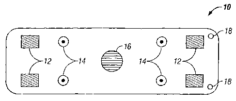
[0023] Figs. 1 and 2 show a monitoring device 10 which includes a plurality of
impedance electrodes 12, a plurality of ECG electrodes 14, a temperature
sensor 16, and a
plurality of suture ports 18. Figs. 1 and 2 show four impedance electrodes 12
that are
spaced from each other and four ECG electrodes 14 that are spaced from each
other,
although fewer (e.g., two impedance electrodes and two ECG electrodes) or more
5
CA 02567769 2006-11-23
WO 2005/110051 PCT/US2005/016396
electrodes may be used in other embodiments. The use of four impedance
electrodes 12
can eliminate electrode interface artifacts and the high electrode tissue
impedance. The
sensors and electrodes are depicted as protruding from the surface, but they
may be flat.
In the specific embodiment shown, the impedance electrodes 12, ECG electrodes
14, and
temperature sensor 16 are built into the body or can of the monitoring device
10. The
monitoring device 10 is desirably a stand-alone, self-powered device with a
battery 20,
which may be rechargeable. There is no need for intra-cardiac electrodes,
although the
addition of intra- or extra-cardiac electrodes may be employed to enhance the
diagnostic
capabilities in alternative embodiments. The monitoring device 10 is compact,
typically
less than half the size of a conventional modern pacemaker. It is designed to
collect, store,
and transmits surrogate blood pressure and/or flow data as well as ECG data.
Various
embodiments may include all or some of the sensors and electrodes depicted.
[0024] The impedance electrodes 12 are configured to transmit electrical
signals and/or
measure the resulting local electrical impedance as it is determined by the
signals passing
through tissue an.d/or blood vessels in the vicinity of the impedance
electrodes 12, as
encompassed by an electrical field of an AC current generated by the impedance
electrodes 12. More particularly, the impedance electrodes 12 can measure
surrogates of
the local blood flow characteristics (e.g., blood flow volume or velocity) of
the local tissue
zone and, more precisely, the pulsatile blood volume change in the muscle in
the local
tissue zone being sampled. The impedance electrodes 12 generate an AC current
with an
electrical field that encompasses the local tissue zone being sampled to
measure voltage
drop therebetween. The voltage drop depends on the impedance corresponding to
the
pulsation ofblood. The blood flow oscillates over time, and intermittently
changes the
impedance over time. For instance, the impedance may represent the relative
magnitudes
of blood flow (increasing or decreasing), or may be a relative measure of the
blood flow
(volume and/or velocity) with respect to an earlier time. The local impedance
change is
expected to be proportional to changes in the pulsatile blood volume change in
the muscle
in the tissue zone being sampled. A minimum of two impedance electrodes 12 are
used.
Additional impedance electrodes 12 allow different impedance or voltage drop
vectors to
be generated to provide a better chance of detecting changes in the blood flow
characteristics.
[0025] In some embodiments, the impedance electrodes 12 are configured to be
disposed near the artery or arteries being monitored, typically separated by
less than about
6
CA 02567769 2006-11-23
WO 2005/110051 PCT/US2005/016396
cm, more desirably about 3-4 cm. The distance depends on the strength of the
electrical
field of the AC current being generated, and can increase with an increase in
the strength
of the electrical field. The artery 30 may be disposed generally parallel to
the spacing
between two impedance electrodes (e.g., Fig. 3A); the impedance electrodes may
be
5 disposed generally transversely across the artery on opposite sides thereof
(e.g., Fig. 3B);
or the impedance electrodes may be disposed at an angle relative to the artery
(e.g., Figs.
3C or 3D). Measurement using the transverse arrangement may be less effective
than
using the longitudinal or parallel arrangement, since blood resistivity
changes with flow,
and decreases in the longitudinal direction and increases in the transverse
direction due to
the lining up of the red cells. The impedance electrodes are described as
extra-vascular
sensors, but may be positioned within the vascular system (intra-vascular) in
other
embodiments.
[0026] It is noted, however, that the impedance electrodes 12 are configured
to be
applied over any target region on a patient's body for detecting a surrogate
marker of
pulsatile blood volume changes or blood flow. The target region may be the
muscle of the
body or some tissue region. In that case, the impedance electrodes 12 need not
be placed
in the vicinity of any arteries.
[0027] The temperature sensor 16 measures local tissue temperature which may
be a
surrogate marker for blood pressure and/or blood flow measurement. The sensor
16 can
detect flow-related temperature differences. A sensitive recording system is
preferably
used. For instance, an abrupt local temperature change may reasonably be
interpreted as
being due to acute changes in local blood flow. Slow temperature changes may
reflect
environmental factors, or a fever, etc. Rapid temperature changes, albeit of a
small
magnitude, is most likely related to blood flow alterations.
[0028] The ECG electrodes 14 increase the clinical utility of the monitoring
device 10
by recording one or more ECG lead vectors. The data collected by the ECG
electrodes 14
may be used to correlate with the data collected by the impedance electrodes
12 and/or the
temperature sensorl6 to assist physicians in distinguishing whether cardiac
arrhythmias
are responsible for hypotensive symptoms or other mechanisms are at fault.
Other sensor
may be used to detect flow change utilizing, for example, laser Doppler
techniques
(photophethysmography) or local detection of hemoglobin by reflectance
methods.
7
CA 02567769 2006-11-23
WO 2005/110051 PCT/US2005/016396
j00291 As seen in Fig. 2, the monitoring device 10 includes a processor 22 and
a
memory 24 for storing data collected by the electrodes and sensor. The
monitoring device
may be programmed to collect and process data using the processor 22 and
memory 24
as desired by the user and/or manufacturer. The memory 24 may store the data
5 temporarily or permanent. The data may be transmitted to a remote site, such
as a
memory device worn by the patient or a server elsewhere, after data transfer
by a
telemetry link with the telemetry component 26. Information may be transmitted
via the
telemetry component 26 between the device 10 and an external system such as a
central
monitoring center. Data may be transmitted automatically or after telemetry
instruction
10 from an external user such as physician or nurse. The device 10 may be
programmed to
store all or some of the recorded data based upon downloaded instructions
delineating
criteria for data storage (e.g., outside upper or lower heart rate
boundaries). A warning
component 28 such as a buzzer or audible alert may be incorporated in order to
warn the
patient of an impending problem.
[0030] In specific embodiments, the monitoring device 10 is inserted into the
body of
the patient under the skin, more typically under the subcutaneous tissue. For
example, the
monitoring device 10 may be inserted subcutaneously under the collar bone to
be disposed
near the subclavian artery. If the monitoring device 10 is inserted to place
the impedance
electrodes 12 against the pectoralis muscles, a surrogate assessment of
skeletal muscle
blood flow will be the target to be monitored. An insertable monitoring device
is more
practical than a wearable one for long term use because it eliminates the need
to attach
electrodes or the like onto the external skin surface of the patient. In yet
another
embodiment, the monitoring device 10 may be incorporated into an implantable
device
such as a cardiac pacemaker, an implantable defibrillator (ICD), or the like
to provide
additional diagnostic or hemodynamic feedback capability (see, e.g., U.S.
Patent No.
5,441,525). Fig. 4 shows a simplified schematic view of monitoring device
components
50 as a part of an implantable device 60.
[0031] While Figs. 1 and 2 show can-mounted surface electrodes, lead-mounted
electrodes may be used. Unipolar and/or bipolar signals can be detected. One
or more
ECG vectors can be provided by the positioning of electrodes on the can or
header, or on
auxiliary leads designed to be positioned in the extra-vascular tissues, or on
intra-vascular
or intra-cardiac electrodes. The leads and can may both be inserted under the
skin, or
either or both the leads and can may be mounted on the body surface.
8
CA 02567769 2006-11-23
WO 2005/110051 PCT/US2005/016396
[0032] Fig. 5 shows another monitoring device 110 having impedance electrodes
112,
ECG electrodes 114, a temperature sensor 116, and suture ports 118. Additional
leads 120
are provided for remote placement. These auxiliary extra-vascular tissue leads
120 are
provided for any of the electrodes 12, 14 and temperature sensor 16 to place
them in closer
proximity with the desired target(s) to be monitored. A needle or the like can
be used to
guide the leads 120 and maiiipulate them subcutaneously to the desired
locations. In
alternative embodiments, the electrodes, sensors, and/or leads may be
detachable rather
than fixed to the body of the device.
[0033] In specific embodiments, the memory capability of the monitoring device
10 is
"looping" (first in, first out) with programmable durations of the "loop"
permitting the
saving of information prior to automatic or patient-activated triggering of
the recordings.
Programmability will be such as to permit all or only a subset of detected
signals to be
stored for subsequent immediate or later transmission to the body surface of
the patient,
and ultimately to medical personnel for interpretation (e.g., by wireless
telephony). For
instance, the monitoring device 10 can be programmed to save data temporally
proximate
certain events (just before and just after), such as an abrupt substantial
change in surrogate
measures of blood flow (e.g., impedance or temperature).
[0034] The patient may be offered a custom-programmed hand-held PDA (personal
digital assistant) or a similar external device 200, as illustrated in Fig. 6.
In Fig. 6, the
monitoring device is inserted under the skin 210 of the patient. The patient
may use the
external device 200 to instruct the implanted or inserted monitoring device 10
to collect
and/or transmit data at such times as the patient feels appropriate (e.g.,
real-time records
recorded during a symptom event or looped records saved by transmitted after
symptoms).
The monitoring device 10 may also be programmed to retain and transmit data
automatically when certain predetermined physiological boundaries are exceeded
(e.g.,
blood flow surrogate or heart rate above or below preset limits).
Communication may be
automatic or initiated by an external user such as a physician or nurse.
[0035] The monitoring device does not require intra-vascular access. For long-
term
(weeks or months) cardiac monitoring, this offers previously unavailable data,
ease of use,
and enhanced safety compared to intra-vascular applications. The result is the
ability to
assess, at least qualitatively, the hemodynamic impact of heart rhythm
disturbances in
free-living individuals. Similarly, the monitoring device offers the potential
to document
9
CA 02567769 2006-11-23
WO 2005/110051 PCT/US2005/016396
heart rhythm and tissue blood flow surrogates (e.g., tissue impedance,
temperature) during
periods of hypotension of non-cardiac cause, thereby helping to assess the
possibility of a
cardiac and/or vascular cardiac etiology during diagnostic evaluation of
patients. This
portable diagnostic device is capable, without use of intra-cardiac
electrodes, of
diagnosing hemodynamic perturbations and ascertaining whether and to what
degree they
are caused by cardiac rhythm disturbances. At the same time, the device can be
enhanced
by adding non-vascular or intra-vascular leads for placing sensors at more
distance sites in
the body, or can be incorporated as a diagnostic element within a conventional
cardiac
pacemaker, ICD, or other implanted diagnostic instrument.
[0036] It is recognized that tissue blood flow may vary with respiration,
posture, altered
cardiac output, or changes in vascular tone. However, for patients in whom
heart
monitoring of the type discussed herein is selected (i.e., those with
suspected arrhythmias
or syncope), an abrupt substantial change in surrogate measures of blood flow
may
reasonably be expected to be due to an arrhythnlia or other abrupt hypotensive
state.
Thus, detection of suspected flow alterations, along with ECG correlation,
will assist
physicians in distinguishing whether cardiac arrhythmias (i.e., abnormally
slow or fast
heart rates) are responsible for hypotensive symptoms or whether other
mechanisms (e.g.,
vasodepressor hypotension without arrhythmia) are at fault. In many instances,
hypotension occurs without evident arrhythmia. The present monitoring device
is
designed to detect this type of clinical problem in free-living individuals.
[0037] From the foregoing, it will be apparent to those skilled in the art
that the present
invention provides, in exemplary non-limiting embodiments, a wide variety of
design
options for the electrodes, sensors, leads, and the like for the monitoring
device. Further,
those skilled in the art will recognize that the present invention may be
manifested in a
variety of forms other than the specific embodiments described and
contemplated herein.
Accordingly, departures in form and detail may be made without departing from
the scope
and spirit of the present invention as described in the appended claims.