Note: Descriptions are shown in the official language in which they were submitted.
CA 02568645 2006-11-24
WO 2005/117800 PCT/US2005/018799
APPARATUS FOR MECHANICALLY VENTILATING A PATIENT
BACKGROUND OF THE INVENTION
The present invention is directed to a physical apparatus used to assist
mechanically ventilating a patient. More specifically, the present invention
provides non-
invasive pressure changes outside a patient's chest wall, allowing mechanical
ventilation without need for invasive endotracheal, orotracheal or tracheal
intubation.
Under normal physiological conditions, humans breathe using "negative pressure
ventilation." In other words, a negative intrathoracic pressure is created by
contraction
of the intercostal muscles (between the ribs), upward and outward expansion of
the
ribs, and downward movement of the muscular diaphragm separating the thorax
from
the abdomen. All these changes act to expand both lungs and thus create a
negative
intrathoracic pressure. The pressure'change enables gas to move from the
outside
atmosphere, through the human air passages, and into the deepest areas of the
human
lung. The natural tendency of the lungs to constrict similarly to a stretched
rubber band,
(elastic recoil), creates an inward intrathoracic pull, such that as soon as
the intercostal
muscles relax, the ribs are pulled inward and downward, and the muscular
diaphragm is
pulled upward. These movements create a positive intrathoracic pressure,
relative to
the outside atmospheric pressure, thus forcing the gas out of the lungs
through the
human air passages, and back into the atmosphere.
1
CA 02568645 2006-11-24
WO 2005/117800 PCT/US2005/018799
By drawing on the natural biomechanics of human breathing, the present
invention very closely simulates human respiratory mechanics and aids
neonatal,
pediatric and adult patients who require respiratory support or assistance.
Many different machines have been designed to deliver gas into the lungs by
creating positive pressure outside the airways, and thereby forcing gas into
the patient's
airways. These machines provide lifesaving benefit, but are not without risks.
For
example, most "positive pressure ventilators" force gas through a small,
artificial tube
placed within the patient's trachea or airway, termed "invasive positive
pressure
ventilation," because the patient's airway is penetrated or invaded by the
artificial tube.
Use of such a tube carries complications such as difficulty in proper
placement, risks of
dislodging, clogging, or causing infection. Additionally, the force with which
each breath
is delivered to the patient can lead to trauma to the lung tissue itself,
including lung
rupture or collapse.
More recently, "noninvasive positive pressure ventilation" has begun being
practiced, which involves using a mask outside a patient's nose or mouth to
deliver the
positive pressure into the lungs. This greatly reduces the risks of improper
placement,
dislodging or clogging of the mask, and virtually eliminates the risk of
severe infection
due to contamination of equipment. However, such form of mechanical
ventilation
functions less than ideally because the gas cannot be directed solely into the
lungs, but
is rather forced into the back of the throat where the gas travels to both the
lungs and
stomach, the relative proportions of gas depending on the resistance of each
pathway.
2
CA 02568645 2006-11-24
WO 2005/117800 PCT/US2005/018799
Furthermore, several noninvasive positive pressure ventiiators require the
patient to
remain confined to bed (e.g., Nasal Continuous Positive Airway Pressure
(NCPAP) or
Bilevel Positive Airway Pressure (BiPAP)), while others might allow the
patient to sit up
or be pushed in a wheelchair, but do not permit full mobility.
Negative pressure ventilators, e.g., iron lungs, are known in which a
patient's
body rests entirely within the chamber with only the patient's head protruding
through a
portal situated around the patient's neck. More recently, negative pressure
ventilator
"shells" have been developed that encompass only the patient's thorax and
abdomen.
For infants, negative pressure chambers are designed to house the entire body
(excluding the head). Both the "shells" and chambers must be attached to a
separate
pressure ventilator via vacuum hose in order to function. However, such
conventional
chambers or ventilators suffer several disadvantages. For example; there is
difficulty in
observing a patient from all angles, with it also being cumbersome to access
the patient
through a door to the chamber. A great deal of space is required to permit the
door to
rest safely and securely on top of the ventilator chamber, when opened.
Placement of
the handle for the front access door to the ventilator chamber has resulted in
confusion
with locking mechanism for creation of the airtight seal of the access door.
This could
result in breaking of the access door handle and/or inadequate closure of the
front
shield and seal formation.
Difficulty has been encountered in including the patient's upper airway within
the
negative pressure chamber. Thus, the upper airway of a patient could be in
danger of
collapse during creation of the vacuum to assist the patient's breathing.
Difficulty in
accessing the interior of the chamber, e.g., during nonoperation, has made it
difficult to
easily clean and launder material in contact with the patient, e.g., an
infant. Although
3
CA 02568645 2006-11-24
WO 2005/117800 PCT/US2005/018799
ventilator chambers have been free-standing on the ground, a separate base or
foundation has been required for practical functioning. Thus, an institution
such as a
hospital must provide such support for'the chamber, while such support might
not meet
standards required by the Food and Drug Administration.
Difficulty has been encountered in providing an adequate seal around the
patient's neck, especially in a small infant, resulting in a high percentage
of vacuum
leaks occurring at low vacuum pressure. This could activate alarms on the
ventilator
itself, forcing an operator to frequently stop and reset the ventilator at low
pressures.
Difficulty in monitoring and maintaining temperature and humidity inside the
ventilator
chamber has also been encountered.
Additional problems encountered with such ventilators include the need to stop
and restart if a seal is broken for longer than an allotted period of time.
Once seals have
been well-established and the ventilator activated, it generally takes 20-30
seconds
(based upon a breath rate of 20 breaths per minute and pressure -7cm H20) to
achieve
the desired negative pressure. Providing sufficient staff to maintain such
ventilators has
also been difficult, while replacement parts were not readily available. As a
result, lead
time in clinical operation of such a ventilator after initial installation is
often more than
one monin.
Developing the ability to utilize "noninvasive negative pressure ventilation"
can
eliminate many of the risks of the positive pressure ventilators.
4
CA 02568645 2006-11-24
WO 2005/117800 PCT/US2005/018799
Accordingly, it is an object of the present invention to improve effective and
safe
use of noninvasive negative pressure ventilation in assisting mechanical
ventilation of a
patient.
It is a more particular object of the present invention to provide a self-
contained,
noninvasive negative pressure mechanical ventilator created in the form of an
air-tight
covering about a patient's torso that will permit full mobility and comfort of
the patient.
It is a further object of the present invention to improve respiratory
mechanics
and mobility, and thereby improve quality of life of patients requiring
mechanical
ventilation.
CA 02568645 2006-11-24
WO 2005/117800 PCT/US2005/018799
SUMMARY OF THE INVENTION
These and other objects are attained by the present invention which is
directed
to an apparatus for mechanically ventilating a patient, comprising two
separate,
substantially rigid components structured and arranged to be movably coupled
with
respect to one another, and a flexible, air-tight covering (e.g., a vest)
structured and
arranged to cover both components when placed about a torso of a patient. When
the
components move away from one another within the air-tight covering, negative
pressure is generated within the covering and causes the patient to draw air
into the
expanding lung cavity. The only active part of the vest is the creation of
negative
intrathoracic pressure by moving the front and back plates away from each
other within
the air-tight vest.
The mechanism that moves the plates away from each other will be timed such
that it will release itself (for example, a pneumatic actuator is spring-
loaded and has a
one-way release valve to let go of the compressed air and thus allow the pin
of the
actuator to return and re-set itself for the next inhalation).
What causes the patient to exhale is the same mechanism by which every other
person exhales, whether spontaneously breathing without a machine, invasive
positive
pressure breathing, or negative pressure breathing that is the natural elastic
recoil of
the lungs themselves.
Similar to stretching giant rubberbands, effort is only required to expand the
lungs (to inhale); once the lungs stop expanding, then they will naturally
recoil (thereby
creating ositive intrathoracic pressure and forcing air from inside the lungs
and airways
6
CA 02568645 2006-11-24
WO 2005/117800 PCT/US2005/018799
to outside the airways). Moving the plates closer to each other does not cause
the
patient to exhale, in and of itself.
The negative pressure ventilator vest allows the patient's own natural luncg
mechanics to control the exhalation ( thus aiding the patient's respirations,
while
operating closely to mimic a patient's own natural, spontaneous respiratory
efforts).
The one-way air-release valve(s) built into the air-tight vest allow for quick-
release of'any air trapped underneath the vest during inhalation (namely from
the area
around the neck of the vest, which cannot realistically be completely air
impenetrable
due to concerns of patient safety and comfort).
Exhalation due to elastic recoil occurs very quickly so trapped air underneath
the
vest should not impede this process. The release valve(s) are placed in the
material of
the vest to quickly release trapped air in preparation for the next
inhalation.
Preferably, means for movably coupling the substantially rigid components
together are provided within the air-tight covering. This means can take the
form of a
pantograph linkage, a U or horseshoe, or a pincer. More particularly, the
components
are formed as two separate, light-weight, concave, rigid half-shells
positioned on the
front and back of a patient's torso, adjacent the chest cavity. Each component
is
positioned with the concave side toward the torso and held in place with soft
straps
placed across the patient's shoulders. Additional straps may be placed around
the
waist, if desired. These separate shells can be formed from any lightweight
material that
will maintain shape, e.g., fiberglass, plastic or plaster, and may be formed
of several
layers adhering together, e.g., as a laminate.
7
CA 02568645 2006-11-24
WO 2005/117800 PCT/US2005/018799
The straps can be formed from cotton, cloth, leather, or any other appropriate
material, and can be fastened together with Velcro , hooks or ties. Different
size shells
can easily be provided in accordance with the present invention.
About one to three pneumatic actuators will be attached to the anterior and
posterior shells on each side of the patient, depending on desired negative
pressure
generation for each patient. These actuators are activated by a pneumatic
system
along the lateral edge of the outer covering or vest, thus eliminating the
need for
electrical or battery-generated power. The pneumatic actuators can be powered
in any
of the following ways. Firstly, compressed gas tubes can be provided with
timed
release-valves to periodically force the pin outwardly from the actuator. When
the valve
is cycled to the "off' position, the compressed gas is no longer directed to
the actuator
and the spring-loaded mechanism then pulls the pin of the actuator back
inwardly. The
air previously inside the barrel of the actuator is simultaneously released
via a one-way
valve built into the actuator. Alternatively, electrically and/or battery
operated
compressors that convert atmospheric gas into compressed gas and then time-
cycle
the compressed gas into the actuator in the same manner, could be used in the
context
ot the present invention.
The air pressure, stroke length, and exerted force of the actuators are
adjustable, allowing for operator control of the patient-specific ventilator
breath rates,
tidal volume generation, and inspiratory time. The stroke of the actuator will
automatically adjust based on anterior and posterior resistance to movement,
thus
allowing the anterior and posterior shells to move equally when the patient is
standing,
8
CA 02568645 2006-11-24
WO 2005/117800 PCT/US2005/018799
and the non-dependent shell to move twice as far when the dependent shell is
immobile, when the patient is lying down (either prone or supine).
The anterior and posterior shells, as well as the pneumatic actuators attached
to
the lateral edges, will all be covered by the air-tight, rubberized, short-
sleeved shirt or
covering, with tight fasteners around both sleeves and the waist area. The
neck area
will also be made of air-tight material, but not fastened as tightly. The
shirt or vest will
have several one-way air-release valves that will contain air during expansion
of the
shells, yet allow for quick escape of air during the period of patient
exhalation when the
shells are moving toward each other.
The inventive vest will sit comparatively or substantially air-tightly about
the
upper torso of a patient. In other words, there will be some slight seepage of
air into
the vest through, e.g., the collar about a patient's neck. However, the one-
way air
release valve permits expelling of this seepage upon the patient's exhalation.
The actuators utilize pneumatic pressure to push apart the anterior and
posterior
shells from each other. When this operation is performed inside the
rubberized, air-tight
shirt, a negative pressure is generated within the shirt that, in turn, pulls
the walls of the
patient's chest upward and outward. This results in negative intrathoracic
pressure,
which then causes the patient to draw air from the higher pressure atmosphere
into the
lungs through the patient's airways. The actuators are set to allow time for
the shells to
come together during the natural "elastic recoil" phase of normal human
exhalation.
During this phase, the one-way valves allow air to exit from inside the air-
tight covering,
thereby readying the apparatus for the next inhalation cycle. Alternatively,
the anterior
9
CA 02568645 2006-11-24
WO 2005/117800 PCT/US2005/018799
and posterior components or shells can be movably coupled by a mechanism
situated
externally of the rubberized shirt or vest.
The inventive apparatus thereby simulates normal, physiologic breathing,
eliminating the need for artificial airway maintenance and allowing each
patient to
achieve full mobility and thereby, normal existence.
The present invention is also directed to a ventilator which helps a patient
such
as a premature infant suffering pulmonary disability to breathe on their own.
The
inventive ventilator is easy to assemble and use, and effective in use, being
of special
advantage to aid premature infants in breathing.
CA 02568645 2006-11-24
WO 2005/117800 PCT/US2005/018799
BRIEF DESCRIPTION OF THE DRAWINGS
The present invention will be described in greater detail with reference to
the
accompanying drawings, in which:
Fig. 1 is a schematic, exploded view of the inventive apparatus;
Fig. 1 A is an enlarged view of encircled area 1 A in Fig 1;
Fig. 2 is a plan view of a portion of the inventive apparatus from the
direction of
arrow 2 in Fig. 1;
Fig. 3 is a plan view, similar to Fig. 2, and illustrating an oppositely-
biased
position of the inventive apparatus from the position shown in Fig. 2;
Fig. 4 is a plan view, similar to Fig. 2 and illustrating an alternative
embodiment
of the inventive apparatus;
Fig. 5 is a plan view, similar to Fig. 3, and illustrating an oppositely-
biased
position of the inventive apparatus from the position shown in Fig. 4;
Fig. 6 is a plan view, similar to Figs. 2 and 4, and illustrating another
alternative
embodiment of the inventive apparatus;
Fig. 7 is a plan view, similar to Figs. 3 and 5, and illustrating an
oppositely-biased
position of the inventive apparatus from the position shown in Fig. 6;
Fig. 8 is a plan view, similar to Figs. 3, 5 and 7 and illustrating a further
alternative embodiment of the present invention.
Fig. 9 illustrates a perspective view of the assembled negative pressure
chamber
ventilator of the present invention;
11
CA 02568645 2006-11-24
WO 2005/117800 PCT/US2005/018799
Fig. 10 is a top plan view of the platform forming part of the inventive
ventilator;
Fig. 11 is a perspective view of the platform shown in Fig. 9;
Fig. 12 is a perspective view of the cover forming part of the inventive
ventilator;
Fig. 13 is a schematic front view of the cover illustrating assembling of a
front
shield thereon;
Fig. 14 is a schematic perspective view illustrating coupling of the cover to
the
platform;
Fig. 15 is a schematic front view of the cover illustrating coupling of a
flexible
collar onto the front shield assembled according to Fig. 13;
Fig. 16 is a schematic view illustrating coupling of a tube from driving
mechanism
to a portal through the cover of the inventive ventilator;
Fig. 17 illustrates an alternative shape of the flexible collar shown in Fig.
15;
Fig. 18 illustrates a side elevational view of another embodiment of the
negative
pressure chamber ventilator in accordance with the present invention;
Fig. 19 is a view in the direction of arrow 19 of Fig. 18 and illustrating an
enlarged view of the hinge arrangement,coupling a door to the ventilator in
closed
PositbT;
Fig. 20 is an inverted view of the hinge arrangement shown in Fig. 19 and
illustrating the door in partially opened position;
Fig. 21 illustrates a schematic view similar to Fig. 13 and illustrating
coupling of a
protective shield upon the front of the ventilator shown in Fig. 18;
12
CA 02568645 2006-11-24
WO 2005/117800 PCT/US2005/018799
Fig. 22 illustrates a protective collar arranged to be coupled about the neck
of a
patient situated within the ventilator shown in Fig. 21 and sealing the vacuum
created
within the ventilator;
Fig. 23 is a schematic, rear perspective view of the ventilator shown in Fig.
21
and illustrating positioning and coupling of ventilation mechanism to the
chamber;
Fig. 24 illustrates storage of the ventilation mechanism prior to coupling to
the
ventilator as shown in Fig. 23;
Fig. 25 illustrates a top plan view of the ventilation mechanism shown in Fig.
23
and illustrating ease of servicing the ventilation mechanism;
Fig. 26 illustrates an enlarged view of part of the ventilation mechanism
shown
in Fig. 25;
Fig. 27 illustrates an enlarged view of another part of the ventilation
mechanism
shown in Fig. 25;
Fig. 28 illustrates a side elevational view of the ventilator as positioned
upon a
support cabinet housing the ventilation mechanism with front cover in position
to
obscure mechanism shown in Fig. 24;
Fig. 29 schematically illustrates arrangement of an orifice through the
chamber to
receive tubing and wires and sealing of the orifice to maintain the vacuum
within the
chamber;
Fig. 30 illustrates a cross-sectional view of a drive belt for the ventilation
mechanism; and
Fig. 31 illustrates the drive belt of Fig. 30 in compressed condition.
13
CA 02568645 2006-11-24
WO 2005/117800 PCT/US2005/018799
DESCRIPTION OF THE PREFERRED EMBODIMENTS
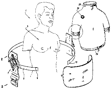
Referring to the drawings in which analogous components are denoted by
analogous reference numerals or characters, the inventive apparatus 1 for
mechanically
ventilating a patient has two components 2 and 3 arranged to reciprocally move
towards and away from one another. These components are positioned about the
torso
4 of a patient, i.e., the chest cavity 5, and secured within an outer elastic
shell 6, e.g., a
vest or shirt, which can be formed of any suitable material such as spandex,
polyester,
etc. A preferred elastic garment that functions especially well as an air-
tight elastic
shell 6 in accordance with the present invention is a Nike Dri-Fit short
sleeve shirt
composed of 82% polyester and 18% spandex. This shirt was coated on the outer
surface thereof with a thin layer of General Electric clear Silicone II 100 %
Window and
Door silicone sealant, manufactured by GE Sealants and Adhesives,
Huntersville, NC
28078, to enhance air-tightness.
The movable components 2 and 3 themselves can be manufactured from any
suitable material, e.g., fiberglass, lightweight plaster, or synthetic plastic
such as
polyethylene terephthalate, polyvinyl chloride, etc. An especially preferred
material is
hardened fiberglass created using a Bondo Home Solutions fiberglass mat
manufactured by the Bondo Corporation (an RPM Company), 3700 Atlanta
Industrial
Parkway, N.W., Atlanta, Georgia 30331 and treated with Everciat (100498)
automotive
fiberglass resin and hardener, manufactured by Fibre Glass-Evercoat, a
division of
Illinois Tool Works, Inc. 660 Cornell Road, Cincinnati, Ohio 45242.
14
CA 02568645 2006-11-24
WO 2005/117800 PCT/US2005/018799
The flexible air-tight covering 6 is placed about the torso 4 of the patient,
i.e.,
around the chest cavity 5, after the substantially rigid components 2 and 3
have been
movably positioned about the torso 4 and chest cavity. Thereby, when
components 2
and 3 move away from one another within the air-tight.covering 6, negative
pressure is
generated within the air-tight covering 6 and influences the torso 4 and chest
cavity 5
of the patient to cause the patient to draw air into the patient's lungs.
Conversely, when
the components 2 and 3 stop moving apart within the air-tight shell 6, the
patient's
natural exhalation mechanism takes over, allowing the patient to expel the air
from
within the patient's lungs.
As shown in Fig. 1, the inventive apparatus 1 comprises means 7 for movably
coupling components 2 and 3 together such that they can reciprocally move
towards
and away from each other. This coupling means 7 can be mounted upon an elastic
band 8 which is then secured around the patient's torso 4, e.g., by Velcro
sections 9
and 10 at ends thereof. As best seen in Fig. 2, the coupling means 7 comprise
a
support 11 mounted upon the band 8, with a turntable 12 rotatably positioned
upon the
support 11 and, in turn, having two substantially cylindrical stops 13 and 14
mounted
thereon. The two movable components 2 and 3 are coupled together through a
pantograph linkage 15 taking the shape of a parallelogram in Fig 2 comprising
four links
or sides 16, 17, 18, 19 rotatably coupled together about four respective pivot
points 20,
21, 22, 23. As shown in Fig. 2, the components 2 and 3 are coupled to
extensions of
respective links 16 and 17 however the components can alternatively be coupled
directly to the pivot points 21 and 23 within the purview of the present
invention.
CA 02568645 2006-11-24
WO 2005/117800 PCT/US2005/018799
An untensioned member 24 is also mounted to the parallelogram linkage 15 to
extend between two opposite pivot points 20 and 22 and straight between the
stops 13
and 14 mounted upon the turntable, in unstressed state as shown in Fig. 2.
Additionally,
a pneumatic actuator 25 is coupled between the support 11 and turntable 12 as
shown
in Figs. 2 and 3. When the pneumatic actuator rotates the turntable 12 with
respect to
the support 11 in a clockwise direction from Fig. 2 to Fig. 3, the space
between the
components 2 and 3 expands due to expansion of the pantograph linkage 15 and a
negative pressure is generated within the elastic shell 6. At the same time,
the member
24 is tensioned and twisted about the two stops 13 and 14 which rotate
together with
the turntable 12 as shown in Fig. 3, thereby enhancing a force biasing the
parallelogram linkage to return to its untensioned state shown in Fig. 2.
Therefore, when pneumatic actuator 25 has expanded to maximum extension as
shown, e.g., by the phantom lines in Fig. 3, the return biasing action of a
spring within
the pneumatic actuator 25 takes over to return the linkage to the unstressed
state
shown in Fig. 2, whereupon the pneumatic actuator retracts to initial position
and once
again begins the next cycle of expansion. Then, the elastic recoil of the
patient's lungs
causes spontaneous exhalation once the compressed air is no longer extending
the pin
of the actuator. This returns the linkage to the unstressed state in
preparation for the
next cycle of expansion.
Two such coupling means 7 have been illustrated in Fig. 1 although the
inventive
apparatus will effectively operate with just one such coupling mechanism as
shown in
Figs. 2 and 3 and with the components coupled on opposite sides, e.g., by just
a driven
16
CA 02568645 2006-11-24
WO 2005/117800 PCT/US2005/018799
pantograph linkage. Although the embodiment illustrated in Fig. 1 shows the
coupling
means positioned within the outer shell 6, nevertheless such coupling means
could
easily be positioned outside the air-tight covering 6 and appropriately
coupled to the
components 2 and 3 within the covering 6 through openings provided in the
covering 6.
As denoted by the dotted lines in Fig. 1, the band 8 is initially positioned
about the torso
4 of a patient. The support member 11 of the coupling means is conveniently
secured
to the band 8 either before or after the band 8 is positioned about the torso
4 of a
patient.
Next, the components 2 and 3 are secured to respective extensions of the
pantograph linkages 15, followed by positioning of the air-tight covering 6
securely
about the torso of the patient, including the chest cavity. The neck, waist,
and sleeve
openings of the covering 6 are sealed by respective straps 26 and buckles 27
as shown
in detail in Fig. 1 A, to provide a secure air-tight enclosure within the
covering 6.
Additionally, a one-way check valve 28 is provided in the covering 6 to
release air from
within the sealed covering 6 and avoid undue build-up of air pressure
therewithin.
Figs. 4 and 5 illustrate and alternative embodiment of the coupling means 7'
which dispenses with the support plate 11 and turntable 12. More particularly,
in this
embodiment, the coupling means 7' comprises two members 29 and 30 forming a
linkage substantially in the shape of a U or horseshoe and pivotally coupled
together at
a pivot point 31 situated substantially at the base of the U or horseshoe. A
respective
movable component 2 and 3 is coupled to a respective pivotal member 29 and 30.
A
pneumatic actuator 25' is provided similarly to the embodiment shown in Figs.
2 and 3
17
CA 02568645 2006-11-24
WO 2005/117800 PCT/US2005/018799
but with the actuator 25' laterally coupled to the pivotal members 29 and 30
above the
pivot point 31 as shown. Additionally, means (not shown) for biasing the
pivotal
members 29 and 30 towards the position shown in Fig. 4, e.g., a coil spring,
can be
provided. The remaining components of the inventive device are the same as
shown in
Figs. 1-3.
The pneumatic actuator 25' operates to push the pivotal members 29 and 30
apart from one another to the position shown in Fig. 5 where the components 2
and 3
are also moved apart, hence creating the negative pressure within the air-
tight covering
6. When the pneumatic actuator 25' reaches the point of maximum extension
shown in
Fig. 5, then the spring action within the pneumatic actuator takes over and
biases the
pivotal arms 29 and 30 back to the closer position shown in Fig. 4 where the
cycle
begins once again.
Figs. 6 and 7 illustrate a further alternative embodiment of the coupling
means 7"
in the shape of a pincer, having two arms 32 and 33 coupled together about
pivot point
34 intermediately positioned between ends of the arms 32 and 33 and with
adjacent
ends of the arms 32 and 33 coupled to the respective components 2 and 3 as
illustrated. The pneumatic cylinder 25" is coupled to the opposite ends of the
respective
arms as shown, with the elastic member 35, e.g. a coil spring, wound about the
pivot
point 34 and coupled to the respective arms 32, 33.
In contrast to the previous two embodiments, expansion of the pneumatic
actuator causes the ends of the arms 32, 33 respectively coupled to the
components 2
and 3 to pivot towards one another and thereby move the components 2 and 3
towards
18
CA 02568645 2006-11-24
WO 2005/117800 PCT/US2005/018799
one another and generate a positive pressure within the air-tight covering 6.
When the
pneumatic actuator 25" reaches its maximum expansion shown in Fig. 6, the
force of
the coil spring 35 takes over and biases the ends of the arms 32, 33 coupled
to the
components 2 and 3 away from one another to the position shown in Fig. 7,
thereby
generating the negative pressure within the air-tight covering 6.
In the embodiment shown in Figs. 6 and 7, the mechanism still functions to
create a negative pressure within the vest, causing the patient to inspire air
into the
lungs. However, in contrast to the previously-described embodiments, recoil of
the coil
spring 35 (and not the pneumatic actuator 25") explicitly generates the
negative
pressure within the vest 6, whereas active expansion of the pneumatic actuator
25"
shown in Fig. 6 enhance the patient's exhalation.
Referring to Fig. 8, the components 2 and 3 can be coupled directly to a
series of
spring-loaded actuators 25', 25", 25"' illustrated in extended or expanded
position.
Compressed gas within these actuator tubes activates all these actuators
simultaneously. In other respects, the mechanism of ventilating a patient
operates
analogously to the other illustrated embodiments supra.
Any suitable, commercially-available pneumatic actuator can be used as the
pneumatic actuator 25 in the inventive apparatus. One such pneumatic actuator
is the
commercially-available HONEYWELL MP909D1201 providing maximum air pressure
30 psi, nominal spring range 3 to 8 psi and a stroke of 2.4 inches.
19
CA 02568645 2006-11-24
WO 2005/117800 PCT/US2005/018799
Therefore, the present apparatus constitutes a self-contained, portable
ventilation system permitting patients using the same to remain fully mobile.
Improved
patient mobility will also improve respiratory mechanics and quality of life.
The inventive
apparatus can be used either intermittently, or continuously throughout the
day or night,
and is always effective whether the patient is standing, sitting or lying
down.
Referring to Figs 9-17, the inventive ventilator I is composed of a cover 2
secured to a platform 3 formed by a clear, plexiglass panel 4 being secured to
aluminum beams 140, 5, 6, 7 by a series of phillips-head screws 8.
Additionally, a
support beam 9 is placed across the panel 4 and secured thereto by phillips-
head
screws. A corrugated rubber seal 10 is positioned about the upper edge of the
platform
3 for sealing a base of the cover 2 when mounted thereon as described in
greater detail
infra. Four right-angle brackets 11, 12, 13, 14 are mounted upon the panel 4
through
the respective phillips-head screw 8 and each comprise an orifice for
receiving a
respective pin 15 mounted upon an adjacent phillips-head screw through a chain
16.
The beams 140, 5, 6, and 7 are formed from hollow aluminum tubing of
substantially
square cross-section
The cover 2 of the inventive ventilator 1 is also formed from clear plexiglass
material and comprises a substantially rectangular-parallelepiped shape with
curved
upper corners and an open bottom, as best seen in the perspective view of Fig.
12.
However, the cover 2 may take any convenient shape in accordance with the
present
invention, e.g., semi-cylindrical, semi-elliptical, and variants thereof.
Separate front 17
and rear 18 panels are affixed to the cover 2 by appropriate adhesive, e.g.,
an epoxy
CA 02568645 2006-11-24
WO 2005/117800 PCT/US2005/018799
glue-silicone combination. The front panel 17 comprises a U-shaped portal 26.
A
hollow aluminum tube or pipe 19, 20 is mounted along bottom lateral edges of
the cover
2, with an aluminum pipe 141 optionally mounted along a bottom edge of the
rear panel
18.
Aluminum braces 21, 22 wrap around the top of the cover 2 and are affixed
thereto by respective phillips-head screws and also to the respective aluminum
pipes
19 and 20 to thereby secure the aluminum pipes 19 and 20 to the cover 2. An
aluminum pipe serving as an additional brace 23 optionally extends across the
front
panel 17 as shown in Fig. 12. Additionally, a portal 24 is provided through
the top of
the cover 2 for coupling to inspiration mechanism. Furthermore, a separate
front
bracing panel 25 approximately rectangular in shape, is mounted across the
front panel
17 of the cover 3 and slightly spaced therefrom, as described further infra.
To assemble the inventive ventilator 1, the cover 2 is simply placed on the
platform 3 with bottom edges resting against the corrugated rubber seal 10.
Next the
respective pins 15 are inserted through the opening in an adjacent right-angle
bracket
11, 12, 13, 14 and then into an open end of a respective aluminum tube or pipe
19 and
20 secured to the cover, to thereby fixedly mount the cover 2 upon the
platform 3, as
illustrated, e.g., in Fig. 14. Then, a separate shield 27 also comprising a U-
shaped
portal 28 but of smaller dimension than U-shaped portal 26 on the front panel
17 of the
cover, is inserted between the front panel 17 and bracing panel 25 as
illustrated, e.g.,
by arrow A in Fig. 13.
21
CA 02568645 2006-11-24
WO 2005/117800 PCT/US2005/018799
Both the bracing panel 25 and front shield 27 are provided with several
squares
29 of material for hook-and-loop, i.e., Velcro fastening with squares 29 of
similar
material placed upon a flexible collar 30 formed of soft plastic. as
illustrated, e.g., in Fig.
15. The flexible collar 30 is also provided with a substantially U-shaped
portal 32 of
smaller dimension than U-shaped portal 28 of shield 27. However, the portal
through
the flexible collar 30 can take any convenient form, e.g., substantially
rectangular as
shown in Fig. 17.
A tube 31 from the inspiration mechanism is coupled to portal 24 as shown,
e.g.,
in Fig. 16. In practice, after the cover 2 is secured onto the platform 3, the
patient, e.g.,
a premature infant, is slid into the ventilator with the infant's head resting
upon the
platform 3 outside the cover 2. Next, the shield 27 is gently and carefully
slid between
the front panel 17- and bracing panel 25 on the cover, with the appropriate
size flexible
collar 30 then conveniently fastened onto the shield 27 by the hook-and-loop
fasteners
29. The brace 25 is then placed over the collar 30 and locked into position by
lock-and-
key mechanism directly into the side panels of the chamber to keep the collar
30 in
position. The tube 31 from the inspiration mechanism can then be coupled to
the portal
24 of the cover 2, if not done previously. The inventive respirator 1 is now
ready for
operation.
Any suitable negative pressure ventilation mechanism can be used with the
inventive ventilator 1. One preferred mechanism is marketed as the NEV -100
Non-
Invasive Ventilator by Respironics, Inc. (www.Respironics.com) and is
disclosed in U.S.
Pat. No. 5,299,599 issued April 5, 2004, the contents of which are
incorporated by
22
CA 02568645 2006-11-24
WO 2005/117800 PCT/US2005/018799
reference herein. The coupling tube 31 is of flexible, corrugated, accordion-
shaped
construction. Specifically, negative pressure is created within the interior
32 of the
ventilator 1 by the inspiration mechanism which causes the patient to inhale;
reduction
of negative pressure during the breathing cycle then allows the patient to
exhale by
natural elastic recoil of the lungs.
Referring to Figs. 18-31 in particular, the inventive ventilator 100 and
chamber
101 eliminates the disadvantages encountered in the prior art devices
described in the
background portion of the present application. The chamber 101 itself is
manufactured
from one-half inch thick Lexan plexiglass, sufficiently sturdy to withstand
the vacuum
pressures required in clinical operation. The walls of the chamber 101 are
thus
transparent on all six sides, allowing medical staff to easily observe the
patient from any
angle at all times, thus improving patient care and safety. The access door
102 used
for inserting and removing a patient into and out of the chamber 101 utilizes
a double-
hinge system 103, allowing a caretaker to easily open the door 102 and place
the door
panel flatly on top of the chamber 101 during non-use. Add_itionally, the
patient, i.e.,
infant is still fully visible, even when the access door 102 is resting on top
of the
chamber 101.
Furthermore, the access door 102 possesses separate locking mechanisms 108
from the door handle 106. These separate locking mechanisms 108 cannot be
accidently misplaced or misaligned. The locking mechanisms 108 are situated
away
from the door handle 106. Additionally, the front door or shield 104 possesses
three
latch-and-hinge locking mechanisms 105, 105', 105" for coupling to the neck
collar 107
23
CA 02568645 2006-11-24
WO 2005/117800 PCT/US2005/018799
of the patient, i.e., infant. The portion of the chamber 101 surrounding the
patient's
neck is specifically designed such that the patient's head is easily
accessible and can
move freely and, at the same time, be quickly removed from the chamber 101, if
necessary. In an explicit improvement over conventional ventilator designs,
the patient's
extrathoracic airway (cervical trachea) is included within the vacuum
mechanism of the
chamber 101.
The portion of the chamber 101 forming the seal around the infant's chin,
i.e.,
the protective collar 107 shown in Fig. 22, is constituted by two mating parts
107' and
107", each composed of a soft bib-like material and easily-disinfected, thinly
coated
polyurethane gel. The ventilator 100 and chamber 101 are designed to operate
as an
integral unit with ventilator controls 110, 110',110" (Fig. 25) easily
accessible from the
front of a housing cabinet 111 supporting the unit as shown, e.g., in Fig. 28.
This
cabinet 111 can be easily opened for simple exchange of ventilator units, if
maintenance is required, as shown in Fig. 24 where covering panel 111' has
been
unhooked.
The ventilator chamber 101 itself is explicitiy designed to include the
extrathoracic airway (cervical trachea) of the patient within the vacuum
portion of the
chamber 101. This allows for dilation of the extrathoracic airway during
creation of the
negative pressure. Poiseuille's Law describes the pressure gradient required
to
maintain laminar flow through a tube:
AP=n)&
r4
where the tube represents the extrathoracic airway of the patient,
24
CA 02568645 2006-11-24
WO 2005/117800 PCT/US2005/018799
AP denotes the pressure differential required to maintain laminar gas flow,
q denotes the viscosity of the fluid (air/oxygen) flowing through the tube,
V denotes the flow of the fluid or gas,
L denotes the length of the tube, and
r denotes the internal radius of the tube.
This radius of the airway is of critical importance in determining the airway
resistance
(APN), with even a tiny decrease in the radius of the upper airway requiring a
tremendous increase in driving pressure of the gas to maintain the same
laminar flow
rate. Once the flow rate becomes high, then the airflow becomes turbulent and
results
in total disorganization of flow, leading to inefficiency in delivery of the
gas. The
compressible nature of the neonatal and infant airway has led to failure of
previously-
available negative pressure ventilators to efficiently function in this
patient population.
A medical grade thermometer 112 is placed inside the chamber 101 to ensure
safety of the temperature environment for the infant. Heat and fluid are quite
easily
dissipated from skin of a newborn infant, with high inflow rates of non-
heated, non-
humidified air also placing some infants at risk. In this regard, the present
invention is
also directed to a method of heating and/or humidifying the gas utilized to
create the
vacuum pressure within the chamber 101. A heating/humidifying unit can be
easily
-co entitat' hE~ .
The inventive negative pressure ventilator as shown, e.g., in Figs. 18-31, is
explicitly designed to provide rapid attaining of desired settings, both at
onset of therapy
and with re-establishing appropriate seals after removing the infant patient
for other
caring. When such patient is removed, the ventilator 100 can be left on and
will
automatically achieve the desired settings within approximately five seconds
after
CA 02568645 2006-11-24
WO 2005/117800 PCT/US2005/018799
establishing the appropriate seals (i.e., closing the access door 102),
without any action
from the operator. If a patient is removed for an extended period, then the
ventilator
100 can be shut off by simply turning a single switch 113 (Fig. 26). When the
patient is
again placed inside the chamber 101, then the desired settings will be easily
attained
upon establishing the proper seals. The ventilator 100 can be safely turned on
either
before or after establishing these seals.
As pointed out above, the upper airway and neck of a patient will be included
within the chamber 101 of the negative pressure system. The head and face of a
patient will be exposed for feeding, care and interaction. A special shield
mechanism
104 near the patient's head allows for easy access to the patient, especially
an infant.
This mechanism 104 can also provide an alternative route for placing or
removing the
infant patient either into or out from the ventilator chamber 101. In
particular, this
special shielding mechanism 104 possesses a three-point locking system 105,
105',
105"' to ensure maintenance of the seal yet permit easy opening. There is a
double-
layered plexiglass sheet 104 which can be pulled upwardly, thus freeing the
two collar
components 107' and 107" which surround the infant's chin. This safety
mechanism
allows the infant head to be completely freed from the ventilator should an
emeraencv
occur. Outer rings 114 of collar components 107', 107" are made of rigid
plexiglass.
There is a four-pin system 11, 15, 16 holding the entire top of the chamber
101
to the base portion 3. In the case of an extreme emergency, such as when the
infant
might need to be accessed for cardiopulmonary resuscitation or urgent
procedures, the
four pins 15 can simply be pulled out and the entire top of the chamber 101
will be
freed from the base 3 within several seconds. The infant's neck will
automatically be
26
CA 02568645 2006-11-24
WO 2005/117800 PCT/US2005/018799
freed from the holding collar mechanism107, with any intravenous or monitoring
systems 150 attached to the infant remaining with the base 3. To replace the
upper
portion of the chamber 101, the lightweight top is simply aligned with the
base 3 and the
four pins 15 reinserted as before.
The support cabinet 111 for the ventilation unit is provided with four support
wheels 151 that can be locked, for easy moving of the entire ventilation
system 100,
101, 111. This mode of ventilation can be used with patients who are not
intubated,
those who are intubated through the mouth or nose, or those who have a
tracheostomy
in place. The ventilator breath rate, inspiratory time and negative pressure
settings can
all be adjusted, either while the machine 100 is functioning, or while it is
turned off.
Adjustments can be made even while_a patient is within the chamber 101.
A pressure gauge 115 is mounted on top of the chamber 101 to continuously
monitor the negative pressures generated within the chamber 101. All of the
mechanical parts are completely separated from the ventilation chamber 101 and
situated, e.g., on the first shelf of the support cabinet as illustrated in
Fig. 24. More
particularly, the electrical connections 117 and vacuum sensors 118 are easily
coupled
to the chamber through a hole 119 in the top of the cabinet 111. The vacuum
hose 120
is connected through a separate hole 121 in the top of the cabinet 111 and
secured in
place by a threading mechanism 122. All three connections 117, 118 and 121 can
be
easily disconnected in the event the chamber 101 or electrical mechanism must
be
exchanged.
The ventilator 100 is wired to operate by a single electrical power cord 123
and
switch 113. The final product includes a three-prong plug 124 with a ground
wire for
27
CA 02568645 2006-11-24
WO 2005/117800 PCT/US2005/018799
patient safety. The operator turns the unit on by the flip of a simple two-way
switch
113, which, when turned to the "on" position, allows the contacts to close,
thus
completing the electrical circuit. The electrical energy is then converted
into mechanical
energy by an electrical motor 125 designed to rotate, e.g., 35 times per
minute. A
capacitance motor unaffected by any power fluctuations is preferably used.
Mechanical operation of the inventive ventilator 100 is based upon a torque-
conversion system constructed in a wheel-and-beit configuration 126. The
engine turns
one axle of the torque converter (the motor-side drive shaft 127) at a steady
rate and
power output. A second axle (the adjustable secondary drive shaft 128) is
synchronized with the first axle 127 by a thick, rubberized symmetrical V-
drive belt 129
located in the middle of each drive shaft 127, 128, surrounded by graduated
side walls
130, 131. The width of each wheel 132, 133 is controlled by a single torque
converter
126 that is attached to a handle 110" outside the machinery box. The operator
can turn
the handle 110" to adjust a threaded bolt 134 that is welded to a sliding
metal plate 135
in turn attached to a ball-bearing roller 136, 136' on both the motor-side
drive shaft 127.
and secondary drive shaft 128 ends. The rollers 136, 136' operate in concert
to
simultaneously move only the distal graduated side of the motor-end wheel 133
and
only the proximal graduated side of the secondary wheel 132 in the same
lateral
direction.
This action serves to concurrently widen one wheel 132 or 133 and
equidistantly
narrow the other 133 or 132. When the handle 110' is turned clockwise, the
graduated
sides of the motor end wheel 133 are brought together, essentially forcing the
rubber
belt 129 to ride higher on this wheel 133 (Fig. 31). This mechanism is
equivalent to
28
CA 02568645 2006-11-24
WO 2005/117800 PCT/US2005/018799
increasing the diameter of the motor side wheel 133. At the same time, the
graduated
sides of the secondary wheel 132 are brought exactly the same distance apart
as the
motor wheel's sides are brought together. As these graduated sides move apart,
the
rubber belt 129 is allowed to slip deeper into the groove created between the
sides of
the secondary wheel 132. This mechanism is equivalent to decreasing the
diameter of
the secondary wheel 132 (Fig. 30).
By creating a torque-conversion system 126 constructed in a wheel-and-belt
configuration, the ventilator 100 can be smoothly adjusted to any desired
setting during
operation without disruption. Tension of the belt 129 will always remain
constant, as the
system is structured to move one edge of each of the wheels 132, 133
equidistantly
and in simultaneously opposite directions.
The adjustable secondary drive shaft 128 is connected to the piston operating
arm 137 and controls the speed and force of rotation of the arm 137. The
relative size
of the two wheels 132, 133 controls both speed and force of such rotation of
the piston
arm 137. The larger the relative diameter of the secondary wheel 132, the
slower the
speed but greater the force, and vice versa. The graduations on each wheel
132, 133
can be made to any desired specification, thus providing any number of
respirations per
minute. For example, the ventilator 100 is easily adjustable to provide 10-40
respirations per minute.
Thus, the rate of respirations can be adjusted by adjusting the torque-
converter
126. Turning the converter 126 clockwise increases the rate and
counterclockwise
decreases the rate. Negative pressure created within the chamber 101 remains
the
same if the pressure sensor solenoid switch 138 is unchanged, a desirable
feature as
29
CA 02568645 2006-11-24
WO 2005/117800 PCT/US2005/018799
an operator generally wants to change only one respiratory parameter at a
time. The
level of the negative pressure generated inside the chamber 101 can be altered
by
adjusting both the torque converter 126 and the pressure solenoid sensor
switch 138.
Duration of inspiration time can be adjusted as a percentage of entire breath.
The proximal side of the secondary drive wheel132 has a metal plate extending
from a
portion of an edge, rotating with the secondary drive shaft 128 and
specifically located
to contact a metal trigger plate 139 electrically wired via a transformer 142
to the
pressure-sensor solenoid switch 138 and a pressure-release valve mechanism
141.
When activated, this release valve 141 eliminates all of the negative pressure
inside the
chamber 101 itself. The length of the protruding metal plate only contacts the
trigger
plate 139 during the "upswing" of the piston operating arm 137 or creation of
the
vacuum and does not contact the trigger plate 139 when the vacuum is no longer
being
created.
When the protruding metal portion of the secondary drive wheel 132 comes into
contact with the metal trigger plate 139 of the pressure release valve
mechanism 141
behind it, the trigger plate 139 is forced to contact the wire 142 and thus
complete an
electrical circuit. Inspiration time can be adjusted by altering the
relationship between
the metal plate on the secondary drive wheel 132 and trigger plate 139 on the
pressure
release valve mechanism 141. An adjustment knob 110 is built into the cabinet
111 for
this modification. The system is fully adjustable to trigger the valve 141
opening at any
fraction of a complete respiratory cycle, and the release valve 141 will
remain open until
the ventilator 100 cycles to the positive pressure side, and when the spring-
mechanism
143 will automatically close the same.
CA 02568645 2006-11-24
WO 2005/117800 PCT/US2005/018799
A pressure hose 118 from inside the patient chamber 101 feeds information to
the pressure sensor/solenoid switch 138. Once the desired negative pressure is
reached within the chamber 101, the pressure sensor/solenoid switch 138
activates a
solenoid valve 145 preventing further negative pressure increases within the
chamber
101, while a separate check-valve 146 maintains the existing negative pressure
within
the chamber 101. An adjustment of the pressure sensor knob on the pressure
sensor/solenoid switch 138 allows for the modification of the desired chamber
pressure.
Another handle adjustment 110 involves a long pin through a hollow portion of
the adjustable secondary drive shaft 128. This arrangement employs a lock-and-
key
design to fit into a rod within the shaft 128 that, when engaged, will rotate
a gear inside
a 90 gear box 147. This adjusts the "throw" of the piston operating arm 137.
The
piston operating arm 137 has two components, a stationary portion welded to
the
secondary drive shaft 128 and a sliding, adjustable portion lengthening the
arm 137
when desired. The lock-and-key system can be engaged and turned to rotate a
gear
within the 90 gear box 147, with the first gear contacting a second gear at
an
orientation of 900 to the original. A long, threaded rod 148 is attached to
the second
gear and in turn, secured to the adjustable portion of the piston operating
arm 137.
When the desired arm length is achieved, the operator disengages the handle
110 to
ensure consistent "throw" of the piston rod 137. The larger the "throw", the
further the
piston rod 137 is pulled during the upswing of the arm 137. The piston rod 149
pulls
the piston 172 within the vacuum cylinder 171 outwardly, thus creating a
negative
pressure within the cylinder 171. Adjusting the "throw" will simultaneously
adjust both
31
CA 02568645 2006-11-24
WO 2005/117800 PCT/US2005/018799
the maximum negative pressure and the time in which this maximum negative
pressure
is achieved.
The vacuum cylinder 171 is attached to a non-compressible hose 120, which is,
in turn, sealed with a threaded lock 122 through a one-way "check" valve 173
to the
inside of the patient chamber 101. When a negative pressure is created in the
vacuum
pipe 174, the atmospheric pressure within the chamber 101 is relatively
higher, and
thus the air molecules are forced out of the patient chamber 101, through the
hose 120,
and into the vacuum chamber 101, creating a negative intrathoracic pressure
relative to
the atmospheric pressure surrounding the entrance to the patients's airway
(the nose
and mouth which are explicitly located outside the vacuum chamber). The
atmospheric
air will then flow into the patient's airways, filling the lungs with the
desired amount of
gas. The one-way "check" valve 173 eliminates the return of any positive
pressure into
the patient chamber 101 itself.
By combining all of the above adjustments, any desired clinical response can
be
achieved with the inventive ventilator. A physician can calculate the fraction
of inspired
oxygen (Fi02) required and place the patient on supplemental oxygen via nasal
cannula, face mask, or tracheal tube, as required. The physician will then
analyze the
patient's physical response to the negative pressure, chest wall movements,
oxygen
saturations, end-tidal carbon dioxide levels, heart rate, respiratory rate and
breathing
function to evaluate the patient's clinical response and adjust settings as
required.
32
CA 02568645 2006-11-24
WO 2005/117800 PCT/US2005/018799
Fig. 29 illustrated a clamp 180 in the shape of an inverted "U" and having a
flexible rubber protrusion 181 designed to mate with an edge of the threshold
182
adjacent to door 102, such that the tubes and wires 150 can securely pass into
the
vacuum chamber with the seal being maintained.
The preceding description of the present invention is merely exemplary and is
not intended to limit the scope thereof in any way.
-33-